Fluorescence Probes and labels Fluorescence Probes and labels for biomedical applications for biomedical applications Susana Susana Sá nchez nchez Donoso Donoso Laboratory for Fluorescence Dynamics. UCI Laboratory for Fluorescence Dynamics. UCI Principles of Fluorescence Techniques 2010 Madrid Principles of Fluorescence Techniques 2010 Madrid May 31 May 31- June 4, 2010, Madrid, Spain June 4, 2010, Madrid, Spain

Welcome message from author
This document is posted to help you gain knowledge. Please leave a comment to let me know what you think about it! Share it to your friends and learn new things together.
Transcript

Fluorescence Probes and labels Fluorescence Probes and labels for biomedical applicationsfor biomedical applications
Susana Susana SSááncheznchez DonosoDonosoLaboratory for Fluorescence Dynamics. UCILaboratory for Fluorescence Dynamics. UCI
Principles of Fluorescence Techniques 2010 MadridPrinciples of Fluorescence Techniques 2010 MadridMay 31May 31-- June 4, 2010, Madrid, SpainJune 4, 2010, Madrid, Spain

Fluorescence ProbesFluorescence Probes
Labeling Labeling ““in vitroin vitro”” Labeling Labeling ““in vivoin vivo””Labeling proteinsLabeling proteins
Ions indicatorsIons indicators
Quantum dotsQuantum dots
Labeling membranesLabeling membranes
Labeling DNALabeling DNA
Genetic IncorporationGenetic Incorporation
Mechanical IncorporationMechanical Incorporation
OutlineOutline
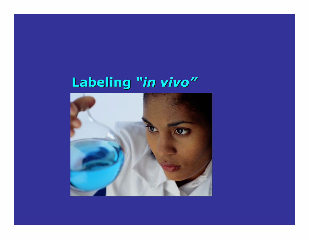
Labeling Labeling ““in vivoin vivo””
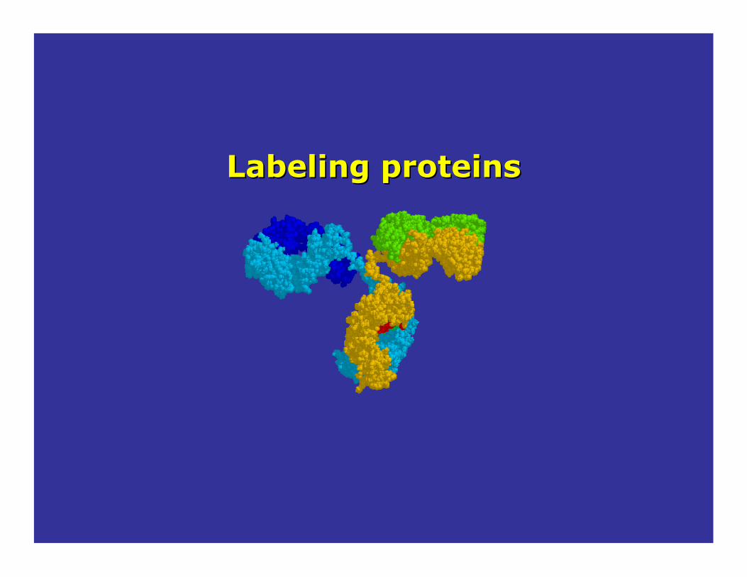
Labeling proteinsLabeling proteins
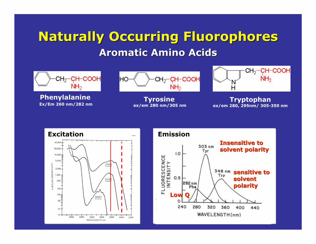
Naturally Occurring Naturally Occurring FluorophoresFluorophores
PhenylalanineEx/Em 260 nm/282 nm
Aromatic Amino AcidsAromatic Amino Acids
Tyrosineex/em 280 nm/305 nm
Tryptophanex/em 280, 295nm/ 305-350 nm
ExcitationExcitationInsensitive to Insensitive to solvent polaritysolvent polarity
sensitive to sensitive to solvent solvent polaritypolarity
Low QLow Q
EmissionEmission

5-Hydroxy-tryptophanex/em 310nm/339 nm
Tryptophan derivatives may be genetically incorporated in a protein
7-azatryptophanex/em 320nm/403nm
Protein Science (1997), 6, 689-697.
Tryptophanex/em 280, 295nm/ 305-350 nm
!! =0.14=0.14 !!= 0.097= 0.097 !! = 0.017= 0.017
••solventsolvent--insensitive insensitive emissionemission
••Large emission Large emission shift in watershift in water
••solventsolvent--sensitive sensitive emissionemission
! =Number of photons emitted/number of photons absorbed
Tryptophan derivativesTryptophan derivatives
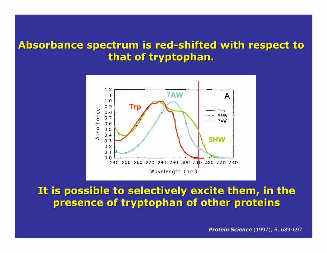
Absorbance spectrum is redAbsorbance spectrum is red--shifted with respect to shifted with respect to that of tryptophan. that of tryptophan.
It is possible to selectively excite them, in the It is possible to selectively excite them, in the presence of tryptophan of other proteinspresence of tryptophan of other proteins
Trp
Protein Science (1997), 6, 689-697.
7AW
5HW

Enzymes CofactorsEnzymes Cofactors
NADH (oxido-reductases)Ex/Em 340/460 nm
FAD (metabolic enzymes
(ex/em 450nm/540 nm)
Porphyrins(ex/em 550 nm/620 nm),

NonNon--covalent Attachmentcovalent Attachment
Extrinsic probesExtrinsic probes(not present in the natural molecule/macromolecule)
Barely fluorescent in pure water but their fluorescence can be strongly enhanced if the environment becomes hydrophobic (hydrophobic patches on proteins)
bis-ANS
1,8-ANS
Developed by G. Weber in 1950’s

Labeling should not change the biological activity of the Labeling should not change the biological activity of the protein.protein.
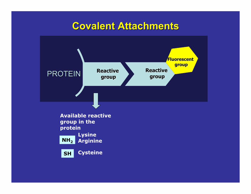
Covalent AttachmentsCovalent Attachments
Reactive Reactive groupgroup
Fluorescent Fluorescent groupgroup
Reactive Reactive groupgroupPROTEINPROTEIN
NH2
SH
Available reactive group in the protein
LysineArginine
Cysteine
Depends on the reactive group in the
protein.
Light sourceLifetime Spectral propertiesAutofluorescence

FITC(488/512) t ! 4.05
Texas Red(595-615), t ! 3.5 ns
Dansyl chloride(335/518) t ! 10 ns
IAEDANS(360/480) t ! 15 ns
BODIPY(493/503), t = 6 ns
Coumarin-3-carboxylic acid -NHS
(445/482), t ! 2 -3 ns

1999“there is a needfor probes with high fluorescence quantum yield and high photostability to allow detection of low-abundance biologicalstructures with great sensitivity and selectivity”
The Alexa-Fluor series
The Journal of Histochemistry & CytochemistryVolume 47(9): 1179–1188, 1999.Molecular Probes, Inc., Eugene, Oregon

Lucifer Yellow
fluorescein
Alexa 350 346/442
Alexa 430 434/539
Alexa 488 495/519
Coumarin-AMCA
rhodamine6G
Alexa 532 531/554
lissaminerhodamine B
Alexa 568 578/603
Texas Red
Alexa 594 590/617
Designed to be more photostable than their commonly used spectral analogues

All Alexa dyes and their conjugates are more fluorescent and more photostable than their
commonly used spectral analogues.
In addition, Alexa dyes are insensitive to pH in the 4–10 range.
The Journal of Histochemistry & CytochemistryVolume 47(9): 1179–1188, 1999. Molecular Probes, Inc., Eugene, Oregon

ALEXA 488
• Cells stained with Alexa Fluor488 or fluoresceinconjugates of goat anti–mouse IgG antibody
• Samples were continuously illuminated and images were collected every 5 seconds with a cooled CCD camera.
PhotostabilityAlexa Fluor 488 v/s fluoresceine
http://www.invitrogen.com/
Photo bleaching profile
FLUORESCEIN
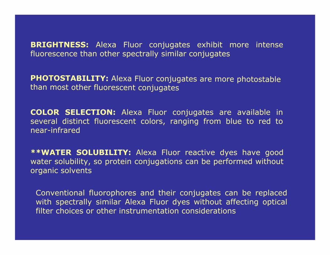
BRIGHTNESS: Alexa Fluor conjugates exhibit more intense fluorescence than other spectrally similar conjugates
PHOTOSTABILITY: Alexa Fluor conjugates are more photostablethan most other fluorescent conjugates
COLOR SELECTION: Alexa Fluor conjugates are available in several distinct fluorescent colors, ranging from blue to red tonear-infrared
**WATER SOLUBILITY: Alexa Fluor reactive dyes have good water solubility, so protein conjugations can be performed without organic solvents
Conventional fluorophores and their conjugates can be replaced with spectrally similar Alexa Fluor dyes without affecting optical filter choices or other instrumentation considerations

Alexa Fluor dyes are available as amine-reactive succinimidyl esters
The Alexa series expanded

http://www.invitrogen.com
Spectral properties of Alexa Fluor dyes
http://www.invitrogen.com
Covalent AttachmentsCovalent Attachments
Reactive Reactive groupgroup
Fluorescent Fluorescent groupgroup
Reactive Reactive groupgroupPROTEINPROTEIN
NH2
SH
Available reactive group in the protein
LysineArginine
Cysteine
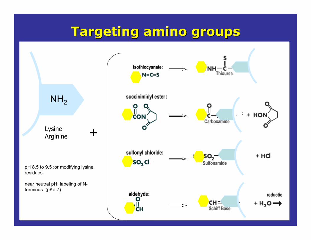
Targeting amino groupsTargeting amino groups
LysineArginine
NH2
+
pH 8.5 to 9.5 :or modifying lysine residues.
near neutral pH: labeling of N-terminus .(pKa 7)

Cysteine
SH +
Targeting Targeting thiolthiol groups:groups:

General labeling protocol for extrinsic labeling
Absorption spectra Protein determination
Activity measurementsSDS or native gelDenaturation exp etc.
Proteinin buffer
Addition of thefluorescent dye
ratio dye/protein
Incubation time
Labeling ratio
[protein][fluorescent dye]
Sample characterization Biological testing
Removal of the free dye

Characterization after the labeling
ProteinProtein--FluoresceinFluorescein A
bsor
banc
e
A="* b* C A="* b* C
Wavelength (nm)
FluoresceinFluoresceinA
bsor
ban
ce
Bradford, Lowry, etc
Wavelength (nm)
Labeling should not Labeling should not change the biological change the biological
activity of the activity of the protein.protein.
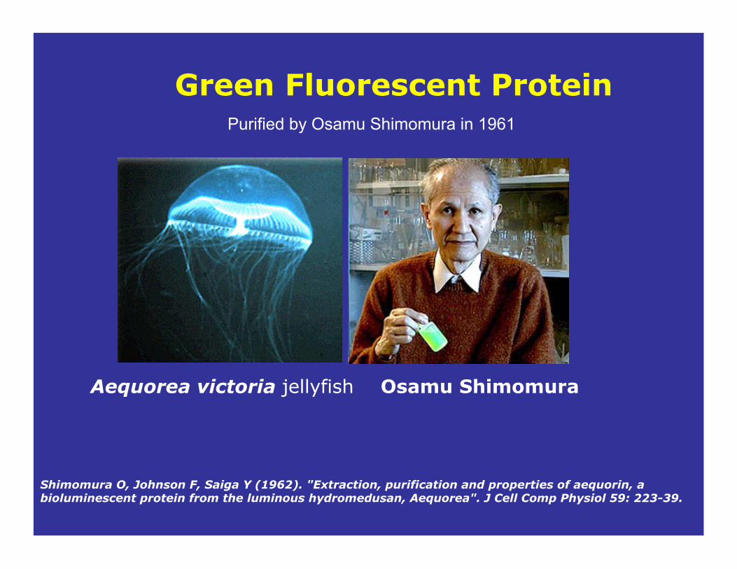
Green Fluorescent Protein
Aequorea victoria jellyfish Osamu Shimomura
Shimomura O, Johnson F, Saiga Y (1962). "Extraction, purification and properties of aequorin, a bioluminescent protein from the luminous hydromedusan, Aequorea". J Cell Comp Physiol 59: 223-39.
Purified by Osamu Shimomura in 1961
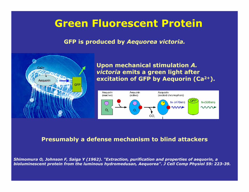
GFP is produced by Aequorea victoria.
Green Fluorescent Protein
Shimomura O, Johnson F, Saiga Y (1962). "Extraction, purification and properties of aequorin, a bioluminescent protein from the luminous hydromedusan, Aequorea". J Cell Comp Physiol 59: 223-39.
Upon mechanical stimulation A. victoria emits a green light after excitation of GFP by Aequorin (Ca2+).
Presumably a defense mechanism to blind attackers

The protein is purified from the luminescent organs in the ring
Green Fluorescent Protein
The ring can be manually removed with scissors
Shimomura. The discovery of aequorin and green fluorescent protein. Journal of Microscopy, 217: 3–15 (2005).

Shimomura. The discovery of aequorin and green fluorescent protein. Journal of Microscopy, 217: 3–15 (2005).
The “ring cutting machine”
•To obtain 1mg of GFP they required 50,000 animals (2.5 tons of jellyfish)
•The job was done in one summer processing 3,000 jellyfish each day.

- 6 am - 8 am: collect jellyfish - Quick breakfast - Cut rings from the jellyfish until noon. - Afternoon devoted to the extraction.- Dinner- 7 pm - 9 pm: collect jellyfish and keep it in an
aquarium for the next day.
Shimomura: daily schedule summer 1961
Our task was to catch and process as many jellyfish as possible.
Shimomura. The discovery of aequorin and green fluorescent protein. Journal of Microscopy, 217: 3–15 (2005).

1974, Morize et alGFP was completely purified and crystallized
1979, Shimomurathe structure of the GFP chromophorewas elucidated
1992, Prasher et alcDNA of GFP was cloned
Matz et al. Fluorescent proteins from nonbioluminescent Anthozoa species. Nat Biotechnol. 17:969-73 (1999)
1994, Chalfie at al and Inouye & Tsuji. cDNA of GFP was expressed in living organisms

• The chromophore is formed spontaneously from
Serine-65, Tyrosine-66, Glycine-67
upon folding of the polypeptide chain, without the need for enzymatic synthesis.
• #-barrel structure, with chromophore housed within the barrel.
GFP chromophore

http://www.scivee.tv/node/11255
The Ser-Tyr-Gly sequence is post-translationally modified to a 4-(p-hydroxybenzylidene)- imidazolidin-5-one structure
The fluorescence is not an intrinsic property of the Ser-Tyr-Gly tripeptide. The cyclizedbackbone of these residues forms the imidazolidone ring.
GFP chromophore

GFP from A. victoriaMatures at low temperatureExcitation peak = 395 nm Emission peak = 509 nmMW:25-30 KDa
Ser65
FPs of different colorsA number of new GFP proteins have been made using site directed mutagenesis to alter the amino acids near the chromophore and thus alter the absorption and fluorescence properties.
http://www.olympusfluoview.com/applications/fpcolorpalette.html
1995 Ole Thastrup EGFP (37°C folding GFP) allowed the use of GFPsin mammalian cells ! = 55,000 M"1cm"1
QY = EGFP is 0.60Excitation peak = 487 nm Emission peak = 507 nm
http://www.olympusfluoview.com/applications/fpcolorpalette.html
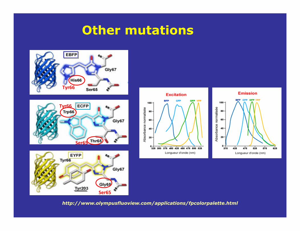
Tyr66
Tyr66
Ser65
Ser65
Other mutations
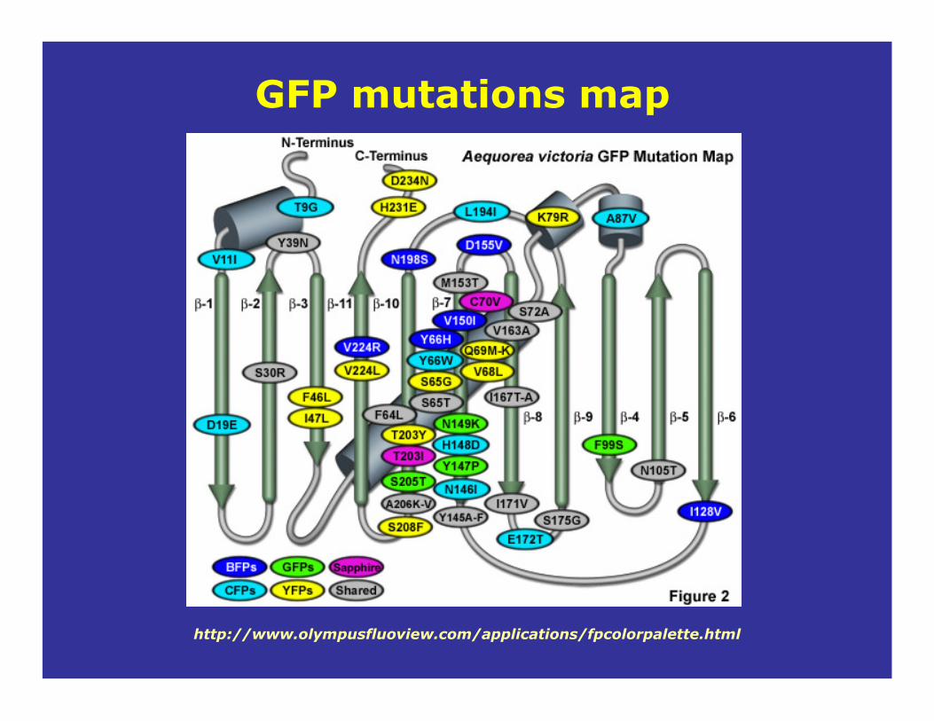
GFP mutations map
http://www.olympusfluoview.com/applications/fpcolorpalette.html
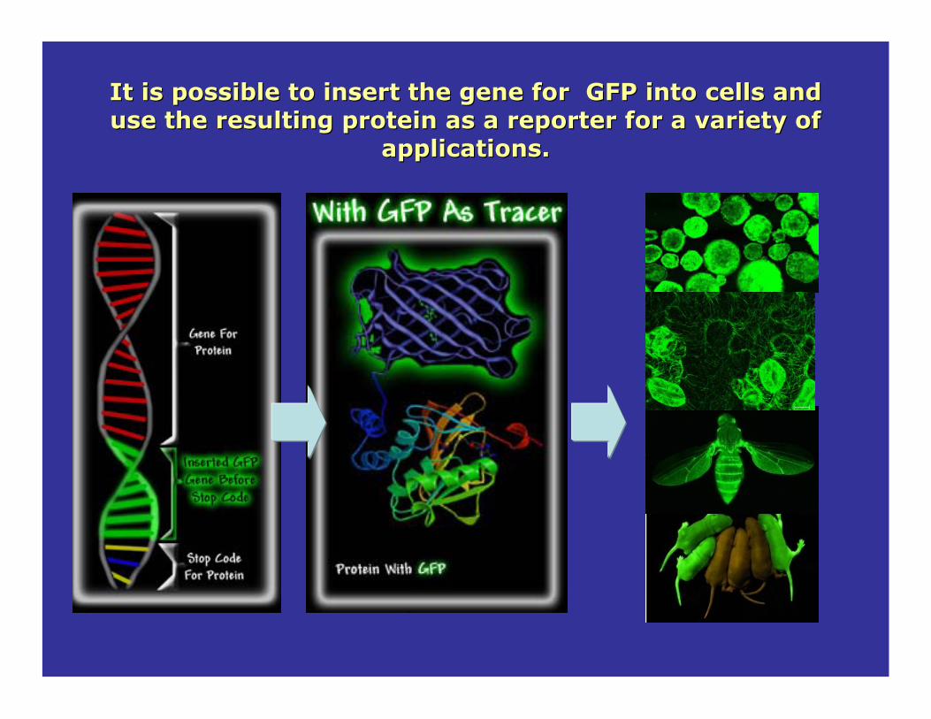
It is possible to insert the gene for GFP into cells and It is possible to insert the gene for GFP into cells and use the resulting protein as a reporter for a variety of use the resulting protein as a reporter for a variety of
applications. applications.

The search for a red-emitting fluorescent protein
from the Aequorea-based fluorescent proteins, the yellow fluorescent proteins (YFPs) remain the most red-shifted
of the GFP derivatives. Em/ex = 520/530 nm
http://zeiss-campus.magnet.fsu.edu/articles/probes/anthozoafps.html
• Provide an important tool for multicolor imaging
• Generate new FRET biosensors with spectral profiles in the longer wavelength regions.
• Cellular auto-fluorescence is significantly reduced at longer wavelength regions.
• Living cells and tissues better tolerate illumination by the longer excitation wavelengths, allowing extend periods for imaging.
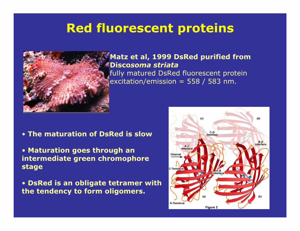
• The maturation of DsRed is slow
• Maturation goes through an intermediate green chromophorestage
• DsRed is an obligate tetramer with the tendency to form oligomers.
Matz et al, 1999 DsRed purified from Discosoma striatafully matured DsRed fluorescent protein excitation/emission = 558 / 583 nm.
Red fluorescent proteins

Pro
tein
fo
ldin
gC
hro
mo
ph
ore
form
ati
on
Chromophoremodification
Aequorea GFP Discosoma DsRed
EGFP mutantMature at 37°CMonomeric
mRFP1 mutantFaster MaturationMonomeric
DsRed forms a chromophoreidentical to GFP to generate its green intermediate
Matures at low temperature
Dimeric
Slow maturation
Tetrameric
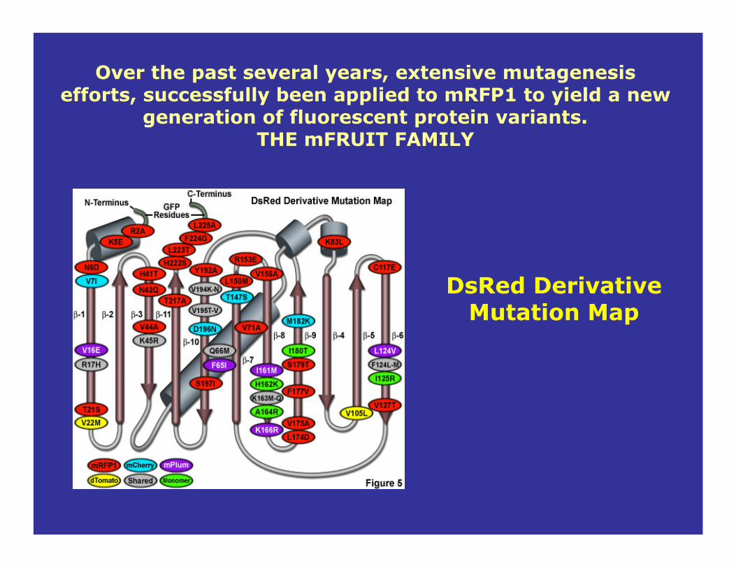
DsRed Derivative Mutation Map
Over the past several years, extensive mutagenesis efforts, successfully been applied to mRFP1 to yield a new
generation of fluorescent protein variants. THE mFRUIT FAMILY

Glutamine 66Tyrosine 67Glycine 68MW= 30-35 KDa
Serine 65Tyrosine 66Glycine 67MW= 20-30 KDa
GFP mCherry
Chromophore
30 Å
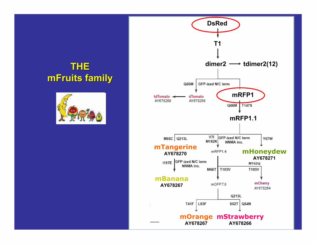
THETHEmFruitsmFruits familyfamily
DsRed
T1
dimer2 tdimer2(12)
mRFP1
mRFP1.1
mTangerineAY678270
mBananaAY678267
mHoneydewAY678271
mOrangeAY678267
mStrawberryAY678266

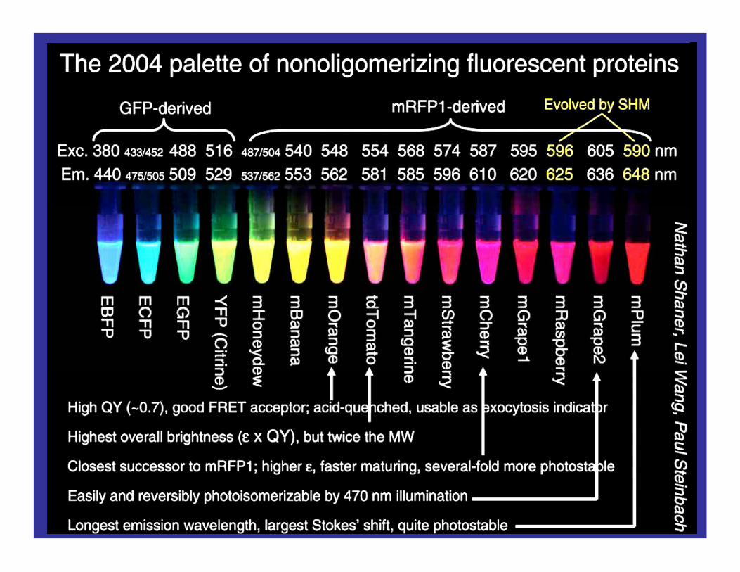
mFruitsmFruits fluorescentfluorescent proteinsproteins

mFruits may replace or be good pairs for FPs in energy transfer experiments
FPsmFruitsmFruits

Labeling DNALabeling DNA
http://info.med.yale.edu/genetics/ward/tavi/n_coupling.html
5’ 3’
3’ 5’
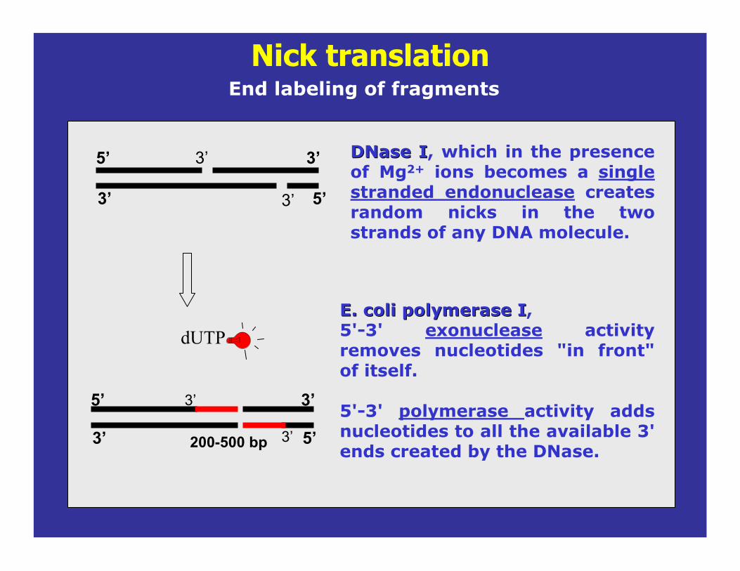
Nick translationEnd labeling of fragments
DNaseDNase II, which in the presence of Mg2+ ions becomes a single stranded endonuclease creates random nicks in the two strands of any DNA molecule.
3’
3’
3’ 5’
5’ 3’
200-500 bp
3’
3’
dUTPE. coli polymerase IE. coli polymerase I, 5'-3' exonuclease activity removes nucleotides "in front" of itself.
5'-3' polymerase activity adds nucleotides to all the available 3' ends created by the DNase.

Polymerase Chain Reaction (PCR)
1- Denaturation step (1min, 95ºC).During the denaturation, the double strand melts open to single stranded DNA
3’ 5’
5’ 3’
3’ 5’
5’ 3’
3’ 5’
5’ 3’
2- Annealing (45 sec, 54ºC).Single stranded DNA primers (18-30 bplong), forward and reverse are synthesized (blue arrows). Then, the primers are allow to anneal to their target sequences.
3’ 5’5’
5’
100-5,000 bp
dUTP
3- Extension (2min, 72ºC). Then Taq polymerase synthesize the new DNA strands. Only dNTP’s.

fluorescein-aha-dUTP from Molecular Probes
Commercially labeled dUTP
dUTP
succinimidyl-ester derivatives of fluorescent dyes
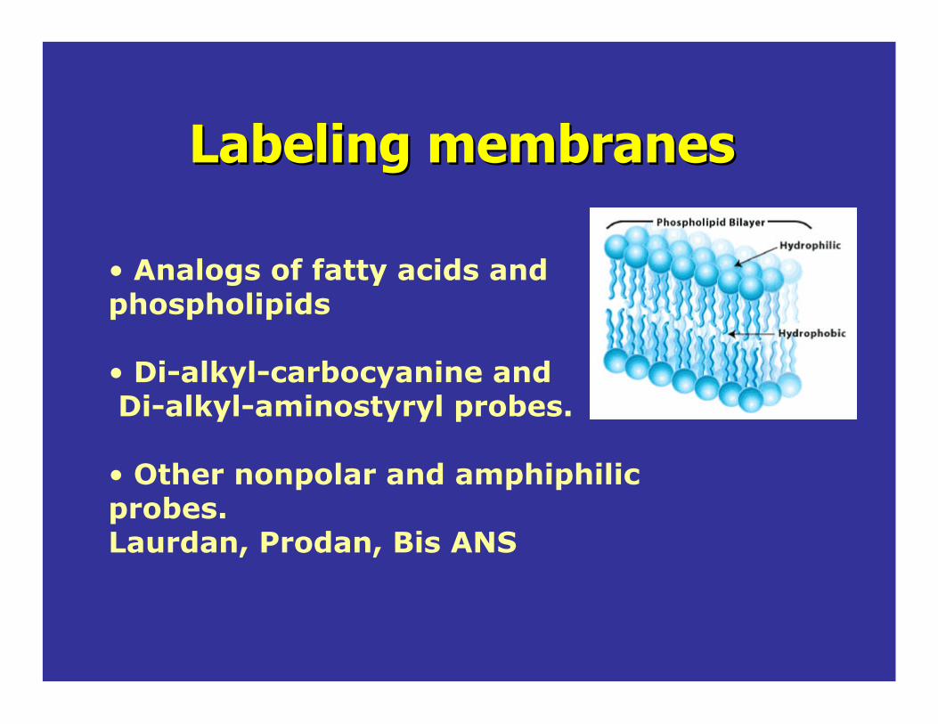
Labeling membranesLabeling membranes
• Analogs of fatty acids and phospholipids
• Di-alkyl-carbocyanine and Di-alkyl-aminostyryl probes.
• Other nonpolar and amphiphilicprobes.Laurdan, Prodan, Bis ANS

Fatty acids analogs and phospholipids
bis-pyrene-PC
NBD
-C6-HPC
N-Rh-PE
BODIPY fa
tty acid

Dil-C16 Dil-C18
DiDi--alkylalkyl--carbocyaninecarbocyanine probes.probes.
Chem.and Phys. of Lipids 141 (2006) 158–168
SM/DOPC/Chol (1:1:1)
Ld
Dil-C18
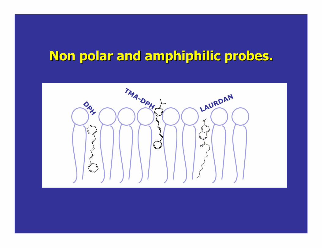
Non polar and Non polar and amphiphilicamphiphilic probes.probes.
DPH LAURDAN
TMA-DPH

DPH1,6-diphenyl-1,3,5-hexatriene
It is oriented parallel to the lipid acyl chain axis .
• DPH shows no partition preference between coexisting gel- and fluid-phase phospholipids
• DPH fluorescence is practically negligible in water and intercalation into membranes is accompanied by strong enhancement of its fluorescence.
• Fluorescence decay data are often analyzed in terms of continuous lifetime distributions and interpreted as being indicative of lipid environment heterogeneity.
Excitation: 350 nmEmission: 452 MeOH
Information on the physical state of the phospholipid bilayer is obtained from the changes in the fluorescence polarizationfluorescence polarization and lifetimelifetime.
Parasassi et al. J. of Biol. Chem. (1984) 259:14011-14017Shinitzky et al. Biochemistry (1971) 10:2106-2113

TMA-DPH1-(4-trimethylammoniumphenyl)-6-phenyl-1,3,5-hexatriene p-toluenesulfonate
Designed to improve the localization of DPH in the membrane, TMA-DPH contains a cationic trimethylammonium substituent that acts as a surface anchor
It partitions from aqueous dispersions into membranes, accompanied by strong fluorescence enhancement.
the duration of plasma membrane surface staining by TMA-DPH before internalization into the cytoplasm is quite prolonged
TMA-DPH fluorescence polarization measurements can be combined with video microscopy to provide spatially resolved images of phospholipid in large liposomes and single cells
Excitation: 355 nmEmission: 430 Meoh
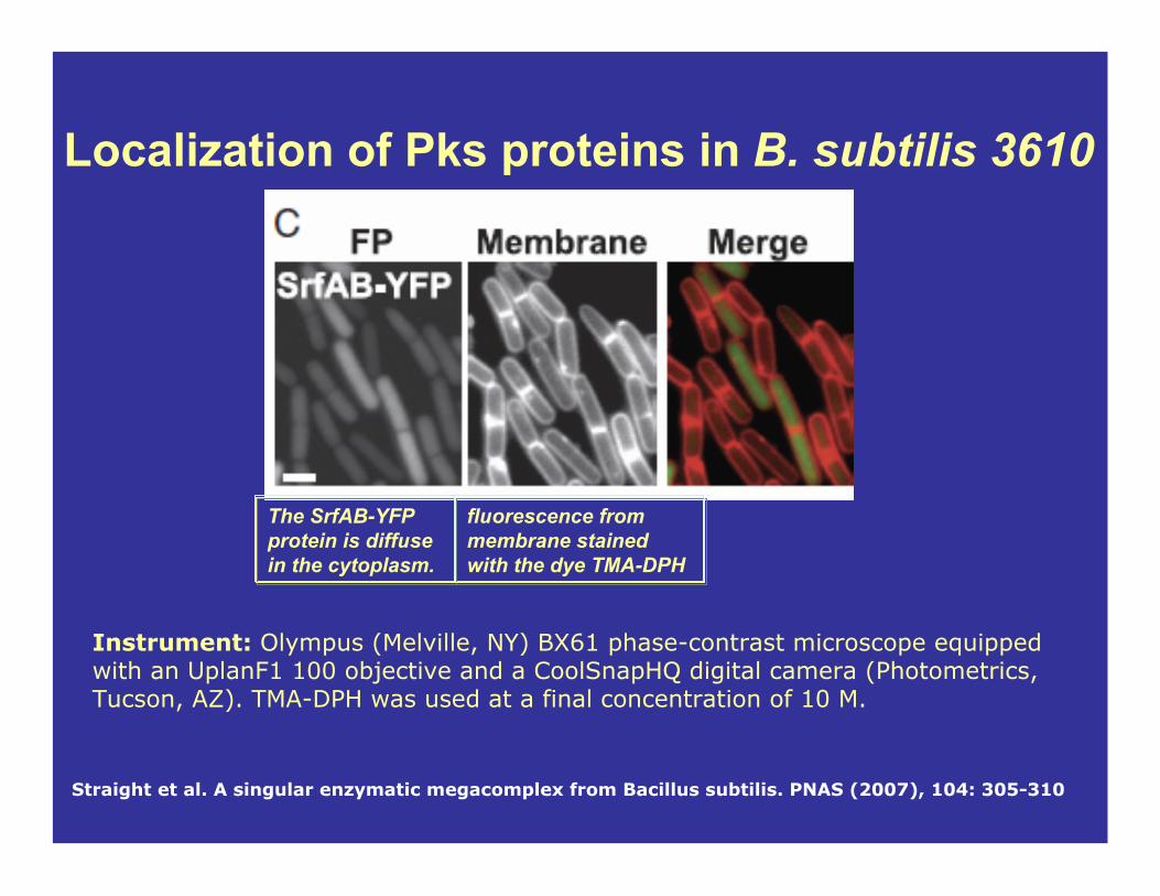
Straight et al. A singular enzymatic megacomplex from Bacillus subtilis. PNAS (2007), 104: 305-310
Localization of Pks proteins in B. subtilis 3610
The SrfAB-YFP protein is diffuse in the cytoplasm.
fluorescence from membrane stained with the dye TMA-DPH
Instrument: Olympus (Melville, NY) BX61 phase-contrast microscope equipped with an UplanF1 100 objective and a CoolSnapHQ digital camera (Photometrics, Tucson, AZ). TMA-DPH was used at a final concentration of 10 M.

350 400 450 500 550 6000.0
0.2
0.4
0.6
0.8
1.0
1.2
Emis
sion
Inte
nsity
wavelength
!"#$%&'("
)*+,*-$./0(1'##*2"$%&'("
Weber, G. and Farris, F. J.Biochemistry, 18, 3075-3078 (1979) .
LAURDAN 6-dodecanoyl-2-dimethylaminonaphthalene
Emission spectra
440 490
Excitation: 364 nmEmission: 497 nm
(Methanol)
(environment-sensitive spectral shifts)
Information on the physical state of the phospholipid bilayer is obtained from the shift in the emission spectra.
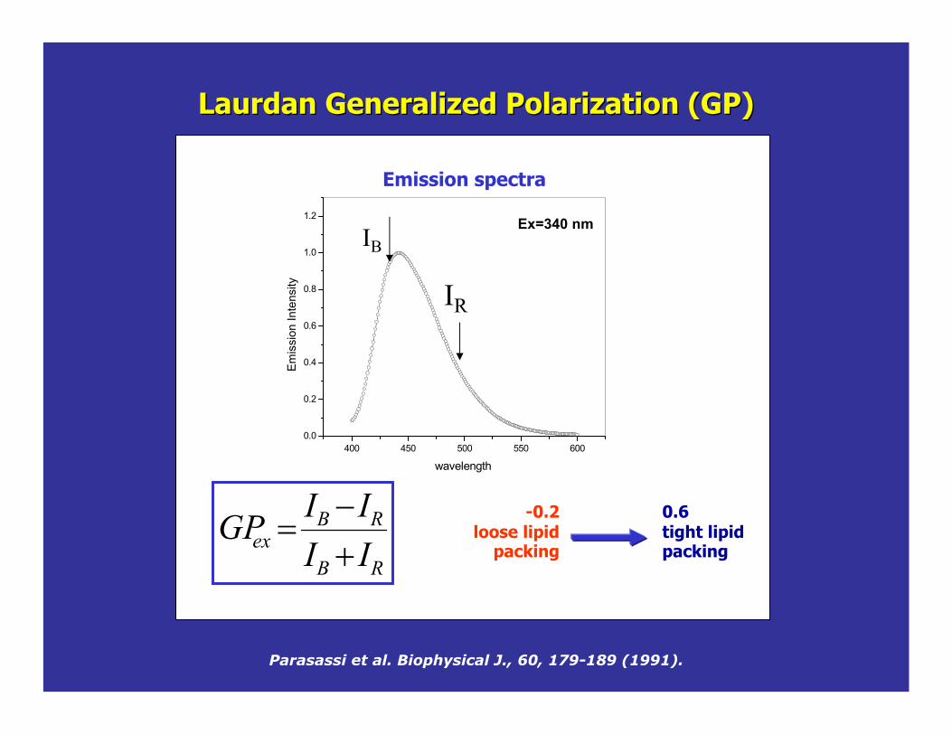
Laurdan Generalized Polarization (GP) Laurdan Generalized Polarization (GP)
RB
RBex II
IIGP$%
&
Parasassi et al. Biophysical J., 60, 179-189 (1991).
0.6tight lipid packing
-0.2loose lipid
packing
400 450 500 550 6000.0
0.2
0.4
0.6
0.8
1.0
1.2
Emis
sion
Inte
nsity
wavelength
IB
IR
Ex=340 nm
Emission spectra

GP in the GP in the cuvettecuvette
30 35 40 45 50 55 60-0.2
-0.1
0.0
0.1
0.2
0.3
0.4
0.5
0.6
GP
temperature (celcius)
Changes of GP for DPPC with temperature, Tm=41ºC.
MLVs, SUVs, LUVs
2Ph- microscopy
-1 GP 1
GP value and spatial resolution
GUVs
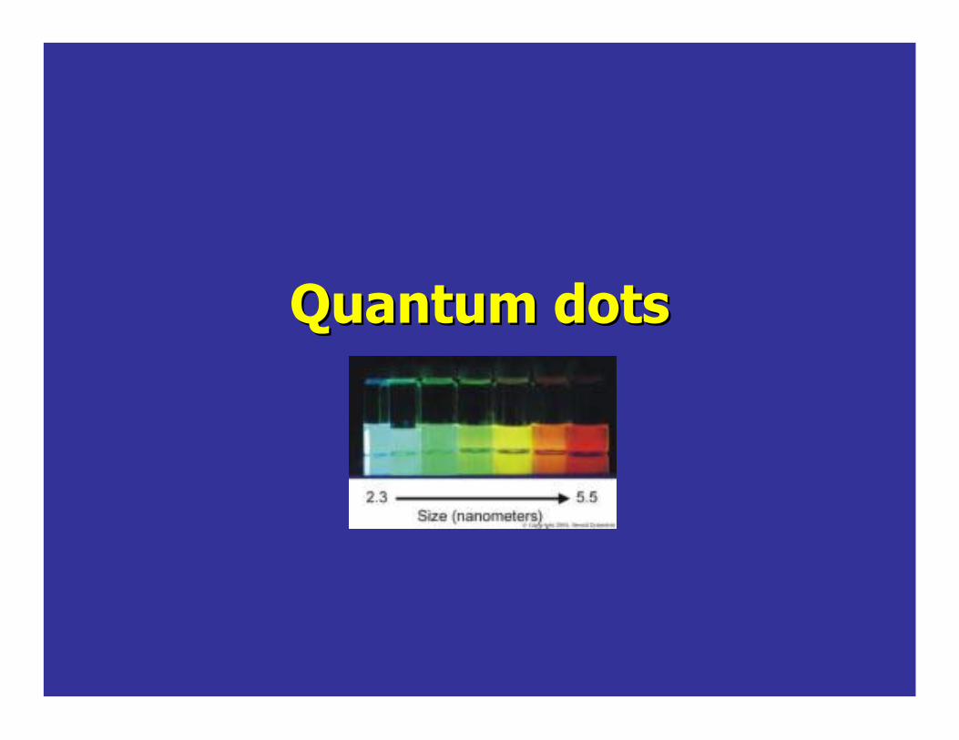
Quantum dotsQuantum dots

In the core emission is typically weak and always unstable.The shell material Zinc Sulfide (ZnS) has been selected to be almost entirely un-reactive and completely insulating for the core.
A layer of organic ligands covalently attached to the surface of the shell. This coating provides a surface for conjugation to biological (antibodies, streptavidin, lectins, nucleic acids) and nonbiological species and makes them “water-soluble”
-Cadmium selenide (CdSe), or Cadmium telluride (CdTe) -Semiconductor material is chosen based upon the emission wavelength.-The size of the particles that tunes the emission wavelength.
SHELLSHELL COATINGCOATING
BIOMOLECULE
BIOMOLECULECORECORE
Quantum DotsQuantum Dots

nanocrystal (NC) sample in PBS
•Q-dots: broad absorption spectra, making it possible to excite all colors of QDssimultaneously with a single excitation light source….
•Q-dots:emission spectra is narrow and symmetrical.
UV handlamp.
relative sizes of the CdSequantum dots in the vials.
The emission is tunable according to their size and material composition
AbsorptionAbsorption
EmissionEmission

Violet excitation Broad range of emissions
Single-color excitation, multicolor emission for easy multiplexingHigh absorbance means increased brightness
Invitrogen
Q-dot Optical SpectraQ-dot Optical Spectra

Nucleus: Qdot® 605 conjugate Microtubules: Alexa Fluor® 488 conjugate
www.Invitrogen.com
Nucleus: Alexa Fluor® 488 conjugate Microtubules: Qdot® 605 conjugate
Photostability of Qdots3T3 cells
Photostability results in sensitivity, and sample permanence

https:/.../news_releases/2008/NR-08-05-02.html
DisadvantagesLarge size and high mass limit their use in applications requiring high diffusional mobility
Advantages:• Broad absorption spectra, making it possible to excite all colors of QDs simultaneously with a single light source - Multiplexing• Narrow and symmetrical emission spectraEmission tunable with size and material composition• Exhibit excellent photo-stability
Qdot Summary
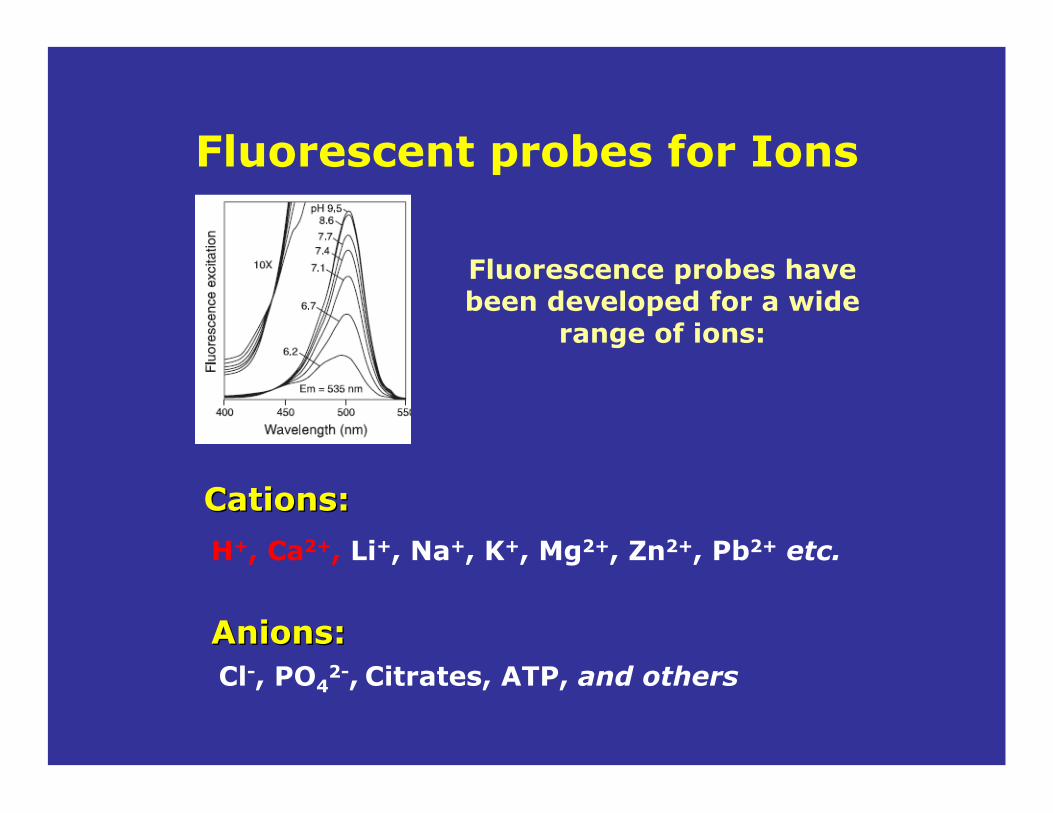
Fluorescent probes for Ions
Fluorescence probes have been developed for a wide
range of ions:
CationsCations::
H+, Ca2+, Li+, Na+, K+, Mg2+, Zn2+, Pb2+ etc.
Anions:Anions:Cl-, PO4
2-, Citrates, ATP, and others

How do we choose the correct probe for ion determination?
1-DISSOCATION CONSTANT (Kd)•Must be compatible with the concentration range of interest.•Calibration. The Kd of the probe is dependent on pH, temperature, viscosity, ionic strength etc.
2- MEASUREMENT MODE•Qualitative or quantitative measurements. •Ratiometric measurements.•Illumination source available.
3- INDICATOR FORM •Cell loading and distribution of the probe.•AM-esters: cleaved by intracellular esterases.

Molecular Probes' pH indicator families, in order of decreasing pKa
Parent Fluorophore pH Range
Typical Measurement
SNARF indicators 6.0–8.0 Emission ratio 580/640 nm HPTS (pyranine) 7.0–8.0 Excitation ratio 450/405 nm BCECF 6.5–7.5 Excitation ratio 490/440 nm Fluoresceins and carboxyfluoresceins
6.0–7.2 Excitation ratio 490/450 nm
LysoSensor Green DND-189 4.5–6.0 Single emission 520 nm Oregon Green dyes 4.2–5.7 Excitation ratio 510/450 nm or
excitation ratio 490/440 nm LysoSensor Yellow/Blue DND-160
3.5–6.0 Emission ratio 450/510 nm
Probes For pH determinationProbes For pH determination

Example: BCECFExample: BCECF
Fluorescence IntensityFluorescence Intensity Fluorescence LifetimeFluorescence Lifetime
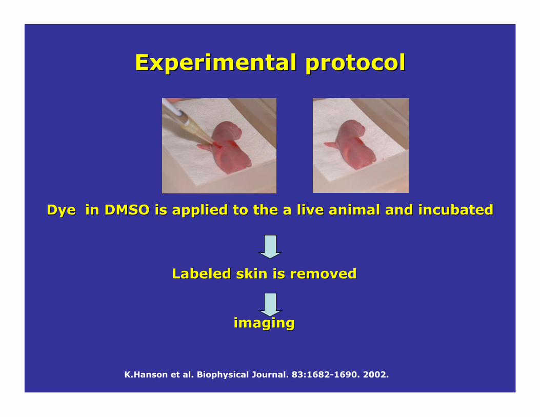
K.Hanson et al. Biophysical Journal. 83:1682-1690. 2002.
Labeled skin is removedLabeled skin is removed
imagingimaging
Dye in DMSO is applied to the a live animal and incubatedDye in DMSO is applied to the a live animal and incubated
Experimental protocolExperimental protocol

0 5 10 15
6.4
6.6
6.8
7.0
SC-SG Junction
Ave
rage
pH
Depth ('m)
4.0 8.0
pHco
rrec
t ed
for (
20 'm
Depth ('m): 0 1.7 3.4 5.1 6.8 10.2
80 300 100 2000100 1000100 1000100 1000 100 1000
Inte
nsi t y
K.Hanson et al. Biophysical Journal. 83:1682-1690. 2002.

UVFURA(Fura-2, Fura-4F, Fura-5F, Fura-6F, Fura-FF)INDO( Indo-1, Indo 5F)
VISIBLEFLUO(Fluo-3, Fluo-4, Fluo5F, Fluo-5N, Fluo-4N) RHOD ( Rhod-2, Rhod-FF, Rhod-5N)CALCIUM GREEN (CG-1, CG-5N,CG-2)OREGON GREEN 488-BAPTA
Probes For Calcium determination
RatiometricRatiometric
NonRatiometric
NonRatiometric

FURA-2FURA-2
Most used in microscopic imagingMost used in microscopic imaging
Good excitation shift with CaGood excitation shift with Ca2+2+
Rationed between 340/350 and 380/385 nmRationed between 340/350 and 380/385 nm
RatiometricRatiometric: 2 excitation/1emission: 2 excitation/1emission

IndoIndo--11
RatiometricRatiometric: 1excitation /2emission: 1excitation /2emission
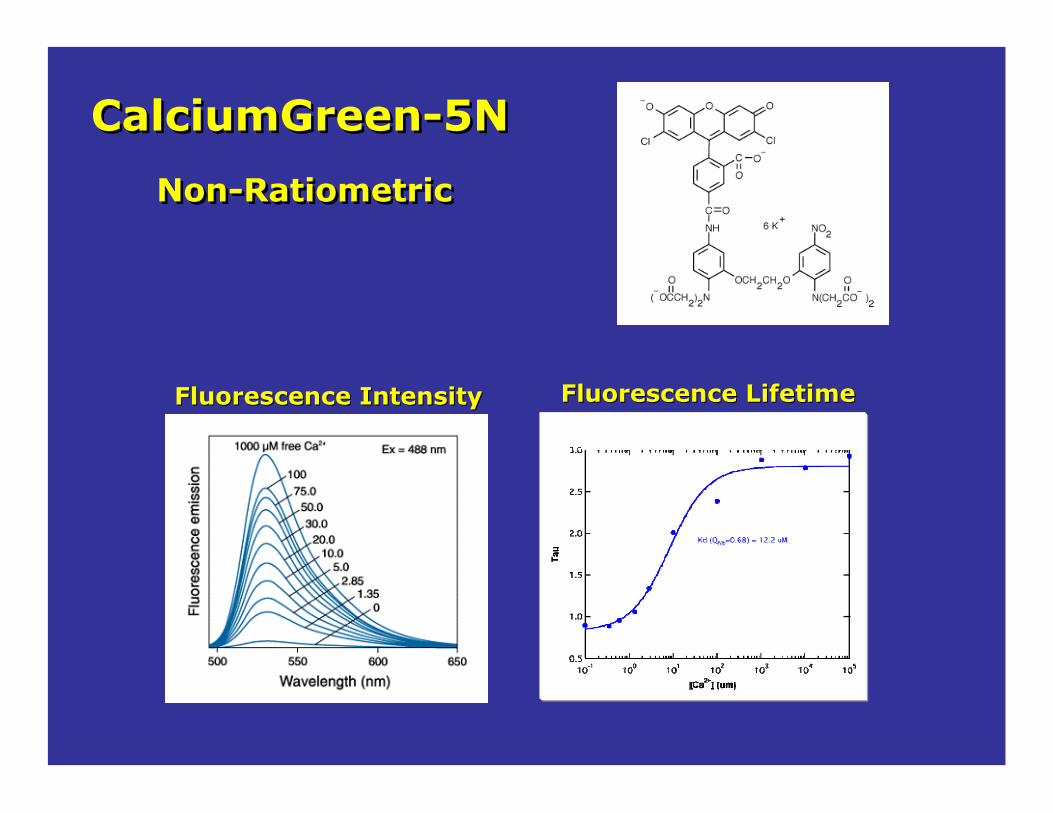
CalciumGreen-5NCalciumGreen-5N
Non-RatiometricNon-Ratiometric
Fluorescence IntensityFluorescence Intensity Fluorescence LifetimeFluorescence Lifetime

Comparing the size of the fluorescence probes and the bio-molecule being labeled
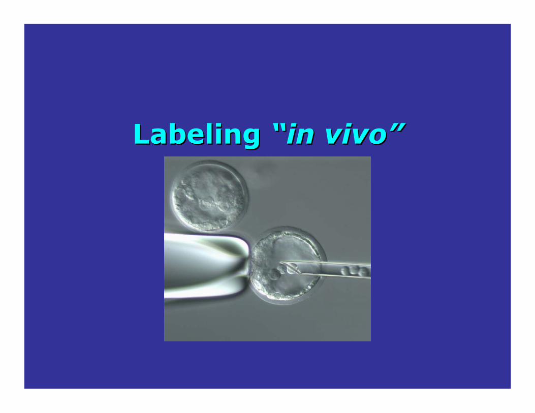
Labeling Labeling ““in vivoin vivo””
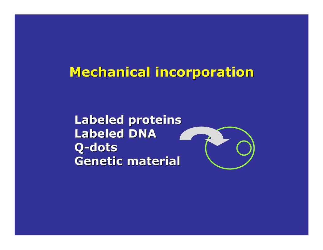
Mechanical incorporationMechanical incorporation
Labeled proteinsLabeled proteinsLabeled DNALabeled DNAQQ--dotsdotsGenetic materialGenetic material

•• Cells are mixed with a labeled Cells are mixed with a labeled compound.compound.
•• The mixture is exposed to pulses of The mixture is exposed to pulses of high electrical voltage. high electrical voltage.
Electroporation
Source: http://dragon.zoo.utoronto.ca/~jlm-gmf/T0301C/technology/introduction.html
Non-homogeneous labelingTransfected cells have to be selected
•• The cell membrane of the host cell The cell membrane of the host cell is penetrable allowing foreign is penetrable allowing foreign compounds to enter the host cell. compounds to enter the host cell.
•• Some of these cells will incorporate Some of these cells will incorporate the molecule of interest (new DNA the molecule of interest (new DNA and express the desired gene). and express the desired gene).

•• Direct injectingDirect injecting foreign DNA into cells. foreign DNA into cells.
Microinjection Microinjection
-Photo of a Microinjection apparatus(courtesy of A. Yanagi)
Source: http://dragon.zoo.utoronto.ca/~jlm-gmf/T0301C/technology/introduction.html
NonNon--homogeneous labelinghomogeneous labelingTransfectedTransfected cells have to be selectedcells have to be selected
•• Under a microscope, a cell is held in Under a microscope, a cell is held in place with gentle suction while being place with gentle suction while being manipulated with the use of a blunt manipulated with the use of a blunt capillary.capillary.
•• A fine pipette is used to insert the A fine pipette is used to insert the DNA into the cytoplasm or nucleus. DNA into the cytoplasm or nucleus.
•• This technique is effective with plant This technique is effective with plant protoplasts and tissues. protoplasts and tissues.

• Biolistics is currently the most widely used in the field of transgenic corn production.
Source: http://dragon.zoo.utoronto.ca
Biolistics
NonNon--homogeneous labelinghomogeneous labelingTransfectedTransfected cells have to be selectedcells have to be selected
• The DNA construct is coated onto fine gold/tungsten particles and then the metal particles are fired into the callus tissue.
• As the cells repair their injuries, they integrate their DNA into their genome, thus allowing for the host cell to transcribe and translate the gene.
• Selection of the transfected cells, is done on the basis of the selectable marker that was inserted into the DNA construct
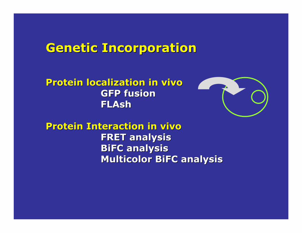
Genetic IncorporationGenetic Incorporation
Protein localization in vivoProtein localization in vivoGFP fusionGFP fusionFLAshFLAsh
Protein Interaction in vivoProtein Interaction in vivoFRET analysisFRET analysisBiFCBiFC analysisanalysisMulticolor Multicolor BiFCBiFC analysisanalysis

GFP
pUG356231 bp
CEN6/ARSH4
yGFP
MET25
URA3AmpR
ori
CYC1
ClaI (3005)
EcoRI (3022)
HindIII (3010)
KpnI (2009)
SacI (3444)
Swa I (5688) PstI (400)
ApaLI (178)
ApaLI (4152)
ApaLI (5398) NcoI (623)
NcoI (2294)
NcoI (2818)
YourYour gene gene (ex: P2b)(ex: P2b)
P2b
pUG35-P2b6549 bp
CEN6/ARSH4
yGFP
MET25
URA3AmpR
oriCYC1
P2b
BamHI (3358)
ClaI (3005)
EcoRI (3022)
HindIII (3010)
PstI (400)
KpnI (2009)
SacI (3762)
Swa I (6006)
ApaLI (178)
ApaLI (4470)
ApaLI (5716) NcoI (623)
NcoI (2294)
NcoI (2818)
GFPP2b
Introduction intodifferent organisms
GFPGFP--fusion proteinsfusion proteins
GFP encodingplasmid
Protein Localization in vivoProtein Localization in vivo

Current Biology 1998, 8:377–385
The human histone H2B gene fused (GFP) and transfected into
human HeLa cells
Homogeneous labeling (if stable line)Homogeneous labeling (if stable line)Regulation of the expression can be a problem for FCSRegulation of the expression can be a problem for FCS
GFPGFP--fusion proteinsfusion proteins
FCSFCSRICSRICSN&BN&B

Receptor domain composed of a few as six natural amino acids that could be genetically incorporated into proteins of interest.
FL AshFL Ash--EDT2 labeling (FLASH tag)EDT2 labeling (FLASH tag)
A small (700-dalton), synthetic, membrane-permeant ligand that could be linked to various spectroscopic probes or crosslinks.
The ligand is non fluorescent until it binds its target, where upon it becomes strongly fluorescent.
++
Protein Localization in vivoProtein Localization in vivo
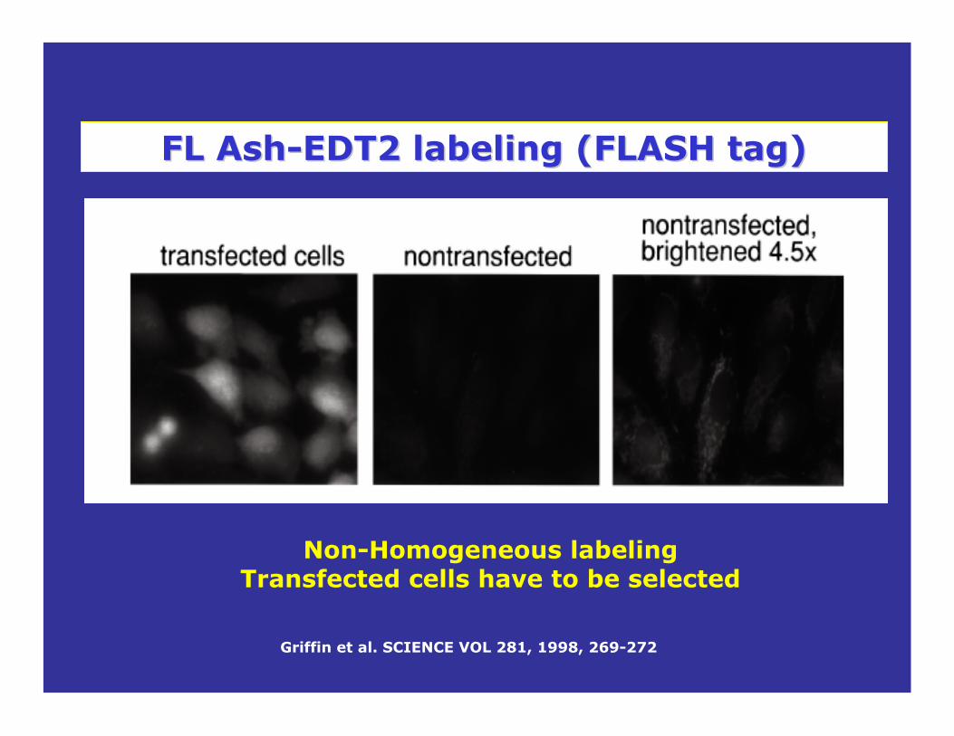
Griffin et al. SCIENCE VOL 281, 1998, 269-272
Non-Homogeneous labelingTransfected cells have to be selected
FL AshFL Ash--EDT2 labeling (FLASH tag)EDT2 labeling (FLASH tag)
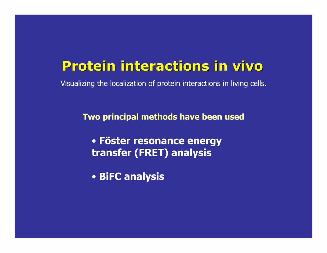
Protein interactions in vivoProtein interactions in vivo
• Föster resonance energy transfer (FRET) analysis
• BiFC analysis
Visualizing the localization of protein interactions in living cells.
Two principal methods have been used

Föster resonance energy transfer (FRET) analysis
Donor Acceptor
Donor intensity decreaseDonor lifetime decrease
Acceptor intensity increase
Based on changes in the fluorescence intensities and lifetimes of two fluorophores that are brought sufficiently close together.
D A D A+
fluorophores apart fluorophores close

(a) INTERMOLECULAR!!FRET:!FRET!between!a!donor!and!acceptor!fluorophore,!each!attached!to!a!different!protein,!reports!protein–proteininteraction.
(b)!INTRAMOLECULAR!!FRET:!two!fluorophores attached!to!the!same!protein.!Changes!in!distance!between!them!reflect!alterations!in!protein!conformation,!which!in!turn!indicates!ligand binding!or!post"translational!modification.
Klaus!Hahn!et!al.!Current!Opinion!in!Cell!Biology!2002,!14:167–172
FRET
Proteins!can!be!labeled!in!vitro!with!small!!fluorescent!dyes.
Mechanically!incorporated
Proteins!can!be!genetically!bounded!to!fluorescent!proteins!
Föster resonance!energy!transfer!(FRET)

(c)!Protein!‘transducer’.!A!protein!is!engineered!to!produce!a!large!change!in!the!distance!between!an!attached!donor!and!acceptor!upon!ligandbinding.!!In!this!example,!calcium!binding!generates!a!hydrophobic!pocket!to!which!the!blue!peptide!binds.!Peptide!binding!brings!the!two!GFP!mutants!together,!producing!FRET.Klaus!Hahn!et!al.!Current!Opinion!in!Cell!Biology!2002,!14:167–172
CFP
calmodulin
YFP
M13 calmodulin-binding peptide
FRET
Binding of Ca2+ makes Calmodulin wrap
around the M-13-domain, increasing the
fluorescence resonance energy transfer (FRET)
between the GFPs.
Miyawaki et al. Nature, 1997: 28: 834-835.
CAMELEON Ca+2 SENSOR

Klaus!Hahn!et!al.!Current!Opinion!in!Cell!Biology!2002,!14:167–172
(d)!Domain/antibody!biosensor.!A!protein!or!antibody!fragment!(blue)!binds!only!to!the!activated!state!of!the!protein.!The!protein!fragment!bears!a!dye!which!undergoes!FRET!when!it!is!brought!in!close!proximity!to!the!GFP!on!the!protein.!In!some!examples,!the!domain!is!part!of!the!same!polypeptide!chain!as!the!protein!(dashed!line)
Rho/Rac BiosensorsDesign of different
fluorescent probes for detection of Rho family GTPase activity in living
cells.

Vadim S. Kraynov, et al. Science 290, 333 (2000)

The Rac nucleotide state biosensor.
Warmer colors indicate higher levels of activation. A broad gradient of Racactivation is visible at the leading edge of the moving cell, together with even higher activation in juxtanuclear structures.
Only a specific subset of the total Racgenerates FRET. This pool of activated protein is sterically accessible to downstream targets such as PAK.
Rac localization (GFP signal) Rac activation (FRET)
Activation of the GTPase Rac in a living motile fibroblast.
Klaus!Hahn!et!al.!Current!Opinion!in!Cell!Biology!2002,!14:167–172
APBD
APBD
Cells expressing GFP-Racare injected with a fragment of p21-activated kinase(PBD) labeled with Alexa-546 dye (PBD-A), which binds selectively to GFP-Rac-GTP.
The Alexa and GFP fluorophoresundergoFRET when brought close together.
Vadim S. Kraynov, et al. Science 290, 333 (2000)

Tom!K.!Kerppola Methods!in!cell!biology,!VOL.!85,!431"470
BiFC analysis!(Bimolecular!Fluorescence!Complementation)
REQUIREMENT: fluorescent protein fragments do not associate with each other efficiently in the absence of an interaction between the proteins fused to the fragments.
CONTROLS: Spontaneous association between the fluorescent protein fragments can be affected by the characteristics of the proteins fused to the fragments. It is therefore essential to test the requirement for a specific interaction interface for complementation by each combination of interaction partners to be studied using the BiFC approach.
THE PRINCIPLE: Based in the association between two fluorescent proteins fragments when by an interaction between proteins fused to the fragments. The individual fragments are non-fluorescent.
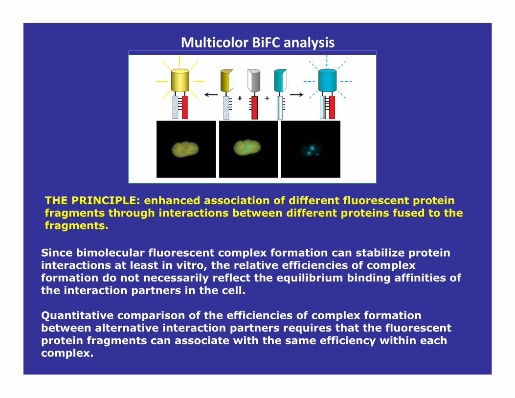
Multicolor!BiFC analysis!
Since bimolecular fluorescent complex formation can stabilize protein interactions at least in vitro, the relative efficiencies of complex formation do not necessarily reflect the equilibrium binding affinities of the interaction partners in the cell.
Quantitative comparison of the efficiencies of complex formationbetween alternative interaction partners requires that the fluorescent protein fragments can associate with the same efficiency within each complex.
THE PRINCIPLE: enhanced association of different fluorescent protein fragments through interactions between different proteins fused to the fragments.
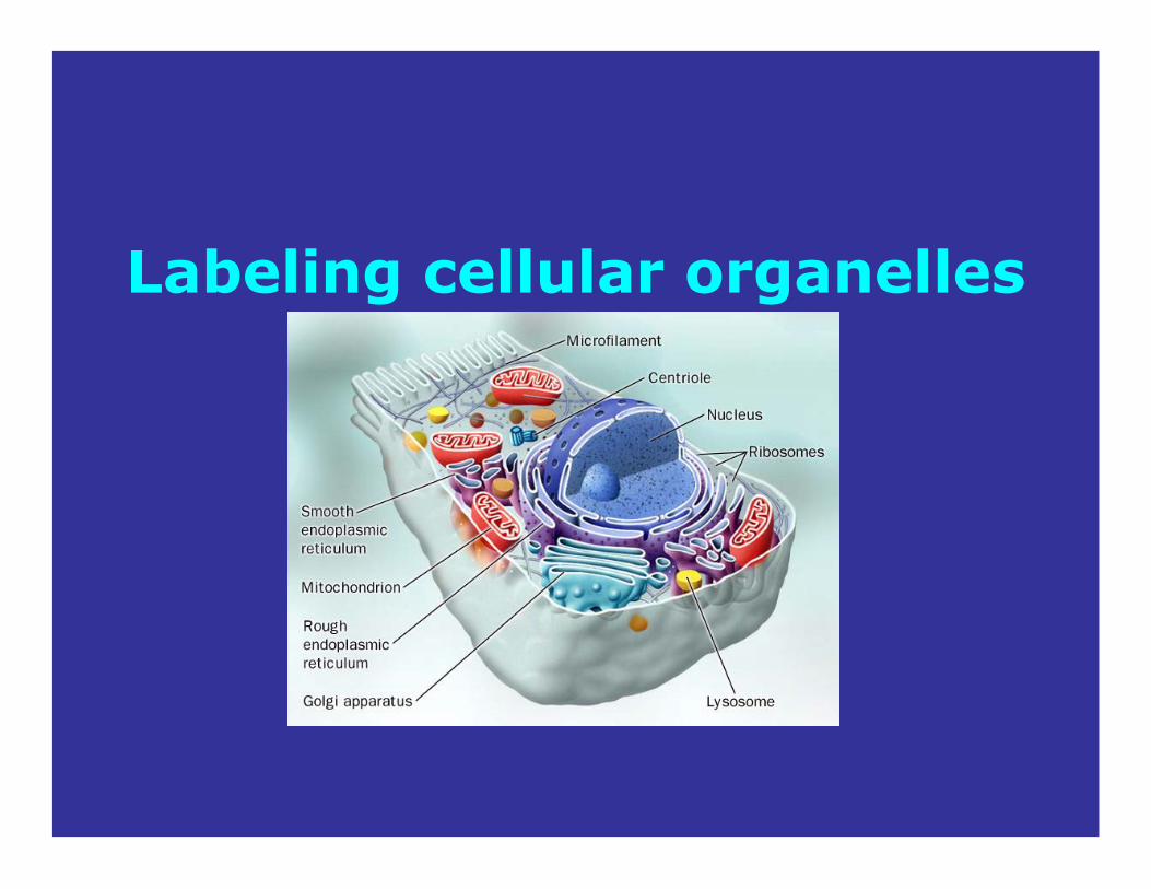
Labeling cellular organelles

www.invitrogen.com
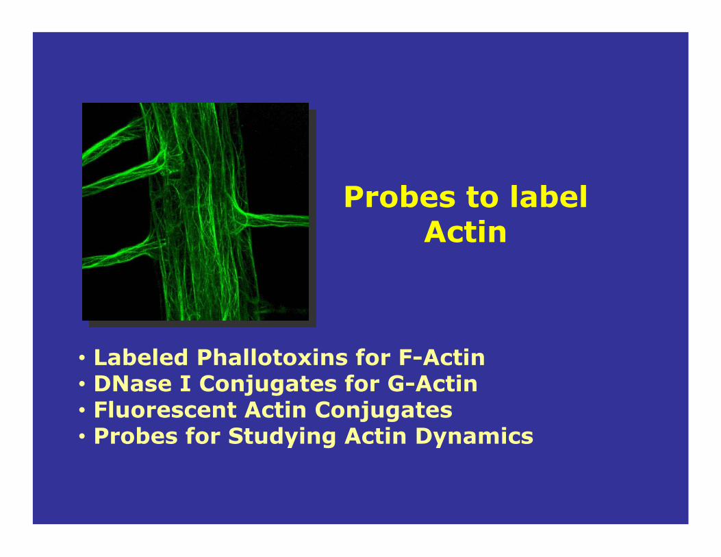
Probes to label Actin
• Labeled Phallotoxins for F-Actin• DNase I Conjugates for G-Actin• Fluorescent Actin Conjugates• Probes for Studying Actin Dynamics

• Phalloidin, a heptapeptide toxin from the poisonous mushroom Amanita phalloides.
• Though highly toxic to liver cells, it has since been found to have little input into the death cap's toxicity as it is not absorbed through the gut
• Binds tightly and specifically to polymerized actin, stabilizing the filaments from a variety of depolymerizing agents and conditions
Phallotoxins bind to filamentous actin (F-actin)
Phallotoxin Conjugatesfor Labeling F-Actin
F-Actin; surface representation of 13 subunit repeat based on Ken Holmes' actin filament model
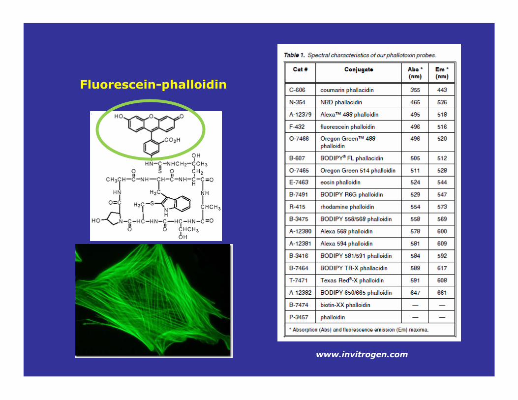
Fluorescein-phalloidin
www.invitrogen.com

The cells were placed under constant illumination on the microscope. Images were acquired at one-second intervals for 30 seconds.
photobleachedto about 20% of its initial value
Intensity stayed at the initial value
The photostability of the Alexa-Fluor dye conjugates is particularly apparent when compared to traditional
fluorophores such as fluorescein
Fluorescein-phalloidin
Alexa Fluor 488-phalloidin
www.invitrogen.com

DNase I Conjugatesfor Labeling G-Actin
G-Actin
fluorescent conjugates of bovine pancreatic DNase I selectively label monomeric G-actin.
www.invitrogen.com

Fluor- phalloidin Alexa488-DNase I
Effect of Latrunculin (inhibits actin polymerization) on Swiss 3T3 cells.
Cramer et al.Cell Motility and the Cytoskeleton 51:27–38 (2002)
EXPERIMENTIncubated in media plusPlus2 M Latrunculin for 30 mFix and double labeled
DNase I conjugates are used in combination with fluorescently labeled phallotoxins to simultaneously visualize G-actin pools
and filamentous F-actin
CONTROLIncubated in media for 30 mFix and double labeled

Probes to label Probes to label MitochondrionMitochondrion
• Rhodamine 123 and Tetrabromorhodamine 123• MitoTacker probes• Antobodies to Mitochondrial proteins• MitoFluor probes• Potential sensitive probes

Rhodamine 123
• Cell-permeant, cationic, • Sequestered by active mitochondria without inducing cytotoxic effects. • Uptake and equilibration takes a few minutes
Staining of rat cortical astrocytes by rhodamine 123
www.invitrogen.com

Mitotracker
Giulia Ossato. LFD-2010
• Cell-permeant mitochondrion-selective dyes which passively diffuse across the plasma membrane and accumulate in active mitochondria • The dye remains associated with the mitochondria after fixation.
MitoTracker Orange CMTMRos
www.invitrogen.com

http://www.med.unipg.it/imagelab/mitoch.html
EXPERIMENT: Mitochondrial behavior in response to UV treatment in NIH 3T3 fibroblasts.
MitoTracker allows direct real-time registration of mitochondrial alterations in response to stressful stimuli
(i.e. oxidative bursts, UV treatment etc.) in "in vitro" cultured cells.
After UV an evident "ballooning" of the mitochondria can be observed.

The mammalian oxidative phosphorylation system, and specific nonoclonal antibodies available from Molecular Probes
Antibodies to Mitochondrial proteinsAntibodies to Mitochondrial proteins
www.invitrogen.com

Antibodies for the different complex are available in different colors
www.invitrogen.com

Contribution to the slidesContribution to the slides
•• Theodore Theodore HazlettHazlett
•• David JamesonDavid Jameson
•• EwaldEwald TerpetschnigTerpetschnig
Related Documents