Engineering Dictyostelium discoideum myosin II for the introduction of site-specific fluorescence probes STUART WAKELIN 1 , PAUL B. CONIBEAR 1 , ROBERT J. WOOLLEY 1 , DAVID N. FLOYD 1 , CLIVE R. BAGSHAW 1, *, MIHA ´ LY KOVA ´ CS 2 and ANDRA ´ S MA ´ LNA ´ SI-CSIZMADIA 2 1 Department of Biochemistry, University of Leicester, Leicester LE1 7RH, United Kingdom; 2 Department of Biochemistry, Eo ¨tvo ¨s Lora ´nd University, H-1117 Budapest, Pa ´zma ´ny Pe´ter se ´ta ´ny 1/C, Hungary Abstract Dictyostelium discoideum is a useful host for the production of constructs for the analysis of structure–function relationships of myosin. Here we describe the use of myosin II constructs containing a single tryptophan residue, at different locations, for probing events at the nucleotide binding site, the relay loop and the communication path between them. GFP fusions have also been expressed at the N- and C-termini of the myosin motor to provide sensitive probes of the actomyosin dissociation reaction in microscope-based kinetic assays. We report on the fluorescence anisotropy of these constructs in the context of their use as resonance energy transfer probes. Introduction The ability to carry out standard molecular genetic procedures and to express milligram quantities of functional myosin constructs has made Dictyostelium discoideum (Dd) a particularly useful organism in the field of cell motility (Ruppel and Spudich, 1996; Kessin, 2001). Here we review and extend our recent work on the introduction of site-specific fluorescent probes for characterising the mechanism of Dd myosin II. Label- ling a protein with fluorescence probes generally in- volves one of three routes (i) introduction of a tryptophan residue (and/or removal on native trypto- phans) to yield a single tryptophan mutant (ii) intro- duction of a cysteine residue (and/or removal on native cysteines) for directed covalent modification (iii) con- struction of a fusion with a green fluorescent protein (or related molecule). In this overview we focus on the first and third approaches. Tryptophan fluorescence Tryptophan residues in proteins are usually the domi- nant source of u.v. absorption and fluorescence emis- sion. Although not an abundant residue, most large proteins contain several tryptophan residues and there- fore the spectroscopic properties reflect the sum of several components (e.g. a myosin motor domain typically contains 4–5 residues). Use of molecular genetic methods to substitute native tryptophan residues with phenylalanine or tyrosine residues to leave a single tryptophan residue per protein molecule is a useful approach because (i) the spectroscopic signature arises from a single known location within the protein (ii) any heterogeneity in the signal may be analysed less ambi- guously than for multi-tryptophan containing proteins (iii) the relative changes in signal on ligand binding may be larger as the background from non-responsive tryptophans is removed. Needless to say in the context of a spectroscopic probe, it is important that the substitution of native tryptophan residues for other aromatic side chains should not significantly alter the overall properties of the protein. Also care is required in selecting the excitation wavelength and slit width to ensure the signal reflects that from the tryptophan residue. The 30 or more tyrosine residues in the myosin motor domain make a significant collec- tive contribution when excited at 280 nm. We generally find that excitation should be at 295 nm or greater, and often employ the 297 nm Hg line from a mercury–xenon lamp. Tryptophan fluorescence has long been used as an empirical probe of myosin conformation during ATPase activity (Bagshaw et al., 1972; Werber et al., 1972; Bagshaw et al., 1974). Most myosin species respond to ATP binding and hydrolysis with a modest but usable change in signal amplitude (5–20% change). Before the impact of molecular biological approaches, the best characterised system was that from vertebrate skeletal myosin subfragment 1, where it was established that tryptophan fluorescence was enhanced (denoted by *) on both the nucleotide binding and hydrolysis steps (Bagshaw et al., 1974; Johnson and Taylor, 1978). A general scheme to account for these transitions was proposed 30 years ago. *To whom correspondence should be addressed: Tel.: +44-116-252- 3454; Fax: +44-116-252-3369; E-mail: [email protected] Journal of Muscle Research and Cell Motility 23: 673–683, 2002. 673 ȑ 2003 Kluwer Academic Publishers. Printed in the Netherlands.

Welcome message from author
This document is posted to help you gain knowledge. Please leave a comment to let me know what you think about it! Share it to your friends and learn new things together.
Transcript

Engineering Dictyostelium discoideum myosin II for the introduction of site-specific
fluorescence probes
STUART WAKELIN1, PAUL B. CONIBEAR1, ROBERT J. WOOLLEY1, DAVID N. FLOYD1,CLIVE R. BAGSHAW1,*, MIHALY KOVACS2 and ANDRAS MALNASI-CSIZMADIA2
1Department of Biochemistry, University of Leicester, Leicester LE1 7RH, United Kingdom; 2Department ofBiochemistry, Eotvos Lorand University, H-1117 Budapest, Pazmany Peter setany 1/C, Hungary
Abstract
Dictyostelium discoideum is a useful host for the production of constructs for the analysis of structure–functionrelationships of myosin. Here we describe the use of myosin II constructs containing a single tryptophan residue,at different locations, for probing events at the nucleotide binding site, the relay loop and the communicationpath between them. GFP fusions have also been expressed at the N- and C-termini of the myosin motor toprovide sensitive probes of the actomyosin dissociation reaction in microscope-based kinetic assays. We reporton the fluorescence anisotropy of these constructs in the context of their use as resonance energy transferprobes.
Introduction
The ability to carry out standard molecular geneticprocedures and to express milligram quantities offunctional myosin constructs has made Dictyosteliumdiscoideum (Dd) a particularly useful organism in thefield of cell motility (Ruppel and Spudich, 1996; Kessin,2001). Here we review and extend our recent work onthe introduction of site-specific fluorescent probes forcharacterising the mechanism of Dd myosin II. Label-ling a protein with fluorescence probes generally in-volves one of three routes (i) introduction of atryptophan residue (and/or removal on native trypto-phans) to yield a single tryptophan mutant (ii) intro-duction of a cysteine residue (and/or removal on nativecysteines) for directed covalent modification (iii) con-struction of a fusion with a green fluorescent protein (orrelated molecule). In this overview we focus on the firstand third approaches.
Tryptophan fluorescence
Tryptophan residues in proteins are usually the domi-nant source of u.v. absorption and fluorescence emis-sion. Although not an abundant residue, most largeproteins contain several tryptophan residues and there-fore the spectroscopic properties reflect the sum ofseveral components (e.g. a myosin motor domaintypically contains 4–5 residues). Use of moleculargenetic methods to substitute native tryptophan residues
with phenylalanine or tyrosine residues to leave a singletryptophan residue per protein molecule is a usefulapproach because (i) the spectroscopic signature arisesfrom a single known location within the protein (ii) anyheterogeneity in the signal may be analysed less ambi-guously than for multi-tryptophan containing proteins(iii) the relative changes in signal on ligand binding maybe larger as the background from non-responsivetryptophans is removed. Needless to say in the contextof a spectroscopic probe, it is important that thesubstitution of native tryptophan residues for otheraromatic side chains should not significantly alterthe overall properties of the protein. Also care isrequired in selecting the excitation wavelength and slitwidth to ensure the signal reflects that from thetryptophan residue. The 30 or more tyrosine residuesin the myosin motor domain make a significant collec-tive contribution when excited at 280 nm. We generallyfind that excitation should be at 295 nm or greater, andoften employ the 297 nm Hg line from a mercury–xenonlamp.Tryptophan fluorescence has long been used as an
empirical probe of myosin conformation during ATPaseactivity (Bagshaw et al., 1972; Werber et al., 1972;Bagshaw et al., 1974). Most myosin species respond toATP binding and hydrolysis with a modest but usablechange in signal amplitude (5–20% change). Before theimpact of molecular biological approaches, the bestcharacterised system was that from vertebrate skeletalmyosin subfragment 1, where it was established thattryptophan fluorescence was enhanced (denoted by *)on both the nucleotide binding and hydrolysis steps(Bagshaw et al., 1974; Johnson and Taylor, 1978). Ageneral scheme to account for these transitions wasproposed 30 years ago.
*To whom correspondence should be addressed: Tel.: +44-116-252-
3454; Fax: +44-116-252-3369; E-mail: [email protected]
Journal of Muscle Research and Cell Motility 23: 673–683, 2002. 673� 2003 Kluwer Academic Publishers. Printed in the Netherlands.

Interestingly, wild-type Dd myosin lacks tryptophanresidues close to the active site (equivalent to W113 andW131 of skeletal muscle myosin) and shows no en-hancement in fluorescence on nucleotide binding (in factthere is a small quench denoted with� (Kuhlman andBagshaw, 1998; Malnasi-Csizmadia et al., 2000)). Anenhancement in tryptophan fluorescence is observed andhas been associated with ATP hydrolysis. Thus thecorresponding scheme for Dd myosin II motor is
Selective chemical labelling studies have enabled parti-cular tryptophans in skeletal muscle myosin to beidentified as the major sensors of conformation duringthe ATPase activity (Park and Burghardt, 2000) but themolecular biological approach has proven more flexible.For example, the latter as shown unambiguously that theconserved relay loop tryptophan (W501 in Ddmyosin II)is the sensor of the ‘hydrolysis’ step 3 in Dd myosin II(Batra and Manstein, 1999; Malnasi-Csizmadia et al.,2000) and vertebrate smooth muscle myosin (Onishiet al., 2000; Yengo et al., 2000). Furthermore, time-resolved studies have shown that the hydrolysis step is acomposite one in which a loosely coupled conforma-tional transition (probably corresponding to the open–closed transition identified by crystallography; (Geevesand Holmes, 1999)) precedes the chemical step and thatthe tryptophan residue actually responds to the former(Malnasi-Csizmadia et al., 2001b; Urbanke and Wray,2001). The detailed kinetic arguments for these conclu-sions are given in the original papers (Malnasi-Csiz-madia et al., 2000; 2001a,b; Kovacs et al., 2002). Rather,here, we put the information in context and highlightsome salient features.
The open–closed transition
While there have been a number of experimentalapproaches that have indicated that the myosin motordomain can bend (Highsmith and Eden, 1990; Tokuna-ga et al., 1991), the crystallisation of the myosin head ininitially two (now three) distinct conformations provid-
ed, perhaps, the most convincing and certainly the mostdetailed description of the sub-domains involved in themovement (Geeves and Holmes, 1999). The originalstructures of Dd myosin in the ADP.BeFx andADP.AlF4 complexes provided the first clues that, upon‘hydrolysis’, the movement of the switch II region wastransmitted towards the C-terminus via the relay helixand allowed the converter domain to roll around thelatter and could impart a significant rotational motionto so-called lever arm (Fisher et al., 1995). Subsequentstudies of smooth muscle and scallop myosin crystalstructures, with an intact light chain(s) supported andextended these ideas (Dominguez et al., 1998; Houdusseet al., 2000). In general, it was found that non-hydro-lysable nucleotides favoured the ‘open’ state in whichthe switch II region was 0.8 nm from the c-phosphate(or equivalent moiety) and the lever arm was exten-ded, while analogues of hydrolysed nucleotide state(ADP.AlF4 and ADP.Vi) favoured the ‘closed’ state andcaused the lever arm to swing through around 70�.Solution studies, with probes designed to sense the leverarm position (Suzuki et al., 1998; Shih et al., 2000),support the idea that these movements are not crystalartifacts, although relating these spectroscopic signals tothe crystal structures remains a challenging task.These structural studies provide a ready explanation
as to why the conserved tryptophan (Dd W501) in therelay loop is sensitive to ATP hydrolysis. Why W501gives a large enhancement in fluorescence (70–100%increase in the single tryptophan mutant) is less easy toexplain in detail, although a number of contributoryfactors can be identified. Comparison of the crystalstructures for the ADP.Vi complex (pdb:1VOM, (Smithand Rayment, 1996) and ADP.BeFx (pdb:1G8X, (Kli-che et al., 2001) suggests the tryptophan becomesslightly less solvent accessible in the closed state,although in other structures (pdb: 1MMD, 1MND,(Fisher et al., 1995) the W501 tryptophan is notresolved, indicating substantial disorder. Collisionalquenching measurements with acrylamide indicate thatW501 is more protected during steady-state ATPhydrolysis (i.e. predominantly the M.ADP.Pi state) thanin the M.ADP state, but the difference is small and lessthan the protecting effect of nucleotide binding to theapo state (Malnasi-Csizmadia et al., 2000). Also, itshould be stressed that, prior to the enhancement inemission intensity on ‘hydrolysis’ i.e. the open–closedtransition, there is a quench in W501 tryptophanfluorescence associated with the nucleotide binding step.Thus the relay loop is sensing events initiated at theactive site earlier in time than the major open–closedtransition. Interestingly, comparable studies on engi-neered smooth muscle constructs indicate that theequivalent W512 tryptophan responds to nucleotidebinding with an enhancement, followed by a furtherenhancement on ‘hydrolysis’ (Yengo et al., 2000). Mostlikely these changes reflect a common movement of therelay loop, but the different residues around the con-served tryptophan result in fluorescence changes of
M + ATP
M.ATP
M*.ATP M**.ADP.Pi
M + ADP M.ADP M*.ADP + Pi M*.ADP.Pi
1 2 3
7 6 5
4
ð1Þ
M + ATP M.ATP M†.ATP M*.ADP.Pi
M + ADP M.ADP M†.ADP + Pi M†.ADP.Pi
1 2 3
7 6 5
4
ð2Þ
674

different sign and magnitude. The complexity of theobserved net emission intensity is also apparent fromfluorescence lifetime studies. In common with practicallyevery other single tryptophan containing protein exam-ined (Lakowicz, 1999), the fluorescence lifetime of W501in all myosin nucleotide states is at least biphasic(Malnasi-Csizmadia et al., 2001a). Together with theexistence of a statically quench component, thesefindings argue that at least three different local confor-mational states of the tryptophan are present for each‘single’ bulk conformation defined for each biochemicalstate. These microstates interconvert slowly on thenanosecond time scale but rapidly on the millisecondtimescale. How these microstates redistribute to give anet enhancement or quench is difficult to predict. It islikely these local conformation states reflect the differentrotamers of the tryptophan residue, as well as differentrotamers of residues in contact with this side chain.Interestingly different rotamers for W501 were ob-
served by crystallography in the two motor domains ofeach unit cell of the Dd myosin motor-a actinin fusionconstruct (M761-2R-R238E, pdb 1G8X, (Kliche et al.,2001). While it might be possible to pin down adominant interaction between the tryptophan residuesand another side chain (or indeed backbone carbonylgroup) that is responsible for the enhancement (or de-quench) (Park and Burghardt, 2002), interpreting theamplitude of the fluorescence changes, even of singletryptophan mutants, at present remains semi-empirical.Although these findings might be regarded as anunnecessary complication of the picture, with littlerelevance to physiological timescales and events, itprovides an appropriate reminder as to the dynamicsof side chains and care required in interpreting interac-tions between residues based on small differences instatic separation.How can tryptophan fluorescence studies be used to
complement structural studies? The main strength of theformer comes from its application as a signal fortransient kinetic studies. Here we have shown that theW501 enhancement occurs with a maximum observedrate constant of 30 s)1 at saturating [ATP] in stopped-flow experiments, in line with the wild-type protein(Malnasi-Csizmadia et al., 2000). This was generallyassumed to provide a measure of the effective hydrolysisstep. In fact, W501 senses a coupled reaction, as alludedto above, with an unfavourable but rapid (�1000 s)1)conformational transition followed by hydrolysis itself,that has an intrinsically higher rate constant than 30 s)1.These processes can only be resolved by perturbationmethods (Malnasi-Csizmadia et al., 2001b), because instopped-flow mixing methods the rapid open–closedtransition is limited by a prior isomerisation associatedwith the binding step. The identification of the observedconformational change as being the open–closed tran-sition rests on the expected change in environment ofW501 and the idea that switch II region has to move tobring the catalytic residues in close contact with the c-phosphate for hydrolysis (i.e. this step must precede
hydrolysis (Geeves and Holmes, 1999) – althoughwhether the steps are kinetically resolvable depends onfortuitous values of the rate constants). The schemeshown in Equation (2) was therefore expanded toseparate the open–closed transition from hydrolysis.
The fact that the combined open–closed transitionand hydrolysis steps are perturbed by temperature andpressure jumps emphasises another important property– these steps are freely reversible, but favour hydrolysis(i.e. M*.ADP.Pi) overall. This idea (for the overallhydrolysis reaction) was proposed 30 years ago basedon quenched flow and isotope exchange studies (Bag-shaw and Trentham, 1973; Bagshaw et al., 1975). Thesignificance of this finding is that little energy is wastedin moving the detached crossbridge into a state ready forthe next crossbridge cycle (but the cocking of thecrossbridge depicted in some text book models isperhaps overly simplistic). The novel recent finding,based in part on the W501 data (Malnasi-Csizmadiaet al., 2001b), is that the detached crossbridge canundergo rapid motions, more than an order of magni-tude faster than the observed hydrolysis rate, to exploreactin binding sites. It is possible that this equilibriumalso exists with weakly attached crossbridges becausethe rate constant for the ATP-induced dissociation ofacto-Dd myosin motor (150 s)1) is several times slowerthan k)3a (Kuhlman and Bagshaw, 1998). Howeveractin may modulate this rate constant, and in astructured system, the open–closed transition of theactin-bound head may be restrained by mechanicalcoupling. The concept of dynamically disordered weaklyattached crossbridges was previously deduced from eprmeasurements (Thomas et al., 1995).The relative insensitivity of W501 to the initial
nucleotide binding events allows the equilibrium orsteady-state fluorescence intensity to provide a measureof the equilibrium constant for the open–closed transi-tion (in combination with the subsequent hydrolysisequilibrium where applicable). In this procedure weassume that the fluorescence emission of the M�.ADPand M*.ADP.AlF4 species represent the fully open andfully closed state respectively. We have extended ouroriginal studies to explore ligands such as PPi, ITP andGTP (Table 1). The latter nucleotide shows little phos-phate burst with other myosin species (White et al.,1993, 1997) yet it results in a marked fluorescenceenhancement of W501 indicating the formation of asmall but significant amount of the closed state M*.GTPduring the steady-state turnover. These estimates requireconfirmation by perturbation methods. For example,the apo M state shows a fluorescence yield between the
M + ATP M.ATP M†.ATP M*.ATP M*.ADP.Pi
M + ADP M.ADP M†.ADP + Pi M†.ADP.Pi
1 2 3a 3b
7 6 5
4
ð3Þ
675

M�.ADP and M*.ADP.AlF4 values that might suggest itis a equilibrium mixture of open:closed states with Keq
of around 0.2. However, pressure jump failed to resolvea transient (Malnasi-Csizmadia et al., 2001b), indicatingthe equilibrium lies strongly towards one (presumablyopen) state. In this case, the difference in fluorescencebetween M and M�.ADP is accounted for by a separateisomerisation step (K6), discussed below. Furthermore,the apo M fluorescence emission peak is red-shiftedcompared with that of the M�.ADP and M*.ADP.AlF4
complexes, and therefore cannot be in an intermediateconformation.Consideration of the crystal structures suggests that
the product phosphate cannot dissociate from the closedstate as the ‘back door’ is obstructed. One mighttherefore consider that the M*.ADP.Pi complex mustreturn to the open state before Pi release (Yount et al.,1995). This idea is emphasised by representing thekinetic scheme in terms of three fundamental myosinstates; apo, open and closed:
In order that the M*.ADP.Pi state is the predominantsteady-state intermediate, it is necessary for either k4 tobe slow (0.05 s)1) or that K4 is rapid but �1 so thatwhen coupled to the Pi release step, the overall rateconstant is slow (i.e. K4k5 ¼ 0.05 s)1). These options aredifficult to distinguish because the M�.ADP.Pi remains
at low concentration throughout. Previous attempts todetermine K4 and K5 as individual equilibrium constantswith vertebrate skeletal myosin, via incorporation of Piinto protein-bound ATP, were only part successfulbecause of uncertainties in the effects of ionic strengthchanges. A model independent estimate of K4K5 ¼0.19 M was obtained and demonstrated the extremelyweak Pi binding to the equivalent of M�.ADP (Mann-herz et al., 1974; Goody et al., 1977).Equation (4) implies that the active site must close to
allow efficient hydrolysis, but open to allow Pi release.Experimental conditions, ATP analogues, chemicalmodifications and mutations may favour the open orclosed state. Modifications that slightly favour the openstate can lead to a slower net hydrolysis rate, but fasterPi release. Given that the latter is the observed rate-limiting step for myosin alone, it is perhaps notsurprising that many minor modifications lead to anincrease in the basal ATPase rate but reduced actin-activation, because the latter may become limited by thehydrolysis step (Sasaki et al., 2002). More extrememutations appear to lock the myosin in open (e.g.G457A) or closed (e.g. E459A) states, and severelyinhibit both basal and actin-activated ATPase (Sasakiet al., 1998). This effect is also illustrated by mutationsthat modify the interface between the N-terminaldomain and converter region. In the wild type Ddmyosin motor there is a charge repulsion between K84and R704 in the open state, but not in the closed statewhere the residues are 3.5 nm apart. In mutations suchas K84M and R704E where the interaction is neutralisedor changed to an attractive force respectively, the levelof fluorescence during the steady-state hydrolysis ofATP indicates a higher proportion of the open state(Malnasi-Csizmadia et al., 2002).Equation (4) indicates how tight binding Pi analogues
such as Vi and AlF4 can drive the formation of anM*.ADP.Pi like state and, in these cases at least, theequilibrium constant equivalent to K4 is �1 for the wildtype myosin motor, in order that almost all of the myosinis trapped in a high fluorescent closed state. For BeFx,K4
appears to be around 1 at ambient temperatures and thusboth open and closed states are nearly equally occupied(Malnasi-Csizmadia et al., 2001b). However, if the Beatom forms a covalent link with the oxygen atom of theb-phosphate of ADP (i.e. it acts as an ATP analog), thenthe rapid transient observed by relaxation methodsmight reflect the open–closed transition associated withstep 3a. This ambiguity can only be removed usingstructural methods, such as nmr, to monitor the extent ofcovalent bond formation (Henry et al., 1993).The favourable characteristics of the tryptophan
emission from the W501þ construct encouraged us tore-examine the formation of the M*.ADP.Pi state bymixing the protein with high concentrations of ADP andPi. Earlier attempts with vertebrate skeletal myosin failedto detect any enhancement in fluorescence above theskeletal M*.ADP state (Equation (1)). Indeed, a slightreduction in fluorescence was noted and interpreted as
Table 1. Equilibrium constants for the open–closed transition in the
presence of different ligands at 20 �C estimated from the fluorescence
emission from W501
Ligand
W501 Fluorescence
change (%) appKoc Koc
ADP �11 <0.05
ATP þ72 �32 �0.4
ATPcS þ14 �0.3 £0.3AMP.PNP þ28 �0.82
ADP.BeFx þ39 �1.43
ADP.AlF4 þ72 >20
PPi �10 <0.02
Pi <�5 <0.06
ITP þ43 �1.7 £1.7GTP þ25 �0.7 £0.7
Tryptophan fluorescence was measured relative to the apo state. These
data were collected using several different preparations over a period
of 3 years on either the steady-state fluorimeter or stopped-flow
apparatus. The data were therefore normalised for each ligand using
the observed fluorescence levels in the presence of ADP and ATP as
reference points. For hydrolysable nucleotides the observed equili-
brium constant (appKoc) is affected by coupling to the hydrolysis step.
2 61 7
3a 4
M*. ATP M*.ADP.Pi closed
M .ATP† M .ADP.Pi† M†.ADP + Pi open
3b
M + ATP M.ATP M.ADP M + ADP apo
5
ð4Þ
676
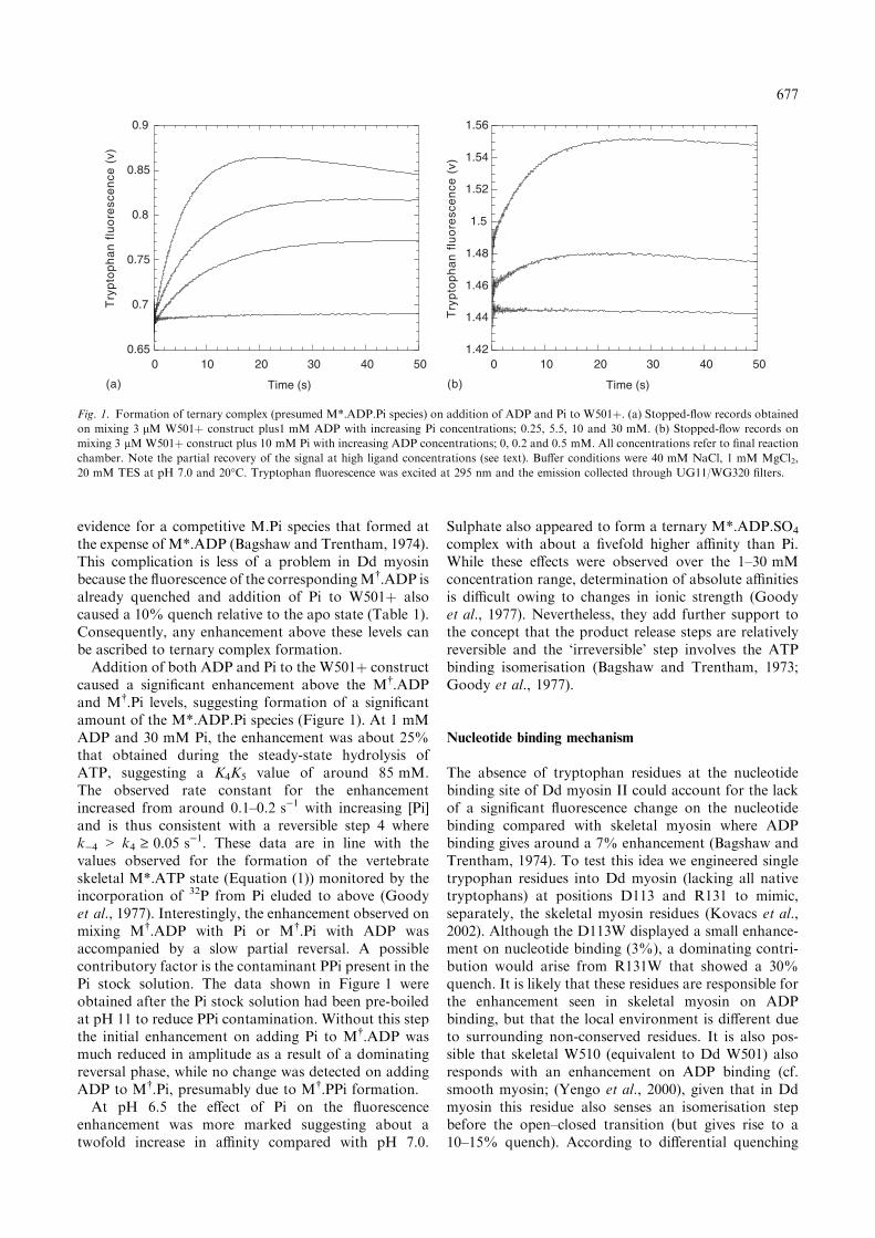
evidence for a competitive M.Pi species that formed atthe expense of M*.ADP (Bagshaw and Trentham, 1974).This complication is less of a problem in Dd myosinbecause the fluorescence of the correspondingM�.ADP isalready quenched and addition of Pi to W501þ alsocaused a 10% quench relative to the apo state (Table 1).Consequently, any enhancement above these levels canbe ascribed to ternary complex formation.Addition of both ADP and Pi to the W501þ construct
caused a significant enhancement above the M�.ADPand M�.Pi levels, suggesting formation of a significantamount of the M*.ADP.Pi species (Figure 1). At 1 mMADP and 30 mM Pi, the enhancement was about 25%that obtained during the steady-state hydrolysis ofATP, suggesting a K4K5 value of around 85 mM.The observed rate constant for the enhancementincreased from around 0.1–0.2 s)1 with increasing [Pi]and is thus consistent with a reversible step 4 wherek)4 > k4 ‡ 0.05 s)1. These data are in line with thevalues observed for the formation of the vertebrateskeletal M*.ATP state (Equation (1)) monitored by theincorporation of 32P from Pi eluded to above (Goodyet al., 1977). Interestingly, the enhancement observed onmixing M�.ADP with Pi or M�.Pi with ADP wasaccompanied by a slow partial reversal. A possiblecontributory factor is the contaminant PPi present in thePi stock solution. The data shown in Figure 1 wereobtained after the Pi stock solution had been pre-boiledat pH 11 to reduce PPi contamination. Without this stepthe initial enhancement on adding Pi to M�.ADP wasmuch reduced in amplitude as a result of a dominatingreversal phase, while no change was detected on addingADP to M�.Pi, presumably due to M�.PPi formation.At pH 6.5 the effect of Pi on the fluorescence
enhancement was more marked suggesting about atwofold increase in affinity compared with pH 7.0.
Sulphate also appeared to form a ternary M*.ADP.SO4
complex with about a fivefold higher affinity than Pi.While these effects were observed over the 1–30 mMconcentration range, determination of absolute affinitiesis difficult owing to changes in ionic strength (Goodyet al., 1977). Nevertheless, they add further support tothe concept that the product release steps are relativelyreversible and the ‘irreversible’ step involves the ATPbinding isomerisation (Bagshaw and Trentham, 1973;Goody et al., 1977).
Nucleotide binding mechanism
The absence of tryptophan residues at the nucleotidebinding site of Dd myosin II could account for the lackof a significant fluorescence change on the nucleotidebinding compared with skeletal myosin where ADPbinding gives around a 7% enhancement (Bagshaw andTrentham, 1974). To test this idea we engineered singletrypophan residues into Dd myosin (lacking all nativetryptophans) at positions D113 and R131 to mimic,separately, the skeletal myosin residues (Kovacs et al.,2002). Although the D113W displayed a small enhance-ment on nucleotide binding (3%), a dominating contri-bution would arise from R131W that showed a 30%quench. It is likely that these residues are responsible forthe enhancement seen in skeletal myosin on ADPbinding, but that the local environment is different dueto surrounding non-conserved residues. It is also pos-sible that skeletal W510 (equivalent to Dd W501) alsoresponds with an enhancement on ADP binding (cf.smooth myosin; (Yengo et al., 2000), given that in Ddmyosin this residue also senses an isomerisation stepbefore the open–closed transition (but gives rise to a10–15% quench). According to differential quenching
0.65
0.7
0.75
0.8
0.85
0.9
0 10 20 30 40 50 0 10 20 30 40 50
Try
pto
ph
an
flu
ore
sce
nce
(v)
Time (s)
1.42
1.44
1.46
1.48
1.5
1.52
1.54
1.56
Try
pto
ph
an
flu
ore
sce
nce
(v)
Time (s)(a) (b)
Fig. 1. Formation of ternary complex (presumed M*.ADP.Pi species) on addition of ADP and Pi to W501þ. (a) Stopped-flow records obtained
on mixing 3 lM W501þ construct plus1 mM ADP with increasing Pi concentrations; 0.25, 5.5, 10 and 30 mM. (b) Stopped-flow records on
mixing 3 lM W501þ construct plus 10 mM Pi with increasing ADP concentrations; 0, 0.2 and 0.5 mM. All concentrations refer to final reaction
chamber. Note the partial recovery of the signal at high ligand concentrations (see text). Buffer conditions were 40 mM NaCl, 1 mM MgCl2,
20 mM TES at pH 7.0 and 20�C. Tryptophan fluorescence was excited at 295 nm and the emission collected through UG11/WG320 filters.
677
measurements (Park and Burghardt, 2000), skeletalW131 is sensitive to nucleotide binding (16–25% en-hancement) but it does not sense subsequent steps,whereas W510 shows an enhancement on both nucleo-tide binding (38% enhancement on ADP) and thehydroylsis (i.e. open–closed) transition (ATP caused a95%, and ADP.AlF4 caused a 102% enhancementrelative to the apo state). Although our Dd tryptophanmutants did not provide a generally applicable solutionas to the contributions from the various tryptophanresidues to net fluorescence changes, the R131W con-struct did provide a useful probe for nucleotide bindingwithout the complications of a signal from the subse-quent open–closed transition.Another mutant F129W provided an even larger
signal that R131W and was the focus of detailedcharacterisation (Kovacs et al., 2002). Although thefluorescence signal was in the opposite direction to thatobserved in skeletal myosin, the Dd F129W constructshowed very similar kinetics of nucleotide binding inyielding biphasic kinetic profiles (Trybus and Taylor,1982). While these ATP binding data could be modelledin terms of two sequential isomerisations (with rateconstants of the order of 1800 and 350 s)1 for DdF129W), there was an indication of a third processwhereby ATP bound weakly in a non-competent con-formation at the active site (Kovacs et al., 2002). It ispossible that this incorrectly bound substrate canreorient at the active site to join the main pathway.There remains considerable uncertainty as to the natureof these alternative pathways (Tesi et al., 1989). As withthe W501þ construct, lifetime-resolved fluorescencestudies with F129W indicate that each biochemicalstable state actually comprises three or more micro-states. These measurements were made possible by theincreased signal-to-background achieved by engineeringa single tryptophan probe as well as improvements inthe dead-time of the stopped-flow apparatus. Theyillustrate a general point that technological advances,besides providing a more precise answer to an existingquestion, often pose new questions, too. It is likely thatprotein–ligand interactions and catalysis are more gen-erally described by an energy landscape rather thanspecific sequential pathway, and thus fitting to discreteexponentials is an approximation. Thus kinetic schemesare a short-hand way of lumping rapidly interconvertingintermediates into single states for the convenience ofthe problem to be addressed while the number of stepsresolved reflects the nature of the experiment.The isomerisations associated with nucleotide binding
sensed by F129W are clearly distinct from the open–closed transition sensed by W501, as illustrated in the‘double’ mutant (F129W/W501þ) where they are clearlyresolved (Kovacs et al., 2002). The binding isomerisa-tions are more rapid and less reversible than theobserved coupled open–closed/hydrolysis transition.We are currently investigating the influence of F-actinon the steps sensed by single tryptophan residues inthe myosin motor domain. Interestingly, a fluorescence
enhancement occurs when a Dd myosin motor lackingall tryptophans (W), (Malnasi-Csizmadia et al., 2000))binds to native vertebrate skeletal actin, indicating oneor more tryptophan residues in actin is perturbed by thisprocess (cf. (Johnson and Taylor, 1978; Yengo et al.,1999)). However the signal-to-noise is sufficient to detectthe contributions from single tryptophan residues withinthe myosin motor, when present, against the back-ground actin signal.
Tryptophan probes in other locations
We have also prepared tryptophan mutants at otherlocations in the Dd myosin motor domain to probe thepath of conformational changes between the nucleotidesite and C-terminus. Single tryptophans were introducedinto a tryptophan-less background (W)) at the F458(W458þ construct), F461 (W461þ) and F692 (W692þ).We also produced a double tryptophan construct havingthe native W501 and F692W mutation (W501/692þ).Based on the atomic structure, the F692 side chainmakes a van der Waals interaction with W501, althoughthese residues are located in separate sub-domains (theconverter and relay loop, respectively). F458 is a keyresidue of the switch II loop. F461 is at the start of thisloop, at the junction of the actin binding cleft and therelay-converter region.The F458W mutation caused at least a 20 times
reduction in the steady-state ATPase activity and didnot show a significant fluorescence emission change onadding nucleotide. We did not characterise it further. Incontrast, the W461þ construct had similar steady-stateMgATPase activity (0.035 s)1) to the wild-type motordomain (0.03 s)1). The fluorescence emission intensity ofW461 was sensitive to nucleotide states, with ADP andATP showing a 12 and 22% quench in the peak emissionat the 345 nm, respectively. The stopped-flow records ofthe reaction of W461þ with ADP could be fitted tosingle exponentials and the observed rate constantdid not saturate up to 400 lM ADP concentration.These data yielded a second-order rate constant of2 · 106 M)1 s)1 and an intercept (dissociation rate con-stant) value of 10 s)1. This represents a slightly fasterbinding process than the wild type, however, theequilibrium binding constant is unchanged. On mixingW461þ with ATP below 150 lM nucleotide concentra-tions, the fluorescence profiles could be fitted by singleexponentials but, at higher substrate concentrations,two phases were distinguished. The fast phase representsa second-order binding reaction (rate constant 0.8 ·106 M)1 s)1) and did not saturate up to 750 lM ATPconcentration, whereas the slow phase reached a max-imum of 30 s)1 above 200 lM ATP. We suppose thatW461 senses both the nucleotide binding and open–closed conformational transition.The steady-state MgATPase activity was not changed
by the F692W mutation. In contrast to the W501þconstruct, which shows a fluorescence enhancement of
678

80% on ATP addition, the fluorescence of the W692þconstruct was reduced by 11% compared with the apoform. ADP reduced emission intensity of W692þ by 7%.There was no significant spectral shift with either ATP orADP. The W501/692þ construct showed a 45% fluores-cence enhancement on adding ATP, but the spectra of theapo and ADP forms were indistinguishable. There wasno spectral shift on adding nucleotide, however, all threeof the spectra (apo, þADP andþATP) were blue-shifted(maximum at 333 nm) compared to the apo form of theW501þ construct. While apo W501þ and apo W692þhad similar fluorescence emission intensities, the apoW501/692þ emission was only 67% of their sum at thespectral maxima. To compare the relative self-quenchingeffect of the nearby tryptophans in the W501þ construct,the areas under the spectra of all three constructs in thepresence and absence of ATP and ADP were integrated.We found that in the apo states, the self-quenching effectin the 501/692þ was 3.30 times while in the presence ofATP (or ADP.AlF4 complex) and ADP it was only 2.60and 2.99 times, respectively. Given the tight packing ofhydrophobic residues in the interface between the relayloop and converter domain, there is little scope forsignificant distant changes between W501 and W692.However orientation changes are likely, which wouldaffect the efficiency of energy transfer between thetryptophans. Indeed, if the tryptophans reside in differentrotamer micro-states, as evidenced by the lifetime andcrystallographic results eluded to above, a redistributionof the relative orientations ofW501 andW692 side chainsin different nucleotide states is a likely outcome.
GFP-motor domain fusions
Green fluorescent protein fusions have proven valuabletools for studying protein function both within cells and
in vitro. Our incentive for characterising such Dd myosinconstructs has been to develop sensitive microscope-based kinetic assays as well as for FRET studies. Inparticular, it is of interest to follow the paths ofconformational changes through the molecule to deter-mine if there is tight coupling between the movement ofswitch 2, the relay loop and the converter domain. Theelegant approach of Suzuki et al. (1998) showed asubstantial FRET change between a BFP and GFPmoiety fused at the N- and C-termini of the Dd myosinII motor domain in different nucleotide states. Forquantitative analysis of such data, information on therotational properties and/or orientation of the fluoro-phores is required in order to assess the appropriate j2
value for the Forster equation. We have initiallycharacterised a single GFP construct fused at the C-terminus of the Dd myosin W501þ motor constructwith an intervening Gly–Gly–Gly linker sequence. Wehave also prepared a YFP fusion attached to the N-terminus of W501þ with no linker. The motor domainwas based on the single tryptophan containing W501þconstruct. These fusions gave a well-resolved tryptophanpeak that responded to ATP binding with kineticssimilar to the W501þ construct alone (Figure 2a). Asexpected, the fluorescence from GFP moiety itself wasinsensitive to ATP binding and hydrolysis (Figure 2b).Furthermore we could detect no change in energytransfer from tryptophan to GFP on addition of ATP.However it is likely that the energy transfer from Trp toGFP is dominated by the two nearby tryptophans in theGFP moiety (van Thor et al., 2002), and any FRETbetween W501 and GFP fluorophore is negligible.Steady-state anisotropy measurements on the isolatedGFP protein and the myosin motor-fusion indicatedthat the fluorophore was relatively immobile on thenanoseconds timescale (Table 2). These data are in linewith earlier reports (Chattoraj et al., 1996) and are to be
1.15
1.2
1.25
1.3
Try
pto
ph
an
flu
ore
sce
nce
(v)
Time (s)
0.85
0.9
0.95
1
GF
P f
luo
resc
en
ce (
v)
Time (s)
0 0.2 0.4 0.6 0.8 1 0 0.2 0.4 0.6 0.8 1
(b)(a)
Fig. 2. Stopped-flow traces of ATP interaction with the W501þ–GFP construct monitored by (a) tryptophan fluorescence and (b) GFP
fluorescence. ATP (25 lM) was mixed with 0.5 lM protein (reaction chamber concentrations) in 40 mM NaCl, 20 mM HEPES and 2 mM
MgCl2 at pH 7.5 and 20�C. Tryptophan fluorescence was monitored by excitation at 295 nm and the emission selected with UG11 and WG320
filters. GFP fluorescence was excited at 490 nm and monitored with an OG515 cut-off filter.
679

expected from the molecular volume of GFP alone.Tethering GFP to the motor domain causes only a slightadditional restriction. The anisotropy of the fusionprotein was insensitive to ATP addition, both in thesteady-state and in polarisation-resolved stopped-flowmeasurements. The value was also unchanged when thefusion protein was bound to actin filaments. Thedifference from the theoretical maximum anisotropyvalue of 0.4, for totally immobilised fluorophoresrandomly distributed in solution, probably arises froma small difference between the absorption and emissiondipoles of the GFP flourophore (Lakowicz, 1999; Boxerand Rosell, 2002). These data indicate that a j2 value of0.67, deduced for pairs of rapidly rotating fluorophores,would be inapplicable to GFP fusions.Actin filaments, decorated with the Dd myosin
motor–GFP fusion, were readily observed under the
fluorescence microscope. When the emitted light wasresolved into two planes of polarisation, there was nomarked difference between the intensity from filamentsaligned along or across the plane of polarisation. Thisobservation argues that either the GFP domains are notwell ordered, or the preferred direction of the dipoles isneither parallel nor perpendicular to the actin filamentaxis, but at some intermediate angle that becomesaveraged out along the actin helix. These possibilitiescan be separated at the level of single molecule fluores-cence (Forkey et al., 2000).The visualisation of GFP–myosin motors bound to
actin by total internal fluorescence microscopy (TIRF)provides a very sensitive method for determining thekinetics of ATP-induced dissociation. Addition of cagedATP at mM concentrations to the GFP–Dd myosin IImotor decorated actin filaments causes little or no loss insignal intensity. However on flash photolysis at 350 nm(Conibear and Bagshaw, 2000), the GFP fusion proteinis quickly released from the actin (Figure 3). Controlexperiments in the absence of caged ATP shows that theflash does not cause significant photobleaching of theGFP moiety. Interestingly, when the same experimentswere performed with YFP-fusion proteins, a smallenhancement was seen in the control that we interpretas photoactivation of the dark state of YFP (Miyawakiand Tsien, 2000). This phenomenon did not prevent theanalysis of the ATP induced dissociation, which was thepredominant transient, but it opens up possibilities oflocal photoactivation as a tool, particularly inside cells.
Table 2. Fluorescence anisotropy values were measured at 490 nm
excitation (fluorescein and GFP) or 510 nm (YFP)
Fluorophore Ligand Anisotropy
GFP – 0.295 ± 0.008
Dd W501+–GFP – 0.311 ± 0.002
Dd W501+–GFP F-actin 0.311 ± 0.003
Dd W501+–GFP ATP 0.311 ± 0.003
YFP–Dd W501þ – 0.311 ± 0.002
Fluorescein – 0.046 ± 0.002
The instrument gave a value of 0.99 for the scattering signal from
dilute glycogen.
Fig. 3. Dissociation of the W501þ–GFP construct from actin filaments on flash photolysis of caged ATP monitored using TIRF microscopy. F-
actin filaments were immobilised on a surface of aged rabbit heavy meromyosin which formed a significant number of ATP resistant rigor bonds
in a flow cell. The actin was perfused with 40 nM W501þ–GFP construct and imaged using TIRF microscopy with excitation at 488 nm with an
argon ion laser. Caged ATP (100 lM) in 20 mMKCl, 10 mMMOPS, 5 mMMgCl2, 0.1 mM EGTA, 10 mMDTT at pH 7.2 and 20�C was added
and flash photolysis used to release about 20 lM ATP (Conibear and Bagshaw, 1996; Conibear and Bagshaw, 2000). (a) Montage of images
taken at time intervals indicated in seconds with the flash at zero time (see movie on CD). (b) Intensity decay of GFP fluorescence measured from
the averaged gray-scale value of an individual filament. The record was fit to a biphasic exponential with rate constants (amplitude) �15 s)1 (0.5)
and 0.6 s)1 (0.5), the fomer being limited by the video acquisition.
680

These assays can be performed with a few microlitres ofsample at 40 nM stock GFP–myosin fusion proteinthereby extending the sensitivity of the flash photolysisapproach (Weiss et al., 2000). Assays of this type offerscope for combined kinetic and motility characterisa-tion, particularly for processive myosin species thatshow a lag in dissociation or discernable sliding beforerelease (Rock et al., 2001).
Discussion
The introduction of site-specific probes in Dd myosinmotor domain has enabled some correlations to be madebetween structural and kinetic events. In particular therelay loop tryptophan, W501 has allowed the kinetics ofthe open–closed transition to be resolved from thehydrolysis step. These studies have also confirmed thereversibility of this transition and provide better timeresolution that previous isotope exchange measure-ments. When bound to actin, the reversal of the open–closed transition could provide a mechanism for thecrossbridge power stroke. However in this case, thereaction needs to be relatively irreversible, by couplingto Pi release, in order that mechanical work can be done.W501 also senses the initial binding isomerisation and
shows that the apo and ADP bound states are con-formationally distinct. This effect has also been observedfor the corresponding W512 residue in smooth musclemyosin constructs (Yengo et al., 2000) and may berelated to the ADP-induced angle change observed inthe regulatory domain when bound to actin filaments(Whittaker et al., 1995). In the case of ATP binding,there is a considerable free energy drop associatedwith binding isomerisation (step 2) that is used, in effect,to drive the dissociation of actomyosin (i.e. the so-calledR to A transition). In principle, this energy could becoupled directly to the crossbridge power stroke. Thusit is possible in the two-step binding of the myosinmotor to actin, there is an inherent conformationaltransition (e.g. change in angle of motor attachment)associated with the A to R transition leading to tightbinding (Geeves et al., 1984). ATP binding must re-verse this step, however, the A.M.ATP intermediatecould be so short lived that the negative tensiondeveloped is insignificant relative to that developed overthe whole cycle (Eisenberg and Greene, 1980). Thisidea has recently been tested by Sleep and coworkers(personal communication) using single motor, opticaltrap measurements. They find there is no inherentstroke associated with tight actin binding to myosin (i.e.rigor bond formation), but rather they suggest thestroke depends on the motor spending a significantproportion of its lifetime in the closed state whendetached i.e. only binding via the closed state leadsto significant net displacement of the actin filament.The fluorescence levels reported by W501 (Table 1)therefore provide a useful means of testing theseideas with different analogs. The open–closed state
equilibrium could be modulated in different myosinisoforms to change the duty ratio, as suggested by theK84M and R704E mutants that probe the interfacebetween the N-terminal domain and the converterregion. The K84 residue is conserved in myosin IIisoforms but not other members of the myosin super-family.Changes in tryptophan fluorescence emission intensity
remain an empirical probe that is difficult to relate to theextent of the associated structural change. FRET probesprovide a more direct structural approach but, in thecase of GFP fusions, their large size prevents rapidrotation of the fluorophore and hence the orientationfactor remains undefined. On the other hand, smallprobes, introduced by covalent modification of engi-neered cysteine residues, are difficult to label as stoi-chiometric FRET pairs within a single polypeptidechain. A combination of a single GFP fusion with anengineered cysteine residue may provide a compromisesolution. Alternatively, pairs of cysteine residues can belabelled with a single probe that that is sensitive toseparation distance and orientation, such as nitroxidespin labels or pyrene eximers (Malnasi-Csizmadia et al.,in press).
Acknowledgements
We thank Nina Bhanji and Ruth Corrigan for technicalassistance. This work was funded by BBSRC and TheWellcome Trust. AMC acknowledges the support of theMagyary Zoltan Foundation.
References
Bagshaw CR and Trentham DR (1973) The reversibility of adenosine
triphosphate cleavage by myosin. Biochem J 133: 323–328.
Bagshaw CR and Trentham DR (1974) The characterization of
myosin-product complexes and of product-release steps during
the magnesium ion-dependent adenosine triphosphatase reaction.
Biochem J 141: 331–349.
Bagshaw CR, Eccleston JF, Trentham DR, Yates DW and Goody RS
(1972) Transient kinetic studies of the Mg2þ-dependent ATPase of
myosin and its proteolytic subfragments. Cold Spring Harbor Symp
Quant Biol 37: 127–135.
Bagshaw CR, Eccleston JF, Eckstein F, Goody RS, Gutfreund H and
Trentham DR (1974) The magnesium ion-dependent adenosine
triphosphatase of myosin. Two-step processes of adenosine tri-
phosphate association and adenosine diphosphate dissociation.
Biochem J 141: 351–364.
Bagshaw CR, Trentham DR, Wolcott RG and Boyer PD (1975)
Oxygen exchange in the gamma-phosphoryl group of protein-
bound ATP during Mg2þ-dependent adenosine triphosphatase
activity of myosin. Proc Natl Acad Sci USA 72: 2592–2596.
Batra R and Manstein DJ (1999) Functional characterisation of
Dictyostelium myosin II with conserved tryptophanyl residue 501
mutated to tyrosine. Biol Chem 380: 1017–1023.
Boxer SG and Rosell FI (2002) Polarized absorption spectra of
orthorhombic crystals of GFP: mapping the transition moments of
the chromophore. Biophys J 82: 1485.
Chattoraj M, King BA, Bublitz GU and Boxer SG (1996) Ultra-fast
excited state dynamics in green fluorescent protein: multiple states
and proton transfer. Proc Natl Acad Sci USA 93: 8362–8367.
681
Conibear PB and Bagshaw CR (1996) Measurement of nucleotide
exchange kinetics with isolated synthetic myosin filaments using
flash photolysis. FEBS Lett 380: 13–16.
Conibear PB and Bagshaw CR (2000) A comparison of optical
geometries for combined flash photolysis and total internal
reflection fluorescence microscopy. J Microsc-Oxf 200: 218–229.
Dominguez R, Freyzon Y, Trybus KM and Cohen C (1998) Crystal
structure of a vertebrate smooth muscle myosin motor domain and
its complex with the essential light chain: visualization of the pre-
power stroke state. Cell 94: 559–571.
Eisenberg E and Greene LE (1980) The relation of muscle biochem-
istry to muscle physiology. Ann Rev Physiol 42: 293–309.
Fisher AJ, Smith CA, Thoden JB, Smith R, Sutoh K, Holden HM and
Rayment I (1995) X-ray structures of the myosin motor domain
of Dictyostelium discoideum complexed with MgADP.BeFx and
MgADP.AlF4. Biochemistry 34: 8960–8972.
Forkey JN, Quinlan ME and Goldman YE (2000) Protein structural
dynamics by single-molecule fluorescence polarization. Prog Bio-
phys Mol Biol 74: 1–35.
Geeves MA and Holmes KC (1999) Structural mechanism of muscle
contraction. Ann Rev Biochem 68: 687–728.
Geeves MA, Goody RS and Gutfreund H (1984) Kinetics of acto-S1
interaction as a guide to a model for the crossbridge cycle. J
Muscle Res Cell Motil 5: 351–361.
Goody RS, Hofmann W and Mannherz GH (1977) The binding
constant of ATP to myosin S1 fragment. Eur J Biochem 78: 317–
324.
Henry GD, Maruta S, Ikebe M and Sykes BD (1993) Observation of
multiple myosin subfragment 1-ADP-fluoroberyllate complexes by19F NMR spectroscopy. Biochemistry 32: 10,451–10,456.
Highsmith S and Eden D (1990) Ligand-induced myosin subfrag-
ment 1 global conformational change. Biochemistry 29: 4087–
4093.
Houdusse A, Szent-Gyorgyi AG and Cohen C (2000) Three confor-
mational states of scallop myosin S1. Proc Natl Acad Sci USA 97:
11,238–11,243.
Johnson KA and Taylor EW (1978) Intermediate states of subfrag-
ment 1 and actosubfragment 1 ATPase: reevaluation of the
mechanism. Biochemistry 17: 3432–3442.
Kessin RH (2001) Dictyostelium: evolution, cell biology, and the
development of multicellularity. In: Bard JBL, Barlow PW and
Kirk DL (eds). Developmental and Cell Biology Series, Cambridge
University Press, Cambridge.
Kliche W, Fujita-Becker S, Kollmar M, Manstein DJ and Kull FJ
(2001) Structure of a genetically engineered molecular motor.
Embo J 20: 40–46.
Kovacs M, Malnasi-Csizmadia A, Woolley RJ and Bagshaw CR
(2002) Analysis of nucleotide binding to dictyostelium myosin II
motor domains containing a single tryptophan near the active site.
J Biol Chem 277: 28,459–28,467.
Kuhlman PA and Bagshaw CR (1998) ATPase kinetics of the
Dictyostelium discoideum myosin II motor domain. J Muscle
Res Cell Motil 19: 491–504.
Lakowicz JR (1999) Principles of Fluorescence Spectroscopy, 2nd edn,
Kluwer Academic/Plenum Press, New York.
Malnasi-Csizmadia A, Kovacs M, Fajer P, Woolley RJ, Conibear PB
and Bagshaw CR. Probes of the myosin cleft that sense actin
binding. Biophys J 84: (in press).
Malnasi-Csizmadia A, Woolley RJ and Bagshaw CR (2000) Resolu-
tion of conformational states of Dictyostelium myosin II motor
domain using tryptophan (W501) mutants: implications for the
open–closed transition identified by crystallography. Biochemistry
39: 16,135–16,146.
Malnasi-Csizmadia A, Kovacs M, Woolley RJ, Botchway SW and
Bagshaw CR (2001a) The dynamics of the relay loop tryptophan
residue in the Dictyostelium myosin motor domain and the origin
of spectroscopic signals. J Biol Chem 276: 19,483–19,490.
Malnasi-Csizmadia A, Pearson DS, Kovacs M, Woolley RJ, Geeves
MA and Bagshaw CR (2001b) Kinetic resolution of a conforma-
tional transition and the ATP hydrolysis step using relaxation
methods with a Dictyostelium myosin II mutant containing a
single tryptophan residue. Biochemistry 40: 12,727–12,737.
Malansi-Csizmadia A, Kovacs M, Woolley RJ, Toth J, Nyitray L and
Bagshaw CR (2002) Fine tuning of ATP hydrolysis rate by
subdomain interactions in myosin. Biophys J 82: 931.
Mannherz HG, Schenck H and Goody RS (1974) Synthesis of ATP
from ADP and inorganic phosphate at the myosin-subfragment 1
active site. Eur J Biochem 48: 287–295.
Miyawaki A and Tsien RY (2000) Monitoring protein conformations
and interactions by fluorescence resonance energy transfer between
mutants of green fluorescent protein. Methods Enzymol 327: 472–
500.
Onishi H, Konishi K, Fujiwara K, Hayakawa K, Tanokura M,
Martinez HM and Morales MF (2000) On the tryptophan residue
of smooth muscle myosin that responds to binding of nucleotide.
Proc Natl Acad Sci USA 97: 11,203–11,208.
Park S and Burghardt TP (2000) Isolating and localizing ATP-sensitive
tryptophan emission in skeletal myosin subfragment 1. Biochem-
istry 39: 11,732–11,741.
Park S and Burghardt TP (2002) Tyrosine mediated tryptophan ATP
sensitivity in skeletal myosin. Biochemistry 41: 1436–1444.
Rock RS, Rice SE, Wells AL, Purcell TJ, Spudich JA and Sweeney HL
(2001) Myosin VI is a processive motor with a large step size. Proc
Natl Acad Sci USA 98: 13,655–13,659.
Ruppel KM and Spudich JA (1996) Structure-function analysis of
the motor domain of myosin. Annu Rev Cell Dev Biol 12: 543–
573.
Sasaki N, Shimada T and Sutoh K (1998) Mutational analysis of the
switch II loop of Dictyostelium myosin II. J Biol Chem 273:
20,334–20,340.
Sasaki N, Ohkura R and Sutoh K (2002) Functional roles of I499 and
F692 of Dictyostelium myosin II at the interface between the
converter domain and two conserved helices. Biophys J 82: 1978.
Shih WM, Gryczynski Z, Lakowicz JR and Spudich JA (2000) A
FRET-based sensor reveals large ATP hydrolysis-induced confor-
mational changes and three distinct states of the molecular motor
myosin. Cell 102: 683–694.
Smith CA and Rayment I (1996) X-ray structure of the magne-
sium(II).ADP.vanadate complex of the Dictyostelium discoideum
myosin motor domain to 1.9 �A resolution. Biochemistry 35: 5404–
5417.
Suzuki Y, Yasunaga T, Ohkura R, Wakabayashi T and Sutoh K
(1998) Swing of the lever arm of a myosin motor at the
isomerization and phosphate-release steps. Nature 396: 380–383.
Tesi C, Bachouchi N, Barman T and Travers F (1989) Cryoenzymic
studies on myosin: transient kinetic evidence for two types of head
with different ATP binding properties. Biochimie 71: 363–372.
Thomas DD, Ramachandran S, Roopnarine O, Hayden DW and
Ostap EM (1995) The mechanism of force generation in myosin: a
disorder-to-order transition, coupled to internal structural chan-
ges. Biophys J 68: 135S–141S.
Tokunaga M, Sutoh K and Wakabayashi T (1991) Structure and
structural change of the myosin head. Adv Biophys 27: 157–167.
Trybus KM and Taylor EW (1982) Transient kinetics of adenosine 5¢-diphosphate and adenosine 5¢-(beta, gamma-imidotriphosphate)
binding to subfragment 1 and actosubfragment 1. Biochemistry 21:
1284–1294.
Urbanke C and Wray J (2001) A fluorescence temperature-jump study
of conformational transitions in myosin subfragment 1. Biochem J
358: 165–173.
van Thor JJ, Gensch T, Hellingwerf KJ and Johnson LN (2002)
Phototransformation of green fluorescent protein with UV and
visible light leads to decarboxylation of glutamate 222. Nat Struct
Biol 9: 37–41.
Weiss S, Chizhov I and Geeves MA (2000) A flash photolysis
fluorescence/light scattering apparatus for use with sub microgram
quantities of muscle proteins. J Muscle Res Cell Motil 21: 423–432.
Werber MM, Szent-Gyorgyi AG and Fasman GD (1972) Fluorescence
studies on heavy meromyosin-substrate interaction. Biochemistry
11: 2872–2883.
682

White HD, Belknap B and Jiang W (1993) Kinetics of binding and
hydrolysis of a series of nucleoside triphosphates by actomyosin-
S1. Relationship between solution rate constants and properties of
muscle fibers. J Biol Chem 268: 10,039–10,045.
White HD, Belknap B and Webb MR (1997) Kinetics of nucleoside
triphosphate cleavage and phosphate release steps by associated
rabbit skeletal actomyosin, measured using a novel fluorescent
probe for phosphate. Biochemistry 36: 11,828–11,836.
Whittaker M, Wilson-Kubalek EM, Smith JE, Faust L, Milligan RA
and Sweeney HL (1995) A 35-A movement of smooth muscle
myosin on ADP release. Nature 378: 748–751.
Yengo CM, Chrin L, Rovner AS and Berger CL (1999) Intrinsic
tryptophan fluorescence identifies specific conformational changes
at the actomyosin interface upon actin binding and ADP release.
Biochemistry 38: 14,515–14,523.
Yengo CM, Chrin LR, Rovner AS and Berger CL (2000) Tryptophan
512 is sensitive to conformational changes in the rigid relay loop of
smooth muscle myosin during the MgATPase cycle. J Biol Chem
275: 25,481–25,487.
Yount RG, Lawson D and Rayment I (1995) Is myosin a ‘back door’
enzyme? Biophys J 68: 44S–47S.
683
Related Documents