Ferroportin is a manganese-responsive protein that decreases manganese cytotoxicity and accumulation By: Zhaobao Yin, Haiyan Jiang, Eun-Sook Y. Lee, Mingwei Ni, Keith M. Erikson , Dejan Milatovic, Aaron B. Bowman, and Michael Aschner Yin, Z., Jiang, H., Lee, E. Ni, M., Erikson, K.M., Milatovic, D., Bowman, A., Aschner, M. (2009) Ferroportin is a manganese responsive protein that decreases manganese cytotoxicity and accumulation. J. Neurochem ePub Dec. 2009. Made available courtesy of Wiley-Blackwell: http://www.wiley.com/ *** Note: Figures may be missing from this format of the document *** Note: The definitive version is available at http://www3.interscience.wiley.com Abstract: Although manganese (Mn) is an essential trace element for human development and growth, chronic exposure to excessive Mn levels can result in psychiatric and motor disturbances, referred to as manganism. However, there are no known mechanism(s) for efflux of excess Mn from mammalian cells. Here, we test the hypothesis that the cytoplasmic iron (Fe) exporter ferroportin (Fpn) may also function as a Mn exporter to attenuate Mn toxicity. Using an inducible human embryonic kidney (HEK293T) cell model, we examined the influence of Fpn expression on Mn-induced cytotoxicity and intracellular Mn concentrations. We found that induction of an Fpn-green fluorescent protein fusion protein in HEK293T cells was cytoprotective against several measures of Mn toxicity, including Mn-induced cell membrane leakage and Mn-induced reductions in glutamate uptake. Fpn-green fluorescent protein mediated cytoprotection correlated with decreased Mn accumulation following Mn exposure. Thus, Fpn expression reduces Mn toxicity concomitant with reduced Mn accumulation. To determine if mammalian cells may utilize Fpn in response to increased intracellular Mn concentrations and toxicity, we assessed endogenous Fpn levels in Mn-exposed HEK293T cells and in mouse brain in vivo. We find that 6 h of Mn exposure in HEK293T cells is associated with a significant increase in Fpn levels. Furthermore, mice exposed to Mn showed an increase in Fpn levels in both the cerebellum and cortex. Collectively, these results indicate that (i) Mn exposure promotes Fpn protein expression, (ii) Fpn expression reduces net Mn accumulation, and (iii) reduces cytotoxicity associated with exposure to this metal. Keywords: cytotoxicity, divalent metal transporter, exporter, ferroportin, iron, manganese. Article: Although manganese (Mn) is an essential trace element for development and multiple physiological functions (Erikson and Aschner 2003; Aschner and Aschner 2005; Golub et al. 2005), chronic exposure to excessive Mn levels can lead to a variety of psychiatric and motor disturbances, termed manganism (Cotzias et al. 1968; Olanow 2004; Aschner et al. 2007; Ellingsen et al. 2008). Generally, exposure to ambient Mn air concentrations in excess of 5 μg Mn/m 3 can lead to Mn-induced symptoms. These exposure levels are encountered in occupational cohorts employed in welding (Bowler et al. 2006; Park et al. 2007), Fe and/or Mn smelting (Myers et al. 2003a), mining (Myers et al. 2003b) as well as the manufacturing of batteries (Bader et al. 1999). Manganese accumulation is modulated by numerous factors, including the brain’s Fe status (Erikson et al. 2002, 2004; Kim et al. 2005; Garcia et al. 2007). Given the essentiality of both Mn and Fe, their uptake and efflux are regulated at multiple levels by several shared transporters to assure optimal ion concentrations within the brain (Jensen et al. 2009; Lee and Beutler 2009). Experiments in animal models with inherent dysfunction in divalent metal transporter 1 (DMT1) have established the shared transporter characteristics of this transporter in regulating the levels of both Mn and Fe brain concentrations. For example, in both the Belgrade rat and in microcytic mice, both of which are characterized by loss-of-function of DMT1, levels of both Mn and Fe are concomitantly reduced (Chua and Morgan 1997; Fleming et al. 1999). In addition, iron deficiency (ID) alone (Erikson et al. 2002, 2004; Kim et al. 2005; Erikson and Aschner 2006) or ID coupled with high Mn levels (Garcia et al. 2007) results in enhanced Mn accumulation in brain, concomitant with increased expression of

Welcome message from author
This document is posted to help you gain knowledge. Please leave a comment to let me know what you think about it! Share it to your friends and learn new things together.
Transcript

Ferroportin is a manganese-responsive protein that decreases manganese cytotoxicity and accumulation
By: Zhaobao Yin, Haiyan Jiang, Eun-Sook Y. Lee, Mingwei Ni, Keith M. Erikson, Dejan Milatovic, Aaron B.
Bowman, and Michael Aschner
Yin, Z., Jiang, H., Lee, E. Ni, M., Erikson, K.M., Milatovic, D., Bowman, A., Aschner, M. (2009) Ferroportin is
a manganese responsive protein that decreases manganese cytotoxicity and accumulation. J. Neurochem
ePub Dec. 2009.
Made available courtesy of Wiley-Blackwell: http://www.wiley.com/
*** Note: Figures may be missing from this format of the document
*** Note: The definitive version is available at http://www3.interscience.wiley.com
Abstract:
Although manganese (Mn) is an essential trace element for human development and growth, chronic exposure
to excessive Mn levels can result in psychiatric and motor disturbances, referred to as manganism. However,
there are no known mechanism(s) for efflux of excess Mn from mammalian cells. Here, we test the hypothesis
that the cytoplasmic iron (Fe) exporter ferroportin (Fpn) may also function as a Mn exporter to attenuate Mn
toxicity. Using an inducible human embryonic kidney (HEK293T) cell model, we examined the influence of
Fpn expression on Mn-induced cytotoxicity and intracellular Mn concentrations. We found that induction of an
Fpn-green fluorescent protein fusion protein in HEK293T cells was cytoprotective against several measures of
Mn toxicity, including Mn-induced cell membrane leakage and Mn-induced reductions in glutamate uptake.
Fpn-green fluorescent protein mediated cytoprotection correlated with decreased Mn accumulation following
Mn exposure. Thus, Fpn expression reduces Mn toxicity concomitant with reduced Mn accumulation. To
determine if mammalian cells may utilize Fpn in response to increased intracellular Mn concentrations and
toxicity, we assessed endogenous Fpn levels in Mn-exposed HEK293T cells and in mouse brain in vivo. We
find that 6 h of Mn exposure in HEK293T cells is associated with a significant increase in Fpn levels.
Furthermore, mice exposed to Mn showed an increase in Fpn levels in both the cerebellum and cortex.
Collectively, these results indicate that (i) Mn exposure promotes Fpn protein expression, (ii) Fpn expression
reduces net Mn accumulation, and (iii) reduces cytotoxicity associated with exposure to this metal.
Keywords: cytotoxicity, divalent metal transporter, exporter, ferroportin, iron, manganese.
Article:
Although manganese (Mn) is an essential trace element for development and multiple physiological functions
(Erikson and Aschner 2003; Aschner and Aschner 2005; Golub et al. 2005), chronic exposure to excessive Mn
levels can lead to a variety of psychiatric and motor disturbances, termed manganism (Cotzias et al. 1968;
Olanow 2004; Aschner et al. 2007; Ellingsen et al. 2008). Generally, exposure to ambient Mn air concentrations
in excess of 5 µg Mn/m3 can lead to Mn-induced symptoms. These exposure levels are encountered in
occupational cohorts employed in welding (Bowler et al. 2006; Park et al. 2007), Fe and/or Mn smelting (Myers
et al. 2003a), mining (Myers et al. 2003b) as well as the manufacturing of batteries (Bader et al. 1999).
Manganese accumulation is modulated by numerous factors, including the brain’s Fe status (Erikson et al. 2002,
2004; Kim et al. 2005; Garcia et al. 2007). Given the essentiality of both Mn and Fe, their uptake and efflux are
regulated at multiple levels by several shared transporters to assure optimal ion concentrations within the brain
(Jensen et al. 2009; Lee and Beutler 2009). Experiments in animal models with inherent dysfunction in divalent
metal transporter 1 (DMT1) have established the shared transporter characteristics of this transporter in
regulating the levels of both Mn and Fe brain concentrations. For example, in both the Belgrade rat and in
microcytic mice, both of which are characterized by loss-of-function of DMT1, levels of both Mn and Fe are
concomitantly reduced (Chua and Morgan 1997; Fleming et al. 1999). In addition, iron deficiency (ID) alone
(Erikson et al. 2002, 2004; Kim et al. 2005; Erikson and Aschner 2006) or ID coupled with high Mn levels
(Garcia et al. 2007) results in enhanced Mn accumulation in brain, concomitant with increased expression of

both DMT1 and transferrin receptor (TfR); Burdo et al. 2003; Erikson and Aschner 2006; Mims and Prchal
2005).
Iron is an essential element for living organisms as it is required for activities of molecules responsible for a
series of decisive physiological events, including oxygen transport, mitochondrial respiration, and DNA
synthesis (Hentze et al. 2004). However, mammals do not have a dedicated Fe-secretory pathway and its
metabolism and stores are largely regulated by intestinal Fe absorption, in part through hepcidin–ferroportin
(Fpn) interaction (Nemeth and Ganz 2006; Darshan and Anderson 2007; Wright and Andrews 2008). Fpn is the
receptor for hepcidin, a polypeptide hormone made by the liver in response to Fe stores and inflammation (Ganz
and Nemeth 2006). Binding of hepcidin to Fpn leads to the internalization and degradation of Fpn (Nemeth et
al. 2004).
Ferroportin (also known as iron-regulated protein 1 or metal tolerance protein 1) is the cytoplamic Fe exporter
responsible for the entry of Fe into plasma, regulating its absorption and recycling (Donovan et al. 2000; McKie
et al. 2000; Montosi et al. 2001). Fpn is present on all physiologically relevant Fe-exporting tissues including
placenta, macrophages, hepatocytes, and intestinal duodenum (Donovan et al. 2000, 2005; Yang et al. 2002;
Knutson et al. 2005). Fpn is ubiquitously expressed in neurons (Moos and Rosengren Nielsen 2006; Moos et al.
2007) actively maintaining their Fe homeostasis. In addition, some Fe is also exported in high molecular weight
forms, such as heme or ferritin. Feline leukemia virus, subgroup C, receptor has been shown to act as a heme
exporter in mammals, and it is predicted to play a role in reducing excess heme levels in erythroid cell precursor
and in heme release from macrophages (Keel et al. 2008). Breast cancer resistance protein/ Abcg2-ATP-binding
cassette sub-family G member2 is another heme transport protein in the plasma membrane that facilitates heme
efflux from immature erythroid cell (Krishnamurthy and Schuetz 2006). Mutations in Fpn cause type VI
hemochromatosis, commonly known as Fpn disease (Pietrangelo 2004), which is predominantly characterized
by Fe accumulation in reticuloendothelial macrophages. Fpn expression is responsive to Fe and inflammatory
stimuli (Abboud and Haile 2000; Zoller et al. 2001; Yang et al. 2002). It is regulated at several levels, including
presumed transcriptional regulation in the duodenal mucosa and macrophages (McKie et al. 2000; Knutson et
al., 2003), translational regulation by the Fe-responsive element/protein regulatory system (Abboud and Haile
2000; Liu et al. 2002; Lymboussaki et al. 2003), and post-translational regulation by the action of hepcidin
(Nemeth and Ganz 2006).
Surprisingly, no studies have addressed the efflux of Mn from mammalian cells. Given the shared
characteristics of Mn and Fe, the present study examined the hypothesis that Fpn, in addition to extracellularly
transporting Fe, also mediates Mn efflux. The objectives of the study were to determine whether (i) an inverse
relationship exists between Fpn protein expression levels and Mn-induced cytotoxicity and intracellular Mn
levels, and (ii) Mn treatment both in vivo and in vitro increases Fpn protein expression. Results from these
studies demonstrate that increased Fpn protein expression in human embryonic kidney (HEK293) cells is
associated with decreased intracellular Mn concentration and attenuated cytotoxicity, characterized by the
reversal of Mn-reduced glutamate uptake and diminished lactate dehydrogenase (LDH) leakage. To our
knowledge, this is the first study to establish a role for Fpn in Mn efflux from mammalian cells.
Materials and methods
Cells and treatments
Wild-type (WT) HEK293T cells (ATCC, Manassas, VA, USA) were maintained in Dulbecco’s modified
Eagle’s medium with 61.5 mg/L Penicillin, 100 mg/L Streptomycin, and 10% fetal bovine serum. Experiments
were carried out in a stable cell line HEK293T-Fpn [HEK293T cells transfected Fpn-green fluorescent protein
(GFP) plasmid], expressing mouse Fpn with a C-terminal GFP under the control of the ecdysone-inducible
promoter in the presence of the inducer, ponasterone A. The cells were a generous gift from Dr Jerry Kaplan
(University of Utah, Salt Lake City, UT, USA). The HEK293T-Fpn-GFP cells were maintained in Dulbecco’s
modified Eagle’s medium with 1 g/L G418, 400 mg/L Zeocin, 1 mg/L Ciprofloxacin, 61.5 mg/L Penicillin, 100
mg/L Streptomycin, and 10% fetal bovine serum. Cells were seeded to confluence for 24 h before treatments.
Cells were treated for 6 h with Mn at 100, 250, or 500 lM in OPTI-MEM [containing Fe(NO3)3 9H2O 4 mg/L,
0.00248 mM; and no transferrin] based on our previous studies (Aschner and Aschner 2005; Milatovic et al.
2007; Stanwood et al. 2009).
Assessments of Mn-induced cytotoxicity
After 6-h Mn treatment, cytotoxicity was evaluated with the LDH assay. The LDH assay (Sigma Chemical Co.,
Saint Louis, MO, USA) were performed according to the manufacturer’s instructions. LDH release was
normalized to controls (100%) adjusted to protein concentration.
SDS-PAGE and western blot analysis
Cells (HEK293T, HEK293T-Fpn-GFP with or without ponasterone A) were cultured for 16 h and harvested
with ice-cold lysis buffer containing: NaCl 150 mM, EDTA 10 mM, Tris-HCl 10 mM, 1% Triton X-100 and
protease inhibitor mixture (Roche Applied Science, Indianapolis, IN, USA). Thirty lg protein were loaded and
electrophoresed on 10% sodium dodecyl sulfate–polyacrylamide gel electrophoresis (SDS–PAGE). Western
blot analysis was performed using either rabbit anti-GFP (1 : 1000; Abcam, Cambridge, MA, USA) or rabbit
anti-Fpn (1 :400; Lifespan Biosciences, Seattle, WA, USA) and detected by enhanced chemiluminescence
technique (Pierce, Rockford, IL, USA).
Analysis of 3H-glutamate uptake
3H-glutamate uptake was examined as previously described (Allen et al. 2001). Briefly, cells in 12-well plates
were washed with fresh sodium-HEPES buffer consisting of: 122 mM NaCl, 3.3 mM KCl, 0.4 mM MgSO4, 1.3
mM CaCl2, 1.2 mM KH2PO4, 10 mM glucose, and 25 mM HEPES adjusted to pH 7.4. In pre-incubation
experiments, cells were pre-treated in Na-HEPES buffer only, or with Na-HEPES buffer containing Mn (100,
250, or 500 lM) for 6 h in a 37°C, 95% air/5% CO2 incubator. Next, the cells were washed with Na-HEPES
buffer, followed by the addition to each well of 0.5 mL of pre-warmed buffer containing 1 lCi/mL of L[G-3H]-
glutamate (American Radiolabeled Chemicals Inc., St. Louis, MO, USA) in the presence of unlabeled glutamate
(at a final concentration of 50 lM). Glutamate uptake was measured at 1 min (a time period reflecting the
ascending portion of cellular uptake) at 25°C. The reactions were terminated by aspirating the buffer, followed
by washes with ice-cold mannitol buffer [290 mM of mannitol, 10 mM of Tris-nitrate, 0.5 mM of Ca(NO3)2;
pH adjusted to 7.4 with KOH]. At the end of the experiment, cells were lysed in 1 mL of 1 M NaOH. An aliquot
of 25 lL was used for protein determination by the bicinchoninic acid protein assay (Pierce, Rockford, IL,
USA). Cell lysates (750 lL) were combined with 75 lL of 10 M HCl and radioactivity was measured in a liquid
scintillation counter (Tri-Carb 2900TR; PerkinElmer Life Science, Waltham, MA, USA). Radioactivity was
corrected for the cellular protein content and the results were normalized to controls (100%).
Measurement of intracellular Mn concentration
Cellular Mn concentrations were measured with atomic absorption spectroscopy (Varian AA240; Varian, Inc.,
Palo Alto, CA, USA). Cell lysates were digested in ultra-pure nitric acid (1 : 2 dilution) for 48 h in a sand bath
(60°C). An aliquot of 100 lL of digested lysate was brought to 1 mL total volume with 2% nitric acid and
analyzed for Mn content. Bovine liver (NBS Standard Reference Material, USDC, Washington, DC, USA) (10
lg Mn/g) was digested in ultra-pure nitric acid and used as an internal standard for analysis (final concentration
5 lg Mn/L).
In vivo animal studies
All experiments were approved by the Vanderbilt University Institutional Animal Care and Use Committees.
C57BL/6 female mice (Jackson Laboratory, Bar Harbor, ME, USA) between 6 and 8 weeks of age were housed
in a 12 h/12 h light/dark cycle at 21 ± 1°C/humidity 50 ± 10%. Mice had free access to water and food pellets
(Rodent Laboratory Chow, Purina Mills Inc., St Louis, MO, USA) containing 10 ppm Mn and 35 ppm Fe. One
group of mice received a single subcutaneous (s.c.) injection at the scruff of the neck with 0 or 100 mg/kg Mn
and a second group received three identical injections of 0 or 100 mg/kg on days 1, 4, and 7. Both groups (4–6
mice in each group) were killed 24 h after the last injection. Brains were removed and cortices and cerebella
isolated on ice. Samples were homogenized and protein concentrations were assayed with the Bradford assay
(Sigma, St. Louis, MO, USA). Fifty lg protein were loaded and applied to 10% SDS–PAGE following identical

procedures to those described for HEK293T cells. The dose and route of Mn administration were based on a
report by Dodd et al. (2005).
Statistical analysis
Differences between various treatment groups both in the in vitro and in vivo studies were analyzed by
ANOVA, followed by Bonferroni’s multiple comparison test with statistical significance set at p < 0.05. In vitro
results were performed in triplicates from at least three independent cultures. In vivo values were derived from
four to six mice in each group. All analyses were carried out with GraphPad Prism 4.02 for Windows
(GraphPad Software, San Diego, CA, USA).
Results
Fpn expression in HEK cells
As shown in Fig. 1, Fpn protein expression was absent in control WT HEK293T cells. In the absence of
treatment with the inducer, ponasterone A, Fpn protein expression was undetected in Fpn-GFP transfected HEK
cells. When ponasterone A was added to the media for 16 h, high level of Fpn protein expression was detected
with both anti-Fpn antibody and anti-GFP antibody from HEK293T-Fpn-GFP cell lysates (Fig. 1 a and b). Fpn
protein expression was also visible in HEK293T-Fpn-GFP cells induced by ponasterone A under fluorescence
microscope, but not in WT HEK293T cells or ponasterone A-uninduced HEK293T-Fpn-GFP cells (Fig. 1 c).

Fpn levels are inversely related with Mn-induced toxicity in HEK cells
Mn-induced cytotoxicity was determined by the LDH release assay. Treatment (6 h) of WT HEK293T and
ponasterone Auninduced HEK293T-Fpn-GFP cells with Mn (100, 250, or 500 lM) led to a concentration-
dependent increase (p < 0.05 to 0.001) in LDH release. In contrast, in ponasterone A-induced HEK293T-Fpn-
GFP cells a significant (p < 0.01) increase in LDH release occurred only at 500 lM Mn. Furthermore, the extent
of LDH leakage was significantly lower in ponasterone A-induced HEK293T-Fpn-GFP cells than in WT
HEK293T (p < 0.05) and in ponasterone A-uninduced HEK293T-FpnGFP cells (p < 0.05 or 0.001) (Fig. 2).
Fpn suppresses the Mn-dependent decrease in HEK cell glutamate uptake
Mn neurotoxicity is, at least in part, caused by altered glutamate metabolism (Brouillet et al. 1993).
Accordingly, studies were designed to test the effect of Mn on glutamate uptake in HEK293T cells and the
relationship between increased Fpn protein expression and glutamate transport in these cells. As shown in Fig.
3, Mn (100, 250, or 500 µM) significantly (p < 0.05 or p < 0.001) decreased the 1-min glutamate uptake in a
concentration-dependent manner in WT HEK293T and ponasterone A-uninduced HEK293T-Fpn-GFP cells. In
contrast, increased Fpn protein expression in ponasterone A-induced HEK293TFpn-GFP cells was associated
with a reversal of the Mn-induced (100 and 250 µM) decrease in glutamate uptake. The effect of 500 pM Mn on
glutamate uptake remained significantly decreased (p < 0.001) in ponasterone A-induced HEK293T-Fpn-GFP
cells compared with WT HEK293T cells. However, glutamate uptake in these cells (ponasterone A-induced
HEK293T-Fpn-GFP cells) was significantly higher versus WT HEK293T (p < 0.05 or 0.001) and ponasterone

A-uninduced HEK293T-FpnGFP cells (p < 0.05 or 0.001) at the same Mn treatments (Fig. 3), corroborating a
protective effect of increased Fpn expression on Mn-induced glutamate uptake inhibition.
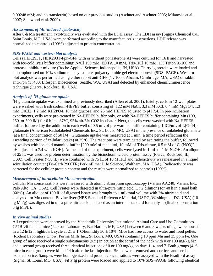
Fpn reduces HEK cell intracellular Mn concentrations
Treatment (6 h) with Mn (100, 250, or 500 µM) resulted in significant (p < 0.01 or 0.001) concentration-
dependent increase in intracellular Mn levels in all three cell types. However, intracellular Mn concentrations
were significantly lower in ponasterone A-induced HEK293T-Fpn-GFP cells versus ponasterone A-uninduced
HEK293T-Fpn-GFP (p < 0.05 or 0.001) and WT HEK293T cells (p < 0.05 or 0.001) at the same Mn treatments
(Fig. 4).
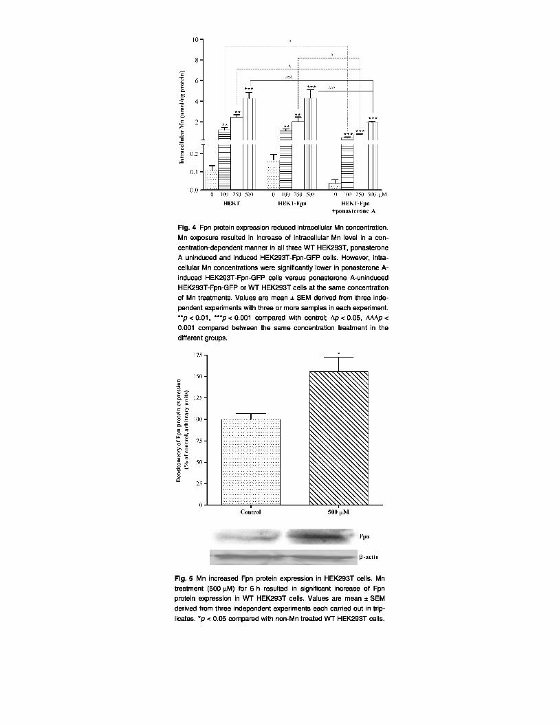
Mn increases Fpn protein expression in HEK293T cells
As shown in Fig. 5, treatment with 500 pM for 6 h significantly increased (p < 0.05) Fpn protein expression in
WT HEK293T cells versus non-Mn exposed cells.
Mn increases Fpn protein expression in mice cerebella and cortices
To corroborate that Mn can increase Fpn protein expression in vivo mice were injected with Mn (s.c., 1–3 doses
of Mn at 100 mg/kg body weight). As shown in Fig. 6, 24 h post-Mn injection, levels of Fpn protein expression
significantly increased (p < 0.01) both in the cortices (Fig. 6a) and cerebella (Fig. 6b) of Mn-treated mice
compared with untreated controls.

Discussion
In the present study, we used WT HEK293T, ponasterone Auninduced HEK293T-Fpn-GFP, and ponasterone
A-induced HEK293T-Fpn-GFP cells to investigate the role of Fpn in Mn efflux, and to ascertain whether
increased Fpn protein expression attenuates the net intracellular Mn concentrations, and its effects on glutamate
uptake and LDH release. Results presented in this study demonstrate, for the first time, that Mn exposure
enhances Fpn protein expression in vitro in WT HEK293T cells and that in vivo s.c. Mn injections promote Fpn
protein expression in mice cortices and cerebella. In addition, increased Fpn protein expression in HEK293T

cells is associated with decreased net intracellular Mn accumulation and attenuated Mn toxicity, exemplified by
reversal of Mn-induced glutamate uptake and diminished cellular LDH release.
Ferroportin is the only known cytoplasmic exporter of Fe in mammalian cells, regulating Fe absorption and
recycling (Abboud and Haile 2000; Knutson and Wessling-Resnick 2003; Donovan et al. 2005). Fpn is densely
expressed on the surface of cells with high capacity for Fe export, such as macrophages and enterocytes
(Abboud and Haile 2000; Delaby et al. 2005), but it is present in almost all cells, including neurons and
oligodendrocytes (Wu et al. 2004; Moos and Rosengren Nielsen 2006; Rouault and Cooperman 2006).
Mutations in the Fpn gene in humans (Pietrangelo 2004) or deletion of the gene in animal models have established the importance of Fpn protein in Fe homeostasis (Donovan et al. 2005). Patients with Fpn mutation
exhibit early Fe overload in the reticuloendocytic macrophages (Montosi et al. 2001) and deletion of the Fpn
gene in the intestinal epithelium of post-natal (a period in which the intestine is the only route for Fe entry)
mice is incompatible with development (Donovan et al. 2005).
Consistent with the shared chemical and physical characteristics of Mn and Fe, animal studies demonstrated that
ID enhances Mn absorption independent of body Mn stores (Chandra and Shukla 1976; Shukla et al. 1976) and
leads to a significant increase in Mn concentrations throughout the rat brain (Erikson et al. 2002, 2004). The
inverse association between body Fe stores and Mn absorption has also been demonstrated in humans (Finley
1999). A G185R mutation in the Belgrade (b/b) rat is associated with complete disruption of DMT1 transport of
Mn across the small-intestine, which is absent in heterozygous +/b rats or +/+ Wistar rats (Knopfel et al. 2005).
Consistent with shared transporters for Fe and Mn, nasal absorption of Mn was significantly attenuated in b/b
rats and the protein level of olfactory DMT1 was significantly elevated in ID b/b rats (Thompson et al. 2007).
The present study demonstrates that net intracellular Mn concentration increased in a concentration-dependent
manner in all three HEK293T cells (Fig. 4). However, in ponasterone A-induced HEK293T-Fpn-GFP cells,
intracellular Mn levels were significantly lower compared with WT HEK293T cells and ponasterone A-
uninduced HEK293T cells treated with the same Mn concentrations (Fig. 4). Although it is possible that Fpn
over-expression caused down-regulation of a Mn importer, such as TfR and DMT1, the most likely explanation
for these observations is that Mn increased Fpn expression (Figs 5 and 6), promoting the efflux of Mn. Notably,
our results also establish that in vivo basal levels of Fpn expression significantly differ among various mouse brain regions (e.g., cerebella vs. cortices; Fig. 6a and b). Whether such differences, and by inference, relatively
low Fpn expression levels account for the propensity of striatal tissue to accumulate large amounts of Mn (e.g.,
6.5-fold increase in striatal Mn levels relative to vehicle mice per method by Dodd et al. 2005) has yet to be
established. Further studies could be profitably directed at establishing the distribution of Fpn expression in
various brain regions to determine whether Fpn expression levels correlate with Mn accumulation.
Consistent with the reduced net Mn concentrations in ponasterone A-induced HEK293T-Fpn-GFP cells, Fpn
expression also decreased LDH leakage (Fig. 2) and restored glutamate uptake (Fig. 3) in these cells. Notably,
an established mechanism of Mn-induced neurotoxicity is associated with attenuated glutamate uptake (Choi
1988; Brouillet et al. 1993; Westergaard et al. 1995; Aschner et al. 2007), resulting in increased extracellular
glutamate concentration and activation of neuronal NMDA receptors (Rosenberg et al. 1992). Notably,
increased Fpn protein expression reversed the Mn-induced inhibition of glutamate uptake (Fig. 3), inherent to
the lower Mn treatments (100 and 250 µM) in ponasterone A-induced HEK293T-Fpn-GFP cells to levels that
were indistinguishable from controls. This, as well as the Fpn associated reversal of the Mn-induced effects on
LDH leakage (Fig. 2) was consistent with the data corroborating indistinguishable intracellular Mn
concentrations in WT HEK293T and ponasterone A-induced HEK293T-Fpn-GFP cells. These results indicate
that increased Fpn protein expression reduces Mn toxicity via stimulation of Mn efflux and concomitant
decrease in net intracellular concentrations of this metal.
In summary, we report the discovery that Mn exposure increases Fpn protein expression in HEK293T cells and
mouse brain. Increased Fpn protein expression in HEK293T cells reduces cellular membrane leakage. Increased
Fpn protein expression also reverses the inhibitory effect of Mn on glutamate uptake. Furthermore, increased

Fpn protein levels reduce intracellular Mn concentrations following exposure, strongly suggesting that Fpn can
actively transport Mn from these cells to decrease Mn cytotoxicity. These results suggest a shared mechanism
for Fe and Mn efflux, paving the way for novel interventions to modulate intracellular levels of these metals.
References
Abboud S. and Haile D. J. (2000) A novel mammalian iron-regulated protein involved in intracellular iron
metabolism. J. Biol. Chem. 275, 19906–19912.
Allen J. W., El-Oqayli H., Aschner M., Syversen T. and Sonnewald U. (2001) Methylmercury has a selective
effect on mitochondria in cultured astrocytes in the presence of [U-13
C]glutamate. Brain Res. 908,149–154.
Aschner J. L. and Aschner M. (2005) Nutritional aspects of manganese homeostasis. Mol. Aspects Med. 26,
353–362.
Aschner M., Guilarte T. R., Schneider J. S. and Zheng W. (2007) Manganese: recent advances in understanding
its transport and neurotoxicity. Toxicol. Appl. Pharmacol. 221, 131–147.
Bader M., Dietz M. C., Ihrig A. and Triebig G. (1999) Biomonitoring of manganese in blood, urine and axillary
hair following low-dose exposure during the manufacture of dry cell batteries. Int. Arch. Occup. Environ.
Health 72, 521–527.
Bowler R. M., Koller W. and Schulz P. E. (2006) Parkinsonism due to manganism in a welder: neurological and
neuropsychological sequelae. Neurotoxicology 27, 327–332.
Brouillet E. P., Shinobu L., McGarvey U., Hochberg F. and Beal M. F. (1993) Manganese injection into the rat
striatum produces excitotoxic lesions by impairing energy metabolism. Exp. Neurol. 120, 89–94.
Burdo J. R., Antonetti D. A., Wolpert E. B. and Connor J. R. (2003) Mechanisms and regulation of transferrin
and iron transport in a model blood-brain barrier system. Neuroscience 121, 883–890.
Chandra S. V. and Shukla G. S. (1976) Role of iron deficiency in inducing susceptibility to manganese toxicity.
Arch. Toxicol. 35, 319–323.
Choi D. W. (1988) Glutamate neurotoxicity and diseases of the nervous system. Neuron 1, 623–634.
Chua A. C. and Morgan E. H. (1997) Manganese metabolism is impaired in the Belgrade laboratory rat. J.
Comp. Physiol. B. 167, 361–369.
Cotzias G. C., Horiuchi K., Fuenzalida S. and Mena I. (1968) Chronic manganese poisoning. Clearance of
tissue manganese concentrations with persistence of the neurological picture. Neurology 18,376–382.
Darshan D. and Anderson G. J. (2007) Liver-gut axis in the regulation of iron homeostasis. World J.
Gastroenterol. 13, 4737–4745.
Delaby C., Pilard N., Hetet G., Driss F., Grandchamp B., Beaumont C. and Canonne-Hergaux F. (2005) A
physiological model to study iron recycling in macrophages. Exp. Cell Res. 310, 43–53.
Dodd C. A., Ward D. L. and Klein B. G. (2005) Basal ganglia accumulation and motor assessment following
manganese chloride exposure in the C57Cl/6 mouse. Int. J. Toxicol. 24, 389–397.
Donovan A., Brownlie A., Zhou Y. et al. (2000) Postional cloning of zebrafish ferroportin1 identifies a
conserved vertebrate iron exporter. Nature 403, 776–781.
Donovan A., Lima C. A., Pinkus J. L., Pinkus G. S., Zon L. I., Robine S. and Andrews N. C. (2005) The iron
exporter ferroportin/Slc40a1 is essential for iron homeostasis. Cell Metab. 1, 191–200.
Ellingsen D. G., Konstantinov R., Bast-Pettersen R., Merkurjeva L., Chashchin M., Thomassen Y. and
Chashchin V. (2008) A neurobehavioral study of current and former welders exposed to manganese.
Neurotoxicology 29, 48–59.
Erikson K. M. and Aschner M. (2003) Manganese neurotoxicity and glutamate-GABA interaction. Neurochem.
Int. 43, 475–480.
Erikson K. M. and Aschner M. (2006) Increased manganese uptake by primary astrocyte cultures with altered
iron status is mediated primarily by divalent metal transporter. Neurotoxicology 27, 125– 130.
Erikson K. M., Shihabi Z. K., Aschner J. L. and Aschner M. (2002) Manganese accumulates in iron-deficient
rat brain regions in a heterogeneous fashion and is associated with neurochemical alterations. Biol. Trace Elem.
Res. 87, 143–156.
Erikson K. M., Syversen T., Steinnes E. and Aschner M. (2004) Globus pallidus: a target brain region for
divalent metal accumulation associated with dietary iron deficiency. J. Nutr. Biochem. 15, 335– 341.

Finley J. W. (1999) Manganese absorption and retention by young women is associated with serum ferritin
concentration. Am. J. Clin. Nutr. 70, 37–43.
Fleming R. E., Migas M. C., Zhou X., Jiang J., Britton R. S., Brunt E. M., Tomatsu S., Waheed A., Bacon B. R.
and Sly W. S. (1999)
Mechanism of increased iron absorption in murine model of hereditary hemochromatosis: increased duodenal
expression of the iron transporter DMT1. Proc. Natl Acad. Sci. USA 96, 3143– 3148.
Ganz T. and Nemeth E. (2006) Iron imports, IV: hepcidin and regulation of iron metabolism. Am. J. Physiol.
Gastrointest. Liver Physiol. 290, G199–G203.
Garcia S. J., Gellein K., Syversen T. and Aschner M. (2007) Iron deficient and manganese supplanted diets alter
metals and transporters in the developing rat brain. Toxicol. Sci. 95, 516–525.
Golub M. S., Hogrefe C. E., Germann S. L., Tran T. T., Beard J. L., Crinella F. M. and Lonnerdal B. (2005)
Neurobehavioral evaluation of rhesus monkey infants fed cow’s milk formula, sow formula or soy formula with
added manganese. Neurotoxicol. Teratol. 27, 615–627.
Hentze M. W., Muchenthaler M. U. and Andrews N. C. (2004) Balancing acts: molecular control of mammalian
iron metabolism. Cell 117, 285–297.
Jensen J. H., Szulc K., Hu C. et al. (2009) Magnetic field correlation as a measure of iron-generated magnetic
field in homogeneities in the brain. Magn. Reson. Med. 61, 481–485.
Keel S. B., Doty R. T., Yang Z. et al. (2008) A heme export protein is required for red blood cell differentiation
and iron homeostasis. Science 319, 825–828.
Kim Y., Park J. K., Choi Y. et al. (2005) Blood manganese concentration is elevated in iron deficiency anemia
patients, whereas globus pallidus signal intensity is minimally affected. Neurotoxicology 26, 107–111.
Knopfel M., Zhao L. and Garrick M. D. (2005) Transport of divalent transition-metal ions is lost in small-
intestinal tissue of b/b Belgrade rats. Biochemistry 44, 3454–3465.
Knutson M. D. and Wessling-Resnick M. (2003) Iron metabolism in the reticuloendothelial system. Crit. Rev.
Biochem. Mol. Biol. 38, 61– 88.
Knutson M. D., Vafa M. R., Haile D. J. and Wessling-Resnick M. (2003) Iron loading and erythrophagocytosis
increase ferroportin 1 (FPN1) expression in J774 macrophages. Blood 102, 4191–4197.
Knutson M. D., Oukka M., Koss L. M., Aydemir F. and Wessling-Resnick M. (2005) Iron release from
macrophages after rythrophagocytosis is up-regulated by ferroportin 1 overexpression and downregulated by
hepcidin. Proc. Natl Acad. Sci. USA 102, 1324–1328.
Krishnamurthy P. and Schuetz J. D. (2006) Role of ABCG2/BCRP in biology and medicine. Annu. Rev.
Pharmacol. Toxicol. 46, 381– 410.
Lee P. L. and Beutler E. (2009) Regulation of hepcidin and iron-overload disease. Annu. Rev. Pathol. 4, 489–
515.
Liu X. B., Hill P. and Haile D. J. (2002) Role of the ferroportin iron-responsive element in iron and nitric oxide
dependent gene regulation. Blood Cells Mol. Dis. 29, 315–326.
Lymboussaki A., Pignatti E., Montosi G., Garuti C., Haile D. J. and Pietrangelo A. (2003) The role of the iron
responsive element in the control of ferroportin1/IREG1/MTP1 gene expression. J. Hepatol. 39, 710–715.
McKie A. T., Marciani P., Rolfs A. et al. (2000) A novel duodenal iron-regulated transporter, IREG1,
implicated in the basolateral transfer of iron to the circulation. Mol. Cell 5, 299–309.
Milatovic D., Yin Z., Gupta R. C., Sidoryk M., Albrecht J., Aschner J. L. and Aschner M. (2007) Manganese
induces oxidative impairment in cultured rat astrocytes. Toxicol. Sci. 98, 198–205.
Mims M. P. and Prchal J. T. (2005) Divalent metal transporter 1. Hematology 10, 339–345.
Montosi G., Donovan A., Totaro A., Garuti C., Pignatti E., Cassanelli S., Trenor C. C., Gasparini P., Andrews
N. C. and Pietrangelo A. (2001) Autosomal-dominant hemochromatosis is associated with a
mutation in the ferroportin (SLC11A3) gene. J. Clin. Invest. 108, 619–623.
Moos T. and Rosengren Nielsen T. (2006) Ferroportin in the postnatal rat brain: implications for axonal
transport and neuronal export of iron. Semin. Pediatr. Neurol. 13, 149–157.
Moos T., Rosengren Nielsen T., Skjørringe T. and Morgan E. H. (2007) Iron trafficking inside the brain. J.
Neurochem. 103, 1730–1740.
Myers J. E., Thompson M. L., Ramushu S. et al. (2003a) The nervous system effects of occupational exposure
on workers in a South African manganese smelter. Neurotoxicology 24, 885–894.

Myers J. E., teWaterNaude J., Fourie M., Zogoe H. B., Naik I., Theodorou P., Tassel H., Daya A. and
Thompson M. L. (2003b) Nervous system effects of occupational manganese exposure on South African
manganese mineworkers. Neurotoxicology 24, 649– 656.
Nemeth E. and Ganz T. (2006) Regulation of iron metabolism by hepcidin. Annu. Rev. Nutr. 26, 323–342.
Nemeth E., Tuttle M. S., Powelson J., Vaughn M. B., Donovan A., Ward M. D., Ganz T. and Kaplan J. (2004)
Hepcidin regulates cellular iron efflux by binding to Ferroportin and inducing its internalization. Science 306,
2090–2093.
Olanow C. W. (2004) Manganese-induced parkinsonism and Parkinson’s disease. Ann. NYAcad. Sci. 1012,
209–223.
Park J. D., Chung Y. H., Kim C. Y. et al. (2007) Comparison of high MRI T1 signals with manganese
concentration in brains of cynomolgus monkeys after 8 months of stainless steel welding-fume exposure. Inhal.
Toxicol. 19, 965–971.
Pietrangelo A. (2004) The Ferroportin disease. Blood Cells Mol. Dis. 32, 131–138.
Rosenberg P. A., Amin S. and Leitner M. (1992) Glutamate uptake disguises neurotoxic potency of glutamate
agonists in cerebral cortex in dissociated cell culture. J. Neurosci. 12, 56–61.
Rouault T. A. and Cooperman S. (2006) Brain iron metabolism. Semin. Pediatr. Neurol. 13, 142–148.
Shukla G. S., Chandra S. V. and Seth P. K. (1976) Effect of manganese on the levels of DNA, RNA, DNase and
RNase in cerebrum, cerebellum and rest of brain regions of rat. Acta Pharmacol. Toxicol. (Copenh). 39, 562–
569.
Stanwood G. D., Leitch D. B., Savchenko V., Wu J., Fitsanakis V. A., Anderson D. J., Stankowski J. N.,
Aschner M. and McLaughlin B. (2009) Manganese exposure is cytotoxic and alters dopaminergic and
GABAergic neurons within the basal ganglia. J. Neurochem. 110,378–389.
Thompson K., Molina R. M., Donaghey T., Schwob J. E., Brain J. D. and Wessling-Resnick M. (2007)
Olfactory uptake of manganese requires DMT1 and is enhanced by anemia. FASEB J. 21,223–230.
Westergaard N., Sonnewald U. and Schousboe A. (1995) Metabolic trafficking between neurons and astrocytes:
the glutamate glutamine cycle revisited. Dev. Neurosci. 17, 203–211.
Wright D. M. and Andrews N. C. (2008) Iron homeostasis and erythropoiesis. Curr. Top. Dev. Biol. 82, 141–
167.
Wu L. J., Leenders A. G., Cooperman S., Meyron-Holtz E., Smith S., Land W., Tsai R. Y., Berger U. V., Sheng
Z. H. and Rouault T. A. (2004) Expression of the iron transporter ferroportin in synaptic vesicles and the blood-
brain barrier. Brain Res. 1001, 108–117.
Yang F., Liu X. B., Quinones M., Melby P. C., Ghio A. and Haile D. J. (2002) Regulation of reticuloendothelial
iron transporter MTP1 (Slc11a3) by inflammation. J. Biol. Chem. 277, 39786– 39791.
Zoller H., Koch R. O., Theurl I., Obrist P., Pietrangelo A., Montosi G., Haile D. J., Vogel W. and Weiss G.
(2001) Expression of the duodenal iron transporters divalent-metal transporter 1 and ferroportin 1 in iron
deficiency and iron overload. Gastroenterology 120, 1412–1419.
Related Documents