– Ferrocene-Based Pyridylphosphine Ligands – Coordination Chemistry of Group 10, 11 and 12 Metals Dissertation zur Erlangung des akademischen Grades eines Doktors der Naturwissenschaften (Dr. rer. nat.) am Fachbereich Mathematik und Naturwissenschaften der Universit ¨ at Kassel von Thorsten Klemann 2010

Welcome message from author
This document is posted to help you gain knowledge. Please leave a comment to let me know what you think about it! Share it to your friends and learn new things together.
Transcript
– Ferrocene-Based Pyridylphosphine Ligands –
Coordination Chemistry
of Group 10, 11 and 12 Metals
Dissertation
zur
Erlangung des akademischen Grades
eines
Doktors der Naturwissenschaften(Dr. rer. nat.)
am Fachbereich
Mathematik und Naturwissenschaften
der Universitat Kassel
von
Thorsten Klemann
2010

By three methods we may learn wisdom: First, by reflection, which is the noblest; se-
cond, by imitation, which is the easiest; and third by experience, which is the bitterest.
(alleged to Confucius)
Der Mensch hat dreierlei Wege Weisheit zu erlangen: Erstens durch Nachdenken, das
ist der Edelste; zweitens durch Nachahmen, das ist der Leichteste; drittens durch Er-
fahrung, das ist der Bitterste.
(Konfuzius zugeschrieben)


The work described in this thesis was carried out in the Institute of Chemistry, University
of Kassel (Germany), since 2008 in the research group of Prof. Siemeling and in the
Department of Inorganic Chemistry, Charles University Prague (Czech Republic), in
2009 and 2010 under supervision of Prof. Stepnicka.
Day of disputation: 01st of December, 2010.
1. Supervisor: Prof. Dr. Ulrich Siemeling
2. Supervisor: Prof. Dr. Petr Stepnicka
i

ACKNOWLEDGEMENTS
Acknowledgements
With the first words I would like to express my sincerest gratitude to my academic te-
acher, Prof. Ulrich Siemeling, for giving me the opportunity to work in his group, his
scientific guidance and for providing the interesting topic of this thesis. Beyond that, he
was marvellous for being not only the supervisor, but a supporting advisor as well.
I had the good fortune to visit for two times the Czech Republic and work under the
supervision of Prof. Petr Stepnicka at Charles University in Prague. I would like to
thank him sincerely for his generousness and support during this time and for the ama-
zing opportunity to experience a different (academic) culture at its best.
In this context the DAAD (Deutscher Akademischer Austauschdienst), the Ministry of
Education of the Czech Republic and the Czech Science Foundation are thanked very
much indeed for their financial support of my visits to Charles University.
I would like to thank all my mater colleagues during my time in the MOC in Kassel,
namely Dr. Frauke Bretthauer, Dr. Jens Hoßbach, Dr. Mario Gatterdam, Dr. Christian
Farber, Dr. Christian Schirrmacher, Ulrich Glebe, Stefan Rittinghaus, Lutz Klapp, Jan
Schroder, Alexander Girod, Tim Fellinger, Stella Helten, Sandra Tripp, Michael Kurle-
mann, Henry Memczak, Tim Koppenrath, Steffen Koppenrath, Alexander Mundstock,
Tim Schulz, Dr. Pavel Turek and also the great people from the Prague group, Dr. Jan
Demel, Jirı Tauchman and Jirı Schulz for creating stimulating and pleasant environ-
ments.
ii

ACKNOWLEDGEMENTS
Furthermore I am indebted to several people, without whom I would not have been able
to conclude my work:
Dr. Maurer for recording all kinds of NMR spectra.
Dr. Bruhn and A. Pilz for collecting and solving the X-ray crystal structures in Kassel,
very special thanks!
Dr. Cısarova for the crystallographic work in Prague.
Dr. Furmeier for mass spectrometry.
Dr. Leibold for DFT calculations.
Jorg Ho for excellent elemental analyses.
Finally I would like to thank my family, first of all my wife Tanja and my parents, Moni-
ka und Ulrich, as well as my sister Patricia, whose mainly non-scientific, but endless
support was absolutely essential to reach this goal.
iii


DECLARATION - ERKLARUNG - PUBLICATIONS
Declaration
The entire body of this work is my own unless stated to the contrary and has not been
submitted previously for any degree at this or any other university.
Kassel, 28th of October, 2010
(Thorsten Klemann)
Erklarung
Ich versichere, dass ich die vorliegende Dissertation selbststandig, ohne unerlaub-
te Hilfe angefertigt habe und keine anderen als die in der Dissertation angegebenen
Hilfsmittel benutzt habe. Alle Stellen, die wortlich oder sinngemaß aus veroffentlichten
Schriften entnommen sind, wurden mit Quellenangaben kenntlich gemacht. Kein Teil
dieser Arbeit ist in einem anderen Promotions- oder Habilitationsverfahren verwendet
worden.
Kassel, am 28. Oktober 2010
(Thorsten Klemann)
Publications
Parts of the work described in this thesis have been published previously:
P. Stepnicka, J. Schulz, T. Klemann, U. Siemeling, I. Cısarova: Synthesis, Structural
Characterization, and Catalytic Evaluation of Palladium Complexes with Homologous
Ferrocene-Based Pyridylphosphine Ligands, Organometallics 2010, 29, 3187.
v

SUMMARY
Summary
The present thesis describes the synthesis of 1,1’-ferrocendiyl-based pyridylphosphine
ligands, the exploration of their fundamental coordination chemistry and preliminary
experiments with selected complexes aimed at potential applications. One main aspect
is the synthesis of the bidentate ferrocene-based pyridylphosphine ligands 1, 2 and 3
(Fig. I).
Fe
1
PPh2
NFe
2
PPh2
NFe
3
PPh2
N
Fig. I: Bidentate 1,1’-ferrocenediyl-based pyridylphosphine ligands.
A specific feature of these ligands is the ball-bearing like flexibility of the ferrocene-
based backbone. An additional flexibility element is the rotation around the C–C single
bonds. Consequently, the donor atoms can realise a wide range of positions with re-
spect to each other and are therefore able to adapt to the coordination requirements of
different metal centres.
The flexibility of the ligand also plays a role in another key aspect of this work, which
concerns the coordination mode, i. e. bridging vs. chelating. In addition to the flexibility,
also the position of the donor atoms to each other is important. This is largely affected
by the position of the pyridyl nitrogen (pyrid-2-yl vs. pyrid-3-yl) and the methylen group
in 3.
Another interesting point is the combination of a soft phosphorus donor atom with a
harder nitrogen donor atom, according to the HSAB principle. This combination ge-
nerates a unique binding profile, since the π-acceptor character of the P site is able
to stabilise a metal centre in a low oxidation state, while the nitrogen σ-donor abili-
vi
SUMMARY
ty can make the metal more susceptible to oxidative addition reactions. A P,N-donor
combination can afford hemilabile binding profiles, which would be ideal for catalysis.
Beyond 1,2-substituted ferrocene derivatives, which are quite successful in catalytic
applications, 1,1’-derivatives are rather underrepresented. While a low-yield synthetic
pathway to ligand 1 was already described in the literature,[1] it was possible to find a
new, improved and simplified synthetic pathway. 2 and 3 were unknown prior to this
work. Satisfactory results in the synthesis of 2 could be achieved by working in analogy
to the new synthetic procedure for 1. The synthesis of 3 has been handled by the group
of Prof. Petr Stepnicka from Charles University, Prague, Czech Republic. The synthesis
of tridentate ligands with an analogous heterodentate arrangement, was investigated
briefly as a sideline of this study.
The major part of this thesis deals with the fundamental coordination chemistry to-
wards transition metals of the groups 10, 11 and 12. Due to the well-established cata-
lytic properties of analogous palladium complexes, the coordination chemistry towards
palladium (group 10) is of particular interest. The metals zinc and cadmium (group 12)
are also of substantial importance because they are redox-inert in their divalent state.
This is relevant in view of electrochemical investigations concerning the utilisation of
the ligands as molecular redox sensors. Also mercury and the monovalent metals sil-
ver and gold (group 11) are included because of their rich coordination chemistry. It
is essential to answer questions concerning aspects of the ligands’ coordination mode
bearing in mind the HSAB principle.
Depending on the metal source used, on the reaction stoichiometry and, in a few cases,
on the experimental conditions, the ligands 1, 2 and 3 exhibit a variety of coordination
modes. Fig. II schematically presents the coordination modes which occurred during
this investigation.
The results of the coordination chemistry experiments are summarised in Tab. I. The
listed metal sources were reacted with one or two equivalent(s) (eq.) of the ligands, af-
fording the complexes 5a to 35. Information about the coordination modes encountered
and aspects of the structural characterisation are given, too.
vii

SUMMARY
n
N
(a)
(e)
(c)
(d)
(b)
P
N
M N
M P
P
N
M P
M
N
P
M
P
N
N
P
M
N
PP
M
N
P
Fig. II: Observed coordination modes of 1, 2 and 3; P,N-chelate “C” (a), monodentate P-
coordination “M” (b), P-coordinated bis(phosphine) complex “B” (c), centrosymmetric
dimer “D” (d) and polymer “P” (e).
Not surprisingly reactions of group 12 metal halides with 1 eq. of 1 afforded the corre-
sponding P,N-chelates, which have been fully characterised with three exceptions only.
It was impossible to crystallise the chelate complexes 5c, 6a and 8a. Interestingly, two
new compounds were obtained in both cases by the crystallisation experiments, viz. the
P-coordinated bis(phosphine) complexes 7 and 9a. Complex 8c crystallises not as a
chelate, but as a dimeric, iodo-bridged, P-coordinated bis(phosphine) complex. Reac-
tions of HgBr2 with 2 eq. of the ligand gave the P-coordinated bis(phosphine) complex
9b. The reaction with 2 eq. of zinc bromide only gave the 1:1 chelate 5b.
Reactions of 2 with group 12 metal bromides gave polymers exclusively. The less pre-
dictable coordination behaviour of 3 can be ascribed to the presence of the methylene
group in this ligand, which makes the ligand more flexible. Reactions of 3 with group 12
metal halides resulted in the P,N-chelates 13, 14, 15 and 16. Due to poor crystallisation
tendencies, the crystallisation of compound 14 was attempted also in a diffusion expe-
riment, which surprisingly did not afford the expected chelate, but the polymer 14a. In
analogous crystallisation experiments performed with the chelates 13 and 16, 3 turned
out to act as a bridging ligand, resulting in the centrosymetric dimer 13a and the po-
lymer 17. Interestingly, the molecular structure of the mercury polymer 17 exhibits a
1:2 (ligand:metal) stoichiometry with halide bridges, even though the experiment was
carried out in a 1:1 stoichiometry.
viii

SUMMARY
In reactions with silver(I) tetrafluoroborate all three ligands coordinated the metal in a
bridging manner. 1 and 3, containing a pyrid-2-yl group, form the polymers 19 and 21.
The pyrid-3-yl containing ligand 2 forms the centrosymmetric dimer 20.
Tab. I: Summary of the results of the coordination chemistry experiments concerning the bi-
dentate ligands 1, 2 and 3.
1 2 3
1 eq. 2 eq. 1 eq. 2 eq. 1 eq. 2 eq.
Zn
Cl2 5a C – – – – –
Br2 5b C 5b C 10 P – 13 C; 13a Da 13a Da
I2 5c Cb – – – – –
Cd
Cl2 6a Cb,c 7 Ba,d – – – –
Br2 6b C – 11 Pb – 14 Cc; 14a Pa –
I2 6c C – – – 15 C; – –
Hg
Cl2 8a Cb,c 9a Ba,d – – – –
Br2 8b C 9b B 12 P – 16 C; 17 Pa,e 18 B
I2 8c Cf – – – – –
Ag BF4 19 P – 20 D – 21 P –
AuCl(tht) 22 M – 23 M – 24 M –
BF4 25 D – 26 Xb – 27 Xb –
Pd
Cl2 28 C 30 B – – 29 Cb 31 B
Cl(LNC) 32 Mb – – – 33 Mb –
(LNC)(MeCN)2ClO4 34 C – – – 35 C –
B = P-coordinated, monodentate bis(phosphine) complex, C = P,N-chelate, D = centrosymmetric
dimer, M = monodentate P-coordination, P = polymer, X = structure not clear, – = no investigations
realised or investigations did not lead to clear results. aOnly characterised by X-ray diffraction. bNo
X-ray data available. cCrystallisation leads to new compound with 2:1 (ligand:metal) stoichiometry.dCrystals obtained by a diffusion experiments with 1:1 stoichiometry. e17 has a 1:2 stoichiometry,
the polymer is formed via halide bridges. fCrystallisation leads to P-coordinated iodo-bridged
dimer.
ix
SUMMARY
The gold complexes 22, 23 und 24 obtained from the reaction of 1, 2 and 3, respectively
with [AuCl(tht)] (tht = tetrahydrothiophene) each contain a monodentate, P-coordinated
Ligand. Further reactions, aimed at the abstraction of the chloro ligand to induce Au-
N coordination resulted in the less soluble and poorly crystalline complexes 25, 26
and 27. Only 25 was structurally characterised. Additional experiments aimed at the
preparation of Au-Ag binuclear complexes failed.
The ligands 1 and 3 react with [PdCl2(cod)] (cod = η2:η2-cycloocta-1,5-diene) in a P-
coordinated monodentate and in a P,N-chelating manner, depending on the reaction
stoichiometry, to form the palladium complexes 28, 29, 30 and 31. Both ligands we-
re also reacted with [Pd(µ-Cl)(LNC)]2 (LNC = [(2-dimethylamino-κN)methyl]phenyl-κC1),
leading to the P-coordinated bis(phosphine) complexes 32 and 33. A similar reaction
with the solvento complex [Pd(LNC)(MeCN)2][ClO4] gave the cationic complexes 34 and
35, in which the ligands exhibited a chelating coordination behavior.
Catalytic investigations of Suzuki-Miyaura cross-coupling reactions showed that com-
plexes 28 and 29 and precatalyst formed in situ from 1 and 3 with Pd(OAc)2 promote
the reaction of 4-bromotoluene with phenylboronic acid efficiently. For the catalyst ba-
sed on ligand 3 similar or slightly better results were obtained than for the correspon-
ding dppf-based (dppf = 1,1’-bis(diphenylphosphino)ferrocene) catalyst. The results
for the catalysts based on 1 are comparable, too, but only slightly inferior to those of
the dppf benchmark. The Pd-catalysed cyanation reaction is more efficiently promo-
ted by the defined precatalyst complexes, with dppf being a ligand superior to both
pyridylphosphines.
The ligands 1, 2 and 3 exhibited a reversible behaviour concerning the ferrocene-based
redox wave in cyclovoltammetric investigations. The chlorogold(I) complexe 22, 23 and
24 showed such a behavior, too. The coordination causes a notable shift of the formal
electrode potential of about ca. 0.2 V with respect to the corresponding uncoordinated
ligand.
x

ZUSAMMENFASSUNG
Zusammenfassung
Die vorliegende Arbeit befasst sich mit der Synthese, der Untersuchung der fundamen-
talen Koordinationschemie und ersten Modellexperimenten zur Anwendungserprobung
von Pyridylphosphin Liganden mit 1,1’-Ferrocendiyl-Ruckgrat. Einer der Hauptaspekte
ist die Synthese der zweizahnigen Liganden 1, 2 und 3 (Abb. I).
Fe
1
PPh2
NFe
2
PPh2
NFe
3
PPh2
N
Abb. I: Zweizahnige Pyridylphosphin-Liganden mit 1,1’-Ferrocendiyl-Ruckgrat.
Ein besonderes Charakteristikum dieser zweizahnigen Liganden ist die kugellagerarti-
ge Beweglichkeit des Ligand-Ruckgrates. Hinzu kommen die Flexibilitatselemente der
C–C-Einfachbindung mit ihrer freien Drehbarkeit. Die Donoratome konnen auf diese
Weise viele verschiedene Positionen zueinander einnehmen und sich so den Koordi-
nationsbedurfnissen unterschiedlichster Metallzentren anpassen.
Ein weiterer wichtiger Aspekt dieser Arbeit, bei dem die Beweglichkeit der Liganden ei-
ne Rolle spielt, ist der Koordinationsmodus der Liganden: verbruckend oder chelatisie-
rend. Nicht nur die Flexibilitat, sondern auch die Position der Donoratome zueinander
ist dabei von Bedeutung. Diese wird hauptsachlich durch die Position des Pyridylstick-
stoffes (pyrid-2-yl vs. pyrid-3-yl) und durch die Methylengruppe in 3 bestimmt.
Ein weiterer interessanter Aspekt ist die Kombination eines, nach dem HSAB-Prinzip
weichen, Phosphordonors mit einem harteren Stickstoffdonor. Diese Kombination er-
offnet interessante Bindungsprofile, da der π-Akzeptorcharakter des P-Donors Metall-
zentren in niedrigen Oxidationsstufen stabilisiert, wahrend die σ-Donorfahigkeit des
N-Donors die Metallzentren anfalliger fur oxidative Additionsreaktionen macht. Ein sol-
xi

ZUSAMMENFASSUNG
ches Donorprofil kann moglicherweise zu hemilabiler Koordination fuhren, was optimal
fur katalytische Zwecke ware.
Neben Ferrocenderivaten mit 1,2-Substitution, die sehr erfolgreich in katalytischen
Bereichen eingesetzt werden, sind 1,1’-Derivate in der wissenschaftlichen Literatur
eher unterreprasentiert. Die Synthese des Liganden 1 war bereits in der Literatur be-
schrieben,[1] allerdings mit geringen Ausbeuten. In der vorliegenden Arbeit konnte ei-
ne neue, verbesserte Syntheseroute gefunden werden. Die Liganden 2 und 3 waren
ganzlich unbekannt. Durch Ubertragung und Modifizierung des neuen Synthesewegs
fur 1 auf die Synthese von 2 konnte eine zufriedenstellende Syntheseroute gefunden
werden. Die Synthese von 3 wurde vom Arbeitskreis um Prof. Petr Stepnicka an der
Karls-Universitat Prag, Tschechische Republik, durchgefuhrt. Auch die Synthese von
dreizahnigen Liganden mit analogem Aufbau war von Interesse, spielte aber im Nach-
hinein nur eine untergeordnete Rolle.
Der Kernteil der Arbeit befasst sich mit der Auslotung der fundamentalen Koordinati-
onschemie der Liganden mit Ubergangsmetallen der Gruppen 10, 11 und 12. Aufgrund
von guten katalytischen Eigenschaften, die analoge Palladiumkomplexe bisher gezeigt
haben, ist die Untersuchung der Koordinationschemie gegenuber Palladium (Gruppe
10) besonders interessant. Auch Zink und Cadmium (Gruppe 12) sind fur diese Arbeit
substanziell, da sie uber keine eigene Redoxchemie verfugen. Letzteres ist im Hinblick
auf elektrochemische Untersuchungen bezuglich der Eignung der Liganden als mole-
kulare Redoxsensoren von Bedeutung. Auch Quecksilber und die einwertigen Metalle
Silber und Gold (Gruppe 11) sind aufgrund ihrer vielseitigen Koordinationschemie in die
Untersuchungen einbezogen worden. Es gilt, Antworten auf Fragestellungen bezuglich
des Koordinationsverhaltens der Liganden unter Berucksichtigung des HSAB-Prinzips
zu klaren.
Abhangig vom eingesetzten Metall, der Reaktionsstochiometrie und, in einigen weni-
gen Fallen, von den experimentellen Bedingungen zeigen die Liganden 1, 2 und 3
unterschiedlichste Koordinationsformen. Die wahrend den Untersuchungen beobach-
teten sind in den schematischen Darstellungen in Abb. II gezeigt.
xii

ZUSAMMENFASSUNG
n
N
(a)
(e)
(c)
(d)
(b)
P
N
M N
M P
P
N
M P
M
N
P
M
P
N
N
P
M
N
PP
M
N
P
Abb. II: Beobachtete Koordinationsformen von 1, 2 und 3; P,N-Chelat ”C“ (a), einzahnige P-
koordination ”M“ (b) , P-koordinierter Bis(phosphin)-Komplex ”B“ (c), zentrosymmetri-
sches Dimer ”D“ (d) und Polymer ”P“ (e).
Die Ergebnisse der koordinationschemischen Experimente sind in Tab. I zusammen-
gefasst. Die aufgelisteten Metalle wurden mit einem oder zwei Aquivalenten (eq.) des
entsprechenden Liganden umgesetzt. Die Komplexverbindungen 5a bis 35 wurden er-
halten. Ebenfalls sind Informationen uber die Koordinationsformen und die strukturelle
Charakterisierung enthalten.
Wenig uberraschend fuhren Reaktionen von Metallhalogeniden der Gruppe 12 mit ei-
nem Aquivalent 1 zu den entsprechenden Chelatkomplexen, die, bis auf drei Ausnah-
men, umfassend charakterisiert wurden. Es gelang nicht, die Komplexe 6a, 5c und
8a zu kristallisieren. Interessanterweise fuhrten Kristallisationsexperimente in diesen
beiden Fallen zu neuen Verbindungen: den einzahnig P-koordinierten Bis(phosphin)-
Komplexen 7 und 9a. Die Kristallisation von 8c fuhrte nicht zum erwarteten Chelat-
komplex, sondern zu einem dimeren iodo-verbruckten P-koordinierten Bis(phosphin)-
Komplex. In Reaktionen von HgBr2 mit zwei Aquivalenten Ligand entstand der einzahnig
P-koordinierte Bis(phosphin)-Komplex 9b. Im Falle des Zinks konnte bei Reaktionen
mit zwei Aquivalenten des Liganden lediglich der Chelatkomplex 5b mit 1:1-Stochio-
metrie isoliert werden.
Reaktionen von 2 mit Metallbromiden der Gruppe 12 ergaben ausschließlich Koordi-
nationspolymere. Hingegen zeigt der flexiblere Ligand 3 ein ambivalentes Verhalten.
xiii

ZUSAMMENFASSUNG
Reaktionen zwischen 3 und Metallhalogeniden der Gruppe 12 resultierten in P,N-
Chelatkomplexen 13, 14, 15 und 16. Da Verbindung 14 keine Tendenz zur Kristallisati-
on zeigte, wurde eine Kristallisation mittels Diffusionsexperiment versucht. Das Ergeb-
Tab. I: Zusammenfassung der Ergebnisse der koordinationschemischen Experimente
bezuglich der zweizahnigen Liganden 1, 2 und 3.
1 2 3
1 eq. 2 eq. 1 eq. 2 eq. 1 eq. 2 eq.
Zn
Cl2 5a C – – – – –
Br2 5b C 5b C 10 P – 13 C; 13a Da 13a Da
I2 5c Cb – – – – –
Cd
Cl2 6a Cb,c 7 Ba,d – – – –
Br2 6b C – 11 Pb – 14 Cc; 14a Pa –
I2 6c C – – – 15 C; – –
Hg
Cl2 8a Cb,c 9a Ba,d – – – –
Br2 8b C 9b B 12 P – 16 C; 17 Pa,e 18 B
I2 8c Cf – – – – –
Ag BF4 19 P – 20 D – 21 P –
AuCl(tht) 22 M – 23 M – 24 M –
BF4 25 D – 26 Xb – 27 Xb –
Pd
Cl2 28 C 30 B – – 29 Cb 31 B
Cl(LNC) 32 Mb – – – 33 Mb –
(LNC)(MeCN)2ClO4 34 C – – – 35 C –
”B“ = P-koordinierter Bis(phosphin)-Komplex; ”C“ = P,N-Chelat; ”D“ = Zentrosymmetrisches Di-
mer, ”M“ = einzahnige P-Koordination, ”P“ = Polymer, ”X“ = Struktur nicht geklart, ”–“ = keine Un-
tersuchungen realisiert oder die Untersuchungen fuhrten zu keinem klaren Ergebnis. aNur durch
Rontgenstruktur charakterisiert. bKeine Rontgenstruktur verfugbar. cKristallisation fuhrte zu einer
neuen Verbindung mit 2:1-Stochiometrie (Ligand:Metall). dKristallisation erfolgte im Diffusions-
experiment mit 1:1-Stochiometrie. e17 weist 1:2-Stochiometrie auf, die Verknupfung erfolgt uber
verbruckende Bromatome. fKristallisation fuhrt zu einem P-koordinierten, iodverbruckten Dimer.
xiv

ZUSAMMENFASSUNG
nis war nicht wie erwartet der Chelatkomplex, sondern uberraschenderweise wurde
das Polymer 14a erhalten. Bei analogen Kristallisationsexperimenten mit den Che-
latkomplexen 13 und 16 zeigte der Ligand 3 verbruckendes Koordinationsverhalten,
mit dem Ergebnis des zentrosymmetrischen Dimers 13a und des Polymers 17. Kurio-
serweise zeigt die Molekulstruktur des Polymers 17 eine halogenverbruckte 1:2-Sto-
chiometrie (Ligand:Metall), obwohl das Experiment in 1:1-Stochiometrie durchgefuhrt
wurde.
Mit Silber(I)-tetrafluoroborat reagieren alle drei Liganden in einer verbruckenden Form.
Die Liganden 1 und 3, die eine Pyrid-2-yl-gruppe enthalten, bilden die Koordinations-
polymere 19 und 21. Ligand 2, welcher eine Pyrid-3-yl-gruppe enthalt, bildet das zen-
trosymmetrische Dimer 20.
Die Goldkomplexe 22, 23 und 24 wurden aus Reaktionen von 1, 2 und 3 mit [AuCl(tht)]
(tht = Tetrahydrothiophen) erhalten und sind alle einzahnig P-koordiniert. Weitere Re-
aktionen, beginnend mit dem Austausch des Chloroliganden durch ein nicht koordi-
nierendes Tetrafluoroborat-Anion, resultieren in den kaum loslichen Komplexen 25,
26 und 27, die nur teilweise Kristallisationstendenzen zeigen. Nur 25 konnte mittels
Rontgendiffraktometrie charakterisiert werden. Zusatzliche Experimente zur Darstel-
lung von Au-Ag binuklearen Komplexen scheiterten.
Die Liganden 1 und 3 reagieren mit [PdCl2(cod)] (cod = η2:η2-Cycloocta-1,5-dien) in ei-
ner P-koordinierenden einzahnigen und in einer P,N-chelatisierenden Weise, abhangig
von der Stochiometrie der Reaktion. Es bilden sich die Palladiumkomplexe 28, 29, 30
und 31. Die Reaktionen beider Liganden mit [Pd(µ-Cl)(LNC)]2 (LNC = [(2-Dimethylamino-
κN)methyl]phenyl-κC1) fuhren zu den entsprechenden Komplexen 32 und 33, in denen
beide Liganden jeweils einzahnige P-Koordination zeigen. Ahnliche Reaktionen mit
dem Solvens-Komplex [Pd(LNC)(MeCN)2][ClO4] ergeben die kationischen Komplexe 34
und 35. Hier hingegen zeigen die Liganden chelatisierendes Koordinationsverhalten.
Katalytische Untersuchungen an der Suzuki-Miyaura Kreuz-Kupplung haben gezeigt,
dass die definierten Komplexe 28 und 29 und die in situ hergestellten Katalysatoren
xv
ZUSAMMENFASSUNG
1/Pd(OAc)2 und 3/Pd(OAc)2 die Reaktion von 4-Bromtoluol mit Phenylboronsaure gut
unterstutzen. Die Katalysatoren, die auf Ligand 3 basieren, liefern mindestens genauso
gute Ergebnisse bezuglich der Ausbeute wie der analoge Komplex mit dppf (dppf =
1,1’-Bis(diphenylphosphino)ferrocen). Die Ausbeuten, die mit Katalysatoren basierend
auf Ligand 1 erreicht wurden sind ebenfalls gut, allerdings ein wenig niedriger als die
vom dppf-Analogon erreichten Werte. Die Pd-katalysierte Cyanierung wird von den
definierten Komplexen mit dppf besser unterstutzt als von beiden Ferrocen-basierten
Pyridylphosphinliganden.
Die Liganden 1, 2 und 3 zeigen in cyclovoltammetrischen Untersuchungen reversibles
Verhalten bezuglich der ferrocenbasierten Redoxwelle. Die Chlorogold(I)-Komplexe 22,
23 und 24 zeigen ebenfalls ein solches Verhalten. Durch die Koordination wird das for-
male Elektrodenpotential deutlich um ca. 0.2 V gegenuber den unkoordinierten Ligan-
den verschoben.
xvi

CONTENTS
Contents
Acknowledgements . . . . . . . . . . . . . . . . . . . . . . . . . . . . . . . . ii
Declaration . . . . . . . . . . . . . . . . . . . . . . . . . . . . . . . . . . . . . v
Erklarung . . . . . . . . . . . . . . . . . . . . . . . . . . . . . . . . . . . . . . v
Publications . . . . . . . . . . . . . . . . . . . . . . . . . . . . . . . . . . . . . v
Summary . . . . . . . . . . . . . . . . . . . . . . . . . . . . . . . . . . . . . . vi
Zusammenfassung . . . . . . . . . . . . . . . . . . . . . . . . . . . . . . . . . xi
Contents . . . . . . . . . . . . . . . . . . . . . . . . . . . . . . . . . . . . . . xvii
1 Introduction 1
1.1 General Introduction — Motivation and Outline . . . . . . . . . . . . . . 1
1.2 Ferrocene . . . . . . . . . . . . . . . . . . . . . . . . . . . . . . . . . . . 5
1.2.1 Physical and Electronic Properties . . . . . . . . . . . . . . . . . 6
1.2.2 Chemical Behaviour . . . . . . . . . . . . . . . . . . . . . . . . . 7
1.3 P,N-Donors . . . . . . . . . . . . . . . . . . . . . . . . . . . . . . . . . . 8
1.3.1 Pyridylphosphines . . . . . . . . . . . . . . . . . . . . . . . . . . 8
1.4 Field of Potential Applications . . . . . . . . . . . . . . . . . . . . . . . . 9
1.4.1 Electrochemical Sensors . . . . . . . . . . . . . . . . . . . . . . 10
1.4.2 Catalytic Application . . . . . . . . . . . . . . . . . . . . . . . . . 11
2 Ferrocene-Based Pyridylphosphine Ligands 13
2.1 Introduction . . . . . . . . . . . . . . . . . . . . . . . . . . . . . . . . . . 13
2.2 Bidentate Ligands (1 - 3) . . . . . . . . . . . . . . . . . . . . . . . . . . . 16
2.2.1 Synthesis and Characterisation of 1 . . . . . . . . . . . . . . . . 16
2.2.2 Synthesis and Characterisation of 2 . . . . . . . . . . . . . . . . 19
2.2.3 Synthesis and Characterisation of 3 . . . . . . . . . . . . . . . . 21
2.3 Tridentate Ligands (4) . . . . . . . . . . . . . . . . . . . . . . . . . . . . 24
2.3.1 Synthesis and Characterisation of a N,P,N-Ligand . . . . . . . . 24
2.3.2 Synthesis and Characterisation of the P,N,P-Ligand 4 . . . . . . 25
2.4 Summary and Conclusion . . . . . . . . . . . . . . . . . . . . . . . . . . 26
xvii

CONTENTS
3 Coordination Chemistry I: Zinc, Cadmium and Mercury 27
3.1 Introduction . . . . . . . . . . . . . . . . . . . . . . . . . . . . . . . . . . 27
3.2 Coordination Chemistry of 1 . . . . . . . . . . . . . . . . . . . . . . . . . 32
3.2.1 Synthesis and Characterisation of Zn Compounds . . . . . . . . 32
3.2.2 Synthesis and Characterisation of Cd Compounds . . . . . . . . 36
3.2.3 Synthesis and Characterisation of Hg Compounds . . . . . . . . 40
3.3 Coordination Chemistry of 2 . . . . . . . . . . . . . . . . . . . . . . . . . 45
3.3.1 Synthesis and Characterisation of the Zn Compound . . . . . . . 46
3.3.2 Synthesis and Characterisation of the Cd Compound . . . . . . 48
3.3.3 Synthesis and Characterisation of the Hg Compound . . . . . . 48
3.4 Coordination Chemistry of 3 . . . . . . . . . . . . . . . . . . . . . . . . . 50
3.4.1 Synthesis and Characterisation of Zn Compounds . . . . . . . . 50
3.4.2 Synthesis and Characterisation of Cd Compounds . . . . . . . . 53
3.4.3 Synthesis and Characterisation of Hg Compounds . . . . . . . . 55
3.5 Coordination Chemistry of 4 . . . . . . . . . . . . . . . . . . . . . . . . . 59
3.6 Summary and Conclusion . . . . . . . . . . . . . . . . . . . . . . . . . . 61
4 Coordination Chemistry II: Silver and Gold 63
4.1 Introduction . . . . . . . . . . . . . . . . . . . . . . . . . . . . . . . . . . 63
4.2 Synthesis and Characterisation of Ag Compounds . . . . . . . . . . . . 66
4.2.1 Ag Complex of 1 . . . . . . . . . . . . . . . . . . . . . . . . . . . 66
4.2.2 Ag Complex of 2 . . . . . . . . . . . . . . . . . . . . . . . . . . . 67
4.2.3 Ag Complex of 3 . . . . . . . . . . . . . . . . . . . . . . . . . . . 69
4.3 Synthesis and Characterisation of Au Compounds . . . . . . . . . . . . 70
4.3.1 Simple Au Complexes of 1, 2 and 3 . . . . . . . . . . . . . . . . 70
4.3.2 Further experiments . . . . . . . . . . . . . . . . . . . . . . . . . 73
4.4 Summary and Conclusion . . . . . . . . . . . . . . . . . . . . . . . . . . 75
5 Coordination Chemistry III: Palladium 77
5.1 Introduction . . . . . . . . . . . . . . . . . . . . . . . . . . . . . . . . . . 77
5.2 Synthesis and Characterisation of Pd-Compounds . . . . . . . . . . . . 80
5.2.1 Simple Palladium Complexes of 1 and 3 . . . . . . . . . . . . . . 80
xviii

CONTENTS
5.2.2 (LNC)-Palladium Complexes of 1 and 3 . . . . . . . . . . . . . . . 85
5.3 Catalytic Evaluation . . . . . . . . . . . . . . . . . . . . . . . . . . . . . 89
5.4 Summary and Conclusion . . . . . . . . . . . . . . . . . . . . . . . . . . 91
6 Experimental 93
6.1 General Techniques and Methods . . . . . . . . . . . . . . . . . . . . . 93
6.2 Synthetic Details . . . . . . . . . . . . . . . . . . . . . . . . . . . . . . . 95
6.2.1 Starting Materials . . . . . . . . . . . . . . . . . . . . . . . . . . 96
6.2.2 Experimental Procedures . . . . . . . . . . . . . . . . . . . . . . 96
7 References 123
Appendix I
Contents . . . . . . . . . . . . . . . . . . . . . . . . . . . . . . . . . . . . . . I
Abbreviations . . . . . . . . . . . . . . . . . . . . . . . . . . . . . . . . . . . . II
List of Figures . . . . . . . . . . . . . . . . . . . . . . . . . . . . . . . . . . . III
List of Tables . . . . . . . . . . . . . . . . . . . . . . . . . . . . . . . . . . . . V
List of numbered Compounds . . . . . . . . . . . . . . . . . . . . . . . . . . . VII
List of X-ray Data . . . . . . . . . . . . . . . . . . . . . . . . . . . . . . . . . . XVII
xix


1 Introduction
1.1 General Introduction — Motivation and Outline
Phosphinylated N-heterocycles, particularly pyridylphosphines, are of great importance
in coordination chemistry and have found numerous applications in catalysis.[2–4] Their
ligand properties can be easily tuned by changing molecular parameters like the type of
the donor groups or their substituents. This explains their high popularity. Pyridylphos-
phines are oligodentate ligands. They have at least two coordination sites. Conse-
quently, two different modes of coordination are possible: bridging versus chelating.
A bridging ligand forms oligonuclear complexes or polymers. A chelating ligand forms
usually mononuclear complexes.
The combination of a soft P and a hard N donor atom[5] makes these ligands typi-
cal hybrid ligands, which can exhibit particularly interesting binding profiles. For suit-
able metal-ligand combinations this can result in a hemilabile binding profile.[6–8] The
term “hemilabile” was coined by Jeffrey and Rauchfuss in 1979.[9] They investigated
the chemistry of phosphine-amine and phosphine-ether ligands with the expectation
that these ligands would bind sufficiently strong to allow isolation of the correspond-
ing complexes. However, the hard ligand component tended to dissociate readily, thus
generating a vacant site for substrate binding.[10] This is ideal for catalysis.
Ligands are usually differentiated by the number and type of donor atoms and the
length and flexibility of the bridging unit. A wide range of ligands with simple organic
backbone units like alkyls and aryls is known.[2] Ligands, which contain a ferrocendiyl-
1

1 INTRODUCTION
FeFe
R1
R2
R1
R2
(c)(a)
Fe
R1(b)
R2
Fig. 1.1: Unsymmetrical disubstitution on ferrocene: 1,2- (a), 1,3- (b) and 1,1’-type (c).
based backbone are a special class. The ferrocene moiety can act in three general
ways as a connector between two substituents or donor groups. First, ferrocene can
be 1,2-disubstituted, second, 1,3-disubstituted and, third, (the more relevant) 1,1’-
disubstituted (Fig. 1.1). Phosphinoferrocenes of the 1,2-type are relatively common and
comprehensively documented.[11–16] Almost all of them are utilised in asymmetric ca-
talysis because of their stereochemical characteristics. Derivatives of the 1,2-type are
planar chiral, unless the two substituents are identical. Ferrocene-based P,N-ligands
of this type such as [2-(diphenylphosphino)ferrocenyl]ethyldimethylamine (PPFA) are
well known and widely applied.[17–19] 1,3-disubstituted ferrocenes have found usage as
electrochemical sensing molecules, photoresponsive molecules and liquid-crystalline
materials, for instance.[20] However, analogous systems with a 1,1’-arrangement are
rare, which is surprising in view of the ubiquity and versatility of 1,1’-substituted biden-
tate ferrocene ligands.[21,22] The only such system that has attracted some attention
is the ferrocene-based pyridylphosphine ligand 1-(diphenylphosphino)-1’-(pyrid-2-yl)-
ferrocene (1) shown in Fig. 1.2.
While the synthesis of 1 was described already by Butler in 1992,[1] nearly no inves-
tigations concerning the coordination chemistry of this ligand have been reported to
Fe
1
PPh2
N
Fig. 1.2: 1-(Diphenylphosphino)-1’-(pyrid-2-yl)ferrocene (1).
2

1.1 GENERAL INTRODUCTION — MOTIVATION AND OUTLINE
Fe
2
PPh2
NFe
3
PPh2
N
Fig. 1.3: Homologous 1,1’-ferrocene-based pyridylphosphines 2 and 3.
date. Only Tani and coworkers utilised 1 in the synthesis of a rhodium bis(chelate)
complex,[23] which later became subject of a quantum-chemical study.[24]
Systematic variations of 1 are its 3-pyridyl isomer, 1-(diphenylphosphino)-1’-(pyrid-3-
yl)ferrocene (2) and the more flexible methylene-spaced analogue 1-(diphenylphos-
phino)-1’-[(pyrid-2-yl)methyl]ferrocene (3) displayed in Fig. 1.3.
Subtle structural differences may have a dramatic influence on the coordination mode,
i. e. chelating versus bridging, and this will be a key issue of this thesis. Ligands like
2,2’-bipyridine, which contain pyrid-2-yl groups in the neighbourhood to a second donor
atom, are known for chelating coordination.[25] 1 contains a pyrid-2-yl group and forms
a rhodium bis(chelate) complex [Rh(cod)(1)],[23] although the number of atoms in the
resulting chelate ring is seven, which is larger than the preferred ring sizes of five and
six atoms. Ligands equipped with pyrid-3-yl groups instead mostly bind in a bridging
coordination mode. For example, 3,3’-bipyridine usually forms polymeric complexes.[26]
The number of atoms in a hypothetical chelate ring formed by 2 would be eight, a
chelate coordination is therefore unlikely. 3 is equipped with a pyrid-2-yl group. If
3 forms a chelate, the chelate ring would contain eight atoms, like in the case of 2.
Nevertheless, both modes, bridging and chelating, might be possible for 3, owing to
the presence of the methylene spacer.
The ball-bearing like characteristics of the 1,1’-ferrocendiyl backbone allows twist an-
gles between 0 ◦ (eclipsed) and 180 ◦ (diametrically opposed) for the substituents. Til-
ting of the ferrocene moiety facilitates small variations in the distance between both
3

1 INTRODUCTION
Cp ring twist (torsion angle)
C-C single bond rotation
Fe
3
PPh2
N
methylene spacer
Fig. 1.4: Flexibility elements in 3.
donor groups. Furthermore, the methylene spacer in 3 leads to additional flexibility
and allows an even larger range of distances between the N-donor and the ligand
backbone. Also the adjustment of the nitrogen donor atom in the pyridyl group can
be influenced by the rotation around the C–C single bond. These factors may cause
a variable, potentially hemilable, coordination behaviour of 3, taking advantage of its
flexibility. In Fig. 1.4 the relevant flexibility elements of 3 are shown.
The primary objective of the research described in this thesis is the preparation of the
ligands 1 - 3 and the subsequent exploration of their fundamental coordination chem-
istry. In particular, the focus will be on transition metals of group 12 (zinc, cadmium,
mercury), group 11 (silver, gold) and group 10 (palladium).
The coordination chemistry of the divalent group 12 metal ions is expected to be partic-
ularly simple. The coordination number is usually four, which is expressed in a typical
tetrahedral coordination geometry, caused by the d10 electron configuration. Further-
more, ZnII and CdII are redox-inert. Metal redox processes would interfere unfavourably
with cyclovoltammetric experiments to explore the utilisation of the ligands as poten-
tial molecular electrochemical sensors. The monovalent group 11 metal ions of silver
and gold are also d10 configurated. The coordination numbers of AgI and AuI are usu-
ally even lower than four, which can give rise to interesting secondary metal-ligand or
metal-metal interactions. Because of the high popularity and the commercial benefit
of palladium complexes in catalytic processes (Nobel prize for chemistry 2010[27]), the
4

1.2 FERROCENE
coordination chemistry towards PdII (group 10) is investigated, too. Again tetracoordi-
nation is expected to occur, in this case, however, in a square-planar geometry owing
to the d8 configuration.
In addition to differences concerning the coordination chemistry of the selected transi-
tion metals in terms of the HSAB principle, the resulting complexes are also expected
to show up variability due to the ligands’ geometric properties. Potential applications
of 1 - 3 as redox-active ligands in electrochemical ion sensing will be addressed utili-
sing redox-inert metal ions like ZnII and CdII. Furthermore the application of palladium
complexes in selected catalytic processes will be investigated.
1.2 Ferrocene
Ferrocene was prepared for the first time in 1951, independently by two different groups.
The discovery was made by serendipity. Kealy and Pauson tried to synthesise ful-
valene by the oxidative coupling of cyclopentadienyl magnesium bromide with ferric
chloride.[28] The high temperature reaction of cyclopentadiene with iron observed by
the group around Miller also afforded ferrocene.[29] Wilkinson[30] and Fischer[31] were
awarded with the Nobel prize for chemistry in 1973 for the elucidation of the ferrocene
structure. The interest in complexes of transition metals with hydrocarbons increased
immediately. Historically, this may be considered as the hour of birth for organo-
transition metal chemistry. Since its discovery, ferrocene has become more and more
remarkable. Today, ferrocene-based compounds are used in applications ranging from
catalysis and material sciences to bioorganometallic chemistry.[11,32]
5

1 INTRODUCTION
1.2.1 Physical and Electronic Properties
Ferrocene is an air-stable, orange crystalline solid which can be easily sublimed in
vacuum or at atmospheric pressure by heating. This is due to its comparatively high
vapour pressure of 2.6 hPa (100 ◦C). It is soluble in all common solvents, except water.
Formally, ferrocene can be considered as being composed of two cyclopentadienyl
anions which bind to the central iron(II) ion in a sandwich-like manner. However, the
bonding is not ionic, but predominantly covalent. The resulting uncharged complex has
18 valence electrons (VE). This noble gas electron configuration of krypton makes fer-
rocene exceptionally stable. Interestingly, the iron(II) centre is easily oxidised to iron(III)
at low potentials (ca. 0.5 V vs. SCE[33]). The ferrocene/ferrocenium redox couple shows
perfectly reversible behaviour because of the stability of the 17 VE ferrocenium cation.
This can be understood by considering the molecular orbital (MO) scheme of ferrocene
(Fig. 1.5), which shows that the highest occupied molecular orbital (HOMO) is essen-
Fig. 1.5: MO scheme of ferrocene in staggered conformation.[33]
6
1.2 FERROCENE
tially non-bonding. The weakly bonding character of the e2- and the nearly non-bonding
character of the a1-symmetric orbitals is a reason for the existence of metallocenes with
less than 18 valence electrons, for example manganocene (17 VE), the isoelectronic
ferrocenium cation (17 VE), chromocene (16 VE) and even vanadocene (15 VE).[33]
Due to their stability and reversible redox behaviour, ferrocene derivatives are well
suited for electrochemical experiments like cyclic voltammetry (CV). Ferrocene is rec-
ommended by the International Union of Pure and Applied Chemistry (IUPAC) as a
reference standard in such experiments.
1.2.2 Chemical Behaviour
Ferrocene undergoes reactions which are characteristic for electron-rich aromatic com-
pounds. It is prone to electrophilic aromatic substitution. This is caused by the high
electron density of the cyclopentadienyl ring systems. In comparison to benzene, fer-
rocene reacts 3 ·106 times faster in these reactions.[33] Ferrocenium instead, which
is formed by the reaction of ferrocene with strongly oxidising electrophiles, is inert to-
wards electrophilic attack. Hence, direct halogenation, nitration etc. of ferrocene is
impossible.
Derivatisation of ferrocene can be also realised conveniently by metallation reactions.
For example, the reaction with two equivalents of butyllithium in the presence of N,-
N,N ’,N ’-tetramethylethylendiamine (TMEDA) and subsequent treatment with an elec-
trophile[34] cleanly leads to symmetrical 1,1’-disubstitution. The preparation of 1,1’-
unsymmetrical ferrocenes is less straightforward. A selective monolithiation of fer-
rocene by simple reaction with one equivalent of buthyllithium is not possible. Today,
suitable synthetic procedures have been developed and new routes to unsymmetrical
substitution are known: 1. selective transmetallation of 1,1’-bis(tri-n-butylstannyl)ferro-
cene, 2. selective lithium-halogen exchange of 1,1’-dibromoferrocene and 3. 1-phenyl-
1-phospha-[1]-ferrocenophane ring opening reaction.[35] The latter two routes have
been used in the work describes in this thesis (for details see Chapter 2).
7

1 INTRODUCTION
1.3 P,N-Donors
P,N-ligands are heterodentate ligands, which combine a hard N donor site with a soft P
donor site in one and the same ligand molecule. This combination generates a unique
binding profile, since the π-acceptor character of the P site is able to stabilise a metal
centre in a low oxidation state, while the nitrogen σ-donor ability can make the metal
more susceptible to oxidative addition reactions. A survey about hard and soft donors in
terms of the HSAB principle is given by Pearson[5,36] and Woodward.[37] Heteroditopic
ligands are of great importance in coordination chemistry, since this combination can
afford hemilabile binding profiles. Not surprisingly, they have found numerous applica-
tions in catalysis.[18,19,38] A classification of P,N-ligands has recently been developed
and reviewed, featuring their catalytic applications by the type of donor groups[17] and
their backbone unit.[2] Because the present work exclusively deals with pyridine type
N-donors and phosphine type P-donors, other donor types will not be described here.
Generally, the pyridine donor is classified as a medium hard donor. Pyridine and am-
monia split d-orbitals of metal cations energetically in a similar manner.[39] Due to its
π-system and the nitrogen sp2-lone pair, pyridine is easier to polarise than ammonia.
Pyridine is a weaker ligand than ammonia; it is placed on the borderline between hard
and soft according to Pearson.[40] The coordination chemistry of pyridine has been
extensively investigated and reviewed.[41]
Phosphine donors are classified as soft in terms of the HSAB principle. They have
π-acceptor character. Usually, the π-acidity of the phosphorus donor atom increases
with the electronegativity of its substituents.
1.3.1 Pyridylphosphines
Pyridylphosphine ligands have been known since 1944.[3,42] Initially, these simple
phosphine derivatives (Fig. 1.6) were only prepared in very low yields. Over the years,
8

1.4 FIELD OF POTENTIAL APPLICATIONS
PN
Br
Fig. 1.6: One of the first pyridylphosphines: Phenyl-p-bromophenyl-2-pyridylphosphine.
the availability of such compounds increased and a wide variety of pyridylphosphine
ligands are known today. Most of them have been used for palladium-catalysed reac-
tions, as mentioned before. 1,2-Ferrocene-based ligands of this type are well known
and have proved useful in this field.[11,12,43] There is still a lack of information concern-
ing the coordination behaviour of 1,1’-ferrocene-based P,N-ligands in the literature.
Chapters 3 - 5 will give detailed insight into the coordination chemistry of known 1,1’-
ferrocene-based P,N-ligands towards group 12 and group 11 metals in comparison to
the new ligands investigated in the present work.
1.4 Field of Potential Applications
Potential uses of ferrocene-based pyridylphosphine ligands are manifold. Within the
scope of this theses, two aspects are of particular interest: The redox chemistry of
ferrocene, or metallocenes in general, has been explored in detail. The utilisation of
ferrocene ligands as electrochemical sensors needs no special mentioning.[44] Also
the use of P,N-ligands in homogeneous catalysis is well documented.[4,17,18,38] Most
of them are simple organic ligands or 1,2-substituted ferrocenes. 1,1’-unsymmetrically
disubstituted ferrocenes instead, are hardly known at all in catalytic chemistry.
9

1 INTRODUCTION
1.4.1 Electrochemical Sensors
A molecular redox sensor is a host molecule which has, according to its redox state,
different chemical affinities to a guest molecule. Such sensors are composed of a
redox-active unit and a receptor. The redox-active unit needs to show reversible elec-
trochemical behaviour. The receptor needs to exhibit strong affinity and also selectivity
for a guest molecule. Furthermore, these two functional units need to interact substan-
tially with one another. Only if these conditions are fulfilled, the coordinated and the
free species will gave rise to separate redox signals in the voltammogram. Only in this
rare and special case can a redox sensor be characterised as highly effective.
The cyclic voltammogram is the result of the CV experiment. In Fig. 1.7 an ideal CV
is displayed. A starting potential E is varied with constant scan rate v to a reversal
potential Eλ and back to the potential E. The measurement category is the current i.
The redox-behaviour of a sample can be classified as reversible if 1. the current ratio
ipa/ipc is equal to 1, which shows that the electrogenerated species is stable (at least
on the CV timescale), 2. ipc ≈√v and 3. the peak-to-peak separation between Epa and
Fig. 1.7: Basic parameters for a cyclic voltammogram. E = starting potential, Eλ = reversal
potental, Epa = anodic peak potential, Epc = cathodic peak potential, ipa = anodic
peak current, ipc = cathodic peak current.
10

1.4 FIELD OF POTENTIAL APPLICATIONS
FeN
NFe
N
N
(a) (b)
Fig. 1.8: 1,1’-Di(pyrid-2-yl)ferrocene (a) and 1,1’-di(pyrid-2-yl)octamethylferrocene (b).
Epc (∆Ep) = 59/n mV (n = number of electrons exchanged per molecule). It must be
stressed, however, that, in measuring the peak-to-peak separation, a deviation of 10-
20 mV from the theoretical value (especially at higher scan rates) does not compromise
the criterion of reversibility.[45–47]
Two examples of pyridyl-functionalised ferrocene-based redox-sensors are displayed
in Fig. 1.8.[48,49] Both ligands show high chemical affinities towards PdII and PtII com-
pounds. Coordination-induced shifts of the half-wave potentials of ca. 120 mV were de-
tected.[50,51] Particularly large shifts of the half-wave potential of up to ca. 400 mV were
detected with the octamethylated ligand (Fig. 1.8 b) for the metal ions ZnII, CdII, CaII or
MgII.[52] These significant shifts characterise 1,1’-di(pyrid-2-yl)octamethylferrocene as
a highly effective ferrocene-based redox sensor.
1.4.2 Catalytic Application
Lowering of the activation barrier in chemical reactions by catalysts is of great com-
mercial importance. More than 80 % of the processes in the chemical industry are
catalysed to optimise their commercial usage. Selectivity of catalysts is important if
more than one product is obtained in the reaction process. If the chosen catalyst pro-
motes only the desired reaction, impurities by side products can be avoided.[53,54]
Ferrocene-based ligands are enormously successful in catalytic applications. One of
the most versatile and popular 1,1’-disubstituted chelate ligand in this context is 1,1’-
11
1 INTRODUCTION
bis(diphenylphosphino)ferrocene (dppf). For example, suitable complexes of dppf are
able to promote C–C, C–N and C–S bond forming reactions.[21,55]
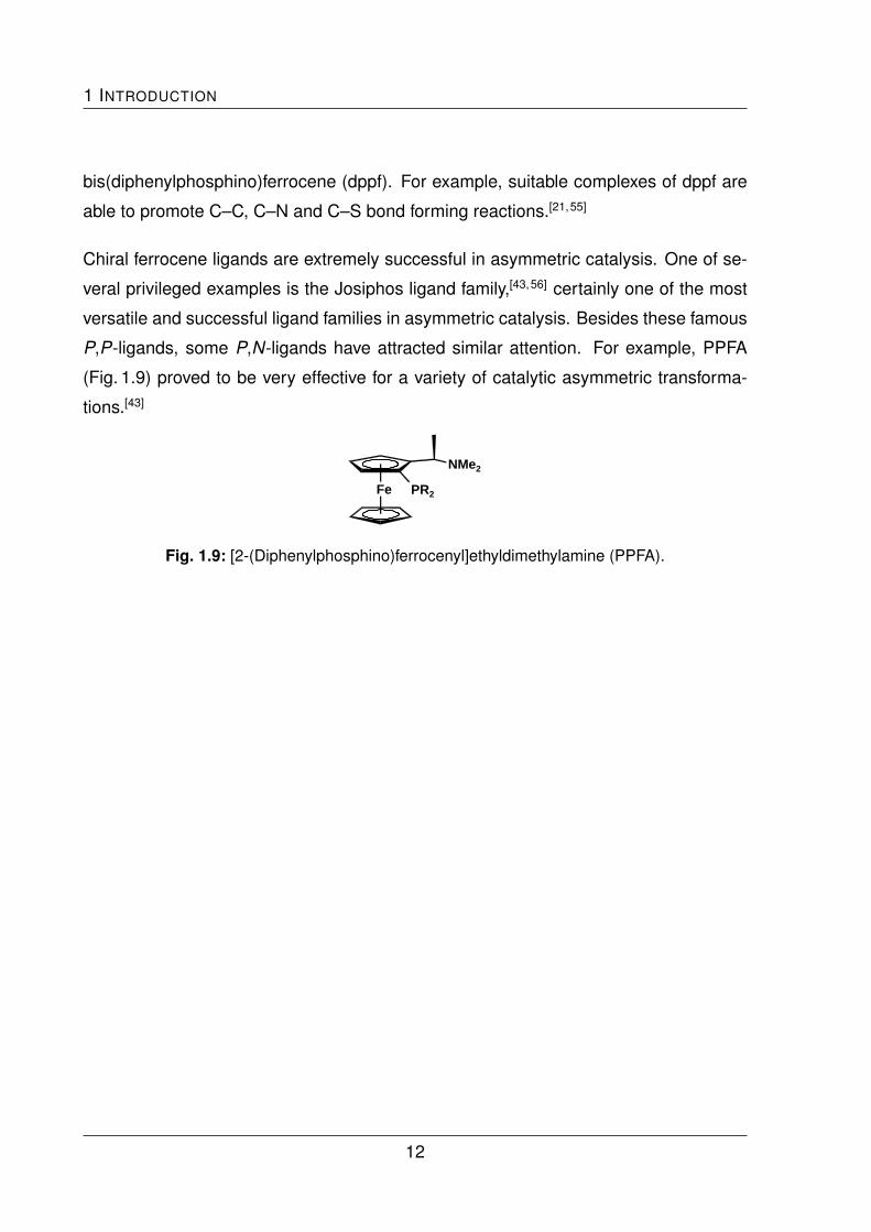
Chiral ferrocene ligands are extremely successful in asymmetric catalysis. One of se-
veral privileged examples is the Josiphos ligand family,[43,56] certainly one of the most
versatile and successful ligand families in asymmetric catalysis. Besides these famous
P,P-ligands, some P,N-ligands have attracted similar attention. For example, PPFA
(Fig. 1.9) proved to be very effective for a variety of catalytic asymmetric transforma-
tions.[43]
Fe
NMe2
PR2
Fig. 1.9: [2-(Diphenylphosphino)ferrocenyl]ethyldimethylamine (PPFA).
12

2 Ferrocene-Based Pyridylphosphine
Ligands
The synthesis and characterisation of bidentate and tridentate 1,1’-unsymmetrical fer-
rocene-based pyridylphosphine ligands is described. Spectroscopic data, results of
X-ray diffraction analyses and electrochemical investigations will be discussed.
2.1 Introduction
Ferrocene ligands bearing a combination of both donor groups, viz. ferrocene-based
pyridylphosphines, gave their debut in the literature in the beginning of the 1990s. 1-
(Diphenylphosphino)-1’-(pyrid-2-yl)ferrocene (1), its 1,2-isomer and other 1-(diphenyl-
phosphino)-1’-(N-heteroaryl)ferrocenes were reported by Butler.[1,57] Selected exam-
ples are shown in Fig. 2.1. Due to the difficulties concerning their synthesis, the interest
in these systems waned away. Today, new more convenient methods for the prepara-
tion of 1,1’-unsymmetrical functionalised systems have been developed. Although the
accessibility of such systems is much easier, ferrocene-based pyridylphosphine ligands
are still rare.
A useful method to prepare 1,1’-unsymmetrical phosphinoferrocene is the P-[1]-ferro-
cenophane ring opening reaction with phenyllithium. Starting from 1-phenyl-1-phospha-
[1]-ferrocenophane, which can be prepared by the reaction of 1,1’-dilithioferrocene with
13

2 FERROCENE-BASED PYRIDYLPHOSPHINE LIGANDS
Fe
PPh2
Fe N
PPh2
N
N
Fe
PPh2
N
N
Fe
PPh2
N
N
(a) (b)
(c)
Fe
1
PPh2
N
Fig. 2.1: 1-(Diphenylphosphino)-1’-(N-heteroaryl)ferrocenes; heteroaryl is pyridine (a),
phenanthroline (b) and bipyridine (c).
dichlorophenylphosphine, 1-(lithio)-1’-diphenylphosphinoferrocene is afforded by the
reaction with phenyllithium. This active species reacts easily with electrophiles (E)
to 1,1’-unsymmetrical phosphinoferrocene derivatives (Fig. 2.2). The ferrocenophane
cleavage reaction, first described by Seyferth and Withers,[58] has been utilised by But-
ler and Cullen for the preparation of 1,1’-bis[(alkyl/aryl)phosphino]ferrocenes.[59]
FeFe
Li
Li
PPh Fe
PPh2
Li
Fe
PPh2
E
Cl2PPh PhLi E
Fig. 2.2: P-[1]-ferrocenophane ring opening reaction.
A method with much wider scope involves the reaction of 1,1’-dilithioferrocene with two
equivalents of tributylstannyl chloride leading to 1,1’-bis(tri-n-butylstannyl)ferrocene,
which is a precursor for the preparation of a wide range of 1,1’-unsymmetrical fer-
rocenes (Fig. 2.3). Adeleke utilised this precursor in the early 1990s to prepare P,S-
ligands based on ferrocene by selective transmetallation at low temperatures.[60]
14

2.1 INTRODUCTION
Fe
Li
Li
Fe
SnBu3
SnBu3
Fe
SnBu3
E1
ClSnBu3
2. E1
1. n-BuLiFe
E2
E1
2. E2
1. n-BuLi
Fig. 2.3: Selective transmetallation of 1,1’-bis(tri-n-butylstannyl)ferrocene.
In 1994 Dong developed an elegant method to synthesise 1,1’-unsymmetrical fer-
rocenes.[61] He established the selective lithium halogen exchange of 1,1’-dibromo-
ferrocene at low temperatures as shown in Fig. 2.4.[62,63] By this method a variety of
unsymmetrical ferrocene derivatives have been synthesised amongst others by Butler
in high yields.[64]
Fe
Li
Li
Fe
Br
Br
Fe
Br
E1
C2H2Br4
2. E1
1. n-BuLiFe
E2
E1
2. E2
1. n-BuLi
Fig. 2.4: Selective lithium halogen exchange of 1,1’-dibromoferrocene.
The focus of this work is on the ferrocene-based pyridylphosphines 1, 2 and 3 (Chapter
1.1). While the synthesis of 1 has already been described by Butler,[1] compounds 2
and 3 are unknown so far. In his publication, Butler reported the coordination chemistry
of a series of ferrocenyl- and ruthenocenylbipyridines, as mentioned before (Fig. 2.1 b,
c). In conjunction with the potential structural modification of these ligands, he tried
the reaction of pyrid-2-ylferrocene with butyllithium and then chlorodiphenylphosphine,
which gave the ferrocene-based pyridylphospine 1-(pyrid-2-yl)-2-(diphenylphosphino)-
ferrocene (Fig. 2.1 a). A different approach resulted in 1, which was utilised indepen-
dently by Tani in the synthesis of the bis-chelate complex [Rh(cod)(1)],[23] which later
became subject of a quantum-chemical study.[24]
The synthetic procedure of 1 is described as the in situ preparation of 1-lithio-1’-
(diphenylphosphino)ferrocene via cleavage reaction of 1-phenyl-1-phospha-[1]-ferro-
cenophane with phenyllithium in diethyl ether at -70 ◦C, followed by the treatment with
15

2 FERROCENE-BASED PYRIDYLPHOSPHINE LIGANDS
Fe PPh Fe
PPh2
Li
Fe
1
PPh2
N
PhLi
Et2O
pyridine
24 h
Fig. 2.5: Synthesis of 1 presented by Butler in 1992.
an excess of pyridine (Fig. 2.5). After hydrolysis the product was purified by column
chromatography, yielding ca. 10 % of an orange oil, which solidified during drying un-
der vacuum. Butler characterised 1 by 1H NMR, MS and elemental analysis. This
low-yield synthetic pathway affords the product contaminated with phosphine oxide im-
purities. This is probably caused by the introduction of the pyridyl group by Ziegler
reaction, which involves an oxidative final step. It was therefore an aim of this work to
improve the synthesis of 1.
2.2 Bidentate Ligands (1 - 3)
Bidentate ligands contain two donor groups. They are able to form chelate complexes
with a single metal centre or cyclic oligomers and polymeric chains. They can also
form simple dinuclear complexes with two metal centres.
2.2.1 Synthesis and Characterisation of 1
By following Butler’s published procedure for the synthesis of 1, his unsatisfactory
results could be reproduced. A moderate improvement of the yield (23 %) could be
achieved by using particularly pure 1-phenyl-1-phospha-[1]-ferrocenophane, which was
obtained by a literature procedure[58] with a revised, non-aqueous work-up. Instead of
treatment with aqueous NH4Cl solution the reaction mixture was filtered, evaporated
16

2.2 BIDENTATE LIGANDS (1 - 3)
to dryness and purified by column chromatography under exclusion of moisture. The
difficulties in the synthesis and handling of the ferrocenophane precursor may explain
the lack of information about ferrocene-based pyridylphosphines in the literature.
As described above, there is the opportunity to prepare 1,1’-unsymmetrical ferrocenes
by selective lithium-halogen exchange. Starting from 1,1’-dibromoferrocene, which is
treated with one equivalent of n-butyllithium at low temperature and subsequently with
chlorodiphenylphosphine, it is quite easy to prepare 1-bromo-1’-(diphenylphosphino)-
ferrocene according to a known procedure[64] (Fig. 2.6). Purification of 1-bromo-1’-
(diphenylphosphino)ferrocene by crystallisation gives brown, air-stable material in re-
producible yields of about 70-80 %. Introduction of the pyridyl group by Ziegler reaction
after selective lithium-halogen exchange gives 1, but still not in acceptable yield.
Fe
Br
Br
Fe
Li
Br
Fe
PPh2
Br
n-BuLi
THF / -70°C
ClPPh2
THF / -70°C
Fig. 2.6: Synthesis of 1-bromo-1’-(diphenylphosphino)ferrocene.
The classic palladium-catalysed Negishi cross-coupling reaction[65–67] turned out to be
a much better method of attaching the N-donor site in the final step of the sequence.
The addition of [ZnCl2(1,4-dioxane)] after the selective lithium-halogen exchange of
1-bromo-1’-(diphenylphosphino)ferrocene leads to the active organozinc compound
in situ. Reaction with 2-bromopyridine (Fig. 2.7) and subsequent column chromato-
graphic work-up afforded pure 1 in satisfactory yield (48 %) as a dark orange oil, which
solidified upon prolonged standing.
1 was found to be air-stable. Its solubility in polar solvents like dichloromethane is
high. Crystals suitable for X-ray diffraction analysis were grown from a concentrated
dichloromethane solution. The results of the structural characterisation are presented
in Fig. 2.8. Structural and spectroscopic data are rather unexceptional. The signal of
the pyridyl H6 in the 1H NMR and the phosphorus signal in the 31P NMR spectrum
17

2 FERROCENE-BASED PYRIDYLPHOSPHINE LIGANDS
2. ZnCl2 Fe
PPh2
ZnCl
1. n-BuLi
Fe
PPh2
BrTHF / -70°C
Fe
1
N
[Pd(PPh3)4]2-BrPy
72 h / 60°C
PPh2
Fig. 2.7: Negishi cross-coupling reaction affording 1.
are interesting diagnostic markers for later coordination chemistry experiments. NMR
spectra of metal complexes of 1 are expected to exhibit shifted signals if the corre-
sponding donor atom is coordinated.
Electrochemical investigations of 1 by cyclic voltammetry revealed the expected ferro-
cene-based oxidation plus an additional irreversible oxidation process at higher poten-
tial (Fig. 2.9 a). The ferrocene-based redox wave of 1 exhibits reversible behaviour, if
the scan is reversed before the second oxidation process starts (Fig. 2.9 b). The ∆EP
value is 65 mV. The formal electrode potential E0’ of the ferrocene-based process is
anodically shifted by ca. 0.55 V with respect to decamethylferrocene (fc*). Comparison
P
Fe
N
C11
C12
C22
C23
C16
C17C1
C6C7
C2
Fig. 2.8: Molecular structure of 1 in the crystal.
18
2.2 BIDENTATE LIGANDS (1 - 3)
-2
-1
0
1
2
3
-0,3 -0,2 -0,1 0,0 0,1 0,2 0,3 0,4 0,5 0,6 0,7 0,8 0,9 1,0 1,1 1,2 1,3
Cur
rent
(µA)
Potential (V)
(a)
fc*
-0,4
-0,2
0,0
0,2
0,4
0,6
0,8
1,0
1,2
1,4
-0,3 -0,2 -0,1 0,0 0,1 0,2 0,3 0,4 0,5 0,6 0,7 0,8
Cur
rent
(µA)
Potential (V)
(b)
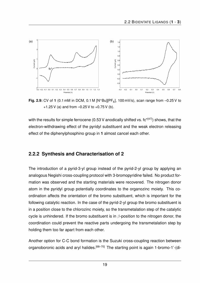
Fig. 2.9: CV of 1 (0.1 mM in DCM, 0.1 M [NnBu][PF6], 100 mV/s), scan range from –0.25 V to
+1.25 V (a) and from –0.25 V to +0.75 V (b).
with the results for simple ferrocene (0.53 V anodically shifted vs. fc*[47]) shows, that the
electron-withdrawing effect of the pyridyl substituent and the weak electron releasing
effect of the diphenylphosphino group in 1 almost cancel each other.
2.2.2 Synthesis and Characterisation of 2
The introduction of a pyrid-3-yl group instead of the pyrid-2-yl group by applying an
analogous Negishi cross-coupling protocol with 3-bromopyridine failed. No product for-
mation was observed and the starting materials were recovered. The nitrogen donor
atom in the pyridyl group potentially coordinates to the organozinc moiety. This co-
ordination affects the orientation of the bromo substituent, which is important for the
following catalytic reaction. In the case of the pyrid-2-yl group the bromo substituent is
in a position close to the chlorozinc moiety, so the transmetalation step of the catalytic
cycle is unhindered. If the bromo substituent is in β-position to the nitrogen donor, the
coordination could prevent the reactive parts undergoing the transmetalation step by
holding them too far apart from each other.
Another option for C-C bond formation is the Suzuki cross-coupling reaction between
organoboronic acids and aryl halides.[68–70] The starting point is again 1-bromo-1’-(di-
19
2 FERROCENE-BASED PYRIDYLPHOSPHINE LIGANDS
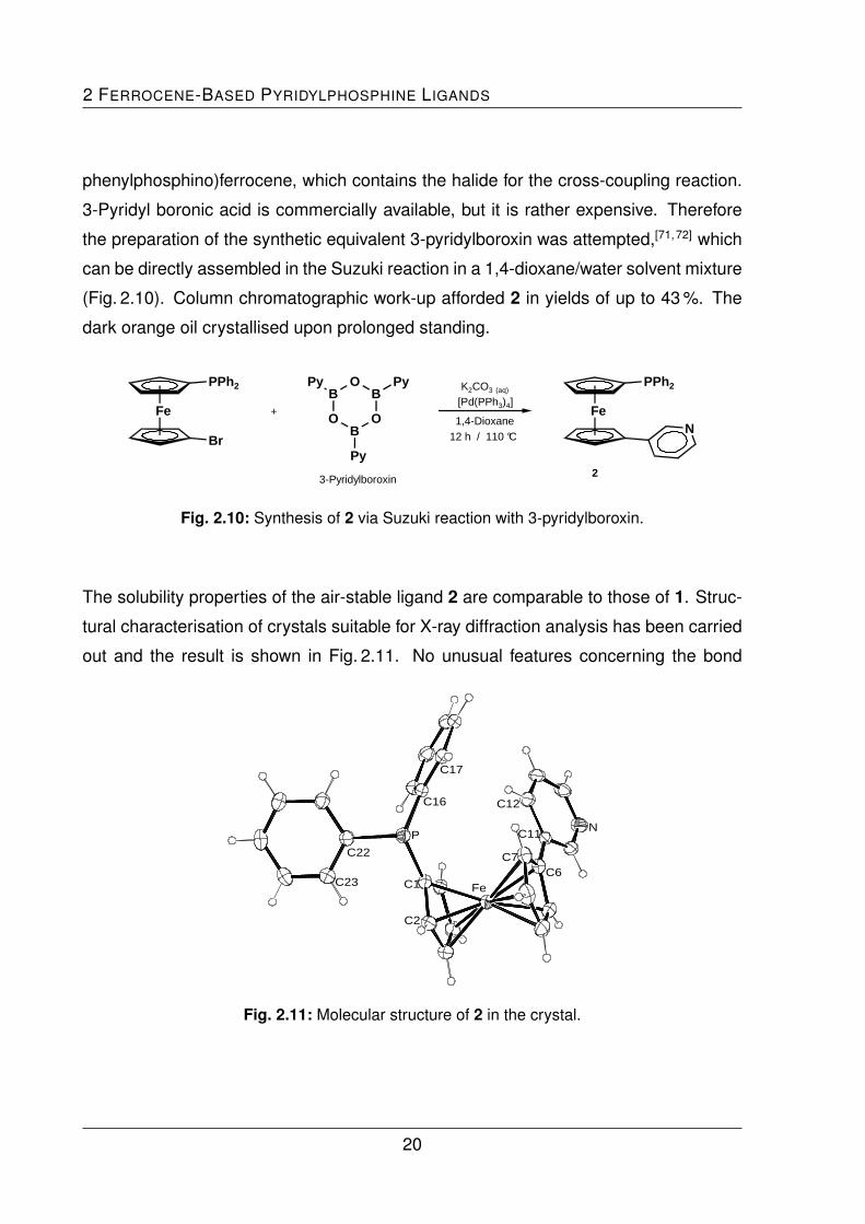
phenylphosphino)ferrocene, which contains the halide for the cross-coupling reaction.
3-Pyridyl boronic acid is commercially available, but it is rather expensive. Therefore
the preparation of the synthetic equivalent 3-pyridylboroxin was attempted,[71,72] which
can be directly assembled in the Suzuki reaction in a 1,4-dioxane/water solvent mixture
(Fig. 2.10). Column chromatographic work-up afforded 2 in yields of up to 43 %. The
dark orange oil crystallised upon prolonged standing.
Fe
PPh2
Br
OB
OB
O
BPy Py
Py
K2CO3 (aq)
[Pd(PPh3)4]
1,4-Dioxane12 h / 110 °C
+
3-Pyridylboroxin
Fe
2
PPh2
N
Fig. 2.10: Synthesis of 2 via Suzuki reaction with 3-pyridylboroxin.
The solubility properties of the air-stable ligand 2 are comparable to those of 1. Struc-
tural characterisation of crystals suitable for X-ray diffraction analysis has been carried
out and the result is shown in Fig. 2.11. No unusual features concerning the bond
C22
C23
P
C16
C17
C11
C12
N
C6C7
FeC1
C2
Fig. 2.11: Molecular structure of 2 in the crystal.
20

2.2 BIDENTATE LIGANDS (1 - 3)
parameters are observed. The 1H NMR spectrum shows four signals typical for a 1,1’-
unsymmetrical substitution at the ferrocene unit and two characteristic low field signals
of the pyridyl protons H2 and H6. A single signal is observed in the 31P NMR spectrum
at –17.7 ppm. The APCI mass spectrum shows peaks at 464 (45%) [MO + H]+ (caused
by reactions during the ionisation process, cf. 31P NMR), 448 m/z (100%) [M + H]+ and
several defined fragments at lower m/z.
2 was also investigated by cyclic voltammetry. Under the same conditions its behaviour
is rather comparable to that of 1: A ferrocene-based oxidation plus an additional irre-
versible oxidation process at higher potential is observed. The ferrocene-based redox
wave is anodically shifted by ca. 0.70 V with respect to fc* and displays reversible be-
haviour (∆EP = 64.5 mV) if the scan is reversed before the second oxidation process
starts. Interestingly, the pyrid-3-yl group causes an anodic shift which is ca. 150 mV
higher than that of 1 containing a pyrid-2-yl group.
2.2.3 Synthesis and Characterisation of 3
The synthesis of 3 was carried out by Jirı Schulz in the group of Prof. Petr Stepnicka at
Charles University, Prague, Czech Republic.
The synthesis of 3 started also with 1-bromo-1’-(diphenylphosphino)ferrocene,[64] which
was first converted into 1-lithio-1’-(diphenylphosphino)ferrocene. Without isolation, this
intermediate was reacted with pyridine-2-carbaldehyde to afford 1-[(pyrid-2-yl)hydroxy-
methyl]-1’-(diphenylphosphino)ferrocene (Fig. 2.12 a). Also a small amount of its cor-
responding ketone was obtained (Fig. 2.12 b). Following an analougous literature pro-
cedure,[73] the secondary alcohol was obtained after aqueous work-up and chromatog-
raphy as an ochre solid in 58 % yield,
The subsequent reduction of the hydroxyl group proved to be a challenging task since
the conventional methods, utilising Me3SiCl / NaI, NaBH4 / CF3CO2H or LiAlH4 / AlCl3
21

2 FERROCENE-BASED PYRIDYLPHOSPHINE LIGANDS
Fe
PPh2
Fe
PPh2
OH O
N N
(a) (b)
Fig. 2.12: 1-[(Pyrid-2-yl)hydroxymethyl]-1’-(diphenylphosphino)ferrocene (a) and its corre-
sponding ketone (b).
failed.[74] In the end, the dehydroxylation was accomplished by reaction with SmI2 /
(Me2N)3PO followed by the treatment with pivalic acid.[75] This synthetic procedure
(Fig. 2.13) gave 3 in yields of up to 55 % as an amber oil, which solidified upon standing.
Fe
PPh2
Br
SmI2/H+
Fe
3
PPh2
N
2. PyCHO1. n-BuLi
THF / -78°C THF
Fe
PPh2
N
OH
Fig. 2.13: Synthetic route to 3.
The air-stable compound 3 is soluble in polar solvents like dichloromethane and diethyl
ether. It was possible to obtain single crystals suitable for X-ray diffraction analysis. The
molecular structure is shown in Fig. 2.14. Bond lengths and angles are as expected.
The 1H NMR spectrum shows four signals for the 1,1’-ferrocendiyl backbone and an
additional signal for the methylene group next to the pyridyl unit between 3.63 ppm and
4.33 ppm. The signal due to the pyridyl H6 is observed at lower field as mentioned
before. The phosphorus NMR spectrum shows a single signal, whose chemical shift of
–16.5 ppm is close to that of the corresponding signals found for 1 and 2.
22

2.2 BIDENTATE LIGANDS (1 - 3)
NC12
C13
C11
C6
C7
Fe
PC23
C24
C17
C18
C1
C2
Fig. 2.14: Molecular structure of 3 in the crystal.
Similar to what was observed for 1 and 2, cyclic voltammetry experiments with 3 re-
sulted in a reversible oxidation wave of the ferrocene unit and an irreversible oxidation
wave at higher potentials (Fig. 2.15 a). The reversible redox wave of the ferrocene unit
appears 0.62 V anodically shifted with respect to fc*, if the scan is reversed before the
second oxidation process starts (Fig. 2.15 b). The result is comparable with that of 1
and 2. The ca. 70 mV higher FeII/FeIII redox potential is caused by the positive inductive
effect of the methylene group. The higher ∆EP of 102 mV is probably caused by the
tenfold higher concentration of 3 in the experiment.
-8
-6
-4
-2
0
2
4
6
8
10
12
14
16
18
20
-0,2 0,0 0,2 0,4 0,6 0,8 1,0 1,2 1,4 1,6 1,8 2,0
Cur
rent
(µA)
Potential (V)
(a)
fc*
-6
-4
-2
0
2
4
6
8
10
12
14
-0,3 -0,2 -0,1 0,0 0,1 0,2 0,3 0,4 0,5 0,6 0,7 0,8 0,9
Cur
rent
(µA)
Potential (V)
(b)
Fig. 2.15: CV of 3 (1.0 mM in DCM, 0.1 M [NnBu][PF6], 100 mV/s), scan range from –0.25 V to
+2.0 V (a) and from –0.25 V to +0.85 V (b).
23

2 FERROCENE-BASED PYRIDYLPHOSPHINE LIGANDS
2.3 Tridentate Ligands (4)
Beyond the bidentate ligands, it is an interesting question what happens if the ligand
offers more than just two donor sites. There could be a “choice” for the metal centre,
which donor atom is preferred. The P,N-motif can be expanded to an N,P,N- and a
P,N,P-motif. Both possibilities were investigated.
2.3.1 Synthesis and Characterisation of a N,P,N-Ligand
The easiest way to synthesise an N,P,N-ligand system, which is very similar to 1, is to
start with the known bis(1’-bromoferrocenyl)phenylphosphine.[64] The Negishi coupling
reaction with two equivalents of 2-bromopyridine is expected to afford the correspond-
ing ligand (Fig. 2.16).
Fe
PPh
ZnCl
Fe
PPh
NFe
PPh
Br
)2 )2 )22. ZnCl2
1. n-BuLi
THF / -70°C
[Pd(PPh3)4]2-BrPy
72 h / 60°C
Fig. 2.16: Synthetic route to bis[1’-(pyrid-2-yl)ferrocenyl]phenylphosphine.
In my hands, it was not possible to reproduce the synthesis of bis(1’-bromoferrocenyl)-
phenylphosphine Butler presented. The final 1H NMR data did not correspond to those
published by Butler. He obtained only one set of signals for the ferrocene unit. In our
case, the NMR spectrum revealed two sets of signals belonging to the ferrocene unit,
which implied inequivalence of both ferrocene units. Further reaction via the Negishi
coupling gave a compound whose identity could not be established unequivocally. Only
later crystallographic investigations of coordination experiments with mercury bromide
revealed the nature of this ligand. It turned out that only one of the two ferrocene units
is substituted with a pyridyl group. It is not clear in which step the problem occurs.
24

2.3 TRIDENTATE LIGANDS (4)
After several attempts this project was put on hold and the synthesis of an alternative
P,N,P-ligand was considered in more detail.
2.3.2 Synthesis and Characterisation of the P,N,P-Ligand 4
The P,N,P-Ligand 4, which contains one pyridine donor and two phosphine donor
atoms, is easily synthesised by Negishi cross-coupling reaction of 2,6-dibromopyridine
with two equivalents of 1-bromo-1’-(diphenylphosphino)ferrocene (Fig. 2.17). Column
chromatography afforded 2,6-bis(1’-diphenylphosphinoferrocenyl)pyridine (4) in yields
of up to 33 %.
Fe
PPh2
ZnCl
Fe
PPh2
Br
Fe
PPh2
Fe
N
PPh2
4
2. ZnCl2
1. n-BuLi
THF / -70°C
[Pd(PPh3)4]2,6-Br2py
60°C / 72 h
Fig. 2.17: Synthesis of 2,6-bis(1’-diphenylphosphanylferrocenyl)pyridine (4).
Characterisation was achieved by NMR spectroscopy, mass spectrometry and elemen-
tal analysis. Only a single signal is observed for both phosphorus atoms in the 31P NMR
spectrum, which demonstrates the symmetric structure of the molecule. The proton
NMR spectrum shows only one set of signals belonging to both ferrocene units. The
result of a single crystal X-ray diffraction study is shown in Fig. 2.18. Crystallisation in
the presence of air causes oxidation of one of the phosphorus atoms. 4 shows only
limited stability towards oxygen in solution.
The cyclic voltammogram of 4 shows two irreversible oxidation processes. The peak
potential positions are anodically shifted with respect to fc* by 0.65 V and 1.21 V, re-
spective.
25

2 FERROCENE-BASED PYRIDYLPHOSPHINE LIGANDS
P1
Fe1N1
Fe2
P2
Fig. 2.18: Molecular structure of 4 in the crystal.
2.4 Summary and Conclusion
While the synthesis of the bidentate ligand 1 was already published,[1] it was possible to
improve the original low yield procedure by establishing a more convenient one, whose
final step is a Negishi cross-coupling instead of a Ziegler reaction in the originally re-
ported synthesis. Key advantages are the air-stable intermediate 1-bromo-1’-(diphenyl-
phosphino)ferrocene and higher yields. The synthesis of the new 3-pyridine isomer 2
by Suzuki cross-coupling reaction has been developed. The homologous ligand 3,
which contains a CH2-linker between ferrocene and its pyrid-2-yl unit was synthesised
via less conventional methods by the cooperation partners from the Czech Republic.
Furthermore, the synthesis of analogous tridentate ligands was attempted. The syn-
thesis of a N,P,N-ligand in analogy to the route to 1 met with limited success. Instead,
the P,N,P-ligand 4 was synthesised using the Negishi methodology established in the
synthesis of 1. Structural characterisation of all ligands was possible by single crystal
X-ray diffraction studies, which revealed no unexceptional features. CV measurements
of 1, 2 and 3 show a reversible ferrocene-based redox process if the scan is reversed
before a second oxidation process starts at higher potentials. Probably the second
oxidation process induces irreversible chemical reactions.
26

3 Coordination Chemistry I:
Zinc, Cadmium and Mercury
This chapter describes the coordination chemistry towards the divalent group 12 metal
ions ZnII, CdII and HgII and discusses the synthesis and characterisation of complexes
of the ligands 1, 2 and 3.
3.1 Introduction
The metals zinc, cadmium and mercury were chosen to explore the fundamental co-
ordination chemistry of the ligands 1, 2 and 3. Aspects of the coordination behaviour
in terms of the HSAB principle on the one hand and questions concerning the issue
of bridging versus chelating coordination mode on the other hand were in the focus
of interest. An important feature of these metals is the redox-inertness of the divalent
MII ions in the case of zinc and cadmium. Metal redox processes would interfere un-
favourably with cyclovoltammetric experiments to explore the utilisation of the ligands
as potential molecular electrochemical sensors.
It is known that pyridine and triphenylphosphine react coordinatively with group 12
metal halides. The molecular structures of simple pyridine complexes of the type
[MX2(PyH)2] (X = Cl, Br, I) of ZnII,[76,77] CdII[78] and HgII[79] show an approximately tetra-
hedral coordination geometry with two exceptions. [CdCl2(PyH)2] and [HgCl2(PyH)2]
27

3 COORDINATION CHEMISTRY I: ZINC, CADMIUM AND MERCURY
exhibit an octahedral coordination geometry in which each metal centre is surrounded
by four halide atoms in equatorial and two pyridines in axial positions. The metal cen-
tres are bridged by both chloro ligands to form a chain of edge-sharing octahedra.
Donor-metal distances are listed in Tab. 3.1.
The corresponding triphenylphosphine complexes [MX2(PPh3)2] with halides of zinc,
cadmium and mercury have also been prepared and structurally characterised in the
past.[80–82] The interligand angles of all three compounds lie between 103 ◦-116 ◦, which
corresponds to an only slightly distorted tetrahedral coordination geometry. The donor-
metal distance (Tab. 3.1) are considerably larger than in the pyridine compounds, which
Tab. 3.1: Selected bond lengths (pm) of [MX2(PyH)2], [MX2(PPh3)2] and [MX2(dppf)] (M = Zn,
Cd, Hg; X = Cl, Br, I).
[MX2(PyH)2] [MX2(PPh3)2] [MX2(dppf)]
Zn–Donora1 (X = Cl) 204.6 261.0
Zn–Donor2 (X = Cl) 205.2 249.9
Zn–Donor1 (X = Br) 203.6
Zn–Donor2 (X = Br) 204.3
Cd–Donor1 (X = Cl) 235 263.3
Cd–Donor2 (X = Cl) 235 264.6
Cd–Donor1 (X = Br) 265.4
Cd–Donor2 (X = Br) 261.8
Cd–Donor1 (X = I) 263.4 263.3
Cd–Donor2 (X = I) 265.7 265.4
Hg–Donor1 (X = Cl) 226.6 250.3 251.0
Hg–Donor2 (X = Cl) 226.6 253.2 251.6
Hg–Donor1 (X = Br) 238.2 255.0
Hg–Donor2 (X = Br) 239.2 249.2
Hg–Donor1 (X = I) 230.6 257.2 255.8
Hg–Donor2 (X = I) 230.6 255.6 257.2
aDonor = N in [MX2(PyH)2], P in [MX2(PPh3)2] and [MX2(dppf)].
28

3.1 INTRODUCTION
is expected with respect to the differences of the covalence radii of both donor atoms
(P = 110 ppm, N = 70 ppm).[39] The largest difference in the donor-metal (M–N, M–P)
distances was obtained in the case of zinc (ca. 50 pm). In the case of cadmium and
mercury the differences have an average value of 25 pm. A single exception is detected
in the HgBr2 complexes. The donor-metal distances here differ only less than 15 ppm.
These facts can be understood, at least to a first approximation, by considering the
HSAB principle. The medium hard ZnII interacts strongly with the medium hard pyri-
dine (comparatively short Zn–N bond), but only comparatively weakly with the soft
phosphine (comparatively long Zn–P bond). The opposite is true in the case of the
soft HgII, which interacts strongly with the soft phosphine (comparatively short Hg–P
bond), but only weakly with pyridine (comparatively long Zn–N bond), which leads to
very similar Hg-donor bond lengths despite the large difference of the covalence radii
of N and P.
The ferrocene-based bis(phosphine) 1,1’-bis(diphenylphosphino)ferrocene (dppf) forms
chelate complexes with several metal halides. Complexes containing group twelve
metals are structurally characterised only in the case of CdII and HgII.[83–86] For both
metals the typical tetrahedral coordination geometry has been found. The donor-metal
distances are in line with those of the simple triphenylphosphine complexes (Tab. 3.1).
Crystallographic data on zinc compounds have not been published. Only a single pub-
lication reports on physicochemical properties of [MCl2(dppf)] (M = Co, Ni, Pd, Pt, Zn,
Cd, Hg).[87] For the pyridine analogue of dppf, 1,1’-di(pyrid-2-yl)ferrocene, surprisingly
no complexes with group 12 metals have been described. A zinc compound of its oc-
tamethyl analogue was reported in conjunction with the detection of divalent ions in
solution.[52]
A ferrocene-based P,O-ligand (Fig. 3.1), which has its donor atoms in structurally sim-
ilar positions like 1, has been investigated in group 12 metal coordination chemistry.[88]
Interestingly, for all three metals of the series, different results concerning their coor-
dination behaviour were obtained. The molecular structure of the complex obtained
with ZnBr2 was identified as a P,O chelate (Fig. 3.2 a), which shows the expected tetra-
hedral coordination geometry (coordination number (CN) = 4). In contrast, the reac-
29

3 COORDINATION CHEMISTRY I: ZINC, CADMIUM AND MERCURY
Fe
PPh2
PO
OEt
OEt
Fig. 3.1: Diethyl [1’-(diphenylphosphino)ferrocenyl]phosphonate.
tion with HgBr2 afforded a symmetric bromo-bridged dimeric bis(phosphine) complex
[{Hg(µ-Br)Br(L-κ2P)}2] (Fig. 3.2 b). Due to the short distance between the phosphoryl
oxygen and the mercury atom of the neighbouring molecule (278.8 pm), the coordina-
tion geometry is better described as a distorted trigonal bipyramid (CN = 4 + 1).
Fe
P
PO
OEt
OEt
PhPh
ZnBr
Br
Fe
P
PO3Et2
PhPh(a) (b)
HgBr
BrHg
Br
Br
P
Fe
P
PEt2O3P
Fig. 3.2: Zinc (a) and mercury (b) complexes of the P,O-ligand shown in Fig. 3.1.
In solution an equilibrium between this bis(phosphine) species, a zinc-analogous P,O-
chelate and a P,O-bridged dimer was observed. The existence of several isomers
in solution (NMR spectroscopy) compared to a single molecular structure in the solid
state (X-ray diffraction analysis) is a typical problem in this context and will become
relevant for the work described in the present thesis.
The reaction with cadmium bromide gave a coordination polymer in which dinuclear
{Cd(µ-Br)Br}2 units are linked to infinite chains by symmetric pairs of the O,P-bridging
phosphinophosphonate ligand (Fig 3.3). The donor set around the cadmium atoms is
classified as a trigonal bipyramid rather than a square pyramid (CN = 5). The Cd–P
bond length of 263.1 pm is in line with the donor-metal distance in [CdBr2(dppf)].
30
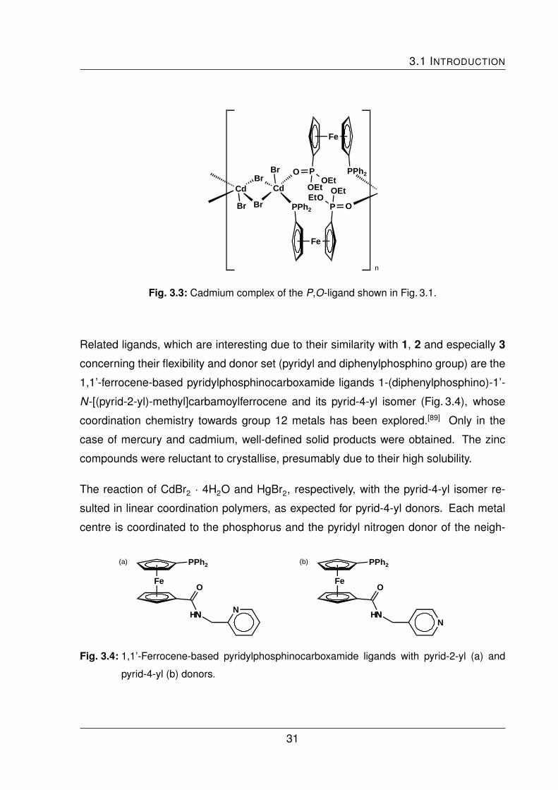
3.1 INTRODUCTION
Fe
PPh2 PO
O
OEtEt
Cd
Br
Cd
Br
BrBr
Fe
PPh2POEt
O
OEt
n
Fig. 3.3: Cadmium complex of the P,O-ligand shown in Fig. 3.1.
Related ligands, which are interesting due to their similarity with 1, 2 and especially 3
concerning their flexibility and donor set (pyridyl and diphenylphosphino group) are the
1,1’-ferrocene-based pyridylphosphinocarboxamide ligands 1-(diphenylphosphino)-1’-
N-[(pyrid-2-yl)-methyl]carbamoylferrocene and its pyrid-4-yl isomer (Fig. 3.4), whose
coordination chemistry towards group 12 metals has been explored.[89] Only in the
case of mercury and cadmium, well-defined solid products were obtained. The zinc
compounds were reluctant to crystallise, presumably due to their high solubility.
The reaction of CdBr2 · 4H2O and HgBr2, respectively, with the pyrid-4-yl isomer re-
sulted in linear coordination polymers, as expected for pyrid-4-yl donors. Each metal
centre is coordinated to the phosphorus and the pyridyl nitrogen donor of the neigh-
Fe
PPh2
N
O
H
(a)
N N
Fe
PPh2
N
O
H
(b)
NN
Fig. 3.4: 1,1’-Ferrocene-based pyridylphosphinocarboxamide ligands with pyrid-2-yl (a) and
pyrid-4-yl (b) donors.
31
3 COORDINATION CHEMISTRY I: ZINC, CADMIUM AND MERCURY
bouring molecule. Both molecular structures are very similar. Interestingly, the donor-
metal distances (P–Hg, N–Hg) in the mercury complex are both identical (243.8 pm),
while they differ markedly in the cadmium species (P–Cd 257.6 pm, N–Cd 230.1 pm).
This is in accord with the HSAB principle (see page 29). The coordination tetrahedron
is strongly distorted in the mercury complex. The interligand angles differ more than
20 ◦ from the ideal 109.5 ◦.[89]
The same reaction with the pyrid-2-yl isomer also afforded well-defined products. How-
ever, the molecular structures established by X-ray crystallography are rather compli-
cated. The cadmium complex is formally a tetramer involving two types of cadmium
centres with different coordination geometries and donor sets. This is a very special
and relatively complicated molecular structure and is therefore not described further. It
indicates that the coordination chemistry of cadmium may become more complicated
than expected initially. The structure determination of the product obtained from the re-
action with HgBr2 revealed a centrosymmetric, doubly halide-bridged dimer commonly
encountered among 1:1 phosphine mercury halide complexes. Because of the steric
demand of the ligand, the coordination polyhedron (CN = 4) is severely distorted.[89]
3.2 Coordination Chemistry of 1
The synthesis of the ferrocene-based pyridylphosphine ligand 1 has been described in
Chapter 2.2.1.
3.2.1 Synthesis and Characterisation of Zn Compounds
The reaction of an ethanolic solution of 1 with zinc halides ZnX2 (X = Cl, Br, I) in a 1:1
molar ratio gave the orange to red chelate complexes [ZnCl2(1)] (5a), [ZnBr2(1)] (5b)
and [ZnI2(1)] (5c) as shown in Fig. 3.5. All three compounds have been found to be
air-stable and show good solubility in polar solvents.
32

3.2 COORDINATION CHEMISTRY OF 1
Fe
1
PPh2
N+ ZnX2
5b (X = Br)5c (X = I)
Fe
5a (X = Cl)
PPh2
N
ZnX2ethanol
14 h
X = Cl, Br, I
Fig. 3.5: Synthesis of Zn compounds of 1 in 1:1 stoichiometry.
One method to elucidate the binding mode in these complexes is an inspection of the
shift of diagnostic signals in the 1H and the 31P NMR spectrum. The chemical shift of
the pyridyl H6 and the shift of the phosphorus signal are given in Tab. 3.2. The signals of
the pyridyl H6 in the 1H NMR spectra are low-field shifted by about 1.1 ppm with respect
to the free ligand. This is caused by deshielding due to the decrease of electron density
in the vicinity of the pyridyl H6 by metal coordination to the pyridyl nitrogen atom. The
phosphorus signals in the 31P NMR spectra are shifted only slightly with respect to
pristine 1. While the phosphorus signals in 5a and 5b show the expected small low-
field shift induced by the zinc coordination, the signal of 5c was observed at slightly
higher field than that of the free ligand.
Single crystal X-ray diffraction analyses gave a detailed picture of the binding mode.
5a and 5b each crystallise with two independent molecules in the unit cell, which differ
slightly from each other. The results for molecule 1 are each shown in Fig. 3.6. For 5c it
was not possible to obtain material suitable for single crystal X-ray diffraction analysis.
Tab. 3.2: Chemical shifts of the diagnostic NMR signals of 5a, 5b and 5c in ppm.
1 5a 5b 5c
signal due to pyridyl H6 (1H NMR) 8.47 9.57 9.57 9.60
phosphorus signal (31P NMR) –17.7 –14.0 –14.4 –19.2
Solvent: CDCl3.
33
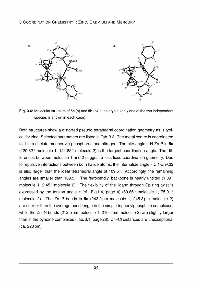
3 COORDINATION CHEMISTRY I: ZINC, CADMIUM AND MERCURY
(a)
N Cl2
Cl1Zn
P
Fe
P Br2
Br1N
Fe Zn
(b)
Fig. 3.6: Molecular structure of 5a (a) and 5b (b) in the crystal (only one of the two independent
species is shown in each case).
Both structures show a distorted pseudo-tetrahedral coordination geometry as is typi-
cal for zinc. Selected parameters are listed in Tab. 3.3. The metal centre is coordinated
to 1 in a chelate manner via phosphorus and nitrogen. The bite angle 6 N-Zn-P in 5a
(120.82 ◦ molecule 1, 124.85 ◦ molecule 2) is the largest coordination angle. The dif-
ferences between molecule 1 and 2 suggest a less fixed coordination geometry. Due
to repulsive interactions between both halide atoms, the interhalide angle 6 Cl1-Zn-Cl2
is also larger than the ideal tetrahedral angle of 109.5 ◦. Accordingly, the remaining
angles are smaller than 109.5 ◦. The ferrocendiyl backbone is nearly untilted (1.39 ◦
molecule 1, 2.45 ◦ molecule 2). The flexibility of the ligand through Cp ring twist is
expressed by the torsion angle τ (cf. Fig 1.4, page 4) (69.86 ◦ molecule 1, 75.01 ◦
molecule 2). The Zn–P bonds in 5a (243.2 pm molecule 1, 245.3 pm molecule 2)
are shorter than the average bond length in the simple triphenylphosphine complexes,
while the Zn–N bonds (212.5 pm molecule 1, 210.4 pm molecule 2) are slightly larger
than in the pyridine complexes (Tab. 3.1, page 28). Zn–Cl distances are unexceptional
(ca. 223 pm).
34
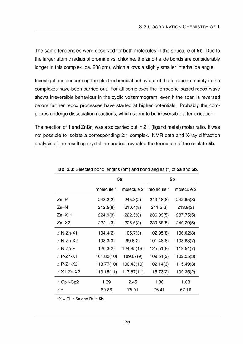
3.2 COORDINATION CHEMISTRY OF 1
The same tendencies were observed for both molecules in the structure of 5b. Due to
the larger atomic radius of bromine vs. chlorine, the zinc-halide bonds are considerably
longer in this complex (ca. 238 pm), which allows a slighly smaller interhalide angle.
Investigations concerning the electrochemical behaviour of the ferrocene moiety in the
complexes have been carried out. For all complexes the ferrocene-based redox-wave
shows irreversible behaviour in the cyclic voltammogram, even if the scan is reversed
before further redox processes have started at higher potentials. Probably the com-
plexes undergo dissociation reactions, which seem to be irreversible after oxidation.
The reaction of 1 and ZnBr2 was also carried out in 2:1 (ligand:metal) molar ratio. It was
not possible to isolate a corresponding 2:1 complex. NMR data and X-ray diffraction
analysis of the resulting crystalline product revealed the formation of the chelate 5b.
Tab. 3.3: Selected bond lengths (pm) and bond angles (◦) of 5a and 5b.
5a 5b
molecule 1 molecule 2 molecule 1 molecule 2
Zn–P 243.2(2) 245.3(2) 243.48(8) 242.65(8)
Zn–N 212.5(8) 210.4(8) 211.5(3) 213.9(3)
Zn–Xa1 224.9(3) 222.5(3) 236.99(5) 237.75(5)
Zn–X2 222.1(3) 225.6(3) 239.68(5) 240.29(5)
6 N-Zn-X1 104.4(2) 105.7(3) 102.95(8) 106.02(8)
6 N-Zn-X2 103.3(3) 99.6(2) 101.48(8) 103.63(7)
6 N-Zn-P 120.3(2) 124.85(16) 125.51(8) 119.54(7)
6 P-Zn-X1 101.82(10) 109.07(9) 109.51(2) 102.25(3)
6 P-Zn-X2 113.77(10) 100.43(10) 102.14(3) 115.49(3)
6 X1-Zn-X2 113.15(11) 117.67(11) 115.73(2) 109.35(2)
6 Cp1-Cp2 1.39 2.45 1.86 1.08
6 τ 69.86 75.01 75.41 67.16
aX = Cl in 5a and Br in 5b.
35
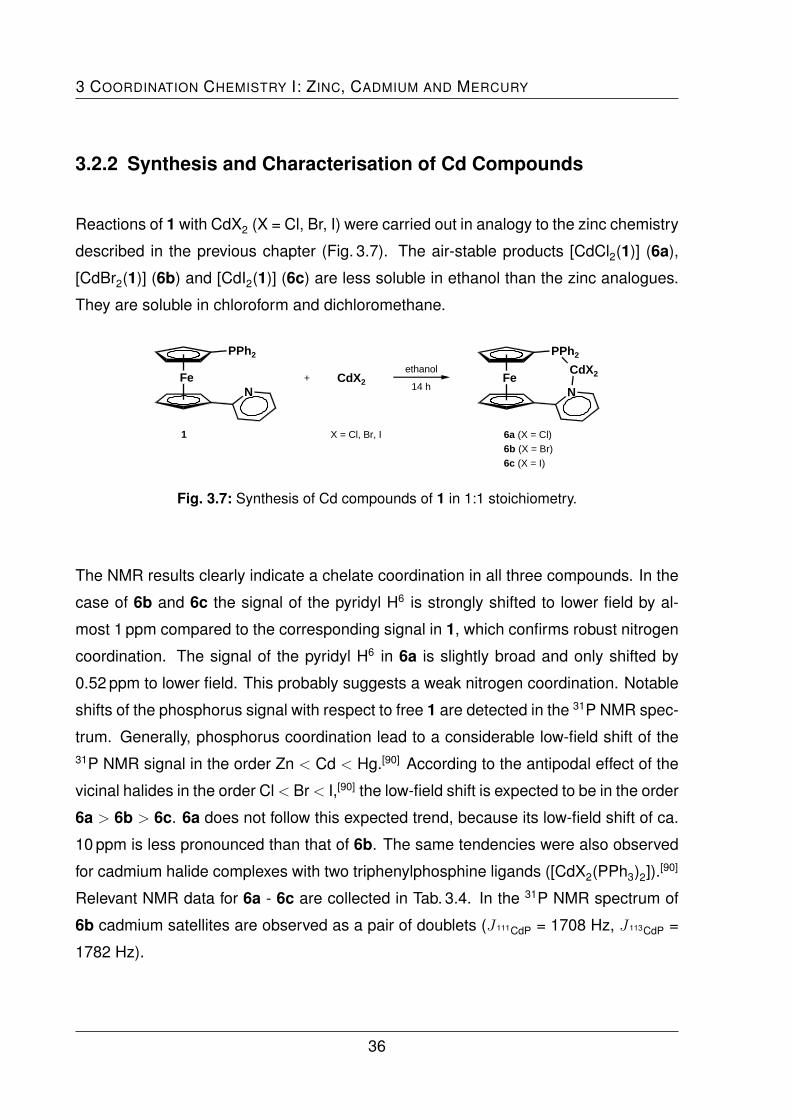
3 COORDINATION CHEMISTRY I: ZINC, CADMIUM AND MERCURY
3.2.2 Synthesis and Characterisation of Cd Compounds
Reactions of 1 with CdX2 (X = Cl, Br, I) were carried out in analogy to the zinc chemistry
described in the previous chapter (Fig. 3.7). The air-stable products [CdCl2(1)] (6a),
[CdBr2(1)] (6b) and [CdI2(1)] (6c) are less soluble in ethanol than the zinc analogues.
They are soluble in chloroform and dichloromethane.
Fe
1
PPh2
N+ CdX2
6b (X = Br)
6c (X = I)
Fe
6a (X = Cl)
PPh2
N
CdX2ethanol
14 h
X = Cl, Br, I
Fig. 3.7: Synthesis of Cd compounds of 1 in 1:1 stoichiometry.
The NMR results clearly indicate a chelate coordination in all three compounds. In the
case of 6b and 6c the signal of the pyridyl H6 is strongly shifted to lower field by al-
most 1 ppm compared to the corresponding signal in 1, which confirms robust nitrogen
coordination. The signal of the pyridyl H6 in 6a is slightly broad and only shifted by
0.52 ppm to lower field. This probably suggests a weak nitrogen coordination. Notable
shifts of the phosphorus signal with respect to free 1 are detected in the 31P NMR spec-
trum. Generally, phosphorus coordination lead to a considerable low-field shift of the31P NMR signal in the order Zn < Cd < Hg.[90] According to the antipodal effect of the
vicinal halides in the order Cl< Br< I,[90] the low-field shift is expected to be in the order
6a > 6b > 6c. 6a does not follow this expected trend, because its low-field shift of ca.
10 ppm is less pronounced than that of 6b. The same tendencies were also observed
for cadmium halide complexes with two triphenylphosphine ligands ([CdX2(PPh3)2]).[90]
Relevant NMR data for 6a - 6c are collected in Tab. 3.4. In the 31P NMR spectrum of
6b cadmium satellites are observed as a pair of doublets (J 111CdP = 1708 Hz, J 113CdP =
1782 Hz).
36

3.2 COORDINATION CHEMISTRY OF 1
Tab. 3.4: Chemical shifts of the diagnostic NMR signals of 6a, 6b and 6c in ppm.
1 6a 6b 6c
signal due to pyridyl H6 (1H NMR) 8.47 8.99 9.35 9.27
phosphorus signal (31P NMR) -17.7 -8.0 -3.6 -8.9
Solvent: CDCl3.
While 6b and 6c could be crystallised easily, 6a resisted crystallisation. Only diffusion
experiments with a dichloromethane solution of 1 layered with a solution of CdCl2 in
diethyl ether afforded material suitable for single-crystal X-ray diffraction (Fig. 3.8). The
obtained inversion symmetric bis(phosphine) structure reflects a 2:1 (ligand:metal) mo-
lar ratio, though the experiment was carried out in a 1:1 molar ratio. The new complex
[CdCl2(1)2] (7) was characterised by X-ray diffraction only.
Two aspects could influence the crystallisation process towards the obtained stoichiom-
etry. First, the poor solubility of CdCl2 in organic solvents could cause concentration
differences at the boundary of both layers. Second, an equilibrium in solution be-
tween the chelated and the bis(phosphine) species may be responsible for this obser-
vation. In solution the equilibrium is almost entirely shifted in favour of the chelate,
which can be confirmed by the low-field shifted broad signal of the pyridyl H6 in the
N
Cl
Cli
Fe
P Cd
Pi
Ni
Fei
Fig. 3.8: Molecular structure of 7 in the crystal.
37

3 COORDINATION CHEMISTRY I: ZINC, CADMIUM AND MERCURY
1H NMR spectrum. However, if the bis(phosphine) species 7 is only slightly less soluble
than the chelate 6a, it crystallises first and is therefore reproduced by the equilibrium.
Generating other cadmium complexes with 2:1 (ligand:metal) stoichiometry failed. At-
tempts with CdBr2 · 4 H2O in ethanol gave probably a mixture of free ligand and the
chelated compound. Crystallisation experiments failed with one exception, which gave
the known metal chelate 6b.
The bond angles in 7 deviate from the ideal tetrahedral angle of 109.5 ◦. The largest
angle is 6 P-Cd-P’ (122.04 ◦), the smallest one in turn is the interhalide angle 6 Cl-Cd-Cl
(103.63 ◦). In comparison with the analogous bis(phosphine) complex [CdCl2(PPh3)2],
this can be ascribed to the steric demand of the pyridylphosphine ligands which contain
an additional bulky ferrocenediyl group. The bond lengths are similar to those of the
corresponding triphenylphosphine complex (Tab. 3.5).
The molecular structure of the chelates 6b and 6c are shown in Fig. 3.9. 6b crystallises
in the monoclinic space group P21/c and 6c in the triclinic space group P 1. The bond
Tab. 3.5: Selected bond lengths (pm) and bond angles (◦) of 7 and [CdCl2(PPh3)2].
7 [CdCl2(PPh3)2]
Cd–P1 258.0(2) 263.3
Cd–P2a 258.0(2) 264.6
Cd–Cl1 244.8(2) 249.0
Cd–Cl2a 244.8(2) 244.2
6 P1-Cd-Cl1 100.55(6) 104.96
6 P1-Cd-Cl2 114.59(5) 105.78
6 P1-Cd-P2 122.04(8) 107.27
6 P2-Cd-Cl1 114.59(5) 112.22
6 P2-Cd-Cl2 100.55(6) 111.55
6 Cl1-Cd-Cl2 103.63(9) 114.37
6 Cp1-Cp2 2.29
6 τ 86.54
aP2, Cl2 = P’, Cl’ generated by symmetry operations in 7.
38

3.2 COORDINATION CHEMISTRY OF 1
(a)
N Br2
Br1Cd
P
Fe
(b)
N
I1
I2Cd
P
Fe
Fig. 3.9: Molecular structure of 6b (a) and 6c (b) in the crystal.
parameters exhibit no unusual features. The donor-metal bond lengths (Tab. 3.6) of
both complexes are very similar.
Tab. 3.6: Selected bond lengths (pm) and bond angles (◦) of 6b and 6c.
6b 6c
Cd–P 261.88(12) 259.32(11)
Cd–N 234.1(4) 237.5(3)
Cd–Xa1 255.96(6) 273.26(4)
Cd–X2 258.00(6) 275.20(5)
6 N-Cd-X1 103.13(9) 102.22(9)
6 N-Cd-X2 102.19(9) 96.02(8)
6 N-Cd-P 118.70(10) 121.60(9)
6 P-Cd-X1 105.60(3) 115.64(3)
6 P-Cd-X2 114.31(3) 103.49(3)
6 X1-Cd-X2 112.58(2) 116.990(15)
6 Cp1-Cp2 2.08 1.32
6 τ 78.41 78.45
aX = Br in 6b and I in 6c.
39

3 COORDINATION CHEMISTRY I: ZINC, CADMIUM AND MERCURY
The halide-metal bonds of 6c are longer due to the larger atomic radius of iodine vs.
bromine. The bond angles in 6b are close to the ideal tetrahedral angle of 109.5 ◦. Only
the bite angle 6 N-Cd-P is wider by ca. 10 ◦. In addition to packing effects, the fixed
ligand geometry might be responsible for this. The coordination tetrahedron of 6c is
more distorted. With a value of only 96.02 ◦ 6 N-Cd-I2 is the smallest angle. Probably
the I2 atom is influenced by the steric demands of the adjacent phenyl ring and the
pyridine ring. The distances between I2 and the nearest carbon atom of the phenyl
ring is 400.6 pm. The distance between I2 and the nearest carbon atom of the pyridine
ring is only 371.5 pm, which is approximately the sum of the van der Waals radii of C
and I (368 pm). Tilt angle and torsion angle a very similar for both complexes.
3.2.3 Synthesis and Characterisation of Hg Compounds
The coordination behaviour of mercury has been studied in analogous reactions with 1
(Fig. 3.10). The results obtained for the reaction with HgCl2 and HgBr2 are very similar
Fe
1
PPh2
N+ HgX2
8b (X = Br)
Fe
8a (X = Cl)
PPh2
N
HgX2ethanol
14 h
X = Cl, Br
Fig. 3.10: Synthesis of Hg compounds of 1 in 1:1 stoichiometry.
to those obtained from the reactions with cadmium halides. The expected chelate
complexes [HgCl2(1)] (8a) and [HgBr2(1)] (8b) were formed. The reaction with HgI2instead afforded the iodo-bridged, P-coordinated dimer [{Hg(µ-I)I(1)}2] (8c) as shown
in Fig. 3.11.
single-crystal X-ray diffraction analyses failed for 8a, because it was impossible to ob-
tain suitable material. Diffusion experiments in a 1:1 molar ratio eventually afforded
40

3.2 COORDINATION CHEMISTRY OF 1
Fe
P
PhPh
Hg
I
I
Hg
N
Fe
P
Ph Ph
N
I
I
Fig. 3.11: Iodo-bridged, P-coordinated dimer 8c.
crystalline material, which turned out to be the 2:1 (ligand:metal) complex [HgCl2(1)2]
(9a), described on page 43.
In the case of 8b, single-crystal X-ray diffraction data clearly establish a distorted pseu-
dotetrahedral chelate structure (Fig. 3.12 and Tab. 3.7). The halide-metal distances are
comparable to those of 6b. However, in accord with the HSAB principle, the Hg–P
Br1
Br2
N
Fe
P
Hg
Fig. 3.12: Molecular structure of 8b in the
crystal.
Tab. 3.7: Selected bond lengths (pm) and
bond
angles (◦) of 8b.
Hg–P 244.62(9)
Hg–N 257.3(3)
Hg–Br1 262.91(5)
Hg–Br2 255.74(5)
6 N-Hg-Br1 90.12(7)
6 N-Hg-Br2 92.37(7)
6 N-Hg-P 118.87(7)
6 P-Hg-Br1 108.03(2)
6 P-Hg-Br2 128.72(3)
6 Br1-Hg-Br2 111.667(16)
6 Cp1-Cp2 3.24
6 τ 76.30
41

3 COORDINATION CHEMISTRY I: ZINC, CADMIUM AND MERCURY
distance is 17 pm shorter than the Cd–P bond length in 6b. On the other hand, the
Hg–N distance is 23 pm longer than the corresponding Cd–N bond. Interestingly, in
8b the Hg–P bond is shorter than the Hg–N bond, which indicates a very strong Hg–P
bond and a very weak Hg–N bond. Due to the vicinity of the bromo ligands to the
bulky phenyl groups, induced by the short Hg–P bond, the N-Hg-Br angles become
particularly acute (ca. 90 ◦).
The molecular structure of 8c, which was obtained by recrystallisation of the initially
formed precipitate, differs from all others mentioned above (Fig. 3.13 and Tab. 3.8).
One ligand molecule is coordinated via its phosphorus donor atom to one HgI2. Two
of these units form an iodo-bridged dimer containing tetracoordinate HgII centres. The
pyridine nitrogen atom remains uncoordinated. Halide bridges are well known in mer-
cury complexes (Chapter 3.1). They arise particularly in the solid state through packing
effects, probably assisted by comparatively weak bonds to the nitrogen donor atom in
accord with the HSAB principle. The bond angles around the HgII centres in 8c show
pseudotetrahedral coordination geometry, which is typical for the coordination num-
ber four and group 12 metals. The atoms Hg, Hgi, I2 and I2i form a diamond, which
is nearly rectangular. Three different Hg–I bond lengths are observed. The bond to
the terminal I1 is the shortest (267.65 pm), the longest bond is that to the bridging I2i
(303.12 pm).
P1
Hg1
I2i
I1i
Hg1i
I2
I1
N1
N1iFe1
Fe1iP1i
Fig. 3.13: Molecular structure of 8c in the crystal (solvent atoms are omitted for clarity).
42

3.2 COORDINATION CHEMISTRY OF 1
Tab. 3.8: Selected bond lengths (pm) and bond angles (◦) of 8c.
bond length bond angles
Hg–P 247.7(3) 6 I1-Hg-I2i 105.40(3)
Hg–I2i 303.12(10) 6 I2-Hg-I2i 95.89(3)
Hg–I1 267.65(10) 6 P-Hg-I2i 97.99(8)
Hg–I2 287.85(11) 6 P-Hg-I1 130.37(8)
6 P-Hg-I2 103.75(8)
6 I1-Hg-I2 116.30(3)
6 Hgi-I2-Hg 84.11(3)
6 τ 146.81
6 Cp1-Cp2 2.80
The NMR data prove the chelate structures of 8a and 8b in solution. The diagnostic
signals in the 1H NMR as well as in the 31P NMR spectrum are shifted to lower field. In
the case of the coordination of mercury dichloride in 8a this effect is most pronounced.
The 199Hg-P coupling constant as reflected by the mercury satellites in the 31P NMR
spectrum of 8a is larger than that observed in the spectrum of 8b. The pyridyl H6
is shifted downfield by nearly 0.9 ppm and the phosphorus signal is detected about
47 ppm shifted downfield with respect to the signal of the free ligand. In 8b and 8c
these shifts are increasingly less pronounced (Tab. 3.9). The coordination-induced shift
observed for the pyridyl H6 signal in 8c is surprising in view of the uncoordinated nature
of the pyridyl group in the crystalline compound. This indicates a highly dynamic equi-
librium in solution between different isomers of 8c, including an N-coordinated chelate.
The crystallisation of 8a by diffusion experiments by layering a ligand solution with
a solution of HgCl2 afforded the bis(phosphine) complex [HgCl2(1)2] (9a), similar to
the crystallisation of the cadmium analogue 6a, which afforded 7. As in the case of
7 an equilibrium in solution between the chelate and the bis(phosphine) complex to-
gether with the poorer solubility of the latter could be responsible for this effect. 9a
was characterised by X-ray diffraction analysis only. The reaction of HgBr2 with two
43

3 COORDINATION CHEMISTRY I: ZINC, CADMIUM AND MERCURY
Tab. 3.9: Chemical shifts of the diagnostic NMR signals of 8a, 8b and 8c in ppm.
1 8a 8b 8c
signal due to pyridyl H6 (1H NMR) 8.47 9.36 9.15 8.89
phosphorus signal (31P NMR) –17.7 32.2 28.5 19.7
J 199HgP in Hz (31P NMR) 7470 6359
Solvent: CDCl3.
equivalents of 1 gave the analogous bis(phosphine) complex [HgBr2(1)2] (9b), which
has been fully characterised. The results of the structure determination carried out for
both 2:1 (ligand:metal) complexes are displayed in Fig 3.14. Both structures show the
P1Cl1
Hg
Cl2
P2
N2
N1
(a)
P1Br2
Hg
Br1P2
N2
N1
(b)
Fig. 3.14: Molecular structures of 9a (a) and 9b (b) in the crystal.
typical, distorted pseudotetrahedral coordination with the P2-Hg-P1 angles being the
largest coordination angles (ca. 124 ◦) (Tab. 3.10). The other angles are unexceptional
and very similar in both complexes. The Hg–P bond lengths are almost identical. Due
to the larger atomic radius of bromine vs. chlorine, the halide-metal bonds of 9b are
longer than those of 9a.
No coordination-induced shift is observed for the pyridyl H6 atom in the 1H NMR spec-
trum. The phosphorus signal is strongly shifted about 32.2 ppm downfield (δ = 14.5 ppm),
indicating phosphorus coordination. This means that the coordination motif of 9b is the
44

3.3 COORDINATION CHEMISTRY OF 2
Tab. 3.10: Selected bond lengths (pm) and bond angles (◦) of 9a and 9b.
9a 9b
Hg–P1 249.46(6) 250.29(11)
Hg–P2 249.49(6) 249.71(12)
Hg–Xa1 255.54(7) 268.18(6)
Hg–X2 255.46(7) 267.75(6)
6 P2-Hg-X1 113.85(2) 102.59(3)
6 P2-Hg-X2 100.29(2) 114.27(3)
6 P2-Hg-P1 123.99(2) 124.28(4)
6 P1-Hg-X1 102.29(2) 114.59(3)
6 P1-Hg-X2 114.23(2) 98.71(3)
6 X1-Hg-X2 100.04(3) 100.26(2)
6 Cp1-Cp2 1.76, 2.90 3.45, 2.08
6 τ 163.95, 152.61 149.74, 164.64
aX = Cl in 9a and Br in 9b.
same in solution and the solid state. There is no indication for an equilibrium of the
type [HgBr2(1-κP)2] ⇀↽ [HgBr2(1-κ2P,N)] + 1 in solution.
3.3 Coordination Chemistry of 2
Ligand 2, synthesised as described in Chapter 2.2.2, was investigated in the coordi-
nation chemistry towards group 12 metal ions as well. The reactions were carried out
exclusively with the metal bromides in a 1:1 molar ratio, similar to the reactions de-
scribed before with 1. In each case a poorly soluble solid precipitated immediately
after the addition of the metal bromide to a solution of 2.
45

3 COORDINATION CHEMISTRY I: ZINC, CADMIUM AND MERCURY
3.3.1 Synthesis and Characterisation of the Zn Compound
The reaction of 2 with ZnBr2 in ethanol gave a yellow-brown solid, which is nearly insol-
uble in dichloromethane and completely insoluble in chloroform and non-polar solvents.
This behaviour and the expected preference of the pyrid-3-yl group for implementing
a bridging coordination mode led to the assumption that a coordination polymer had
formed. The structural link-up of [ZnBr2(2)]n (10) was identified by X-ray diffraction
analysis of crystals obtained from a diffusion experiment. The result (Fig. 3.15) shows
the expected polymer chain, with the metal centre coordinated by the ligand in a P,N-
bridging coordination mode.
The interligand angles around the metal centre do not differ much from the ideal tetra-
hedral angle of 109.5 ◦ (Tab. 3.11). The smallest angle is 6 N’-Zn-P (104.2 ◦) and the
largest is the interhalide angle 6 Br1-Zn-Br2 (119.2 ◦), probably caused by repulsive
interactions. The structural parameters approximate those of the analogous chelate 5b
with one main exception. The N-Zn-P angle in 5b (120.3 ◦ molecule 1, 124.8 ◦ molecule
2) is more than 15 ◦ larger, which is probably caused by the more fixed geometry in the
chelate. The observed bond lengths are unexceptional. The difference between the
donor-metal bond lengths of ca. 40 pm is caused by the different covalence radii of
nitrogen (70 pm) and phosphorus (110 pm). The Zn–N’ bond and the Zn–P bond are in
N
FeBr1
Br2Zn
N'
Fe'
P'
Zn'
Br2'
Br1'
Fig. 3.15: Section of the polymer chain in the crystal structure of 10 (H atoms are omitted for
clarity).
46

3.3 COORDINATION CHEMISTRY OF 2
Tab. 3.11: Selected bond lengths (pm) and angles (◦) of 10.
bond lengths bond angles
Zn–P 246.28(14) 6 N’-Zn-P 104.22(12)
Zn–N’ 207.7(4) 6 N’-Zn-Br1 105.01(11)
Zn–Br1 237.71(7) 6 P-Zn-Br1 116.07(4)
Zn–Br2 238.23(7) 6 N’-Zn-Br2 104.51(12)
6 P-Zn-Br2 106.18(4)
6 Br1-Zn-Br2 119.20(3)
6 Cp1-Cp2 4.21
6 τ 73.84
good agreement with those of the simple pyridine and triphenylphosphine complexes
described in Chapter 3.1.
Because of the poor solubility of 10, its NMR characterisation was carried out in the
donor solvent dimethylsulfoxide (DMSO), which dissolves 10 by depolymerisation. In
the 1H NMR spectrum, two characteristic signals due to the pyridyl protons H2 and H6
were observed at 8.38 ppm and 8.67 ppm. Both are nearly unshifted in comparison
with those of 2, which is indicative of essentially uncoordinated pyridyl groups in solu-
tion. Two signals are observed in the 31P NMR spectrum, a large one at –18.9 ppm,
exhibiting almost the same chemical shift as that of the free ligand and a small one
at 25.3 ppm, markedly shifted downfield. These observation are compatible with the
presence of two species in the solution: uncoordinated ligand and a P-coordinated
species. However, the shift of the phosphorus signal should be weaker in the case of a
Zn-coordination than the shift observed in the 31P NMR spectrum. Therefore the signal
at 25.3 ppm is rather due to the corresponding P-oxide of 2.
The dissolution of coordination polymers in donor solvents usually coincides with sub-
stitutional displacement of the original ligand. In this case probably the solvato complex
[ZnBr2(DMSO)2][91] has formed in the presence of an excess of the donor solvent. The
47

3 COORDINATION CHEMISTRY I: ZINC, CADMIUM AND MERCURY
NMR data indicate that a P-coordinated species, probably [ZnBr2(DMSO)(2-κP)], is
present. This is contraintuitive in view of the HSAB classification of ZnII as an acid bor-
derline between hard and soft, which should prefer the pyridyl group (also borderline)
instead of the soft phosphine donor.
3.3.2 Synthesis and Characterisation of the Cd Compound
From the reaction with CdBr2 · 4 H2O, an orange solid was obtained, whose solubility
and NMR spectroscopic behaviour turned out to be very similar to that of 10. 1:1
stoichiometry was confirmed by elemental analysis. A polymeric structure analogous
to that of 10, [CdBr2(2)]n (11), is therefore very likely (Fig. 3.16).
P
NCd
Br
BrP
NCd
Br
Br
N
N n
Fig. 3.16: Potential structural association in 11.
It was not possible to grow single crystals suitable for X-ray diffraction, even by diffusion
experiments. 11 showed no tendency to crystallise. Mass spectrometric investigations
met with limited success. During the ionisation in ESI and MALDI mass spectrometry
the polymeric framework was destroyed; only peaks of the free ligand were observed.
3.3.3 Synthesis and Characterisation of the Hg Compound
An insoluble, orange solid was also obtained from the reaction of 2 with HgBr2 in a 1:1
molar ratio. In contrast to the cadmium analogue described before, it was possible to
obtain crystals suitable for structural characterisation in this case. The result (Fig. 3.17)
48

3.3 COORDINATION CHEMISTRY OF 2
Br1
Br2Hg
P
Fe
N
N'
Fe'
P'
Hg'
Br1'
Br2'
Fig. 3.17: Section of the polymer chain in the crystal structure of 12 (H atoms are omitted for
clarity).
supports the preference of 2 for realising a coordination polymer in the solid state by
acting as a bridging ligand. Even though 12 is a coordination polymer ([HgBr2(1)]n),
bond lengths and bond angles (Tab. 3.12) are comparable to those of the chelate 8b
(Tab. 3.7, page 41), with the notable exception of the N-Hg-P angle, which is much
larger in the chelate (118.87 ◦ in 8b vs. 107.63 ◦ in 12). Furthermore, the Hg–N bond
length in 8b (257.3 pm) is 14 pm larger than the Hg–N’ distance in 12 (243.9 pm). Prob-
ably, the larger values in 8b is caused by the more fixed geometry of the chelating
ligand. Also the distortion of the pseudotetrahedral coordination geometry is rather
Tab. 3.12: Selected bond lengths (pm) and angles (◦) of 12.
bond lengths bond angles
Hg–P 244.23(10) 6 N’-Hg-P 107.63(9)
Hg–N’ 243.9(3) 6 N’-Hg-Br1 94.97(8)
Hg–Br1 257.27(4) 6 P-Hg-Br1 123.16(3)
Hg–Br2 258.47(4) 6 N’-Hg1-Br2 93.37(8)
6 P-Hg-Br2 116.89(3)
6 Br1-Hg-Br2 112.81(2)
6 Cp1-Cp2 2.50
6 τ 73.86
49

3 COORDINATION CHEMISTRY I: ZINC, CADMIUM AND MERCURY
similar. Bond length and angles are also comparable with the linear coordination poly-
mer of the 1,1’-ferrocene-based pyriylphosphinocarboxamide ligands (Chapter 3.1).
Obviously, 12 is more stable during the mass spectrometric ionisation process. In ad-
dition to the peak due to the ligand fragment, also peaks of coordinated fragments can
be observed in a MALDI mass spectrometry experiment. Interestingly, the phosphorus
NMR spectrum shows only a single broadened signal at 19.3 ppm. Probably the Hg–P
bond is sufficiently strong to resist dissociation in DMSO solution. According to the
HSAB principle, mercury has the highest affinity to a phosphorus donor in the series
of the group 12 metals. Not surprisingly, the 1H NMR spectrum shows no coordination
induced shift of the signals due to the pyridyl protons H2 and H6.
3.4 Coordination Chemistry of 3
The synthesis of 3 as described in Chapter 2.2.3 and the synthesis of the complexes
13, 14, 16 and 18 was carried out by Jirı Schulz during his stay as a visiting scholar at
the University of Kassel.
3.4.1 Synthesis and Characterisation of Zn Compounds
The reaction of ZnBr2 with 3 in a 1:1 molar ratio in ethanol and subsequent evapora-
tion gave a yellow, air-stable solid, whose composition according to elemental analysis
was [ZnBr2(3)] (13). The solid was recrystallised from chloroform and the single crys-
tals subjected to an X-ray diffraction study (Fig. 3.18 and Tab. 3.13), which revealed
the anticipated chelate structure, exhibiting a distorted pseudotetrahedral coordina-
tion environment. The Zn–P bond (243.89 pm) is shorter than in the related complex
[ZnBr2(PPh3)2] (Tab. 3.1, page 28), but in line with those of the chelate 5b, formed with
ligand 1 (Tab. 3.3, page 35). Due to the higher flexibility of 3 vs. 1, the Zn–N bond in 13
(208.7) is slightly shorter than in 5b (211.5 pm molecule 1, 213.9 molecule 2). The bite
50

3.4 COORDINATION CHEMISTRY OF 3
Br1Br2
Fe
P
Zn
N
Fig. 3.18: Molecular structure of 13 in the
crystal.
Tab. 3.13: Selected bond lengths (pm) and
bond
angles (◦) of 13.
Zn–P 243.89(9)
Zn–N 208.7(3)
Zn–Br1 240.40(5)
Zn–Br2 239.04(5)
6 N-Zn-Br1 103.32(7)
6 N-Zn-Br2 102.34(8)
6 N-Zn-P 122.64(8)
6 P-Zn-Br1 108.05(3)
6 P-Zn-Br2 106.58(3)
6 Br1-Zn-Br2 114.19(2)
6 Cp1-Cp2 5.25
6 τ 7.48
angle and the interhalide angle in 13 are similar to the corresponding average value of
molecule 1 and 2 of 5b. The 1,1’-ferrocenediyl backbone unit of the ligand is slightly
tilted (5.25 ◦).
An NMR spectroscopic investigation revealed that the signal of the pyridyl H6 is markedly
shifted to lower field (9.66 ppm), whereas the signal in the phosphorus NMR spectrum
is observed nearly unshifted at –18.1 ppm. The unshifted phosphorus signal indicates
that the metal centre is probably dissociated from the phosphorus donor in solution.
Interestingly, if the isolation was carried out by storing the diluted reaction mixture
for several days to obtain crystalline material directly from the ethanolic solution, a
different structural result was obtained. This procedure afforded a centrosymmetric
dimer containing ligand 3 in a bridging coordination mode (13a). Also a mixed-solvent
diffusion experiment in 2:1 (ligand:metal) molar ratio (2 eq. 3 in DCM layered with 1
eq. ZnBr2 in ethanol) afforded crystals of the 1:1 centrosymmetric dimer 13a. Due to
51

3 COORDINATION CHEMISTRY I: ZINC, CADMIUM AND MERCURY
FeN
Zni
Br2i
Br1i
Pi
FeiNi
Zn
Br2
Br1
P
Fig. 3.19: Molecular structure of 13a in the crystal (H and solvent atoms are omitted for clarity).
the better structural refinement, the structure obtained from the diffusion experiment is
discussed (Fig. 3.19 and Tab. 3.14).
The bond lengths and angles compare well to those of the corresponding chelate 13
(Tab. 3.13) with two exceptions. The N-Zn-P angle in the corresponding chelate 13
(122.6 ◦) is considerably larger than in the centrosymmetric dimer 13a (100.6 ◦), which
Tab. 3.14: Selected bond lengths (pm) and angles (◦) of 13a.
bond lengths bond angles
Zn–P 245.5(2) 6 Ni-Zn-Br1 103.48(16)
Zn–Ni 209.7(6) 6 Ni-Zn-Br2 121.3(2)
Zn–Br1 240.03(12) 6 Ni-Zn-P 100.5(2)
Zn–Br2 237.90(12) 6 P-Zn-Br1 104.85(6)
6 P-Zn-Br2 112.58(6)
6 Br1-Zn-Br2 112.31(5)
6 Cp1-Cp2 3.67
6 τ 85.47
52

3.4 COORDINATION CHEMISTRY OF 3
is caused by the more rigid character in the chelate, because both coordinated donor
atoms are located in a single ligand molecule. Consequently, more space is available
for the coligands, which is reflected by a widening of the angle 6 N-Zn1-Br2. Its value
is 102.3 ◦ in the chelate and 121.3 ◦ in the centrosymmetric dimer.
3.4.2 Synthesis and Characterisation of Cd Compounds
The reaction of 3 with an ethanolic solution of CdBr2 · 4 H2O in a 1:1 molar ratio gave a
precipitate of the composition [CdBr2(3)], according to elemental analysis. NMR spec-
troscopic investigations showed a low-field shifted signal for the pyridyl H6. However,
the phosphorus signal could not been detected.
Unfortunately, characterisation of the precipitated solid by X-ray diffraction was not pos-
sible, because crystals could not be obtained despite several attempts of simple recrys-
tallisation from CH2Cl2 and CHCl3 and liquid phase diffusion of non-polar solvents into
a dichloromethane solution of the precipitate. Also, layering of an ethanolic solution of
CdBr2 · 4 H2O with an ethanolic solution of the ligand proved to be ineffective. Finally,
crystals were obtained in a diffusion experiment using two different solvents. A solution
of 0.05 mmol 3 in dichloromethane (ca. 0.5 mL) was layered first with pure ethanol and
then with an ethanolic solution (ca. 0.5 mL) of 0.05 mmol CdBr2 · 4 H2O. The crystals
obtained do not contain the expected chelate 14, but the corresponding coordination
polymer 14a (Fig. 3.20).
Instead of forming complicated, halide-bridged polynuclear complexes with CN = 5 as
that described in Chapter 3.1, the coordination geometry remains tetrahedral with the
coordination number four. The interligand angles deviate only less than 10 ◦ from the
ideal tetrahedral angle of 109.5 ◦ (Tab. 3.15). The Cd–P’ bond length in 14a (263.7 pm)
is only slightly longer than the Cd–P bond length in the comparable chelate 6b (261.9
pm), containing ligand 1. Due to the more fixed geometry of the chelating ligand in
6b, the N-Cd-P angle is larger (118.7 ◦) than that of the polymer 14a (109.13). Con-
sequently, the bromo ligands are forced closer to each other in the chelate, which is
53
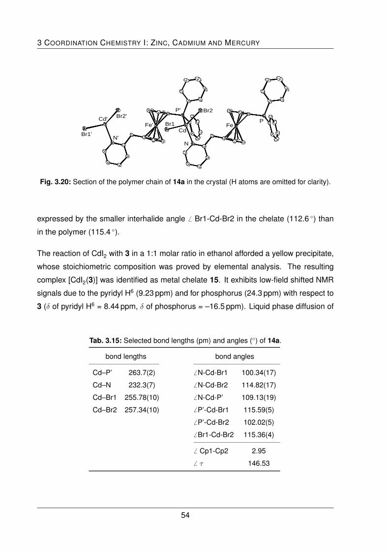
3 COORDINATION CHEMISTRY I: ZINC, CADMIUM AND MERCURY
Br2'Cd'
Br1'N'
Fe' Br1
P' Br2
Cd
N
FeP
Fig. 3.20: Section of the polymer chain of 14a in the crystal (H atoms are omitted for clarity).
expressed by the smaller interhalide angle 6 Br1-Cd-Br2 in the chelate (112.6 ◦) than
in the polymer (115.4 ◦).
The reaction of CdI2 with 3 in a 1:1 molar ratio in ethanol afforded a yellow precipitate,
whose stoichiometric composition was proved by elemental analysis. The resulting
complex [CdI2(3)] was identified as metal chelate 15. It exhibits low-field shifted NMR
signals due to the pyridyl H6 (9.23 ppm) and for phosphorus (24.3 ppm) with respect to
3 (δ of pyridyl H6 = 8.44 ppm, δ of phosphorus = –16.5 ppm). Liquid phase diffusion of
Tab. 3.15: Selected bond lengths (pm) and angles (◦) of 14a.
bond lengths bond angles
Cd–P’ 263.7(2) 6 N-Cd-Br1 100.34(17)
Cd–N 232.3(7) 6 N-Cd-Br2 114.82(17)
Cd–Br1 255.78(10) 6 N-Cd-P’ 109.13(19)
Cd–Br2 257.34(10) 6 P’-Cd-Br1 115.59(5)
6 P’-Cd-Br2 102.02(5)
6 Br1-Cd-Br2 115.36(4)
6 Cp1-Cp2 2.95
6 τ 146.53
54
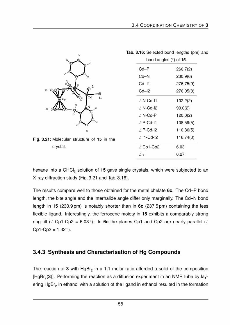
3.4 COORDINATION CHEMISTRY OF 3
Fe
P I2
I1Cd
N
Fig. 3.21: Molecular structure of 15 in the
crystal.
Tab. 3.16: Selected bond lengths (pm) and
bond angles (◦) of 15.
Cd–P 260.7(2)
Cd–N 230.9(6)
Cd–I1 276.75(9)
Cd–I2 276.05(8)
6 N-Cd-I1 102.2(2)
6 N-Cd-I2 99.0(2)
6 N-Cd-P 120.0(2)
6 P-Cd-I1 108.59(5)
6 P-Cd-I2 110.36(5)
6 I1-Cd-I2 116.74(3)
6 Cp1-Cp2 6.03
6 τ 6.27
hexane into a CHCl3 solution of 15 gave single crystals, which were subjected to an
X-ray diffraction study (Fig. 3.21 and Tab. 3.16).
The results compare well to those obtained for the metal chelate 6c. The Cd–P bond
length, the bite angle and the interhalide angle differ only marginally. The Cd–N bond
length in 15 (230.9 pm) is notably shorter than in 6c (237.5 pm) containing the less
flexible ligand. Interestingly, the ferrocene moiety in 15 exhibits a comparably strong
ring tilt (6 Cp1-Cp2 = 6.03 ◦). In 6c the planes Cp1 and Cp2 are nearly parallel ( 6
Cp1-Cp2 = 1.32 ◦).
3.4.3 Synthesis and Characterisation of Hg Compounds
The reaction of 3 with HgBr2 in a 1:1 molar ratio afforded a solid of the composition
[HgBr2(3)]. Performing the reaction as a diffusion experiment in an NMR tube by lay-
ering HgBr2 in ethanol with a solution of the ligand in ethanol resulted in the formation
55

3 COORDINATION CHEMISTRY I: ZINC, CADMIUM AND MERCURY
P
Br1
Br2Hg
NFe
Fig. 3.22: Molecular structure of 16 in the
crystal.
Tab. 3.17: Selected bond lengths (pm) and
bond angles (◦) of 16.
Hg–P 244.3(2)
Hg–N 237.2(7)
Hg–Br1 261.20(13)
Hg–Br2 262.78(10)
6 N-Hg-Br1 93.6(2)
6 N-Hg-Br2 97.3(2)
6 N-Hg-P 123.8(2)
6 P-Hg-Br1 116.21(6)
6 P-Hg-Br2 112.92(6)
6 Br1-Hg-Br2 110.31(4)
6 Cp1-Cp2 7.32
6 τ 8.51
of single crystals. The result of the X-ray diffraction analysis exhibits the anticipated
chelate structure of 16 (Fig. 3.22 and Tab. 3.17).
The tetrahedral coordination geometry is significantly distorted. The bite angle 6 N-
Hg-P is the largest of the coordination angles (123 ◦) and the N-Hg-Br1 angle is the
smallest one (93.6 ◦). In accord with the HSAB principle the Hg–P distance is shorter
than that of the analogous cadmium complex 15. The tendencies observed in the com-
parison of the cadmium chelates 6c and 15, are paralleled in the comparison of 16
with 8b, containing ligand 1. The Hg–P distances are almost equal in both complexes
(244.3 pm in 16, 244.6 pm in 8b) and the Hg–N distance in 8b (257.3 pm) is consider-
ably larger than that of 16 (237.2 pm), due to the more flexible pyridyl group in 16. Also,
the relatively strong ring tilt of the ferrocene moiety was observed ( 6 Cp1-Cp2 = 7.32 ◦
in 16 and 3.24 ◦ in 8b). The tendency is inverted for the bite angle 6 N-Hg-P, which is
larger in 16 (123.3 ◦) than in 8b (118.9 ◦). Probably the larger atomic radius of mercury
vs. cadmium is responsible for this.
56
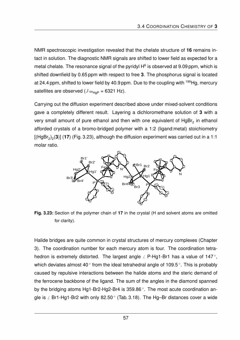
3.4 COORDINATION CHEMISTRY OF 3
NMR spectroscopic investigation revealed that the chelate structure of 16 remains in-
tact in solution. The diagnostic NMR signals are shifted to lower field as expected for a
metal chelate. The resonance signal of the pyridyl H6 is observed at 9.09 ppm, which is
shifted downfield by 0.65 ppm with respect to free 3. The phosphorus signal is located
at 24.4 ppm, shifted to lower field by 40.9 ppm. Due to the coupling with 199Hg, mercury
satellites are observed (J 199HgP = 6321 Hz).
Carrying out the diffusion experiment described above under mixed-solvent conditions
gave a completely different result. Layering a dichloromethane solution of 3 with a
very small amount of pure ethanol and then with one equivalent of HgBr2 in ethanol
afforded crystals of a bromo-bridged polymer with a 1:2 (ligand:metal) stoichiometry
[(HgBr2)2(3)] (17) (Fig. 3.23), although the diffusion experiment was carried out in a 1:1
molar ratio.
Br1'
Hg2'
Br3'Br4'
Br2'
Hg1'
P'
Fe'N'
Br4Br3
Hg1Hg2
Br2Br1
P
FeN
Fig. 3.23: Section of the polymer chain of 17 in the crystal (H and solvent atoms are omitted
for clarity).
Halide bridges are quite common in crystal structures of mercury complexes (Chapter
3). The coordination number for each mercury atom is four. The coordination tetra-
hedron is extremely distorted. The largest angle 6 P-Hg1-Br1 has a value of 147 ◦,
which deviates almost 40 ◦ from the ideal tetrahedral angle of 109.5 ◦. This is probably
caused by repulsive interactions between the halide atoms and the steric demand of
the ferrocene backbone of the ligand. The sum of the angles in the diamond spanned
by the bridging atoms Hg1-Br2-Hg2-Br4 is 359.86 ◦. The most acute coordination an-
gle is 6 Br1-Hg1-Br2 with only 82.50 ◦ (Tab. 3.18). The Hg–Br distances cover a wide
57

3 COORDINATION CHEMISTRY I: ZINC, CADMIUM AND MERCURY
Tab. 3.18: Selected bond lengths (pm) and angles (◦) of 17.
bond lengths bond angles bond angles
Hg1–Br1 247.14(12) 6 P-Hg1-Br1 147.82(8) 6 Br1-Hg1-Br4 96.77(4)
Hg2–Br2 265.19(12) 6 P-Hg1-Br2 110.52 6 Br3-Hg2-Br4 126.49(5)
Hg2–Br3 253.19(14) 6 P-Hg1-Br4 110.21(7) 6 Br3-Hg2-Br2 117.81(5)
Hg2–Br4 260.02(13) 6 N’-Hg2-Br3 99.2(2) 6 Br4-Hg2-Br2 98.26(4)
Hg1–Br4 286.48(12) 6 N’-Hg2-Br4 117.3(2) 6 Br1-Hg1-Br2 82.50
Hg1–Br2 315.0 6 N’-Hg2-Br2 93.9(2) 6 Hg2-Br4-Hg1 93.13(4)
Hg1–P 241.9(3)
Hg2–N’ 236.9(8) 6 Cp1-Cp2 2,93 6 τ 73.74
range from ca. 247.1 pm - 315.0 pm. The Hg2–Br distances lies between ca. 253.2 pm
and 265.2 pm, the shortest bond being that to the terminal Br3. For the phosphine
coordinated Hg1 a very short bond to the terminal Br1 of only 241.9 pm is observed
plus two very long ones (ca. 286.5 pm and 315.0 pm).
The “inverted” complex, where two equivalents of 3 are coordinated only to one equiva-
lent of mercury bromide [HgBr2(3)2] (18), has also been prepared and characterised.
Liquid phase diffusion of hexane into a chloroform solution of the complex afforded
the solvate 18 · CHCl3, which was structurally characterised by X-ray diffraction as the
expected bis(phosphine) complex (Fig. 3.24 and Tab. 3.19), which is an analogue of 9b
containing ligand 1 instead of 3. All bond parameters compare well with those of the
bis(phosphine) complex 9b. However, while 9b crystallises in the monoclinic space
group P21/n, 18 is orthorhombic (Iba2).
58

3.5 COORDINATION CHEMISTRY OF 4
Tab. 3.19: Selected bond lengths (pm) and angles (◦) of 18.
bond lengths bond angles
Hg–P1 251.0(2) 6 P1-Hg-Br1 99.21(5)
Hg–P2 251.2(2) 6 P1-Hg-Br2 115.33(5)
Hg–Br1 266.15(8) 6 P1-Hg-P2 124.01(5)
Hg–Br2 266.40(8) 6 P2-Hg-Br1 113.23(5)
6 P2-Hg-Br2 100.58(5)
6 Br1-Hg-Br2 103.16(3)
3.5 Coordination Chemistry of 4
During the coordination experiments with mercury and zinc halides, it turned out that
the resulting solids were not the expected well-defined complexes, but very likely ill-
defined mixtures. It was not possible to grow crystals suitable for X-ray diffraction
analysis and the NMR spectra turned out to be too complex for a meaningful analysis.
For example, immediately after the addition of ZnCl2 to a dichloromethane solution of
the ligand, the colour changed from orange to a very dark red. Even though coordina-
N2
Fe2
P2
Hg1
Br1
Br2
P1
Fe1
N1
Fig. 3.24: Molecular structure of 18 in the crystal (H and solvent atoms are omitted for clarity).
59
3 COORDINATION CHEMISTRY I: ZINC, CADMIUM AND MERCURY
tion had obviously occurred, no defined complex could be isolated. DFT calculations
performed by Dr. Leibold in our group suggest that tetrahedral coordination is sterically
hindered if all three donor atoms of the ligand are involved in the coordination to a sin-
gle zinc atom. More detailed, and probably very time-consuming, investigations will be
needed to understand the coordination behaviour of 4. In view of the time constrictions
of the present work, experiments with 4 were abandoned at this preliminary stage.
60

3.6 SUMMARY AND CONCLUSION
3.6 Summary and Conclusion
The ferrocene-based pyridylphosphine ligands 1 - 4 were investigated in terms of the
coordination chemistry towards the group 12 metals zinc, cadmium and mercury, which
are used in form of the metal halide salts of the type MX2 (M = Zn, Cd, Hg and X = Cl,
Br, I). The results of the experiments are summarised in Tab. 3.20. Depending on the
ligand and the reaction stoichiometry the ligands 1, 2 and 3 react with different salts in
a P,N-chelating, P,N-bridging or a P-coordinated monodentate coordination mode.
Tab. 3.20: Results of the coordination chemistry experiments concerning 1, 2 and 3.
1 2 3
1 eq. 2 eq. 1 eq. 2 eq. 1 eq. 2 eq.
Zn
Cl2 5a C – – – – –
Br2 5b C 5b C 10 P – 13 C; 13a Da 13a Da
I2 5c Cb – – – – –
Cd
Cl2 6a Cb,c 7 Ba,d – – – –
Br2 6b C – 11 Pb – 14 Cc; 14a Pa –
I2 6c C – – – 15 C; – –
Hg
Cl2 8a Cb,c 9a Ba,d – – – –
Br2 8b C 9b B 12 P – 16 C; 17 Pa,e 18 B
I2 8c Cf – – – – –
C = P,N-chelate, P = polymer, D = centrosymmetric dimer, B = P-coordinated
bis(phosphine) complex, – = no investigations realised or investigations did not lead to clear
results. aOnly characterised by X-ray diffraction. bNo X-ray data available. cCrystallisation
leads to new compound with 2:1 (ligand:metal) stoichiometry. d Crystals obtained by diffu-
sion experiments with 1:1 stoichiometry. e17 has a 1:2 stoichiometry, the polymer is formed
via halide bridges. fCrystallisation leads to isomeric iodo-bridged dimer.
61

3 COORDINATION CHEMISTRY I: ZINC, CADMIUM AND MERCURY
Reactions in a 1:1 molar ratio with 1 resulted mostly in P,N-chelated complexes, reac-
tions with 2 led to polymeric structures. The less predictable coordination behaviour of
3 can be ascribed to the presence of the methylene group in this ligand, which makes
the ligand more flexible.
Reactions with two equivalents of the ligand gave P-coordinated, monodentate bis-
(phosphine) complexes with the ligands 1 and 3 in the case of mercury. Zinc complexes
with a monodentate coordination mode were not observed; for 1 only the 1:1 chelating
complex and for 3 the centrosymmetric dimer was isolated.
The coordination chemistry of 4 was not explored in detail. Preliminary experiments
show low crystallisation tendencies and very complicated spectral data.
62

4 Coordination Chemistry II:
Silver and Gold
The coordination chemistry of the ferrocene-based ligands 1 - 3 towards silver(I) and
gold(I) is described. The synthesis, characterisation and electrochemical investigations
concerning the potential application of 1 - 3 as molecular redox sensor are presented.
4.1 Introduction
Silver and gold have not only a cultural importance by being used as currency or for
objects in arts and jewellery, they have also great importance in chemistry. Numer-
ous applications in catalysis[92–94] and medicine[95] have been reported in the scientific
literature.
Silver is classified in terms of the HSAB principle as a relatively soft acid with a versatile
coordination chemistry. Oxidation states from (I) to (III) and coordination numbers
from one to ten are known. However, AgI compounds with the coordination numbers
three (mostly distorted trigonal planar) and four (usually distorted tetrahedral) are most
common. The metal-donor distances vary from 202 pm to 246 pm for monodentate
nitrogen donors and from 236 pm to 270 pm in the case of phosphorus donors.[96–98]
Even though silver shows such a diverse and well-investigated coordination chemistry,
complexes with the popular structural analogue of the ferrocene-based pyridylphos-
63

4 COORDINATION CHEMISTRY II: SILVER AND GOLD
Fe
PPh2
PPh2
Ag PPh3
ClO4
Fig. 4.1: 1,1’-[Bis(diphenylphosphino)ferrocene]-triphenylphosphine-silver(I).
phines 1, 2 and 3, 1,1’-bis(diphenylphosphino)ferrocene (dppf), are rare. The structure
of [Ag(dppf)2][BF4] has been reported by Long and coworkers.[99] One silver atom is co-
ordinated by two chelating dppf molecules. The complex has a tetrahedral coordination
geometry as expected for the coordination number four. The coordination tetrahedron
is distorted due to the steric demand of the eight phenyl groups. The chelate bite an-
gles are only 97.7 ◦ and 105.5 ◦. The Ag–P distances range from 255.3 pm to 266.2 pm.
A related AgI dppf complex with the coordination number three is shown in Fig. 4.1.[100]
The metal centre is coordinated by one dppf molecule in a chelate manner and by
one molecule of triphenylphosphine. Coordination angles are close to 120 ◦ (sum of
angles 359.94 ◦) and the silver atom is in a trigonal planar coordination environment.
The donor-metal bond lengths have an average of 244.5 pm, which is shorter than in
the bis(dppf) complex, owing to the lower coordination number.
A search in the cambridge structural database (CSD) for silver compounds with pyridyl-
substituted ferrocenes, gave just a single hit, viz. a silver complex of 1,1’-bis(pyrid-2-
yl)ferrocene.[101] The silver atom is chelated by the two nitrogen donors in a quasilinear
dicoordinated fashion ( 6 N1-Ag-N2 = 163.1 ◦). The perchlorate counter ions are not
connected to the metal centre. Due to the smaller atomic radius of nitrogen vs. phos-
phorus, the Ag–N distances (214.3 pm and 218.7 pm) are significantly shorter than
those between Ag–P in the dppf complex (244.5 pm).
The chemistry of gold is dominated by the oxidation states (I) and (III), having the elec-
tron configuration [Xe] 4f14 5d10 6s0 and [Xe] 4f14 5d8 6s0, respectively. Compounds
64

4.1 INTRODUCTION
of AuI assume the coordination number two with a linear coordination geometry and
a 14 VE configuration in most cases. AuI complexes with a coordination number of
three (16 VE, trigonal planar) or four (18 VE, tetrahedral) are also known, but less
common,[102] since the participation of the energetically high lying 6py and 6pz orbitals
causes high promotion energies. The general form of gold(I) complexes is [LAuX] ( L=
neutral ligand; X = anionic ligand) and gold(I) exhibits a strong preference for large and
soft (easily polarisable) donor ligands L.[103] Particularly stable gold(I) complexes are
formed with organic P- or S-donor ligands.
Several gold complexes with the archetypal phosphinoferrocene ligand dppf are found
in the literature.[104] The simplest one was prepared by the reaction of dppf with two
equivalents of [AuCl(tht)]. The resulting binuclear complex [(AuCl)2(dppf)] exhibits a
linear dicoordinate arrangement for both gold atoms (Fig. 4.2). Short Au–P distances
Fe
P
P
PhPh
Ph PhAu
Au Cl
Cl
Fig. 4.2: Linear coordination geometry in [(AuCl)2(dppf)].
(222.8 pm) underline the stability of this complex. Interestingly, [(AuCl)2(dppf)] has
been described to lack electrochemical activity. Either, electron transfer is very slow
or this complex is indeed inert to electrochemical oxidation or reduction.[105] It seems
likely that the AuCl fragments possess such a high electron withdrawing ability to make
the HOMO levels of the ferrocene moiety inaccessible.[106] This AuI complex appears to
be the first example of an electrochemically inert ferrocene derivative. Gold complexes
of pyridyl-substituted ferrocene ligands are not known.
65
4 COORDINATION CHEMISTRY II: SILVER AND GOLD
4.2 Synthesis and Characterisation of Ag Compounds
The synthesis and characterisation of silver complexes with the ferrocene-based py-
ridylphosphine ligands 1 - 3 is described. This was carried out in analogy to the syn-
thesis of an AgI compound with an imidazole-based P,N-ligand.[107]
4.2.1 Ag Complex of 1
The reaction of the ligand, dissolved in methanol, and one equivalent of solid AgBF4 at
room temperature yielded [Ag(1)][BF4] (19), which was isolated by a solvent exchange
to dichloromethane and following precipitation with hexane. ESI mass spectrometry
and elemental analysis confirm the stoichiometry.
The signal of the pyridyl H6 is located at 8.87 ppm in the 1H NMR spectrum (CHCl3),
which means a low-field shift of about 0.4 ppm with respect to pristine 1. The 31P NMR
spectrum shows a single signal, observed as a doublet (Ag-P coupling). Due to in-
termolecular dynamic processes, the signal is broadened. This is typical for 31P NMR
spectra of Ag-P complexes at room temperature. In low temperature NMR spectra
two doublets would be observed usually, one due to the coupling with 107Ag and the
other one due to the coupling with 109Ag.[90,108] The phosphorus signal here is located
low-field shifted with respect to uncoordinated 1 (δ = –17.7 ppm) at δ = 6.8 ppm. The
averaged coupling constant of the broad doublet has a value of J 107AgP,109AgP ≈ 712 Hz.
Recrystallisation of 19 from chloroform gave material suitable for structural charac-
terisation by X-ray diffraction analysis. Under these conditions 19 crystallises as a
polymeric solvate with two molecules of CHCl3 per asymmetric unit (Fig. 4.3).
Compared to known complexes (Chapter 4.1), the donor–metal distances of 237.1 pm
(Ag–P) and 217.4 pm (Ag–N) are rather short. The N-Ag-P angle (164 ◦) deviates from
a linear coordination geometry. The distortion is probably caused by weak interactions
66

4.2 SYNTHESIS AND CHARACTERISATION OF AG COMPOUNDS
Fe P
Ag
N Fe'
P'
Ag'
N'
Fig. 4.3: Section of the polymer chain in the crystal structure of 19 (H, counter ion and solvent
atoms are omitted for clarity).
Tab. 4.1: Selected bond lengths (pm) and bond angles (◦) of 19.
Ag–P 237.08(8)
Ag–N 217.4(3)
Ag–F4 273.7
6 P-Ag-N 164.21(6)
between the silver atom and one flourine atom of the BF4– counter ion (F4 in the crystal
structure), whose distance is only 273.7 pm (Tab. 4.1), which is smaller than the sum
of the van der Waals radii of silver and fluorine (ca. 308 pm). Such a coordination motif
(CN = 2 + 1) is frequently observed in AgI chemistry.[109,110]
4.2.2 Ag Complex of 2
The reaction of 2 with one equivalent of AgBF4 in methanol gave an orange precipitate,
which was filtered off, rinsed with methanol, washed with diethyl ether and hexane and
finally dried in vacuum. The resulting complex [Ag(2)][BF4] (20) was characterised by
67

4 COORDINATION CHEMISTRY II: SILVER AND GOLD
FeN
Ag P
Fe'
N'
Ag'P'
Fig. 4.4: Molecular structure of 20 in the crystal (H, counter ion and solvent atoms are omitted
for clarity).
the same methods as 19 before. The elemental analysis confirms the stoichiometry.
The ESI mass spectrum exhibits a peak at higher m/z than the simplest coordinated
monomeric cation [C27H22AgFeNP]+. This indicates that the product is not monomeric.
In the 1H NMR spectrum (CHCl3) one of the pyridyl protons next to the nitrogen atom
(H2, H6) gives rise to a signal at 9.00 ppm, low-field shifted with respect to the free
ligand 2. The signal due to the other pyridyl proton of these is observed at 8.08 ppm,
shifted in the opposite field direction. The 31P NMR spectrum shows a single signal,
observed broad and split into a doublet (δ = 11.1 ppm), in accord with the Ag-P coupling
(J 107AgP,109AgP ≈ 691 Hz).
Liquid phase diffusion of hexane into a chloroform solution of 20 afforded crystals suit-
able for X-ray diffraction analysis, which revealed a centrosymmetric dimeric molecular
structure with one molecule of CHCl3 per asymmetric unit (Fig. 4.4).
The metal-donor distances are very similar to those of 19 (Tab. 4.2). Again, a 2 + 1
coordination is observed. The angle 6 P-Ag-N deviates even more from linearity, due
to a stronger interaction with the anion, which is reflected by a comparatively smaller
Ag–F distance of only 266.6 pm as opposed to 273.7 pm in the case of 19.
68

4.2 SYNTHESIS AND CHARACTERISATION OF AG COMPOUNDS
Tab. 4.2: Selected bond lengths (pm) and bond angles (◦) of 20.
Ag–P 235.91(10)
Ag–N 217.3(3)
Ag–F4 266.6
6 P-Ag-N 156.61(10)
4.2.3 Ag Complex of 3
The analogous reaction of 3 with AgBF4 in methanol gave [Ag(3)][BF4] · 0.5 C6H14
(21), according to elemental analysis after precipitation with hexane. Single crystals
were obtained from chloroform. An X-ray diffraction analysis revealed the polymeric
structure of the solvate [Ag(3)][BF4] · CHCl3 (Fig. 4.5 and Tab. 4.3).
Bond parameters are similar to those of the the two silver complexes describes before.
Not surprisingly, the strongest similarity occurs between 19 and 21, which are both
coordination polymers. The P-Ag-N angle and Ag–F4 distances are almost identical.
A P,N-coordination in solution is indicated by NMR spectroscopy (CHCl3). The diag-
nostic pyrdiyl H6 signal is observed at 8.77 ppm, 0.33 ppm low-field shifted in compar-
P'
Ag'
Fe'
N'
P
Ag
Fe
N
Fig. 4.5: Section of the polymer chain in the crystal structure of 21 (H, counter ion and solvent
atoms are omitted for clarity).
69

4 COORDINATION CHEMISTRY II: SILVER AND GOLD
Tab. 4.3: Selected bond lengths (pm) and bond angles (◦) of 21.
Ag–P 235.8(2)
Ag–N 216.2(6)
Ag–F4 275.2
6 P-Ag-N 166.9(2)
ison with pristine 3. The signal observed in the 31P NMR spectrum is also broad and
split into a doublet, located at 7.1 ppm. The doublet is located at a similar chemical
shift than this of 19 (6.8 ppm).
4.3 Synthesis and Characterisation of Au Compounds
4.3.1 Simple Au Complexes of 1, 2 and 3
The ligands were reacted with one equivalent of [AuCl(tht)] in dichloromethane at
room temperature. Precipitation with hexane, washing and drying in vacuum afforded
[AuCl(1)] (22), [AuCl(2)] (23) and [AuCl(3)] (24) as orange to yellow solids, whose stoi-
chiometry was proved by elemental analysis.
4.3.1.1 Characterisation
All three products were structurally characterised by single crystal X-ray diffraction
analysis. Suitable crystals were obtained in all cases by liquid phase diffusion of diethyl
ether into a chloroform or dichloromethane solution of the complexes. The results are
shown in Fig. 4.6. The typical linear coordination geometry with the coordination num-
ber two was found in all cases (Chapter 4.1). The structures are very similar, which is
not surprising, because the nitrogen donor remains uncoordinated.
70

4.3 SYNTHESIS AND CHARACTERISATION OF AU COMPOUNDS
Cl
AuP
Fe
N
(a)
PAu
ClFe
N
(b) (c)
Fe
P
AuCl
N
Fig. 4.6: Molecular structures of 22 (a), 23 (b) and 24 (c) in the crystal.
The Au–P and the Au–Cl distances differ only marginally (Tab. 4.4). The same is true
for the angle 6 P-Au-Cl. Close intermolecular contacts or aurophilic interactions are not
observed. The Au–P and Au–Cl distances in 22, 23 and 24 are slightly larger than in
the corresponding dppf complex [(AuCl)2(dppf)]. In turn the P-Au-Cl angles are smaller
than in the dppf analogue.
Tab. 4.4: Selected bond lengths (pm) and bond angles (◦) of 22, 23 and 24.
22 23 24 [(AuCl)2(dppf)]
Au–P 225.0(9) 223.66(11) 223.71(16) 222.8
Au–Cl 230.8(10) 230.55(10) 230.1(2) 228.4
6 P-Au-Cl 176.2(4) 175.33(4) 176.75(6) 178.51
A monodentate P-coordination in solution is indicated by a pronounced shift of the
phosphorus signal in the 31P NMR spectra of 22, 23 and 24. The signals are located
close to 28 ppm, downfield shifted by approximately 45 ppm with respect to the re-
spective free ligand (δ = ca. –17 ppm). The 1H NMR signals diagnostic for nitrogen
coordination remain unshifted.
4.3.1.2 Electrochemical Investigations
22 was investigated in standard cyclic voltammetry, which revealed a ferrocene-based
oxidation plus an additional irreversible oxidation process at higher potential (Fig. 4.7 a).
71

4 COORDINATION CHEMISTRY II: SILVER AND GOLD
In view of the presumptive redox-inertness of [(AuCl)2(dppf)] (Chapter 4), this com-
plex surprisingly exhibited reversible behaviour concerning the ferrocene-based re-
dox waves, if the scan was reversed before the start of the second oxidation process
(Fig. 4.7 b). The redox potential for the ferrocene-based process of 22 has a value ofE0’
= 0.91 V vs. decamethylferrocene (fc*) at a scan rate of 100 mV/s, which corresponds
to a remarkable coordination-induced anodic shift of ca. 0.36 V with respect to pris-
tine 1. Compared with the well established redox sensors 1,1’-di(pyrid-2-yl)ferrocene
and 1,1’-di(pyrid-2-yl)octamethylferrocene, introduced in Chapter 1.4.1, this is a re-
spectable result.
23 and 24 were also subjected to CV. Both exhibited reversible behaviour for the
ferrocene-based redox process, if the scan was reversed before the onset of a fur-
ther oxidation process at higher potentials. For 23 the ∆EP value is 61 mV. After the
reference standard fc* had been added, the reversible behaviour disappeared. Hence,
the half wave potential could not been referenced. In the case of 24 the formal elec-
trode potential could be identified for the ferrocene-based redox process. It has a value
of E0’ = 0.85 V vs. fc*. Again, this corresponds to a remarkable coordination-induced
anodic shift of ca. 0.23 V with respect to the free ligand 3.
-0,1
0,0
0,1
0,2
-1,0 -0,8 -0,6 -0,4 -0,2 0,0 0,2 0,4 0,6 0,8 1,0 1,2 1,4 1,6 1,8
Cur
rent
(µA)
Potential (V)
(a)
fc*
-1,0
-0,8
-0,6
-0,4
-0,2
0,0
0,2
0,4
0,6
0,8
1,0
1,2
1,4
1,6
1,8
-0,4 -0,3 -0,2 -0,1 0,0 0,1 0,2 0,3 0,4 0,5 0,6 0,7 0,8 0,9 1,0 1,1 1,2 1,3
Cur
rent
(µA)
Potential (V)
(b)
fc*
Fig. 4.7: CV of 22 (0.1 mM, DCM, 0.1 M [NnBu][PF6], 100 mV/s), scan range from –1.0 V to
+1.7 V (a) and from –0.25 V to +1.2 V (b).
72
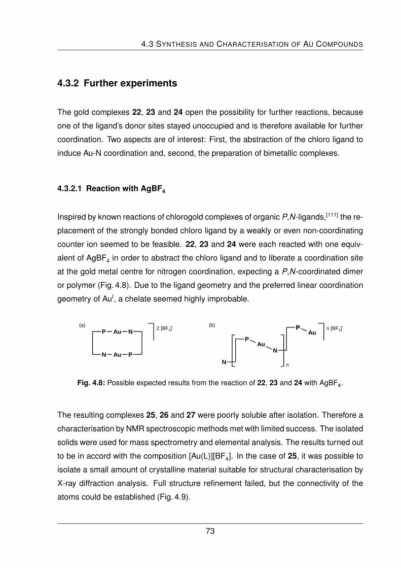
4.3 SYNTHESIS AND CHARACTERISATION OF AU COMPOUNDS
4.3.2 Further experiments
The gold complexes 22, 23 and 24 open the possibility for further reactions, because
one of the ligand’s donor sites stayed unoccupied and is therefore available for further
coordination. Two aspects are of interest: First, the abstraction of the chloro ligand to
induce Au-N coordination and, second, the preparation of bimetallic complexes.
4.3.2.1 Reaction with AgBF4
Inspired by known reactions of chlorogold complexes of organic P,N-ligands,[111] the re-
placement of the strongly bonded chloro ligand by a weakly or even non-coordinating
counter ion seemed to be feasible. 22, 23 and 24 were each reacted with one equiv-
alent of AgBF4 in order to abstract the chloro ligand and to liberate a coordination site
at the gold metal centre for nitrogen coordination, expecting a P,N-coordinated dimer
or polymer (Fig. 4.8). Due to the ligand geometry and the preferred linear coordination
geometry of AuI, a chelate seemed highly improbable.
P
NAu
PP
N
PAu
n
n [BF4]-
N
Au N
Au P
2 [BF4]-(a) (b)
Fig. 4.8: Possible expected results from the reaction of 22, 23 and 24 with AgBF4.
The resulting complexes 25, 26 and 27 were poorly soluble after isolation. Therefore a
characterisation by NMR spectroscopic methods met with limited success. The isolated
solids were used for mass spectrometry and elemental analysis. The results turned out
to be in accord with the composition [Au(L)][BF4]. In the case of 25, it was possible to
isolate a small amount of crystalline material suitable for structural characterisation by
X-ray diffraction analysis. Full structure refinement failed, but the connectivity of the
atoms could be established (Fig. 4.9).
73

4 COORDINATION CHEMISTRY II: SILVER AND GOLD
N'
Au
P
Fe N
Au'
P'
Fe'
Fig. 4.9: Molecular structure of 25 in the crystal (H and counter ion atoms are omitted for clarity,
remaining electron density of probably disordered solvent could not be refined).
A P,N-coordinated dimer was found. The gold atoms are in a nearly linear coordination
environment (Tab. 4.5). The Au–P distance of 224.5 ppm is indistinguishable from that
of the related compound [(AuCl)2(dppf)] (Chapter 4.1). The Au–N’ bond in 25 is about
8 pm longer than the corresponding bond in the pyridine complexes [AuX(C5H5N)] (X =
Cl, I).[112] Aurophilic interactions are absent in 25.
Tab. 4.5: Selected bond lengths (pm) and bond angles (◦) of 25.
Au–P 224.5(5)
Au–N’ 214.1(2)
6 P-Au-N’ 174.0(5)
74
4.4 SUMMARY AND CONCLUSION
4.3.2.2 Bimetallic Complexes
As a sideline of these investigations the preparation of heterobimetallic complexes of
1 and 3 with gold and silver was attempted. An interesting aspect of such complexes
are potential metallophilic interactions. Reaction of the simple chlorogold complexes
22 and 24 with AgBF4 in the presence of one equivalent of a phosphine ligand (PMe3,
PCy3, 1) gave no crystalline material. Also, reactions of [Au(tht)2][BF4] with two equiva-
lents of 1 and 3, respectively, followed by the addition of silver tetrafluoroborate, failed.
Crystallisation experiments with AgSbF6 as silver source instead of AgBF4 failed, too.
4.4 Summary and Conclusion
The ligands 1, 2 and 3 were examinated in coordination reactions with AuI and AgI.
The reactions with AgBF4 in a 1:1 molar ratio gave the corresponding complexes 19,
20 and 21. All three ligands show a bridging coordination mode. 1 and 3, containing a
pyrid-2-yl group, form polymers , while the pyrid-3-yl containing ligand 2 forms a cyclic
dimer. The complexes 22, 23 and 24 obtained from the reactions of 1 - 3 with [AuCl(tht)]
afforded monodentate, P-coordinated phosphine complexes. These complexes show
a reversible behaviour concerning the ferrocene-based redox wave in the cyclovoltam-
metric investigations. Further reactions, aimed at the abstraction of the chloro ligand to
induce Au-N coordination resulted in the less soluble and poorly crystalline complexes
25, 26 and 27. Only 25 was structurally characterised. Additional experiments aimed
at the preparation of Au-Ag binuclear complexes failed.
75

5 Coordination Chemistry III:
Palladium
This chapter deals with the coordination chemistry of ligands 1 and 3 towards palla-
dium(II). The synthesis and characterisation of relevant complexes is described. Due
to the intrinsic attractiveness of P,N-donors in catalytic applications, the utilisation of
these complexes in palladium-catalysed C–C bond forming reactions is scrutinised.
5.1 Introduction
It is well known that palladium readily coordinates both, donors containing phosphorus
and nitrogen.[113–115] Many organic P,N-ligands form palladium complexes, when they
are reacted with the popular Pd source [PdCl2(cod)] (cod = η2:η2-cycloocta-1,5-diene).
They simply substitute the chelating diolefin ligand in this reaction. A typical example
is shown in Fig. 5.1(a).[116] Concerning the palladium chemistry of ferrocene-based lig-
ands, it is essential to address 1,1’-bis(diphenylphosphino)ferrocene (dppf). It is one
of the most useful and popular 1,1’-disubstituted chelate ligands in this field of chem-
istry. Details of the coordination and catalytic chemistry have been comprehensively
reviewed.[21,55] Dppf and the complex [PdCl2(dppf)], Fig. 5.1 b, are used in the following
for comparison in structural chemistry and catalytic tests, respectively.
The number of palladium complexes of ferrocene-based ligands closely related to 1
and 3 is rather small. A rare example is the square-planar trans-[PdCl2(bipyppf)]
77

5 COORDINATION CHEMISTRY III: PALLADIUM
NN
N
PPh
Pd
Cl Cl
Ph
(a) (b)
Fe
P
P
Pd
PhPh
PhPh
ClCl
Fig. 5.1: PdCl2 complexes of P-functionalised donors.
(bipyppf = 1-(diphenylphosphino)-1’-(2,2’-bipyridyl-6-yl)ferrocene), which is a chelate
of a bipyridyl-substituded phosphinoferrocene. The structure of this complex has been
established by X-ray diffraction analysis (Fig. 5.2).[117] Selected distances and angles
are given in Tab. 5.3 on page 83.
Fe
P
N
N
PhPh
PdCl
Cl
Fig. 5.2: trans-[PdCl2(bipyppf)].
Ligands which are closely related to 1 and 3 concerning their flexibility elements and
donor groups (pyridyl and diphenylphosphino group) are 1-(diphenylphosphino)-1’-N-
[(pyrid-2-yl)methyl]carbamoylferrocene, Fig. 5.3 a, and its ethylene-spaced analogue 1-
(diphenylphosphino)-1’-N-[2-(pyrid-2-yl)ethyl]carbamoylferrocene, Fig. 5.3(b).[118]
Fe
PPh2
N
O
H
(a) (b)
Fe
PPh2
N
O
HN NNNN
Fig. 5.3: 1,1’-Ferrocene-based pyridylphosphinocarboxamide ligands.
78

5.1 INTRODUCTION
(a)
(b)
P
N
P
N
Pd
Cl
Cl
P
N
PdCl
Cl
(c)
P
N
N
P
PdCl
Cl
PdCl
Cl
Fig. 5.4: Coordination behaviour of the pyridylphosphinocarboxamide ligands shown in Fig 5.3:
monodentate bis(phosphine) complex (a), chelate (b) and centrosymmetric dimer (c).
The reaction of [PdCl2(cod)] with two equivalents of the P,N-ligand afforded a trans-
square-planar, bis(phosphine) complex in both cases. Fig. 5.4 shows possible binding
modes of both pyridylphosphinocarboxamide ligands. When the reaction was carried
out in 1:1 molar ratio, both ligands showed different behaviour. On the one hand,
the ligand with the shorter methylene bridge, 1-(diphenylphosphino)-1’-N-[(pyrid-2-yl)-
methyl]carbamoylferrocene, forms a trans-chelate complex. On the other hand, the
ligand with the longer ethylene bridge, 1-(diphenylphosphino)-1’-N-[2-(pyrid-2-yl)ethyl]-
carbamoylferrocene, forms a centrosymmetric, dimer (Fig. 5.4 c). Similar behaviour
was observed for ZnBr2 complexes containing 3 (Capter 3.4.1).
Selected structural data are collected in Tab. 5.1. The coordination angles deviate less
than 6 ◦ from the value of 90 ◦ for an undistorted square-planar arrangement. The Pd–
N distances are close to ca. 210 pm, while the Pd–P distances are ca. 224 pm in the
chelate and the dimer. This effect (small Pd–N, larger Pd–P distances) is caused by the
different covalence radii of both atoms. The Pd–P bond lengths are even ca. 232 pm
in the bis(phosphine) complex, due to the larger trans influence of the P-donor. Pd–Cl
bond lengths (ca. 230 pm) are essentially identically in all three complexes.
79

5 COORDINATION CHEMISTRY III: PALLADIUM
Tab. 5.1: Selected bond lengths (ppm) and angles (◦) of the Pd complexes of pyridylphos-
phinocarboxamide ligands shown in Fig 5.3.
bis(phosphine) complex chelate centrosymmetric dimer
Pd–P 232.5(1) 224.3(1) 224.3(1)
Pd–N – 212.4(2) 210.7(4)
Pd–Cl(1) 229.6(1) 229.6(1) 230.0(1)
Pd–Cl(2) – 230.2(1) 229.8(1)
6 N-Pd-Cl(1) – 88.85(6) 85.8(1)
6 N-Pd-Cl(2) – 88.55(6) 90.0(1)
6 P-Pd-Cl(1) 86.22(3) 92.78(3) 95.57(5)
6 P-Pd-Cl(2) – 89.95(3) 88.77(5)
5.2 Synthesis and Characterisation of Pd-Compounds
The synthesis of compounds 29, 31, 33 and 35 was carried out by the group members
of Prof. Petr Stepnicka at Charles University, Prague. All analytical, X-ray crystallo-
graphic and catalytic data of the palladium complexes were collected in the laboratories
of the cooperation partners in the Czech Republic.
5.2.1 Simple Palladium Complexes of 1 and 3
Starting from [PdCl2(cod)], it was first attempted to synthesise palladium complexes of
1 and 3 in a 1:1 and 1:2 metal to ligand molar ratio. For 1:1 stoichiometry, both ligands
form chelate complexes, viz. [PdCl2(1)] (28) and [PdCl2(3)] (29). The analogous reac-
tion with two equivalents of 1 or 3 afforded the respective bis(phosphine) complexes
trans-[PdCl2(1)2] (30) and trans-[PdCl2(3)2] (31) as shown in Fig. 5.5. The complexes
were isolated as air-stable solids and characterised by spectroscopic methods and el-
emental analysis. Due to the poor solubility of the bis(phosphine) complexes 30 and
80

5.2 SYNTHESIS AND CHARACTERISATION OF PD-COMPOUNDS
Fe
P
[PdCl2(cod)] + m L
()n
N
Pd
Ph
Ph
Cl
Cl
Fe
P Fe
PPd
Ph
Cl
ClPh
Ph Ph
(CH2)nPy
(CH2)nPy
m = 1
m = 2
30 (n = 0), 31 (n = 1)
28 (n = 0), 29 (n = 1)
Fig. 5.5: Synthesis of simple Pd complexes 28, 29, 30 and 31, (n = 0: L = 1, n = 1: L = 3).
31, NMR characterisation was carried out with in situ generated compounds. Except
for 29, the molecular structures were determinated by single crystal X-ray diffraction
analysis.
The coordination of the phosphorus donor atom in both chelates is manifested by a low-
field shift of the phosphorus signal in the 31P NMR spectrum of 34.1 ppm for 28 and
38.7 ppm for 29 with respect to the free ligand 1 or 3 (Tab. 5.2). The signal of the pyridyl
H6 is only shifted by about 0.12 ppm to lower field in 28 and into the opposite field di-
rection for 29. Interestingly, the signals of the ferrocene moiety in the chelated complex
28 are markedly shifted from the usual region. They are located between 4.63 ppm
and 6.64 ppm, probably influenced by ring currents of the nearby aromatic rings. This
Tab. 5.2: Chemical shifts of the diagnostic NMR signals of 28, 29, 30 and 31 in ppm.
1 28 30 3 29 31
signal due to pyridyl H6 (1H NMR) 8.47 8.55 8.49 8.44 8.32 8.48
phosphorus signal (31P NMR) –17.7 16.4 16.2 –16.5 22.2 15.9
Solvent: CD2Cl2, except for 1 (CDCl3).
81

5 COORDINATION CHEMISTRY III: PALLADIUM
is a result of the chelating coordination, which leads to conformationally locked, C1-
symmetric structures. These chiral structures make the CH groups of the ferrocene
moiety diastereotopic and anisochronic. In the analogous chelate 29, the protons of
the ferrocene CH groups and the methylene protons become also diastereotopic and
anisochronic. Their signals were detected between 2.32 ppm and 5.03 ppm.
In contrast, the signals of the CH groups of the ferrocene moiety of the bis(phosphine)
complexes 30 and 31 are similar to those of free ligands. They were observed to be
degenerate, indicating conformational flexibility and equivalence of the ligands. In both
bis(phosphine) complexes the phosphorus signal was also low-field shifted with respect
of the corresponding free ligand (33.9 ppm for 30 and 32.4 ppm for 31), as shown in
Table 5.2. Not surprisingly, the NMR signals due to the pyridyl H6 of the uncoordinated
pyridyl donor remained similar to those of the free ligand.
The molecular structure of 28 is shown in Fig. 5.6, as the result of the X-ray diffrac-
tion analysis. Unlike trans-[PdCl2(bipyppf)],[117] complex 28 shows a cis-P,N-geometry,
which apparently reflects the tighter disposition of the donor atoms. The typical square-
planar coordination geometry of PdII was found. The coordination angles do not differ
Fe Pd
Cl1
Cl2
P
N
Fig. 5.6: Molecular structure of 28 in the crystal.
82

5.2 SYNTHESIS AND CHARACTERISATION OF PD-COMPOUNDS
Tab. 5.3: Selected bond lengths (pm) and bond angles (◦) of 28 and comparable complexes.
28 [PdCl2(dppf)]·CHCl3 [PdCl2(bipyppf)]·[PdCl2(cod)]
Pd–P 224.42(7) 228.3(1) 223.5(1)
Pd–Na 204.50(2) 230.1(1) 212.6(4)
Pd–Cl1 236.38(7) 234.7(1) 231.5(1)
Pd–Cl2 230.62(7) 234.8(1) 232.2(1)
6 Na-Pd-Cl1 88.44(7)
6 Na-Pd-Cl2 175.30(6)
6 Na-Pd-P 89.77(7) 99.07(5) 173.92(10)
6 P-Pd-Cl1 177.27(2)
6 P-Pd-Cl2 90.67(2)
6 Cl1-Pd-Cl2 91.28(2) 87.80(1) 176.09(4)
6 Cp1-Cp2 5.71
6 τ 17.40
aN = P2 donor atom in the compared dppf complex.
significantly from the ideal right angle (Tab. 5.3). The bond lengths, in particular the
palladium-donor distances, compare well to those of the dppf analogue [PdCl2(dppf-
κ2P,P’)] · CHCl3[119] and other known bis(phosphine) complexes.[88,120–123] The differ-
ences in the Pd–Cl bond length can be accounted for by a bigger trans influence of
the second phosphine donor instead of a nitrogen donor atom in 28.[124] The trans
influence can also be observed in the bipyppf analogue (Tab. 5.3), which causes a
larger Pd–N bond than in the cis complex 28 (212.6 pm vs. 204.5 pm). The widened
bite angle in the dppf complex arises from the less flexible geometry of the dppf com-
pared with 1, in which the position of the nitrogen donor atom becomes more flexible
due to the rotation around the C–C single bond between the pyridyl group and the
ferrocene moiety. The Pd–N distance is considerably shorter than in the mentioned
trans-[PdCl2(1-(2,2’-bipyrid-6-yl)-1’-diphenylphosphinoferrocene)].
83

5 COORDINATION CHEMISTRY III: PALLADIUM
Ni
Fei
Cl
CliPd
P
Fe
N
Fig. 5.7: Molecular structure of 31 in the crystal.
The molecular structures of 30 and 31, obtained from X-ray diffraction analysis, share
many common features (Fig. 5.7, 5.8 and Tab. 5.4). The symmetry of the triclinic
spacegroup P 1 in the crystal structure of 31 causes an ideal planar coordination ge-
ometry with the Pd atoms residing on the crystallographic inversion centre. The bulky
pyridylphosphinoferrocene ligands are situated in trans positions, while making their
PC3 moieties mutually staggered as viewed along the P–P’ direction. This feature was
encountered in the structurally characterised bis(phosphine) complexes mentioned be-
fore.[88,120–123]
ClFe
N
Ni
Fei
P
Cli
Pi
Pd
(a)(b)
Cli
Cl
Pd
P
NNi
PiFei
Fe
Fig. 5.8: Molecular structure of the independent molecule 1 (a) and 2 (b) of 30 in the crystal
(solvate atoms have been omitted for clarity).
84
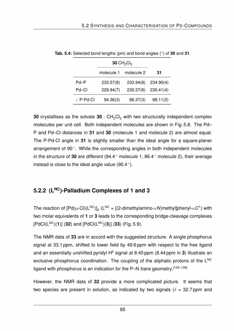
5.2 SYNTHESIS AND CHARACTERISATION OF PD-COMPOUNDS
Tab. 5.4: Selected bond lengths (pm) and bond angles (◦) of 30 and 31.
30·CH2Cl2
molecule 1 molecule 2 31
Pd–P 233.57(8) 233.94(8) 234.90(4)
Pd–Cl 229.94(7) 230.37(8) 230.41(4)
6 P-Pd-Cl 94.36(3) 86.37(3) 88.11(2)
30 crystallises as the solvate 30 · CH2Cl2 with two structurally independent complex
molecules per unit cell. Both independent molecules are shown in Fig. 5.8. The Pd–
P and Pd–Cl distances in 31 and 30 (molecule 1 and molecule 2) are almost equal.
The P-Pd-Cl angle in 31 is slightly smaller than the ideal angle for a square-planer
arrangement of 90 ◦. While the corresponding angles in both independent molecules
in the structure of 30 are different (94.4 ◦ molecule 1, 86.4 ◦ molecule 2), their average
instead is close to the ideal angle value (90.4 ◦).
5.2.2 (LNC)-Palladium Complexes of 1 and 3
The reaction of [Pd(µ-Cl)(LNC)]2 (LNC = [(2-dimethylamino-κN)methyl]phenyl-κC1) with
two molar equivalents of 1 or 3 leads to the corresponding bridge-cleavage complexes
[PdCl(LNC)(1)] (32) and [PdCl(LNC)(3)] (33) (Fig. 5.9).
The NMR data of 33 are in accord with the suggested structure. A single phosphorus
signal at 33.1 ppm, shifted to lower field by 49.6 ppm with respect to the free ligand
and an essentially unshifted pyridyl H6 signal at 8.45 ppm (8.44 ppm in 3) illustrate an
exclusive phosphorus coordination. The coupling of the aliphatic protons of the LNC
ligand with phosphorus is an indication for the P–N trans geometry.[125–130]
However, the NMR data of 32 provide a more complicated picture. It seems that
two species are present in solution, as indicated by two signals (δ = 32.7 ppm and
85

5 COORDINATION CHEMISTRY III: PALLADIUM
Fe
P
N
PdCl
NPd
NPd
Cl Cl
(CH2)nPy
2 eq. of 1 or 3
32 (n = 0), 33 (n = 1)
Fig. 5.9: Reaction of [Pd(µ-Cl)(LNC)]2 with two molar equivalents of ligands 1 and 3.
32.9 ppm) in the 31P NMR spectrum. The diagnostic pyridyl H6 also gives rise to two
signals, an essentially unshifted one at 8.45 ppm and a second one, shifted to lower
field, at 8.92 ppm, which indicates pyridyl coordination. Most likely this is due to an
equilibrium between the primary bridge-cleavage product 32 and a dissociated form
34a (Fig. 5.10), which exhibits a chelate structure.
Fe
P
NN
Pdsolvent
Cl
Fe
P
N
PdCl
Py
32 34a
Fig. 5.10: Solvent constrained equilibrium between 32 and 34a.
This assumption is strongly supported by NMR data concerning the signals of the fer-
rocene CH groups. These become diastereotopic in 34a due to a fixed chelate geom-
etry, which is in accord with eight well separated signals observed for them. Moreover,
the involvement of a dissociation process is in line with the observation that the NMR
spectra recorded in CD3CN as a donor solvent suggested an additional species to be
present. The strong solvent dependence of the equilibrium occurs probably due to the
better stabilisation of the cationic complex by polar solvents, which is in accord with
86

5.2 SYNTHESIS AND CHARACTERISATION OF PD-COMPOUNDS
a shift of the equilibrium in favour of the neutral complex 32, observed if the NMR is
measured in non-polar C6D6.
A similar reaction of both ligands with the solvato complex [Pd(LNC)(MeCN)2][ClO4] in a
1:1 molar ratio afforded the cationic complexes [Pd(LNC)(1)][ClO4] (34) and [Pd(LNC)(3)]
[ClO4] (35), displayed in Fig. 5.11.
N
ClO4
N
PdNCMe
NCMe
Ligand 1 or 3
Fe
P
()n
Ph
PhN
Pd
ClO4
34 (n = 0), 35 (n = 1)
Fig. 5.11: Reaction of 1 and 3 with [Pd(LNC)(MeCN)2][ClO4].
NMR characterisation of the products indicated a cis-chelating coordination of both
ligands. The constrained geometry makes several protons of the compound, especially
the ferrocene CH groups, diastereotopic. The behaviour is comparable with that of
the simple Pd chelate complexes 28 and 29. The shifts of the pyridyl H6 signal (δ
= 8.98 ppm vs. 8.47 ppm in free 1) and the phosphorus signal (δ = 32.6 ppm vs. –
17.7 ppm in free 1) are consistent with the P,N-coordination. The phosphorus signal
of 35 is also shifted (δ = 34.3 ppm), whereas the pyridyl H6 signal is located at slightly
higher field (δ = 8.38 ppm vs. 8.44 ppm in free 3), an effect already known from 29.
The ESI mass spectra showed only the respective cation [Pd(LNC)(L)]+ (L = 1, 3). The
presence of the perchlorate counter ion was proven by IR spectroscopy, elemental
analysis verified the stoichiometry.
The results of the structural characterisation of 34 and 35 are shown in Fig. 5.12. Both
coordination environments of the metal centres are distorted square-planar. The donor-
metal distances of 34 and 35 are in accord with those of related complexes.[125–130] The
bite angle of LNC (6 N2-Pd-C29) in 34 and 35 is the most acute coordination angle and
87

5 COORDINATION CHEMISTRY III: PALLADIUM
(a)
P
Pd C29
N2
N1
Fe
(b)
P
Fe
N1
Pd
C29
N2
Fig. 5.12: Molecular structures of 34 (a) and 35 (b) in the crystal (counterion and solvent
molecules have been omitted for clarity).
very similar in both compounds (Tab. 5.5). The bite angles of the ferrocene ligands ( 6
P-Pd-N) differ considerably from each other. The higher flexibility of 3, induced by the
Tab. 5.5: Selectet distances (pm) and angles (◦)
of 34 and 35.
34 35
Pd–P 225.71(9) 224.91(7)
Pd–N1 217.4(3) 214.2(2)
Pd–N2 216.6(3) 213.8(2)
Pd–C29 201.3(3) 203.2(2)
6 P-Pd-N1 99.13(8) 91.90(6)
6 P-Pd-C29 91.8(1) 94.41(8)
6 N1-Pd-N2 89.2(1) 91.07(7)
6 N2-Pd-C29 81.5(1) 81.93(9)
6 Cp1-Cp2 3.05 2.91
6 τ 57.67 39.92
88

5.3 CATALYTIC EVALUATION
methylene group, allows a closer approach to the ideal right angle (91.9 ◦), while the
constrained geometry of 1 requires a more obtuse bite angle (99.1 ◦).
5.3 Catalytic Evaluation
The catalytic properties of palladium complexes of 1 and 3 were evaluated in Pd-
catalysed C–C bond forming reactions, namely in Suzuki-Miyaura arylation[68,131,132]
and cyanation[133,134] of 4-bromotoluene as a deactivated model substrate. The experi-
ments were performed simultaneously with both donors. The very successful ferrocene
ligand dppf[32] was used for comparison. Complexes of the type [PdCl2(L)] (L = 1, 3,
dppf) were used as well-defined precatalyst and compared to precatalysts generated
in situ from palladium(II) acetate and the appropriate ligand.
The Suzuki-Miyaura cross-coupling reaction of 4-bromotoluene with phenylboronic acid
(Fig. 5.13) was carried out in 1,4-dioxane at 80 ◦C using 0.5 or 0.25 mol% palladium cat-
alyst. Potassium carbonate was used as base. The obtained results showed that the
defined catalyst as well as the in situ generated complexes boost the coupling reac-
tion efficiently at 0.5 mol% Pd loading (Tab. 5.6). At 80 ◦C the reaction proceeded with
similar conversions, regardless of the nature of the ligand. Raising the reaction tem-
perature up to 100 ◦C, better conversions and discrimination by the ligand is achieved.
The conversions increased for [PdCl2(L)] in the order of the ligands dppf ≈ 1 < 3. The
in situ generated catalyst (Pd(OAc)2/L) showed similar trends with a higher activity in
general. Both reactivity trends can be attributed to changed electronic properties and
the ease of reduction of the metal source to a catalytically active species.[135]
BrPhB(OH)2
[Pd]/K2CO3
Fig. 5.13: Suzuki-Miyaura cross-coupling reaction of 4-bromotoluene.
89

5 COORDINATION CHEMISTRY III: PALLADIUM
Tab. 5.6: Catalytic results for Suzuki cross-coupling reaction at 0.5 mol% palladium loading.
yield of 4-methylbiphenyl (%)a
[PdCl2(L)]b [PdCl2(L)]b Pd(OAc)2/Lc
at 80 ◦C at 100 ◦C at 100 ◦C
time (h) dppf 1 3 dppf 1 3 dppf 1 3
2 36 35 35 37 42 52 82 89 90
4 41 39 41 42 46 57 93 94 97
6 44 42 44 50 51 61 96 96 99
8 45 47 47 56 55 64 97 96 100
12 50 51 51 65 61 71 99 97 100
24 63 59 60 84 75 85 100 98 100
aThe reactions were performed with 0.5 mol% of palladium using
2.0 mmol of 4-bromotoluene, 2.2 mmol of phenylboronic acid and
4.4 mmol of K2CO3 in dry dioxane (10 mL). bDefined precatalyst.cIn situ generated catalyst.
Benzonitriles, the products of palladium-catalysed cyanation of aryl halides, are ver-
satile synthetic intermediates and therefore important commodity chemicals. The cya-
nation reaction remains still less explored. The reaction with a CN− source, a base
and a catalyst (sometimes an additional cocatalyst like a zinc salt is necessary) pro-
ceeds satisfactorily mostly only under harsh temperature conditions. The cyanation test
reaction, Fig. 5.14, of the deactivated substrate 4-bromotoluene with potassium hexa-
cyanoferrate(II), Na2CO3 and 0.5 mol% catalyst in N,N-dimethylformamide (DMF) or
N-methyl-2-pyrrolidone (NMP) was performed at 130 ◦C for 16 h (Tab. 5.7). The overall
conversions in NMP were high, but the reaction was accompanied by undesired re-
ductive dehalogenation of the substrate to give toluene (Tab. 5.7 in parentheses). Both
Br CNK4[Fe(CN)6]
[Pd]/Na2CO3
Fig. 5.14: Palladium-catalysed cyanation reaction of 4-bromotoluene.
90

5.4 SUMMARY AND CONCLUSION
Tab. 5.7: Catalytic results for the palladium-catalysed cyanation reaction of 4-bromotoluene.
conversion to 4-cyanotoluene (%)a,b
catalyst NMP DMF
[PdCl2(dppf)] 93(7) 100(n.d.)
[PdCl2(1)] (28) 92(8) 100(n.d.)
[PdCl2(3)] (29) 87(9) 100(n.d.)
Pd(AcO)2/dppf 92(8) 91(9)
Pd(AcO)2/1 64(22) 90(6)
Pd(AcO)2/3 71(20) 90(6)
aConditions: 4-bromotoluene (2.0 mmol), K4[Fe(CN)6] (0.5 mmol), Na2CO3
(2.0 mmol) and 0.5 mol% of a Pd catalyst in dry solvent (3 mL) at 130 ◦C for 16 h.
Conversions were determined by integration of 1H NMR signals. bNMR yield of
toluene is given in parentheses; n.d. = toluene not detected.
dppf catalysts, [PdCl2(dppf)] and in situ generated Pd(OAc)2/dppf, performed similarly
and with the best conversions. For the catalysts based on ligands 1 and 3 the de-
fined complexes gave better results than the mixtures. Changing the solvent to DMF,
the reaction proceeded with complete conversions for all three defined catalysts. The
catalyst mixtures are only slightly inferior with conversions of about 90 %.
5.4 Summary and Conclusion
The ferrocene-based pyridylphosphine ligands 1 and 3 were examined with respect
to their coordination chemistry towards palladium(II). The ligands react with dichloro-
(cod)palladium(II) in a P-coordinated monodentate and in a P,N-chelating manner,
depending on the reaction stoichiometry, to form the simple Pd complexes 28, 29, 30
and 31.
91

5 COORDINATION CHEMISTRY III: PALLADIUM
Both ligands were also reacted with [Pd(µ-Cl)(LNC)]2 (LNC = [(2-dimethylamino-κN)me-
thyl]phenyl-κC1), leading to the corresponding complexes 32 and 33. Similar reactions
with the preformed solvato complex [Pd(LNC)(MeCN)2][ClO4] gaves the cationic com-
plexes 34 and 35. The reaction behaviour of 1 and 3 in these reactions compared
well to that observed in the reactions with [PdCl2(cod)]. The resulting compounds were
fully characterised by standard methods. 28, 30, 31, 34 and 35 were analysed by single
crystal X-ray diffraction. Structural differences can be attributed mainly to the presence
of the methylene group in ligand 3, which makes this ligand more flexible than 1.
Catalytic investigations of Suzuki-Miyaura cross-coupling reactions showed that com-
plexes 28 and 29 and precatalyst formed in situ from 1 and 3 with Pd(OAc)2 promote
the reaction of 4-bromotoluene with phenylboronic acid efficiently. The catalyst based
on ligand 3 works similar to or slightly better than the corresponding dppf-based cata-
lyst. The results for the 1-based catalysts are comparable, too, but only slightly inferior
to those of the dppf benchmark. The Pd-catalysed cyanation reaction is more efficiently
promoted by the defined precatalyst complexes with dppf being a ligand superior to
both pyridylphosphines.
92
6 Experimental
6.1 General Techniques and Methods
All preparations involving air-sensitive compounds were carried out under an atmo-
sphere of dry nitrogen (N50) using standard Schlenk techniques or a conventional
glove box (Argon N50). Solvents and reagents were appropriately dried and purified
by conventional methods and stored under inert gas atmosphere.
Safety note
Caution! Although I have not encountered any problems it should be noted that per-
chlorate salts of metal complexes with organic ligands are potentially explosive! Only
small quantities of these materials should be prepared and they should be handled
with the necessary respect and caution.
NMR Spectroscopy
NMR spectra were recorded on different spectrometers: 1. Varian Unity INOVA (500
MHz), 2. Varian Unity INOVA (400 MHz) 3. Varian NMR System (500 MHz) and 4. Var-
ian 400-MR (400 MHz). 13C and 31P NMR data were collected by proton decoupled
methods. Chemical shifts δ are given in ppm and are referenced to the shift of the
residual protioimpurities of the solvents used relative to trimethylsilane (TMS) for 1H
and to the solvent signal for 13C.[136,137] In the case of 31P NMR samples in CDCl3 were
indirectly referenced to triphenyl phosphane (δ = -6 ppm, relative to 85% phosphoric
93

6 EXPERIMENTAL
acid in D2O[138]). Samples in DMSO-d6 were also indirectly referenced (85% phospho-
ric acid in DMSO−d6 δ = 0 ppm). Coupling constants were given as absolute value in
Hz. Data were analysed by the computer programs Mest-ReC and Spinworks.
Mass spectrometry
ESI and APCI Mass spectra were recorded on a Bruker Esquire 3000 spectrome-
ter (ESI) and a quadrupole ion-trap spectrometer (ESI and APCI) Finnigan LCQ DECA
(ThermoQuest, San Jose, USA).
HR-ESI Mass spectra were recorded on a micrOTOF spectrometer (Bruker Daltonics,
Bremen, Germany).
MALDI Mass spectra were recorded on a BiFlex IV (Bruker Daltonics, Bremen, Ger-
many) spectrometer equipped with an N2-Laser with a wavelength of 337 nm and 3 ns
pulse duration. DCTB (2-[(2E)-3-(4-tert-butylphenyl)-2-methylprop-2-enyliden]malono-
nitril) was used as matrix. Mass calibration was performed immediately prior to the
measurements using polystyrole standard (Ag-Adduct).
Elemental Analysis
Analysis were carried out by the microanalytical laboratories of the Institute of Thermal
Energy Management at the University of Kassel and the department of Chemistry,
Charles University, Prague.
Crystal Structure Analysis
X-ray crystallographic data collection was performed using a Stoe & Cie “IPDS II”
diffractometer with an Eu doped BaFCl area detector and a Nonius KappaCCD diffrac-
tometer. Graphite-monochromatised MoKα radiation (λ0 = 0.71073 A) was used in
each case. The datasets were corrected for Lorentz and polarisation effects. A nu-
merical absorption correction was applied. The program SHELXS-97[139] was used for
structure solution by direct methods. Refinement was carried out using the program
SHELXL-97[139] by full-matrix least-squares against F 2. All non H atoms were refined
94

6.2 SYNTHETIC DETAILS
anisotropically. H atoms were included to the model at calculated positions accord-
ing to the riding model. Graphics were generated with the program ORTEP-3 win,[140]
showing the displacement elipsoides enclosing the 30 % probability level.
Electrochemistry
Electrochemistry was performed in a home-built cylindrical single-compartment cell
with Ag pseudo-reference electrode and Pt working and auxiliary electrodes. The elec-
troactive species was investigated in 0.1 mM or 1.0 mM solution in CH2Cl2 (99.9% Extra
Dry, Acros Organics) with 0.1 M [NnBu4][PF6] as supporting electrolyte at a scan rate of
100 mV/s at room temperature and under nitrogen atmosphere. The supporting elec-
trolyte was recrystalised three times from dry ethanol before use. Decametylferrocene
(fc*) was used as internal standard. An appropriate amount of decamethylferrocene
was added after the scans had been recorded. Electrochemical data were acquired
with a computer-controlled Princeton Applied potentiostat model Versa Stat II utilising
the Princeton PowerSuite (vers. 2.58) software package.
Chromatography
For column chromatography (CC) silica gel 60 M 0.04-0.063 mm / 230-400 mesh ASTM
for CC (Chery-Nagel GmbH & Co. KG) or neutral alumina 0.05-0.2 mm (ICN Alumina
N - Super I) were used. Thin layer chromatography (TLC) was performed on standard
aluminium sheets with silica gel 60 F254 (Merck KGaA). Detection was carried out by
fluorescence quenching at 254 nm and 366 nm.
6.2 Synthetic Details
All non-indexed compounds were either present in our workgroup or commercially
available and used as received.
95

6 EXPERIMENTAL
6.2.1 Starting Materials
Following compounds were synthesised by literature procedures:
1,1’-(Ferrocediyl)phenylphosphane[58]
(Note: Work-up procedure was changed: The reaction mixture was filtered through
celite and evaporated under vacuum. Subsequent CC using neutral alumina under
inert conditions with dry hexane and subsequent with dry diethyl ether afforded pure
product.)
1,1’-Dibromoferrocene[141]
1,1’-Dilithioferrocene[141]
1-Bromo-1’-diphenylphosphinoferrocene[64]
1-Lithio-1’-diphenylphosphinoferrocene[59]
3-Pyridylboroxin[72]
Chloro(tetrahydrothiophene)gold(I)[142]
Bis(1’-bromoferrocenyl)phenylphosphane[64]
Chloro(η2:η2-cycloocta-1,5-diene)palladium(II)[143]
Di-µ-chloro-bis[(2-dimethylamino-κN)methyl]phenyl-κC1dipalladium(II) (LNC)[144]
Bis(acetonitrile-κN)[(2-dimethylamino-κN)methyl]phenyl-κC1palladium(II) perchlorate[125]
6.2.2 Experimental Procedures
6.2.2.1 Ligand Synthesis
1-(Pyrid-2-yl)-1’-diphenylphosphinoferrocene (1)
A solution of 1.6 M n-buthyllithium in hexane (12.0 mmol, 7.5 mL) was slowly added
at –70 ◦C to a solution of 1-bromo-1’-diphenylphosphinoferrocene of (11 mmol, 5.0 g)
in THF (40 mL) via syringe. After 45 min of stirring at –70 ◦C [ZnCl2(1,4-dioxane)]
(11 mmol, 2.5 g) was introduced in one portion and stirring was continued at room
96

6.2 SYNTHETIC DETAILS
temperature for 3 h. A solution of [Pd(PPh3)4] (1 mol%, 0.11 mmol, 127 mg) and 2-
bromopyridine (11 mmol, 1.1 mL) in THF (5 mL) was added. The resulting mixture was
warmed to 60 ◦C and stirred for 72 h. The reaction mixture was evaporated using a
rotary evaporator and the residue was dissolved in dichloromethane (DCM) (20 mL ).
The solution was washed with water (2 x 20 mL), dried with Na2SO4 and evaporated
to dryness. The residue was purified by CC using neutral alumina first with hexane
to remove non-polar side products and subsequently with DCM to elute the product.
Subsequent evaporation gave 1 as a dark orange oil, which crystallised upon stand-
ing. Yield: 2.39 g (48 %). Crystallisation: Crystals suitable for single-crystal X-ray
diffraction analysis were obtained from a highly concentrated DCM solution by slow
evaporation in the air. 1H NMR (CDCl3): 3.96 (m, 2H, Fc), 4.21 (m, 2H, Fc), 4.27 (m,
2H, Fc), 4.83 (m, 2H, Fc), 7.04 (m, 1H, Py), 7.27 (m, 1H, Py), 7.29-7.38 (m, 10H, Ph),
7.51 (m, 1H, Py), 8.47 (m, 1H, Py). 13C NMR (CDCl3): 68.41 (s), 71.41 (s), 73.10 (d,
JPC = 4 Hz), 74.34 (d, JPC = 14 Hz), 76.95 (d, JPC = 7 Hz), 84.59 (s), 120.48 (s), 120.79
(s), 128.29 (d, JPC = 7 Hz), 128.64 (s), 133.64 (d, JPC = 20 Hz), 136.05 (s), 139.14 (d,
JPC = 10 Hz), 149.41 (s), 158.67 (s). 31P NMR (CDCl3): –17.7 (s). MS/APCI(+) (m/z
(%)): 464 (40) [MO + H]+, 448 (100) [M + H]+, 263 (29) [C15H12FeN]+.
1-(Pyrid-3-yl)-1’-diphenylphosphinoferrocene (2)
In a Schlenk-tube 1-bromo-1’-diphenylphosphinoferrocene (4 mmol, 1.8 g) was dis-
solved in 1,4-dioxane (25 mL). 3-Pyridylboroxin (2 mmol, 0.63 g), K2CO3 (20 mmol,
2.8 g) dissolved in water (10 mL) (c(K2CO3): 2.0 mol/L) and solid [Pd(PPh3)4] (2.5 mol%,
0.1 mmol, 115 mg) were added successively. The reaction mixture was heated up to
120 ◦C (bath temp.) and stirred for 14 h. The organic layer was separated and evapo-
rated using a rotary evaporator. The residue was dissolved in DCM (20 mL). The solu-
tion was washed with water (2 x 20 mL) and the aqueous layer extracted with DCM (2
x 10 mL). The combined organic phases were dried with Na2SO4 and filtered. Neutral
alumina (5 g) was added and volatile compounds were removed. This material sub-
jected to CC with neutral alumina, eluting first with petroleum ether 40 ◦C - 60 ◦C (PE) /
diethyl ether 5:1 to remove non-polar side products and subsequently with chloroform.
97

6 EXPERIMENTAL
Evaporation gave 2 as an orange oil, which solidified upon standing. Yield: 0,76 g
(43 %). Crystallisation: Slow evaporation of a highly concentrated DCM solution in the
air gave crystals suitable for single-crystal X-ray diffraction analysis. 1H NMR (CDCl3):
3.96 (m, 2H, Fc), 4.22 (m, 2H, Fc), 4.26 (m, 2H, Fc), 4.56 (m, 2H, Fc), 7.15 (m, 1H, Py),
7.30-7.36 (m, 10H, Ph), 7.58 (m, 1H, Py), 8.40 (m, 1H, Py), 8.61 (m, 1H, Py). 1H NMR
(DMSO−d6): 3.88 (s, 2H, Fc), 4.22 (s, 2H, Fc), 4.27 (s, 2H, Fc), 4.78 (s, 2H, Fc), 7.27
(m, 1H, Py), 7.30-7.37 (m, 10H, Ph), 7.59 (m, 1H, Py), 8.40 (m, 1H, Py), 8.61 (m, 1H,
Py). 13C NMR (CDCl3): 67.67 (d, JPC = 1 Hz), 70.94 (d, JPC = 1 Hz), 73.08 (d, JPC = 4
Hz), 74.52 (d, JPC = 15 Hz), 82.56 (s), 123.38 (s), 128.33 (d, JPC = 7 Hz), 128.73 (s),
133.37 (s), 133.59 (d, JPC = 20 Hz), 134.82 (s), 138.96 (d, JPC = 10 Hz), 147.33 (s),
147.56 (s). 31P NMR (CDCl3): –17.8 (s). 31P NMR (DMSO−d6): –18.8 (s). MS/APCI(+)
(m/z (%)): 464 (45) [MO + H]+, 448 (100) [M + H]+, 370 (21) [C21H17FeNP]+, 263 (15)
[C15H12FeN]+. Elemental analysis (%) calculated for C27H22FeNP (477.3): C 72.50, H
4.96, N 3.13. Found: C 73.34, H 4.94, N 3.23.
1-[(pyrid-2-yl)methyl]-1’-(diphenylphosphino)ferrocene (3)
The synthesis of 3 was carried out by Jirı Schulz in the group of Prof. Petr Stepnicka at
Charles University, Prague, Czech Republic.
Precursor: A stirred solution of 1-bromo-1’-diphenylphosphinoferrocene (10 mmol,
4.49 g) in THF (50 mL) was treated with a 2.5 M solution of n-butyllithium in hexane
(10 mmol, 4.0 mL) at ca. –78 ◦C (dry ice/ethanol bath). After 15 min neat pyridine-2-
carboxaldehyde (12 mmol, 1.29 g) was added and the resulting mixture was stirred at
–78 ◦C for 15 min and then at room temperature for 90 min. The reaction mixture was
quenched by addition of water and extracted with DCM after 30 min. The extract was
washed successively with saturated aqueous NaHCO3 and NaCl solutions, dried with
MgSO4, filtered and evaporated under vacuum. The residue was purified by CC on
silica gel using an ethyl acetate:hexane mixture 1:5 (v/v) to elute first a yellow band
containing (diphenylphosphino)ferrocene (1.04 g, 28 %) and then a minor red band
containing the corresponding ketone (cf. Fig 2.12, page 22) (burgundy red solid; 27 mg,
98

6.2 SYNTHETIC DETAILS
ca. 0.6 %). Then, the mobile phase was changed to ethyl acetate:hexane 1:1 (v/v) to
elute the alcohol 1-[(pyrid-2-yl)hydroxymethyl]-1’-(diphenylphosphino)ferrocene, which
was isolated as an ochre solid following evaporation. Yield: 2.78 g (58 %). 1H NMR
(CDCl3): 4.01 (td, J = 2.4, 1.3 Hz, 1H, Fc), 4.04 (td, J = 2.4, 1.3 Hz, 1H, Fc), 4.10 (m,
2H, Fc), 4.14 (m, 1H, Fc), 4.20 (dt, J = 2.5, 1.3 Hz, 1H, Fc), 4.21 (br s, 1H, CHOH),
4.40 (td, J = 2.4, 1.2 Hz, 1H, Fc), 4.41 (td, J = 2.4, 1.2 Hz, 1H, Fc), 5.36 (s, 1H, CHOH),
7.16 (ddd, JHH = 7.5, 4.9, 1.2 Hz, 1H, Py), 7.26 (dm, JHH ≈ 8 Hz, 1H, Py), 7.28-7.41
(m, 10H, Ph), 7.63 (td, JHH = 7.7, 1.7 Hz, 1H, Py), 8.51 (ddd, JHH = 4.9, 1.8, 1.0 Hz,
1H, Py). 13C NMR (CDCl3): 66.79 (s), 68.32 (s), 69.37 (s), 69.33 (s), 71.21 (s, CHOH),
71.65 (d, JPC ≈ 5 Hz), 71.68 (d, JPC ≈ 5 Hz), 73.39 (d, JPC = 13 Hz), 73.62 (d, JPC
= 15 Hz), 76.22 (d, JPC = 6 Hz), 92.89 (s), 120.79 (s), 122.49 (s), 128.14 (d, JPC = 6
Hz), 128.49 (s), 128.58 (s), 133.48 (vt, J ′ = 20 Hz), 136.76 (s), 138.77 (d, JPC = 9 Hz),
138.95 (d, JPC = 9 Hz), 147.90 (s), 161.02 (s). 31P NMR (CDCl3): –16.3 (s). Elemental
analysis (%) calculated for C18H24FeNOP (477.3): C 70.45, H 5.07, N 2.94. Found: C
70.27, H 5.18, N 2.84.
Ligand: Commercial SmI2 solution (10 mmol, 100 mL of 0.1 M in THF) was added
to a stirred mixture of 1-[(pyrid-2-yl)hydroxymethyl]-1’-(diphenylphosphino)ferrocene (4
mmol, 1.91 g), (Me2N)3PO (HMPA; 40 mmol, 7.0 mL) and 10 mL dry THF during 10 min,
yielding a dark brown mixture. After stirring for 5 min, a solution of pivalic acid (5.0 mmol,
514 mg in 5 mL THF) was added, causing the color of the reaction mixture to turn back
to the initial orange-yellow. The mixture was stirred for another 15 min, diluted with
diethyl ether and saturated aqueous NaCl. The organic layer was separated, and the
aqueous layer was extracted with diethyl ether, adding little hexane to facilitate phase
separation. The combined organic layers were washed with saturated aqueous NaCl,
dried with MgSO4, filtered and evaporated. The residue was passed through a short
silica gel column eluting with ethyl acetate:hexane (1:1 v/v) to remove some polar by-
products and inorganic residuals. The first yelloworange band was collected and evap-
orated to afford crude product, which was immediately purified by chromatography
on an alumina column using diethyl ether:hexane (4:1 v/v) as the eluent. Evapora-
99

6 EXPERIMENTAL
tion of the first yellow band followed by careful evaporation and drying under vacuum
(80 ◦C/0.1 Torr) gave 3 as a dark orange-brown oil, which slowly crystallised when
stored at 4 ◦C. Yield: 1.02 g (55 %). Crystallisation: Crystals suitable for single-crystal
X-ray diffraction analysis were grown by recrystallisation from diethyl ether. 1H NMR
(CD2Cl2): 3.63 (s, 2H, CH2Py), 3.97 (m, 2H, Fc), 4.05 (m, 2H, Fc), 4.07 (m, 2H, Fc),
4.33 (m, 2H, Fc), 7.02 (m, 1H, Py), 7.06 (m, 1H, Py), 7.28-7.40 (m, 10H, Ph), 7.54 (m,
1H, Py), 8.44 (m, 1H, Py). 13C NMR (CD2Cl2): 38.65 (s), 69.36 (s), 70.31 (s), 72.18 (d,
JPC = 4 Hz), 73.96 (d, JPC = 15 Hz), 76.34 (d, JPC = 6 Hz), 87.50 (s), 121.44 (s), 122.73
(s), 128.48 (d, JPC = 7 Hz), 128.80 (s), 133.86 (d, JPC = 20 Hz), 136.50 (s), 139.92 (d,
JPC = 9 Hz), 149.43 (s), 161.44 (s). 31P NMR (CD2Cl2): –16.5 (s). MS/ESI(+) (m/z):
462 [M + H]+, 484 [M + Na]+. HR-ESI: m/z calculated for C28H24FeNP [M+] 461.0996.
Found: 461.1000.
2,6-Bis(1’-diphenylphosphinoferrocenyl)pyridine (4)
1-Bromo-1’-diphenylphosphinoferrocene (6.4 mmol, 2.89 g) was dissolved in THF (40
mL) and the solution cooled to –70 ◦C. 1.6 M n-buthyllithium in hexane (6.5 mmol,
4.1 mL) was added by syringe. The mixture was stirred at this temperature for 0.5 h.
Solid [ZnCl2(1,4-dioxane)] (7.1 mmol, 1.58 g) was added. After 0.5 h the mixture was
allowed to warm to room temperature and stirred further for a 2.5 h. Subsequently 2,6-
dibromopyridine (3.1 mmol, 0.73 g) and [Pd(PPh3)4] (2 mol% per coupling, 0.12 mmol,
133 mg) were added. The resulting mixture was warmed to 60 ◦C and stirred for 72 h.
A small amount of silica gel was added and the reaction mixture was evaporated using
a rotary evaporator to afford a pourable solid, which was subjected to CC on silica gel,
eluting first with PE / diethyl ether 10:1 to remove non-polar side products and then
with DCM (+0.5% of triethylamine) to elute the product. Subsequent evaporation gave
4 · 0.25 CH2Cl2 as an orange foamy solid. Yield: 0.824 g (33 %). Crystallisation: Ma-
terial suitable for single-crystal X-ray diffraction analysis was obtained in a NMR tube
by layering a highly concentrated DCM solution with a small amount of pure DCM and
then with hexane under inert gas atmosphere to avoid oxidation. 1H NMR (CDCl3):
3.94 (s, 4H, Fc), 4.17 (s, 4H, Fc), 4.26 (s, 4H, Fc), 4.88 (s, 4H, Fc), 7.01 (m, 2H, Py),
100

6.2 SYNTHETIC DETAILS
7.30-7.38 (m, 21H, Ph + Py). 13C NMR (CDCl3): 68.50 (s), 71.20 (d, JPC = 1 Hz), 73.23
(d, JPC = 4 Hz), 74.28 (d, JPC = 15 Hz), 76.72 (d, JPC = 7 Hz), 85.19 (s), 117.04 (s),
128.28 (d, JPC = 7 Hz), 128.61 (s), 133.63 (d, JPC = 20 Hz), 135.93 (s), 139.15 (d, JPC
= 10 Hz), 157.60 (s). 31P NMR (CDCl3): –17.7 (s). MS/MALDI(+) (m/z (%)): 815 (100)
[M]+. Elemental analysis (%) calculated for C49H39Fe2NP2 · 0.25 CH2Cl2 (836.7): C
70.70, H 4.76, N 1.67. Found: C 70.85, H 4.78, N 1.64.
6.2.2.2 Group 12 Metal Complexes
General Procedure
Solid metal halide MX2 (M = Zn, Cd, Hg and X = Cl, Br, I) (0.1 mmol) was added to a so-
lution of one (0.1 mmol) or two equivalents (0.2 mmol) of the ligand in ethanol (10 mL).
The mixture was stirred at room temperature for 14 h in a 50 mL Schlenk flask. If pre-
cipitation of the product occurred, the solvent volume was reduced in vacuum to less
than half. If no precipitation occurred the solvent was removed in vacuum, the residue
was dissolved in DCM and hexane was added to precipitate the product. In either case
the suspension was filtered and the solid residue was washed with diethyl ether (2 mL)
and subsequently with hexane (5 mL). Finally, drying under reduced pressure afforded
the respective product. Diffusion Experiments to obtain crystals directly from the reac-
tion were carried out usually in an NMR tube with 0.05 mmol substance in 0.5 mL of
respective solvent. Potential deviations are given in the detailed description.
[ZnCl2(1)] (5a)
Reactants: 1 (0.1 mmol, 44.7 mg), ZnCl2 (0.1 mmol, 13.6 mg). Work-up: The solvent
was evaporated in vacuum and the residue dissolved in DCM (2 mL). 5a was precipi-
tated with hexane (25 mL). Yield: 55 mg (95 %). Crystallisation: Crystals suitable for
single-crystal X-ray diffraction analysis were obtained by layering a DCM solution of 1
with a solution of ZnCl2 in diethyl ether. 1H NMR (CDCl3): 4.30 (m, 4H, Fc), 4.58 (m, 2H,
Fc), 5.19 (m, 2H, Fc), 7.42 (m, 8H, Ph + Py), 7.76 (m, 5H, Ph + Py), 9.57 (m, 1H, Py).13C NMR (CDCl3): 71.41 (s), 71.59 (s), 73.39 (d, JPC = 6 Hz), 75.63 (d, JPC = 10 Hz),
101

6 EXPERIMENTAL
84.34 (s), 123.10 (s), 125.22 (s), 128.98 (d, JPC = 10 Hz), 131.22 (s), 133.79 (d, JPC
= 13 Hz), 139.32 (s), 152.78 (s), 158.46 (s). 31P NMR (CDCl3): –14.0 (s). MS/ESI(+)
(m/z (%)): 605 (10) [M + Na]+, 546 (20) [M – Cl]+, 510 (35) [C27H22FeNPZn]+, 464
(30) [C27H22FeNPO]+, 448 (100) [C27H22FeNP]+. Elemental analysis (%) calculated
for C27H22Cl2FeNPZn (583.6): C 55.57, H 3.80, N 2.40. Found: C 56.27, H 3.84, N
2.57.
[ZnBr2(1)] (5b)
Reactants: 1 (0.1 mmol, 44.7 mg), ZnBr2 (0.1 mmol, 22.5 mg). Work-up: The solvent
was evaporated in vacuum and the residue dissolved in DCM (2 mL). 5a · 0.25 C6H14
was obtained by adding hexane (15 mL), filtering off, washing and drying in vacuum.
Yield: 41 mg (61 %). Crystallisation: Single-crystals suitable for X-ray diffraction anal-
ysis were obtained analogue to the procedure for 5a. 1H NMR (CDCl3): 4.23 (m, 2H,
Fc), 4.54 (m, 2H, Fc), 4.60 (m, 2H, Fc), 5.02 (m, 2H, Fc), 7.43 (m, 8H, Ph + Py), 7.78
(m, 5H, Ph + Py), 9.57 (m, 1H, Py). 13C NMR (CDCl3): 71.32 (s), 71.40 (s), 73.69
(br d, JPC ≈ 5 Hz), 75.97 (d, JPC = 12 Hz), 84.75 (s), 123.12 (s), 125.10 (s), 128.96
(d, JPC = 10 Hz), 131.22 (s), 133.82 (d, JPC = 12 Hz), 139.29 (s), 153.46 (s), 157.93
(s). 31P NMR (CDCl3): –14.4 (s). MS/ESI(+) (m/z (%)): 592 (15) [M – Br]+, 510 (15)
[C27H22FeNPZn]+, 464 (22) [C27H22FeNPO]+, 448 (100) [C27H22FeNP]+. Elemental
analysis (%) calculated for C27H22Br2FeNPZn · 0.25 C6H14(694.0): C 49.32, H 3.70, N
2.02. Found: C 49.73, H 3.73, N 2.16.
[ZnI2(1)] (5c)
Reactants: 1 (0.1 mmol 44.7 mg), ZnI2 (0.1 mmol, 31.9 mg). Work-up: The solvent
volume was reduced to more than half. The product was precipitated by the addition
of hexane (10 mL). Yield: Yield: 49 mg (65 %). 1H NMR (CDCl3): 4.18 (m, 2H, Fc),
4.60 (m, 2H, Fc), 4.69 (m, 2H, Fc), 4.94 (m, 2H, Fc), 7.38 (m, 1H, Py), 7.41-7.52
(m, 7H, Ph + Py), 7.74-7.83 (m, 5H, Ph + Py), 9.60 (m, 1H, Py). 13C NMR (CDCl3):
70.95 (s), 71.34 (s), 73.79 (d, JPC = 10 Hz), 76.14 (d, JPC = 12 Hz), 77.36 (s), 84.88
(s), 122.87 (s), 124.67 (s), 128.91 (d, JPC = 10 Hz), 130.60 (d, JPC = 39 Hz), 131.18
102

6.2 SYNTHETIC DETAILS
(s), 134.01 (d, JPC = 11 Hz), 139.26 (s), 153.95 (s), 157.45 (s). 31P NMR (CDCl3):
–19.2 (s). MS/ESI(+) (m/z (%)): 767 (7) [M]+, 510 (33) [C27H22FeNPZn]+, 447 (100)
[C27H22FeNP]+. Elemental analysis (%) calculated for C27H22FeI2NPZn (766.5): C
42.31, H 2.89, N 1.83. Found: C 42.29, H 3.12, N 1.80.
[CdCl2(1)] (6a)
Reactants: 1 (0.1 mmol, 44.7 mg), CdCl2 (0.1 mmol, 18.3 mg). Work-up: The formed
precipitate was filtered off, washed and dried. Yield: 14 mg (22 %). 1H NMR (CDCl3):
4.12 (s, 2H, Fc), 4.27 (s, 2H, Fc), 4.43 (s, 2H, Fc), 5.04 (s, 2H, Fc), 7.25 (m, 1H,
Py), 7.37 (m, 1H, Py), 7.41 (m, 6H, Ph), 7.59 (m, 4H, Ph), 7.65 (m, 1H, Py), 8.99 (br
s, 1H, Py). 13C NMR (CDCl3): 68.28 (s), 71.56 (s), 73.23 (d, JPC = 6 Hz), 75.15 (d,
JPC = 13 Hz), 77.36 (s), 84.73 (s), 122.82 (s), 128.93 (s), 129.18 (d, JPC = 10 Hz),
130.03 (s), 131.22 (s), 133.77 (d, JPC = 14 Hz), 138.69 (s), 152.03 (s), 158.26 (s),
167.90 (s). 31P NMR (CDCl3): –8.0 (br s). Elemental analysis (%) calculated for
C27H22CdCl2FeNP (630.6): C 51.43, H 3.52, N 2.22. Found: C 52.44, H 3.52, N 2.33.
[CdBr2(1)] (6b)
Reactants: 1 (0.1 mmol, 44.7 mg), CdBr2 · 4 H2O (0.1 mmol, 34.4 mg). Work-up: The
yellow-orange precipitate was filtered off, washed and dried. Yield: 43 mg (60 %).
Crystallisation: Single-crystals suitable for X-ray diffraction analysis were obtained by
slow evaporation of a DCM solution of the crude product. 1H NMR (CDCl3): 4.21 (m,
2H, Fc), 4.27 (m, 2H, Fc), 4.60 (m, 2H, Fc), 5.19 (m, 2H, Fc), 7.38 (m, 1H, Py), 7.44-
7.52 (m, 7H, Ph + Py), 7.72 (m, 1H, Py), 7.75 (m, 4H, Ph), 9.35 (m, 1H, Py). 13C NMR
(CDCl3): 71.05 (s), 71.44 (s), 73.44 (d, JPC = 7 Hz), 75.62 (d, JPC = 13 Hz), 77.36 (s),
85.06 (s), 123.19 (s), 124.48 (s), 129.22 (d, JPC = 10 Hz), 129.42 (s), 131.58 (d, JPC =
2 Hz), 133.80 (d, JPC = 13 Hz), 139.12 (s), 152.87 (s), 157.88 (s). 31P NMR (CDCl3): –
3.6 (J 111CdP = 1708 Hz, J 113CdP = 1782 Hz). MS/MALDI(+) (m/z (%)): 639 (21) [M – Br]+,
446 (100) [C27H22FeNP]+. Elemental analysis (%) calculated for C27H22Br2CdFeNP
(719.5): C 45.07, H 3.08, N 1.95. Found: C 45.32, H 3.00, N 2.07.
103

6 EXPERIMENTAL
[CdI2(1)] (6c)
Reactants: 1 (0.1 mmol, 44.7 mg), CdI2 (0.1 mmol, 36.6 mg). Work-up: The solvent
volume was reduced in vacuum. The addition of hexane (10 mL) gave 6c · 0.5 C6H14.
Yield: 25 mg (34 %). Crystallisation: Single-crystals suitable for X-ray diffraction anal-
ysis were obtained by layering a CDCl3 solution of 6c with hexane. 1H NMR (CDCl3):
4.21 (m, 2H, Fc), 4.34 (m, 2H, Fc), 4.57 (m, 2H, Fc), 5.06 (m, 2H, Fc), 7.34-7.40 (m,
2H, Py), 7.43-7.52 (m, 6H, Ph), 7.67-7.74 (m, 5H, Ph + Py), 9.27 (m, 1H, Py). 13C NMR
(CDCl3): 70.66 (s), 71.31 (s), 73.55 (d, JPC = 6 Hz), 75.76 (d, JPC = 12 Hz), 77.40 (s),
85.42 (s), 122.88 (s), 123.94 (s), 129.07 (d, JPC = 10 Hz), 131.21 (d, JPC = 44 Hz),
131.23 (s), 133.90 (d, JPC = 13 Hz), 134.18(s), 138.67 (s), 153.08 (s), 157.56 (s).31P NMR (CDCl3): –8.9 (bs). MS/ESI(+) (m/z (%)): 838 (3) [M + Na]+, 688 (15) [M
– I]+, 464 (34) [C27H22FeNPO]+, 448 (100) [C27H22FeNP]+. Elemental analysis (%)
calculated for C27H22CdFeI2NP · 0.5 C6H14 (856.6): C 42.07 , H 3.41, N 1.64. Found:
C 42.63, H 2.98, N 1.73.
[CdCl2(1)2] (7)
Reactants: 1 (0.1 mmol, 44.7 mg), CdCl2 (0.1 mmol, 18.3 mg). Crystallisation: Single-
crystals suitable for X-ray diffraction analysis were obtained analogue to the procedure
in 5a.
[HgCl2(1)] (8a)
Reactants: 1 (0.1 mmol, 44.7 mg), HgCl2 (0.1 mmol, 27.1 mg). Work-up: The solvent
volume was reduced in vacuum. The precipitate was filtered off, washed and dried in
vacuum. Yield: 61 mg (84 %). 1H NMR (CDCl3): 4.25 (s, 2H, Fc), 4.32 (s, 2H, Fc), 4.60
(s, 2H, Fc), 5.19 (s, 2H, Fc), 7.30 (m, 1H, Py), 7.36 (m, 1H, Py), 7.56 (m, 6H, Ph), 7.64
(m, 1H, Py), 7.80 (m, 4H, Ph), 9.36 (m, 1H, Py). 13C NMR (CDCl3): 71.44 (s), 71.47
(s), 74.08 (d, JPC = 9 Hz), 75.53 (d, JPC = 14 Hz), 77.36 (s), 86.36 (s), 122.91 (s),
123.25 (s), 127.75 (d, JPC = 52 Hz), 129.70 (d, JPC = 11 Hz), 132.69 (d, JPC = 3 Hz),
133.61 (d, JPC = 13 Hz), 137.72 (s), 152.21 (s), 156.97 (s). 31P NMR (CDCl3): 32.2
(J 199HgP = 7470 Hz). MS/ESI(+) (m/z (%)): 684 (100) [M – Cl]+, 447 (35) [C27H22FeNP]+.
104
6.2 SYNTHETIC DETAILS
Elemental analysis (%) calculated for C27H22Cl2FeHgNP (718.8): C 45.12, H 3.08, N
1.95. Found: C 45.28, H 3.15, N 1.94.
[HgBr2(1)] (8b)
Reactants: 1 (0.1 mmol, 44.7 mg), HgBr2 (0.1 mmol, 36.0 mg). Work-up: The formed
precipitate was filtered off, washed and dried in vacuum. Yield: 67 mg (83 %). Crys-
tallisation: Layering a solution of 8b in DCM with a small amount of pure DCM
and then with diethyl ether gave single-crystals suitable for X-ray diffraction analysis.1H NMR (CDCl3): 4.33 (m, 2H, Fc), 4.38 (m, 2H, Fc), 4.57 (m, 2H, Fc), 5.11 (m, 2H, Fc),
7.24 (m, 1H, Py), 7.30 (m, 1H, Py), 7.52-7.62 (m, 7H, Ph + Py), 7.77 (m, 4H, Ph), 9.15
(m, 1H, Py). 13C NMR (CDCl3): 70.75 (s), 71.62 (s), 74.47 (d, JPC = 9 Hz), 75.52 (d,
JPC = 13 Hz), 86.46 (s), 122.46 (s), 122.54 (s), 129.58 (d, JPC = 9 Hz), 132.49 (d, JPC
= 2 Hz), 133.64 (d, JPC = 13 Hz), 137.35 (s), 152.04 (s), 156.84 (s). 31P NMR (CDCl3):
28.5 (J 199HgP = 6359 Hz). MS/MALDI(+) (m/z (%)): 728 (80) [M – Br]+, 447 (100)
[C27H22FeNP]+. Elemental analysis (%) calculated for C27H22Br2FeHgNP (807.7): C
40.15, H 2.75, N 1.73. Found: C 40.21, H 2.58, N 1.84.
[HgI2(1)] (8c)
Reactants: 1 (0.1 mmol, 44.7 mg), HgI2 (0.1 mmol, 45.4 mg). Work-up: The addition
of hexane (10 mL) completed the precipitation. The product was filtered and washed.
Subsequent drying under reduced pressure gave 8c · 0.25 C6H14. Yield: 72 mg (80 %).
Crystallisation: Single-crystals suitable for X-ray diffraction analysis were obtained
from a CHCl3 solution. 1H NMR (CDCl3): 4.23 (m, 2H, Fc), 4.37 (m, 2H, Fc), 4.44
(m, 2H, Fc), 4.98 (m, 2H, Fc), 7.18 (m, 1H, Py), 7.24 (m, 1H, Py), 7.45-7.55 (m, 6H,
Ph), 7.57 (m, 1H, Py), 7.66 (m, 4H, Ph), 8.89 (m, 1H, Py). 13C NMR (CDCl3): 70.03
(s), 70.30 (s), 71.71 (s), 74.44 (d, JPC = 8 Hz), 75.43 (d, JPC = 13 Hz), 86.39 (s),
121.57 (s), 122.12 (s), 129.5 (d, JPC = 12 Hz), 131.95 (s), 133.68 (d, JPC = 12 Hz),
136.93 (s), 151.72 (s), 156.85 (s). 31P NMR (CDCl3): 19.74 (br s). MS/ESI(+) (m/z
(%)): 776 (33) [M – I]+, 448 (100) [C27H22FeNP]+. Elemental analysis (%) calculated
for C27H22FeHgI2NP · 0.25 C6H14 (923.2): C 37.08, H 2.78, N 1.52. Found: C 37.65, H
105

6 EXPERIMENTAL
2.61, N 1.62.
[HgCl2(1)2] (9a)
Reactants: 1 (0.1 mmol, 44.7 mg), HgCl2 (0.1 mmol, 27.1 mg). Crystallisation: Single-
crystals suitable for X-ray diffraction analysis were obtained analogue to the procedure
for 5a.
[HgBr2(1)2] (9b)
Reactants: 1 (0.2 mmol, 89.4 mg), HgBr2 (0.1 mmol, 36.0 mg). Work-up: The precipi-
tate was filtered off, washed with ethanol and dried in vacuum. Yield: 107 mg (85 %).
Crystallisation: Layering of a DCM solution of 9b with a small amount pure DCM and
then with diethyl ether gave crystals suitable for X-ray diffraction analysis. 1H NMR
(CDCl3): 4.12 (m, 4H, Fc), 4.19 (m, 4H, Fc), 4.36 (m, 4H, Fc), 4.86 (m, 4H, Fc), 7.05
(m, 2H, Py), 7.15 (m, 2H, Py), 7.36 (m, 8H, Ph + Py), 7.43-7.51 (m, 8H, Ph), 7.35-7.45
(m, 6H, Ph + Py), 7.61 (m, 8H, Ph), 8.48 (m, 2H, Py). 13C NMR (CDCl3): 69.19 (s),
70.42 (s), 72.94 (s), 74.76 (s), 77.39 (s), 85.41 (s), 120.85 (s), 121.13 (s), 128.99 (s),
131.27 (s), 133.71 (s), 136.29 (s), 149.55 (s), 157.74 (s). 31P NMR (CDCl3): 14.5 (s).
MS/ESI(+) (m/z (%)): 448 (100) [C27H22FeNP]+, 728 (19) [C27H22FeNPHgBr]+, 1175 (4)
[M – Br]+, 1254 (3) [M]+. Elemental analysis (%) calculated for C54H44Br2Fe2HgN2P2
(1254.9): C 51.68, H 3.53, N 2.23. Found: C 51.32, H 3.49, N 2.05.
[ZnBr2(2)]n (10)
Reactants: 2 (0.1 mmol, 44.7 mg), ZnBr2 (0.1 mmol, 22.5 mg). Work-up: After the
addition of solid ZnBr2, immediately a yellow-brown solid precipitated. The reaction
mixture was stirred for a further 6 h. The solvent volume was reduced in vacuum.
Filtration, an ethanol wash and drying under reduced pressure gave 10 as a yellow
solid. Yield: 42 mg (53 %). Crystallisation: Crystals suitable for X-ray diffraction anal-
ysis were obtained by layering a DCM solution (0.5 mL) of 2 (0.05 mmol) with a small
amount pure ethanol and then with an ethanolic solution (0.5 mL) of ZnBr2 (0.05 mmol)
106

6.2 SYNTHETIC DETAILS
in an NMR tube. 1H NMR (DMSO−d6): 3.88 (s, 2H, Fc), 4.22 (s, 2H, Fc), 4.28 (s, 2H,
Fc), 4.77 (s, 2H, Fc), 7.31 (m, 5H, Ph + Py), 7.34 (m, 6H, Ph), 7.79 (m, 1H, Py), 8.38
(m, 1H, Py), 8.67 (m, 1H, Py). 13C NMR (DMSO−d6): 67.52 (s), 70.55 (s), 72.83 (d,
JPC = 4 Hz), 74.02 (d, JPC = 15 Hz), 76.42 (d, JPC = 8 Hz), 81.98 (s), 123.62 (s), 128.34
(d, JPC = 7 Hz), 128.68 (s), 133.04 (d, JPC = 20 Hz), 133.41 (s), 134.40 (s), 138.53
(d, JPC = 10 Hz), 146.78 (s), 146.87 (s). 31P NMR (DMSO−d6): –18.9 (s), 25.3 (s).
MS/MALDI(+) (m/z (%)): 447 (100) [C27H22FeNP]+.
[CdBr2(2)]n (11)
Reactants: 2 (0.1 mmol, 44.7 mg), CdBr2 · 4 H2O (0.1 mmol, 34.4 mg). Work-up: After
the addition of CdBr2 · 4 H2O, immediately an orange solid precipitated. Over-night
stirring, filtration, ethanol wash and drying under reduced pressure gave 11 as a yellow
solid. Yield: 30 mg (38 %). 1H NMR (DMSO−d6): 3.89 (s, 2H, Fc), 4.22 (s, 2H, Fc),
4.28 (s, 2H, Fc), 4.77 (s, 2H, Fc), 7.29 (m, 5H, Ph + Py), 7.34 (m, 6H, Ph), 7.78
(m, 1H, Py), 8.38 (m, 1H, Py), 8.67 (s, 1H, Py). 13C NMR (DMSO−d6): 67.46 (s),
70.51 (s), 72.80 (d, JPC = 4 Hz), 73.98 (d, JPC = 15 Hz), 76.38 (d, JPC = 8 Hz), 82.03
(s), 123.49 (s), 128.31 (d, JPC = 7 Hz), 128.65 (s), 132.92 (s), 133.18 (d, JPC = 20
Hz), 134.25 (s), 138.49 (s), 146.89 (s), 146.94 (s). 31P NMR (DMSO−d6): –18.8 (s),
25.3 (s). MS/MALDI(+) (m/z (%)): 447 (100) [C27H22FeNP]+. Elemental analysis (%)
calculated for (C27H22Br2CdFeNP)n (719.5, n = 1): C 45.07, H 3.08, N 1.95. Found: C
44.77, H 3.02, N 1.88.
[HgBr2(2)]n (12)
Reactants: 2 (0.1 mmol, 44.7 mg) and HgBr2 (0.1 mmol, 36.0 mg). Work-up: Immedi-
ately after the addition of HgBr2 an orange solid precipitated. Over-night stirring, filtra-
tion, ethanol wash and drying under reduced pressure gave 12 as a yellow solid. Yield:
55 mg (68 %). Crystallisation: Layering a DCM solution (0.5 mL) of 2 (0.05 mmol)
with a small amount pure DCM and then with an ethanolic (0.5 mL) solution of HgBr2
(0.05 mmol) gave crystals suitable for X-ray diffraction analysis. 1H NMR (DMSO−d6):
4.41-4.46 (m, 6H, Fc), 4.85 (s, 2H, Fc), 7.53 (m, 5H, Ph + Py), 7.61 (m, 7H, Ph +
107

6 EXPERIMENTAL
Py), 8.36 (m, 1H, Py), 8.57 (m, 1H, Py). 13C NMR (DMSO−d6): 68.11 (s), 71.56 (s),
73.55 (d, JPC = 13 Hz), 74.65 (d, JPC = 7 Hz), 83.48 (s), 123.53 (s), 129.35 (d, JPC
= 11 Hz), 130.76 (d, JPC = 10 Hz), 132.28 (s), 133.01 (d, JPC = 13 Hz), 133.15 (s),
146.85 (s), 147.37 (s). 31P NMR (DMSO−d6): 19.3 (br s). MS/MALDI(+) (m/z (%)):
808 (8) [C27H22Br2FeHgNP]+, 728 (29) [C27H22BrFeHgNP]+, 447 (100) [C27H22FeNP]+.
Elemental analysis (%) calculated for (C27H22Br2FeHgNP)n (807.7, n = 1): C 40.15, H
2.75, N 1.73. Found: C 40.15, H 2.72, N 1.79.
[ZnBr2(3)] (13)
Reactants: 3 (0.05 mmol, 23.1 mg), ZnBr2 (0.05 mmol, 11.3 mg). Work-up: A Solution
of ZnBr2 in ethanol (5 mL) was mixed with solution of the ligand in ethanol (5 mL) and
stirred for 0.5 h. The solvent was removed under reduced pressure. The residue was
dissolved in DCM (1 mL) and dropped into hexane to afford 13 · 0.25 CH2Cl2. Yield:
30 mg (90 %). Crystallisation: Liquid phase diffusion of hexane into a chloroform
solution (0.7 mL) of 13 gave crystals suitable for X-ray diffraction analysis. 1H NMR
(CDCl3): 3.85 (br s, 2H), 4.01 (s, 2H), 4.37-4.59 (br m, 6H), (Fc + CH2Py); 7.39 (m,
5H, Ph + Py), 7.45 (m, 2H, Ph), 7.52 (m, 1H, Py), 7.55-7.82 (br m, 4H, Ph), 7.96 (m,
1H, Py), 9.66 (m, 1H, Py). 13C NMR (CDCl3): 37.45 (s), 68.96 (s), 69.83 (s), 72.92 (s),
74.24 (s), 77.36 (s), 87.01 (s), 123.58 (s), 127.19 (s), 128.85 (d, JPC = 10 Hz), 130.16
(d, JPC = 40 Hz), 131.06 (s), 133.91 (d, JPC = 12 Hz), 140.56 (s), 150.96 (s), 159.96
(s). 31P NMR (CDCl3): –18.1 (s). MS/ESI(+) (m/z (%)): 524 (100) [C28H24FeNPZn]+.
Elemental analysis (%) calculated for C28H24Br2FeZnNP · 0.25 CH2Cl2 (707.7): C
47.94, H 3.49, N 1.98. Found: C 47.59, H 3.58, N 1.92.
[ZnBr2(3)]n (13a)
Reactants: 3 (0.1 mmol, 46.1 mg), ZnBr2 (0.1 mmol, 22.5 mg). Work-up: The solvent
volume was reduced in vacuum to half. To obtain crystals directly from the reaction
mixture, it was stored several 72 h. The obtained crystalline material was filtered and
dried in vacuum. Yield: 42 mg (61 %). Crystallisation: The product was suitable for
characterisation by X-ray diffraction analysis.
108

6.2 SYNTHETIC DETAILS
[CdBr2(3)] (14)
Reactants: 3 (0.05 mmol, 23.1 mg), CdBr2 · 4 H2O (0.05 mmol, 17.2 mg). Work-up:
A solution of CdBr2 · 4 H2O was mixed with a solution of the ligand in ethanol (both
10 mL) and stirred for 1 h. The resulting precipitate was isolated by suction, washed
with ethanol and dried in vacuum. Yield: 34 mg (94 %). 1H NMR (CDCl3): 3.96 (m,
2H), 4.00 (m, 2H), 4.29 (m, 2H), 4.48 (m, 2H), 4.52 (m, 2H), (Fc + CH2Py); 7.35 (m, 1H,
Py), 7.39 - 7.53 (m, 7H, Ph + Py), 7.62 (m, 4H, Ph), 7.92 (m, 1H, Py), 9.45 (m, 1H, Py).13C NMR (CDCl3): 38.35 (s), 68.64 (s), 70.31 (s), 72.93 (d, JPC = 7 Hz), 74.51 (d, JPC
= 12 Hz), 77.39 (s), 87.12 (s), 123.61 (s), 126.38 (s), 129.16 (d, JPC = 10 Hz), 131.33
(s), 133.71 (d, JPC = 14 Hz), 140.26 (s), 150.77 (s), 160.50 (s). 31P NMR (CDCl3): It
was not possible to detect the phosphorus signal. Elemental analysis (%) calculated
for C28H24Br2CdFeNP (733.5): C 45.85, H 3.30, N 1.91. Found: C 45.68, H 3.27, N
1.85.
[CdBr2(3)]n (14a)
Reactants: 3 (0.05 mmol, 23.1 mg), CdBr2 · 4 H2O (0.05 mmol, 17.2 mg). Crystallisa-
tion: Crystals were obtained by layering a solution of 3 (0.05 mmol) in DCM (0.5 mL)
with pure ethanol and than with an ethanolic solution (0.5 mL) of CdBr2 · 4 H2O (0.05
mmol).
[CdI2(3)] (15)
Reactants: 3 (0.1 mmol, 46.1 mg), HgBr2 (0.1 mmol, 36.0 mg). Work-up: The synthe-
sis was performed in analogy to that of 14. Yield: 37.3 mg (90 %). Crystallisation:
Liquid phase diffusion of hexane into a chloroform solution (0.7 mL) of 15 gave crystals
suitable for X-ray diffraction analysis. 1H NMR (CDCl3): 4.04 (s, 2H), 4.07 (s, 2H), 4.17
(s, 2H), 4.39 (s, 2H), 4.55 (s, 2H), (Fc + CH2Py); 7.22 (m, 1H, Py), 7.35 (m, 1H, Py),
7.50 (m, 6H, Ph), 7.73 (m, 5H, Ph + Py), 9.23 (m, 1H, Py). 13C NMR (CDCl3): 37.63
(s), 69.25 (s), 70.64 (s), 73.69 (d, JPC = 8 Hz), 74.65 (d, JPC = 12 Hz), 77.39 (s), 88.49
(s), 122.90 (s), 125.11 (s), 129.48 (d, JPC = 12 Hz), 132.27 (s), 133.56 (d, JPC = 13 Hz),
138.45 (s), 150.16 (s), 159.88 (s). 31P NMR (CDCl3): 24.3 (s). Elemental analysis
109

6 EXPERIMENTAL
(%) calculated for C28H24CdFeI2NP (827.5): C 40.64, H 2.92, N 1.69. Found: C 40.40,
H 2.93, N 1.68.
[HgBr2(3)] (16)
Reactants: 3 (0.05 mmol, 23.1 mg), HgBr2 (0.05 mmol, 18.0 mg). Work-up: A solution
of HgBr2 was mixed with a solution of 3 in ethanol (both 10 mL) and stirred for 1 h. The
resulting precipitate was isolated by suction, washed with ethanol and dried in vacuum.
Yield: 37 mg (91 %). Crystallisation: Crystallisation was performed by layering HgBr2
(0.01 mmol) in ethanol (0.1 mL) with the ligand (0.01 mmol) in ethanol (0.1 mL). 1H NMR
(CDCl3): 4.07 (s, 4H), 4.10 (s, 2H), 4.12 (s, 2H), 4.55 (s, 2H), (Fc + CH2Py); 7.19 (m,
1H, Py), 7.30 (m, 1H, Py), 7.51 (m, 6H, Ph), 7.69-7.72 (m, 5H, Ph + Py), 9.09 (m, 1H,
Py). 13C NMR (CDCl3): 37.65 (s), 69.84 (s), 70.69 (s), 73.84 (d, JPC = 9 Hz), 74.61
(d, JPC = 13 Hz), 77.39 (s), 88.38 (s), 122.73 (s), 124.82 (s), 128.83 (d, JPC = 51 Hz),
129.50 (d, JPC = 12 Hz), 132.31 (d, JPC = 3 Hz), 133.55 (d, JPC = 13 Hz), 138.29 (s),
149.93 (s), 159.92 (s). 31P NMR (CDCl3): 24.4 (J 199HgP = 6321 Hz). MS/ESI(+) (m/z
(%)): 742 (76) [C28H24BrFeHgNP]+, 462 (100) [C28H24FeNP]+. Elemental analysis (%)
calculated for C28H24Br2FeHgNP (821.7): C 40.93, H 2.94, N 1.71. Found: C 40.93, H
2.94, N 1.69.
[(HgBr2)2(3)]n (17)
Reactants: 3 (0.1 mmol, 46.1 mg), HgBr2 (0.1 mmol, 36.0 mg). Crystallisation: Crys-
tals were obtained in analogy to the procedure for 14a.
[HgBr2(3)2] (18)
Reactants: 3 (0.2 mmol, 92.3 mg), HgBr2 (0.1 mmol, 36.0 mg). Work-up: Immediately
after the addition of the metal salt a yellow solid precipitated. The Reaction mixture
was stirred 14 h. The precipitate was filtered, washed with ethanol (10 mL) and diethyl
ether (20 mL) and dried in a stream of nitrogen and under vacuum. Yield: 71.1 mg
(55 %). Crystallisation: Layering a solution of 9b in chloroform (0.7 mL) with hexane
110

6.2 SYNTHETIC DETAILS
gave single-crystals suitable for X-ray diffraction analysis. 1H NMR (CDCl3): 3.48 (s,
4H, CH2Py), 3.92 (s, 4H, Fc), 4.06 (s, 4H, Fc), 4.19 (s, 4H, Fc), 4.30 (s, 4H, Fc), 7.04
(m, 4H, Py), 7.36 (m, 8H, Ph), 7.44 (m, 4H, Ph), 7.51 (m, 2H, Py), 7.66 (m, 8H, Ph),
8.44 (s, 2H, Py). 13C NMR (CDCl3): 37.98 (s), 70.53 (s), 70.99 (s), 73.31 (s), 74.44 (s),
77.36 (s), 88.12 (s), 121.34 (s), 122.99 (s), 128.93 (s), 131.23 (s), 133.75 (s), 136.67
(s), 148.92 (s), 160.79 (s). 31P NMR (CDCl3): 15.4 (s). MS/APCI(+) (m/z (%)): 1283 (3)
[M]+, 1202 (5) [M – Br]+, 741 (46) [C28H24BrFeHgNP]+, 462 (100) [C28H24FeNP]+. Ele-
mental analysis (%) calculated for C56H48Br2Fe2HgN2P2 (1283.0): C 52.42, H 3.77, N
2.18. Found: C 52.68, H 3.25, N 2.19.
6.2.2.3 Gold and Silver Complexes
[Ag(1)][BF4] (19)
1 (0.1 mmol, 44.7 mg) was dissolved in methanol (4 mL). Solid AgBF4 (0.1 mmol, 19.5
mg) was added. The reaction mixture was stirred in the dark at room temperature for
5 h. The solvent was removed in vacuum. The residue was dissolved in DCM (2 mL).
A tan yellow product was recovered by the addition of hexane (15 mL). Filtration and
washing with diethyl ether and hexane (both 3 mL) gave 19, which was finally dried in
vacuum. Yield: 54.8 mg (85 %). Crystallisation: Recrystallisation from a highly con-
centrated CHCl3 solution gave suitable crystals for characterisation by X-ray diffraction
analysis. 1H NMR (CDCl3): 4.16 (s, 2H, Fc), 4.34 (s, 2H, Fc), 4.55 (s, 2H, Fc), 4.90 (s,
2H, Fc), 7.37 (m, 1H, Py), 7.47 (m, 7H, Ph + Py), 7.61 (m, 4H, Ph), 7.75 (m, 1H, Py),
8.87 (s, 1H, Py). 13C NMR (CDCl3): Due to the poor solubility 13C NMR characterisa-
tion met with limited success, despite prolonged recording. 31P NMR (CDCl3): 6.8 (br
d, J 107AgP,109AgP ≈ 712 Hz). MS/ESI(+) (m/z (%)): 556 (100) [C27H22AgFeNP]+. MS/ESI(-
) (m/z (%)): 87 (100) [BF4]-. Elemental analysis (%) calculated for C27H22AgBF4FeNP
(641.9): C 50.52, H 3.45, N 2.18. Found: C 50.50, H 3.66, N 2.17.
[Ag(2)][BF4] (20)
2 (0.1 mmol, 44.7 mg) was dissolved in methanol (4 mL). Solid AgBF4 (0.1 mmol, 19.5
111

6 EXPERIMENTAL
mg) was added. After 14 h of stirring in the dark at room temperature, an orange
solid precipitated, which was filtered, rinsed with methanol (1 mL), washed with small
amounts of diethyl ether and hexane and dried in vacuum. Yield: 41.9 mg (65 %).
Crystallisation: Single-crystals suitable for X-ray diffraction analysis were obtained
by layering a chloroform solution (0.7 mL) with a small amount of chloroform and then
with hexane. 1H NMR (CDCl3): 4.05 (s, 2H, Fc), 4.17 (s, 2H, Fc), 4.75 (s, 2H, Fc),
4.87 (s, 2H, Fc), 7.43 (m, 6H, Ph), 7.60 (m, 4H, Ph), 7.66 (s, 1H, Py), 7.92 (s, 1H,
Py), 8.08 (s, 1H, Py), 9.00 (br s, 1H, Py). 13C NMR (CDCl3): Due to the poor solu-
bility 13C NMR characterisation met with limited success, despite prolonged recording.31P NMR (CDCl3): 11.1 (br d, J 107AgP,109AgP ≈ 691 Hz). MS/ESI(+) (m/z (%)): 556 (100)
[C27H22AgFeNP]+, 697 (40) [C37H30AgFeN2P]+. MS/ESI(-) (m/z (%)): 87 (100) [BF4]-.
Elemental analysis (%) calculated for C54H44Ag2B2F8Fe2N2P2 (1283.9): C 50.52, H
3.45, N 2.18. Found: C 50.49, H 3.46, N 2.30.
[Ag(3)][BF4] (21)
Solid AgBF4 (0.1 mmol, 19.5 mg) and 3 (0.1 mmol, 46.2 mg) were dissolved in methanol
(5 mL) and stirred in the dark for 3 h. Then hexane (10 mL) was added and the pre-
cipitate was collected by suction and washing. Drying in vacuum gave 21 · 0.5 C6H14.
Yield: 58.0 mg (89 %). Crystallisation: Single-crystals suitable for X-ray diffraction
analysis were obtained by layering a chloroform solution (0.7 mL) with a small amount
of chloroform and then with hexane. 1H NMR (CDCl3): 3.93 (s, 2H), 4.06 (s, 2H), 4.13
(s, 2H), 4.23 (s, 2H), 4.54 (s, 2H), (Fc + CH2Py); 7.30 (m, 1H, Py), 7.39-7.44 (m, 7H,
Ph + Py), 7.50 (m, 4H, Ph), 7.79 (m, 1H, Py), 8.77 (s, 1H, Py). 13C NMR (CDCl3): Due
to the poor solubility 13C NMR characterisation met with limited success, despite pro-
longed recording. 31P NMR (CDCl3): 7.1 (br d, J 107AgP,109AgP ≈ 702 Hz). MS/ESI(+) (m/z
(%)): 558 (100) [C28H24AgFeNP]+, 461 (45) [C28H24FeNP]+. Elemental analysis (%)
calculated for C28H24AgBF4FeNP · 0.5 C6H14(698.1): C 53.34, H 4.33, N 2.01. Found:
C 53.38, H 4.25, N 2.25.
112

6.2 SYNTHETIC DETAILS
[AuCl(1)] (22)
1 (0.1 mmol, 44.7 mg) was added to a solution of [AuCl(tht)] (0.1 mmol, 32.06 mg) in
DCM (5 mL). The mixture was stirred at room temperature for 3 h. The solvent volume
was reduced to half. Hexane (25 mL) was added to precipitate the product. The solid
residue was filtered off, washed with diethyl ether and hexane (both 2 mL) and dried
under reduced pressure. Yield: 26.0 mg (38 %). Crystallisation: Crystalline material
was obtained by recrystallisation from diethyl ether. 1H NMR (CDCl3): 4.18 (s, 2H, Fc),
4.35 (s, 2H, Fc), 4.44 (s, 2H, Fc), 5.00 (s, 2H, Fc), 7.09 (m, 1H, Py), 7.39 (m, 1H,
Py), 7.44 (m, 4H, Ph), 7.49 (m, 1H, Py), 7.57 (m, 6H, Ph), 8.48 (m, 1H, Py). 13C NMR
(CDCl3): 69.25 (s), 70.05 (d, JPC = 74 Hz), 72.28 (s), 74.78 (d, JPC = 9 Hz), 74.95 (d,
JPC = 14 Hz), 85.71 (s), 120.80 (s), 121.45 (s), 129.70 (d, JPC = 12 Hz), 130.64 (s),
131.19 (s), 131.81 (d, JPC = 64 Hz), 133.71 (d, JPC = 14 Hz), 136.79 (s), 149.46 (s),
157.10 (s). 31P NMR (CDCl3): 27.6 (s). MS/APCI(+) (m/z (%)): 679 (5) [M]+, 644 (100)
[M – Cl]+. Elemental analysis (%) calculated for C27H22AuClFeNP (679.7): C 47.71,
H 3.26, N 2.06. Found: C 47.52, H 3.25, N 2.09.
[AuCl(2)] (23)
2 (0.1 mmol, 44.7 mg) was added to a solution of [AuCl(tht)] (0.1 mmol, 32.0 mg) in
DCM (5 mL). The mixture was stirred at room temperature for 6 h. Hexane (25 mL) was
added to precipitate the product. The solid residue was filtered off, washed with di-
ethyl ether and hexane (both 2 mL) and dried under reduced pressure. Yield: 33.4 mg
(49 %). Crystallisation: Single-crystals were obtained by layering a chloroform solu-
tion (0.7 mL) of 23 with diethyl ether. 1H NMR (CDCl3): 4.20 (s, 2H, Fc), 4.42 (s, 2H,
Fc), 4.44 (s, 2H, Fc), 4.71 (s, 2H, Fc), 7.20 (s, 1H, Py), 7.44 (m, 4H, Ph), 7.49-7.58
(m, 6H, Ph), 7.65 (m, 1H, Py), 8.42 (s, 1H, Ph), 8.54 (s, 1H, Py). 13C NMR (CDCl3):
68.34 (s), 70.37 (d, JPC = 73 Hz), 72.03 (s), 74.78 (d, JPC = 9 Hz), 75.06 (d, JPC = 14
Hz), 83.76 (s), 123.77 (s), 129.13 (d, JPC = 12 Hz), 129.31 (d, JPC = 12 Hz), 130.50
(s), 131.02 (s), 131.93 (d, JPC = 3 Hz), 133.59 (s), 133.68 (d, JPC = 14 Hz), 147.29 (s),
147.87 (s). 31P NMR (CDCl3): 28.26 (s). MS/ESI(+) (m/z (%)): 644 (100) [M – Cl]+.
Elemental analysis (%) calculated for C27H22AuClFeNP (679.7): C 47.71, H 3.26, N
113

6 EXPERIMENTAL
2.06. Found: C 47.43, H 3.27, N 2.15.
[AuCl(3)] (24)
3 (0.1 mmol, 46.1 mg) was added to a solution of [AuCl(tht)] (0.1 mmol, 32.0 mg) in
DCM (5 mL). The mixture was stirred at room temperature for 6 h. Hexane (25 mL)
was added to precipitate the product. The solid residue was filtered off, washed with
diethyl ether and hexane (both 2 mL) and dried under reduced pressure. Yield: 33.4 mg
(49 %). Crystallisation: Single-crystals were obtained by layering a DCM solution
(0.7 mL) of 24 with hexane. 1H NMR (CDCl3): 3.67 (s, 2H, Fc), 4.11 (m, 2H, Fc), 4.27
(m, 4H, CH2Py + Fc), 4.51 (m, 2H, Fc), 7.10 (m, 2H, Py), 7.41-7.52 (m, 6H, Ph), 7.54-
7.62 (m, 5H, Ph + Py), 8.47 (m, 1H, Py). 13C NMR (CDCl3): 38.01 (s), 68.91 (d, JPC
= 74 Hz), 70.02 (s), 71.01 (s), 73.54 (d, JPC = 9 Hz), 74.19 (d, JPC = 14 Hz), 88.23
(s), 121.36 (s), 122.78 (s), 128.95 (d, JPC = 12 Hz), 130.98 (d, JPC = 63 Hz), 131.62
(d, JPC = 3 Hz), 133.47 (d, JPC = 14 Hz), 136.58 (s), 149.01 (s), 160.27 (s). 31P NMR
(CDCl3): 29.2(s). MS/ESI(+) (m/z (%)): 695 (15) [M]+, 658 (100) [M – Cl]+. Elemental
analysis (%) calculated for C28H24AuClFeNP (693.7): C 48.48, H 3.49, N 2.02. Found:
C 48.39, H 3.53, N 1.84.
[Au(1)]2[BF4]2 (25)
1 (0.1 mmol, 44.7 mg) was added to a solution of [AuCl(tht)] (0.1 mmol, 32.0 mg) in
DCM (5 mL). The mixture was stirred for 1.5 h at room temperature in the dark. Solid
AgBF4 (0.11 mmol, 21.34 mg) was added. The mixture was stirred at room tempera-
ture for 14 h. DCM (10 mL) was added to dissolve potentially precipitated product. The
mixture was filtered and the filter cake was washed with DCM (5 mL). The filtrate was
evaporated in vacuum and mixed with 10 mL diethyl ether to precipitate the product
as an orange micro-crystalline powder. The product was filtered, washed with diethyl
ether (2 mL) and dried in vacuum. Yield: 28.3 mg (39 %). Crystallisation: Crys-
talline material was obtained from a hot cloroform solution. NMR: Due to the poor
solubility NMR characterisation met with limited success. MS/ESI(+) (m/z (%)): 1091
(85) [M – Au]+, 644 (100) [C27H22AuFeNP]+. Elemental analysis (%) calculated for
114

6.2 SYNTHETIC DETAILS
C54H44Au2B2F8Fe2N2P2 (1462.1): C 44.36, H 3.03, N 1.92. Found: C 44.76, H 3.07, N
2.01.
[Au(2)]n[BF4]n (26)
2 (0.1 mmol, 44.7 mg) was added to a solution of [AuCl(tht)] (0.1 mmol, 32.0 mg) in
DCM (5 mL). The mixture was stirred for 3 h at room temperature in the dark. Solid
AgBF4 (0.11 mmol, 21.34 mg) and DCM (5 mL) were added . The mixture was stirred
at room temperature for 14 h. The mixture was filtered and the filter cake was washed
with DCM (5 mL). The filtrate was concentrated in vacuum and mixed with hexane
(10 mL) to precipitate the product. Filtration, washing with diethyl ether (2 mL)and hex-
ane (2 mL) and drying in vacuum afforded 26 · 0.5 CH2Cl2. Yield: 54.8 mg (75 %).
NMR: Due to the poor solubility NMR characterisation met with limited success. Ele-
mental analysis (%) calculated for (C27H22AuBF4FeNP)n · 0.5n CH2Cl2 (773.5, n = 1):
C 42.07, H 3.00, N 1.81. Found: C 42.84, H 2.98, N 1.94. MS/ESI(+) (m/z (%)): 644
(100) [C27H22AuFeNP]+.
[Au(3)]n[BF4]n (27)
3 (0.1 mmol, 46.1 mg) was added to a solution of [AuCl(tht)] (0.1 mmol, 32.0 mg) in
DCM (5 mL). The mixture was stirred for 1.5 h at room temperature. Solid AgBF4
(0.11 mmol, 21.34 mg) in THF (1 mL) was added and stirred for 1 h. Then precipi-
tate was filtered. The filter cake was washed with DCM (5 mL). The filtrate was highly
concentrated in vacuum and mixed with hexane (10 mL) to precipitate the product. 27 ·2 CH2Cl2 was filtered, washed with diethyl ether (2 mL) and hexane (2 mL) and dried in
vacuum. Yield: 8.7 mg (27 %). NMR: Due to the poor solubility NMR characterisation
met with limited success. Elemental analysis (%) calculated for (C28H24AuBF4FeNP)n
· 2n CH2Cl2 (915.0, n = 1): C 39.38, H 3.08, N 1.53 . Found: C 40.34, H 3.26, N 1.61.
115

6 EXPERIMENTAL
6.2.2.4 Palladium Complexes
The synthesis of compounds 29, 31, 33, 35 was carried out by the group members of
Prof. Petr Stepnicka at Charles University, Prague. All analytical, X-ray crystallographic
and catalytic data of the palladium complexes were collected in the laboratories of the
cooperation partners in the Czech Republic.
[PdCl2(1)] (28)
[PdCl2(cod)] (0.1 mmol, 28.5 mg) and 1 (0.1 mmol, 44.5 mg) were mixed with DCM
(2 mL). The mixture was stirred at room temperature for 3 h, during which the prod-
uct separated as an orange solid. Pentane (2 mL) was added and the product was
filtered off, washed with diethyl ether and pentane and dried under vacuum to give 28
· 0.2 CH2Cl2 as an orange solid. Yield: 60 mg (96 %). Crystallisation: Single crystals
were obtained from a solution of DCM layered with ethanol. 1H NMR (CD2Cl2): 4.63
(dq, J = 1.2, 1.2 Hz, 1H, Fc), 4.76 (dt, J = 1.3, 2.6 Hz, 1H, Fc), 4.77 (dt, J = 1.3, 2.6
Hz, 1H, Fc), 4.85 (dt, J = 1.3, 2.6 Hz, 1H, Fc), 4.89 (m, 2H, Fc), 5.81 (m, 1H, Fc),
6.64 (dq, J = 1.3, 1.3 Hz, 1H, Fc), 6.67 (ddd, J = 7.5, 5.9, 1,6 Hz, 1H, Py), 6.95-7.75
(m, 12H, Ph + Py), 8.55 (dm, J = 5.9 Hz, 1H, Py). 13C NMR (CD2Cl2): 64.84 (d, JPC
= 61 Hz), 70.47 (s), 73.25 (s), 73.83 (d, JPC = 3 Hz), 73.96 (d, JPC = 6 Hz), 74.46
(s), 75.01 (d, JPC = 11 Hz), 79.07 (d, JPC = 21 Hz), 91.06 (d, JPC = 2 Hz), 123.69 (s),
128.26 (d, JPC = 12 Hz), 128.81 (s), 128.83 (d, JPC = 10 Hz), 128.28 (s), 129.44 (d,
JPC = 59 Hz), 130.49 (d, JPC = 9 Hz), 130.94 (d, JPC = 9 Hz), 131.55 (d, JPC = 3 Hz),
134.44 (d, JPC = 10 Hz), 137.68 (s), 153.92 (s), 157.51 (s). 31P NMR (CD2Cl2): 16.4
(s). MS/ESI(+) (m/z): 587 [M – Cl]+, 552 [M – Cl – HCl]+. Elemental analysis (%)
calculated for C27H22Cl2FeNPPd · 0.2 CH2Cl2 (641.6): C 50.92, H 3.52, N 2.18. Found:
C 51.06, H 3.48, N 2.18.
[PdCl2(3)] (29)
A solution of 3 (0.2 mmol, 93 mg) and [PdCl2(cod)] (0.2 mmol, 57 mg) in DCM (5 mL)
was stirred at room temperature for 1.5 h and then filtered through a PTFE syringe
116

6.2 SYNTHETIC DETAILS
filter (0.45µm pore size). The filtrate was diluted with DCM (5 mL), layered with diethyl
ether (8 mL) and the mixture was allowed to crystallise at room temperature for several
days. The separated product was filtered off, washed with pentane and dried under
vacuum. Evaporation of the mother liquor followed by crystallisation (DCM (3 mL) +
diethyl ether:pentane 1:1 (8 mL) and isolation as above afforded a second crop of the
product. Combined yield of 29 · 0.5 CH2Cl2: 120 mg (88 %). 1H NMR (CD2Cl2): 2.32
(br s, 1H, Fc), 3.64 (br s, 1H, Fc), 4.04 (dt, J = 2.6, 1.3 Hz, 1H, Fc), 4.08 (d, 2JHH =
14.8 Hz, 1H, CH2Py), 4.24 (dt, J = 1.4, 2.6 Hz, 1H, Fc), 4.27-4.31 (m, 2H, Fc), 4.36
(br m, 1H, Fc), 5.03 (dt, J = 2.4, 1.4 Hz, 1H, Fc), 5.42 (d, 2JHH = 14.8 Hz, 1H, CH2Py),
7.10 (ddd, J ≈ 7.3, 5.7, 1.5 Hz, 1H, Py), 7.38-7.82 (partly broad m, 11H, Ph + Py), 7.87
(td, J = 7.7, 1.6 Hz, 1H, Py), 8.32 (d of unresolved t, J = 5.9 Hz, 1H, Py). 13C NMR
(CD2Cl2): 39.14 (s), 69.31 (d, JPC = 61 Hz), 69.31 (s), 70.01 (s), 70.03 (s), 72.22 (d,
JPC = 7 Hz), 73.30 (d, JPC = 12 Hz), 74.41 (d, JPC = 8 Hz), 74.87 (br s), 75.66 (d, JPC
= 8 Hz), 87.35 (s), 123.70 (s), 127.64 (s), 128.09 (d, JPC = 12 Hz), 129.46 (d, JPC = 49
Hz), 129.48 (11), 129.67 (d, JPC = 64 Hz), 131.42 (d, JPC = 3 Hz), 132.01 (d, JPC = 2
Hz), 134.03 (br d, JPC ≈ 11 Hz), 134.21 (br d, JPC ≈ 9 Hz), 139.29 (s), 151.96 (d, J ≈1 Hz), 163.39 (s). 31P NMR (CD2Cl2): 22.2 (s). MS/ESI(+) (m/z): 602 [M – Cl]+, 566
[M – Cl – HCl]+. MS/ESI(-) (m/z): 674 [M + Cl]- Elemental analysis (%) calculated for
C28H24Cl2FeNPPd · 0.5 CH2Cl2 (681.1): C 50.16, H 3.79, N 1.78. Found: C 50.26, H
3.70, N 2.06.
[PdCl2(1)2] (30)
[PdCl2(cod)] (0.1 mmol, 28.5 mg) and 1 (0.21 mmol, 89.5 mg) were mixed with DCM
(2 mL). The resulting mixture was stirred at room temperature for 3 h to give red precip-
itate. Pentane (2 mL) was added to ensure complete precipitation. The separated solid
was filtered off, washed successively with diethyl ether and pentane and dried under
vacuum to afford 30 · 0.2 CH2Cl2 as a red solid. Yield: 101 mg, 94 %. Crystallisation:
Crystalline material resulted when the initially separated product was dissolved by ad-
dition of more DCM. The resulting solution was layered with diethyl ether and allowed
to crystallise by liquid-phase diffusion. Because the isolated (crystallised) complex is
117

6 EXPERIMENTAL
only poorly soluble in DCM, the NMR spectra were recorded for in situ generated 30.
The NMR spectra indicated that 30 is formed cleanly, the liberated cycloocta-1,5-diene
and a tiny amount of 28 being the only side products. 1H NMR (CD2Cl2): 4.18 (vt, J ′ ≈1.9 Hz, 2H, Fc), 4.53 (br m, 2H, Fc), 4.84 (vt, J ′ ≈ 1.9 Hz, 2H, Fc), 5.10 (vt, J ′ ≈ 1.9
Hz, 2H, Fc), 7.10 (ddd, J = 7.4, 4.9, 1.1 Hz, 1H, Py), 7.36-7.50 (m, 7H, Ph + Py), 7.57
(td, J = 7.8, 2.0 Hz, 1H, Py), 7.62-7.69 (m, 4H, Ph), 8.49 (ddd, J = 4.9, 1.8, 0.9 Hz,
1H, Py). 13C NMR (CD2Cl2): 69.30 (s), 74.78 (apparent t, J ’ = 27 Hz), 73.44 (s), 74.89
(apparent t, J ’ = 4 Hz), 76.41 (apparent t, J ’ = 5 Hz), 85.96 (s), 120.79 (s), 121.38
(s), 128.12 (apparent t, J ’ = 5 Hz), 130.71 (s), 131.72 (apparent t, J ’ = 25 Hz), 134.58
(apparent t, J ’ = 5 Hz), 136.38 (s), 149.83 (s), 158.31 (s). 31P NMR (CD2Cl2): 16.2 (s).
MS/ESI(+) (m/z): 1035 [M – Cl]+, 802 [M – 2Cl – FeC5H4C5H4N]+, 589 [M – Cl – 1]+.
Elemental analysis (%) calculated for C54H44Cl2Fe2N2P2Pd · 0.2 CH2Cl2 (1088.8): C
59.78, H 4.11, N 2.57. Found: C 59.50, H 4.13, N 2.52.
[PdCl2(3)2] (31)
3 (0.2 mmol, 93 mg) and [PdCl2(cod)] (0.1 mmol, 28.5 mg) were dissolved in DCM
(4 mL) to give a deep red solution, which was stirred for 1 h and then filtered through
a 0.45µm PTFE syringe filter. The filtrate was diluted with DCM (1 mL) and layered
with diethyl ether (ca. 10 mL). The mixture was left crystallising by liquid-phase diffu-
sion over several days. The separated product was filtered off, washed with pentane
and dried under vacuum. Yield of 31 · 0.2 CH2Cl2: 87 mg (78 %), well-developed ruby
red crystals. The NMR spectra were recorded on a solution of in situ generated com-
pound due to its limited solubility. 1H NMR (CD2Cl2): 3.81 (br s, 2H, CH2Py, Fc), 4.32
(vt, J ′ = 1.8 Hz, 2H, Fc), 4.36 (unresolved vt, 2H, Fc), 4.51 (br m, 4H, Fc), 7.03-7.11
(m, 2H, Py), 7.31-7.69 (m, 11H, Ph + Py), 8.48 (br d, J = 3.1 Hz, Py). 13C NMR
(CD2Cl2): 38.42 (s), 70.55 (s), 71.00 (s), 72.92 (apparent t, J ’ = 4 Hz), 75.91 (appar-
ent t, J ’ = 5 Hz), 87.51 (s), 121.20 (s), 122.60 (s), 127.68 (apparent t, J ’ = 4 Hz),
130.18 (s), 131.41 (apparent t, J ’ = 25 Hz), 134.15 (apparent t, J ’ = 6 Hz), 136.38 (s),
149.05 (s), 160.81 (s). 31P NMR (CD2Cl2): 15.9 (s). MS/ESI(+) (m/z): 1064 [M – Cl]+,
816 [M – 2Cl – C5H4CH2N]+, 602 [PdCl(3)]+. Elemental analysis (%) calculated for
118

6.2 SYNTHETIC DETAILS
C56H48Cl2Fe2N2P2Pd · 0.2 CH2Cl2 (1116.9): C 60.43, H 4.37, N 2.51. Found: C 60.54,
H 4.37, N 2.40.
[PdCl(LNC)(1)] (32)
[Pd(µ-Cl)(LNC)]2 (0.05 mmol, 27.5 mg) and 1 (0.1 mmol, 44.5 mg) were dissolved in
DCM (2 mL). The resulting solution was stirred for 2 h and then evaporated to dry-
ness. The residue was washed successively with diethyl ether:pentane (1:1 v/v) and
pentane and dried under vacuum to give 32 as an orange solid. Yield: 67 mg (87 %).
(The compound retains traces of DCM, analysing as 32 · 0.5 CH2Cl2). Selected data of
32 1H NMR (CD2Cl2): 3.93 (d, J = 3.4 Hz, 2H, CH2Py), 4.09 (m, 2H, Fc), 4.15 (d, J =
2.2 Hz, 2H, NCH2), 4.36 (m, 2H, Fc), 4.90 (vt, J ′ = 1.9 Hz, 2H, Fc), 5.07 (vt, J ′ = 1.9 Hz,
2H, Fc), 8.45 (d of unresolved m, J = 4.2 Hz, 1H, Py). Selected data of 34a 1H NMR
(CD2Cl2): 3.79 (d, J = 14.2 Hz, 1H, CH2Py), 3.98 (d, J = 14.2 Hz, 1H, CH2Py), 4.05
(m, 1H, Fc), 4.05 (dd, J = 13.4, ca. 2.5 Hz, 1H, NCH2), 4.21 (dd, J = 13.4, 2.1 Hz, 1H,
NCH2), 4.31 (m, 1H, Fc), 4.36 (m, 1H, Fc), 4.48 (m, 1H, Fc), 4.98 (d of vt, J ′ = 1.3, 2.6
Hz, 1H, Fc), 5.00 (d of vt, J ′ = 1.3, 2.6 Hz, 1H, Fc), 5.63 (d of vt, J ′ = 2.6, 1.3 Hz, 1H,
Fc), 6.23 (d of vt, J ′ = 2.6, 1.3 Hz, 1H, Fc), 8.92 (dd, J = 5.6, 1.1 Hz, 1H, Py). 31P NMR
(CD2Cl2): 32.7 (s), 32.9 (s). MS/ESI(+) (m/z): 687 [M – Cl]+. Elemental analysis (%)
calculated for C36H34ClFeN2PPd · 0.5 CH2Cl2 (765.8): C 57.24, H 4.61, N 3.66. Found:
C 57.27, H 4.67, N 3.53.
[PdCl(LNC)(3)] (33)
[Pd(µ-Cl)(LNC)]2 (0.05 mmol, 27.5 mg) and 1 (0.1 mmol, 46.5 mg) were dissolved in
DCM (3 mL). The solution was stirred for 1 h, filtered (0.45µm PTFE filter) and evap-
orated to dryness. The solid residue was washed with diethyl ether and pentane and
dried under vacuum to afford 33 as an orange solid. Yield: 62 mg (84 %). 1H NMR
(CD2Cl2): 2.80 (d, 4JPH = 2.7 Hz, 3H, NMe2), 3.85 (s, 2H, CH2Py), 4.11 (d, 4JPH = 2.3
Hz, 2H, NCH2), 4.33 (br s, 4H, Fc), 4.41 (vt, J ′ = 1.8 Hz, 2H, Fc), 4.52 (vt, J ′ = 1.8
Hz, 2H, Fc), 6.31 (ddd, J = 7.7, 6.3, 1.2 Hz, 1H, C6H4), 6.39 (td, J = 7.7, ca. 1.3H,
1H, C6H4), 6.83 (td, J = 7.3, 1.2 Hz, 1H, C6H4), 7.02 (dd, J = 7.4, 1.5 Hz, 1H, C6H4),
119

6 EXPERIMENTAL
7.06-7.12 (m, 2H, Py), 7.32-7.62 (m, 11H, Ph + Py), 8.45 (ddd, J = 4.9, 1.8, 0.8 Hz,
1H, Py). 13C NMR (CD2Cl2): 38.68 (s, CH2Py), 50.28 (d, JPC = 2 Hz), 70.99 (s), 71.38
(s), 73.22 (d, JPC = 7 Hz), 73.48 (d, JPC = 59 Hz), 73.86 (d, JPC = 4 Hz), 76.21 (d, JPC =
10 Hz), 88.17 (s), 121.53 (s), 122.80 (s), 122.84 (s), 124.00 (s), 125.04 (d, JPC = 6 Hz),
128.12 (d, JPC = 10 Hz), 130.80 (d, JPC = 2 Hz), 132.55 (d, JPC = 49 Hz), 134.74 (d,
JPC = 12 Hz), 136.60 (s), 138.79 (d, JPC = 10 Hz), 149.07 (d, JPC = 2 Hz), 149.43 (s),
152.66 (s), 161.25 (s). 31P NMR (CD2Cl2): 33.1 (s). MS/ESI(+) (m/z): 701 [M – Cl]+.
Elemental analysis (%) calculated for C37H36ClFeN2PPd (737.3): C 60.27, H 4.92, N
3.80. Found: C 61.10, H 5.15, N 3.48.
[Pd(LNC)(1)][CLO4] (34)
[Pd(LNC)(MeCN)2][ClO4] (0.1 mmol, 42 mg) and 1 (0.1 mmol, 44.5 mg) were dissolved in
DCM (2 mL) and the mixture was stirred for 2 h. Pentane (2 mL) was added to complete
precipitation of the product. The separated solid was filtered off, washed with diethyl
ether and pentane and dried under vacuum to give 34 as an orange solid. Yield: 67 mg
(85 %). Crystallisation: Crystalline material resulted when the initially precipitated 35
was dissolved by adding more DCM and the solution allowed to crystallise by liquid-
phase diffusion of diethyl ether. 1H NMR (CD2Cl2): 2.03 (d, 4JPH = 3.4 Hz, 3H, NMe),
2.55 (d, 4JPH = 1.8 Hz, 3H, NMe), 3.02 (m, 1H, Fc), 3.61 (dd, 2JHH = 13.4, 4JPH = 3.7
Hz, 1H, NCH2), 3.72 (m, 1H, Fc), 4.30 (m, 1H, Fc), 4.59 (tdd, J = 2.6, 1.2, 0.4 Hz,
1H, Fc), 4.71 (m, 1H, Fc), 4.82 (d, 2JHH = 13.4 Hz, 1H, NCH2), 4.90-4.93 (m, 2H, Fc),
6.37-6.46 (m, 2H, C6H4), 6.84 (m, 1H, Fc; partly obscured by the neighboring signal),
ca. 6.84-6.90 (m, 1H, C6H4), 7.07 (d of unresolved m, J ≈ 7.4 Hz, 1H, C6H4), 7.24-7.69
(m, 9H of Ph + Py), 7.69 (ddd, J = 8.1, 1.4, 0.7 Hz, 1H, Py), 7.89 (ddd, J = 8.1, 7.6, 1.7
Hz, 1H, Py), 7.99-8.06 (m, 2H, Ph), 8.98 (ddd, J = 5.5, 1.6, 0.7 Hz, 1H, Py). 13C NMR
(CD2Cl2): 49.39 (d, JPC = 2 Hz), 49.41 (d, JPC = 2 Hz), 70.65 (s), 71.75 (d, JPC = 7 Hz),
72.54 (d, JPC = 60 Hz), 72.63 (s), 73.15 (d, JPC = 3 Hz), 74.24 (s), 76.07 (d, JPC = 8 Hz),
76.17 (d, JPC = 8 Hz), 77.42 (d, JPC = 4 Hz), 78.68 (d, JPC = 12 Hz), 81.37 (s), 123.38
(s), 123.68 (s), 125.09 (s), 125.28 (s), 126.13 (d, JPC = 6 Hz), 128.08 (d, JPC = 46 Hz),
128.65 (br d, JPC ≈ 12 Hz), 129.97 (d, JPC = 11 Hz), 130.36 (d, JPC = 56 Hz), 130.90
(d, JPC = 2 Hz), 131.42 (d, JPC = 2 Hz), 136.11 (d, JPC = 14 Hz), 139.80 (d, JPC = 12
120

6.2 SYNTHETIC DETAILS
Hz), 140.02 (s), 147.56 (d, JPC = 2 Hz), 148.49 (d, JPC = 2 Hz), 150.13 (s), 161.04 (s).31P NMR (CD2Cl2): 32.6 (s). MS/ESI(+) (m/z): 687 [Pd(LNC)(1)]+. Elemental analysis
(%) calculated for C36H34ClFeN2O4PPd (787.3): C 54.92, H 4.35, N 3.56. Found: C
54.76, H 4.25, N 3.49
[Pd(LNC)(3)][CLO4] (35)
[Pd(LNC)(MeCN)2][ClO4] (0.1 mmol, 42 mg) and 3 (0.1 mmol, 44.5 mg) were dissolved
in DCM (3 mL). The solution was stirred for 1 h and filtered (0.45µm PTFE filter). The
filtrate was diluted with DCM (3 mL) and overlayered with diethyl ether (12 mL). Crys-
tallisation at room temperature for several days afforded crystalline product, which was
filtered off, washed with diethyl ether and dried under vacuum. Yield: 74 mg (92 %),
rusty brown crystals. 1H NMR (CD2Cl2): 2.25 (d, 4JPH = 3.3 Hz, 3H, NMe), 2.54 (d,4JPH = 2.1 Hz, 3H, NMe), 2.80 (br s, 1H, CH of Fc), 3.84 (br s, 1H, CH of Fc), 3.98 (dd,2JHH = 13.4, 4JPH = 3.2 Hz, 1H, NCH2), 3.98 (dt, J ′ = 2.7, 1.3 Hz, 1H, Fc), 4.25 (dt, J ′
= 1.3, 2.4 Hz, 1H, Fc), 4.28 (dt, J = 1.3, 2.6 Hz, 1H, Fc), 4.33 (d, 2JHH = 14.3 Hz, 1H,
CH2Py), 4.37 (m, 1H, Fc), 4.48 (m, 1H, Fc), 4.48 (br d, 2JHH = 13.4 Hz, 1H, NCH2),
4.63 (dt, J ′ = 2.4, 1.3 Hz, 1H, Fc), 5.03 (d, 2JHH = 14.3 Hz, 1H, CH2Py), 6.35-6.43 (m,
2H, C6H4), 6.85-6.92 (m, 1H, C6H4), 7.06-7.10 (m, 1H, C6H4), 7.27-7.63 (m, 9H, Ph +
Py), 7.69 (dt, J = 7.9, 1.1 Hz, 1H, Py), 7.77-7.84 (m, 2H, Ph), 8.03 (td, J = 7.8, 1.7 Hz,
H4 of Py), 8.38 (ddd, J = 5.5, 1.6, 0.6 Hz, 1H, Py). 13C NMR (CD2Cl2): 39.28 (s), 50.71
(d, JPC = 2 Hz), 50.32 (d, JPC = 2 Hz), 70.01 (s), 70.32 (s), 70.48 (s), 72.27 (s), 72.46
(d, JPC = 7 Hz), 72.83 (d, JPC = 59 Hz), 72.86 (d, JPc = 3 Hz), 73.37 (d, JPc = 10 Hz),
74.62 (d, JPc = 7 Hz), 75.45 (d, JPc = 10 Hz), 85.96 (s), 123.51 (s), 125.07 (s), 125.30
(s), 125.69 (d, JPc = 5 Hz), 127.81 (s), 128.45 (d, JPc = 12 Hz), 129.48 (d, JPc = 11
Hz), 130.18 (d, JPc = 48 Hz), 130.57 (d, JPc = 56 Hz), 131.50 (d, JPc = 2 Hz), 131.97
(d, JPc = 2 Hz), 134.35 (d, JPc = 12 Hz), 134.88 (d, JPc = 12 Hz), 139.79 (d, JPc = 12
Hz), 140.02 (s), 148.32 (d, JPc = 2 Hz), 148.53 (d, JPc = 2 Hz), 150.18 (s), 160.98 (s).31P NMR (CD2Cl2): 34.3 (s). MS/ESI(+) (m/z): 701 [Pd(LNC)(3)]+. Elemental analysis
(%) calculated for C37H36ClFeN2O4PPd (801.3): C 55.45, H 4.53, N 3.50. Found: C
55.15, H 4.63, N 3.38.
121


7 REFERENCES
7 References
[1] I. R. Butler, Organometallics 1992, 11, 74.
[2] S. Maggini, Coord. Chem. Rev. 2009, 253, 1793.
[3] G. R. Newkome, Chem. Rev. 1993, 93, 2067.
[4] G. Chelucci, G. Orru, G. A. Pinna, Tetrahedron 2003, 59, 9471.
[5] R. G. Pearson, J. Am. Chem. Soc. 1963, 85, 3533.
[6] A. Bader, E. Lindner, Coord. Chem. Rev. 1991, 108, 27.
[7] P. Braunstein, F. Naud, Angew. Chem. Int. Ed. 2001, 40, 680.
[8] K.-J. Wei, J. Ni, Y. Liu, Inorg. Chem. 2010, 49, 1834.
[9] J. C. Jeffrey, T. B. Rauchfuss, Inorg. Chem. 1979, 18, 2658.
[10] T. B. Rauchfuss, F. T. Patino, D. M. Roundhill, Inorg. Chem. 1975, 4, 652.
[11] A. Togni, T. Hayashi, Eds., Ferrocenes: Homogeneous Catalysis, Organic Syn-
thesis, Materials Science, 1. ed., VCH, Weinheim, 1995.
[12] A. Togni, N. Bieler, U. Burckhardt, C. Kollner, G. Pioda, R. Schneider, Pure Appl.
Chem. 1999, 71, 1531.
[13] T. J. Colacot, Chem. Rev. 2003, 103, 3101.
123

7 REFERENCES
[14] L.-X. Dai, T. Tu, S.-L. You, W.-P. Deng, X.-L. Hou, Acc. Chem. Res. 2003, 36,
659.
[15] P. Barbaro, C. Bianchini, G. Giambastiani, S. L. Parisel, Coord. Chem. Rev. 2004,
248, 2131.
[16] C. J. Richards, A. J. Locke, Tetrahedron: Asymmetry 1998, 9, 2377.
[17] P. J. Guiry, C. P. Saunders, Adv. Synth. Catal. 2004, 346, 497.
[18] P. Espinet, K. Soulantica, Coord. Chem. Rev. 1999, 293, 499.
[19] F. Speiser, P. Braunstein, L. Saussine, Acc. Chem. Res. 2005, 38, 784.
[20] P. Stepnicka, M. Lamac in Ferrocenes: Ligands, Materials and Biomolecules,
Ed.: P. Stepnicka, 1. ed., Wiley, Chichester, Ch. 7, 2008.
[21] K. S. Gan, T. S. A. Hor in Ferrocenes: Homogeneous Catalysis, Organic Syn-
thesis, Materials Science, Eds.: A. Togni, T. Hayashi, 1. ed., VCH, Weinheim,
Ch. 1, 1995.
[22] U. Siemeling, T.-C. Auch, Chem. Soc. Rev. 2005, 34, 584.
[23] T. Yoshida, K. Tani, T. Yamagata, Y. Tatsuno, T. Saito, J. Chem. Soc. Chem.
Commun. 292 1990.
[24] Y. Zhang, J. C. Lewis, R. G. Bergman, J. A. Ellman, E. Oldfield, Organometallics
2006, 25, 3515.
[25] W. W. Brandt, F. P. Dwyer, E. C. Gyarfas, Chem. Rev. 1954, 54, 959.
[26] G. F. Cavichiolia, D. P. Martina, A. J. Ursinia, R. L. LaDuca, J. Mol. Struct. 2008,
881, 107.
[27] R. F. Heck, E.-I. Negishi, A. Suzuki, The Nobel Prize in Chemistry “for palladium-
catalysed cross couplings in organic synthesis”, 2010.
124

7 REFERENCES
[28] T. J. Kealy, P. L. Pausen, Nature 1951, 168, 1039.
[29] S. A. Miller, J. A. Tebboth, J. F. Tremaine, J. Chem. Soc. 632 1952.
[30] G. Wilkinson, M. Rosenblum, M. C. Whiting, R. B. Woodward, J. Am. Chem. Soc.
1952, 74, 2125.
[31] E. O. Fischer, W. Pfab, Z. Naturforsch. B 1952, 7, 377.
[32] P. Stepnicka, Ed., Ferrocenes: Ligands, Materials and Biomolecules, 1. ed., Wi-
ley, Chichester, 2008.
[33] C. Elschenbroich, A. Salzer, Organometallchemie, 6. ed., B. G. Teubner,
Stuttgart, 2008.
[34] M. D. Rausch, D. J. Ciapenelli, J. Organomet. Chem. 1967, 10, 127.
[35] R. C. J. Atkinson, V. C. Gibson, N. J. Long, Chem. Soc. Rev. 2004, 33, 313.
[36] R. G. Pearson, J. Songstad, J. Am. Chem. Soc. 1967, 89, 1827.
[37] S. Woodward, Tetrahedron 2002, 58, 1017.
[38] P. Braunstein, J. Organometal. Chem. 2004, 689, 3953.
[39] A. F. Hollemann, E. Wiberg, N. Wiberg, Lehrbuch der anorganischen Chemie,
102. ed., Walter de Gruyter, Berlin, New York, 2007.
[40] J. E. Huheey, E. A. Keiter, R. L. Keiter, Anorganische Chemie - Prinzipien von
Struktur und Reaktivitat, Walter de Gruyter, Berlin, New York, 2003.
[41] J. Reedijk, Comprehensive Coordination Chemistry, 1. ed., Pergamon Press,
1987.
[42] W. C. Davies, F. K. Mann, J. Chem. Soc. 276 1944.
125

7 REFERENCES
[43] H.-U. Blaser, W. Chen, F. Camponovo, A. Togni in Ferrocenes: Ligands, Materi-
als and Biomolecules, Ed.: P. Stepnicka, 1. ed., Wiley, Chichester, Ch. 6, 2008.
[44] S. R. Bayly, P. D. Beer, G. Z. Chen in Ferrocenes: Ligands, Materials and
Biomolecules, Ed.: P. Stepnicka, 1. ed., Wiley, Chichester, Ch. 8, 2008.
[45] B. Speiser, Chem. Unserer Zeit 1981, 15, 62.
[46] J. Heinze, Angew. Chem. 1984, 96, 823.
[47] P. Zanello, Inorganic Electrochemistry. Theory, Practice and Application, The
Royal Society of Chemistry, Cambridge, 2003.
[48] U. Siemeling, U. Vorfeld, B. Neumann, H.-G. Stammler, Chem. Ber. 1995, 128,
481.
[49] K. Schlogl, M. Fried, Monatsh. Chem. 1963, 94, 537.
[50] J. G. P. Delis, P. W. N. M. van Leeuwen, K. Vrieze, N. Veldman, A. L. Spek,
J. Fraanje, K. Goubitz, J. Organomet. Chem. 1996, 514, 125.
[51] B. Neumann, U. Siemeling, H.-G. Stammler, U. Vorfeld, J. G. P. Delis, P. W. N. M.
van Leeuwen, K. Vrieze, J. Fraanje, K. Goubitz, F. F. de Biani, P. Zanello, J.
Chem. Soc. Dalton Trans. 4705 1997.
[52] U. Siemeling, B. Neumann, H.-G. Stammler, A. Salmon, Z. Anorg. Allg. Chem.
2002, 628, 2315.
[53] F. Schuth, Chem. Unserer Zeit 2006, 40, 92.
[54] M. Roper, Chem. Unserer Zeit 2006, 40, 126.
[55] S. W. Chien, T. S. A. Hor in Ferrocenes: Ligands, Materials and Biomolecules,
Ed.: P. Stepnicka, 1. ed., Wiley, Chichester, Ch. 2, 2008.
[56] H.-U. Blaser, W. Brieden, B. Pugin, F. Spindler, M. Studer, A. Togni, Top. Catal.
126

7 REFERENCES
2002, 19, 3.
[57] I. R. Butler, Polyhedron 1992, 11, 3117.
[58] D. Seyferth, H. P. Withers(Jr), Organometallics 1982, 1, 1275.
[59] I. R. Butler, W. R. Cullen, T.-J. Kim, S. J. Rettig, J. Trotter, Organometallics 1985,
4, 972.
[60] J. A. Adeleke, Y.-W. Chen, L.-K. Liu, Organometallics 1992, 11, 2543.
[61] L.-L. Lai, T.-Y. Dong, J. Chem. Soc. Chem. Commun. 2347 1994.
[62] T.-Y. Dong, P.-H. Ho, C.-K. Chang, J. Chin. Chem. Soc. 2000, 47-3, 421.
[63] T.-Y. Dong, C.-K. Chang, J. Chin. Chem. Soc. 1998, 45, 577.
[64] I. R. Butler, R. L. Davies, Synthesis 1350 1996.
[65] J. J. Li, Name Reactions, 3. ed., Springer, 2006.
[66] E. Erdik, Tetrahedron 1992, 48, 9577.
[67] E.-I. Negishi, Acc. Chem. Res. 1982, 15, 340.
[68] N. Miyaura, A. Suzuki, Chem. Rev. 1995, 95, 2457.
[69] A. Suzuki, J. Chem. Soc. Chem. Commun. 4759 2005.
[70] A. Suzuki, Proc. Jpn. Acad.,Ser. B 2004, 80, 359.
[71] D. Cai, R. D. Larsen, P. J. Reider, Tetrahedron Lett. 2002, 43, 4285.
[72] W. Li, D. P. Nelson, M. S. Jensen, R. S. Hoerrner, D. Cai, R. D. Larsen, P. J.
Reider, J. Org. Chem. 2002, 67, 5394.
[73] R. J. Kloetzing, A. Krasovskiy, P. Knochel, Chem. Eur. J. 2007, 13, 215.
127

7 REFERENCES
[74] R. C. Larock, Comprehensive Organic Transformations, A Guide to Functional
Groups Preparations, 2. ed., Wiley-VCH, New York, 1999.
[75] R. Gleiter, D. B. Werz, Organometallics 2005, 24, 4316.
[76] W. L. Steffen, G. J. Palenik, Acta Cryst. 1976, B32, 298.
[77] C. A. Grapperhaus, T. Tuntulani, J. H. Reibenspies, M. Y. Darensbourg, Inorg.
Chem. 1998, 37, 4052.
[78] H. Paulus, Z. Anorg. Allg. Chem. 1969, 369, 38.
[79] A. J. Canty, C. L. Raston, B. W. Skelton, A. H. White, J. Chem. Soc. Dalton Trans.
15 1982.
[80] S. Vongtragool, B. Gorshunov, M. Dressel, J. Krzystek, D. M. Eichhorn, J. Telser,
Inorg. Chem. 2003, 42, 1788.
[81] A. F. Cameron, K. P. Forrest, G. Ferguson, J. Chem. Soc. (A) 1286 1971.
[82] T. S. Lobana, M. K. Sandhu, M. R. Snow, E. R. T. Tiekink, Acta Cryst. 1988, C44,
179.
[83] Y.-Y. Niu, Y.-L. Song, S.-X. Liu, X.-Q. Xin, Private communication to Cambridge
Structural Database 2002 (CSD refcode CADCEQ).
[84] J. McGinley, V. McKee, C. J. McKenzie, Private communication to Cambridge
Structural Database 1998 (CSD refcode NIDWIG).
[85] Z.-X. Huang, X.-L. Wang, S.-X. Liu, Private communication to Cambridge Struc-
tural Database 2002 (CSD refcode TAFNOE).
[86] X.-L. Wang, Z.-X. Huang, S.-X. Liu, Private communication to Cambridge Struc-
tural Database 2001 (CSD refcode XOKSOF).
[87] B. Corain, B. Longato, G. Favero, D. Ajo, G. Pilloni, U. Russo, F. R. Kreissl, Inorg.
128

7 REFERENCES
Chim. Acta 1989, 157, 259.
[88] P. Stepnicka, I. Cısarova, R. Gyepes, Eur. J. Inorg. Chem. 926 2006.
[89] J. Kuhnert, I. Cısarova, M. Lamac, P. Stepnicka, J. Chem. Soc. Dalton Trans.
2454 2008.
[90] S. Berger, S. Braun, H.-O. Kalinowski, NMR-Spektroskopie von Nichtmetallen -31P-NMR-Spektroskopie, 1. ed., Georg Thieme Verlag, Stuttgart, 1993.
[91] R. A. Edwards, O. P. Gladkikh, M. Nieuwenhuyzen, C. J. Wilkins, Z. Kritallogr.
1999, 214, 111.
[92] A. S. K. Hashmi, Angew. Chem. Int. Ed. 2008, 47, 6754.
[93] A. S. K. Hashmi, M. Rudolph, Chem. Soc. Rev. 2008, 37, 1766.
[94] A. S. K. Hashmi, Angew. Chem. Int. Ed. 2010, 49, 5232.
[95] I. Ott, Coord. Chem. Rev. 2009, 253, 1670.
[96] C. E. Holloway, M. Melnik, W. A. Nevin, W. Liu, J. Coord. Chem. 1995, 35, 85.
[97] S. M. Cortez, R. G. Raptis, Coord. Chem. Rev. 1997, 162, 495.
[98] S. M. Cortez, R. G. Raptis, Coord. Chem. Rev. 1998, 169, 363.
[99] N. J. Long, J. Martin, G. Opromolla, A. J. P. White, D. J. Williams, P. Zanello, J.
Chem. Soc. Dalton Trans. 1981 1999.
[100] M. C. Gimeno, P. G. Jones, A. Laguna, C. Sarroca, J. Chem. Soc. Dalton Trans.
1473 1995.
[101] K. Tani, T. Mihana, T. Yamagata, T. Saito, Private communication to Cambridge
Structural Database 1991 (CSD refcode SOXNUO).
129

7 REFERENCES
[102] M. C. Gimeno, A. Laguna, Chem. Rev. 1997, 97, 511.
[103] H. Schmidbaur, Chem. Soc. Rev. 1995, 24, 391.
[104] M. C. Gimeno, A. Laguna, C. Sarroca, Inorg. Chem. 1993, 32, 5926.
[105] A. Houlton, R. Roberts, J. Silver, R. Parish, J. Organomet. Chem. 1991, 418,
269.
[106] P. Zanello in Ferrocenes: Homogeneous Catalysis, Organic Synthesis, Materials
Science, Eds.: A. Togni, T. Hayashi, 1. ed., VCH, Weinheim, Ch. 7, 1995.
[107] M. A. Jalil, T. Yamada, S. Fujinami, T. Honjo, H. Nishikawa, Polyhedron 2001, 20,
627.
[108] E. C. Alyea, J. Malito, J. H. Nelson, Inorg. Chem. 1987, 26, 4294.
[109] M.-X. Li, K.-K. Cheung, A. Mayr, J. Solid State Chem. 2000, 152, 247.
[110] B. Djordjevic, O. Schuster, H. Schmidbaur, Z. Naturforsch. B 2006, 61, 6.
[111] R. J. Bowen, J. Coates, E. M. Coyanis, D. Defayay, M. A. Fernandes, M. Layh,
R. M. Moutloali, Inorg. Chim. Acta 2009, 362, 3172.
[112] H.-N. Adams, W. Hiller, J. Strahle, Z. Anorg. Allg. Chem. 1982, 485, 81.
[113] F. R. Hartley, The Chemistry of Platinum and Palladium, Applied Science, Lon-
don, 1973.
[114] C. J. Barnard, M. J. H. Russel in Comprehensive Coordination Chemistry, Eds.:
G. Wilkinson, R. D. Gillard, J. A. McCleverty, Pergamon Press, Oxford, Vol. 5,
Ch. 51, 1987.
[115] A. T. Hutton, C. P. Morley in Comprehensive Coordination Chemistry, Eds.:
G. Wilkinson, R. D. Gillard, J. A. McCleverty, Pergamon Press, Oxford, Vol. 5,
Ch. 51.9, 1987.
130

7 REFERENCES
[116] T. Schareina, R. Kempe, Angew. Chem. Int. Ed. 2002, 41, 1521.
[117] I. R. Butler, M. Kalaji, L. Nehrlich, M. Hursthouse, A. I. Karaulov, K. M. A. Malik,
J. Chem. Soc. Chem. Commun. 459 1995.
[118] J. Kuhnert, M. Dusek, J. Demel, H. Lang, P. Stepnicka, J. Chem. Soc. Dalton
Trans. 2802 2007.
[119] T. Hayashi, M. Konishi, Y. Kobori, M. Kumada, T. Higuchi, K. Hirotsuf, J. Am.
Chem. Soc. 1984, 106, 158.
[120] V. C. Gibson, N. J. Long, A. J. P. White, C. K. Williams, D. J. Williams, M. Fontani,
P. Zanello, J. Chem. Soc. Dalton Trans. 3280 2002.
[121] P. Stepnicka, I. Cısarova, Collect. Czech. Chem. Commun. 2006, 71, 215.
[122] M. B. Hursthouse, D. E. Hibbs, I. R. Butler, Private communication to Cambridge
Structural Database 2003 (CSD refcode GATGAF).
[123] M. B. Hursthouse, S. J. Coles, I. R. Butler, Private communication to Cambridge
Structural Database 2003 (CSD refcode EKAQUC).
[124] T. G. Appleton, H. C. Clark, L. E. Manzer, Coord. Chem. Rev. 1973, 10, 335.
[125] P. Stepnicka, Inorg. Chem. Commun. 2004, 7, 423.
[126] J.-F. Ma, Y. Yamamoto, Inorg. Chim. Acta 2000, 299, 164.
[127] P. Stepnicka, I. Cısarova, Organometallics 2003, 22, 1728.
[128] P. Stepnicka, M. Lamac, I. Cısarova, Polyhedron 2004, 23, 921.
[129] P. Stepnicka, I. Cısarova, Inorg. Chem. 2006, 45, 8785.
[130] P. Stepnicka, M. Krupa, M. Lamac, I. Cısarova, J. Organomet. Chem. 2009, 694,
2987.
131

7 REFERENCES
[131] A. de Meijere, F. Diederich in Metal-Catalyzed Cross-Coupling Reactions, 2. ed.,
Wiley-VCH, Weinheim, Ch. 2, 2004.
[132] A. Suzuki, J. Organomet. Chem. 1999, 576, 147.
[133] M. Sundermeier, A. Zapf, M. Beller, Eur. J. Inorg. Chem. 3513 2003.
[134] G. P. Ellis, T. M. Romney-Alexander, Chem. Rev. 1987, 87, 779.
[135] D. Astruc, Inorg. Chem. 2007, 46, 1884.
[136] H. E. Gottlieb, V. Kotlyar, A. Nudelman, J. Org. Chem. 1997, 62, 7512.
[137] G. R. Fulmer, A. J. M. Miller, N. H. Sherden, H. E. Gottlieb, A. Nudelman, B. M.
Stoltz, J. E. Bercaw, K. I. Goldberg, Organometallics 2010, 29, 2176.
[138] M. Hesse, H. Meier, B. Zeeh, Spektroskopische Methoden in der organischen
Chemie, 7. ed., Georg Thieme Verlag, Stuttgart, 2005.
[139] G. M. Sheldrick, Acta Cryst. 2008, A64, 112.
[140] L. J. Farrugia, J. Appl. Cryst 1997, 30, 565.
[141] A. Shafir, M. P. Power, G. D. Whitener, J. Arnold, Organometallics 2000, 19,
3978.
[142] R. Uson, A. Laguna, M. Laguna, Inorg. Synth. 1989, 26, 85.
[143] D. Drew, J. R. Doyle, Inorg. Synth. 1972, 13, 47.
[144] A. C. Cope, E. C. Friedrich, J. Am. Chem. Soc. 1968, 90, 909.
132

Appendix
Contents
Contents . . . . . . . . . . . . . . . . . . . . . . . . . . . . . . . . . . . . . . I
Abbreviations . . . . . . . . . . . . . . . . . . . . . . . . . . . . . . . . . . . . II
List of Figures . . . . . . . . . . . . . . . . . . . . . . . . . . . . . . . . . . . III
List of Tables . . . . . . . . . . . . . . . . . . . . . . . . . . . . . . . . . . . . V
List of numbered Compounds . . . . . . . . . . . . . . . . . . . . . . . . . . . VII
List of X-ray Data . . . . . . . . . . . . . . . . . . . . . . . . . . . . . . . . . . XVII
I

APPENDIX
Abbreviations
Ac acetylAPCI atmosphere pressure chemical ionisationbr broadenedbipyppf 1-diphenylphosphino-1’(2,2’-bipyrid-6-yl)ferrocene-κ2P,NBu buthylca. circaCC column chromatographycf. confer, LatinCg centroids of the Cp ringsCp cyclopentadienyl ring planeCN coordination numberCSD cambridge structural databasecod η2:η2-cycloocta-1,5-dieneCV cyclic voltammetry (cyclic voltammogram respectively)d doubletDCM dichloromethaneDCTB 2-[(2E)-3-(4-tert-butylphenyl)-2-methylprop-2-enyliden]malononitrilDMF N,N-dimethylformamideDMSO dimethylsulfoxidedppf 1,1’-bis(diphenylphosphino)ferroceneE electrophileE potentailEλ reversal potentailESI electrospray ionisationeq. equivalent(s)etc. ed cetera, LatinFc 1,1’-ferrocenediylfc ferrocenefc* decamethylferroceneFig. Figureh hour(s)HMPA (Me2N)3POHOMO highest occupied molecular orbitalHR-ESI high resolution electrospray ionisationHSAB hard and soft acids and basesi currenti. e. id est, LatinIR infra redIUPAC International Union of Pure and Applied ChemistryJ coupling constant
II

LIST OF FIGURES
L ligandLNC [(2-dimethylamino-κN)methyl]phenyl-κC1
m multipletMALDI matrix assisted laser desorption ionisationMe methylmin minute(s)MO molecular orbitalMS mass spectrometryNMP N-methyl-2-pyrrolidoneNMR nuclear magnetic resonancePE petroleum ether 40 ◦C - 60 ◦CPh phenylPPFA [2-(diphenylphosphino)ferrocenyl]ethyldimethylaminePTFE polytetrafluorethylenePy pyridyl (C5H4N)py pyriddiyl (C5H3N)s singletSCE saturated calomel electrodet tripletτ torsion angle of ferrocene (C1-Cg1-Cg2-C6)Tab. TableTHF tetrahydrofuranetht tetrahydrothipheneTLC thin layer chromatographyTMEDA N,N,N ’,N ’-tetramethylethylendiamineTMS trimethylsilanev scan rateVE valence electronsviz. videlicet, Latinvs. versusvt virtual triplet
List of Figures
I Bidentate 1,1’-ferrocenediyl-based pyridylphosphine ligands. . . . . . . viII Observed coordination modes of 1, 2 and 3. . . . . . . . . . . . . . . . . viiiI Zweizahnige Pyridylphosphin-Liganden mit 1,1’-Ferrocendiyl-Ruckgrat. xiII Beobachtete Koordinationsformen von 1, 2 und 3. . . . . . . . . . . . . xiii
1.1 Unsymmetrical disubstitution on ferrocene: 1,2- (a), 1,3- (b) and 1,1’-type (c). . . . . . . . . . . . . . . . . . . . . . . . . . . . . . . . . . . . . 2
III

APPENDIX
1.2 1-(Diphenylphosphino)-1’-(pyrid-2-yl)ferrocene (1). . . . . . . . . . . . . 21.3 Homologous 1,1’-ferrocene-based pyridylphosphines 2 and 3. . . . . . . 31.4 Flexibility elements in 3. . . . . . . . . . . . . . . . . . . . . . . . . . . . 41.5 MO scheme of ferrocene in staggered conformation.[33] . . . . . . . . . 61.6 Phenyl-p-bromophenyl-2-pyridylphosphine. . . . . . . . . . . . . . . . . 91.7 Basic parameters for a cyclic voltammogram. . . . . . . . . . . . . . . . 101.8 1,1’-Di(pyrid-2-yl)ferrocene (a) and 1,1’-di(pyrid-2-yl)octamethylferrocene
(b). . . . . . . . . . . . . . . . . . . . . . . . . . . . . . . . . . . . . . . . 111.9 [2-(Diphenylphosphino)ferrocenyl]ethyldimethylamine (PPFA). . . . . . . 12
2.1 1-(Diphenylphosphino)-1’-(N-heteroaryl)ferrocenes. . . . . . . . . . . . . 142.2 P-[1]-ferrocenophane ring opening reaction. . . . . . . . . . . . . . . . . 142.3 Selective transmetallation of 1,1’-bis(tri-n-butylstannyl)ferrocene. . . . . 152.4 Selective lithium halogen exchange of 1,1’-dibromoferrocene. . . . . . . 152.5 Synthesis of 1 presented by Butler in 1992. . . . . . . . . . . . . . . . . 162.6 Synthesis of 1-bromo-1’-(diphenylphosphino)ferrocene. . . . . . . . . . 172.7 Negishi cross-coupling reaction affording 1. . . . . . . . . . . . . . . . . 182.8 Molecular structure of 1 in the crystal. . . . . . . . . . . . . . . . . . . . 182.9 Cyclic voltammogram of 1 in DCM. . . . . . . . . . . . . . . . . . . . . . 192.10 Synthesis of 2 via Suzuki reaction with 3-pyridylboroxin. . . . . . . . . . 202.11 Molecular structure of 2 in the crystal. . . . . . . . . . . . . . . . . . . . 202.12 1-[(Pyrid-2-yl)hydroxymethyl]-1’-(diphenylphosphino)ferrocene (a) and its
corresponding ketone (b). . . . . . . . . . . . . . . . . . . . . . . . . . . 222.13 Synthetic route to 3. . . . . . . . . . . . . . . . . . . . . . . . . . . . . . 222.14 Molecular structure of 3 in the crystal. . . . . . . . . . . . . . . . . . . . 232.15 Cyclic voltammogram of 3 in DCM. . . . . . . . . . . . . . . . . . . . . . 232.16 Synthetic route to bis[1’-(pyrid-2-yl)ferrocenyl]phenylphosphine. . . . . . 242.17 Synthesis of 2,6-bis(1’-diphenylphosphanylferrocenyl)pyridine (4). . . . 252.18 Molecular structure of 4 in the crystal. . . . . . . . . . . . . . . . . . . . 26
3.1 Diethyl [1’-(diphenylphosphino)ferrocenyl]phosphonate. . . . . . . . . . 303.2 Zinc (a) and mercury (b) complexes of the P,O-ligand shown in Fig. 3.1. 303.3 Cadmium complex of the P,O-ligand shown in Fig. 3.1. . . . . . . . . . . 313.4 1,1’-Ferrocene-based pyridylphosphinocarboxamide ligands. . . . . . . 313.5 Synthesis of Zn compounds of 1 in 1:1 stoichiometry. . . . . . . . . . . . 333.6 Molecular structure of 5a (a) and 5b (b) in the crystal (only one of the
two independent species is shown in each case). . . . . . . . . . . . . . 343.7 Synthesis of Cd compounds of 1 in 1:1 stoichiometry. . . . . . . . . . . 363.8 Molecular structure of 7 in the crystal. . . . . . . . . . . . . . . . . . . . 373.9 Molecular structure of 6b (a) and 6c (b) in the crystal. . . . . . . . . . . 393.10 Synthesis of Hg compounds of 1 in 1:1 stoichiometry. . . . . . . . . . . 403.11 Iodo-bridged, P-coordinated dimer 8c. . . . . . . . . . . . . . . . . . . . 413.12 Molecular structure of 8b in the crystal. . . . . . . . . . . . . . . . . . . 41
IV

LIST OF FIGURES
3.13 Molecular structure of 8c in the crystal. . . . . . . . . . . . . . . . . . . 423.14 Molecular structures of 9a (a) and 9b (b) in the crystal. . . . . . . . . . . 443.15 Section of the polymer chain in the crystal structure of 10. . . . . . . . . 463.16 Potential structural association in 11. . . . . . . . . . . . . . . . . . . . . 483.17 Section of the polymer chain in the crystal structure of 12. . . . . . . . . 493.18 Molecular structure of 13 in the crystal. . . . . . . . . . . . . . . . . . . 513.19 Molecular structure of 13a in the crystal. . . . . . . . . . . . . . . . . . . 523.20 Section of the polymer chain of 14a in the crystal. . . . . . . . . . . . . 543.21 Molecular structure of 15 in the crystal. . . . . . . . . . . . . . . . . . . 553.22 Molecular structure of 16 in the crystal. . . . . . . . . . . . . . . . . . . 563.23 Section of the polymer chain of 17 in the crystal. . . . . . . . . . . . . . 573.24 Molecular structure of 18 in the crystal. . . . . . . . . . . . . . . . . . . 59
4.1 1,1’-[Bis(diphenylphosphino)ferrocene]-triphenylphosphine-silver(I). . . 644.2 Linear coordination geometry in [(AuCl)2(dppf)]. . . . . . . . . . . . . . . 654.3 Section of the polymer chain in the crystal structure of 19. . . . . . . . . 674.4 Molecular structure of 20 in the crystal. . . . . . . . . . . . . . . . . . . 684.5 Section of the polymer chain in the crystal structure of 21. . . . . . . . . 694.6 Molecular structures of 22 (a), 23 (b) and 24 (c) in the crystal. . . . . . . 714.7 Cyclic voltammogram of 22 in DCM. . . . . . . . . . . . . . . . . . . . . 724.8 Possible expected results from the reaction of 22, 23 and 24 with AgBF4. 734.9 Molecular structure of 25 in the crystal. . . . . . . . . . . . . . . . . . . 74
5.1 PdCl2 complexes of P-functionalised donors. . . . . . . . . . . . . . . . 785.2 trans-[PdCl2(bipyppf)]. . . . . . . . . . . . . . . . . . . . . . . . . . . . . 785.3 1,1’-Ferrocene-based pyridylphosphinocarboxamide ligands. . . . . . . 785.4 Coordination behaviour of the pyridylphosphinocarboxamide ligands. . . 795.5 Synthesis of simple Pd complexes 28, 29, 30 and 31. . . . . . . . . . . 815.6 Molecular structure of 28 in the crystal. . . . . . . . . . . . . . . . . . . 825.7 Molecular structure of 31 in the crystal. . . . . . . . . . . . . . . . . . . 845.8 Molecular structure of the independent molecule 1 (a) and 2 (b) of 30 in
the crystal. . . . . . . . . . . . . . . . . . . . . . . . . . . . . . . . . . . 845.9 Reaction of [Pd(µ-Cl)(LNC)]2 with two molar equivalents of ligands 1 and 3. 865.10 Solvent constrained equilibrium between 32 and 34a. . . . . . . . . . . 865.11 Reaction of 1 and 3 with [Pd(LNC)(MeCN)2][ClO4]. . . . . . . . . . . . . 875.12 Molecular structures of 34 (a) and 35 (b) in the crystal. . . . . . . . . . . 885.13 Suzuki-Miyaura cross-coupling reaction of 4-bromotoluene. . . . . . . . 895.14 Palladium-catalysed cyanation reaction of 4-bromotoluene. . . . . . . . 90
V

APPENDIX
List of Tables
I Summary of the results of the coordination chemistry experiments con-cerning the bidentate ligands 1, 2 and 3. . . . . . . . . . . . . . . . . . . ix
I Zusammenfassung der Ergebnisse der koordinationschemischen Exper-imente bezuglich der zweizahnigen Liganden 1, 2 und 3. . . . . . . . . . xiv
3.1 Selected bond lengths (pm) of [MX2(PyH)2], [MX2(PPh3)2] and [MX2(dppf)](M = Zn, Cd, Hg; X = Cl, Br, I). . . . . . . . . . . . . . . . . . . . . . . . 28
3.2 Chemical shifts of the diagnostic NMR signals of 5a, 5b and 5c in ppm. 333.3 Selected bond lengths (pm) and bond angles (◦) of 5a and 5b. . . . . . 353.4 Chemical shifts of the diagnostic NMR signals of 6a, 6b and 6c in ppm. 373.5 Selected bond lengths (pm) and bond angles (◦) of 7 and [CdCl2(PPh3)2]. 383.6 Selected bond lengths (pm) and bond angles (◦) of 6b and 6c. . . . . . 393.7 Selected bond lengths (pm) and bond angles (◦) of 8b. . . . . . . . . . . 413.8 Selected bond lengths (pm) and bond angles (◦) of 8c. . . . . . . . . . . 433.9 Chemical shifts of the diagnostic NMR signals of 8a, 8b and 8c in ppm. 443.10 Selected bond lengths (pm) and bond angles (◦) of 9a and 9b. . . . . . 453.11 Selected bond lengths (pm) and angles (◦) of 10. . . . . . . . . . . . . . 473.12 Selected bond lengths (pm) and angles (◦) of 12. . . . . . . . . . . . . . 493.13 Selected bond lengths (pm) and bond angles (◦) of 13. . . . . . . . . . . 513.14 Selected bond lengths (pm) and angles (◦) of 13a. . . . . . . . . . . . . 523.15 Selected bond lengths (pm) and angles (◦) of 14a. . . . . . . . . . . . . 543.16 Selected bond lengths (pm) and bond angles (◦) of 15. . . . . . . . . . . 553.17 Selected bond lengths (pm) and bond angles (◦) of 16. . . . . . . . . . . 563.18 Selected bond lengths (pm) and angles (◦) of 17. . . . . . . . . . . . . . 583.19 Selected bond lengths (pm) and angles (◦) of 18. . . . . . . . . . . . . . 593.20 Results of the coordination chemistry experiments concerning 1, 2 and 3. 61
4.1 Selected bond lengths (pm) and bond angles (◦) of 19. . . . . . . . . . . 674.2 Selected bond lengths (pm) and bond angles (◦) of 20. . . . . . . . . . . 694.3 Selected bond lengths (pm) and bond angles (◦) of 21. . . . . . . . . . . 704.4 Selected bond lengths (pm) and bond angles (◦) of 22, 23 and 24. . . . 714.5 Selected bond lengths (pm) and bond angles (◦) of 25. . . . . . . . . . . 74
5.1 Selected bond lengths (ppm) and angles (◦) of the Pd complexes ofpyridylphosphinocarboxamide ligands shown in Fig 5.3. . . . . . . . . . 80
5.2 Chemical shifts of the diagnostic NMR signals of 28, 29, 30 and 31 in ppm. 815.3 Selected bond lengths (pm) and bond angles (◦) of 28 and comparable
complexes. . . . . . . . . . . . . . . . . . . . . . . . . . . . . . . . . . . 835.4 Selected bond lengths (pm) and bond angles (◦) of 30 and 31. . . . . . 855.5 Selectet distances (pm) and angles (◦) of 34 and 35. . . . . . . . . . . . 885.6 Catalytic results for Suzuki cross-coupling reaction at 0.5 mol% palla-
dium loading. . . . . . . . . . . . . . . . . . . . . . . . . . . . . . . . . . 90
VI

LIST OF NUMBERED COMPOUNDS
5.7 Catalytic results for the palladium-catalysed cyanation reaction of 4-bromo-toluene. . . . . . . . . . . . . . . . . . . . . . . . . . . . . . . . . . . . . 91
List of numbered Compounds
1 1-(Pyrid-2-yl)-1’-diphenylphosphinoferrocene
Fe
PPh2
N
C27H22FeNP M = 447.2891 g/mol
2 1-(Pyrid-3-yl)-1’-diphenylphosphinoferrocene
Fe
PPh2
N
C27H22FeNP M = 447.2891 g/mol
3 1-[(Pyrid-2-yl)methyl]-1’-diphenylphosphinoferrocene
Fe
PPh2
N
C28H24FeNP M = 461.3260 g/mol
4 2,6-Bis(1’-diphenylphosphinoferroceneyl)pyridine
Fe
PPh2
Fe
N
PPh2
C49H39Fe2NP2 M = 815.4969 g/mol
5a [ZnCl2(1-(Pyrid-2-yl-κN)-1’-(diphenylphosphino-κP)ferrocene)]
VII

APPENDIX
Fe
PPh2
N
ZnCl2
C27H22Cl2FeNPZn M = 583.5851 g/mol
5b [ZnBr2(1-(Pyrid-2-yl-κN)-1’-(diphenylphosphino-κP)ferrocene)]
Fe
PPh2
N
ZnBr2
C27H22Br2FeNPZn M = 672.4871 g/mol
5c [ZnI2(1-(Pyrid-2-yl-κN)-1’-(diphenylphosphino-κP)ferrocene)]
Fe
PPh2
N
ZnI2
C27H22FeI2NPZn M = 766.4791 g/mol
6a [CdCl2(1-(Pyrid-2-yl-κN)-1’-(diphenylphosphino-κP)ferrocene)]
Fe
PPh2
N
CdCl2
C27H22CdCl2FeNP M = 630.6051 g/mol
6b [CdBr2(1-(Pyrid-2-yl-κN)-1’-(diphenylphosphino-κP)ferrocene)]
Fe
PPh2
N
CdBr2
C27H22Br2CdFeNP M = 719.5071 g/mol
6c [CdI2(1-(Pyrid-2-yl-κN)-1’-(diphenylphosphino-κP)ferrocene)]
VIII

LIST OF NUMBERED COMPOUNDS
Fe
PPh2
N
CdI2
C27H22CdFeI2NP M = 813.4991 g/mol
7 [CdCl2(1-(Pyrid-2-yl)-1’-(diphenylphosphino-κP)ferrocene)2]
Fe
P Fe
PCd
Ph
Cl
ClPh
Ph Ph
N
N
C54H44CdCl2FeNP M = 977.0768 g/mol
8a [HgCl2(1-(Pyrid-2-yl-κN)-1’-(diphenylphosphino-κP)ferrocene)]
Fe
PPh2
N
HgCl2
C27H22Cl2FeHgNP M = 718.7951 g/mol
8b [HgBr2(1-(Pyrid-2-yl-κN)-1’-(diphenylphosphino-κP)ferrocene)]
Fe
PPh2
N
HgBr2
C27H22Br2FeHgNP M = 807.6971 g/mol
8c [HgI2(1-(Pyrid-2-yl)-1’-(diphenylphosphino-κP)ferrocene)]2
Fe
P
PhPh
Hg
I
I
Hg
N
Fe
P
Ph Ph
N
I
I
C27H22FeHgI2NP M = 901.6891 g/mol
IX

APPENDIX
9a [HgCl2(1-(Pyrid-2-yl)-1’-(diphenylphosphino-κP)ferrocene)2]
Fe
P Fe
PHg
Ph
Cl
ClPh
Ph Ph
N
N
C54H44Cl2Fe2HgN2P2 M = 1166.0943 g/mol
9b [HgBr2(1-(Pyrid-2-yl)-1’-(diphenylphosphino-κP)ferrocene)2]
Fe
P Fe
PHg
Ph
Br
BrPh
Ph Ph
N
N
C54H44Br2Fe2HgN2P2 M = 1254.9963 g/mol
10 [ZnBr2(1-(Pyrid-3-yl-κN)-1’-(diphenylphosphino-κP)ferrocene)]n
Fe
P
N
PhPh
Zn
n
Br Br
(C27H22Br2FeNPZn)n M = 672.4871 g/mol (n = 1)
11 [CdBr2(1-(Pyrid-3-yl-κN)-1’-(diphenylphosphino-κP)ferrocene)]n
Fe
P
N
PhPh
Cd
n
Br Br
(C27H22Br2CdFeNP)n M = 719.5071 g/mol (n = 1)
12 [HgBr2(1-(Pyrid-3-yl-κN)-1’-(diphenylphosphino-κP)ferrocene)]n
X

LIST OF NUMBERED COMPOUNDS
Fe
P
N
PhPh
Hg
n
Br Br
(C27H22Br2FeHgNP)n M = 807.6971 g/mol (n = 1)
13 [ZnBr2(1-[(Pyrid-2-yl-κN)methyl]-1’-(diphenylphosphino-κP)ferrocene)]
Fe
PPh2
NZnBr2
C28H24Br2FeNPZn M = 686.5140 g/mol
13a [ZnBr2(1-[(Pyrid-2-yl-κN)methyl]-1’-(diphenylphosphino-κP)ferrocene)]2
Fe
P
PhPh
N
Fe
P
N
Ph Ph
Zn
ZnBr
Br
Br
Br
C56H48Br4Fe2N2P2Zn2 M = 1373.0280 g/mol
14 [CdBr2(1-[(Pyrid-2-yl-κN)methyl]-1’-(diphenylphosphino-κP)ferrocene)]
Fe
PPh2
NCdBr2
C28H24Br2CdFeNP M = 733.5340 g/mol
14a [CdBr2(1-[(Pyrid-2-yl-κN)methyl]-1’-(diphenylphosphino-κP)ferrocene)] n
XI

APPENDIX
Fe
P
PhPh
n
Cd
N
BrBr
(C28H24Br2CdFeNP)n M = 733.5340 g/mol (n = 1)
15 [CdI2(1-[(Pyrid-2-yl-κN)methyl]-1’-(diphenylphosphino-κP)ferrocene)]
Fe
PPh2
NCdI2
C28H24CdFeI2NP M = 827.5260 g/mol
16 [HgBr2(1-[(Pyrid-2-yl-κN)methyl]-1’-(diphenylphosphino-κP)ferrocene)]
Fe
PPh2
NHgBr2
C28H24Br2FeHgNP M = 821.7240 g/mol
17 [(HgBr2)2(1-[(Pyrid-2-yl-κN)methyl]-1’-(diphenylphosphino-κP)ferrocene)]n
Fe
P
PhPh
n
Hg
NBr
Br
HgBr
Br
(C28H24Br4FeHg2NP)n M = 1182.1220 g/mol (n = 1)
18 [HgBr2(1-[(Pyrid-2-yl)methyl]-1’-(diphenylphosphino-κP)ferrocene)2]
XII

LIST OF NUMBERED COMPOUNDS
Fe
P Fe
PHg
Ph
Br
BrPh
Ph Ph
N
N
C56H48Br2Fe2HgN2P2 M = 1283.0500 g/mol
19 [Ag(1-(Pyrid-2-yl-κN)-1’-(diphenylphosphino-κP)ferrocene)]n[BF4]n
Fe
P
PhPh
n
N
Ag
n BF4
(C27H22AgBF4FeNP)n M = 641.9783 g/mol (n = 1)
20 [Ag(1-(Pyrid-3-yl-κN)-1’-(diphenylphosphino-κP)ferrocene)]2[BF4]2
Fe
P
PhPh
N
Fe
P
Ph Ph
NAg
Ag
2 BF4
C54H44Ag2B2F8Fe2N2P2 M = 1238.9475 g/mol
21 [Ag(1-[(Pyrid-2-yl-κN)methyl]-1’-(diphenylphosphino-κP)ferrocene)]n[BF4]n
Fe
P
PhPh
n
Ag
n BF4
N
(C28H24AgBF4FeNP)n M = 656.0006 g/mol (n = 1)
XIII

APPENDIX
22 [AuCl(1-(Pyrid-2-yl)-1’-(diphenylphosphino-κP)ferrocene)]
Fe
PPh2
AuClN
C27H22FeNPAuCl M = 679.7091 g/mol
23 [AuCl(1-(Pyrid-3-yl)-1’-(diphenylphosphino-κP)ferrocene)]
Fe
PPh2
AuClN
C27H22FeNPAuCl M = 679.7091 g/mol
24 [AuCl(1-[(Pyrid-2-yl)methyl]-1’-(diphenylphosphino-κP)ferrocene)]
Fe
PPh2
AuCl
N
C28H24FeNPAuCl M = 693.7455 g/mol
25 [Au2(1-(Pyrid-2-yl-κN)-1’-(diphenylphosphino-κP)ferrocene)2][BF4]2
Fe
P
PhPh
Fe
P
Ph Ph
2 BF4
N
N
Au
Au
C54H44Fe2N2P2Au2B2F8 M = 1462.1405 g/mol
26 [Au(1-(Pyrid-3-yl)-1’-(diphenylphosphino)ferrocene)]n[BF4]n
27 [Au(1-[(Pyrid-2-yl)methyl]-1’-(diphenylphosphino)ferrocene]n[BF4]n
XIV
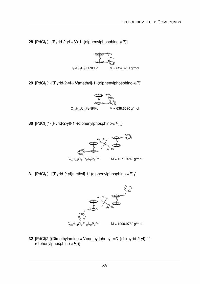
LIST OF NUMBERED COMPOUNDS
28 [PdCl2(1-(Pyrid-2-yl-κN)-1’-(diphenylphosphino-κP)]
Fe
PPh2
N
PdCl2
C27H22Cl2FeNPPd M = 624.6251 g/mol
29 [PdCl2(1-[(Pyrid-2-yl-κN)methyl]-1’-(diphenylphosphino-κP)]
Fe
PPh2
NPdCl2
C28H24Cl2FeNPPd M = 638.6520 g/mol
30 [PdCl2(1-(Pyrid-2-yl)-1’-(diphenylphosphino-κP)2]
Fe
P Fe
PPd
Ph
Cl
ClPh
Ph Ph
N
N
C54H44Cl2Fe2N2P2Pd M = 1071.9243 g/mol
31 [PdCl2(1-[(Pyrid-2-yl)methyl]-1’-(diphenylphosphino-κP)2]
Fe
P Fe
PPd
Ph
Cl
ClPh
Ph Ph
N
N
C56H48Cl2Fe2N2P2Pd M = 1099.9780 g/mol
32 [PdCl(2-[(Dimethylamino-κN)methyl]phenyl-κC1)(1-(pyrid-2-yl)-1’-(diphenylphosphino-κP))]
XV

APPENDIX
Fe
P
N
PdCl N
C36H34ClFeN2PPd M = 723.3731 g/mol
33 [PdCl(2-[(Dimethylamino-κN)methyl]phenyl-κC1)(1-[(pyrid-2-yl)methyl]-1’-(diphenylphosphino-κP)]
Fe
P
N
PdCl
N
C37H36ClFeN2PPd M = 737.4000 g/mol
34 [Pd(2-[(Dimethylamino-κN)methyl]phenyl-κC1)(1-(pyrid-2-yl-κN)-1’-(diphenylphosphino-κP))][ClO4]
NFe
PPh
Ph Pd
ClO4
N
C36H34ClFeN2O4PPd M = 787.3707 g/mol
34a [(Pd(2-[(Dimethylamino-κN)methyl]phenyl-κC1)(1-(pyrid-2-yl-κN)-1’-(diphenylphosphino-κP))][Cl]
NFe
PPh
Ph Pd
Cl
N
C36H34ClFeN2PPd M = 723.3731 g/mol
XVI

List of X-ray Data
35 [Pd(2-[(Dimethylamino-κN)methyl]phenyl-κC1)(1-[(pyrid-2-yl-κN)methyl]-1’-(diphenylphosphino-κP)][ClO4]
NFe
PPh
PhN
Pd
ClO4
C37H36ClFeN2O4PPd M = 801.3976 g/mol
List of X-ray Data
X-ray Data of 1
Table 1. Crystal data and structure refinement for i0540a.
Code i0540aEmpirical formula C27H22FeNPFormula weight 447.28Temperature 173(2) KWavelength 0.71073 Acrystal system Triclinicspacegoup P 1Unit cell dimensions a = 10.1437(9) A α = 72.013(6)◦
b = 11.0401(9) A β = 85.964(7)◦
c = 10.9432(9) A γ = 66.324(6)◦
Volume 1065.47(16) A3
Z 2Density (calculated) 1.394 g/cm3
Absorption coefficient 0.797 mm−1
F (000) 464Crystal size 0.49 mm × 0.39 mm × 0.24 mmθ-range for data collection 1.96→ 25.22◦
Index ranges -12→ h→12, -13→ k →13, -13→ l→13Reflections collected 9178Independent reflections 3636 [Rint = 0.0340]Reflections observed 3430Absorption correction IntegrationMax. and min. transmission 0.8225 and 0.7162Refinement method full-matrix least-squares against F 2
Data / restraints / parameters 3636 / 0 / 271Goodness-of-fit on F 2 1.088Final R indices [I > 2σ(I)] R1 = 0.0264, wR2 = 0.0732R indices (all data) R1 = 0.0278, wR2 = 0.0739Largest diff. peak and hole 0.311 and -0.357 e/A3
Table 2. Atomic coordinates (· 104) and equivalent isotropic displacement parameters (A2 · 103) for i0540a. Ueq is defined as onethird of the trace of the orthogonalized Uij tensor.
XVII

APPENDIX
x y z Ueq
C(1) 678(2) 2320(2) 9675(1) 32(1)C(2) 1691(2) 2407(2) 10467(2) 38(1)C(3) 3027(2) 1242(2) 10570(2) 44(1)C(4) 2863(2) 441(2) 9844(2) 43(1)C(5) 1421(2) 1097(2) 9288(2) 36(1)C(6) 2583(2) 2893(2) 6713(1) 32(1)C(7) 1673(2) 4106(2) 7056(2) 36(1)C(8) 2508(2) 4344(2) 7877(2) 45(1)C(9) 3920(2) 3284(2) 8055(2) 46(1)C(10) 3974(2) 2383(2) 7349(2) 37(1)C(11) 2223(2) 2334(2) 5786(1) 30(1)C(12) 795(2) 2719(2) 5375(2) 36(1)C(13) 527(2) 2249(2) 4431(2) 40(1)C(14) 1685(2) 1394(2) 3918(2) 42(1)C(15) 3064(2) 1028(2) 4392(2) 41(1)C(16) -2092(2) 2810(2) 10638(1) 30(1)C(17) -1400(2) 1791(2) 11776(2) 39(1)C(18) -2182(2) 1330(2) 12773(2) 48(1)C(19) -3676(2) 1879(2) 12650(2) 49(1)C(20) -4383(2) 2880(2) 11509(2) 47(1)C(21) -3602(2) 3344(2) 10518(2) 37(1)C(22) -1870(2) 3452(2) 7947(1) 32(1)C(23) -2504(2) 4652(2) 6903(2) 38(1)C(24) -3120(2) 4600(2) 5833(2) 48(1)C(25) -3090(2) 3359(2) 5783(2) 51(1)C(26) -2471(2) 2159(2) 6817(2) 47(1)C(27) -1880(2) 2204(2) 7900(2) 37(1)N 3352(2) 1486(1) 5305(1) 37(1)P -1161(1) 3632(1) 9352(1) 31(1)Fe 2428(1) 2436(1) 8681(1) 30(1)
X-ray Data of 2
Table 1. Crystal data and structure refinement for i0653.
Code i0653Empirical formula C27H24FeNPFormula weight 449.29Temperature 173(2) KWavelength 0.71073 Acrystal system Triclinicspacegroup P 1Unit cell dimensions a = 10.1847(12) A α = 72.890(9)◦
b = 11.0585(12) A β = 85.100(10)◦
c = 10.9491(13) A γ = 65.974(8)◦
Volume 1075.6(2) A3
Z 2Density (calculated) 1.387 g/cm3
Absorption coefficient 0.789 mm−1
F (000) 468Crystal size 0.25 × 0.21 × 0.02 mmθ-range for data collection 1.95→ 24.99◦
Index ranges -12→ h→10, -12→ k →12, -12→ l→13Reflections collected 7001Independent reflections 3555 [Rint = 0.0534]Reflections observed 2598Absorption correction IntegrationMax. and min. transmission 0.9589 and 0.5543
XVIII
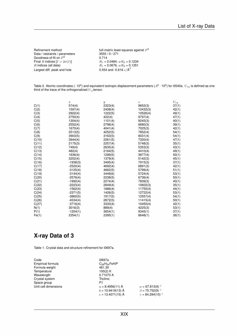
List of X-ray Data
Refinement method full-matrix least-squares against F 2
Data / restraints / parameters 3555 / 0 / 271Goodness-of-fit on F 2 0.714Final R indices [I > 2σ(I)] R1 = 0.0484, wR2 = 0.1234R indices (all data) R1 = 0.0676, wR2 = 0.1351Largest diff. peak and hole 0.554 and -0.616 e/A3
Table 2. Atomic coordinates (· 104) and equivalent isotropic displacement parameters (A2 · 103) for i0540a. Ueq is defined as onethird of the trace of the orthogonalized Uij tensor.
x y z Ueq
C(1) 574(4) 2323(4) 9652(3) 37(1)C(2) 1597(4) 2408(4) 10432(3) 42(1)C(3) 2922(4) 1222(5) 10526(4) 49(1)C(4) 2750(4) 422(4) 9797(4) 47(1)C(5) 1304(4) 1101(4) 9245(3) 40(1)C(6) 2552(4) 2798(4) 6680(3) 39(1)C(7) 1670(4) 4041(4) 7025(3) 42(1)C(8) 2513(5) 4252(5) 7852(4) 54(1)C(9) 3903(5) 3163(5) 8031(4) 54(1)C(10) 3944(4) 2261(5) 7320(4) 47(1)C(11) 2175(3) 2257(4) 5748(3) 35(1)C(12) 749(4) 2635(4) 5353(3) 43(1)C(13) 482(4) 2164(5) 4410(4) 49(1)C(14) 1636(4) 1299(5) 3877(4) 50(1)C(15) 3252(4) 1379(4) 5142(3) 45(1)C(16) -1938(3) 3495(4) 7915(3) 37(1)C(17) -2520(4) 4692(4) 6891(3) 42(1)C(18) -3125(4) 4662(5) 5799(4) 51(1)C(19) -3144(4) 3448(6) 5724(4) 53(1)C(20) -2576(4) 2238(5) 6736(4) 50(1)C(21) -1992(4) 2274(4) 7839(3) 40(1)C(22) -2223(4) 2849(4) 10602(3) 35(1)C(23) -1562(4) 1886(4) 11755(4) 44(1)C(24) -2371(5) 1426(5) 12722(4) 53(1)C(25) -3860(5) 1917(5) 12557(4) 54(1)C(26) -4534(4) 2872(5) 11415(4) 50(1)C(27) -3716(4) 3333(4) 10450(4) 42(1)N(1) 3016(3) 889(4) 4225(3) 53(1)P(1) -1254(1) 3654(1) 9345(1) 37(1)Fe(1) 2354(1) 2395(1) 8648(1) 36(1)
X-ray Data of 3
Table 1. Crystal data and structure refinement for i0697a.
Code i0697aEmpirical formula C28H24FeNPFormula weight 461.30Temperature 100(2) KWavelength 0.71073 ACrystal system TriclinicSpace group P1Unit cell dimensions a = 8.4956(11) A α = 67.813(9) ◦
b = 10.9419(13) A β = 75.752(9) ◦
c = 13.4071(15) A γ = 84.294(10) ◦
XIX

APPENDIX
Volume 1118.5(2) A3
Z 2Density (calculated) 1.370 g/cm3
Absorption coefficient 0.761 mm−1
F (000) 480Crystal size 0.57 × 0.40 × 0.15 mmθ-range for data collection 1.68→ 25.00 ◦
Index ranges -8→ h→10, -13→ k →13, -15→ l→15Reflections collected 8622Independent reflections 3867 [Rint = 0.0501]Reflections observed 3663Absorption correction IntegrationMax. and min. transmission 0.8761 and 0.7248Refinement method Full-matrix least-squares on F 2
Data / restraints / parameters 3867 / 0 / 280Goodness-of-fit on F 2 1.056Final R indices [I > 2σ(I)] R1 = 0.0323, wR2 = 0.0892R indices (all data) R1 = 0.0336, wR2 = 0.0900Largest diff. peak and hole 0.383 and -0.445 e/A3
Table 2. Atomic coordinates (· 104) and equivalent isotropic displacement parameters (A2 · 103) for i0697a. Ueq is defined as onethird of the trace of the orthogonalized Uij tensor.
x y z Ueq
C(1) 1481(2) 2903(2) 2602(1) 25(1)C(2) 3077(2) 3098(2) 1879(1) 26(1)C(3) 3663(2) 4350(2) 1739(1) 29(1)C(4) 2440(2) 4941(2) 2362(2) 30(1)C(5) 1098(2) 4052(2) 2897(2) 28(1)C(6) 2781(2) 2888(2) 5029(1) 26(1)C(7) 2839(2) 1635(2) 4902(1) 29(1)C(8) 4371(2) 1504(2) 4217(1) 31(1)C(9) 5284(2) 2671(2) 3927(2) 30(1)C(10) 4313(2) 3520(2) 4427(1) 28(1)C(11) 1378(2) 3449(2) 5666(2) 31(1)C(12) 1611(2) 3315(2) 6792(1) 26(1)C(13) 761(2) 2383(2) 7768(2) 31(1)C(14) 985(2) 2321(2) 8778(2) 35(1)C(15) 2062(2) 3197(2) 8786(2) 33(1)C(16) 2863(2) 4093(2) 7775(2) 31(1)C(17) 1576(2) 244(2) 2764(1) 25(1)C(18) 2144(2) -755(2) 3618(2) 33(1)C(19) 3275(3) Y-1689(2) 3387(2) 40(1)C(20) 3829(2) -1633(2) 2305(2) 39(1)C(22) 2140(2) 283(2) 1677(2) 28(1)C(23) -1019(2) 1935(2) 2039(1) 24(1)C(24) -2040(2) 974(2) 2071(2) 29(1)C(25) -3010(2) 1272(2) 1305(2) 32(1)C(26) -2992(2) 2539(2) 501(2) 32(1)C(27) -2016(2) 3505(2) 478(2) 30(1)C(28) -1038(2) 3210(2) 1239(1) 27(1)N(1) 2664(2) 4163(2) 6792(1) 29(1)P(1) 150(1) 1483(1) 3123(1) 24(1)Fe(1) 3149(1) 3150(1) 3389(1) 23(1)C(21) 3262(2) -656(2) 1450(2) 33(1)
XX
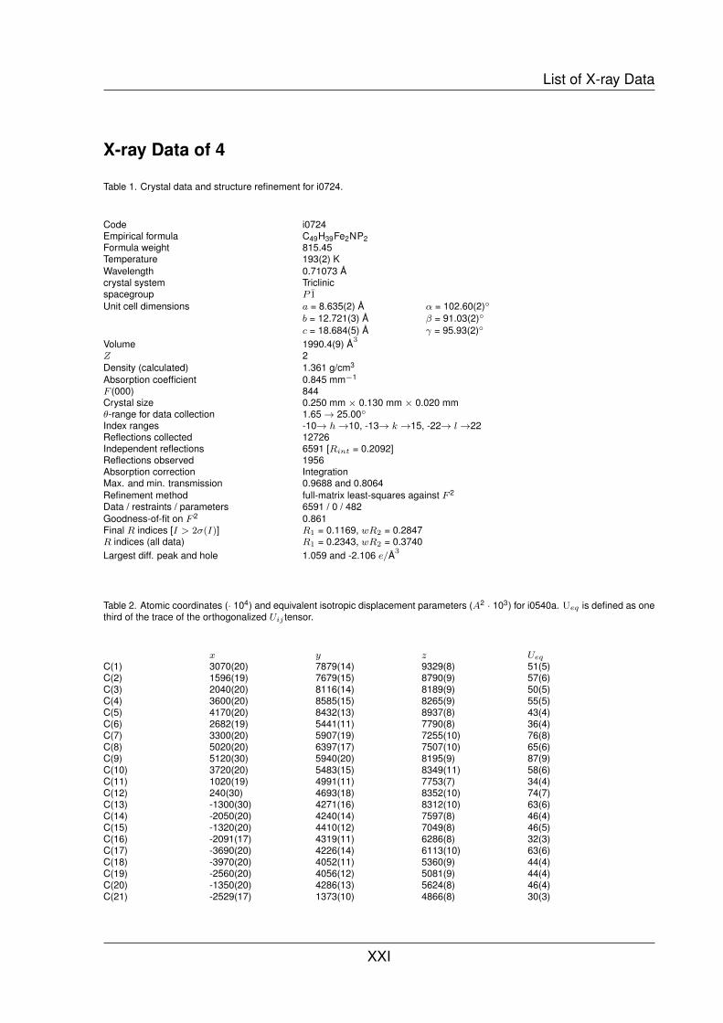
List of X-ray Data
X-ray Data of 4
Table 1. Crystal data and structure refinement for i0724.
Code i0724Empirical formula C49H39Fe2NP2Formula weight 815.45Temperature 193(2) KWavelength 0.71073 Acrystal system Triclinicspacegroup P 1Unit cell dimensions a = 8.635(2) A α = 102.60(2)◦
b = 12.721(3) A β = 91.03(2)◦
c = 18.684(5) A γ = 95.93(2)◦
Volume 1990.4(9) A3
Z 2Density (calculated) 1.361 g/cm3
Absorption coefficient 0.845 mm−1
F (000) 844Crystal size 0.250 mm × 0.130 mm × 0.020 mmθ-range for data collection 1.65→ 25.00◦
Index ranges -10→ h→10, -13→ k →15, -22→ l→22Reflections collected 12726Independent reflections 6591 [Rint = 0.2092]Reflections observed 1956Absorption correction IntegrationMax. and min. transmission 0.9688 and 0.8064Refinement method full-matrix least-squares against F 2
Data / restraints / parameters 6591 / 0 / 482Goodness-of-fit on F 2 0.861Final R indices [I > 2σ(I)] R1 = 0.1169, wR2 = 0.2847R indices (all data) R1 = 0.2343, wR2 = 0.3740Largest diff. peak and hole 1.059 and -2.106 e/A3
Table 2. Atomic coordinates (· 104) and equivalent isotropic displacement parameters (A2 · 103) for i0540a. Ueq is defined as onethird of the trace of the orthogonalized Uij tensor.
x y z Ueq
C(1) 3070(20) 7879(14) 9329(8) 51(5)C(2) 1596(19) 7679(15) 8790(9) 57(6)C(3) 2040(20) 8116(14) 8189(9) 50(5)C(4) 3600(20) 8585(15) 8265(9) 55(5)C(5) 4170(20) 8432(13) 8937(8) 43(4)C(6) 2682(19) 5441(11) 7790(8) 36(4)C(7) 3300(20) 5907(19) 7255(10) 76(8)C(8) 5020(20) 6397(17) 7507(10) 65(6)C(9) 5120(30) 5940(20) 8195(9) 87(9)C(10) 3720(20) 5483(15) 8349(11) 58(6)C(11) 1020(19) 4991(11) 7753(7) 34(4)C(12) 240(30) 4693(18) 8352(10) 74(7)C(13) -1300(30) 4271(16) 8312(10) 63(6)C(14) -2050(20) 4240(14) 7597(8) 46(4)C(15) -1320(20) 4410(12) 7049(8) 46(5)C(16) -2091(17) 4319(11) 6286(8) 32(3)C(17) -3690(20) 4226(14) 6113(10) 63(6)C(18) -3970(20) 4052(11) 5360(9) 44(4)C(19) -2560(20) 4056(12) 5081(9) 44(4)C(20) -1350(20) 4286(13) 5624(8) 46(4)C(21) -2529(17) 1373(10) 4866(8) 30(3)
XXI

APPENDIX
C(22) -1163(18) 1671(12) 5397(8) 34(3)C(23) -1900(20) 1754(11) 6089(9) 45(4)C(24) -3520(20) 1555(13) 6006(9) 49(5)C(25) -3850(20) 1375(11) 5229(7) 33(4)C(26) 5311(19) 7546(13) 10295(8) 43(4)C(27) 5920(20) 6500(15) 10140(9) 57(5)C(28) 7500(30) 6480(20) 10209(11) 72(6)C(29) 8450(20) 7420(20) 10494(11) 68(6)C(30) 7890(20) 8470(20) 10652(11) 76(7)C(31) 6250(20) 8431(15) 10583(9) 58(5)C(32) 2680(20) 8718(14) 10799(8) 46(4)C(33) 2830(30) 8656(15) 11558(9) 73(7)C(34) 2380(30) 9500(19) 12108(9) 92(9)C(35) 1910(30) 10453(19) 11879(10) 79(7)C(36) 1730(30) 10485(14) 11144(9) 68(6)C(37) 2030(20) 9473(15) 10608(9) 56(5)C(38) -2401(17) -239(12) 3576(7) 32(3)C(39) -2600(30) -699(15) 2807(10) 69(6)C(40) -2780(30) -1903(12) 2614(11) 65(6)C(41) -2770(20) -2518(18) 3071(12) 76(7)C(42) -2640(20) -2113(14) 3874(10) 59(6)C(43) -2420(20) -884(13) 4036(10) 49(5)C(44) -127(18) 1603(12) 3808(7) 38(4)C(45) 967(17) 855(12) 3893(8) 36(4)C(46) 2490(20) 1151(12) 3880(8) 48(5)C(47) 3080(20) 2135(16) 3882(9) 57(5)C(48) 2040(30) 2958(17) 3776(10) 67(6)C(49) 430(20) 2603(11) 3774(9) 43(4)N(1) 202(16) 4871(10) 7094(6) 39(3)P(1) 3134(5) 7465(4) 10142(2) 50(1)P(2) -2269(5) 1239(3) 3849(2) 40(1)Fe(1) 3465(3) 6994(2) 8272(1) 41(1)Fe(2) -2673(3) 2860(2) 5587(1) 36(1)
X-ray Data of 5a
Table 1. Crystal data and structure refinement for i0531.
Code i0531Empirical formula C27H22Cl2FeNPZnFormula weight 583.55Temperature 203(2) KWavelength 0.71073 Acrystal system Monoclinicspacegroup P21/cUnit cell dimensions a = 19.2132(16) A α = 90◦
b = 9.1831(6) A β = 101.679(7)◦
c = 28.737(3) A γ = 90◦
Volume 4965.3(7) A3
Z 8Density (calculated) 1.561 g/cm3
Absorption coefficient 1.846 mm−1
F (000) 2368θ-range for data collection 1.62→ 25.00◦
Index ranges -21→ h→22, -10→ k →7, -25→ l→34Reflections collected 9793Independent reflections 5901 [Rint = 0.0825]Reflections observed 2875Absorption correction Integration
XXII

List of X-ray Data
Refinement method full-matrix least-squares against F 2
Data / restraints / parameters 5901 / 0 / 595Goodness-of-fit on F 2 0.772Final R indices [I > 2σ(I)] R1 = 0.0506, wR2 = 0.1039R indices (all data) R1 = 0.1090, wR2 = 0.1163Largest diff. peak and hole 0.587 and -0.370 e/A3
Table 2. Atomic coordinates (· 104) and equivalent isotropic displacement parameters (A2 · 103) for i0531. Ueq is defined as onethird of the trace of the orthogonalized Uij tensor.
x y z Ueq
C(1) 2950(5) 4940(11) 6767(4) 41(3)C(2) 2236(5) 5221(11) 6825(4) 44(3)C(3) 2172(6) 6771(11) 6859(4) 46(3)C(4) 2836(6) 7411(12) 6840(4) 52(4)C(5) 3308(6) 6277(12) 6782(4) 50(3)C(6) 2824(5) 6496(10) 5605(4) 36(3)C(7) 2418(5) 5146(11) 5593(4) 38(3)C(8) 1729(5) 5543(13) 5648(4) 48(3)C(9) 1690(5) 7038(13) 5694(4) 52(3)C(10) 2380(5) 7652(11) 5681(4) 44(3)C(11) 3570(5) 6617(12) 5552(4) 44(3)C(12) 3762(5) 7891(11) 5351(5) 52(4)C(13) 4449(6) 7984(13) 5260(5) 64(4)C(14) 4895(6) 6861(14) 5353(5) 71(4)C(15) 4696(6) 5615(12) 5561(5) 61(4)C(16) 3826(5) 2504(13) 7166(4) 42(3)C(17) 4034(6) 3341(16) 7585(4) 65(4)C(18) 4437(7) 2748(17) 7994(5) 73(5)C(19) 4611(6) 1343(16) 8007(5) 61(4)C(20) 4442(7) 493(15) 7623(6) 67(4)C(21) 4040(6) 1095(14) 7194(5) 59(4)C(22) 2491(5) 2020(12) 6522(4) 42(3)C(23) 2235(5) 1527(11) 6924(4) 47(3)C(24) 1605(6) 747(13) 6855(5) 55(4)C(25) 1254(6) 414(12) 6419(5) 55(4)C(26) 1505(6) 880(11) 6021(4) 45(3)C(27) 2140(5) 1634(11) 6086(4) 40(3)C(28) 1930(5) 3820(10) 3263(4) 37(3)C(29) 2624(5) 4117(11) 3156(4) 42(3)C(30) 2632(5) 5602(11) 3024(3) 38(3)C(31) 1980(7) 6237(12) 3050(4) 65(4)C(32) 1535(5) 5164(11) 3200(4) 39(3)C(33) 2193(5) 6136(10) 4328(4) 35(3)C(34) 2549(5) 4790(10) 4399(4) 36(3)C(35) 3220(5) 4962(10) 4288(4) 40(3)C(36) 3290(5) 6403(11) 4142(4) 45(3)C(37) 2630(5) 7166(11) 4145(4) 44(3)C(38) 1450(5) 6475(10) 4385(4) 37(3)C(39) 1285(5) 7851(10) 4504(4) 39(3)C(40) 611(6) 8106(12) 4596(4) 51(4)C(41) 158(6) 7038(12) 4613(5) 57(4)C(42) 341(5) 5639(12) 4478(4) 49(3)C(43) 1108(5) 1056(11) 3122(4) 38(3)C(44) 775(5) -65(12) 3290(4) 46(3)C(45) 411(6) -1088(13) 2986(5) 58(4)C(46) 338(6) -937(12) 2501(5) 56(4)C(47) 639(5) 265(14) 2319(4) 53(4)C(48) 1026(5) 1247(11) 2624(4) 39(3)C(49) 2481(5) 1204(10) 3710(4) 37(3)C(50) 2725(5) 434(11) 3346(4) 47(3)C(51) 3399(6) -256(11) 3470(4) 49(3)
XXIII

APPENDIX
C(52) 3800(5) -182(12) 3913(4) 50(3)C(53) 3554(6) 562(12) 4272(5) 51(4)C(54) 2884(5) 1201(11) 4163(4) 45(3)N(1) 4027(4) 5477(9) 5662(3) 45(3)N(2) 982(4) 5321(9) 4367(3) 41(3)Cl(1) 5066(1) 2952(3) 6380(1) 53(1)Cl(2) 3582(2) 1946(3) 5380(1) 60(1)Cl(3) 1359(1) 1864(3) 4819(1) 51(1)Cl(4) -115(1) 2867(3) 3756(1) 55(1)Fe(1) 2465(1) 6224(2) 6230(1) 39(1)Fe(2) 2443(1) 5402(1) 3699(1) 34(1)Zn(1) 3951(1) 3427(1) 5991(1) 38(1)Zn(2) 1017(1) 3135(1) 4153(1) 36(1)P(1) 3284(1) 3191(3) 6622(1) 39(1)P(2) 1661(1) 2248(3) 3554(1) 35(1)
X-ray Data of 5b
Table 1. Crystal data and structure refinement for i0544.
Code i0544Empirical formula C27H22Br2FeNPZnFormula weight 672.47Temperature 173(2) KWavelength 0.71073 Acrystal system Monoclinicspacegroup P21Unit cell dimensions a = 14.6245(6) A α = 90◦
b = 9.2633(4) A β = 102.744(3)◦
c = 19.3704(7) A γ = 90◦
Volume 2559.49(18) A3
Z 4Density (calculated) 1.745 g/cm3
Absorption coefficient 4.705 mm−1
F (000) 1328Crystal size 0.33 mm × 0.23 mm × 0.16 mmθ-range for data collection 1.43→ 25.19◦
Index ranges -17→ h→17, -11→ k →11, -22→ l→23Reflections collected 16734Independent reflections 8771 [Rint = 0.0449]Reflections observed 8329Absorption correction IntegrationMax. and min. transmission 0.4668 and 0.3410Refinement method full-matrix least-squares against F 2
Data / restraints / parameters 8771 / 1 / 596Goodness-of-fit on F 2 0.990Final R indices [I > 2σ(I)] R1 = 0.0210, wR2 = 0.0512R indices (all data) R1 = 0.0225, wR2 = 0.0516Absolute structure parameter 0.00Extinction coefficient 0.00070(15)Largest diff. peak and hole 0.340 and -0.362 e/A3
Table 2. Atomic coordinates (· 104) and equivalent isotropic displacement parameters (A2 · 103) for i0544. Ueq is defined as onethird of the trace of the orthogonalized Uij tensor.
x y z Ueq
XXIV

List of X-ray Data
C(1) -3521(2) 8108(3) 7810(2) 26(1)C(2) -3704(2) 6772(4) 7428(2) 27(1)C(3) -3449(3) 6949(4) 6765(2) 33(1)C(4) -3119(3) 8391(4) 6724(2) 34(1)C(5) -3160(2) 9110(4) 7365(2) 31(1)C(6) -3723(2) 8420(3) 8505(2) 26(1)C(7) -3985(2) 9817(4) 8654(2) 34(1)C(8) -4216(3) 10122(4) 9295(2) 42(1)C(9) -4206(3) 9008(5) 9768(2) 49(1)C(10) -3935(3) 7657(4) 9604(2) 44(1)C(11) -1512(2) 5685(3) 8081(2) 27(1)C(12) -1339(2) 6988(4) 8497(2) 32(1)C(13) -996(2) 8055(4) 8082(2) 36(1)C(14) -956(2) 7437(4) 7419(2) 37(1)C(15) -1274(2) 5976(4) 7408(2) 32(1)C(16) -1329(2) 2931(3) 8849(2) 29(1)C(17) -1692(3) 1773(4) 9172(2) 39(1)C(18) -1097(3) 761(4) 9560(2) 45(1)C(19) -132(3) 887(4) 9638(2) 46(1)C(20) 229(3) 2036(5) 9324(2) 45(1)C(21) -364(3) 3057(4) 8935(2) 36(1)C(22) -2552(2) 3192(3) 7489(2) 27(1)C(23) -3499(2) 3157(3) 7144(2) 30(1)C(24) -3765(3) 2484(4) 6488(2) 33(1)C(25) -3112(3) 1832(4) 6175(2) 37(1)C(26) -2178(3) 1822(4) 6526(2) 35(1)C(27) -1894(2) 2490(4) 7176(2) 32(1)C(28) 3675(2) 272(4) 7119(2) 28(1)C(29) 3710(2) 1517(4) 7570(2) 29(1)C(30) 3607(3) 1027(4) 8247(2) 35(1)C(31) 3518(3) -504(4) 8226(2) 37(1)C(32) 3567(2) -983(4) 7537(2) 35(1)C(33) 3828(2) 250(4) 6388(2) 28(1)C(34) 4286(3) -943(4) 6181(2) 41(1)C(35) 4506(3) -978(5) 5525(2) 47(1)C(36) 4267(3) 190(5) 5080(2) 43(1)C(37) 3796(2) 1329(4) 5300(2) 33(1)C(38) 1408(2) 1754(4) 7033(2) 28(1)C(39) 1331(2) 1384(4) 7743(2) 33(1)C(40) 1231(2) -145(4) 7772(2) 38(1)C(41) 1247(3) -717(4) 7088(2) 39(1)C(42) 1359(2) 441(4) 6639(2) 35(1)C(43) 607(2) 4222(4) 6196(2) 33(1)C(44) 572(3) 5679(4) 5994(2) 41(1)C(45) -241(3) 6276(5) 5600(2) 50(1)C(46) -1027(3) 5424(5) 5389(2) 51(1)C(47) -1000(3) 3972(6) 5561(2) 56(1)C(48) -184(3) 3371(5) 5970(2) 43(1)C(49) 1901(2) 4631(3) 7522(2) 28(1)C(50) 2803(2) 5008(4) 7877(2) 32(1)C(51) 2953(3) 5799(4) 8498(2) 37(1)C(52) 2201(3) 6238(4) 8774(2) 40(1)C(53) 1295(3) 5908(4) 8420(2) 39(1)C(54) 1140(2) 5095(4) 7802(2) 36(1)N(1) -3669(2) 7342(3) 8986(2) 31(1)N(2) 3572(2) 1380(3) 5944(1) 28(1)P(1) -2158(1) 4183(1) 8318(1) 25(1)P(2) 1693(1) 3507(1) 6729(1) 26(1)Fe(7) 2464(1) 418(1) 7481(1) 27(1)Fe(8) -2307(1) 7328(1) 7574(1) 25(1)Zn(1) -3254(1) 5156(1) 8979(1) 26(1)Zn(2) 2912(1) 3400(1) 6060(1) 25(1)Br(1) -4704(1) 3918(1) 8651(1) 36(1)Br(2) -2419(1) 4833(1) 10180(1) 39(1)Br(3) 2079(1) 4077(1) 4892(1) 36(1)
XXV

APPENDIX
Br(4) 4194(1) 5002(1) 6441(1) 35(1)
X-ray Data of 6b
Table 1. Crystal data and structure refinement for i0501.
Code i0501Empirical formula C27H22Br2CdFeNPFormula weight 719.50Temperature 203(2) KWavelength 0.71073 Acrystal system Monoclinicspacegroup P21/cUnit cell dimensions a = 14.8184(13) A α = 90◦
b = 9.3495(6) A β = 91.972(7)◦
c = 18.8737(17) A γ = 90◦
Volume 2613.3(4) A3
Z 4Density (calculated) 1.829 g/cm3
Absorption coefficient 4.504 mm−1
F (000) 1400Crystal size 0.32 mm × 0.20 mm × 0.02 mmθ-range for data collection 1.37→ 25.00◦
Index ranges -17→ h→17, -11→ k →11, -22→ l→22Reflections collected 16395Independent reflections 4593 [Rint = 0.0789]Reflections observed 3022Absorption correction IntegrationMax. and min. transmission 0.9124 and 0.3289Refinement method full-matrix least-squares against F 2
Data / restraints / parameters 4593 / 0 / 298Goodness-of-fit on F 2 0.779Final R indices [I > 2σ(I)] R1 = 0.0310, wR2 = 0.0601R indices (all data) R1 = 0.0550, wR2 = 0.0632Largest diff. peak and hole 0.728 and -0.450 e/A3
Table 2. Atomic coordinates (· 104) and equivalent isotropic displacement parameters (A2 · 103) for i0501. Ueq is defined as onethird of the trace of the orthogonalized Uij tensor.
x y z Ueq
C(1) 6736(3) 1948(5) 251(2) 43(1)C(2) 6381(3) 2980(5) 747(3) 50(1)C(3) 5659(3) 2341(6) 1097(3) 54(1)C(4) 5545(3) 935(6) 830(3) 49(1)C(5) 6196(3) 690(5) 317(2) 40(1)C(6) 7581(3) -464(5) 1640(2) 39(1)C(7) 6961(3) -46(5) 2156(2) 40(1)C(8) 7139(3) 1388(5) 2346(2) 48(1)C(9) 7860(3) 1873(5) 1952(3) 49(1)C(10) 8151(3) 756(5) 1498(2) 41(1)C(11) 7465(3) 2198(5) -244(2) 41(1)C(12) 7617(3) 3583(5) -498(3) 48(1)C(13) 8275(3) 3812(6) -979(3) 57(1)C(14) 8776(4) 2667(6) -1222(3) 56(1)C(15) 8597(3) 1349(6) -946(3) 52(1)
XXVI

List of X-ray Data
C(16) 6534(3) -2970(5) 1267(2) 36(1)C(17) 5893(3) -3076(5) 719(3) 41(1)C(18) 5060(3) -3690(5) 824(3) 46(1)C(19) 4856(3) -4200(5) 1491(3) 51(1)C(20) 5493(3) -4114(6) 2042(3) 54(1)C(21) 6328(3) -3514(5) 1937(3) 47(1)C(22) 8458(3) -3211(5) 1599(2) 38(1)C(23) 8703(3) -2957(5) 2310(2) 41(1)C(24) 9346(3) -3808(6) 2655(3) 49(1)C(25) 9726(3) -4925(6) 2308(3) 52(1)C(26) 9493(3) -5209(5) 1607(3) 52(1)C(27) 8861(3) -4349(5) 1248(3) 45(1)N(1) 7964(2) 1071(4) -468(2) 42(1)P(1) 7618(1) -2097(1) 1129(1) 36(1)Fe(1) 6826(1) 1206(1) 1278(1) 37(1)Br(1) 9635(1) -2066(1) -324(1) 55(1)Br(2) 6914(1) -2450(1) -1130(1) 52(1)Cd(1) 7985(1) -1359(1) -168(1) 40(1)
X-ray Data of 6c
Table 1. Crystal data and structure refinement for i0731.
Code i0731Empirical formula C27H22CdFeI2NPFormula weight 813.48Temperature 193(2) KWavelength 0.71073 Acrystal system Triclinicspacegroup P 1Unit cell dimensions a = 10.4638(10) A α = 72.904(6)◦
b = 10.5429(8) A β = 69.694(7)◦
c = 14.1699(11) A γ = 70.125(7)◦
Volume 1351.0(2) A3
Z 2Density (calculated) 2.000 g/cm3
Absorption coefficient 3.684 mm−1
F (000) 772Crystal size 0.36 mm × 0.19 mm × 0.04 mmθ-range for data collection 1.56→ 25.00◦
Index ranges -12→ h→12, -12→ k →12, -16→ l→15Reflections collected 8799Independent reflections 4481 [Rint = 0.0464]Reflections observed 3978Absorption correction IntegrationMax. and min. transmission 0.8511 and 0.2744Refinement method full-matrix least-squares against F 2
Data / restraints / parameters 4481 / 0 / 299Goodness-of-fit on F 2 1.111Final R indices [I > 2σ(I)] R1 = 0.0282, wR2 = 0.0758R indices (all data) R1 = 0.0324, wR2 = 0.0835Extinction coefficient 0.0041(4)Largest diff. peak and hole 0.793 and -0.845 e/A3
Table 2. Atomic coordinates (· 104) and equivalent isotropic displacement parameters (A2 · 103) for i0731. Ueq is defined as onethird of the trace of the orthogonalized Uij tensor.
XXVII

APPENDIX
x y z Ueq
C(1) 6107(4) 7162(4) 1238(3) 31(1)C(2) 4692(4) 7543(5) 1132(3) 41(1)C(3) 4453(5) 6395(5) 977(4) 43(1)C(4) 5687(5) 5282(5) 989(4) 43(1)C(5) 6712(4) 5743(4) 1152(3) 35(1)C(6) 5416(4) 4699(4) 3651(3) 36(1)C(7) 4923(4) 6126(5) 3756(3) 38(1)C(8) 3539(5) 6665(5) 3633(3) 47(1)C(9) 3145(5) 5610(6) 3466(4) 54(1)C(10) 4296(5) 4394(5) 3477(4) 45(1)C(11) 6765(4) 3727(4) 3766(3) 33(1)C(12) 6915(5) 2314(5) 4009(4) 45(1)C(13) 8142(6) 1401(5) 4186(4) 52(1)C(14) 9223(5) 1897(5) 4157(4) 48(1)C(15) 9030(5) 3316(5) 3897(4) 41(1)C(16) 5555(4) 9526(4) 2092(3) 34(1)C(17) 5271(4) 9609(4) 3106(3) 38(1)C(18) 4180(5) 10652(5) 3512(4) 45(1)C(19) 3366(5) 11638(5) 2917(4) 47(1)C(20) 3669(5) 11583(5) 1889(5) 52(1)C(21) 4745(5) 10539(5) 1479(4) 44(1)C(22) 7975(4) 8927(4) 333(3) 34(1)C(23) 8734(5) 8136(5) -410(4) 47(1)C(24) 9549(6) 8663(7) -1356(4) 66(2)C(25) 9596(6) 10012(7) -1557(5) 67(2)C(26) 8869(5) 10808(5) -823(5) 61(2)C(27) 8059(5) 10264(5) 128(5) 50(1)N(1) 7850(3) 4213(3) 3668(3) 34(1)P(1) 6959(1) 8121(1) 1565(1) 29(1)Fe(1) 4934(1) 5965(1) 2347(1) 32(1)Cd(1) 8408(1) 6330(1) 2768(1) 29(1)I(1) 8628(1) 7283(1) 4287(1) 41(1)I(2) 10892(1) 5202(1) 1428(1) 40(1)
X-ray Data of 7
Table 1. Crystal data and structure refinement for i0609a.
Code i0609aEmpirical formula C54H44CdCl2Fe2N2P2Formula weight 1077.85Temperature 173(2) KWavelength 0.71073 Acrystal system Orthorhombicspacegroup Fdd2Unit cell dimensions a = 49.456(5) A α = 90◦
b = 22.4070(14) A β = 90◦
c = 8.2534(7) A γ = 90◦
Volume 9146.0(13) A3
Z 8Density (calculated) 1.566 g/cm3
Absorption coefficient 1.314 mm−1
F (000) 4368Crystal size 0.43 mm × 0.04 mm × 0.03 mmθ-range for data collection 1.65→ 24.99◦
Index ranges -58→ h→58, -24→ k →24, -9→ l→9Reflections collected 14627Independent reflections 3764 [Rint = 0.0896]
XXVIII

List of X-ray Data
Reflections observed 2968Absorption correction IntegrationMax. and min. transmission 0.9679 and 0.6208Refinement method full-matrix least-squares against F 2
Data / restraints / parameters 3764 / 1 / 285Goodness-of-fit on F 2 0.919Final R indices [I > 2σ(I)] R1 = 0.0408, wR2 = 0.0759R indices (all data) R1 = 0.0568, wR2 = 0.0797Absolute structure parameter 0.01(3)Largest diff. peak and hole 0.470 and -0.950 e/A3
Table 2. Atomic coordinates (· 104) and equivalent isotropic displacement parameters (A2 · 103) for i0609a. Ueq is defined as onethird of the trace of the orthogonalized Uij tensor.
x y z Ueq
C(1) 1096(1) -465(3) 1267(8) 38(2)C(2) 831(1) -536(3) 579(7) 39(2)C(3) 692(1) -992(3) 1410(8) 43(2)C(4) 867(1) -1220(3) 2617(9) 43(2)C(5) 1115(1) -915(3) 2545(8) 39(2)C(6) 555(1) 393(3) 3213(7) 34(2)C(7) 477(1) -65(3) 4335(7) 38(2)C(8) 699(1) -179(3) 5361(9) 45(2)C(9) 914(1) 192(3) 4920(7) 39(2)C(10) 830(1) 551(3) 3604(7) 38(2)C(11) 1305(1) -30(4) 862(7) 38(2)C(12) 1249(1) 455(3) -167(8) 43(2)C(13) 1449(1) 865(4) -493(8) 48(2)C(14) 1705(1) 782(4) 179(10) 51(2)C(15) 1740(1) 295(4) 1166(9) 50(2)C(16) 134(1) 1270(3) 2691(8) 33(1)C(17) 4(1) 1702(3) 1770(7) 38(2)C(18) -166(1) 2114(3) 2478(9) 45(2)C(19) -213(1) 2096(4) 4149(9) 45(2)C(20) -85(1) 1667(3) 5088(10) 44(2)C(21) 83(1) 1263(4) 4393(8) 41(2)C(22) 537(1) 1118(3) 269(8) 29(1)C(23) 693(1) 1601(3) 804(8) 37(2)C(24) 832(1) 1954(3) -302(8) 42(2)C(25) 815(1) 1833(3) -1972(9) 43(2)C(26) 665(1) 1356(3) -2494(8) 43(2)C(27) 528(1) 991(3) -1400(7) 39(2)N(1) 1552(1) -118(3) 1528(7) 45(2)P(1) 332(1) 692(1) 1684(2) 31(1)Cl(1) 215(1) -715(1) -1664(2) 43(1)Fe(1) 803(1) -326(1) 2992(1) 33(1)Cd(1) 0 0 170(1) 33(1)
X-ray Data of 8b
Table 1. Crystal data and structure refinement for i0507a.
Code i0507aEmpirical formula C27H22Br2FeHgNPFormula weight 807.69Temperature 100(2) K
XXIX

APPENDIX
Wavelength 0.71073 Acrystal system Triclinicspacegroup P 1Unit cell dimensions a = 10.1471(10) A α = 104.921(8)◦
b = 10.3258(10) A β = 92.316(8)◦
c = 13.6179(13) A γ = 110.214(8)◦
Volume 1280.6(2) A3
Z 2Density (calculated) 2.095 g/cm3
Absorption coefficient 9.752 mm−1
F (000) 764Crystal size 0.18 mm × 0.16 mm × 0.05 mmθ-range for data collection 1.56→ 25.00◦
Index ranges -12→ h→11, -12→ k →12, -16→ l→16Reflections collected 9514Independent reflections 4485 [Rint = 0.0411]Reflections observed 4156Absorption correction IntegrationMax. and min. transmission 0.5998 and 0.2161Refinement method full-matrix least-squares against F 2
Data / restraints / parameters 4485 / 0 / 298Goodness-of-fit on F 2 1.002Final R indices [I > 2σ(I)] R1 = 0.0224, wR2 = 0.0529R indices (all data) R1 = 0.0249, wR2 = 0.0536Largest diff. peak and hole 1.628 and -1.305 e/A3
Table 2. Atomic coordinates (· 104) and equivalent isotropic displacement parameters (A2 · 103) for i0507a. Ueq is defined as onethird of the trace of the orthogonalized Uij tensor.
x y z Ueq
C(1) -2609(4) 1202(4) 3776(3) 20(1)C(2) -2932(5) -227(4) 3898(3) 24(1)C(3) -1626(5) -422(4) 4059(3) 27(1)C(4) -493(5) 863(4) 4046(3) 28(1)C(5) -1072(4) 1870(4) 3870(3) 23(1)C(6) -1175(4) 455(4) 1364(3) 20(1)C(7) -2702(4) -132(4) 1237(3) 22(1)C(8) -3124(5) -1542(4) 1384(3) 30(1)C(9) -1891(5) -1818(4) 1611(3) 31(1)C(10) -685(5) -597(4) 1599(3) 27(1)C(11) -262(4) 1865(4) 1247(3) 20(1)C(12) 1112(4) 2072(4) 1010(3) 25(1)C(13) 1915(4) 3355(4) 812(3) 28(1)C(14) 1327(4) 4390(4) 843(3) 26(1)C(15) -34(4) 4119(4) 1099(3) 26(1)C(16) -4079(4) 3048(4) 4694(3) 21(1)C(17) -2884(5) 3870(4) 5439(3) 28(1)C(18) -3033(6) 4670(5) 6404(3) 37(1)C(19) -4360(6) 4660(5) 6612(3) 35(1)C(20) -5542(5) 3867(4) 5867(4) 34(1)C(21) -5413(4) 3068(4) 4899(3) 26(1)C(22) -5473(4) 553(4) 2882(3) 22(1)C(23) -5963(4) 241(4) 1838(3) 24(1)C(24) -7189(5) -953(4) 1374(3) 31(1)C(25) -7956(5) -1802(4) 1955(4) 36(1)C(26) -7499(5) -1474(4) 3007(4) 35(1)C(27) -6264(5) -307(4) 3465(3) 29(1)N -822(3) 2902(3) 1323(2) 22(1)P -3800(1) 2016(1) 3468(1) 18(1)Fe -1857(1) 6(1) 2683(1) 19(1)Br(1) -799(1) 5905(1) 3491(1) 24(1)
XXX

List of X-ray Data
Br(2) -3938(1) 3837(1) 806(1) 27(1)Hg -2731(1) 3476(1) 2344(1) 19(1)
X-ray Data of 8c
Table 1. Crystal data and structure refinement for i0800.
Code i0800Empirical formula C56H46Cl6Fe2Hg2I4N2P2Formula weight 2042.07Temperature 100(2) KWavelength 0.71073 Acrystal system Triclinicspacegroup P 1Unit cell dimensions a = 9.5405(11) A α = 79.405(11)◦
b = 9.8309(13) A β = 86.852(10)◦
c = 16.591(2) A γ = 89.624(10)◦
Volume 1527.2(3) A3
Z 1Density (calculated) 2.220 g/cm3
Absorption coefficient 7.851 mm−1
F (000) 952Crystal size 0.11 mm × 0.11 mm × 0.01 mmθ-range for data collection 1.25→ 25.00◦
Index ranges -10→ h→11, -11→ k →11, -19→ l→19Reflections collected 10365Independent reflections 5303 [Rint = 0.0879]Reflections observed 3609Absorption correction IntegrationMax. and min. transmission 0.9181 and 0.4544Refinement method full-matrix least-squares against F 2
Data / restraints / parameters 5303 / 0 / 335Goodness-of-fit on F 2 0.931Final R indices [I > 2σ(I)] R1 = 0.0538, wR2 = 0.1220R indices (all data) R1 = 0.0832, wR2 = 0.1345Extinction coefficient 0.0026(3)Largest diff. peak and hole 1.149 and -1.866 e/A3
Table 2. Atomic coordinates (· 104) and equivalent isotropic displacement parameters (A2 · 103) for i0800. Ueq is defined as onethird of the trace of the orthogonalized Uij tensor.
x y z Ueq
C(1) 10464(13) 1964(12) 3040(8) 41(3)C(2) 9356(15) 1160(14) 3513(10) 54(3)C(3) 9138(14) -23(12) 3186(8) 42(3)C(4) 10069(14) 3(13) 2489(9) 46(3)C(5) 10905(14) 1232(12) 2409(8) 43(3)C(6) 6849(13) 1624(13) 1934(8) 41(3)C(7) 7879(14) 1733(13) 1265(8) 43(3)C(8) 8617(14) 3005(13) 1190(9) 46(3)C(9) 8063(14) 3679(12) 1832(8) 44(3)C(10) 6958(12) 2834(12) 2296(8) 39(3)C(11) 5788(13) 492(13) 2221(8) 42(3)C(12) 4820(12) 530(13) 2862(8) 39(3)C(13) 3828(14) -496(13) 3061(9) 47(3)
XXXI
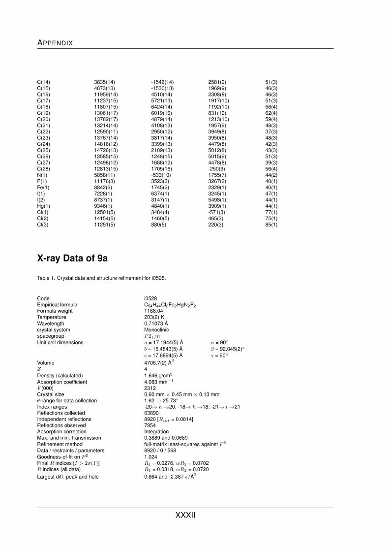
APPENDIX
C(14) 3835(14) -1546(14) 2581(9) 51(3)C(15) 4873(13) -1530(13) 1969(9) 46(3)C(16) 11959(14) 4510(14) 2308(8) 46(3)C(17) 11237(15) 5721(13) 1917(10) 51(3)C(18) 11807(15) 6424(14) 1192(10) 56(4)C(19) 13061(17) 6019(16) 831(10) 62(4)C(20) 13782(17) 4879(14) 1213(10) 59(4)C(21) 13214(14) 4108(13) 1957(9) 48(3)C(22) 12590(11) 2950(12) 3949(8) 37(3)C(23) 13767(14) 3817(14) 3950(8) 48(3)C(24) 14816(12) 3399(13) 4479(8) 42(3)C(25) 14726(13) 2109(13) 5012(8) 43(3)C(26) 13585(15) 1248(15) 5015(9) 51(3)C(27) 12496(12) 1688(12) 4478(8) 39(3)C(28) 12813(15) 1705(16) -250(9) 56(4)N(1) 5858(11) -533(10) 1755(7) 44(2)P(1) 11176(3) 3523(3) 3267(2) 40(1)Fe(1) 8842(2) 1745(2) 2329(1) 40(1)I(1) 7228(1) 6374(1) 3245(1) 47(1)I(2) 8737(1) 3147(1) 5498(1) 44(1)Hg(1) 9346(1) 4840(1) 3909(1) 44(1)Cl(1) 12501(5) 3484(4) -571(3) 77(1)Cl(2) 14154(5) 1460(5) 465(3) 75(1)Cl(3) 11251(5) 880(5) 220(3) 85(1)
X-ray Data of 9a
Table 1. Crystal data and structure refinement for i0528.
Code i0528Empirical formula C54H44Cl2Fe2HgN2P2Formula weight 1166.04Temperature 203(2) KWavelength 0.71073 Acrystal system Monoclinicspacegroup P21/nUnit cell dimensions a = 17.1944(5) A α = 90◦
b = 15.4843(5) A β = 92.045(2)◦
c = 17.6894(5) A γ = 90◦
Volume 4706.7(2) A3
Z 4Density (calculated) 1.646 g/cm3
Absorption coefficient 4.083 mm−1
F (000) 2312Crystal size 0.60 mm × 0.45 mm × 0.13 mmθ-range for data collection 1.62→ 25.73◦
Index ranges -20→ h→20, -18→ k →18, -21→ l→21Reflections collected 63890Independent reflections 8920 [Rint = 0.0814]Reflections observed 7954Absorption correction IntegrationMax. and min. transmission 0.3889 and 0.0689Refinement method full-matrix least-squares against F 2
Data / restraints / parameters 8920 / 0 / 568Goodness-of-fit on F 2 1.024Final R indices [I > 2σ(I)] R1 = 0.0276, wR2 = 0.0702R indices (all data) R1 = 0.0318, wR2 = 0.0720Largest diff. peak and hole 0.864 and -2.387 e/A3
XXXII

List of X-ray Data
Table 2. Atomic coordinates (· 104) and equivalent isotropic displacement parameters (A2 · 103) for i0528. Ueq is defined as onethird of the trace of the orthogonalized Uij tensor.
x y z Ueq
C(1) 2736(1) 9636(2) -1452(2) 31(1)C(2) 2600(2) 10322(2) -1997(2) 36(1)C(3) 3113(2) 10192(2) -2601(2) 41(1)C(4) 3561(2) 9439(2) -2447(2) 43(1)C(5) 3331(2) 9091(2) -1746(2) 37(1)C(6) 2166(2) 8472(2) -3474(2) 39(1)C(7) 1590(2) 9135(2) -3382(2) 43(1)C(8) 1206(2) 8973(2) -2696(2) 49(1)C(9) 1534(2) 8221(2) -2362(2) 49(1)C(10) 2128(2) 7905(2) -2831(2) 43(1)C(11) 2705(2) 8389(2) -4101(2) 40(1)C(12) 2656(2) 8941(2) -4720(2) 57(1)C(13) 3154(2) 8809(2) -5316(2) 64(1)C(14) 3692(2) 8155(2) -5260(2) 57(1)C(15) 3710(2) 7652(2) -4626(2) 55(1)C(16) 2579(1) 10319(2) 68(1) 31(1)C(17) 2923(2) 11076(2) -179(2) 43(1)C(18) 3096(2) 11742(2) 335(2) 52(1)C(19) 2918(2) 11658(2) 1082(2) 53(1)C(20) 2603(2) 10895(2) 1338(2) 54(1)C(21) 2432(2) 10224(2) 837(2) 42(1)C(22) 1223(1) 9662(2) -765(1) 32(1)C(23) 962(2) 10491(2) -967(2) 38(1)C(24) 178(2) 10635(2) -1141(2) 48(1)C(25) -347(2) 9959(2) -1118(2) 50(1)C(26) -102(2) 9140(2) -904(2) 46(1)C(27) 684(2) 8982(2) -725(2) 38(1)C(28) 4457(2) 7999(2) 732(2) 34(1)C(29) 4337(2) 8908(2) 864(2) 38(1)C(30) 4980(2) 9216(2) 1315(2) 44(1)C(31) 5500(2) 8517(2) 1466(2) 45(1)C(32) 5185(2) 7759(2) 1104(2) 40(1)C(33) 4160(2) 8694(2) 2902(2) 38(1)C(34) 4609(2) 7910(2) 2965(2) 45(1)C(35) 4179(2) 7241(2) 2588(2) 48(1)C(36) 3464(2) 7602(2) 2293(2) 46(1)C(37) 3454(2) 8495(2) 2486(2) 41(1)C(38) 4362(2) 9547(2) 3231(2) 39(1)C(39) 4994(2) 9660(2) 3732(2) 48(1)C(40) 5117(2) 10463(2) 4060(2) 60(1)C(41) 4610(2) 11133(2) 3881(2) 63(1)C(42) 3999(2) 10965(2) 3368(2) 60(1)C(43) 3788(2) 6264(2) 619(2) 38(1)C(44) 3122(2) 5993(2) 988(2) 42(1)C(45) 3131(2) 5230(2) 1407(2) 52(1)C(46) 3803(2) 4726(2) 1441(2) 62(1)C(47) 4448(2) 4974(2) 1060(2) 64(1)C(48) 4455(2) 5741(2) 649(2) 52(1)C(49) 4233(2) 7216(2) -734(2) 36(1)C(50) 3877(2) 6684(2) -1290(2) 46(1)C(51) 4161(2) 6673(3) -2015(2) 62(1)C(52) 4783(2) 7196(3) -2202(2) 66(1)C(53) 5133(2) 7720(3) -1663(2) 60(1)C(54) 4865(2) 7727(2) -929(2) 47(1)Cl(1) 1695(1) 8068(1) 1181(1) 46(1)Cl(2) 1768(1) 6776(1) -719(1) 54(1)Fe(1) 2383(1) 9145(1) -2486(1) 33(1)Fe(2) 4415(1) 8229(1) 1856(1) 34(1)Hg(1) 2482(1) 7998(1) -16(1) 34(1)N(1) 3236(2) 7744(2) -4044(2) 48(1)
XXXIII

APPENDIX
N(2) 3864(2) 10198(2) 3043(2) 51(1)P(1) 2255(1) 9457(1) -579(1) 29(1)P(2) 3795(1) 7331(1) 183(1) 32(1)
X-ray Data of 9b
Table 1. Crystal data and structure refinement for i0634.
Code i0634Empirical formula C54H44Br2Fe2HgN2P2Formula weight 1254.96Temperature 173(2) KWavelength 0.71073 Acrystal system Monoclinicspacegroup P21/nUnit cell dimensions a = 17.4068(8) A α = 90◦
b = 15.5806(5) A β = 92.161(4)◦
c = 17.5380(8) A γ = 90◦
Volume 4753.1(3) A3
Z 4Density (calculated) 1.754 g/cm3
Absorption coefficient 5.612 mm−1
F (000) 2456Crystal size 0.42 mm × 0.283 mm × 0.17 mmθ-range for data collection 1.62→ 25.00◦
Index ranges -20→ h→20, -18→ k →17, -20→ l→20Reflections collected 30291Independent reflections 8305 [Rint = 0.0776]Reflections observed 7436Absorption correction IntegrationMax. and min. transmission 0.7179 and 0.3150Refinement method full-matrix least-squares against F 2
Data / restraints / parameters 8305 / 0 / 569Goodness-of-fit on F 2 1.085Final R indices [I > 2σ(I)] R1 = 0.0474, wR2 = 0.1142R indices (all data) R1 = 0.0513, wR2 = 0.1165Extinction coefficient 0.00075(10)Largest diff. peak and hole 4.258 and -2.843 e/A3
Table 2. Atomic coordinates (· 104) and equivalent isotropic displacement parameters (A2 · 103) for i0634. Ueq is defined as onethird of the trace of the orthogonalized Uij tensor.
x y z Ueq
C(1) 9469(3) 2001(3) 5772(3) 29(1)C(2) 10201(3) 2237(4) 6144(3) 35(1)C(3) 10521(3) 1495(4) 6491(3) 40(1)C(4) 10006(3) 795(4) 6357(3) 40(1)C(5) 9361(3) 1101(4) 5910(3) 36(1)C(6) 9213(3) 1298(4) 7964(3) 33(1)C(7) 9691(3) 2066(4) 8030(3) 38(1)C(8) 9271(3) 2752(4) 7670(3) 42(1)C(9) 8548(3) 2431(4) 7377(3) 40(1)C(10) 8514(3) 1538(4) 7558(3) 34(1)C(11) 9394(3) 441(4) 8271(3) 35(1)C(12) 10023(3) 299(4) 8765(3) 45(1)
XXXIV

List of X-ray Data
C(13) 10134(4) -518(5) 9077(3) 52(2)C(14) 9621(4) -1164(5) 8870(4) 55(2)C(15) 9015(4) -967(5) 8359(4) 57(2)C(16) 8790(3) 3718(4) 5665(3) 36(1)C(17) 9448(3) 4251(4) 5676(4) 47(1)C(18) 9440(4) 5024(5) 6074(4) 58(2)C(19) 8801(4) 5267(4) 6469(4) 57(2)C(20) 8142(3) 4758(4) 6440(3) 46(1)C(21) 8140(3) 3985(4) 6029(3) 37(1)C(22) 9228(3) 2769(4) 4294(3) 32(1)C(23) 9838(3) 2254(4) 4088(3) 42(1)C(24) 10098(3) 2269(5) 3349(3) 52(2)C(25) 9750(4) 2814(5) 2809(3) 58(2)C(26) 9146(3) 3344(5) 3020(3) 55(2)C(27) 8875(3) 3321(4) 3753(3) 42(1)C(28) 7743(2) 378(3) 3566(2) 28(1)C(29) 8315(3) 943(4) 3271(3) 35(1)C(30) 8549(3) 611(4) 2560(3) 40(1)C(31) 8119(3) -153(4) 2407(3) 39(1)C(32) 7624(3) -309(3) 3015(3) 33(1)C(33) 7144(3) 1522(4) 1529(3) 36(1)C(34) 6590(3) 855(4) 1627(3) 40(1)C(35) 6208(3) 1000(4) 2320(3) 43(1)C(36) 6521(3) 1757(4) 2663(3) 43(1)C(37) 7094(3) 2087(4) 2184(3) 37(1)C(38) 7673(3) 1620(4) 904(3) 37(1)C(39) 7630(3) 1084(5) 264(3) 47(1)C(40) 8110(4) 1226(5) -328(3) 57(2)C(41) 8647(4) 1892(4) -269(4) 53(2)C(42) 8657(4) 2368(4) 378(3) 50(1)C(43) 6258(2) 337(3) 4270(2) 28(1)C(44) 6000(3) -480(4) 4040(3) 36(1)C(45) 5222(3) -613(4) 3861(3) 42(1)C(46) 4696(3) 56(4) 3904(3) 43(1)C(47) 4949(3) 858(4) 4139(3) 39(1)C(48) 5727(3) 1007(4) 4331(3) 34(1)C(49) 7602(2) -337(4) 5087(3) 30(1)C(50) 7981(3) -1062(4) 4823(3) 39(1)C(51) 8157(3) -1746(4) 5321(4) 46(1)C(52) 7957(3) -1710(4) 6073(3) 46(1)C(53) 7596(4) -985(4) 6343(3) 49(1)C(54) 7423(3) -301(4) 5855(3) 40(1)N(36) 8880(3) -183(3) 8068(3) 47(1)N(54) 8192(3) 2265(3) 968(3) 43(1)P(1) 8806(1) 2655(1) 5230(1) 28(1)P(2) 7279(1) 535(1) 4459(1) 26(1)Fe(1) 9462(1) 1777(1) 6911(1) 30(1)Fe(2) 7377(1) 860(1) 2530(1) 30(1)Br(1) 6710(1) 1875(1) 6329(1) 38(1)Br(2) 6772(1) 3265(1) 4338(1) 45(1)Hg(1) 7510(1) 1971(1) 5055(1) 30(1)
X-ray Data of 10
Table 1. Crystal data and structure refinement for i0818.
Code i0818Empirical formula C27H22Br2FeNPZnFormula weight 672.47Temperature 173(2) K
XXXV

APPENDIX
Wavelength 0.71073 Acrystal system Monoclinicspacegroup P21/cUnit cell dimensions a = 15.3284(10) A α = 90◦
b = 9.0578(6) A β = 96.220(5)◦
c = 18.4976(10) A γ = 90◦
Volume 2553.1(3) A3
Z 4Density (calculated) 1.749 g/cm3
Absorption coefficient 4.716 mm−1
F (000) 1328Crystal size 0.13 mm × 0.12 mm × 0.09 mmθ-range for data collection 1.34→ 25.00◦
Index ranges -18→ h→18, -10→ k →10, -21→ l→21Reflections collected 16069Independent reflections 4461 [Rint = 0.0776]Reflections observed 3378Absorption correction IntegrationMax. and min. transmission 0.6663 and 0.5464Refinement method full-matrix least-squares against F 2
Data / restraints / parameters 4461 / 0 / 298Goodness-of-fit on F 2 0.930Final R indices [I > 2σ(I)] R1 = 0.0408, wR2 = 0.0861R indices (all data) R1 = 0.0595, wR2 = 0.0915Largest diff. peak and hole 2.014 and -0.747 e/A3
Table 2. Atomic coordinates (· 104) and equivalent isotropic displacement parameters (A2 · 103) for i0818. Ueq is defined as onethird of the trace of the orthogonalized Uij tensor.
x y z Ueq
C(1) 6518(3) 3105(5) 1737(3) 23(1)C(2) 6360(3) 2789(5) 2476(3) 26(1)C(3) 5646(3) 3682(6) 2647(3) 30(1)C(4) 5351(3) 4552(6) 2021(3) 31(1)C(5) 5880(3) 4205(5) 1458(3) 27(1)C(6) 7728(3) 5575(5) 3084(3) 24(1)C(7) 6960(3) 6392(5) 3218(3) 29(1)C(8) 6677(4) 7204(5) 2570(3) 32(1)C(9) 7257(3) 6875(5) 2036(3) 30(1)C(10) 7902(3) 5870(5) 2350(3) 24(1)C(11) 8266(3) 4614(5) 3618(3) 22(1)C(12) 9182(3) 4716(5) 3706(3) 29(1)C(13) 9661(3) 3825(6) 4221(3) 31(1)C(14) 9224(3) 2859(6) 4638(3) 27(1)C(15) 7874(3) 3632(5) 4057(3) 26(1)C(16) 8177(3) 1614(5) 1879(3) 27(1)C(17) 8983(3) 2371(6) 1954(3) 32(1)C(18) 9625(4) 1979(6) 2504(3) 40(1)C(19) 9479(4) 872(6) 2983(3) 41(1)C(20) 8683(4) 94(7) 2909(4) 47(2)C(21) 8044(4) 474(6) 2355(3) 33(1)C(22) 6739(3) 536(5) 866(3) 23(1)C(23) 7206(3) -476(5) 486(3) 28(1)C(24) 6788(4) -1743(5) 174(3) 31(1)C(25) 5917(4) -2010(6) 256(3) 36(1)C(26) 5453(4) -1022(5) 650(3) 31(1)C(27) 5867(3) 244(6) 950(3) 30(1)N(1) 8335(3) 2767(4) 4558(2) 26(1)P(1) 7293(1) 2239(1) 1205(1) 24(1)Fe(1) 6656(1) 4984(1) 2357(1) 21(1)Zn(1) 7675(1) 3708(1) 165(1) 24(1)
XXXVI

List of X-ray Data
Br(1) 8679(1) 5680(1) 444(1) 32(1)Br(2) 6330(1) 4184(1) -570(1) 28(1)
X-ray Data of 12
Table 1. Crystal data and structure refinement for i0626.
Code i0626Empirical formula C27H22Br2FeHgNPFormula weight 807.69Temperature 173(2) KWavelength 0.71073 Acrystal system Monoclinicspacegroup P21/cUnit cell dimensions a = 16.0531 A α = 90◦
b = 8.9781(4) A β = 105.934(4)◦
c = 19.1000(11) A γ = 90◦
Volume 2647.0(2) A3
Z 4Density (calculated) 2.027 g/cm3
Absorption coefficient 9.436 mm−1
F (000) 1528Crystal size 0.60 mm × 0.45 mm × 0.09 mmθ-range for data collection 1.32→ 25.23◦
Index ranges -19→ h→17, -10→ k →10, -22→ l→22Reflections collected 16588Independent reflections 4720 [Rint = 0.0518]Reflections observed 4423Absorption correction IntegrationMax. and min. transmission 0.3925 and 0.0502Refinement method full-matrix least-squares against F 2
Data / restraints / parameters 4720 / 0 / 299Goodness-of-fit on F 2 1.167Final R indices [I > 2σ(I)] R1 = 0.0261, wR2 = 0.0723R indices (all data) R1 = 0.0285, wR2 = 0.0734Extinction coefficient 0.00250(16)Largest diff. peak and hole 0.933 and -0.731 e/A3
Table 2. Atomic coordinates (· 104) and equivalent isotropic displacement parameters (A2 · 103) for i0626. Ueq is defined as onethird of the trace of the orthogonalized Uij tensor.
x y z Ueq
C(1) 1483(2) 2106(4) -1282(2) 31(1)C(2) 835(2) 1015(5) -1230(2) 35(1)C(3) 350(3) 657(5) -1948(3) 43(1)C(4) 684(3) 1484(5) -2448(3) 41(1)C(5) 1378(2) 2387(5) -2047(2) 35(1)C(6) 2737(2) -403(4) -2097(2) 34(1)C(7) 2868(3) -679(4) -1334(2) 37(1)C(8) 2216(3) -1701(5) -1254(3) 42(1)C(9) 1691(3) -2073(5) -1965(3) 45(1)C(10) 2008(3) -1272(4) -2481(3) 40(1)C(11) 3268(2) 599(4) -2426(2) 34(1)C(12) 4139(2) 867(5) -2087(2) 40(1)C(13) 4604(2) 1808(6) -2420(3) 45(1)
XXXVII

APPENDIX
C(14) 4190(2) 2469(5) -3077(2) 41(1)C(15) 2902(3) 1314(4) -3079(2) 34(1)C(16) 3166(3) 3605(4) -767(3) 37(1)C(17) 3975(2) 3006(5) -426(3) 42(1)C(18) 4705(3) 3532(6) -613(4) 55(1)C(19) 4626(3) 4606(6) -1131(4) 63(2)C(20) 3825(3) 5211(6) -1472(4) 64(2)C(21) 3091(3) 4718(5) -1290(3) 50(1)C(22) 1665(2) 4639(4) -327(2) 33(1)C(23) 2110(3) 5594(5) 228(2) 37(1)C(24) 1688(3) 6830(5) 412(3) 41(1)C(25) 832(3) 7126(5) 37(3) 45(1)C(26) 397(3) 6193(5) -509(3) 47(1)C(27) 805(3) 4937(5) -698(2) 41(1)Fe(1) 1642(1) 182(1) -1796(1) 31(1)N(1) 3347(2) 2227(4) -3406(2) 36(1)P(1) 2212(1) 2965(1) -514(1) 30(1)Hg(1) 2549(1) 1318(1) 545(1) 33(1)Br(1) 3680(1) -782(1) 733(1) 47(1)Br(2) 1301(1) 720(1) 1114(1) 47(1)
X-ray Data of 13
Table 1. Crystal data and structure refinement for i772.
Code i772Empirical formula C28H24Br2FeNPZnFormula weight 686.49Temperature 218(2) KWavelength 0.71073 Acrystal system Monoclinicspacegroup P21/nUnit cell dimensions a = 9.2769(5) A α = 90◦
b = 18.5543(9) A β = 93.655(4)◦
c = 15.1266(8) A γ = 90◦
Volume 2559.49(18) A3
Z 4Density (calculated) 1.745 g/cm3
Absorption coefficient 4.705 mm−1
F (000) 1328Crystal size 0.33 mm × 0.23 mm × 0.04 mmθ-range for data collection 1.74→ 25.00◦
Index ranges -11→ h→9, -22→ k →22, -17→ l→17Reflections collected 14373Independent reflections 4562 [Rint = 0.0445]Reflections observed 3659Absorption correction IntegrationMax. and min. transmission 0.7870 and 0.2810Refinement method full-matrix least-squares against F 2
Data / restraints / parameters 4562 / 0 / 307Goodness-of-fit on F 2 0.981Final R indices [I > 2σ(I)] R1 = 0.0296, wR2 = 0.0685R indices (all data) R1 = 0.0419, wR2 = 0.0714Largest diff. peak and hole 0.999 and -0.646 e/A3
Table 2. Atomic coordinates (· 104) and equivalent isotropic displacement parameters (A2 · 103) for i772. Ueq is defined as onethird of the trace of the orthogonalized Uij tensor.
XXXVIII
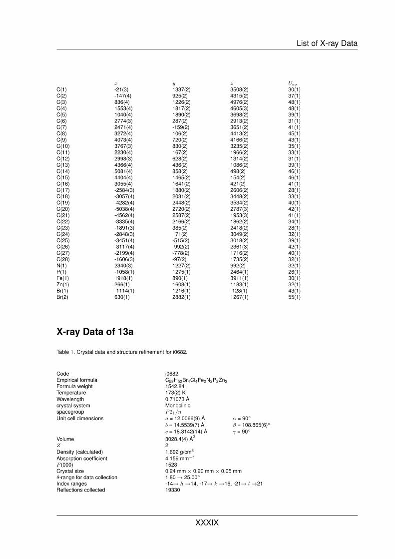
List of X-ray Data
x y z Ueq
C(1) -21(3) 1337(2) 3508(2) 30(1)C(2) -147(4) 925(2) 4315(2) 37(1)C(3) 836(4) 1226(2) 4976(2) 48(1)C(4) 1553(4) 1817(2) 4605(3) 48(1)C(5) 1040(4) 1890(2) 3698(2) 39(1)C(6) 2774(3) 287(2) 2913(2) 31(1)C(7) 2471(4) -159(2) 3651(2) 41(1)C(8) 3272(4) 106(2) 4413(2) 45(1)C(9) 4073(4) 720(2) 4166(2) 43(1)C(10) 3767(3) 830(2) 3235(2) 35(1)C(11) 2230(4) 167(2) 1966(2) 33(1)C(12) 2998(3) 628(2) 1314(2) 31(1)C(13) 4366(4) 436(2) 1086(2) 39(1)C(14) 5081(4) 858(2) 498(2) 46(1)C(15) 4404(4) 1465(2) 154(2) 46(1)C(16) 3055(4) 1641(2) 421(2) 41(1)C(17) -2584(3) 1880(2) 2606(2) 28(1)C(18) -3057(4) 2031(2) 3448(2) 33(1)C(19) -4282(4) 2448(2) 3534(2) 40(1)C(20) -5038(4) 2720(2) 2787(3) 42(1)C(21) -4562(4) 2587(2) 1953(3) 41(1)C(22) -3335(4) 2166(2) 1862(2) 34(1)C(23) -1891(3) 385(2) 2418(2) 28(1)C(24) -2848(3) 171(2) 3049(2) 32(1)C(25) -3451(4) -515(2) 3018(2) 39(1)C(26) -3117(4) -992(2) 2361(3) 42(1)C(27) -2199(4) -778(2) 1716(2) 40(1)C(28) -1606(3) -97(2) 1735(2) 32(1)N(1) 2340(3) 1227(2) 992(2) 32(1)P(1) -1058(1) 1275(1) 2464(1) 26(1)Fe(1) 1918(1) 890(1) 3911(1) 30(1)Zn(1) 266(1) 1608(1) 1183(1) 32(1)Br(1) -1114(1) 1216(1) -128(1) 43(1)Br(2) 630(1) 2882(1) 1267(1) 55(1)
X-ray Data of 13a
Table 1. Crystal data and structure refinement for i0682.
Code i0682Empirical formula C58H52Br4Cl4Fe2N2P2Zn2Formula weight 1542.84Temperature 173(2) KWavelength 0.71073 Acrystal system Monoclinicspacegroup P21/nUnit cell dimensions a = 12.0066(9) A α = 90◦
b = 14.5539(7) A β = 108.865(6)◦
c = 18.3142(14) A γ = 90◦
Volume 3028.4(4) A3
Z 2Density (calculated) 1.692 g/cm3
Absorption coefficient 4.159 mm−1
F (000) 1528Crystal size 0.24 mm × 0.20 mm × 0.05 mmθ-range for data collection 1.80→ 25.00◦
Index ranges -14→ h→14, -17→ k →16, -21→ l→21Reflections collected 19330
XXXIX

APPENDIX
Independent reflections 5342 [Rint = 0.1594]Reflections observed 3458Absorption correction IntegrationMax. and min. transmission 0.8180 and 0.5191Refinement method full-matrix least-squares against F 2
Data / restraints / parameters 5342 / 0 / 334Goodness-of-fit on F 2 0.940Final R indices [I > 2σ(I)] R1 = 0.0663, wR2 = 0.1610R indices (all data) R1 = 0.0983, wR2 = 0.1777Largest diff. peak and hole 1.307 and -0.889 e/A3
Table 2. Atomic coordinates (· 104) and equivalent isotropic displacement parameters (A2 · 103) for i0682. Ueq is defined as onethird of the trace of the orthogonalized Uij tensor.
x y z Ueq
C(1) 7552(6) 3232(5) 4979(4) 44(2)C(2) 7368(7) 4020(5) 5397(4) 46(2)C(3) 8006(7) 3896(6) 6194(4) 52(2)C(4) 8618(7) 3036(6) 6277(4) 53(2)C(5) 8354(7) 2630(6) 5534(4) 52(2)C(6) 5163(7) 3011(5) 5847(4) 48(2)C(7) 5958(8) 2711(9) 6565(5) 79(3)C(8) 6424(8) 1856(9) 6452(8) 89(4)C(9) 5956(9) 1614(7) 5675(8) 83(3)C(10) 5174(7) 2310(5) 5301(5) 52(2)C(11) 4461(7) 3890(6) 5673(5) 56(2)C(12) 3447(7) 3868(5) 5995(4) 48(2)C(13) 2622(7) 3197(7) 5801(5) 61(2)C(14) 1675(7) 3168(6) 6085(5) 59(2)C(15) 1616(7) 3844(5) 6597(5) 53(2)C(16) 2485(7) 4516(6) 6791(5) 55(2)C(17) 8392(6) 3235(5) 3699(4) 45(2)C(18) 9373(7) 3720(6) 4183(5) 55(2)C(19) 10341(8) 3882(7) 3943(6) 68(2)C(20) 10332(8) 3599(7) 3221(6) 69(3)C(21) 9398(8) 3127(6) 2756(5) 62(2)C(22) 8396(7) 2951(6) 2972(4) 52(2)C(23) 6571(7) 1935(5) 3729(4) 49(2)C(24) 7393(8) 1205(6) 3859(5) 59(2)C(25) 6992(10) 304(6) 3746(6) 73(3)C(26) 5785(11) 106(6) 3490(6) 73(3)C(27) 4979(9) 838(6) 3349(5) 66(2)C(28) 5383(7) 1738(6) 3473(5) 52(2)C(29) 1072(16) 1091(14) 3935(10) 150(7)N(1) 3387(5) 4541(4) 6498(3) 46(1)P(1) 7050(2) 3117(1) 3955(1) 43(1)Fe(1) 6842(1) 2836(1) 5799(1) 48(1)Zn(1) 5620(1) 4250(1) 3206(1) 47(1)Br(1) 5513(1) 3990(1) 1890(1) 60(1)Br(2) 3753(1) 4108(1) 3387(1) 64(1)Cl(1) 2397(5) 1433(6) 3935(4) 226(4)Cl(2) 769(3) 1117(3) 4779(2) 118(1)
X-ray Data of 14a
Table 1. Crystal data and structure refinement for i0569.
XL
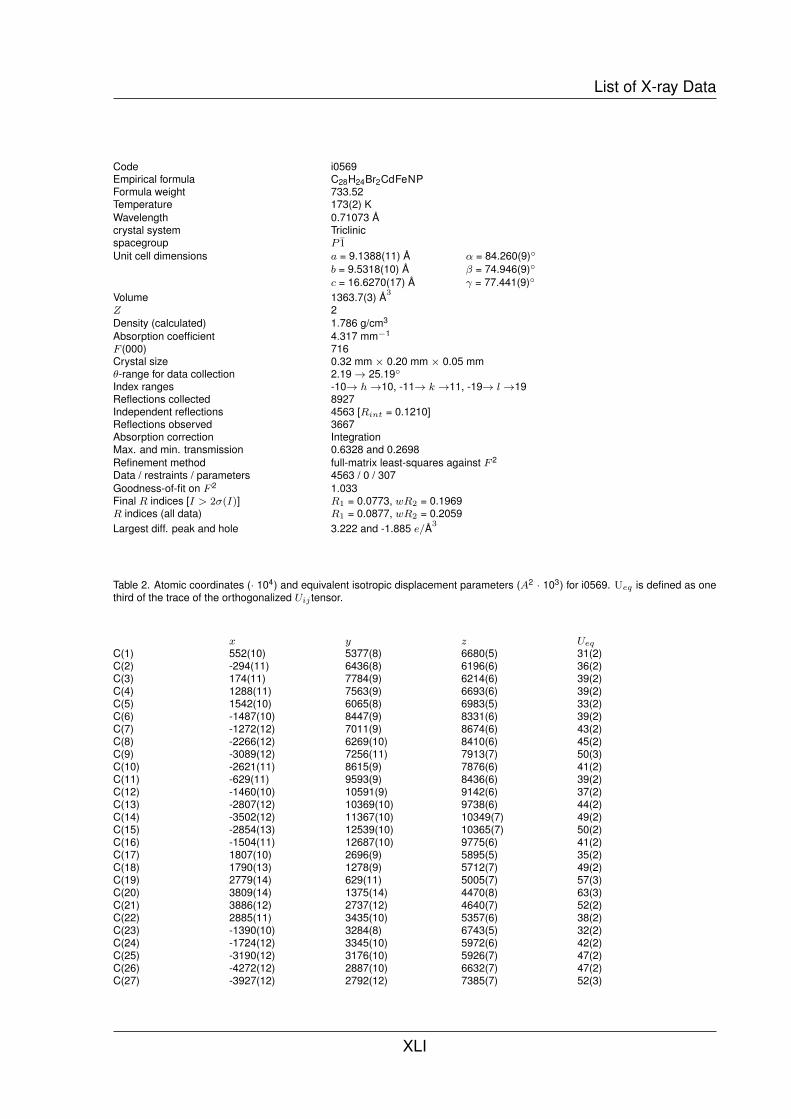
List of X-ray Data
Code i0569Empirical formula C28H24Br2CdFeNPFormula weight 733.52Temperature 173(2) KWavelength 0.71073 Acrystal system Triclinicspacegroup P 1Unit cell dimensions a = 9.1388(11) A α = 84.260(9)◦
b = 9.5318(10) A β = 74.946(9)◦
c = 16.6270(17) A γ = 77.441(9)◦
Volume 1363.7(3) A3
Z 2Density (calculated) 1.786 g/cm3
Absorption coefficient 4.317 mm−1
F (000) 716Crystal size 0.32 mm × 0.20 mm × 0.05 mmθ-range for data collection 2.19→ 25.19◦
Index ranges -10→ h→10, -11→ k →11, -19→ l→19Reflections collected 8927Independent reflections 4563 [Rint = 0.1210]Reflections observed 3667Absorption correction IntegrationMax. and min. transmission 0.6328 and 0.2698Refinement method full-matrix least-squares against F 2
Data / restraints / parameters 4563 / 0 / 307Goodness-of-fit on F 2 1.033Final R indices [I > 2σ(I)] R1 = 0.0773, wR2 = 0.1969R indices (all data) R1 = 0.0877, wR2 = 0.2059Largest diff. peak and hole 3.222 and -1.885 e/A3
Table 2. Atomic coordinates (· 104) and equivalent isotropic displacement parameters (A2 · 103) for i0569. Ueq is defined as onethird of the trace of the orthogonalized Uij tensor.
x y z Ueq
C(1) 552(10) 5377(8) 6680(5) 31(2)C(2) -294(11) 6436(8) 6196(6) 36(2)C(3) 174(11) 7784(9) 6214(6) 39(2)C(4) 1288(11) 7563(9) 6693(6) 39(2)C(5) 1542(10) 6065(8) 6983(5) 33(2)C(6) -1487(10) 8447(9) 8331(6) 39(2)C(7) -1272(12) 7011(9) 8674(6) 43(2)C(8) -2266(12) 6269(10) 8410(6) 45(2)C(9) -3089(12) 7256(11) 7913(7) 50(3)C(10) -2621(11) 8615(9) 7876(6) 41(2)C(11) -629(11) 9593(9) 8436(6) 39(2)C(12) -1460(10) 10591(9) 9142(6) 37(2)C(13) -2807(12) 10369(10) 9738(6) 44(2)C(14) -3502(12) 11367(10) 10349(7) 49(2)C(15) -2854(13) 12539(10) 10365(7) 50(2)C(16) -1504(11) 12687(10) 9775(6) 41(2)C(17) 1807(10) 2696(9) 5895(5) 35(2)C(18) 1790(13) 1278(9) 5712(7) 49(2)C(19) 2779(14) 629(11) 5005(7) 57(3)C(20) 3809(14) 1375(14) 4470(8) 63(3)C(21) 3886(12) 2737(12) 4640(7) 52(2)C(22) 2885(11) 3435(10) 5357(6) 38(2)C(23) -1390(10) 3284(8) 6743(5) 32(2)C(24) -1724(12) 3345(10) 5972(6) 42(2)C(25) -3190(12) 3176(10) 5926(7) 47(2)C(26) -4272(12) 2887(10) 6632(7) 47(2)C(27) -3927(12) 2792(12) 7385(7) 52(3)
XLI

APPENDIX
C(28) -2516(11) 3017(8) 7457(6) 38(2)N(1) -823(9) 11732(7) 9169(5) 35(2)P(1) 493(2) 3487(2) 6842(1) 30(1)Fe(1) -748(1) 7034(1) 7399(1) 31(1)Br(2) 3375(1) 10052(1) 7602(1) 41(1)Br(1) 2227(1) 13885(1) 9023(1) 43(1)Cd(1) 1336(1) 12262(1) 8189(1) 33(1)
X-ray Data of 15
Table 1. Crystal data and structure refinement for i0791.
Code i0791Empirical formula C28H24CdFeI2NPFormula weight 672.47Temperature 100(2) KWavelength 0.71073 Acrystal system Monoclinicspacegroup P21/nUnit cell dimensions a = 9.4757(8) A α = 90◦
b = 19.0085(11) A β = 94.513(6)◦
c = 15.4717(12) A γ = 90◦
Volume 2778.1(4) A3
Z 4Density (calculated) 1.978 g/cm3
Absorption coefficient 3.585 mm−1
F (000) 1576Crystal size 0.27 mm × 0.05 mm × 0.05 mmθ-range for data collection 1.70→ 25.00◦
Index ranges -11→ h→11, -22→ k →22, -18→ l→15Reflections collected 14403Independent reflections 84897 [Rint = 0.1141]Reflections observed 3388Absorption correction IntegrationMax. and min. transmission 0.8484 and 0.5150Refinement method full-matrix least-squares against F 2
Data / restraints / parameters 4897 / 0 / 307Goodness-of-fit on F 2 0.893Final R indices [I > 2σ(I)] R1 = 0.0460, wR2 = 0.1027R indices (all data) R1 = 0.0726, wR2 = 0.1101Largest diff. peak and hole 0.765 and -0.915 e/A3
Table 2. Atomic coordinates (· 104) and equivalent isotropic displacement parameters (A2 · 103) for i0791. Ueq is defined as onethird of the trace of the orthogonalized Uij tensor.
x y z Ueq
C(1) 5051(8) 1319(4) 1422(6) 28(2)C(2) 5121(8) 903(5) 648(6) 30(2)C(3) 4181(9) 1229(5) -28(6) 34(2)C(4) 3530(9) 1819(5) 345(6) 36(2)C(5) 4024(9) 1882(4) 1221(6) 31(2)C(6) 2192(8) 321(4) 1986(6) 26(2)C(7) 1268(9) 887(4) 1686(6) 31(2)C(8) 956(8) 801(5) 768(6) 35(2)C(9) 1668(9) 197(5) 488(6) 34(2)
XLII

List of X-ray Data
C(10) 2461(9) -102(5) 1244(6) 34(2)C(11) 2745(8) 154(4) 2888(6) 28(2)C(12) 1920(8) 544(4) 3555(5) 25(2)C(13) 560(8) 306(4) 3737(6) 30(2)C(14) -176(8) 662(5) 4351(6) 33(2)C(15) 446(9) 1247(5) 4768(6) 37(2)C(16) 1729(9) 1457(5) 4569(7) 36(2)C(17) 7568(8) 1845(4) 2346(5) 26(2)C(18) 8319(9) 2081(4) 3096(6) 32(2)C(19) 9539(8) 2497(4) 3027(6) 32(2)C(20) 9975(8) 2685(4) 2210(6) 32(2)C(21) 9211(9) 2448(5) 1473(7) 39(2)C(22) 7996(8) 2026(4) 1518(6) 27(2)C(23) 6874(8) 372(4) 2508(5) 25(2)C(24) 6542(8) -120(4) 3145(5) 28(2)C(25) 7131(9) -794(4) 3173(7) 36(2)C(26) 8068(9) -990(4) 2538(6) 34(2)C(27) 8415(9) -505(4) 1910(6) 35(2)C(28) 7837(8) 171(4) 1882(6) 26(2)N(1) 2509(7) 1108(3) 3967(5) 28(2)P(1) 6059(2) 1249(1) 2449(1) 26(1)Fe(1) 3087(1) 920(1) 1011(1) 26(1)I(1) 6272(1) 1158(1) 5276(1) 33(1)I(2) 3938(1) 2994(1) 3703(1) 34(1)Cd(1) 4701(1) 1593(1) 3788(1) 27(1)
X-ray Data of 16
Table 1. Crystal data and structure refinement for i0776.
Code i0776Empirical formula C28H24Br2FeHgNPFormula weight 821.71Temperature 218(2) KWavelength 0.71073 Acrystal system Monoclinicspacegroup P21/nUnit cell dimensions a = 9.3132(8) A α = 90◦
b = 18.5685(17) A β = 94.473(7)◦
c = 15.3200(13) A γ = 90◦
Volume 2641.3(4) A3
Z 4Density (calculated) 2.066 g/cm3
Absorption coefficient 9.459 mm−1
F (000) 1560Crystal size 0.39 mm × 0.04 mm × 0.04 mmθ-range for data collection 1.73→ 25.00◦
Index ranges -178→ h→11, -18→ k →22, -18→ l→18Reflections collected 11523Independent reflections 4552 [Rint = 0.0746]Reflections observed 2932Absorption correction IntegrationMax. and min. transmission 0.7314 and 0.3539Refinement method full-matrix least-squares against F 2
Data / restraints / parameters 4552 / 0 / 307Goodness-of-fit on F 2 0.884Final R indices [I > 2σ(I)] R1 = 0.0334, wR2 = 0.0841R indices (all data) R1 = 0.0669, wR2 = 0.0901Largest diff. peak and hole 1.067 and -1.043 e/A3
XLIII
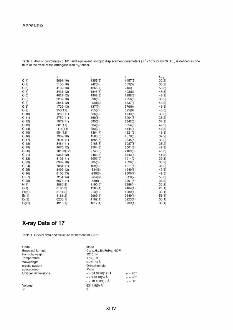
APPENDIX
Table 2. Atomic coordinates (· 104) and equivalent isotropic displacement parameters (A2 · 103) for i0776. Ueq is defined as onethird of the trace of the orthogonalized Uij tensor.
x y z Ueq
C(1) 5091(10) 1355(5) 1457(5) 30(2)C(2) 5150(10) 942(6) 659(5) 38(2)C(3) 4132(13) 1266(7) 24(6) 53(3)C(4) 3431(12) 1846(6) 403(6) 48(3)C(5) 4024(12) 1908(6) 1288(6) 43(3)C(6) 2237(10) 298(5) 2056(5) 33(2)C(7) 2541(12) -130(6) 1337(6) 44(3)C(8) 1736(13) 137(7) 576(6) 48(3)C(9) 956(11) 755(7) 825(6) 45(3)C(10) 1269(11) 856(6) 1749(5) 39(2)C(11) 2759(11) 160(6) 3004(5) 36(2)C(12) 1972(11) 585(5) 3642(5) 34(2)C(13) 601(11) 384(6) 3854(6) 42(2)C(14) -114(11) 782(7) 4444(6) 48(3)C(15) 554(12) 1384(7) 4821(6) 49(3)C(16) 1902(10) 1568(6) 4576(5) 39(2)C(17) 7694(11) 1885(5) 2345(5) 33(2)C(18) 8464(11) 2168(5) 3087(6) 38(2)C(19) 9670(12) 2584(6) 3001(6) 42(3)C(20) 10123(12) 2740(6) 2189(6) 45(3)C(21) 9367(12) 2483(6) 1443(6) 41(2)C(22) 8152(11) 2057(5) 1514(5) 35(2)C(23) 6982(10) 385(5) 2520(5) 30(2)C(24) 7884(11) 169(6) 1911(5) 36(2)C(25) 8492(12) -534(6) 1949(6) 42(3)C(26) 8156(13) -998(6) 2600(7) 49(3)C(27) 7254(14) -784(6) 3228(7) 53(3)C(28) 6673(11) -88(6) 3201(5) 37(2)N(1) 2585(8) 1190(5) 3996(4) 35(2)P(1) 6190(3) 1283(1) 2464(1) 28(1)Fe(1) 3114(2) 913(1) 1080(1) 33(1)Br(1) 4181(2) 2969(1) 3849(1) 56(1)Br(2) 6238(1) 1182(1) 5223(1) 53(1)Hg(1) 4915(1) 1617(1) 3739(1) 36(1)
X-ray Data of 17
Table 1. Crystal data and structure refinement for i0573.
Code i0573Empirical formula C29.50H24Br4FeHg2NOPFormula weight 1216.14Temperature 173(2) KWavelength 0.71073 Acrystal system Orthorhombicspacegroup PccnUnit cell dimensions a = 34.9720(12) A α = 90◦
b = 9.9410(5) A β = 90◦
c = 18.1638(8) A γ = 90◦
Volume 6314.8(5) A3
Z 8
XLIV

List of X-ray Data
Density (calculated) 2.558 g/cm3
Absorption coefficient 15.292 mm−1
F (000) 4456Crystal size 0.49 mm × 0.17 mm × 0.14 mmθ-range for data collection 2.13→ 25.26◦
Index ranges -41→ h→42, -11→ k →11, -20→ l→20Reflections collected 37537Independent reflections 5554 [Rint = 0.0991]Reflections observed 4558Absorption correction IntegrationMax. and min. transmission 0.1673 and 0.0059Refinement method full-matrix least-squares against F 2
Data / restraints / parameters 5554 / 0 / 358Goodness-of-fit on F 2 1.056Final R indices [I > 2σ(I)] R1 = 0.0553, wR2 = 0.1376R indices (all data) R1 = 0.0679, wR2 = 0.1483Extinction coefficient 0.00018(4)Largest diff. peak and hole 4.404 and -2.301 e/A3
Table 2. Atomic coordinates (· 104) and equivalent isotropic displacement parameters (A2 · 103) for i0573. Ueq is defined as onethird of the trace of the orthogonalized Uij tensor.
x y z Ueq
C(1) 1317(3) 2894(9) 5980(6) 35(2)C(2) 1317(3) 1436(10) 5953(7) 39(2)C(3) 1075(4) 954(11) 6517(7) 46(3)C(4) 926(4) 2122(12) 6902(7) 49(3)C(5) 1079(3) 3320(11) 6563(7) 43(3)C(6) 575(3) 1511(9) 4829(6) 35(2)C(7) 564(3) 2946(10) 4825(7) 40(2)C(8) 327(3) 3393(10) 5420(6) 39(2)C(9) 191(3) 2218(10) 5795(7) 42(3)C(10) 337(3) 1081(10) 5423(6) 38(2)C(11) 771(3) 591(10) 4281(6) 38(2)C(12) 548(3) 356(9) 3582(6) 34(2)C(13) 217(3) 1080(11) 3410(7) 44(3)C(14) 20(3) 769(11) 2768(7) 46(3)C(15) 166(3) -199(11) 2288(7) 44(3)C(16) 502(3) -841(10) 2493(6) 39(2)C(17) 2014(3) 4516(10) 5793(7) 41(3)C(18) 2138(3) 3898(12) 6449(8) 52(3)C(19) 2491(4) 4279(15) 6756(8) 60(3)C(20) 2718(4) 5205(13) 6389(9) 61(4)C(21) 2593(4) 5788(13) 5745(9) 62(4)C(22) 2241(4) 5461(12) 5443(8) 52(3)C(23) 1705(3) 3092(10) 4575(7) 41(2)C(24) 1543(3) 3352(16) 3894(8) 60(4)C(25) 1652(5) 2480(20) 3294(8) 77(5)C(26) 1914(4) 1413(16) 3412(8) 65(4)C(27) 2072(4) 1268(12) 4077(7) 51(3)C(28) 1978(4) 2066(11) 4668(7) 46(3)C(29) 2609(5) 6629(19) 8446(10) 66(5)C(30) 2500 7500 7810(20) 154(18)Br(1) 599(1) 7293(1) 5608(1) 48(1)Br(2) 792(1) 5857(1) 3591(1) 49(1)Br(3) 1527(1) 8144(1) 2040(1) 56(1)Br(4) 1576(1) 8137(1) 4562(1) 45(1)Fe 779(1) 2225(1) 5810(1) 33(1)Hg(1) 1151(1) 5949(1) 5180(1) 56(1)Hg(2) 1224(1) 7985(1) 3304(1) 47(1)O(30) 2806(4) 5968(11) 8285(5) 62(3)P 1564(1) 4032(3) 5376(2) 37(1)
XLV

APPENDIX
N 695(2) -565(8) 3119(5) 33(2)
X-ray Data of 18
Table 1. Crystal data and structure refinement for i0683.
Code i0683Empirical formula C57H49Br2Cl2Fe2HgN2P2Formula weight 1402.38Temperature 173(2) KWavelength 0.71073 Acrystal system Orthorhombicspacegroup I b a 2Unit cell dimensions a = 13.7805(6) A α = 90◦
b = 40.4138(16) A β = 90◦
c = 19.1500(9) A γ = 90◦
Volume 10665.1(8) A3
Z 8Density (calculated) 1.747 g/cm3
Absorption coefficient 5.158 mm−1
F (000) 5504Crystal size 0.31 mm × 0.20 mm × 0.15 mmθ-range for data collection 1.56→ 25.00◦
Index ranges -16→ h→16, -48→ k →47, -22→ l→22Reflections collected 33209Independent reflections 9405 [Rint = 0.0449]Reflections observed 8569Absorption correction IntegrationMax. and min. transmission 0.5756 and 0.4185Refinement method full-matrix least-squares against F 2
Data / restraints / parameters 9405 / 1 / 622Goodness-of-fit on F 2 1.026Final R indices [I > 2σ(I)] R1 = 0.0382, wR2 = 0.0963R indices (all data) R1 = 0.0430, wR2 = 0.1010Absolute structure parameter -0.008(6)Largest diff. peak and hole 2.009 and -1.340 e/A3
Table 2. Atomic coordinates (· 104) and equivalent isotropic displacement parameters (A2 · 103) for i0683. Ueq is defined as onethird of the trace of the orthogonalized Uij tensor.
x y z Ueq
C(1) -5866(6) -3892(2) -2097(4) 33(2)C(2) -5360(5) -4009(2) -1502(4) 37(2)C(3) -4698(6) -4256(2) -1718(5) 48(2)C(4) -4776(6) -4296(2) -2463(5) 46(2)C(5) -5503(5) -4070(2) -2692(4) 36(2)C(6) -6938(6) -4728(2) -2509(5) 41(2)C(7) -7552(5) -4497(2) -2168(5) 41(2)C(8) -7349(6) -4506(3) -1428(5) 52(2)C(9) -6615(7) -4755(2) -1321(5) 56(2)C(10) -6377(6) -4889(2) -1968(6) 52(2)C(11) -6889(6) -4795(2) -3285(6) 57(2)C(12) -7612(6) -5049(2) -3561(5) 48(2)C(13) -8483(8) -5109(3) -3272(8) 98(5)C(14) -9106(9) -5341(3) -3566(8) 92(4)
XLVI

List of X-ray Data
C(15) -8868(7) -5484(3) -4162(6) 64(3)C(16) -7966(9) -5419(3) -4428(7) 81(3)C(17) -7558(5) -3610(2) -1398(5) 36(2)C(18) -8509(5) -3726(2) -1487(5) 44(2)C(19) -9097(6) -3779(2) -902(5) 51(2)C(20) -8762(7) -3711(2) -245(5) 55(2)C(21) -7828(9) -3588(3) -155(5) 53(2)C(22) -7231(8) -3532(2) -713(6) 43(2)C(23) -6078(5) -3181(2) -1991(4) 33(2)C(24) -5069(5) -3170(2) -2098(4) 41(2)C(25) -4609(7) -2862(3) -2070(5) 52(2)C(26) -5118(7) -2577(2) -1919(5) 54(2)C(27) -6121(7) -2593(2) -1793(5) 49(2)C(28) -6585(6) -2894(2) -1835(5) 43(2)C(29) -5797(5) -3170(2) -4484(4) 33(2)C(30) -5247(5) -3062(2) -5092(4) 37(2)C(31) -4551(5) -2828(2) -4858(5) 40(2)C(32) -4669(5) -2779(2) -4124(5) 43(2)C(33) -5435(5) -2985(2) -3898(4) 39(2)C(34) -6711(5) -2296(2) -4177(4) 38(2)C(35) -7359(5) -2520(2) -4511(5) 42(2)C(36) -7107(6) -2529(2) -5246(4) 44(2)C(37) -6311(7) -2309(2) -5344(5) 48(2)C(38) -6074(6) -2165(2) -4687(4) 41(2)C(39) -6722(6) -2199(2) -3408(5) 48(2)C(40) -7003(5) -1826(2) -3324(5) 39(1)C(41) -6279(4) -1586(2) -3298(5) 40(2)C(42) -6561(6) -1256(2) -3245(5) 47(2)C(43) -7534(6) -1183(2) -3228(6) 51(2)C(44) -8196(5) -1440(2) -3261(6) 52(2)C(45) -7502(5) -3434(2) -5200(5) 33(2)C(46) -7180(8) -3505(2) -5877(5) 43(2)C(47) -7766(7) -3440(3) -6461(5) 48(2)C(48) -8684(6) -3299(2) -6363(4) 44(2)C(49) -9004(6) -3230(2) -5694(5) 49(2)C(50) -8427(5) -3291(2) -5117(4) 42(2)C(51) -6045(6) -3869(2) -4618(4) 36(2)C(52) -6538(7) -4165(2) -4778(5) 48(2)C(53) -6025(9) -4458(2) -4831(6) 63(3)C(54) -5026(8) -4464(2) -4710(5) 63(3)C(55) -4527(7) -4183(2) -4564(5) 55(2)C(56) -5022(6) -3878(2) -4522(5) 46(2)C(57) -8823(9) -4640(4) -6301(9) 94(4)N(1) -7329(6) -5200(3) -4146(5) 69(2)N(2) -7947(4) -1753(2) -3303(4) 42(1)P(1) -6760(2) -3565(1) -2148(1) 29(1)P(2) -6706(2) -3484(1) -4457(1) 30(1)Cl(1) -7981(3) -4338(1) -6555(3) 131(2)Cl(2) -8894(5) -4642(3) -5399(4) 208(4)Cl(3) -9962(3) -4555(1) -6679(3) 130(2)Fe(1) -6107(1) -4386(1) -1980(1) 35(1)Fe(2) -5943(1) -2671(1) -4646(1) 32(1)Br(1) -8731(1) -3006(1) -3059(1) 44(1)Br(2) -8844(1) -4006(1) -3595(1) 49(1)Hg(1) -7588(1) -3523(1) -3312(1) 32(1)
X-ray Data of 19
Table 1. Crystal data and structure refinement for i0812.
XLVII

APPENDIX
Code i0812Empirical formula C29H24AgBCl6F4FeNPFormula weight 880.69Temperature 100(2) KWavelength 0.71073 Acrystal system Monoclinicspacegroup P21/cUnit cell dimensions a = 10.5259(10) A α = 90◦
b = 11.9401(16) A β = 93.193(7)◦
c = 26.573(3) A γ = 90◦
Volume 3334.6(6) A3
Z 4Density (calculated) 1.754 g/cm3
Absorption coefficient 1.596 mm−1
F (000) 1744Crystal size 0.31 mm × 0.10 mm × 0.08 mmθ-range for data collection 1.53→ 25.00◦
Index ranges -12→ h→11, -14→ k →14, -31→ l→30Reflections collected 21156Independent reflections 5720 [Rint = 0.0773]Reflections observed 4955Absorption correction IntegrationMax. and min. transmission 0.8971 and 0.7251Refinement method full-matrix least-squares against F 2
Data / restraints / parameters 5720 / 0 / 397Goodness-of-fit on F 2 0.691Final R indices [I > 2σ(I)] R1 = 0.0307, wR2 = 0.0842R indices (all data) R1 = 0.0377, wR2 = 0.0903Largest diff. peak and hole 0.974 and -0.867 e/A3
Table 2. Atomic coordinates (· 104) and equivalent isotropic displacement parameters (A2 · 103) for i0812. Ueq is defined as onethird of the trace of the orthogonalized Uij tensor.
x y z Ueq
C(1) 7802(3) 1202(2) 2107(1) 17(1)C(2) 8269(3) 705(2) 1656(1) 19(1)C(3) 9504(3) 255(2) 1783(1) 21(1)C(4) 9806(3) 481(2) 2308(1) 22(1)C(5) 8777(3) 1059(2) 2511(1) 20(1)C(6) 7059(3) -1746(2) 1936(1) 19(1)C(7) 8301(3) -2151(2) 2110(1) 23(1)C(8) 8477(3) -1903(2) 2638(1) 25(1)C(9) 7376(3) -1345(2) 2793(1) 23(1)C(10) 6496(3) -1245(2) 2362(1) 20(1)C(11) 3485(3) 3147(2) 3594(1) 19(1)C(12) 2758(3) 3310(2) 4009(1) 23(1)C(13) 3284(3) 3144(2) 4498(1) 25(1)C(14) 4558(3) 2823(2) 4562(1) 27(1)C(15) 5241(3) 2679(2) 4136(1) 25(1)C(16) 5093(3) 1244(2) 1791(1) 20(1)C(17) 4077(3) 770(2) 2035(1) 25(1)C(18) 3129(3) 189(2) 1746(1) 28(1)C(19) 3176(3) 99(2) 1223(1) 29(1)C(20) 4177(3) 582(2) 981(1) 27(1)C(21) 5134(3) 1154(2) 1261(1) 22(1)C(22) 6589(3) 3259(2) 1835(1) 20(1)C(23) 5581(3) 3794(2) 1564(1) 25(1)C(24) 5776(3) 4792(2) 1311(1) 31(1)C(25) 6983(3) 5265(2) 1325(1) 30(1)C(26) 7992(3) 4753(2) 1596(1) 29(1)C(27) 7795(3) 3750(2) 1853(1) 25(1)
XLVIII

List of X-ray Data
C(28) 7974(3) -490(3) 4704(1) 29(1)C(29) 11089(3) -3200(3) 4735(1) 29(1)N(1) 4725(2) 2821(2) 3657(1) 20(1)P(1) 6345(1) 1953(1) 2174(1) 17(1)Fe(1) 8166(1) -458(1) 2222(1) 16(1)Ag(1) 5763(1) 2362(1) 3003(1) 21(1)Cl(1) 12442(1) -4072(1) 4837(1) 40(1)Cl(2) 11436(1) -2096(1) 4323(1) 42(1)Cl(3) 9783(1) -3978(1) 4484(1) 39(1)Cl(4) 8897(1) -1032(1) 5226(1) 29(1)Cl(5) 7648(1) -1550(1) 4243(1) 55(1)Cl(6) 6524(1) 52(1) 4903(1) 38(1)B(1) 8933(3) 2377(3) 3873(1) 24(1)F(1) 8229(2) 1864(2) 4242(1) 34(1)F(2) 9533(2) 3318(2) 4091(1) 33(1)F(3) 9830(2) 1632(2) 3710(1) 32(1)F(4) 8121(2) 2717(2) 3463(1) 36(1)
X-ray Data of 20
Table 1. Crystal data and structure refinement for i0703.
Code i0703Empirical formula C56H46Ag2B2Cl6F8Fe2N2P2Formula weight 1522.65Temperature 173(2) KWavelength 0.71073 Acrystal system Monoclinicspacegroup P21/nUnit cell dimensions a = 9.1606(5) A α = 90◦
b = 21.4054(9) A β = 93.684(5)◦
c = 15.4766(9) A γ = 90◦
Volume 3028.5(3) A3
Z 2Density (calculated) 1.670 g/cm3
Absorption coefficient 1.488 mm−1
F (000) 1512Crystal size 0.50 mm × 0.11 mm × 0.09 mmθ-range for data collection 1.63→ 25.16◦
Index ranges -10→ h→10, -23→ k →25, -18→ l→18Reflections collected 19521Independent reflections 5387 [Rint = 0.0304]Reflections observed 4463Absorption correction IntegrationMax. and min. transmission 0.8975 and 0.6853Refinement method full-matrix least-squares against F 2
Data / restraints / parameters 5387 / 0 / 361Goodness-of-fit on F 2 1.070Final R indices [I > 2σ(I)] R1 = 0.0409, wR2 = 0.1115R indices (all data) R1 = 0.0499, wR2 = 0.1157Largest diff. peak and hole 1.418 and -0.746 e/A3
Table 2. Atomic coordinates (· 104) and equivalent isotropic displacement parameters (A2 · 103) for i0703. Ueq is defined as onethird of the trace of the orthogonalized Uij tensor.
x y z Ueq
XLIX

APPENDIX
C(1) 4127(4) -96(2) -2005(2) 40(1)C(2) 3997(5) -706(2) -2379(3) 47(1)C(3) 5326(4) -109(2) -1354(2) 34(1)C(4) 6849(4) -596(2) -3698(2) 40(1)C(5) 7897(4) 157(2) -2771(3) 39(1)C(6) 5918(4) -729(2) -1337(2) 36(1)C(7) 6675(5) 424(2) -3240(3) 45(1)C(8) 5096(5) -1093(2) -1972(3) 43(1)C(9) 6028(5) -32(2) -3811(3) 46(1)C(10) 8026(4) -484(2) -3044(2) 34(1)C(11) 6992(4) 364(2) -193(2) 33(1)C(12) 5853(4) 431(2) -832(2) 33(1)C(13) 5759(6) 1510(2) -424(3) 54(1)C(14) 6898(5) 1403(2) 182(3) 47(1)C(15) 5224(5) 1019(2) -935(3) 45(1)C(16) 11534(4) 1762(2) 2475(3) 37(1)C(17) 9231(4) 706(2) 4115(3) 39(1)C(18) 8608(5) 1715(2) 3518(3) 43(1)C(19) 9437(4) 1168(2) 3502(2) 35(1)C(20) 11731(5) 1980(2) 1638(3) 44(1)C(21) 12118(5) 2111(2) 3188(3) 48(1)C(22) 12520(6) 2526(2) 1524(4) 57(1)C(23) 7334(5) 1316(2) 4719(3) 49(1)C(24) 7550(5) 1788(2) 4122(3) 50(1)C(25) 8191(5) 781(2) 4723(3) 48(1)C(26) 13077(6) 2864(2) 2233(4) 62(1)C(27) 12882(6) 2654(2) 3065(4) 60(1)C(28) 17172(6) 3434(2) 1468(3) 55(1)Ag(1) 9163(1) 729(1) 1365(1) 44(1)B(1) 15901(6) 3908(3) 3950(3) 48(1)F(1) 14458(4) 3856(2) 3726(3) 94(1)F(2) 16659(5) 3491(3) 3461(3) 122(2)F(3) 16328(7) 3852(3) 4773(3) 148(2)F(4) 16299(6) 4500(2) 3710(4) 134(2)Fe(1) 6030(1) -337(1) -2552(1) 32(1)N(1) 7508(4) 840(2) 306(2) 37(1)P(1) 10616(1) 1024(1) 2612(1) 33(1)Cl(1) 17364(2) 4210(1) 1134(1) 90(1)Cl(2) 18890(2) 3065(1) 1573(2) 101(1)Cl(3) 15983(2) 3043(1) 701(1) 74(1)
X-ray Data of 21
Table 1. Crystal data and structure refinement for i0760a.
Code i0760aEmpirical formula C29H24AgBCl3F4FeNPFormula weight 774.34Temperature 298(2) KWavelength 0.71073 Acrystal system Monoclinicspacegroup P21/cUnit cell dimensions a = 13.2362(14) A α = 90◦
b = 11.5572(13) A β = 91.336(11)◦
c = 20.694(3) A γ = 90◦
Volume 3164.7(7) A3
Z 4Density (calculated) 1.625 g/cm3
Absorption coefficient 1.425 mm−1
L

List of X-ray Data
F (000) 1540θ-range for data collection 1.54→ 25.00◦
Index ranges -14→ h→15, -5→ k →13, -23→ l→9Reflections collected 2375Independent reflections 2074 [Rint = 0.0254]Reflections observed NoneAbsorption correction IntegrationRefinement method full-matrix least-squares against F 2
Data / restraints / parameters 2074 / 0 / 370Goodness-of-fit on F 2 0.790Final R indices [I > 2σ(I)] R1 = 0.0321, wR2 = 0.0495R indices (all data) R1 = 0.0737, wR2 = 0.0557Largest diff. peak and hole 0.175 and -0.150 e/A3
Table 2. Atomic coordinates (· 104) and equivalent isotropic displacement parameters (A2 · 103) for i0760a. Ueq is defined as onethird of the trace of the orthogonalized Uij tensor.
x y z Ueq
C(1) 1748(4) 5341(7) 3059(6) 53(3)C(2) 2338(5) 5126(10) 3611(6) 67(3)C(3) 1699(6) 4764(12) 4128(6) 74(4)C(4) 686(6) 4878(11) 3871(7) 74(4)C(5) 726(5) 5159(9) 3223(6) 60(3)C(6) 1216(5) 2026(9) 3633(6) 55(3)C(7) 736(6) 2271(10) 3038(7) 70(4)C(8) 1517(10) 2580(12) 2614(8) 91(4)C(9) 2445(9) 2506(12) 2930(9) 91(5)C(10) 2277(5) 2174(9) 3559(7) 68(4)C(11) 733(4) 1626(9) 4262(6) 55(3)C(12) 941(4) 360(7) 4391(5) 51(3)C(13) 1805(4) 11(10) 4746(5) 67(3)C(14) 2013(5) -1137(10) 4840(6) 80(4)C(15) 1353(5) -1938(9) 4604(6) 68(4)C(16) 497(4) -1574(8) 4269(6) 59(3)C(17) 2113(4) 7262(8) 2223(6) 51(3)C(18) 1867(4) 7976(9) 2740(6) 57(3)C(19) 1805(5) 9161(10) 2674(7) 64(4)C(20) 1999(5) 9659(9) 2086(7) 70(4)C(21) 2263(7) 9000(11) 1590(8) 90(6)C(22) 2309(6) 7836(11) 1648(7) 73(4)C(23) 3446(4) 5330(7) 2203(6) 56(3)C(24) 3713(5) 4426(9) 1811(6) 64(4)C(25) 4713(6) 4104(12) 1782(7) 105(5)C(26) 5413(6) 4650(12) 2112(9) 120(6)C(27) 5194(5) 5559(13) 2533(7) 96(5)C(28) 4174(4) 5896(9) 2554(6) 77(4)C(29) -5035(6) 6980(13) 5248(7) 97(5)B(1) -2360(6) 7053(14) 4256(8) 69(4)N(1) 296(3) -444(6) 4154(4) 50(2)F(1) -3038(3) 6740(6) 3781(4) 106(3)F(2) -2810(3) 7284(7) 4807(4) 121(3)F(3) -1859(3) 8044(7) 4053(4) 104(3)F(4) -1616(3) 6250(7) 4323(4) 121(3)P(1) 2121(1) 5721(2) 2259(1) 46(1)Fe(1) 1532(1) 3688(1) 3359(1) 54(1)Ag(1) 990(1) 4990(1) 1462(1) 57(1)Cl(1) -5964(2) 6616(6) 4794(4) 262(5)Cl(2) -4998(2) 6383(7) 6026(4) 251(4)Cl(3) -5100(2) 8463(6) 5431(4) 246(4)
LI

APPENDIX
X-ray Data of 22
Table 1. Crystal data and structure refinement for i0777.
Code i0777Empirical formula C27H22AuClFeNPFormula weight 679.69Temperature 218(2) KWavelength 0.71073 Acrystal system Monoclinicspacegroup P21/nUnit cell dimensions a = 13.8398(9) A α = 90◦
b = 10.7636(5) A β = 107.505(5)◦
c = 16.6681(12) A γ = 90◦
Volume 2368.0(3) A3
Z 4Density (calculated) 1.907 g/cm3
Absorption coefficient 6.997 mm−1
F (000) 1312Crystal size 0.17 mm × 0.15 mm × 0.06 mmθ-range for data collection 1.68→ 25.00◦
Index ranges -16→ h→16, -12→ k →11, -19→ l→16Reflections collected 10860Independent reflections 4149 [Rint = 0.0607]Reflections observed 3172Absorption correction IntegrationMax. and min. transmission 0.6590 and 0.3593Refinement method full-matrix least-squares against F 2
Data / restraints / parameters 4149 / 0 / 289Goodness-of-fit on F 2 0.999Final R indices [I > 2σ(I)] R1 = 0.0327, wR2 = 0.0741R indices (all data) R1 = 0.0461, wR2 = 0.0766Largest diff. peak and hole 1.310 and -2.150 e/A3
Table 2. Atomic coordinates (· 104) and equivalent isotropic displacement parameters (A2 · 103) for i0777. Ueq is defined as onethird of the trace of the orthogonalized Uij tensor.
x y z Ueq
C(1) 5872(4) 44(6) 3688(4) 36(1)C(2) 4808(4) 217(7) 3564(4) 45(2)C(3) 4279(5) -414(8) 2808(5) 54(2)C(4) 4991(5) -949(7) 2457(5) 53(2)C(5) 5978(5) -679(6) 2986(4) 42(2)C(6) 5888(5) 1821(7) 1781(4) 45(2)C(7) 5977(5) 2556(7) 2522(5) 51(2)C(8) 4979(6) 2831(9) 2556(6) 66(2)C(9) 4285(5) 2257(9) 1858(6) 69(2)C(10) 4828(5) 1634(9) 1373(5) 63(2)C(11) 6722(5) 1316(7) 1490(4) 46(2)N(13) 6456(5) 829(7) 710(4) 58(2)C(13) 7210(7) 382(8) 447(5) 65(2)C(14) 8219(6) 383(8) 904(5) 58(2)C(15) 8468(6) 885(8) 1691(5) 60(2)C(16) 7042(5) -710(7) 5337(4) 41(2)C(17) 7686(5) -549(9) 6159(5) 58(2)C(18) 7778(6) -1519(11) 6754(5) 72(3)C(19) 7235(7) -2587(10) 6536(6) 73(3)C(20) 6608(7) -2770(9) 5717(6) 66(2)C(21) 6519(5) -1843(7) 5131(5) 50(2)
LII

List of X-ray Data
C(22) 7980(4) 857(6) 4358(4) 39(2)C(23) 8366(5) 2051(7) 4411(4) 44(2)C(24) 9285(5) 2289(8) 4247(5) 50(2)C(25) 9791(5) 1321(8) 4013(4) 51(2)C(26) 9420(5) 117(7) 3966(4) 47(2)C(27) 8518(4) -132(7) 4140(4) 44(2)C(28) 7724(5) 1348(7) 2000(5) 53(2)P(1) 6810(1) 591(2) 4616(1) 36(1)Cl(1) 5923(2) 3887(2) 5937(1) 57(1)Fe(1) 5214(1) 939(1) 2580(1) 41(1)Au(1) 6334(1) 2233(1) 5233(1) 39(1)
X-ray Data of 23
Table 1. Crystal data and structure refinement for i0702.
Code i0702Empirical formula C27H22AuClFeNPFormula weight 679.69Temperature 173(2) KWavelength 0.71073 Acrystal system Triclinicspacegroup P 1Unit cell dimensions a = 8.6932(8) A α = 94.337(8)◦
b = 8.9534(9) A β = 103.354(8)◦
c = 16.2110(16) A γ = 107.402(8)◦
Volume 1157.14(19) A3
Z 2Density (calculated) 1.951 g/cm3
Absorption coefficient 7.159 mm−1
F (000) 656Crystal size 0.46 mm × 0.37 mm × 0.18 mmθ-range for data collection 1.31→ 25.18◦
Index ranges -10→ h→10, -9→ k →10, -19→ l→19Reflections collected 6997Independent reflections 3841 [Rint = 0.0361]Reflections observed 3716Absorption correction IntegrationMax. and min. transmission 0.3441 and 0.1085Refinement method full-matrix least-squares against F 2
Data / restraints / parameters 3841 / 0 / 290Goodness-of-fit on F 2 1.070Final R indices [I > 2σ(I)] R1 = 0.0249, wR2 = 0.0654R indices (all data) R1 = 0.0260, wR2 = 0.0659Extinction coefficient 0.0025(4)Largest diff. peak and hole 1.321 and -1.531 e/A3
Table 2. Atomic coordinates (· 104) and equivalent isotropic displacement parameters (A2 · 103) for i0702. Ueq is defined as onethird of the trace of the orthogonalized Uij tensor.
x y z Ueq
C(1) 5486(5) 2080(4) 2901(3) 29(1)C(2) 4006(5) 1173(5) 2231(3) 35(1)C(3) 2672(6) 1698(5) 2328(3) 41(1)C(4) 3277(6) 2917(5) 3049(3) 41(1)
LIII

APPENDIX
C(5) 5007(6) 3165(5) 3405(3) 34(1)C(6) 4063(6) 5420(4) 1635(3) 30(1)C(7) 3985(6) 4217(5) 969(3) 35(1)C(8) 5576(7) 4007(6) 1124(3) 42(1)C(9) 6638(6) 5052(5) 1871(3) 39(1)C(10) 5713(6) 5919(4) 2195(3) 34(1)C(11) 2730(6) 6090(4) 1694(3) 30(1)C(12) 1558(6) 6129(5) 955(3) 38(1)N(16) 354(5) 6791(5) 945(3) 43(1)C(13) 315(6) 7463(5) 1697(3) 40(1)C(14) 1404(7) 7484(5) 2465(3) 40(1)C(16) 7508(5) 250(4) 3661(2) 28(1)C(17) 6277(6) -373(5) 4066(3) 38(1)C(18) 6345(7) -1593(6) 4541(3) 45(1)C(19) 7643(7) -2218(5) 4600(3) 43(1)C(20) 8882(7) -1602(5) 4198(3) 43(1)C(21) 8836(6) -368(5) 3730(3) 35(1)C(22) 7691(5) 1222(4) 2019(2) 30(1)C(23) 6737(6) -318(5) 1621(3) 37(1)C(24) 6832(8) -851(6) 803(3) 49(1)C(25) 7857(8) 168(7) 400(3) 55(2)C(26) 8781(7) 1699(7) 800(3) 48(1)C(27) 8734(6) 2242(5) 1625(3) 35(1)C(15) 2623(6) 6777(5) 2462(3) 37(1)Au(1) 9659(1) 4076(1) 3774(1) 27(1)Cl(1) 11984(1) 6170(1) 4515(1) 36(1)Fe(1) 4622(1) 3546(1) 2161(1) 27(1)P(1) 7534(1) 1917(1) 3074(1) 26(1)
X-ray Data of 24
Table 1. Crystal data and structure refinement for i0763.
Code i0763Empirical formula C28H24AuClFeNPFormula weight 693.72Temperature 223(2) KWavelength 0.71073 Acrystal system Monoclinicspacegroup P21/cUnit cell dimensions a = 9.972(4) A α = 90◦
b = 22.351(9) A β = 105.81(3)◦
c = 11.648(4) A γ = 90◦
Volume 2497.9(15) A3
Z 4Density (calculated) 1.845 g/cm3
Absorption coefficient 6.635 mm−1
F (000) 1344Crystal size 0.600 mm × 0.553 mm × 0.260 mmθ-range for data collection 1.82→ 25.00◦
Index ranges -11→ h→11, -26→ k →26, -12→ l→13Reflections collected 15428Independent reflections 4384 [Rint = 0.1663]Reflections observed 4081Absorption correction IntegrationMax. and min. transmission 0.2415 and 0.0605Refinement method full-matrix least-squares against F 2
Data / restraints / parameters 4384 / 0 / 299Goodness-of-fit on F 2 1.102
LIV

List of X-ray Data
Final R indices [I > 2σ(I)] R1 = 0.0521, wR2 = 0.1370R indices (all data) R1 = 0.0550, wR2 = 0.1400Extinction coefficient 0.0027(5)Largest diff. peak and hole 2.484 and -3.026 e/A3
Table 2. Atomic coordinates (· 104) and equivalent isotropic displacement parameters (A2 · 103) for i0763. Ueq is defined as onethird of the trace of the orthogonalized Uij tensor.
x y z Ueq
C(1) 8634(6) 1292(3) 3611(5) 38(1)
C(2) 9212(7) 1885(4) 3832(6) 49(2)C(3) 10655(8) 1812(5) 4410(7) 66(2)C(4) 10950(7) 1219(5) 4534(6) 60(2)C(5) 9719(6) 867(4) 4069(5) 51(2)C(6) 10211(6) 1207(3) 7150(5) 42(1)C(7) 8944(7) 918(5) 6667(6) 64(2)C(8) 7939(7) 1377(7) 6211(6) 91(4)C(9) 8591(11) 1951(7) 6411(6) 83(4)C(10) 9979(8) 1840(4) 7003(6) 58(2)C(11) 11543(7) 904(3) 7816(5) 48(2)C(12) 11761(6) 940(3) 9144(5) 44(1)N(14) 12020(6) 1488(3) 9632(5) 52(1)C(37) 12175(9) 1526(5) 10842(7) 72(2)C(38) 12085(8) 1047(5) 11541(6) 64(2)C(39) 11821(10) 493(5) 11025(7) 72(2)C(17) 6902(6) 1055(3) 1280(4) 35(1)C(18) 8024(6) 1259(3) 886(6) 44(1)C(19) 7950(8) 1228(4) -323(6) 52(2)C(20) 6805(8) 1002(4) -1117(6) 57(2)C(21) 5687(7) 794(4) -728(5) 50(2)C(22) 5737(6) 825(3) 470(5) 43(1)C(23) 5823(5) 1731(3) 2927(5) 35(1)C(24) 5763(6) 2247(3) 2224(6) 47(1)C(25) 5001(7) 2735(3) 2339(7) 57(2)C(26) 4259(8) 2733(4) 3209(7) 62(2)C(27) 4264(7) 2223(4) 3895(6) 56(2)C(28) 5028(6) 1726(4) 3770(5) 47(2)C(36) 11665(9) 441(4) 9805(7) 62(2)P(1) 6892(1) 1088(1) 2837(1) 32(1)Cl(1) 5528(2) -681(1) 4062(2) 52(1)Fe(1) 9462(1) 1418(1) 5381(1) 43(1)Au(1) 6193(1) 230(1) 3478(1) 37(1)
X-ray Data of 25
Table 1. Crystal data and structure refinement for i0696.
Code i0696Empirical formula C62H44B2F8Fe2N2O4P2Formula weight 1622.18Temperature 173(2) KWavelength 0.71073 Acrystal system Monoclinicspacegroup P21/cUnit cell dimensions a = 13.332(2) A α = 90◦
LV
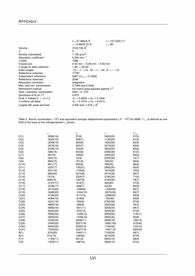
APPENDIX
b = 27.486(4) A β = 107.523(11)◦
c = 8.8823(12) A γ = 90◦
Volume 3103.7(8) A3
Z 2Density (calculated) 1.736 g/cm3
Absorption coefficient 5.293 mm−1
F (000) 1586Crystal size 0.35 mm × 0.08 mm × 0.06 mmθ-range for data collection 1.48→ 25.00◦
Index ranges -15→ h→15, -32→ k →32, -9→ l→10Reflections collected 17797Independent reflections 5467 [Rint = 0.1642]Reflections observed 2296Absorption correction IntegrationMax. and min. transmission 0.7484 and 0.4200Refinement method full-matrix least-squares against F 2
Data / restraints / parameters 5467 / 0 / 379Goodness-of-fit on F 2 0.973Final R indices [I > 2σ(I)] R1 = 0.0924, wR2 = 0.1934R indices (all data) R1 = 0.1914, wR2 = 0.2313Largest diff. peak and hole 2.405 and -1.078 e/A3
Table 2. Atomic coordinates (· 104) and equivalent isotropic displacement parameters (A2 · 103) for i0696. Ueq is defined as onethird of the trace of the orthogonalized Uij tensor.
x y z Ueq
C(1) -2880(16) 27(8) 2450(20) 57(5)C(2) -3220(15) 338(7) 1130(20) 61(5)C(3) -3048(15) 820(8) 1620(30) 62(5)C(4) -2576(16) 823(7) 3270(20) 63(6)C(5) -2435(14) 335(8) 3800(20) 63(6)C(6) -336(15) 843(8) 2390(30) 61(5)C(7) -95(16) 408(8) 3300(20) 64(6)C(8) -364(15) 10(9) 2320(30) 74(7)C(9) -894(15) 181(8) 730(30) 66(6)C(10) -851(17) 692(8) 780(20) 68(6)C(11) -223(17) 1353(7) 2860(20) 60(5)C(12) -831(19) 1733(8) 1970(30) 75(6)C(13) -680(20) 2210(8) 2410(30) 80(7)C(14) 73(19) 2330(7) 3740(30) 71(6)C(15) 666(19) 1957(9) 4720(30) 78(7)C(16) -3127(17) -818(7) 420(20) 57(5)C(17) -2226(17) -948(7) 20(30) 64(6)C(18) -2310(20) -1068(9) -1530(30) 83(7)C(19) -3240(20) -1044(10) -2670(30) 86(7)C(20) -4130(20) -917(10) -2280(30) 95(9)C(21) -4084(19) -818(8) -720(40) 83(8)C(22) -4231(18) -760(8) 2760(30) 67(6)C(23) -4865(18) -398(9) 3030(30) 76(7)C(24) -5856(19) -501(11) 3260(30) 84(7)C(25) -6200(20) -970(13) 3250(30) 97(9)C(26) -5580(30) -1326(12) 2950(40) 119(11)C(27) -4600(20) -1238(10) 2660(30) 90(8)C(28) -4230(30) 2028(11) 2890(70) 166(19)C(29) -3590(30) 2227(16) 1080(100) 210(40)C(30) -6940(30) 2630(14) 1740(40) 133(12)C(31) -7020(50) 2227(18) -160(110) 260(40)B(1) -970(20) 1463(11) 7150(30) 69(7)N(1) 512(14) 1483(6) 4210(20) 67(5)F(1) -1183(11) 951(4) 6903(15) 89(4)F(2) -1333(11) 1687(4) 5684(15) 87(4)
LVI

List of X-ray Data
F(3) 70(11) 1527(5) 7884(16) 99(4)F(4) -1563(12) 1638(5) 8123(15) 92(4)P(1) -2975(5) -632(2) 2453(6) 60(1)Fe(1) -1667(2) 437(1) 2186(3) 60(1)Au(1) -1668(1) -1032(1) 4207(1) 68(1)O(1) -3610(40) 2420(30) -340(90) 390(60)O(2) -6840(50) 2564(17) -1080(80) 340(30)
X-ray Data of 28
Table 1. Crystal data and structure refinement for tkpd1.
Code tkpd1Empirical formula C27H22Cl2FeNPPdFormula weight 624.58Temperature 150(2) KWavelength 0.71073 Acrystal system Monoclinicspacegroup P21/nUnit cell dimensions a = 15.4619(2) A α = 90◦
b = 10.5555(2) A β = 114.4480(10)◦
c = 15.9837(3) A γ = 90◦
Volume 2374.77(7) A3
Z 4Density (calculated) 1.747 g/cm3
Absorption coefficient 1.678 mm−1
F (000) 1248Crystal size 0.25 mm × 0.23 mm × 0.10 mmθ-range for data collection 1.54→ 27.49◦
Index ranges -19→ h→20, -13→ k →13, -20→ l→20Reflections collected 43867Independent reflections 5433 [Rint = 0.0461]Reflections observed 4613Absorption correction IntegrationMax. and min. transmission 0.864 and 0.665Refinement method full-matrix least-squares against F 2
Data / restraints / parameters 5433 / 0 / 298Goodness-of-fit on F 2 1.063Final R indices [I > 2σ(I)] R1 = 0.0292, wR2 = 0.0673R indices (all data) R1 = 0.0388, wR2 = 0.0723Largest diff. peak and hole 1.130 and -0.687 e/A3
Table 2. Atomic coordinates ((· 104)) and equivalent isotropic displacement parameters (A2 · 103) for tkpd1. Ueq is defined as onethird of the trace of the orthogonalized Uij tensor.
x y z Ueq
Pd 662(1) 19(1) 3986(1) 15(1)Fe 1593(1) 1286(1) 2343(1) 20(1)Cl(1) -928(1) -697(1) 3398(1) 24(1)Cl(2) 1218(1) -2034(1) 4135(1) 22(1)P 2156(1) 764(1) 4587(1) 16(1)N 159(1) 1835(2) 3746(1) 17(1)C(1) 2521(2) 878(2) 3662(2) 20(1)C(2) 2417(2) -178(2) 3067(2) 25(1)C(3) 2705(2) 208(3) 2373(2) 29(1)
LVII

APPENDIX
C(4) 2984(2) 1491(3) 2521(2) 26(1)C(5) 2879(2) 1918(2) 3314(2) 22(1)C(6) 315(2) 1986(2) 2268(2) 21(1)C(7) 201(2) 721(3) 1921(2) 25(1)C(8) 475(2) 704(3) 1171(2) 33(1)C(9) 753(2) 1944(3) 1049(2) 32(1)C(10) 655(2) 2737(3) 1719(2) 27(1)C(12) 58(2) 2519(2) 2991(2) 20(1)C(13) -95(2) 2345(2) 4379(2) 23(1)C(14) -451(2) 3554(3) 4302(2) 29(1)C(15) -550(2) 4277(3) 3548(2) 31(1)C(16) -300(2) 3749(3) 2890(2) 26(1)C(17) 3033(2) -249(2) 5441(2) 20(1)C(18) 3927(2) -401(3) 5441(2) 25(1)C(19) 4590(2) -1203(3) 6089(2) 30(1)C(20) 4358(2) -1828(3) 6731(2) 31(1)C(21) 3460(2) -1667(3) 6728(2) 28(1)C(22) 2802(2) -889(2) 6087(2) 23(1)C(23) 2323(2) 2317(2) 5127(2) 18(1)C(24) 2671(2) 2412(3) 6080(2) 23(1)C(25) 2794(2) 3594(3) 6498(2) 28(1)C(26) 2579(2) 4684(3) 5973(2) 30(1)C(27) 2204(2) 4600(3) 5021(2) 28(1)C(28) 2070(2) 3428(2) 4601(2) 22(1)
X-ray Data of 30
Table 1. Crystal data and structure refinement for tkpd2.
Code tkpd2Empirical formula C55H46Cl4Fe2N2P2ZnPdFormula weight 1156.78Temperature 150(2) KWavelength 0.71073 Acrystal system Triclinicspacegroup P 1Unit cell dimensions a = 11.2998(3) A α = 85.8700(11)◦
b = 12.3849(2) A β = 81.0170(11)◦
c = 18.7197(4) A γ = 70.1510(10)◦
Volume 2433.39(9) A3
Z 2Density (calculated) 1.579 g/cm3
Absorption coefficient 1.280 mm−1
F (000) 1172Crystal size 0.35 mm × 0.35 mm × 0.13 mmθ-range for data collection 1.75→ 27.53◦
Index ranges -14→ h→14, -16→ k →16, -23→ l→24Reflections collected 44051Independent reflections 11175 [Rint = 0.0485]Reflections observed 8567Absorption correction IntegrationMax. and min. transmission 0.857 and 0.655Refinement method full-matrix least-squares against F 2
Data / restraints / parameters 11175 / 0 / 598Goodness-of-fit on F 2 0.990Final R indices [I > 2σ(I)] R1 = 0.0389, wR2 = 0.0886R indices (all data) R1 = 0.0620, wR2 = 0.1006Largest diff. peak and hole 1.003 and -0.897 e/A3
LVIII

List of X-ray Data
Table 2. Atomic coordinates ((· 104)) and equivalent isotropic displacement parameters (A2 · 103) for tkpd2. Ueq is defined as onethird of the trace of the orthogonalized Uij tensor.
x y z Ueq
Pd(1) 5000 0 5000 19(1)Pd(2) 0 0 10000 18(1)Fe(1) 3547(1) 3152(1) 6301(1) 23(1)Fe(2) -3007(1) 2667(1) 9033(1) 21(1)P(1) 2967(1) 1370(1) 5149(1) 20(1)P(2) -2111(1) 102(1) 10024(1) 19(1)Cl(1) 4565(1) -1129(1) 5966(1) 27(1)Cl(2) 391(1) -842(1) 8891(1) 28(1)C(1) 2915(3) 2748(2) 5426(2) 22(1)C(2) 3855(3) 3294(3) 5203(2) 27(1)C(3) 3399(4) 4390(3) 5513(2) 32(1)C(4) 2194(3) 4545(3) 5928(2) 32(1)C(5) 1886(3) 3538(3) 5882(2) 27(1)C(6) 3477(3) 3454(3) 7372(2) 28(1)C(7) 3398(3) 2343(3) 7289(2) 30(1)C(8) 4511(3) 1693(3) 6837(2) 32(1)C(9) 5279(3) 2391(3) 6638(2) 34(1)C(10) 4650(3) 3473(3) 6968(2) 31(1)N(1) 1284(3) 4408(3) 7872(2) 38(1)C(12) 2494(3) 4404(3) 7778(2) 28(1)C(13) 376(4) 5292(3) 8221(2) 35(1)C(14) 652(4) 6184(3) 8496(2) 35(1)C(15) 1896(4) 6139(3) 8402(2) 39(1)C(16) 2823(3) 5250(3) 8049(2) 32(1)C(17) 2325(3) 1653(3) 4293(2) 25(1)C(18) 1590(3) 2728(3) 4084(2) 34(1)C(19) 1144(4) 2907(4) 3421(2) 44(1)C(20) 1471(4) 2011(4) 2953(2) 45(1)C(21) 2221(3) 931(4) 3151(2) 38(1)C(22) 2644(3) 753(3) 3819(2) 31(1)C(23) 1729(3) 1055(3) 5793(2) 26(1)C(24) 1926(3) 796(3) 6512(2) 30(1)C(25) 1012(4) 561(3) 7015(2) 40(1)C(26) -103(4) 575(4) 6810(2) 48(1)C(27) -319(4) 819(4) 6103(2) 47(1)C(28) 596(3) 1067(3) 5589(2) 35(1)C(31) -2924(3) 1020(2) 9334(2) 22(1)C(32) -2418(3) 1087(3) 8586(2) 25(1)C(33) -3396(3) 1862(3) 8228(2) 31(1)C(34) -4495(3) 2289(3) 8738(2) 34(1)C(35) -4216(3) 1777(3) 9426(2) 27(1)C(36) -2546(3) 4027(2) 8518(2) 24(1)C(37) -3578(3) 4417(3) 9084(2) 30(1)C(38) -3168(4) 3905(3) 9747(2) 33(1)C(39) -1885(3) 3194(3) 9599(2) 27(1)C(40) -1497(3) 3254(3) 8843(2) 26(1)N(2) -3554(3) 5267(2) 7592(2) 38(1)C(42) -2585(3) 4338(3) 7749(2) 26(1)C(43) -3604(4) 5564(3) 6889(2) 48(1)C(44) -2749(4) 4956(4) 6332(2) 47(1)C(45) -1762(4) 3995(4) 6495(2) 46(1)C(46) -1675(4) 3691(3) 7216(2) 38(1)C(47) -3229(3) 592(3) 10846(2) 22(1)C(48) -3933(3) -57(3) 11213(2) 25(1)C(49) -4789(3) 370(3) 11824(2) 31(1)C(50) -4954(3) 1439(3) 12076(2) 33(1)C(51) -4268(3) 2102(3) 11708(2) 34(1)C(52) -3408(3) 1675(3) 11099(2) 27(1)C(53) -2174(3) -1317(2) 9881(2) 21(1)C(54) -2761(3) -1535(3) 9337(2) 26(1)
LIX

APPENDIX
C(55) -2777(3) -2642(3) 9257(2) 33(1)C(56) -2221(3) -3519(3) 9719(2) 37(1)C(57) -1631(4) -3307(3) 10255(2) 37(1)C(58) -1590(3) -2214(3) 10338(2) 30(1)C(90) 1895(4) -1992(4) 5964(2) 54(1)Cl(91) 2471(2) -2936(1) 6663(1) 81(1)Cl(92) 2413(1) -2590(1) 5121(1) 65(1)
X-ray Data of 31
Table 1. Crystal data and structure refinement for ps307b.
Code ps307bEmpirical formula C56H48Cl2Fe2N2P2PdFormula weight 1099.90Temperature 150(2) KWavelength 0.71073 Acrystal system Triclinicspacegroup P 1Unit cell dimensions a = 9.94370(10) A α = 66.8117(7)◦
b = 10.99230(10) A β = 72.6510(6)◦
c = 12.0044(2) A γ = 77.0771(7)◦
Volume 1142.79(2 A3
Z 1Density (calculated) 1.598 g/cm3
Absorption coefficient 1.245 mm−1
F (000) 560Crystal size 0.43 mm × 0.42 mm × 0.20 mmθ-range for data collection 1.90→ 27.57◦
Index ranges -12→ h→12, -14→ k →14, -15→ l→15Reflections collected 41724Independent reflections 5249 [Rint = 0.0315]Reflections observed 4978Absorption correction IntegrationMax. and min. transmission 0.815 and 0.615Refinement method full-matrix least-squares against F 2
Data / restraints / parameters 5249 / 0 / 295Goodness-of-fit on F 2 1.065Final R indices [I > 2σ(I)] R1 = 0.0218, wR2 = 0.0514R indices (all data) R1 = 0.0235, wR2 = 0.0524Largest diff. peak and hole 0.390 and -0.639 e/A3
Table 2. Atomic coordinates ((· 104)) and equivalent isotropic displacement parameters (A2 · 103) for ps307b. Ueq is defined asone third of the trace of the orthogonalized Uij tensor.
x y z Ueq
Pd 10000 0 0 13(1)Fe 5894(1) 2006(1) 1620(1) 15(1)Cl 8798(1) -1402(1) -254(1) 21(1)P 8502(1) 1801(1) -1030(1) 13(1)N 3191(2) 2500(2) 5675(1) 32(1)C(1) 6706(2) 1970(1) -142(1) 16(1)C(2) 5785(2) 3189(2) -169(1) 18(1)C(3) 4414(2) 2851(2) 556(2) 22(1)C(4) 4466(2) 1441(2) 1028(2) 22(1)
LX

List of X-ray Data
C(5) 5873(2) 885(2) 610(1) 19(1)C(6) 5138(2) 2639(2) 3115(1) 22(1)C(7) 5338(2) 1224(2) 3531(1) 22(1)C(8) 6787(2) 801(2) 3090(1) 23(1)C(9) 7498(2) 1953(2) 2391(2) 24(1)C(10) 6482(2) 3090(2) 2409(2) 24(1)C(11) 3747(2) 3467(2) 3408(2) 33(1)C(12) 3445(2) 3606(2) 4672(2) 25(1)C(13) 2948(2) 2606(2) 6794(2) 31(1)C(14) 2931(2) 3768(2) 6977(2) 31(1)C(15) 3155(2) 4906(2) 5939(2) 35(1)C(16) 3414(2) 4819(2) 4770(2) 30(1)C(17) 8310(2) 1591(1) -2404(1) 17(1)C(18) 9544(2) 1323(2) -3244(2) 22(1)C(19) 9460(2) 1214(2) -4333(2) 29(1)C(20) 8147(2) 1339(2) -4574(2) 32(1)C(21) 6919(2) 1581(2) -3737(2) 30(1)C(22) 6994(2) 1720(2) -2651(2) 21(1)C(23) 8982(2) 3499(1) -1644(1) 16(1)C(24) 8952(2) 4121(2) -816(1) 20(1)C(25) 9255(2) 5427(2) -1261(2) 23(1)C(26) 9623(2) 6120(2) -2531(2) 26(1)C(27) 9673(2) 5509(2) -3357(2) 27(1)C(28) 9348(2) 4212(2) -2921(2) 21(1)
X-ray Data of 32
Table 1. Crystal data and structure refinement for tkpd3.
Code tkpd3Empirical formula C36H34ClFeN2O4PPdFormula weight 787.32Temperature 150(2) KWavelength 0.71073 Acrystal system Monoclinicspacegroup P21/cUnit cell dimensions a = 17.2610(6) A α = 90◦
b = 10.9697(2) A β = 92.5390(14)◦
c = 16.9970(5) A γ = 90◦
Volume 3215.19(16) A3
Z 4Density (calculated) 1.627 g/cm3
Absorption coefficient 1.187 mm−1
F (000) 1600Crystal size 0.13 mm × 0.10 mm × 0.03 mmθ-range for data collection 2.20→ 26.07◦
Index ranges -21→ h→21, -13→ k →12, -19→ l→20Reflections collected 42208Independent reflections 6352 [Rint = 0.0876]Reflections observed 4458Absorption correction IntegrationMax. and min. transmission 0.956 and 0.865Refinement method full-matrix least-squares against F 2
Data / restraints / parameters 6352 / 0 / 417Goodness-of-fit on F 2 1.023Final R indices [I > 2σ(I)] R1 = 0.0393, wR2 = 0.0677R indices (all data) R1 = 0.0763, wR2 = 0.0782Largest diff. peak and hole 0.570 and -0.561 e/A3
LXI
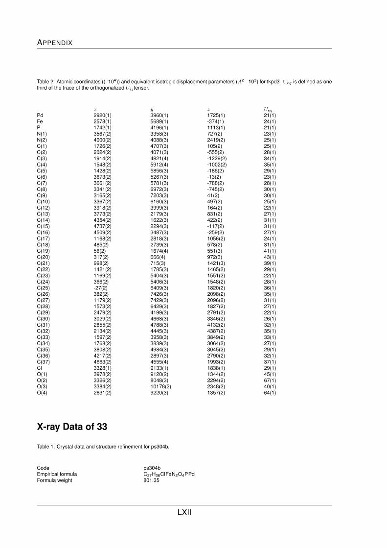
APPENDIX
Table 2. Atomic coordinates ((· 104)) and equivalent isotropic displacement parameters (A2 · 103) for tkpd3. Ueq is defined as onethird of the trace of the orthogonalized Uij tensor.
x y z Ueq
Pd 2920(1) 3960(1) 1725(1) 21(1)Fe 2578(1) 5689(1) -374(1) 24(1)P 1742(1) 4196(1) 1113(1) 21(1)N(1) 3567(2) 3358(3) 727(2) 23(1)N(2) 4000(2) 4088(3) 2419(2) 25(1)C(1) 1726(2) 4707(3) 105(2) 25(1)C(2) 2024(2) 4071(3) -555(2) 28(1)C(3) 1914(2) 4821(4) -1229(2) 34(1)C(4) 1548(2) 5912(4) -1002(2) 35(1)C(5) 1428(2) 5856(3) -186(2) 29(1)C(6) 3673(2) 5267(3) -13(2) 23(1)C(7) 3661(2) 5781(3) -788(2) 28(1)C(8) 3341(2) 6972(3) -745(2) 30(1)C(9) 3165(2) 7203(3) 41(2) 30(1)C(10) 3367(2) 6160(3) 497(2) 25(1)C(12) 3918(2) 3999(3) 164(2) 22(1)C(13) 3773(2) 2179(3) 831(2) 27(1)C(14) 4354(2) 1622(3) 422(2) 31(1)C(15) 4737(2) 2294(3) -117(2) 31(1)C(16) 4509(2) 3487(3) -259(2) 27(1)C(17) 1168(2) 2818(3) 1056(2) 24(1)C(18) 485(2) 2739(3) 578(2) 31(1)C(19) 56(2) 1674(4) 551(3) 41(1)C(20) 317(2) 666(4) 972(3) 43(1)C(21) 998(2) 715(3) 1421(3) 39(1)C(22) 1421(2) 1785(3) 1465(2) 29(1)C(23) 1169(2) 5404(3) 1551(2) 22(1)C(24) 366(2) 5406(3) 1548(2) 28(1)C(25) -27(2) 6409(3) 1820(2) 36(1)C(26) 382(2) 7426(3) 2098(2) 35(1)C(27) 1179(2) 7429(3) 2096(2) 31(1)C(28) 1573(2) 6429(3) 1827(2) 27(1)C(29) 2479(2) 4199(3) 2791(2) 22(1)C(30) 3029(2) 4668(3) 3346(2) 26(1)C(31) 2855(2) 4788(3) 4132(2) 32(1)C(32) 2134(2) 4445(3) 4387(2) 35(1)C(33) 1597(2) 3958(3) 3849(2) 33(1)C(34) 1768(2) 3839(3) 3064(2) 27(1)C(35) 3808(2) 4984(3) 3045(2) 29(1)C(36) 4217(2) 2897(3) 2790(2) 32(1)C(37) 4663(2) 4555(4) 1993(2) 37(1)Cl 3328(1) 9133(1) 1838(1) 29(1)O(1) 3978(2) 9120(2) 1344(2) 45(1)O(2) 3326(2) 8048(3) 2294(2) 67(1)O(3) 3384(2) 10178(2) 2348(2) 40(1)O(4) 2631(2) 9220(3) 1357(2) 64(1)
X-ray Data of 33
Table 1. Crystal data and structure refinement for ps304b.
Code ps304bEmpirical formula C37H36ClFeN2O4PPdFormula weight 801.35
LXII

List of X-ray Data
Temperature 120(2) KWavelength 0.71073 Acrystal system Monoclinicspacegroup C2/cUnit cell dimensions a = 22.9503(3) A α = 90◦
b = 18.1430(2) A β = 125.6086(5)◦
c = 19.2860(2) A γ = 90◦
Volume 6528.85(13) A3
Z 8Density (calculated) 1.631 g/cm3
Absorption coefficient 1.171 mm−1
F (000) 3264Crystal size 0.37 mm × 0.25 mm × 0.23 mmθ-range for data collection 1.57→ 27.51◦
Index ranges -29→ h→29, -23→ k →23, -25→ l→25Reflections collected 70441Independent reflections 7503 [Rint = 0.0390]Reflections observed 6652Absorption correction Gaussian integrationMax. and min. transmission 0.853 and 0.707Refinement method full-matrix least-squares against F 2
Data / restraints / parameters 7503 / 0 / 429Goodness-of-fit on F 2 1.039Final R indices [I > 2σ(I)] R1 = 0.0281, wR2 = 0.0662R indices (all data) R1 = 0.0341, wR2 = 0.0700Largest diff. peak and hole 1.048 and -0.848 e/A3
Table 2. Atomic coordinates ((· 104)) and equivalent isotropic displacement parameters (A2 · 103) for ps304b. Ueq is defined asone third of the trace of the orthogonalized Uij tensor.
x y z Ueq
Pd 3311(1) 2649(1) 2945(1) 15(1)Fe 3372(1) 698(1) 4447(1) 19(1)P 2414(1) 2002(1) 2809(1) 16(1)N(1) 3882(1) 2704(1) 4303(1) 17(1)N(2) 4094(1) 3332(1) 2995(1) 19(1)C(1) 2568(1) 1417(1) 3657(1) 19(1)C(2) 2291(1) 682(1) 3537(1) 24(1)C(3) 2442(1) 431(1) 4322(2) 28(1)C(4) 2808(1) 1001(1) 4933(1) 25(1)C(5) 2893(1) 1612(1) 4536(1) 20(1)C(6) 4346(1) 917(1) 4661(1) 22(1)C(7) 3956(1) 383(1) 4002(2) 26(1)C(8) 3821(1) -235(1) 4342(2) 31(1)C(9) 4124(1) -89(1) 5214(2) 29(1)C(10) 4449(1) 620(1) 5410(1) 24(1)C(11) 4625(1) 1630(1) 4559(1) 23(1)C(12) 4445(1) 2284(1) 4878(1) 18(1)C(13) 3685(1) 3271(1) 4581(1) 20(1)C(14) 4040(1) 3443(1) 5434(2) 25(1)C(15) 4633(1) 3026(1) 6026(1) 26(1)C(16) 4835(1) 2445(1) 5746(1) 23(1)C(17) 1946(1) 1373(1) 1902(1) 19(1)C(18) 1224(1) 1204(1) 1489(1) 24(1)C(19) 881(1) 729(1) 792(2) 31(1)C(20) 1265(2) 413(1) 520(2) 36(1)C(21) 1990(2) 557(1) 948(2) 33(1)C(22) 2331(1) 1042(1) 1635(2) 26(1)C(23) 1786(1) 2677(1) 2735(1) 17(1)C(24) 1802(1) 3404(1) 2505(1) 22(1)C(25) 1390(1) 3948(1) 2528(1) 26(1)
LXIII
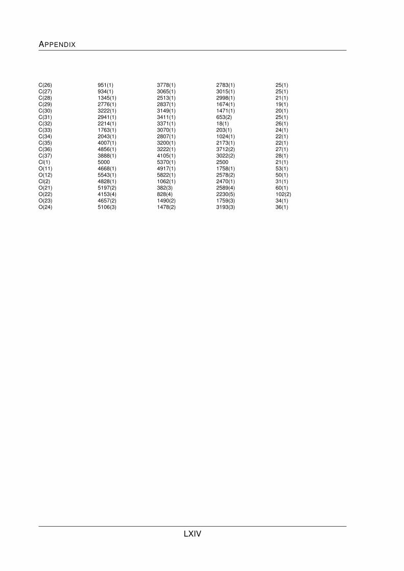
APPENDIX
C(26) 951(1) 3778(1) 2783(1) 25(1)C(27) 934(1) 3065(1) 3015(1) 25(1)C(28) 1345(1) 2513(1) 2998(1) 21(1)C(29) 2776(1) 2837(1) 1674(1) 19(1)C(30) 3222(1) 3149(1) 1471(1) 20(1)C(31) 2941(1) 3411(1) 653(2) 25(1)C(32) 2214(1) 3371(1) 18(1) 26(1)C(33) 1763(1) 3070(1) 203(1) 24(1)C(34) 2043(1) 2807(1) 1024(1) 22(1)C(35) 4007(1) 3200(1) 2173(1) 22(1)C(36) 4856(1) 3222(1) 3712(2) 27(1)C(37) 3888(1) 4105(1) 3022(2) 28(1)Cl(1) 5000 5370(1) 2500 21(1)O(11) 4668(1) 4917(1) 1758(1) 53(1)O(12) 5543(1) 5822(1) 2578(2) 50(1)Cl(2) 4828(1) 1062(1) 2470(1) 31(1)O(21) 5197(2) 382(3) 2589(4) 60(1)O(22) 4153(4) 828(4) 2230(5) 102(2)O(23) 4657(2) 1490(2) 1759(3) 34(1)O(24) 5106(3) 1478(2) 3193(3) 36(1)
LXIV
Related Documents