Anterior Segment Cataract © TOUCH MEDICAL MEDIA 2014 37 Abstract The use of the femtosecond laser in ophthalmic surgery over the last decade has resulted in the development of innovative procedures. The ultra-short infrared laser pulses of the femtosecond laser can be applied precisely and predictably with minimal collateral tissue damage, making it an ideal tool for highly precise ophthalmic surgery. Flap creation in laser in situ keratomileusis (LASIK) is the most common use of this laser. It can also be used for other corneal refractive procedures, lamellar and full-thickness corneal transplantation and cataract surgery. This article summarises recent advanced applying femtosecond laser technology in ophthalmology. Keywords Femtosecond, laser, laser in situ keratomileusis, LASIK, cataract, femtosecond laser-assisted corneal surgery, femtosecond laser-assisted keratoplasty, intracorneal ring segment, astigmatism keratotomy, femtosecond laser-assisted cataract surgery Disclosure: Duna A Raoof and Roni M Shtein have no conflicts of interest to declare. No funding was received for the publication of this article. Received: 16 January 2013 Accepted: 20 February 2013 Citation: European Ophthalmic Review, 2014;8(1):37–9 Correspondence: Roni M Shtein, Associate Professor of Ophthalmology and Visual Sciences, University of Michigan WK Kellogg Eye Center, 1000 Wall Street, Ann Arbor, MI 48105, US. E: [email protected] Femtosecond Lasers in Ophthalmology Duna A Raoof 1 and Roni M Shtein 2 1. Cornea Fellow, Massachusetts Eye and Ear Infirmary, Boston, Massachusetts, US; 2. Associate Professor, Kellogg Eye Center, University of Michigan, Michigan, US The versatility, predictability and unique properties of the femtosecond laser have allowed its application in multiple avenues of anterior segment surgery. Femtosecond lasers generate ultra-short pulses utilising small amounts of energy and minimising damage to any of the surrounding tissues. Here, we summarise the surgical techniques that have been developed in ophthalmic surgery utilising the femtosecond laser since its approval by the US Food and Drug Administration (FDA) in 2001. The femtosecond laser was initially introduced for creation of corneal flaps for laser in situ keratomileusis (LASIK). Since then, the use of femtosecond lasers has expanded to other corneal surgeries and, recently, to cataract surgery. Femtosecond Laser Principles The femtosecond laser is an infrared laser (wavelength: 1,053 nm) with ultra-short pulse duration (10 – 15 s). Given its short pulse duration, the femtosecond laser has the ability to deliver laser energy with minimal collateral damage to the adjacent tissue. Thermal damage to neighbouring tissue in the cornea has been measured to be in the order of 1 μm. 1 The tissue interaction this laser utilises is known as photo-disruption, a process in which small volumes of tissue are vapourised resulting in the formation of cavitation gas (carbon dioxide and water) bubbles. 2 Furthermore, the femtosecond laser is unique in that it can be focused anywhere within or behind the cornea and is capable, to a certain extent, of passing through optically hazy media, such as an oedematous cornea. The laser may be applied in multiple geometric patterns including vertical, spiral or zig-zag cuts. Femtosecond Laser Systems There are multiple commercially available femtosecond laser models: • Intralase FS™ (Abbott Medical Optics, Abbott Park, Illinois); • Femtec ® (20/10 Perfect Vision, Heidelberg, Germany); • VisuMax Femtosecond System ® (Carl Zeiss Meditec, Jena, Germany); • Femto LDV™ (Ziemer Group, Port, Switzerland); and • Wavelight FS200 ® (Alcon, Fort Worth, Texas). Systems designed specifically for cataract surgery include: • LenSx (Alcon, Fort Worth, Texas); • Catalys (OptiMedica ® , Sunnyvale, California); • LensAR (LensAR Inc., Orlando, Florida); and • VICTUS (Technolas and Bausch and Lomb). The early femtosecond laser systems operated with a low repetition rate (15 kHz) and thus required higher energy to operate. The new devices have an increased repetition rate (as high as 150 kHz), which leads to utilisation of less energy and shorter procedure duration. In addition, the devices vary in their programmed and customisable geometric cut patterns. Each laser system has distinctive features allowing it to be popularised for use in specific procedures. Refractive Surgery Laser In Situ Keratomileusis Flaps In ophthalmic surgery, the femtosecond laser was first popularised as an alternative to the mechanical microkeratome for the creation of LASIK flaps. The femtosecond laser is applied to the corneal stroma at a pre- calculated depth. Flap creation using the femtosecond laser has been compared with creation using the mechanical microkeratome. Reports have shown reduced higher-order aberrations 3 and enhanced flap thickness predictability. 4 In addition, the femtosecond laser offers more options in terms of flap thickness, side cut angle, hinge specifications and firing patterns. DOI: 10.17925/EOR.2014.08.01.37

Welcome message from author
This document is posted to help you gain knowledge. Please leave a comment to let me know what you think about it! Share it to your friends and learn new things together.
Transcript
Anterior Segment Cataract
© TouCh MEdiCal MEdia 2014 37
AbstractThe use of the femtosecond laser in ophthalmic surgery over the last decade has resulted in the development of innovative procedures. The
ultra-short infrared laser pulses of the femtosecond laser can be applied precisely and predictably with minimal collateral tissue damage,
making it an ideal tool for highly precise ophthalmic surgery. Flap creation in laser in situ keratomileusis (LASIK) is the most common use
of this laser. It can also be used for other corneal refractive procedures, lamellar and full-thickness corneal transplantation and cataract
surgery. This article summarises recent advanced applying femtosecond laser technology in ophthalmology.
KeywordsFemtosecond, laser, laser in situ keratomileusis, LASIK, cataract, femtosecond laser-assisted corneal surgery, femtosecond laser-assisted
keratoplasty, intracorneal ring segment, astigmatism keratotomy, femtosecond laser-assisted cataract surgery
Disclosure: Duna A Raoof and Roni M Shtein have no conflicts of interest to declare. No funding was received for the publication of this article.
Received: 16 January 2013 Accepted: 20 February 2013 Citation: European Ophthalmic Review, 2014;8(1):37–9
Correspondence: Roni M Shtein, Associate Professor of Ophthalmology and Visual Sciences, University of Michigan WK Kellogg Eye Center, 1000 Wall Street, Ann Arbor, MI
48105, US. E: [email protected]
Femtosecond Lasers in Ophthalmology
Duna A Raoof1 and Roni M Shtein2
1. Cornea Fellow, Massachusetts Eye and Ear Infirmary, Boston, Massachusetts, US; 2. Associate Professor, Kellogg Eye Center, University of Michigan, Michigan, US
The versatility, predictability and unique properties of the femtosecond
laser have allowed its application in multiple avenues of anterior
segment surgery. Femtosecond lasers generate ultra-short pulses
utilising small amounts of energy and minimising damage to any of the
surrounding tissues.
Here, we summarise the surgical techniques that have been developed
in ophthalmic surgery utilising the femtosecond laser since its approval
by the US Food and Drug Administration (FDA) in 2001. The femtosecond
laser was initially introduced for creation of corneal flaps for laser in situ
keratomileusis (LASIK). Since then, the use of femtosecond lasers has
expanded to other corneal surgeries and, recently, to cataract surgery.
Femtosecond Laser PrinciplesThe femtosecond laser is an infrared laser (wavelength: 1,053 nm) with
ultra-short pulse duration (10–15 s). Given its short pulse duration,
the femtosecond laser has the ability to deliver laser energy with
minimal collateral damage to the adjacent tissue. Thermal damage
to neighbouring tissue in the cornea has been measured to be in the
order of 1 μm.1 The tissue interaction this laser utilises is known as
photo-disruption, a process in which small volumes of tissue are
vapourised resulting in the formation of cavitation gas (carbon dioxide
and water) bubbles.2 Furthermore, the femtosecond laser is unique in
that it can be focused anywhere within or behind the cornea and is
capable, to a certain extent, of passing through optically hazy media,
such as an oedematous cornea. The laser may be applied in multiple
geometric patterns including vertical, spiral or zig-zag cuts.
Femtosecond Laser SystemsThere are multiple commercially available femtosecond laser models:
• Intralase FS™ (Abbott Medical Optics, Abbott Park, Illinois);
• Femtec® (20/10 Perfect Vision, Heidelberg, Germany);
• VisuMax Femtosecond System® (Carl Zeiss Meditec, Jena,
Germany);
• Femto LDV™ (Ziemer Group, Port, Switzerland); and
• Wavelight FS200® (Alcon, Fort Worth, Texas).
Systems designed specifically for cataract surgery include:
• LenSx (Alcon, Fort Worth, Texas);
• Catalys (OptiMedica®, Sunnyvale, California);
• LensAR (LensAR Inc., Orlando, Florida); and
• VICTUS (Technolas and Bausch and Lomb).
The early femtosecond laser systems operated with a low repetition rate
(15 kHz) and thus required higher energy to operate. The new devices
have an increased repetition rate (as high as 150 kHz), which leads to
utilisation of less energy and shorter procedure duration. In addition,
the devices vary in their programmed and customisable geometric cut
patterns. Each laser system has distinctive features allowing it to be
popularised for use in specific procedures.
Refractive SurgeryLaser In Situ Keratomileusis FlapsIn ophthalmic surgery, the femtosecond laser was first popularised as an
alternative to the mechanical microkeratome for the creation of LASIK
flaps. The femtosecond laser is applied to the corneal stroma at a pre-
calculated depth. Flap creation using the femtosecond laser has been
compared with creation using the mechanical microkeratome. Reports
have shown reduced higher-order aberrations3 and enhanced flap
thickness predictability.4 In addition, the femtosecond laser offers more
options in terms of flap thickness, side cut angle, hinge specifications
and firing patterns.
Shtein_EU_FINAL.indd 37 15/07/2014 14:52
DOI: 10.17925/EOR.2014.08.01.37
38
Anterior Segment Cataract
EuropEan ophthalmiC rEviEw
Complications associated with the use of femtosecond laser for LASIK
flap creation are rare. The opaque bubble layer (OBL) forms along the
cutting plane and may limit the ability of the surgeon or the excimer
laser eye tracker to locate the pupil for centration. This can occur when
cavitation bubbles escape into the deep corneal stroma, although most
disappear spontaneously. Transient light-sensitivity syndrome (TLSS) is
characterised by photophobia and pain that may appear days to weeks
following LASIK with femtosecond laser flap creation.5 It typically
resolves after aggressive treatment with topical steroids.
Intracorneal Ring SegmentsIntracorneal ring segments (INTACS) are thin semicircular inserts
made of polymethylmethacrylate that are implanted in the corneal
stroma to shorten the arc length of the central corneal surface and
result in corneal surface flattening. Intracorneal ring segments have
been used to treat corneal ectatic disorders such as keratoconus and
post-LASIK ectasia, as well as myopia. The femtosecond laser may be
programmed to create tunnels for INTACS implantation. This technique
has been shown to be similar to manual tunnel dissection in terms of
visual and refractive outcomes.6,7 The consistency of depth, uniformity
of cut and the minimal trauma induced when creating the channels
using the femtosecond laser can make insertion of the INTACS easier
and minimise the duration of the procedure.7
Astigmatic KeratotomyFemtosecond laser-assisted astigmatic keratotomy has been primarily
described for treatment of high astigmatism following penetrating
keratoplasty.8,9 As the femtosecond laser is capable of creating incisions
with high precision and reproducibility, it can be used to control the
desired length, shape and depth of incisions for astigmatic keratotomy.
Multiple studies have reported enhanced predictability and reduced
complications rates, such as microperforation and decentration, in
femtosecond laser-assisted astigmatic keratotomy compared with
manual techniques.10,11 Axial topographic maps are used to identify the
steep meridians and a standardised nomogram is used to generate a
surgical plan with paired incisions for each patient.
Femtosecond Laser-assisted Lenticule ExtractionFemtosecond laser-assisted lenticule extraction (also known as FLEx)
is used to correct myopia. The technique involves making two lamellar
cuts in the cornea that intersect in the periphery, thus creating a
lenticule that is removed. The lenticule is extracted through a traditional,
femtosecond laser-created corneal flap. The removal of the lenticule
reduces the curvature of the cornea, subsequently reducing myopia.
In a study by Blum et al., 6-month results demonstrated that FLEx is
both a safe and promising procedure for treatment of myopia.12
Small Incision Lenticule ExtractionThis technique is similar to FLEx in that a corneal lenticule is extracted
to correct myopia. However, the lenticule is removed though a small
femtosecond laser-created side cut rather than a flap. As this procedure
does not involve creation of a flap, it may result in lower incidence of
dry eye and ectasia, and it also eliminates the potential of flap-related
complications. Reports show promising results in correcting myopia at
6 months.13,14
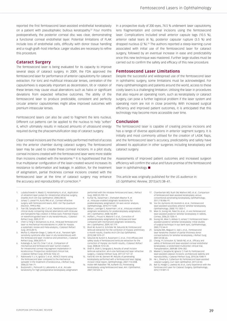
Corneal TransplantFemtosecond Laser-assisted KeratoplastyThe femtosecond laser was approved for the creation of full and
partial-thickness corneal incisions for keratoplasty in 2005. Prior
to keratoplasty surgery, the desired incision pattern is first applied
to the donor cornea then a corresponding pattern is applied to the
recipient cornea using the femtosecond laser. The recipient incisions
are left incomplete in order to facilitate transfer of the patient to
the operating room. The uncut bridge is then dissected in the
operating room and the keratoplasty is completed in similar manner
to traditional keratoplasty surgery (see Figure 1).
Performing femtosecond laser-assisted keratoplasty (FLAK) has a
few advantages over traditional penetrating keratoplasty. Different
patterns of trephination cuts may be applied, such as top-hat, zig-zag
or mushroom shapes. These configurations result in greater surface
area of graft–host contact, which translates to shorter healing time
and faster suture removal.15–17 The mushroom configuration may be
advantageous for keratoconus by providing a larger anterior refractive
surface, while a top-hat pattern may be preferred in endothelial disease
in order to replace more endothelial cells.
Anterior Lamellar KeratoplastyAnterior lamellar keratoplasty consists of transplantation of the anterior
layer of the cornea where only the anterior lamella is diseased, such
as anterior corneal scars, degenerations or dystrophies. Advantages of
anterior lamellar keratoplasty include its less-invasive nature and reduced
risk of rejection. Precise manual lamellar dissection is challenging,
however. In a study by Yoo et al., the depth of the anterior corneal
pathology was determined using anterior segment optical coherence
tomography, and the femtosecond laser was used to prepare the donor
tissue and recipient eye to successfully perform femtosecond laser-
assisted anterior lamellar keratoplasty.18 The only reported complications
included dry eye – otherwise no incidences of graft rejection, infection
or epithelial ingrowth were reported. Outcomes with femtosecond-laser
anterior lamellar keratoplasty need to be evaluated further to determine
benefits over standard anterior lamellar keratoplasty.
Endothelial KeratoplastyDescemet’s stripping endothelial keratoplasty has become the
standard procedure for isolated posterior pathology, such as Fuch’s
endothelial dystrophy and pseudophakic bullous keratopathy. The
femtosecond laser has been utilised experimentally in preparation
of donor tissue for endothelial keratoplasty as well as in vivo rabbit
models.19,20 Initial reports demonstrated preparation of a donor cornea
using the femtosecond laser is safe.21 Cheng et al. subsequently
Figure 1: Femtosecond Laser-assisted Keratoplasty with a Mushroom-shaped Pattern
Shtein_EU_FINAL.indd 38 15/07/2014 14:52
EuropEan ophthalmic rEviEw 39
Femtosecond Lasers in Ophthalmology
reported the first femtosecond laser-assisted endothelial keratoplasty
on a patient with pseudophakic bullous keratopathy.22 Four months
postoperatively, the posterior corneal disc was clear, demonstrating
a functional corneal endothelial layer. Potential limitations of FLAK
include loss of endothelial cells, difficulty with donor tissue handling
and a rough graft–host interface. Larger studies are necessary to refine
this procedure.
Cataract Surgery The femtosecond laser is being evaluated for its capacity to improve
several steps of cataract surgery. In 2009, the FDA approved the
femtosecond laser for performance of anterior capsulotomy for cataract
extraction. For toric and multifocal intraocular lenses, centration of the
capsulorhexis is especially important as decentration, tilt or rotation of
these lenses may cause visual aberrations such as halos or significant
deviations from expected refractive outcomes. The ability of the
femtosecond laser to produce predictable, consistent and perfectly
circular anterior capsulotomies might allow improved outcomes with
premium intraocular lenses.
Femtosecond lasers can also be used to fragment the lens nucleus.
Different cut patterns can be applied to the nucleus to help ‘soften’
it, which ultimately results in reduced amounts of ultrasound energy
required during the phacoemulsification step of cataract surgery.
Clear corneal incisions are the most widely performed method of access
into the anterior chamber during cataract surgery. The femtosecond
laser may be used to create these corneal incisions. In a pilot study,
corneal incisions created with the femtosecond laser were more stable
than incisions created with the keratome.23 It is hypothesised that the
true multiplanar configuration of the laser-created wound increases its
resistance to deformation and leakage. In addition, for the treatment
of astigmatism, partial thickness corneal incisions created with the
femtosecond laser at the time of cataract surgery may enhance
the accuracy and reproducibility of correction.24
In a prospective study of 200 eyes, 74.5 % underwent laser capsulotomy,
lens fragmentation and corneal incisions using the femtosecond
laser. Complications included small anterior capsule tags (10.5 %),
anterior radial tears (4 %), posterior capsular rupture (3.5 %) and
dropped nucleus (2 %).25 The authors reported a steep-learning curve
associated with initial use of the femtosecond laser for cataract
surgery, followed by an eventual increase in ease and predictability
once this new technique was mastered. Further large studies must be
carried out to confirm the safety and efficacy of this new procedure.
Femtosecond Laser LimitationsDespite the successful and widespread use of the femtosecond laser
in ophthalmic surgery, some limitations must be acknowledged. For
many ophthalmologists and patients around the world, access to these
costly lasers is a challenging limitation. Utilising the laser in procedures
that also require an operating room, such as keratoplasty or cataract
surgery, can pose a further logistical problem if the laser system and
operating room are not in close proximity. With increased surgical
efficiency and improved patient outcomes, it is anticipated that this
technology may become more accessible over time.
ConclusionThe femtosecond laser is capable of creating precise incisions and
has a range of diverse applications in anterior segment surgery. It is
initially and most commonly utilised for the creation of LASIK flaps,
and the femtosecond laser’s accuracy, predictability and safety have
allowed its application in other surgeries including keratoplasty and
cataract surgery.
Assessments of improved patient outcomes and increased surgeon
efficiency will confirm the value and future promise of the femtosecond
laser in ophthalmology. n
This article was originally published for the US audience in:
US Ophthalmic Review, 2013;6(1):38–41.
1. Lubatschowski H, Maatz G, Heisterkamp A, et al., Application of ultrashort laser pulses for intrastromal refractive surgery, Graefes Arch Clin Exp Ophthalmol, 2000;238:33.
2. Juhasz T, Loesel FH, Kurtz RM, et al., Corneal refractive surgery with femtosecond lasers, IEEE J Sel Top Quantum Electron, 1999;5:902.
3. Tran DB, Sarayba MA, Bor Z, et al., Randomized prospective clinical study comparing induced aberrations with IntraLase and hansatome flap creation in fellow eyes: Potential impact on wavefront-guided laser in situ keratomileusis, J Cataract Refract Surg, 2005;31:97.
4. Chen S, Feng Y, Stonjanovic A, et al., IntraLase femtosecond laser vs mechanical microkeratome in LASIK for myopia: a systematic review and meta-analysis, J Cataract Refract Surg, 2012;28:15.
5. Muñoz G, Albarrán-Diego C, Sakla HF, et al., Transient light-sensitivity syndrome after laser in situ keratomileusis with the femtosecond laser: incidence and prevention, J Cataract Refract Surg, 2006;2075–9.
6. Kubaloglu A, Sari ES, Cinar Y, et al., Comparison of mechanical and femtosecond laser tunnel creation for intrastromal corneal ring segment implantation in keratoconus: Prospective randomized clinical trial, J Cataract Refract Surg, 2010;36:1556–61.
7. Rabinowitz Y, Li X, Ignacio T, et al., INTACS inserts using the femtosecond laser compared to the mechanical spreader in the treatment of keratoconus, J Refract Surg, 2006;22:764–71.
8. Buzzonetti L, Petrocelli G, Laborante A, et al., Arcuate keratotomy for high postoperative keratoplasty astigmatism
performed with the intralase femtosecond laser, J Refract Surg, 2009;25:709–14.
9. Kumar NL, Kaiserman I, Shehadeh-Mashor R, et al., IntraLase-enabled astigmatic keratotomy for postkeratoplasty astigmatism: On-axis vector analysis, Ophthalmology, 2010;117:1228.
10. Bahar I, Levinger E, Kaiserman I, et al., IntraLase enabled astigmatic keratotomy for postkeratoplasty astigmatism, Am J Ophthalmol, 2008;146:897.
11. Hoffart L, Proust H, Matonti F, et al., Correction of postkeratoplasty astigmatism by femtosecond laser compared with mechanized astigmatic keratotomy, Am J Ophthalmol, 2009;147:779.
12. Blum M, Kunert K, Schröder M, Sekundo W, Femtosecond lenticule extraction for the correction of myopia: preliminary 6-month results, Graefes Arch Clin Exp Ophthalmol, 2010;248:1019–27.
13. Sekundo W, Kunert K, Russmann C, et al., First efficacy and safety study of femtosecond lenticule extraction for the correction of myopia: six-month results, J Cataract Refract Surg, 2008;34:1513–20.
14. Shah R, Shah S, Sengupta S, Results of small incision lenticule extraction: all-in-one femtosecond laser refractive surgery, J Cataract Refract Surg, 2011;37:127–37.
15. Farid M, Kim M, Steinert RF, Results of penetrating keratoplasty performed with a femtosecond laser zigzag incision initial report, Ophthalmology, 2007;114:2208.
16. Holzer MP, Rabsilber TM, Auffarth GU, Penetrating keratoplasty using femtosecond laser, Am J Ophthalmol, 2007;143:524–6.
17. Chamberlain WD, Rush SW, Mathers WD, et al., Comparison of femtosecond laser-assisted keratoplasty versus conventional penetrating keratoplasty, Ophthalmology, 2011;118:486–91.
18. Yoo SH, Kymionis GD, Koreishi A, et al., Femtosecond laser-assisted sutureless anterior lamellar keratoplasty, Ophthalmology, 2008;115:1303–7.
19. Mian SI, Soong HK, Patel SV, et al., In vivo femtosecond laser-assisted posterior lamellar keratoplasty in rabbits, Cornea, 2006;25:1205–9.
20. Soong HK, Mian S, Abbasi O, Juhasz T, Femtosecond laser–assisted posterior lamellar keratoplasty: Initial studies of surgical technique in eye bank eyes, Ophthalmology, 2005;112:44–9.
21. Sarayba MA, Maguen E, Salz J, et al., Femtosecond laser keratome creation of partial thickness donor corneal buttons for lamellar keratoplasty, J Refract Surg, 2007;23:58–65.
22. Cheng, YY, Schouten JS, Tahzib NG, et al., Efficacy and safety of femtosecond laser-assisted corneal endothelial keratoplasty: a randomized multicenter clinical trial, Transplantation, 2009;88:1294–302.
23. Masket S, Sarayba M, Ignacio T, Fram N, Femtosecond laser-assisted cataract incisions: architectural stability and reproducibility, J Cataract Refract Surg, 2010;36:1048–9.
24. He L, Sheehy K, Culbertson W, Femtosecond laser-assisted cataract surgery, Curr Opin Ophthalmol, 2011;22:43.
25. Bali SJ, Hodge C, Lawless M, et al., Early Experience with the Femtosecond Laser for Cataract Surgery, Ophthalmology, 2012;119:891–9.
Shtein_EU_FINAL.indd 39 15/07/2014 14:52
Related Documents