1 1 2 Femtosecond laser-assisted cataract surgery versus 3 standard phacoemulsification cataract surgery 4 5 Case-control study from the European Registry of Quality 6 Outcomes for Cataract and Refractive Surgery 7 8 Sonia Manning 1 , MD, FRCSI (Ophth) 9 Peter Barry 2 , FRCS, FRCOphth 10 Ype Henry 3 , MD, FEBO 11 Paul Rosen 4 , FRCS, FRCOphth 12 Ulf Stenevi 5 , MD, PhD 13 David Young 6 , PhD 14 Mats Lundström 7 , MD, PhD 15 16 1. Department of Ophthalmology, University Hospital Waterford, Waterford, 17 Ireland 18 2. Department of Ophthalmology, St. Vincent's University Hospital Group, 19 Dublin, Ireland 20 3. Department of Ophthalmology, Vumc, Amsterdam, The Netherlands 21 4. Oxford Eye Hospital, Oxford, United Kingdom 22 5. Department of Ophthalmology, Sahlgren's University Hospital, Molndal, 23 Sweden 24

Welcome message from author
This document is posted to help you gain knowledge. Please leave a comment to let me know what you think about it! Share it to your friends and learn new things together.
Transcript
1
1 2
Femtosecond laser-assisted cataract surgery versus 3
standard phacoemulsification cataract surgery 4
5
Case-control study from the European Registry of Quality 6
Outcomes for Cataract and Refractive Surgery 7
8
Sonia Manning1, MD, FRCSI (Ophth) 9
Peter Barry2, FRCS, FRCOphth 10
Ype Henry3, MD, FEBO 11
Paul Rosen4, FRCS, FRCOphth 12
Ulf Stenevi5, MD, PhD 13
David Young6, PhD 14
Mats Lundström7, MD, PhD 15
16
1. Department of Ophthalmology, University Hospital Waterford, Waterford, 17
Ireland 18
2. Department of Ophthalmology, St. Vincent's University Hospital Group, 19
Dublin, Ireland 20
3. Department of Ophthalmology, Vumc, Amsterdam, The Netherlands 21
4. Oxford Eye Hospital, Oxford, United Kingdom 22
5. Department of Ophthalmology, Sahlgren's University Hospital, Molndal, 23
Sweden 24
2
6. Department of Mathematics and Statistics, University of Strathclyde, Glasgow, 25
United Kingdom 26
7. Department of Clinical Sciences, Ophthalmology, Faculty of Medicine, Lund 27
University, Lund, Sweden 28
29 30 The ESCRS FLACS Study Collaborators: 31
Michael Lawless, Gerard Sutton, Tim Roberts, Christopher Hodge, Erik Mertens, 32
Werner Ingels, Pavel Stodulka, Michaela Netukova, Detlef Holland, Tim Herbst, 33
Zoltan Z. Nagy, Tamas Filkon, Roberto Bellucci, Miriam Cargnoni, Massimo Gualdi, 34
Luca Gualdi, Edoardo Ligabue, Leonardo Mastropasqua, Luca Vecchiarino, 35
Giuseppe Perone, Filippo Incarbone, Rudy Nuijts, Frank van den Biggelar, José 36
Guӫll, Mar Mas, Bilgehan Sezgin Asena, Sinan Goker, Buket Ayoglu, Sheraz Daya, 37
Crista Sunga, Marcela Espinosa-Lagana, Julian Stevens, Janet Barlett 38
39
3
Presented in part at the at the XXXIII Congress of the ESCRS, Barcelona, 7th 40
September 2015 41
42
This study was funded by the European Society of Cataract and Refractive 43
Surgeons. 44
45
FINANCIAL DISCLOSURE: No author has any financial or proprietary interest in 46
any material, or method, mentioned. 47
48
ABSTRACT 49
50
PURPOSE: To compare the visual, refractive and adverse outcomes of femtosecond 51
laser-assisted cataract surgery (FLACS) to conventional phacoemulsification 52
cataract surgery (CPCS). 53
54
SETTING: Cataract surgery clinics in 9 European countries and Australia (FLACS) 55
and in 18 European countries and Australia (CPCS). 56
57
DESIGN: Multicenter consecutive case control study from the European Registry of 58
Quality Outcomes for Cataract and Refractive Surgery. 59
60
METHODS: Eyes undergoing FLACS were matched to eyes undergoing CPCS, for 61
preoperative corrected distance visual acuity (CDVA), age and preoperative risk 62
factors. The two groups were compared for intraoperative and postoperative 63
complications, postoperative CDVA, absolute biometry prediction error (BPE), 64
4
preoperative and postoperative corneal astigmatism and surgically induced 65
astigmatism (SIA). Follow-up was 7-60 days. 66
67
RESULTS: A total of 2,814 FLACS cases were matched to 4,987 CPCS cases. The 68
majority were female (57%) with mean age 66 years and baseline logMAR CDVA 69
0.32 (6/12-1). Posterior capsule complications were similar (FLACS: 0.4 %; CPCS: 70
0.7%). Postoperative logMAR CDVA differed by one letter (FLACS: 0.05 [6/6-3]; 71
CPCS: 0.03 [6/6-2]). At follow-up, FLACS versus CPCS compared as follows: worse 72
postoperative CDVA (by 5 letters or more): 1% versus 0.4%; % CDVA 0.3 (6/12) or 73
better: 87.8% versus 90.4% ; absolute BPE: 0.43 D versus 0.40 D; % within ± 0.5D 74
of target: 72% versus 74.3%; postoperative complications : 3.4% versus 2.3%. 75
76
CONCLUSION: FLACS does not have superior visual and refractive outcomes, but 77
does have superior corneal astigmatic treatment outcomes, compared to CPCS. 78
Intraoperative complications are similar and low in both groups. Postoperative 79
complications are lower in CPCS. 80
81
82
83
84
5
INTRODUCTION 85
86
Femtosecond laser-assisted cataract surgery (FLACS) has been under the spotlight 87
since the first publication of its use in clinical practice, in 2009. (Nagy 2009) 88
Femtosecond lasers can perform the anterior capsulotomy, lens fragmentation and 89
corneal incision construction, as well as corneal astigmatic treatment 90
91
There has been significant excitement in the peer-reviewed (Mamalis 2013; 92
Lindstrom 2011) and non-peer-reviewed (Duke Med Health News Nov 13; Duke 93
Med Health News Jan 2012) ophthalmic literature, regarding the potential 94
advantages of FLACS over conventional phacoemulsification cataract surgery 95
(CPCS). 96
97
Successful outcome in cataract surgery is measured in terms of visual outcome 98
(visual acuity) (Lundstrom 2012; Jaycock 2009; Hahn 2011), refractive outcome 99
(biometry prediction error [BPE] of postoperative refraction) (Lundstrom 2012; 100
Hahn 2011), rate of complications (with the rate of torn posterior capsule being used 101
as a benchmark standard against which cataract surgeons measure 102
themselves)(Lundstrom 2012; Johnston 2010) and, more recently, patient-reported 103
outcome measures (PROMs). (McAlinden 2011; Lamoureux 2011) 104
105
Even though several studies have shown that FLACS demonstrates better 106
reproducibility in terms of capsulotomy diameter and centration (Nagy 2011; 107
Friedman 2011; Kranitz 2011; Auffarth 2013; Reddy 2013; Mastropasqua 2013), 108
corneal wound construction (Mastropasqua 2014; Grewal 2014) and decreased 109
6
ultrasound energy and time (Takacs 2012; Conrad-Hengerer 2012 a; Abell 2013; 110
Conrad-Hengerer 2013 c; Reddy 2013; Daya 2014), there is no evidence, to date, 111
showing that visual and refractive outcomes achieved with FLACS, are superior, in a 112
clinically meaningful way, to those achieved with CPCS. (Kránitz 2012; Miháltz 113
2011; Abell 2013; Roberts 2012; Lawless 2012; Filkorn 2012) 114
115
In addition, even though posterior capsule complication rates with FLACS are 116
reported as similar to the lowest published rates for CPCS (Roberts 2013), these 117
findings need to be balanced against the fact that these FLACS studies excluded 118
cases with small pupil and other difficult cases, which carry a higher risk of posterior 119
capsule rupture. 120
121
So, even though there is a plethora of published reports about FLACS in the 122
literature, there is lack of evidence regarding its superiority over CPCS. The authors 123
believe such evidence can be delivered by a carefully constructed case-control 124
study, using the European Registry of Quality Outcomes for Cataract and Refractive 125
Surgery (EUREQUO), a well-established multinational cataract and refractive 126
surgery database. EUREQUO has contributed to the formulation of evidence-based 127
guidelines for CPCS (Lundström 2012) and has provided data on visual outcomes 128
in a real-life clinical setting. (Lundström 2013) 129
130
The superiority of FLACS over CPCS, has not been shown. This study aims to 131
compare the visual, refractive and adverse outcomes of a consecutive series of 132
FLACS cases to carefully matched cases of CPCS as reported in EUREQUO. 133
134 MATERIALS AND METHODS 135
7
136
Ophthalmic surgeons from Europe and Australia, with known clinical experience in 137
FLACS, were invited, to participate in the study. The laser platform was not identified 138
in order to avoid bias. The surgeons had to have performed at least 50 cases of 139
FLACS to account for the learning curve associated with a new procedure. The 140
FLACS cases reported had to be consecutive and a case was included from the 141
moment docking was attempted. 142
143
The EUREQUO web form was used as the case report form for all cases. The 144
patients were informed about registration of their data in EUREQUO and were free to 145
accept or refuse participation in the study, without their decision affecting their 146
treatment. A dedicated, site-specific, registry manager, trained by the European 147
Society of Cataract and Refractive Surgeons, ensured that reporting guidelines were 148
met and consecutive FLACS cases were reported. Local institutional ethics 149
committee approval was obtained for each participating clinic. 150
151
The EUREQUO web form normally used for recording CPCS preoperative, 152
intraoperative and postoperative data underwent expansion, to allow recording of 153
parameters specific to FLACS. (Lundstrom 2012) A number of FLACS-specific 154
parameters were extracted for each FLACS case, in addition to the regular 155
parameters related to CPCS: 156
157
Demographic data: age; gender. 158
159
8
Preoperative data: corrected distance visual acuity (CDVA) in logarithm of the 160
minimum angle of resolution (logMAR) (calculated from the decimal notation in the 161
database) [with Snellen equivalent]; target refraction [D]; keratometry (K) readings; 162
ocular co-morbidity (glaucoma; AMD; diabetic retinopathy; amblyopia; other); 163
surgical difficulty (previous corneal refractive surgery; white cataract; 164
pseudoexfoliation; previous vitrectomy; corneal opacity; small pupil; other). 165
166
Intra operative data: steps of the cataract operation for which the laser platform was 167
used (corneal incision, corneal astigmatic treatment, capsulotomy, nucleus 168
fragmentation); type of intraocular lens (IOL) (acrylic hydrophilic; acrylic hydrophobic; 169
hydrogel; PMMA; silicone; no IOL); additional IOL specification (accommodative; 170
toric; multifocal; multifocal toric); surgical complications common to both procedures 171
(torn posterior capsule; vitreous loss; iris damage; dropped nucleus; other); FLACS-172
specific complications (procedure abandoned and reason, conversion to CPCS or 173
extracapsular cataract extraction, incision-related complications, capsulotomy-174
related complications, lens fragmentation-related complications, other laser-related 175
complications). 176
177
Postoperative data: CDVA in logMAR (calculated from the decimal notation in the 178
database) [with Snellen equivalent]; K-readings; postoperative refraction; 179
postoperative complications (uveitis; corneal edema; early posterior capsule 180
opacification; uncontrolled intraocular pressure; IOL explantation; other). 181
182
9
FLACS cases were recruited between December 1st 2013 and May 31st 2015. 183
CPCS cases were recruited retrospectively from the CPCS cases reported in the 184
EUREQUO database in 2014. 185
186
Statistical analysis 187
188
The criteria for matching CPCS cases to FLACS cases included: exact matching for 189
preoperative logMAR CDVA in the eye to be operated on; age matched within 2 190
years; same number of ocular co-morbidities (see preoperative data); same number 191
of surgical difficulty variables (see preoperative data). We aimed to match two CPCS 192
cases for each FLACS case. 193
194
All statistical calculations were performed using IBM SPSS, version 22, IBM Ltd, 195
Chicago, Ill. Demographic data were analyzed using descriptive statistics. CPCS 196
cases were compared to FLACS cases for age, gender, preoperative and 197
postoperative CDVA, intraoperative and postoperative complications, absolute 198
biometry prediction error (BPE), preoperative and postoperative corneal astigmatism 199
and surgically induced astigmatism (SIA) by Naeser polar value. The chi-square test 200
was used for categorical variables and the 2-tail Student’s t-test for numerical 201
variables. 202
203
Unchanged postoperative CDVA was defined as postoperative CDVA within 0.10 204
logMAR of preoperative CDVA, according to Bailey et al. (Bailey 1991) (1 Snellen 205
line of 5 letters). Accordingly, better postoperative CDVA was defined as CDVA that 206
had increased by more than 0.10 logMAR from the preoperative value and worse 207
10
postoperative CDVA was defined as CDVA that had deteriorated by more than 0.10 208
logMAR from the preoperative value. The percentage of CPCS and FLACS cases 209
with better, unchanged and worse postoperative CDVA were examined as was the 210
percentage of CPCS and FLACS cases with BPE within ± 0.5 D and within ± 1.0 D of 211
target. Follow up period in the database ends 2 months after surgery. Multivariate 212
analyses of relationships between the dichotomized visual outcome and the other 213
variables were performed by logistic regression. 214
215
Refractive surprise was defined as a BPE outside ± 2 D of target. Corneal 216
astigmatism [mean K] was defined as [mean Ksteep] - [mean Kflat], both before and 217
after surgery. Clinically significant residual postoperative corneal astigmatism was 218
defined as corneal astigmatism ≥ 1.5 D. Multivariate analyses of relationships 219
between postoperative corneal astigmatism and other variables were performed by 220
logistic regression. Multivariate analyses of relationships between SIA and other 221
variables were performed by linear regression. In all analyses, a p-value of 0.05 or 222
less was considered significant. 223
224
RESULTS 225
226
Surgeons from 10 countries (Australia; Belgium; Czech Republic; Germany; 227
Hungary; Italy; the Netherlands; Spain; Turkey; United Kingdom) contributed data 228
from FLACS cases, between December 2013 and May 2015. Surgeons from 19 229
countries (Australia; Austria; Belgium; Czech Republic; Denmark; Germany; Greece; 230
Hungary; Iceland; Ireland; Italy; Lithuania; the Netherlands; Norway; Slovak 231
Republic; Spain; Switzerland; Turkey, United Kingdom) contributed data from CPCS 232
11
cases, between January and December 2014. The number of FLACS cases and 233
matched CPCS controls are given in Figure 1. The achieved 1:1.8 case-control ratio 234
did not reach the intended 1:2 case-control ratio. The preoperative characteristics of 235
the two groups are given in Table 1. 236
237
In the 2,814 FLACS cases with matched CPCS controls, a femtosecond laser was 238
used to carry out the corneal incisions in 34.7% of cases, the capsulotomy in 99.4% 239
of cases and the nucleus fragmentation in 94.7% of cases. In addition, 4.5% of 240
FLACS cases had corneal astigmatism treated by the femtosecond laser at the time 241
of cataract surgery. 242
243
Intra operative complications of FLACS and CPCS are given in Table 2. FLACS-244
specific complications are given in Table 3. Data on type of IOL implanted are given 245
in Table 4. 246
247
Postoperative outcomes, including visual outcomes, refractive outcomes and 248
postoperative complications, are given in Table 5, for all FLACS cases compared to 249
all CPCS cases. Due to the high rate of use of multifocal IOLs in the FLACS group 250
(see Table 4), we compared postoperative outcomes between FLACS cases and 251
CPCS cases, including only cases where monofocal IOLs were used. 252
253
Multivariate logistic regression analysis results for the association between worse 254
postoperative CDVA after FLACS or CPCS and significant preoperative, 255
intraoperative and postoperative variables, are reported in Table 6A for monofocal 256
IOLs only and in table 6B for all cases. Multivariate logistic regression analysis 257
12
results for the association between refractive outcome of FLACS or CPCS and 258
significant preoperative and intraoperative variables are reported in Table 7. 259
Multivariate logistic regression analysis results for the association between 260
postoperative corneal astigmatism after FLACS or CPCS and significant 261
preoperative and intraoperative variables are reported in Table 8. 262
263
In a multivariate linear regression analysis for the association between postoperative 264
SIA (reported by the Naeser polar value) after FLACS or CPCS and significant 265
preoperative and intraoperative variables, a higher Naeser polar value was predicted 266
(standardized beta coefficient [CI]) by poorer preoperative logMAR CDVA (0.69 267
[0.114 – 0.224]), previous astigmatic treatment (0.71 [0.207 – 0.406]), any ocular co-268
morbidity (0.52 [0.068 – 0.169]), CPCS (0.061 [0.069 – 0.155]), previous corneal 269
refractive surgery (0.58 [0.262 – 0.587]) and female gender (0.028 [0.11 – 0.091]). 270
271
DISCUSSION 272
273
The intention of this study was to compare FLACS to CPCS, in terms of visual 274
outcome, refractive outcome and complications, by means of a case-control study 275
using data from EUREQUO. 276
277
The intended 1:2 case-control ratio was not achieved despite the large number of 278
CPCS cases submitted to EUREQUO during the study period (over 295,000 cases), 279
because there were not enough CPCS controls in the database with matching 280
preoperative CDVA and matching (young) age. The trend for FLACS patients to 281
have better preoperative CDVA has been reported before (Ewe 2015) and may 282
13
indicate surgeon or patient preference for FLACS, or possibly, socioeconomic 283
influences on the selected mode of surgery. The trend for younger age in FLACS, 284
compared to CPCS patients, which was overcome with meticulous matching, has not 285
been reported in previous comparative studies. (Abell 2013; Abell 2014; Mayer 286
2014b; Ewe 2015). However, age may be a confounding factor for other 287
characteristics in the FLACS group, such as previous corneal refractive surgery and 288
preference for non-monofocal IOLs. 289
290
There was a difference in the type of detailed? ocular co-morbidities and surgical 291
difficulty variables between the two groups. There were more patients with diabetic 292
retinopathy in the CPCS than the FLACS group and more patients with amblyopia in 293
the FLACS than the CPCS group. Other studies comparing FLACS to CPCS either 294
excluded patients with coexistent ocular disease (Conrad-Hengerer 2015) or did not 295
report preoperative ocular co-morbidities. (Ewe 2015). In one study where patients 296
with ocular co-morbidities, other than corneal were included, the preoperative and 297
postoperative CDVA did not differ in the two groups. (Abell 2013). The difference in 298
diabetic retinopathy rates in this study may indicate surgeon preference for eyes with 299
less disease for the newer surgical technique, while the difference in amblyopia rates 300
may indicate surgeon preference for eyes with a wider visual safety margin for the 301
newer surgical technique. The FLACS group had a much higher rate of previous 302
corneal refractive surgery and pseudoexfoliation, while the CPCS group had a much 303
higher rate of white cataracts, small pupils and other surgical difficulty variables 304
(such as deep-set eyes, patients with kyphosis or other inability to position for 305
surgery etc). 306
307
14
The higher rate of previous corneal refractive surgery in the FLACS group is, 308
clinically, very significant. A recent study showed that CPCS patients with previous 309
corneal refractive surgery are younger and at much higher risk of worse 310
postoperative CDVA than patients without previous corneal refractive surgery, 311
especially when they have good preoperative CDVA. (Manning 2015) Studies 312
comparing FLACS to CPCS to date, either excluded patients with previous corneal 313
refractive surgery (Abell 2013) or did not report on that preoperative characteristic. 314
(Ewe 2015) It is possible that FLACS surgeons also perform corneal refractive 315
surgery, so they have an over representation of patients with previous corneal 316
refractive surgery, who subsequently undergo cataract surgery. 317
318
There were more white cataracts in the CPCS than in the FLACS group. This is likely 319
because laser is unable to penetrate through opaque lens material, so that the laser 320
cannot perform the step of lens fragmentation. Also, even though anterior 321
capsulotomy in white cataracts is technically feasible with FLACS, the rate of 322
capsule related complications such as radial tears, capsular tags and incomplete 323
capsulotomy buttons, is still high in such cases. (Conrad-Hengerer 2014) 324
325
The rate of pseudoexfoliation was higher in the FLACS compared to the CPCS 326
group. However, the two groups were not matched for race. In addition, there can be 327
up to 50% clinical under-diagnosis of pseudoexfoliation, according to a 328
histopathologic study of 40 eyes with late in-the-bag subluxation or dislocation. (Liu 329
2015) 330
331
In contrast, the rate of small pupils was higher in the CPCS compared to the FLACS 332
15
group. This is because laser capsulotomy requires direct line of site to the capsule 333
and a safety zone of 1000 µm between iris and capsule to avoid inadvertent laser 334
damage to the iris and subsequent intraoperative pupil miosis. Techniques to assist 335
FLACS in eyes with a small pupil have been described. (Conrad-Hengerer 2013) 336
However, in such cases, it is recommended that both the FLACS treatment and the 337
manual part of the cataract operation be performed in the same sterile room, without 338
moving the patient, to reduce the risk of infection. This may limit the use of FLACS in 339
eyes with small pupils to surgeons with access to that particular operating theatre 340
arrangement. The particular operating theatre organization of each participating 341
FLACS clinic in this study is not known. However, there were no cases of 342
postoperative endophthalmitis in either study group. 343
344
Other surgical difficulty variables, not specified in the EUREQUO database, but 345
grouped under the term “other”, were higher in the CPCS than in the FLACS group. 346
The reason could be that FLACS surgeons avoid these cases as they affect the 347
ability to obtain successful docking, such as narrow palpebral fissure, deep set orbit, 348
severe blepharospasm, pterygia and conjunctival chalasis, or variables that affect 349
the ability to position the patient underneath the laser, such as cervical kyphosis and 350
inability of the patient to stay still. 351
352
The laser was used for the capsulotomy in over 99% of FLACS cases, for nucleus 353
fragmentation in 95% of cases, for corneal incisions in 35% of cases and for 354
astigmatic incisions in 5% of cases. This breakdown is different from the results of 355
the most recent ESCRS and ASCRS members’ survey, where astigmatic incisions 356
were used in over 70% of cases. (Duffey 2015) It may also reflect the steps of 357
16
CPCS which surgeons find more challenging (Travella 2011), or the steps during 358
which cataract surgeons are more likely to encounter posterior capsule rupture 359
(nuclear dismantling, and cortical aspiration) and which they would, therefore, like to 360
be automated. 361
362
Overall, the rate of complications was higher in FLACS than in CPCS cases. 363
However, there are a number of FLACS-specific minor complications, such as 364
imperforate corneal incisions, capsular tags and bridges and incomplete laser 365
capsulotomies, which cannot occur during CPCS cataract surgery. This explains the 366
higher overall rate of complications with FLACS. For this reason, during the analysis 367
we also excluded FLACS-specific complications and we compared the rate of torn 368
posterior capsule, with or without vitreous loss, with or without dropped nucleus 369
(complications which are likely to affect the visual and refractive outcome) in the two 370
groups. The rates of these complications were low and similar in both groups. Also 371
they were similar to other large series of CPCS (Lundstrom 2012, Sparrow 2011) 372
and FLACS (Roberts 2013, Chee 2015) cases. 373
374
The rate of FLACS-specific complications was 2%. This included complications that 375
are unlikely to affect the final visual and refractive outcomes of the surgery 376
(imperforate corneal incisions, capsular tags and bridges and incomplete 377
capsulotomies), but are more likely to lengthen the surgery a little, because they 378
require the surgeon to manually complete those steps not fully completed by the 379
laser. The concern that FLACS is more time-consuming than CPCS and may affect 380
patient flow and volumes has been previously expressed. (Feldman 2015, Hatch 381
2013, Donaldson 2013, Lubahn 2014) 382
17
383
The rate of use of non-monofocal IOLs was much higher in the FLACS than in the 384
CPCS group. The choice of IOL to be implanted was at the discretion of the surgeon, 385
in consultation with the patient, according to the routines of each participating clinic. 386
High rates of non-monofocal IOL implantation in FLACS have been reported before 387
(Ewe 2015), while some studies have found similar, albeit high rates of non-388
monofocal IOL use in both FLACS and CPCS cases. (Chee 2015) This may suggest 389
that FLACS patients have different preconceptions, demands and expectations from 390
their cataract surgery than CPCS patients and may be being treated in a different 391
healthcare system. 392
393
Improvement in CDVA was defined as a gain of more than 0.1 logMAR (one line or 5 394
letters on the chart) and deterioration as loss of more than 0.1 logMAR. These 395
definitions were used in order to ensure that clinically meaningful changes in CDVA 396
were captured. A meta-analysis of 9 randomized controlled trials comparing FLACS 397
to CPCS found that CDVA was better in the FLACS group, but only by one logMAR 398
letter. (Chen 2015) Similarly, a non-randomized cohort study of 1105 FLACS eyes 399
with 410 matched historical controls, found that UDVA was better in the FLACS 400
group, but by less than one logMAR letter. (Chee 2015) These differences are not 401
clinically meaningful. Indeed, in this study, there was significant and clinically 402
meaningful improvement in postoperative CDVA of 2 ½ to 3 lines, following surgery 403
by either method. The improvement was similar in both groups, with the FLACS 404
group gaining, on average, one logMAR letter more than the CPCS group. There 405
was a difference in the proportion of patients with better, unchanged or worse 406
postoperative CDVA, with the CPCS group performing better in these categories. 407
18
Multivariate regression analysis revealed that worse postoperative CDVA was 408
associated with better preoperative CDVA, ocular co-morbidity, FLACS, posterior 409
capsule opacification (PCO), uveitis and other postoperative complications. Given 410
the fact that the two groups were exactly matched for preoperative CDVA, a possible 411
reason why the FLACS group had more cases with worse postoperative CDVA than 412
the CPCS group is the higher rate of postoperative complications. 413
414
Postoperative complications including corneal oedema, early PCO reducing visual 415
acuity, uveitis requiring treatment and uncontrolled intraocular pressure, were higher 416
in the FLACS than the CPCS group. A study of 1105 FLACS eyes with 6 weeks 417
follow-up, found similar rates of corneal oedema, and higher rates of posterior 418
capsule opacification and raised intraocular pressure, than our study. (Chee 2015) 419
Even though this study (Chee 2015) contained matched historical CPCS cases, a 420
comparison of postoperative complications between groups was not done. The 421
meta-analysis of nine randomized controlled trials, including 989 eyes (512 FLACS 422
and 477 CPCS) found no difference in postoperative endothelial cell counts and 423
central corneal thickness past the first day of follow-up and no difference in the rate 424
of macular oedema and elevated intraocular pressure. (Chen 2015) Both intraocular 425
surgery and the delivery of laser energy to intraocular tissues are pro-inflammatory, 426
through disruption of the blood-aqueous barrier. Our data suggest that FLACS may 427
be a little more pro-inflammatory than CPCS, leading to higher rates of corneal 428
oedema, early PCO reducing visual acuity, uveitis requiring treatment and 429
uncontrolled intraocular pressure. One prospective comparative study found that 430
prostaglandin levels in the aqueous of patients increased following FLACS compared 431
to CPCS. (Schultz 2013) However, in a prospective intra individual study of 204, the 432
19
levels of postoperative laser flare photometry as a measure of postoperative 433
intraocular inflammation were higher 2 hours following the procedure, in the CPCS 434
than in the FLACS group. (Conrad-Hengerer 2014b) The rates of postoperative 435
complications in the CPCS group were low, compared to a previous EUREQUO-436
based study. (Lundstrom 2012) 437
438
Absolute BPE (also called mean absolute error) was 0.43D in the FLACS group and 439
0.40D in the CPCS group, with the difference being statistically but not clinically 440
significant. The percent of eyes within ± 0.5 D and within ± 1.0 D of target was higher 441
in the CPCS than the FLACS group (74% versus 72% and 94% versus 92%, 442
respectively). Multivariate regression analysis revealed that younger age, poor 443
preoperative CDVA, previous corneal refractive surgery, ocular co-morbidity and 444
FLACS was related to BPE outside ± 1D of target. The refractive outcomes of other 445
studies comparing FLACS to CPCS are variable. A prospective multicenter 446
comparative cohort study of 1876 eyes (988 FLACS versus 888 CPCS) with 6 moths 447
follow-up found that CPCS had better refractive results than FLACS (absolute BPE 448
of 0.35 D versus 0.41D and 83% within ± 0.5 D versus 72%). (Ewe 2015) A 449
nonrandomized cohort study of 1105 FLACS eyes with 420 matched, historical 450
controls with 6 weeks follow-up, found no difference in the absolute BPE between 451
the two groups (0.33 D versus 0.30 D). (Chee 2015) A prospective randomized intra 452
individual cohort study of 200 eyes with 6 months follow-up found that in the FLACS 453
group 92% and 100% of eyes were within ± 0.5 D and ± 1.0 D of target, respectively, 454
the highest reported rates in the peer-reviewed literature to-date. (Conrad-Hengerer 455
2015) Overall, the published refractive results for FLACS are very good and within 456
the accepted benchmark standards for CPCS. (Lundstrom 2012) In our study, the 457
20
superior refractive results in the CPCS group could be explained by smaller 458
proportion of eyes with previous corneal refractive surgery. 459
460
Corneal astigmatism was considered clinically meaningful if the mean K was ≥ 0.25 461
D, because this is the smallest amount that can be corrected by glasses or contact 462
lenses. Cases that received FLACS corneal astigmatic treatment, were analyzed 463
separately from FLACS cases that did not. When compared to CPCS cases, FLACS 464
cases without corneal astigmatic treatment had similar preoperative astigmatism to 465
CPCS cases (0.93 D versus 0.97 D). In contrast, FLACS cases that received corneal 466
astigmatic treatment had much higher preoperative astigmatism (1.30 D). CPCS 467
cases with high preoperative astigmatic treatment were not analyzed separately. 468
Postoperative corneal astigmatism was statistically lower, but clinically similar in both 469
FLACS and CPCS groups (0.89 D versus 0.95 D). In addition, corneal astigmatism 470
did not change significantly following cataract surgery in either group, except in the 471
FLACS subgroup that received corneal astigmatic treatment (1.30 D preoperatively 472
and 0.87 D postoperatively). This represented 4.5% of all FLACS cases. Our results 473
are very similar to a previous retrospective interventional case series of 54 eyes that 474
underwent FLACS including corneal astigmatic treatment. (Chan 2015) In our study 475
almost double the number of CPCS, compared to FLACS eyes had residual 476
postoperative cylinder of 1.5 D or higher (18.4% versus 9.2%). Surgically induced 477
astigmatism in the two groups was measured by the Naeser polar value at the 478
surgical meridian, which indicates the power of the efficacy of the surgical procedure. 479
(Naeser 1997) The Naeser polar value was smaller in the FLACS groups by 0.06 D. 480
This difference increased to 0.1 D, when all cases that received a toric IOL, FLACS 481
corneal astigmatic treatment or previous corneal refractive surgery were excluded. 482

21
483
There are limitations to this study. It is registry based, not a randomized controlled 484
trial. FLACS is in its infancy whilst CPCS is tried and tested. There was no 485
standardization of visual acuity testing, nor was there independent validation of 486
entered data. The allocation to femto was at the discretion of the surgeon. We did 487
not measure circularity or centration of the rhexis, effective lens position, or record 488
the femto platform used, phacoemulsification energy used, endothelial cell counts. 489
Although these parameters are relevant, but because there were no comparators in 490
the EUREQUO database for matching, we could not include them in this study. 491
492
In conclusion, in a case-control study in the real-life clinical setting, both FLACS and 493
CPCS have excellent visual outcomes and low complications. This study dispels the 494
claims that FLACS is a major advance and superior to the non-laser method. FLACS 495
has superior astigmatic outcomes, whilst CPCS has slightly better visual outcomes. 496
Intraoperative complications are similar and low in both groups. Postoperative 497
complications are higher in the FLACS group and specifically the FLACS patients 498
had a higher incidence of postoperative visual acuity worse than that prevailing 499
preoperatively, due specifically to corneal edema, early PCO and uveitis requiring 500
treatment. Future sophistication of FLACS may eliminate these differences. 501
502
ACKNOWLEDGEMENTS 503
504
The authors are indebted to the pioneering participating surgeons for their 505
enthusiasm and support of this early study. We also acknowledge all surgeons 506
reporting to the EUREQUO database. 507

22
508
509
510
511
512
513
514
23
REFERENCES 515 516 517 518 Nagy 2009 519 Initial clinical evaluation of an intraocular femtosecond laser in cataract surgery. 520 Nagy Z, Takacs A, Filkorn T, Sarayba M. 521 J Refract Surg. 2009 Dec;25(12):1053-60 522 523 Mamalis 2011 524 Femtosecond laser: the future of cataract surgery? 525 Mamalis N. 526 J Cataract Refract Surg. 2011 Jul;37(7):1177-8 527 528 Lindstrom 2011 529 The Future of Laser-assisted Refractive Cataract Surgery 530 Lindstrom R. M. 531 J Refract Surg. 2011 Aug;27(8): 552-553 532 533 Vann 2012 534 Game-changer for cataract surgery. Femtosecond lasers enable more efficient, 535 faster and safer cataract surgery and are expected to be the standard of care within 536 about 5 years. 537 Vann R. R. 538 Duke Med Health News. 2012 Jan;18(1):1-2 539 540 [No authors listed 2013] 541 New laser method could revolutionize cataract surgery. The femtosecond laser will 542 enable safer, more precise surgery. 543 Duke Med Health News. 2013 Nov;19(11):3-4 544 545 Lundström 2012 546 Lundström M, Barry P, Henry Y, Rosen P, Stenevi U. 547 Evidence-based guidelines for cataract surgery: guidelines based on data in the 548 European Registry of Quality Outcomes for Cataract and Refractive Surgery 549 database. 550 J Cataract Refract Surg. 2012 Jun;38(6):1086-93 551 552 Jaycock 2009 553 Jaycock P, Johnston RL, Taylor H, Adams M, Tole DM, Galloway P, Canning C, 554 Sparrow JM; UK EPR user group. 555 The Cataract National Dataset electronic multi-centre audit of 55,567 operations: 556 updating benchmark standards of care in the United Kingdom and internationally. 557 Eye (Lond). 2009 Jan;23(1):38-49 558 559 Hahn 2011 560 Hahn U, Krummenauer F, Kölbl B, Neuhann T, Schayan-Araghi K, Schmickler S, von 561 Wolff K, Weindler J, Will T, Neuhann I. 562 Determination of valid benchmarks for outcome indicators in cataract surgery: a 563 multicenter, prospective cohort trial. 564
24
Ophthalmology. 2011 Nov;118(11):2105-12. doi: 10.1016/j.ophtha.2011.05.011. 565 Epub 2011 Aug 19 566 567 Johnston 2010 568 Johnston RL, Taylor H, Smith R, Sparrow JM. 569 The Cataract National Dataset electronic multi-centre audit of 55,567 operations: 570 variation in posterior capsule rupture rates between surgeons. 571 Eye (Lond). 2010 May;24(5):888-93 572 573 McAlinden 2011 574 McAlinden C, Gothwal VK, Khadka J, Wright TA, Lamoureux EL, Pesudovs K. 575 A head-to-head comparison of 16 cataract surgery outcome questionnaires. 576 Ophthalmology. 2011 Dec;118(12):2374-81 577 578 Lamoureux 2011 579 Lamoureux EL, Fenwick E, Pesudoversus K, Tan D. 580 The impact of cataract surgery on quality of life. 581 Curr Opin Ophthalmol. 2011 Jan;22(1):19-27 582 583 Nagy 2011 584 Comparison of intraocular lens decentration parameters after femtosecond and 585 manual capsulotomies. 586 J Refract Surg. 2011 Aug;27(8):564-9 587 588 Friedman 2011 589 Femtosecond laser capsulotomy. 590 Friedman NJ, Palanker DV, Schuele G, Andersen D, Marcellino G, Seibel BS, Batlle 591 J, Feliz R, Talamo JH, Blumenkranz MS, Culbertson WW. 592 J Cataract Refract Surg. 2011 Jul;37(7):1189-98 593 Erratum in: J Cataract Refract Surg. 2011 Sep;37(9):1742 594 595 Kranitz 2011 596 Femtosecond laser capsulotomy and manual continuous curvilinear capsulorrhexis 597 parameters and their effects on intraocular lens centration. 598 Kránitz K, Takacs A, Miháltz K, Kovács I, Knorz MC, Nagy ZZ. 599 J Refract Surg. 2011 Aug;27(8):558-63 600 601 Auffarth 2013 602 Comparison of the maximum applicable stretch force after femtosecond laser-603 assisted and manual anterior capsulotomy. 604 Auffarth GU, Reddy KP, Ritter R, Holzer MP, Rabsilber TM. 605 J Cataract Refract Surg. 2013 Jan;39(1):105-9 606 607 Reddy 2013 608 Effectiveness and safety of femtosecond laser-assisted lens fragmentation and 609 anterior capsulotomy versus the manual technique in cataract surgery. 610 Reddy KP, Kandulla J, Auffarth GU. 611 J Cataract Refract Surg. 2013 Sep;39(9):1297-306 612 613 Mastropasqua 2013 614
25
Scanning electron microscopy evaluation of capsulorhexis in femtosecond laser-615 assisted cataract surgery. 616 Mastropasqua L,Toto L,Calienno R,Mattei PA,Mastropasqua A,Vecchiarino L,DiIorio 617 D. 618 J Cataract Refract Surg. 2013 Oct;39(10):1581-6 619 620 Mastropasqua 2014 621 Femtosecond laser versus manual clear corneal incision in cataract surgery. 622 J Refract Surg. 2014 Jan;30(1):27-33. 623 Mastropasqua L, Toto L, Mastropasqua A, Vecchiarino L, Mastropasqua R, Pedrotti 624 E, Di Nicola M. 625 626 Grewal 2014 627 Comparison of morphologic features of clear corneal incisions created with a 628 femtosecond laser or a keratome. 629 J Cataract Refract Surg. 2014 Apr;40(4):521-30. doi: 10.1016/j.jcrs.2013.11.028. 630 Epub 2014 Feb 22. 631 Grewal DS, Basti S. 632 633 Takacs 2012 634 Central corneal volume and endothelial cell count following femtosecond laser-635 assisted refractive cataract surgery compared to conventional CPCSemulsification. 636 Takács AI, Kovács I, Miháltz K, Filkorn T, Knorz MC, Nagy ZZ. 637 J Refract Surg. 2012 Jun;28(6):387-91 638 639 Conrad-Hengerer 2012 a 640 Effect of femtosecond laser fragmentation on effective CPCSemulsification time in 641 cataract surgery. 642 Conrad-Hengerer I, Hengerer FH, Schultz T, Dick HB. 643 J Refract Surg. 2012 Dec;28(12):879-83 644 645 Conrad-Hengerer 2013 c 646 Corneal endothelial cell loss and corneal thickness in conventional compared with 647 femtosecond laser-assisted cataract surgery: three-month follow-up. 648 Conrad-Hengerer I, Al Juburi M, Schultz T, Hengerer FH, Dick HB. 649 J Cataract Refract Surg. 2013 Sep;39(9):1307-13 650 651 Mayer 2014 652 Impact of crystalline lens opacification on effective CPCSemulsification time in 653 femtosecond laser-assisted cataract surgery. 654 Mayer WJ, Klaproth OK, Hengerer FH, Kohnen T. 655 Am J Ophthalmol. 2014 Feb;157(2):426-432.e1 656 Erratum in: Am J Ophthalmol. 2014 Apr;157(4):920 657 658 Daya 2014 659 Translenticular hydrodissection, lens fragmentation, and influence on ultrasound 660 power in femtosecond laser-assisted cataract surgery and refractive lens exchange. 661 Daya SM, Nanavaty MA, Espinosa-Lagana MM. 662 J Cataract Refract Surg. 2014 Jan;40(1):37-43 663 664
26
Kránitz 2012 665 Intraocular lens tilt and decentration measured by Scheimpflug camera following 666 manual or femtosecond laser-created continuous circular capsulotomy. 667 Kránitz K, Miháltz K, Sándor GL, Takacs A, Knorz MC, Nagy ZZ. 668 J Refract Surg. 2012 Apr;28(4):259-63 669 670 Miháltz 2011 671 Internal aberrations and optical quality after femtosecond laser anterior capsulotomy 672 in cataract surgery. 673 Miháltz K, Knorz MC, Alió JL, Takács AI, Kránitz K, Kovács I, Nagy ZZ. 674 J Refract Surg. 2011 Oct;27(10):711-6 675 676 Abell 2013 677 Toward zero effective CPCSemulsification time using femtosecond laser 678 pretreatment. 679 Abell RG, Kerr NM, Vote BJ. 680 Ophthalmology. 2013 May;120(5):942-8 681 682 Roberts 2012 683 Femtosecond laser cataract surgery: technology and clinical practice. 684 Roberts TV, Lawless M, Chan CC, Jacobs M, Ng D, Bali SJ, Hodge C, Sutton G. 685 Clin Experiment Ophthalmol. 2013 Mar;41(2):180-6 686 687 Lawless 2012 688 Outcomes of femtosecond laser cataract surgery with a diffractive multifocal 689 intraocular lens. 690 Lawless M, Bali SJ, Hodge C, Roberts TV, Chan C, Sutton G. 691 J Refract Surg. 2012 Dec;28(12):859-64 692 693 Filkorn 2012 694 Comparison of IOL power calculation and refractive outcome after laser refractive 695 cataract surgery with a femtosecond laser versus conventional CPCSemulsification. 696 Filkorn T, Kovács I, Takács A, Horváth E, Knorz MC, Nagy ZZ. 697 J Refract Surg. 2012 Aug;28(8):540-4 698 699 Lundström 2013 700 Visual outcome of cataract surgery; study from the European Registry of Quality 701 Outcomes for Cataract and Refractive Surgery. 702 Lundström M, Barry P, Henry Y, Rosen P, Stenevi U. 703 J Cataract Refract Surg. 2013 May;39(5):673-9 704 705 Roberts 2013 706 Surgical outcomes and safety of femtosecond laser cataract surgery: a prospective 707 study of 1500 consecutive cases. 708 Roberts TV, Lawless M, Bali SJ, Hodge C, Sutton G. 709 Ophthalmology. 2013 Feb;120(2):227-33 710 711 Bailey 1991 712 Clinical grading and the effects of scaling. 713 Bailey IL, Bullimore MA, Raasch TW, Taylor HR. 714
27
Invest Ophthalmol Vis Sci. 1991 Feb;32(2):422-32. 715 716 Ewe 2015 717 A Comparative Cohort Study of Visual Outcomes in Femtosecond Laser-Assisted 718 versus CPCSemulsification Cataract Surgery. 719 Ewe SY, Abell RG, Oakley CL, Lim CH, Allen PL, McPherson ZE, Rao A, Davies PE, 720 Vote BJ. 721 Ophthalmology. 2015 Oct 31. pii: S0161-6420(15)01095-7. doi: 722 10.1016/j.ophtha.2015.09.026. [Epub ahead of print] 723 724 Conrad-Hengerer 2015 725 Comparison of visual recovery and refractive stability between femtosecond laser-726 assisted cataract surgery and standard CPCSemulsification: six-month follow-up. 727 Conrad-Hengerer I, Al Sheikh M, Hengerer FH, Schultz T, Dick HB. 728 J Cataract Refract Surg. 2015 Jul;41(7):1356-64. 729 730 Manning 2015 731 Cataract surgery outcomes in corneal refractive surgery eyes 732 Study from the European Registry of Quality Outcomes for Cataract and Refractive 733 Surgery 734 Manning S, Barry P, Henry Y, Rosen P, Stenevi U, Lundström M. 735 J Cataract Refract Surg. 2015 Nov;41(11): 2358–2365 736 737 Conrad-Hengerer 2014 738 Femtosecond laser-assisted cataract surgery in intumescent white cataracts. 739 Conrad-Hengerer I, Hengerer FH, Joachim SC, Schultz T, Dick HB. 740 J Cataract Refract Surg. 2014 Jan;40(1):44-50 741 742 Liu 2015 743 Pathologic evidence of pseudoexfoliation in cases of in-the-bag intraocular lens 744 subluxation or dislocation. 745 Liu E, Cole S, Werner L, Hengerer F, Mamalis N, Kohnen T 746 J Cataract Refract Surg. 2015 May;41(5):929-35 747 748 749 Conrad-Hengerer 2013 750 Femtosecond laser-assisted cataract surgery in eyes with a small pupil. 751 Conrad-Hengerer I, Hengerer FH, Schultz T, Dick HB. 752 J Cataract Refract Surg. 2013 Sep;39(9):1314-20. doi: 10.1016/j.jcrs.2013.05.034. 753 754 Travella 2011 755 Characterizing the learning curve in CPCSemulsification. 756 Taravella MJ, Davidson R, Erlanger M, Guiton G, Gregory D. 757 J Cataract Refract Surg. 2011 Jun;37(6):1069-75. 758 759 Sparrow 2011 760 The cataract national data set electronic multi-centre audit of 55,567 operations: 761 case-mix adjusted surgeon's outcomes for posterior capsule rupture. 762 Sparrow JM, Taylor H, Qureshi K, Smith R, Johnston RL; UK EPR user group. 763 Eye (Lond). 2011 Aug;25(8):1010-5. 764
http://www.ncbi.nlm.nih.gov/pubmed/?term=Hengerer%20FH%5BAuthor%5D&cauthor=true&cauthor_uid=26287876
28
765 Feldman 2015 766 Femtosecond laser will not be a standard method for cataract extraction ten years 767 from now. 768 Feldman BH. 769 Surv Ophthalmol. 2015 Jul-Aug;60(4):360-5. doi: 10.1016/j.survophthal.2015.02.002. 770 Epub 2015 Feb 17. 771 772 Hatch 2014 773 Laser-assisted cataract surgery: benefits and barriers. 774 Hatch KM, Talamo JH. 775 Curr Opin Ophthalmol. 2014 Jan;25(1):54-61. 776 777 Donaldson 2013 778 Femtosecond laser-assisted cataract surgery. 779 Donaldson KE, Braga-Mele R, Cabot F, Davidson R, Dhaliwal DK, Hamilton R, 780 Jackson M, Patterson L, Stonecipher K, Yoo SH; ASCRS Refractive Cataract 781 Surgery Subcommittee. 782 J Cataract Refract Surg. 2013 Nov;39(11):1753-63. 783 784 Lubahn 2014 785 Operating times of experienced cataract surgeons beginning femtosecond laser-786 assisted cataract surgery. 787 Lubahn JG, Donaldson KE1, Culbertson WW1, Yoo SH2. 788 J Cataract Refract Surg. 2014 Nov;40(11):1773-6. 789 790 Abell 2015 791 Femtosecond laser-assisted cataract surgery versus standard CPCSemulsification 792 cataract surgery: outcomes and safety in more than 4000 cases at a single center. 793 Abell RG, Darian-Smith E, Kan JB, Allen PL, Ewe SY, Vote BJ. 794 J Cataract Refract Surg. 2015 Jan;41(1):47-52. 795 796 Conrad-Hengerer 2014b 797 Femtosecond laser-induced macular changes and anterior segment inflammation in 798 cataract surgery. 799 Conrad-Hengerer I, Hengerer FH, Al Juburi M, Schultz T, Dick HB. 800 J Refract Surg. 2014 Apr;30(4):222-6. doi: 10.3928/1081597X-20140321-01. 801 802 Chen 2015 803 Efficacy and safety of femtosecond laser-assisted cataract surgery versus 804 conventional CPCSemulsification for cataract: a meta-analysis of randomized 805 controlled trials. 806 Chen X, Xiao W, Ye S, Chen W, Liu Y. 807 Sci Rep. 2015 Aug 13;5:13123. 808 809 Schultz 2013 810 Changes in prostaglandin levels in patients undergoing femtosecond laser-assisted 811 cataract surgery. 812 Schultz T, Joachim SC, Kuehn M, Dick HB. 813 J Refract Surg. 2013 Nov;29(11):742-7. 814
29
815 Chan 2015 816 Vector Analysis of Corneal Astigmatism After Combined Femtosecond-Assisted 817 CPCSemulsification and Arcuate Keratotomy. 818 Chan TC, Cheng GP, Wang Z, Tham CC, Woo VC, Jhanji V. 819 Am J Ophthalmol. 2015 Aug;160(2):250-255.e2. doi: 10.1016/j.ajo.2015.05.004. 820 Epub 2015 May 14. 821 822 Naeser 1997 823 Correlation between polar values and vector analysis. 824 Naeser K, Behrens JK. 825 J Cataract Refract Surg. 1997 Jan-Feb;23(1):76-81. 826 827 OTHER CITED MATERIAL 828 829 Duffey 2015 830 Duffy RJ, “U.S. Trends in refractive surgery: 2015 ISRS survey”, presented at the 831 AAO Refractive Subspecialty Day, Las Vegas, Nevada, USA, November 2015 832 833 834 835 836 837
30
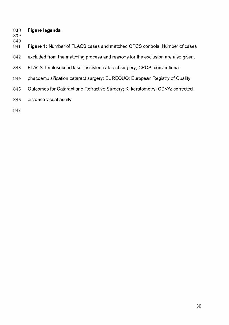
Figure legends 838 839 840 Figure 1: Number of FLACS cases and matched CPCS controls. Number of cases 841
excluded from the matching process and reasons for the exclusion are also given. 842
FLACS: femtosecond laser-assisted cataract surgery; CPCS: conventional 843
phacoemulsification cataract surgery; EUREQUO: European Registry of Quality 844
Outcomes for Cataract and Refractive Surgery; K: keratometry; CDVA: corrected-845
distance visual acuity 846
847
Related Documents