Fatty Acid Desaturase Gene Variants, Cardiovascular Risk Factors, and Myocardial Infarction in the Costa Rica Study The Harvard community has made this article openly available. Please share how this access benefits you. Your story matters Citation Aslibekyan, S., M. K. Jensen, H. Campos, C. D. Linkletter, E. B. Loucks, J. M. Ordovas, R. Deka, E. B. Rimm, and A. Baylin. 2012. Fatty acid desaturase gene variants, cardiovascular risk factors, and myocardial infarction in the costa rica study. Frontiers in Genetics 3:72. Published Version doi:10.3389/fgene.2012.00072 Citable link http://nrs.harvard.edu/urn-3:HUL.InstRepos:10433482 Terms of Use This article was downloaded from Harvard University’s DASH repository, and is made available under the terms and conditions applicable to Other Posted Material, as set forth at http:// nrs.harvard.edu/urn-3:HUL.InstRepos:dash.current.terms-of- use#LAA

Welcome message from author
This document is posted to help you gain knowledge. Please leave a comment to let me know what you think about it! Share it to your friends and learn new things together.
Transcript
Fatty Acid Desaturase GeneVariants, Cardiovascular
Risk Factors, and MyocardialInfarction in the Costa Rica Study
The Harvard community has made thisarticle openly available. Please share howthis access benefits you. Your story matters
Citation Aslibekyan, S., M. K. Jensen, H. Campos, C. D. Linkletter, E. B.Loucks, J. M. Ordovas, R. Deka, E. B. Rimm, and A. Baylin. 2012.Fatty acid desaturase gene variants, cardiovascular risk factors, andmyocardial infarction in the costa rica study. Frontiers in Genetics3:72.
Published Version doi:10.3389/fgene.2012.00072
Citable link http://nrs.harvard.edu/urn-3:HUL.InstRepos:10433482
Terms of Use This article was downloaded from Harvard University’s DASHrepository, and is made available under the terms and conditionsapplicable to Other Posted Material, as set forth at http://nrs.harvard.edu/urn-3:HUL.InstRepos:dash.current.terms-of-use#LAA
ORIGINAL RESEARCH ARTICLEpublished: 03 May 2012
doi: 10.3389/fgene.2012.00072
Fatty acid desaturase gene variants, cardiovascular riskfactors, and myocardial infarction in the Costa Rica StudyS. Aslibekyan1, M. K. Jensen2, H. Campos2, C. D. Linkletter 1, E. B. Loucks1, J. M. Ordovas3, R. Deka4,
E. B. Rimm2 and A. Baylin1,5*
1 Department of Community Health, Brown University, Providence, RI, USA2 Department of Nutrition, Harvard School of Public Health, Boston, MA, USA3 Jean Mayer USDA Human Nutrition Research Center on Aging, Tufts University, Boston, MA, USA4 Department of Environmental Health, University of Cincinnati College of Medicine, Cincinnati, OH, USA5 Department of Epidemiology, University of Michigan School of Public Health, Ann Arbor, MI, USA
Edited by:
Jill Barnholtz-Sloan, Case WesternReserve University School ofMedicine, USA
Reviewed by:
Cheryl L. Thompson, Case WesternReserve University, USAMichael Scheurer, Baylor College ofMedicine, USA
*Correspondence:
A. Baylin, Department ofEpidemiology, University of MichiganSchool of Public Health, 1420Washington Heights, Ann Arbor, MI48109-2029, USA.e-mail: [email protected]
Genetic variation in fatty acid desaturases (FADS) has previously been linked to long-chain polyunsaturated fatty acids (PUFAs) in adipose tissue and cardiovascular risk. Thegoal of our study was to test associations between six common FADS polymorphisms(rs174556, rs3834458, rs174570, rs2524299, rs174589, rs174627), intermediate cardiovas-cular risk factors, and non-fatal myocardial infarction (MI) in a matched population basedcase–control study of Costa Rican adults (n = 1756). Generalized linear models and multipleconditional logistic regression models were used to assess the associations of interest.Analyses involving intermediate cardiovascular risk factors and MI were also conductedin two replication cohorts, The Nurses’ Health Study (n = 1200) and The Health Profes-sionals Follow-Up Study (n = 1295). In the Costa Rica Study, genetic variation in the FADScluster was associated with a robust linear decrease in adipose gamma-linolenic, arachi-donic, and eicosapentaenoic fatty acids, and significant or borderline significant increasesin the eicosadienoic, eicosatrienoic, and dihomo-gamma-linolenic fatty acids. However, theassociations with adipose tissue fatty acids did not translate into changes in inflammatorybiomarkers, blood lipids, or the risk of MI in the discovery or the replication cohorts. In con-clusion, fatty acid desaturase polymorphisms impact long-chain PUFA biosynthesis, buttheir overall effect on cardiovascular health likely involves multiple pathways and meritsfurther investigation.
Keywords: desaturases, polyunsaturated fatty acids, inflammation, lipids, myocardial infarction
INTRODUCTIONLong-chain omega-3 polyunsaturated fatty acids (PUFAs),obtained either through dietary intake or synthesized endoge-nously from alpha-linolenic acid (ALA), have long been knownto be protective against heart disease, diabetes, and other chronicoutcomes (Harris, 2010). Previous studies showed that in popula-tions with low intake of marine fatty acids, dietary intake of ALAalso confers cardioprotective benefits, either by itself or throughconversion to long-chain PUFAs (Baylin et al., 2003).
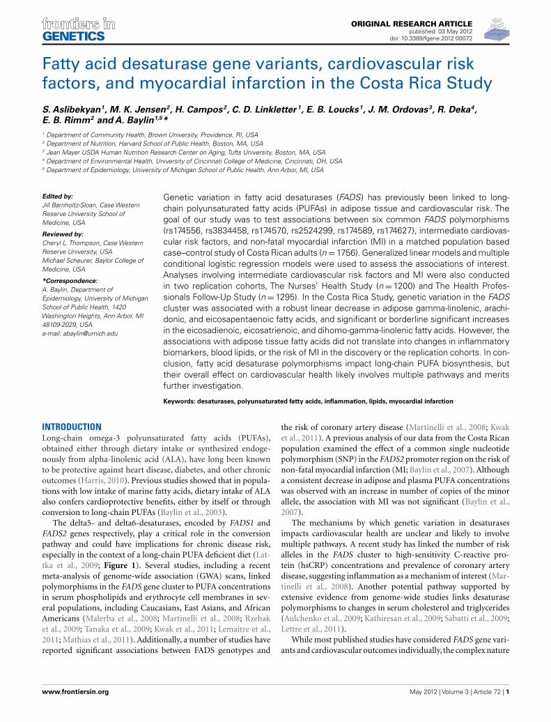
The delta5- and delta6-desaturases, encoded by FADS1 andFADS2 genes respectively, play a critical role in the conversionpathway and could have implications for chronic disease risk,especially in the context of a long-chain PUFA deficient diet (Lat-tka et al., 2009; Figure 1). Several studies, including a recentmeta-analysis of genome-wide association (GWA) scans, linkedpolymorphisms in the FADS gene cluster to PUFA concentrationsin serum phospholipids and erythrocyte cell membranes in sev-eral populations, including Caucasians, East Asians, and AfricanAmericans (Malerba et al., 2008; Martinelli et al., 2008; Rzehaket al., 2009; Tanaka et al., 2009; Kwak et al., 2011; Lemaitre et al.,2011; Mathias et al., 2011). Additionally, a number of studies havereported significant associations between FADS genotypes and
the risk of coronary artery disease (Martinelli et al., 2008; Kwaket al., 2011). A previous analysis of our data from the Costa Ricanpopulation examined the effect of a common single nucleotidepolymorphism (SNP) in the FADS2 promoter region on the risk ofnon-fatal myocardial infarction (MI; Baylin et al., 2007). Althougha consistent decrease in adipose and plasma PUFA concentrationswas observed with an increase in number of copies of the minorallele, the association with MI was not significant (Baylin et al.,2007).
The mechanisms by which genetic variation in desaturasesimpacts cardiovascular health are unclear and likely to involvemultiple pathways. A recent study has linked the number of riskalleles in the FADS cluster to high-sensitivity C-reactive pro-tein (hsCRP) concentrations and prevalence of coronary arterydisease, suggesting inflammation as a mechanism of interest (Mar-tinelli et al., 2008). Another potential pathway supported byextensive evidence from genome-wide studies links desaturasepolymorphisms to changes in serum cholesterol and triglycerides(Aulchenko et al., 2009; Kathiresan et al., 2009; Sabatti et al., 2009;Lettre et al., 2011).
While most published studies have considered FADS gene vari-ants and cardiovascular outcomes individually, the complex nature
www.frontiersin.org May 2012 | Volume 3 | Article 72 | 1
Aslibekyan et al. Fatty acid desaturases and cardiovascular risk
FIGURE 1 | Biosynthetic pathway of polyunsaturated fatty acids.
of biological mechanisms underlying the association betweendesaturases and cardiovascular risk warrants a more compre-hensive approach. The objective of this study is to evaluate theassociation between genetic variation in the FADS cluster, adiposetissue fatty acids, serum lipids, and inflammatory biomarkers, andthe risk of MI in a Costa Rican population. By considering inter-mediate outcomes as well as the endpoint of MI, this study aimsto provide novel mechanistic insights into the relation betweendesaturases, dietary fatty acids, and cardiovascular disease.
MATERIALS AND METHODSSTUDY POPULATIONThe population of the Costa Rica Study, described in detail in priorpublications, included 4,548 unrelated Hispanics who resided inthe Central Valley of Costa Rica between 1994 and 2004 (Baylinand Campos, 2004; Kabagambe et al., 2007). Cases of first non-fatal acute MI were ascertained by two independent cardiologistsin the participating hospitals and deemed eligible if they metthe World Health Organization criteria, survived hospitalization,were under 75 years of age on the day of their first MI, and ableto answer the questionnaire (Tunstallpedoe et al., 1994). Eligiblecases were matched by 5-year age group, sex, and area of resi-dence to population controls. Participation was 98% for cases and88% for controls. The study population is appropriate for inves-tigating genetic markers of disease due to its origin in a smallnumber of founders and low rates of migration (Baylin et al.,2007).
The original sample size was 2,274 cases and 2,274 controls. Par-ticipants missing information on outcomes, exposure, or covari-ates were excluded from the analysis. Excluded participants did not
significantly differ from those who remained in the analyses on keydemographic, genetic, and dietary variables. Data were missingdue to lost laboratory samples as well as non-differential responsepatterns with respect to the exposure, outcome, or covariates.All participants provided written informed consent. The studywas approved by the Human Subjects Committee of the HarvardSchool of Public Health and the University of Costa Rica.
The replication study populations consisted of the NursesHealth Study (NHS) and Health Professionals Follow-Up Study(HPFS) participants. The NHS enrolled 121,701 female nursesaged 30–55 who returned a mailed questionnaire in 1976 regard-ing lifestyle and medical history. The HPFS enrolled 51,529 malesaged 40–75 who returned a similar questionnaire in 1986. Par-ticipants of both cohorts have received follow-up questionnairesbiennially to record newly diagnosed illnesses. Detailed descrip-tions of the study cohorts have been published previously (Colditzet al., 1997).
In both replication cohorts, nested case–control studies weredesigned using incident coronary heart disease (CHD), with non-fatal MI and fatal CHD as the outcome. Diagnosis of MI wasconfirmed on the basis of the criteria of the World Health Organi-zation (Tunstallpedoe et al., 1994). Fatal CHD was confirmed by anexamination of hospital or autopsy records. Among participantswho provided blood samples and who were free of diagnosed car-diovascular disease or cancer at blood draw, 474 women and 454men with incident CHD between blood draw and June of 2004were identified. Using risk-set sampling (Prentice and Breslow,1978), controls were selected randomly and matched in a 1:2 ratioon age, smoking, and month of blood return, yielding final samplesizes of 1200 for NHS and 1295 for HPFS.
Frontiers in Genetics | Applied Genetic Epidemiology May 2012 | Volume 3 | Article 72 | 2
Aslibekyan et al. Fatty acid desaturases and cardiovascular risk
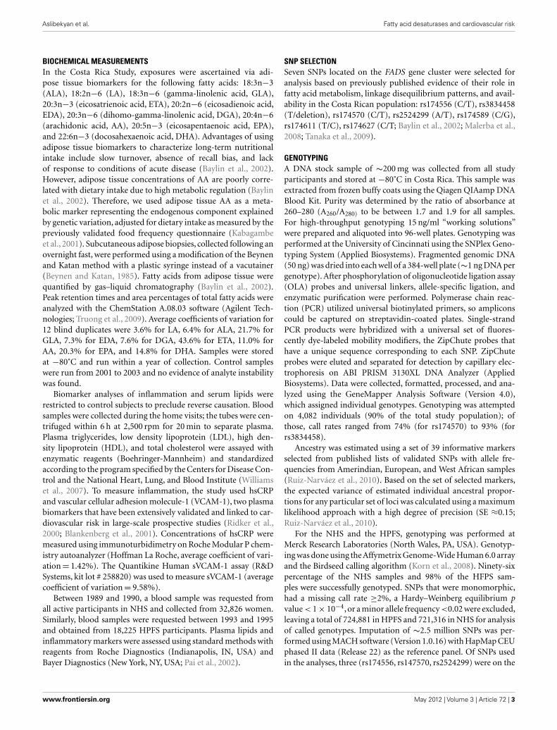
BIOCHEMICAL MEASUREMENTSIn the Costa Rica Study, exposures were ascertained via adi-pose tissue biomarkers for the following fatty acids: 18:3n−3(ALA), 18:2n−6 (LA), 18:3n−6 (gamma-linolenic acid, GLA),20:3n−3 (eicosatrienoic acid, ETA), 20:2n−6 (eicosadienoic acid,EDA), 20:3n−6 (dihomo-gamma-linolenic acid, DGA), 20:4n−6(arachidonic acid, AA), 20:5n−3 (eicosapentaenoic acid, EPA),and 22:6n−3 (docosahexaenoic acid, DHA). Advantages of usingadipose tissue biomarkers to characterize long-term nutritionalintake include slow turnover, absence of recall bias, and lackof response to conditions of acute disease (Baylin et al., 2002).However, adipose tissue concentrations of AA are poorly corre-lated with dietary intake due to high metabolic regulation (Baylinet al., 2002). Therefore, we used adipose tissue AA as a meta-bolic marker representing the endogenous component explainedby genetic variation, adjusted for dietary intake as measured by thepreviously validated food frequency questionnaire (Kabagambeet al., 2001). Subcutaneous adipose biopsies, collected following anovernight fast, were performed using a modification of the Beynenand Katan method with a plastic syringe instead of a vacutainer(Beynen and Katan, 1985). Fatty acids from adipose tissue werequantified by gas–liquid chromatography (Baylin et al., 2002).Peak retention times and area percentages of total fatty acids wereanalyzed with the ChemStation A.08.03 software (Agilent Tech-nologies; Truong et al., 2009). Average coefficients of variation for12 blind duplicates were 3.6% for LA, 6.4% for ALA, 21.7% forGLA, 7.3% for EDA, 7.6% for DGA, 43.6% for ETA, 11.0% forAA, 20.3% for EPA, and 14.8% for DHA. Samples were storedat −80˚C and run within a year of collection. Control sampleswere run from 2001 to 2003 and no evidence of analyte instabilitywas found.
Biomarker analyses of inflammation and serum lipids wererestricted to control subjects to preclude reverse causation. Bloodsamples were collected during the home visits; the tubes were cen-trifuged within 6 h at 2,500 rpm for 20 min to separate plasma.Plasma triglycerides, low density lipoprotein (LDL), high den-sity lipoprotein (HDL), and total cholesterol were assayed withenzymatic reagents (Boehringer-Mannheim) and standardizedaccording to the program specified by the Centers for Disease Con-trol and the National Heart, Lung, and Blood Institute (Williamset al., 2007). To measure inflammation, the study used hsCRPand vascular cellular adhesion molecule-1 (VCAM-1), two plasmabiomarkers that have been extensively validated and linked to car-diovascular risk in large-scale prospective studies (Ridker et al.,2000; Blankenberg et al., 2001). Concentrations of hsCRP weremeasured using immunoturbidimetry on Roche Modular P chem-istry autoanalyzer (Hoffman La Roche, average coefficient of vari-ation = 1.42%). The Quantikine Human sVCAM-1 assay (R&DSystems, kit lot # 258820) was used to measure sVCAM-1 (averagecoefficient of variation = 9.58%).
Between 1989 and 1990, a blood sample was requested fromall active participants in NHS and collected from 32,826 women.Similarly, blood samples were requested between 1993 and 1995and obtained from 18,225 HPFS participants. Plasma lipids andinflammatory markers were assessed using standard methods withreagents from Roche Diagnostics (Indianapolis, IN, USA) andBayer Diagnostics (New York, NY, USA; Pai et al., 2002).
SNP SELECTIONSeven SNPs located on the FADS gene cluster were selected foranalysis based on previously published evidence of their role infatty acid metabolism, linkage disequilibrium patterns, and avail-ability in the Costa Rican population: rs174556 (C/T), rs3834458(T/deletion), rs174570 (C/T), rs2524299 (A/T), rs174589 (C/G),rs174611 (T/C), rs174627 (C/T; Baylin et al., 2002; Malerba et al.,2008; Tanaka et al., 2009).
GENOTYPINGA DNA stock sample of ∼200 mg was collected from all studyparticipants and stored at −80˚C in Costa Rica. This sample wasextracted from frozen buffy coats using the Qiagen QIAamp DNABlood Kit. Purity was determined by the ratio of absorbance at260–280 (A260/A280) to be between 1.7 and 1.9 for all samples.For high-throughput genotyping 15 ng/ml “working solutions”were prepared and aliquoted into 96-well plates. Genotyping wasperformed at the University of Cincinnati using the SNPlex Geno-typing System (Applied Biosystems). Fragmented genomic DNA(50 ng) was dried into each well of a 384-well plate (∼1 ng DNA pergenotype). After phosphorylation of oligonucleotide ligation assay(OLA) probes and universal linkers, allele-specific ligation, andenzymatic purification were performed. Polymerase chain reac-tion (PCR) utilized universal biotinylated primers, so ampliconscould be captured on streptavidin-coated plates. Single-strandPCR products were hybridized with a universal set of fluores-cently dye-labeled mobility modifiers, the ZipChute probes thathave a unique sequence corresponding to each SNP. ZipChuteprobes were eluted and separated for detection by capillary elec-trophoresis on ABI PRISM 3130XL DNA Analyzer (AppliedBiosystems). Data were collected, formatted, processed, and ana-lyzed using the GeneMapper Analysis Software (Version 4.0),which assigned individual genotypes. Genotyping was attemptedon 4,082 individuals (90% of the total study population); ofthose, call rates ranged from 74% (for rs174570) to 93% (forrs3834458).
Ancestry was estimated using a set of 39 informative markersselected from published lists of validated SNPs with allele fre-quencies from Amerindian, European, and West African samples(Ruiz-Narváez et al., 2010). Based on the set of selected markers,the expected variance of estimated individual ancestral propor-tions for any particular set of loci was calculated using a maximumlikelihood approach with a high degree of precision (SE ≈0.15;Ruiz-Narváez et al., 2010).
For the NHS and the HPFS, genotyping was performed atMerck Research Laboratories (North Wales, PA, USA). Genotyp-ing was done using the Affymetrix Genome-Wide Human 6.0 arrayand the Birdseed calling algorithm (Korn et al., 2008). Ninety-sixpercentage of the NHS samples and 98% of the HFPS sam-ples were successfully genotyped. SNPs that were monomorphic,had a missing call rate ≥2%, a Hardy–Weinberg equilibrium pvalue < 1 × 10⁻⁴, or a minor allele frequency <0.02 were excluded,leaving a total of 724,881 in HPFS and 721,316 in NHS for analysisof called genotypes. Imputation of ∼2.5 million SNPs was per-formed using MACH software (Version 1.0.16) with HapMap CEUphased II data (Release 22) as the reference panel. Of SNPs usedin the analyses, three (rs174556, rs147570, rs2524299) were on the
www.frontiersin.org May 2012 | Volume 3 | Article 72 | 3

Aslibekyan et al. Fatty acid desaturases and cardiovascular risk
Affymetrix chip, while two (rs174589 and rs174627) were imputedwith r2 values > 0.85.
STATISTICAL ANALYSISData were analyzed using the SAS software package (Version 9.2;SAS Institute Inc, Cary, NC, USA). To assess the significance ofdifferences in general characteristics and potential confounders,we used paired t -tests for continuous variables, McNemar’s testsfor categorical variables, and Fisher’s exact test for minor allele fre-quencies. The ALLELE procedure was used to test for deviationsfrom Hardy–Weinberg equilibrium among controls. Of all SNPs,only rs174611 was found to be in violation of the Hardy–Weinbergequilibrium and removed from all subsequent analyses.
Linear regression models were fit among controls to evalu-ate the association between each FADS cluster SNP, adipose tis-sue long-chain PUFAs, and plasma hsCRP, VCAM-1, and serumlipids. Least square means and 95% confidence intervals wereused to report the relation between the outcomes and FADSgenetic variants. Normal probability plots were constructed, log-transformations were carried out for non-normally distributedvariables (GLA, hsCRP, and triglycerides), and geometric meanswere reported. The intermediate risk factors models were adjustedfor age, sex, and residence area, while the PUFA models were addi-tionally adjusted for dietary intake of all nine fatty acids as assessedby the food frequency questionnaire. Adipose tissue PUFAs andintermediate risk factors were modeled as continuous variables.The relation between the SNPs and the dichotomous MI out-come was modeled using conditional logistic regression, adjusted
for age, sex, and residence area. Additionally, models fit to theCosta Rica Study data were adjusted for ancestry due to evidenceof population stratification (Ruiz-Narváez et al., 2010); modelsfit to the NHS and the HPFS data were not adjusted for ances-try as the cohorts were found to be genetically homogeneous.Least square means of adipose fatty acids, blood lipids, and inflam-matory markers were estimated using PROC GLM, adjusted forpotential confounders listed above. P values from single SNPanalyses were adjusted for multiple comparisons using the falsediscovery rate controlling procedure (Benjamini and Hochberg,1995).
Linear and conditional logistic regression analyses with individ-ual SNPs as predictors and inflammation markers, blood lipids,and MI as outcomes as described above were replicated in theNHS and HPFS. Genotype information at the rs3834458 locuswas not available in the replication cohorts. Because minor allelefrequencies varied considerably between the discovery and repli-cation cohorts, a meta-analysis was not attempted. All analyseswere adjusted for sex and age.
RESULTSThe general characteristics of the three populations are summa-rized by case/control status in Table 1. None of the selected SNPsdiffered significantly in minor allele frequency by disease status.Cases were more likely to report MI risk factors, specifically smok-ing and history of chronic disease. Additionally, cases in the CostaRica Study had significantly lower adipose tissue concentrationsof ALA and LA. Ancestral admixture proportions did not vary by
Table 1 | General characteristics of the study populations.
Variable Costa Rica Study Nurses’ Health Study Health Professionals
Follow-Up Study
Cases
(n = 878)
Controls
(n = 878)
Cases
(n = 403)
Controls
(n = 797)
Cases
(n = 435)
Controls
(n = 860)
Age, years 58.2 ± 10.9 58.1 ± 11.0 60.2 ± 6.3 59.6 ± 6.5 64.5 ± 8.6 64.2 ± 8.6
% Female 26 26 100 100 0 0
% History of hypertension 39 32 50 27 37 29
% History of hypercholesterolemia 31 29 53 41 49 40
% History of diabetes 23 15 15 6 9 4
% Current smokers 38 21 27 25 10 9
BMI, kg/m2 26.0 ± 4.0 26.5 ± 4.3 26.6 ± 5.4 25.1 ± 4.3 26.0 ± 3.2 25.6 ± 3.3
DIETARY INTAKE OF FATTY ACIDS, g
Alpha-linolenic 1.71 ± 0.86 1.61 ± 0.77 – – – –
Linoleic 19.1 ± 9.6 17.9 ± 7.9 – – – –
ADIPOSETISSUE FATTY ACIDS, %TOTAL
Alpha-linolenic 0.64 ± 0.21 0.68 ± 0.21 – – – –
Linoleic 15.4 ± 3.7 16.0 ± 3.8 – – – –
MINOR ALLELE FREQUENCY, %
rs174556 (C/T) 45 45 31 28 30 29
rs3834458 (T/deletion) 48 48 – – – –
rs174570 (C/T) 37 39 13 13 13 14
rs2524299 (A/T) 28 26 12 11 11 13
rs174589 (G/C) 30 29 22 21 20 20
rs174627(C/T) 12 11 18 19 17 17
Frontiers in Genetics | Applied Genetic Epidemiology May 2012 | Volume 3 | Article 72 | 4
Aslibekyan et al. Fatty acid desaturases and cardiovascular risk
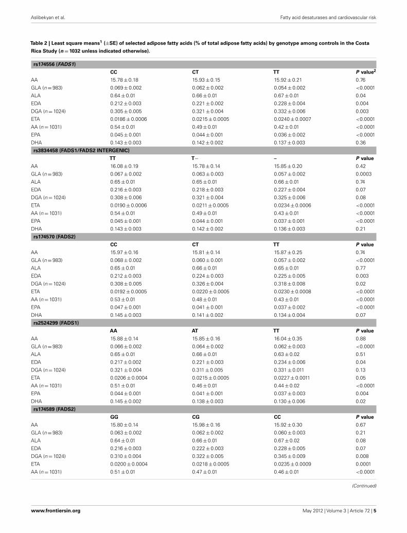
Table 2 | Least square means1 (±SE) of selected adipose fatty acids (% of total adipose fatty acids) by genotype among controls in the Costa
Rica Study (n = 1032 unless indicated otherwise).
rs174556 (FADS1)
CC CT TT P value2
AA 15.78 ± 0.18 15.93 ± 0.15 15.92 ± 0.21 0.76
GLA (n = 983) 0.069 ± 0.002 0.062 ± 0.002 0.054 ± 0.002 <0.0001
ALA 0.64 ± 0.01 0.66 ± 0.01 0.67 ± 0.01 0.04
EDA 0.212 ± 0.003 0.221 ± 0.002 0.228 ± 0.004 0.004
DGA (n = 1024) 0.305 ± 0.005 0.321 ± 0.004 0.332 ± 0.006 0.003
ETA 0.0186 ± 0.0006 0.0215 ± 0.0005 0.0240 ± 0.0007 <0.0001
AA (n = 1031) 0.54 ± 0.01 0.49 ± 0.01 0.42 ± 0.01 <0.0001
EPA 0.045 ± 0.001 0.044 ± 0.001 0.036 ± 0.002 <0.0001
DHA 0.143 ± 0.003 0.142 ± 0.002 0.137 ± 0.003 0.36
rs3834458 (FADS1/FADS2 INTERGENIC)
TT T− – P value
AA 16.08 ± 0.19 15.78 ± 0.14 15.85 ± 0.20 0.42
GLA (n = 983) 0.067 ± 0.002 0.063 ± 0.003 0.057 ± 0.002 0.0003
ALA 0.65 ± 0.01 0.65 ± 0.01 0.66 ± 0.01 0.74
EDA 0.216 ± 0.003 0.218 ± 0.003 0.227 ± 0.004 0.07
DGA (n = 1024) 0.308 ± 0.006 0.321 ± 0.004 0.325 ± 0.006 0.08
ETA 0.0190 ± 0.0006 0.0211 ± 0.0005 0.0234 ± 0.0006 <0.0001
AA (n = 1031) 0.54 ± 0.01 0.49 ± 0.01 0.43 ± 0.01 <0.0001
EPA 0.045 ± 0.001 0.044 ± 0.001 0.037 ± 0.001 <0.0001
DHA 0.143 ± 0.003 0.142 ± 0.002 0.136 ± 0.003 0.21
rs174570 (FADS2)
CC CT TT P value
AA 15.97 ± 0.16 15.81 ± 0.14 15.87 ± 0.25 0.74
GLA (n = 983) 0.068 ± 0.002 0.060 ± 0.001 0.057 ± 0.002 <0.0001
ALA 0.65 ± 0.01 0.66 ± 0.01 0.65 ± 0.01 0.77
EDA 0.212 ± 0.003 0.224 ± 0.003 0.225 ± 0.005 0.003
DGA (n = 1024) 0.308 ± 0.005 0.326 ± 0.004 0.318 ± 0.008 0.02
ETA 0.0192 ± 0.0005 0.0220 ± 0.0005 0.0230 ± 0.0008 <0.0001
AA (n = 1031) 0.53 ± 0.01 0.48 ± 0.01 0.43 ± 0.01 <0.0001
EPA 0.047 ± 0.001 0.041 ± 0.001 0.037 ± 0.002 <0.0001
DHA 0.145 ± 0.003 0.141 ± 0.002 0.134 ± 0.004 0.07
rs2524299 (FADS1)
AA AT TT P value
AA 15.88 ± 0.14 15.85 ± 0.16 16.04 ± 0.35 0.88
GLA (n = 983) 0.066 ± 0.002 0.064 ± 0.002 0.062 ± 0.003 <0.0001
ALA 0.65 ± 0.01 0.66 ± 0.01 0.63 ± 0.02 0.51
EDA 0.217 ± 0.002 0.221 ± 0.003 0.234 ± 0.006 0.04
DGA (n = 1024) 0.321 ± 0.004 0.311 ± 0.005 0.331 ± 0.011 0.13
ETA 0.0206 ± 0.0004 0.0215 ± 0.0005 0.0227 ± 0.0011 0.05
AA (n = 1031) 0.51 ± 0.01 0.46 ± 0.01 0.44 ± 0.02 <0.0001
EPA 0.044 ± 0.001 0.041 ± 0.001 0.037 ± 0.003 0.004
DHA 0.145 ± 0.002 0.138 ± 0.003 0.130 ± 0.006 0.02
rs174589 (FADS2)
GG CG CC P value
AA 15.80 ± 0.14 15.98 ± 0.16 15.92 ± 0.30 0.67
GLA (n = 983) 0.063 ± 0.002 0.062 ± 0.002 0.060 ± 0.003 0.21
ALA 0.64 ± 0.01 0.66 ± 0.01 0.67 ± 0.02 0.08
EDA 0.216 ± 0.003 0.222 ± 0.003 0.228 ± 0.005 0.07
DGA (n = 1024) 0.310 ± 0.004 0.322 ± 0.005 0.345 ± 0.009 0.008
ETA 0.0200 ± 0.0004 0.0218 ± 0.0005 0.0235 ± 0.0009 0.0001
AA (n = 1031) 0.51 ± 0.01 0.47 ± 0.01 0.46 ± 0.01 <0.0001
(Continued)
www.frontiersin.org May 2012 | Volume 3 | Article 72 | 5
Aslibekyan et al. Fatty acid desaturases and cardiovascular risk
Table 2 | Continued
EPA 0.043 ± 0.001 0.042 ± 0.001 0.039 ± 0.002 0.09
DHA 0.140 ± 0.002 0.140 ± 0.003 0.150 ± 0.005 0.16
rs174627 (FADS2/FADS3 INTERGENIC)
CC CT TT P value
AA 15.91 ± 0.12 15.75 ± 0.22 15.87 ± 0.93 0.78
GLA (n = 983) 0.062 ± 0.001 0.061 ± 0.002 0.065 ± 0.009 0.79
ALA 0.65 ± 0.01 0.65 ± 0.01 0.68 ± 0.05 0.88
EDA 0.220 ± 0.002 0.218 ± 0.004 0.204 ± 0.017 0.58
DGA (n = 1024) 0.317 ± 0.003 0.319 ± 0.007 0.371 ± 0.028 0.15
ETA 0.0211 ± 0.0004 0.0211 ± 0.0007 0.0205 ± 0.0030 0.98
AA (n = 1031) 0.49 ± 0.02 0.47 ± 0.01 0.53 ± 0.04 0.21
EPA 0.043 ± 0.001 0.042 ± 0.002 0.043 ± 0.007 0.95
DHA 0.141 ± 0.002 0.142 ± 0.004 0.145 ± 0.015 0.96
1Models were adjusted for age/sex/residence (by matching), ancestry, and dietary fatty acids.2Unadjusted for multiple testing.
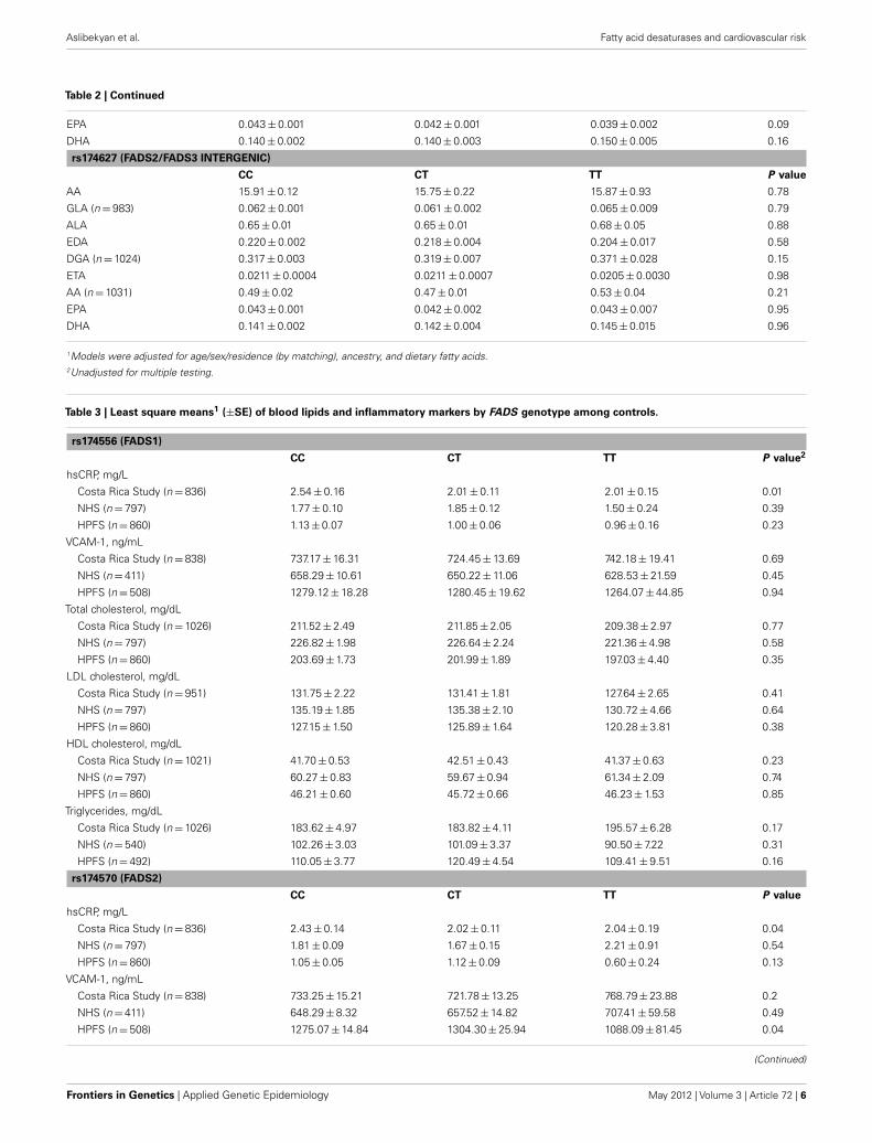
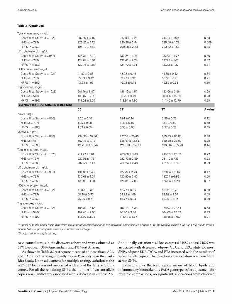
Table 3 | Least square means1 (±SE) of blood lipids and inflammatory markers by FADS genotype among controls.
rs174556 (FADS1)
CC CT TT P value2
hsCRP, mg/L
Costa Rica Study (n = 836) 2.54 ± 0.16 2.01 ± 0.11 2.01 ± 0.15 0.01
NHS (n = 797) 1.77 ± 0.10 1.85 ± 0.12 1.50 ± 0.24 0.39
HPFS (n = 860) 1.13 ± 0.07 1.00 ± 0.06 0.96 ± 0.16 0.23
VCAM-1, ng/mL
Costa Rica Study (n = 838) 737.17 ± 16.31 724.45 ± 13.69 742.18 ± 19.41 0.69
NHS (n = 411) 658.29 ± 10.61 650.22 ± 11.06 628.53 ± 21.59 0.45
HPFS (n = 508) 1279.12 ± 18.28 1280.45 ± 19.62 1264.07 ± 44.85 0.94
Total cholesterol, mg/dL
Costa Rica Study (n = 1026) 211.52 ± 2.49 211.85 ± 2.05 209.38 ± 2.97 0.77
NHS (n = 797) 226.82 ± 1.98 226.64 ± 2.24 221.36 ± 4.98 0.58
HPFS (n = 860) 203.69 ± 1.73 201.99 ± 1.89 197.03 ± 4.40 0.35
LDL cholesterol, mg/dL
Costa Rica Study (n = 951) 131.75 ± 2.22 131.41 ± 1.81 127.64 ± 2.65 0.41
NHS (n = 797) 135.19 ± 1.85 135.38 ± 2.10 130.72 ± 4.66 0.64
HPFS (n = 860) 127.15 ± 1.50 125.89 ± 1.64 120.28 ± 3.81 0.38
HDL cholesterol, mg/dL
Costa Rica Study (n = 1021) 41.70 ± 0.53 42.51 ± 0.43 41.37 ± 0.63 0.23
NHS (n = 797) 60.27 ± 0.83 59.67 ± 0.94 61.34 ± 2.09 0.74
HPFS (n = 860) 46.21 ± 0.60 45.72 ± 0.66 46.23 ± 1.53 0.85
Triglycerides, mg/dL
Costa Rica Study (n = 1026) 183.62 ± 4.97 183.82 ± 4.11 195.57 ± 6.28 0.17
NHS (n = 540) 102.26 ± 3.03 101.09 ± 3.37 90.50 ± 7.22 0.31
HPFS (n = 492) 110.05 ± 3.77 120.49 ± 4.54 109.41 ± 9.51 0.16
rs174570 (FADS2)
CC CT TT P value
hsCRP, mg/L
Costa Rica Study (n = 836) 2.43 ± 0.14 2.02 ± 0.11 2.04 ± 0.19 0.04
NHS (n = 797) 1.81 ± 0.09 1.67 ± 0.15 2.21 ± 0.91 0.54
HPFS (n = 860) 1.05 ± 0.05 1.12 ± 0.09 0.60 ± 0.24 0.13
VCAM-1, ng/mL
Costa Rica Study (n = 838) 733.25 ± 15.21 721.78 ± 13.25 768.79 ± 23.88 0.2
NHS (n = 411) 648.29 ± 8.32 657.52 ± 14.82 707.41 ± 59.58 0.49
HPFS (n = 508) 1275.07 ± 14.84 1304.30 ± 25.94 1088.09 ± 81.45 0.04
(Continued)
Frontiers in Genetics | Applied Genetic Epidemiology May 2012 | Volume 3 | Article 72 | 6
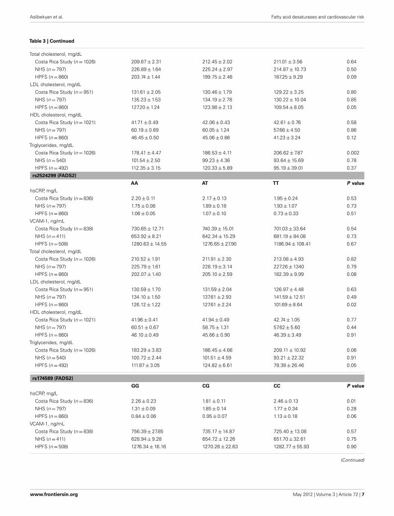
Aslibekyan et al. Fatty acid desaturases and cardiovascular risk
Table 3 | Continued
Total cholesterol, mg/dL
Costa Rica Study (n = 1026) 209.67 ± 2.31 212.45 ± 2.02 211.01 ± 3.56 0.64
NHS (n = 797) 226.89 ± 1.64 225.24 ± 2.97 214.87 ± 10.73 0.50
HPFS (n = 860) 203.74 ± 1.44 199.75 ± 2.46 187.25 ± 9.29 0.09
LDL cholesterol, mg/dL
Costa Rica Study (n = 951) 131.61 ± 2.05 130.46 ± 1.79 129.22 ± 3.25 0.80
NHS (n = 797) 135.23 ± 1.53 134.19 ± 2.78 130.22 ± 10.04 0.85
HPFS (n = 860) 127.20 ± 1.24 123.98 ± 2.13 109.54 ± 8.05 0.05
HDL cholesterol, mg/dL
Costa Rica Study (n = 1021) 41.71 ± 0.49 42.06 ± 0.43 42.61 ± 0.76 0.58
NHS (n = 797) 60.19 ± 0.69 60.05 ± 1.24 57.66 ± 4.50 0.86
HPFS (n = 860) 46.45 ± 0.50 45.06 ± 0.86 41.23 ± 3.24 0.12
Triglycerides, mg/dL
Costa Rica Study (n = 1026) 178.41 ± 4.47 186.53 ± 4.11 206.62 ± 7.87 0.002
NHS (n = 540) 101.54 ± 2.50 99.23 ± 4.36 93.64 ± 15.69 0.78
HPFS (n = 492) 112.35 ± 3.15 120.33 ± 5.89 95.19 ± 39.01 0.37
rs2524299 (FADS2)
AA AT TT P value
hsCRP, mg/L
Costa Rica Study (n = 836) 2.20 ± 0.11 2.17 ± 0.13 1.95 ± 0.24 0.53
NHS (n = 797) 1.75 ± 0.08 1.89 ± 0.18 1.93 ± 1.07 0.73
HPFS (n = 860) 1.06 ± 0.05 1.07 ± 0.10 0.73 ± 0.33 0.51
VCAM-1, ng/mL
Costa Rica Study (n = 838) 730.65 ± 12.71 740.39 ± 15.01 701.03 ± 33.64 0.54
NHS (n = 411) 653.92 ± 8.21 642.34 ± 15.29 681.19 ± 84.08 0.73
HPFS (n = 508) 1280.63 ± 14.55 1276.65 ± 27.90 1186.94 ± 108.41 0.67
Total cholesterol, mg/dL
Costa Rica Study (n = 1026) 210.52 ± 1.91 211.91 ± 2.30 213.08 ± 4.93 0.82
NHS (n = 797) 225.79 ± 1.61 228.19 ± 3.14 227.26 ± 1340 0.79
HPFS (n = 860) 202.07 ± 1.40 205.10 ± 2.59 182.39 ± 9.99 0.08
LDL cholesterol, mg/dL
Costa Rica Study (n = 951) 130.59 ± 1.70 131.59 ± 2.04 126.97 ± 4.48 0.63
NHS (n = 797) 134.10 ± 1.50 137.61 ± 2.93 141.59 ± 12.51 0.49
HPFS (n = 860) 126.12 ± 1.22 127.61 ± 2.24 101.69 ± 8.64 0.02
HDL cholesterol, mg/dL
Costa Rica Study (n = 1021) 41.96 ± 0.41 41.94 ± 0.49 42.74 ± 1.05 0.77
NHS (n = 797) 60.51 ± 0.67 58.75 ± 1.31 57.62 ± 5.60 0.44
HPFS (n = 860) 46.10 ± 0.49 45.66 ± 0.90 46.39 ± 3.49 0.91
Triglycerides, mg/dL
Costa Rica Study (n = 1026) 183.29 ± 3.83 186.45 ± 4.66 209.11 ± 10.92 0.06
NHS (n = 540) 100.72 ± 2.44 101.51 ± 4.59 93.21 ± 22.32 0.91
HPFS (n = 492) 111.87 ± 3.05 124.82 ± 6.61 78.38 ± 26.46 0.05
rs174589 (FADS2)
GG CG CC P value
hsCRP, mg/L
Costa Rica Study (n = 836) 2.26 ± 0.23 1.81 ± 0.11 2.46 ± 0.13 0.01
NHS (n = 797) 1.31 ± 0.09 1.85 ± 0.14 1.77 ± 0.34 0.28
HPFS (n = 860) 0.84 ± 0.06 0.95 ± 0.07 1.13 ± 0.18 0.06
VCAM-1, ng/mL
Costa Rica Study (n = 838) 756.39 ± 27.85 735.17 ± 14.87 725.40 ± 13.08 0.57
NHS (n = 411) 628.94 ± 9.28 654.72 ± 12.26 651.70 ± 32.61 0.75
HPFS (n = 508) 1276.34 ± 16.16 1270.26 ± 22.63 1282.77 ± 55.93 0.90
(Continued)
www.frontiersin.org May 2012 | Volume 3 | Article 72 | 7
Aslibekyan et al. Fatty acid desaturases and cardiovascular risk
Table 3 | Continued
Total cholesterol, mg/dL
Costa Rica Study (n = 1026) 207.66 ± 4.16 212.08 ± 2.25 211.34 ± 1.99 0.63
NHS (n = 797) 225.22 ± 7.42 220.30 ± 2.44 229.60 ± 1.79 0.009
HPFS (n = 860) 195.14 ± 5.62 200.88 ± 2.23 203.72 ± 1.52 0.24
LDL cholesterol, mg/dL
Costa Rica Study (n = 951) 126.31 ± 3.79 130.24 ± 1.98 132.01 ± 1.77 0.36
NHS (n = 797) 128.04 ± 6.94 130.41 ± 2.28 137.73 ± 1.67 0.02
HPFS (n = 860) 120.75 ± 4.87 124.70 ± 1.94 127.12 ± 1.32 0.31
HDL cholesterol, mg/dL
Costa Rica Study (n = 1021) 41.87 ± 0.88 42.22 ± 0.48 41.88 ± 0.42 0.84
NHS (n = 797) 65.53 ± 3.12 59.77 ± 1.92 59.99 ± 0.75 0.21
HPFS (n = 860) 43.63 ± 1.96 46.72 ± 0.78 45.85 ± 0.53 0.30
Triglycerides, mg/dL
Costa Rica Study (n = 1026) 201.76 ± 8.97 186.19 ± 4.57 183.06 ± 3.98 0.09
NHS (n = 540) 100.87 ± 2.76 95.79 ± 3.49 103.66 ± 15.33 0.20
HPFS (n = 492) 113.02 ± 3.50 113.94 ± 4.95 114.45 ± 12.79 0.99
rs174627 (FADS2/FADS3 INTERGENIC)
CC CT TT P value
hsCRP, mg/L
Costa Rica Study (n = 836) 2.25 ± 0.10 1.84 ± 0.14 2.95 ± 0.72 0.12
NHS (n = 797) 1.75 ± 0.09 1.88 ± 0.15 1.57 ± 0.40 0.58
HPFS (n = 860) 1.09 ± 0.05 0.98 ± 0.08 0.97 ± 0.23 0.45
VCAM-1, ng/mL
Costa Rica Study (n = 838) 734.33 ± 10.86 727.08 ± 20.49 685.99 ± 80.80 0.80
NHS (n = 411) 660.18 ± 9.12 636.57 ± 12.53 639.80 ± 33.07 0.28
HPFS (n = 508) 1286.08 ± 15.42 1245.81 ± 24.13 1360.67 ± 65.90 0.14
Total cholesterol, mg/dL
Costa Rica Study (n = 1026) 211.77 ± 1.64 209.06 ± 3.08 210.53 ± 12.92 0.72
NHS (n = 797) 227.65 ± 1.75 222.73 ± 2.59 231.10 ± 7.33 0.23
HPFS (n = 860) 202.56 ± 1.47 202.24 ± 2.40 201.93 ± 6.09 0.99
LDL cholesterol, mg/dL
Costa Rica Study (n = 951) 131.48 ± 1.46 127.79 ± 2.73 129.84 ± 11.62 0.47
NHS (n = 797) 135.68 ± 1.64 132.90 ± 2.42 137.24 ± 6.85 0.60
HPFS (n = 860) 125.93 ± 1.28 126.81 ± 2.08 124.04 ± 5.28 0.87
HDL cholesterol, mg/dL
Costa Rica Study (n = 1021) 41.80 ± 0.35 42.77 ± 0.65 43.96 ± 2.73 0.30
NHS (n = 797) 60.10 ± 0.73 59.82 ± 1.09 62.63 ± 3.07 0.69
HPFS (n = 860) 46.25 ± 0.51 45.77 ± 0.84 43.34 ± 2.12 0.39
Triglycerides, mg/dL
Costa Rica Study (n = 1026) 185.32 ± 6.55 190.18 ± 6.34 178.07 ± 22.41 0.63
NHS (n = 540) 102.45 ± 2.66 96.80 ± 3.80 104.69 ± 12.53 0.43
HPFS (n = 492) 112.80 ± 3.24 114.44 ± 5.57 138.56 ± 17.60 0.21
1Models fit to the Costa Rican data were adjusted for age/sex/residence (by matching) and ancestry. Models fit to the Nurses’ Health Study and the Health Profes-
sionals Follow-Up Study data were adjusted for sex and age.2Unadjusted for multiple testing.
case–control status in the discovery cohort and were estimated at58% European, 38% Amerindian, and 4% West African.
As shown in Table 2, least square means of adipose tissue ALAand LA did not vary significantly by FADS genotype in the CostaRica Study. Upon adjustment for multiple testing, variation at thers174627 locus was not associated with any of the fatty acid out-comes. For all the remaining SNPs, the number of variant allelecopies was significantly associated with a decrease in adipose AA.
Additionally, variation at all loci except rs174589 and rs174627 wasassociated with decreased adipose GLA and EPA, while for mostSNPs, adipose EDA, DGA, and ETA increased with the number ofvariant allele copies. The direction of association was consistentacross SNPs.
Table 3 shows the least square means of blood lipids andinflammatory biomarkers by FADS genotype. After adjustment formultiple comparisons, no significant associations were observed
Frontiers in Genetics | Applied Genetic Epidemiology May 2012 | Volume 3 | Article 72 | 8
Aslibekyan et al. Fatty acid desaturases and cardiovascular risk
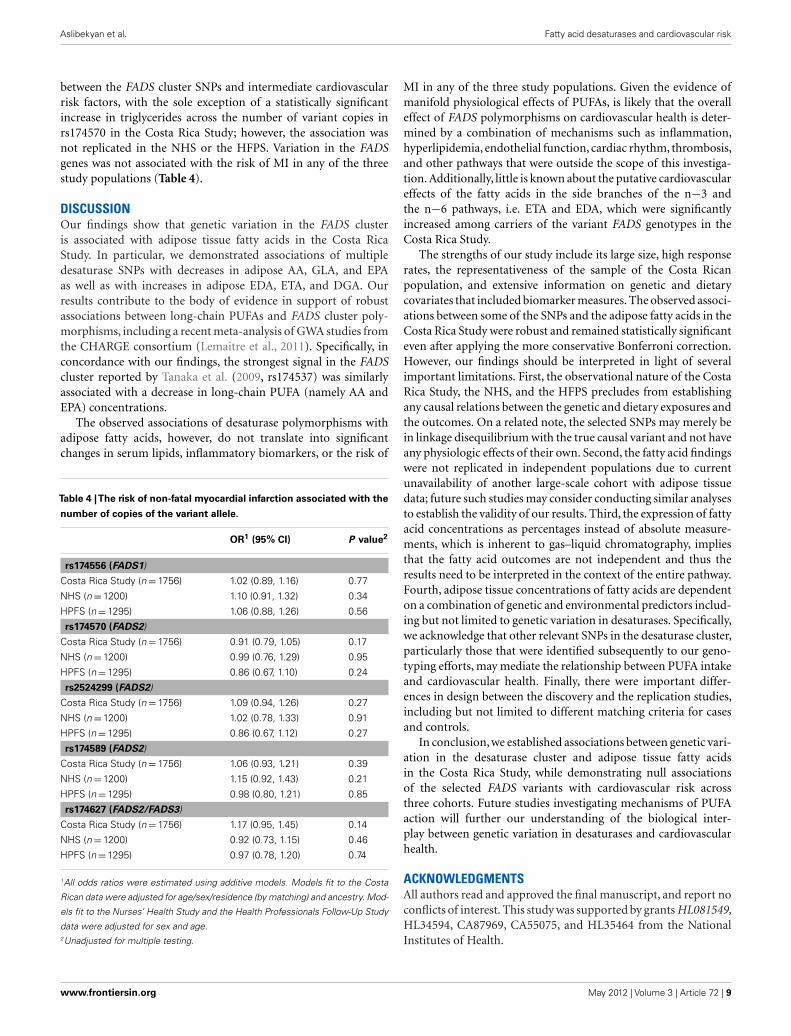
between the FADS cluster SNPs and intermediate cardiovascularrisk factors, with the sole exception of a statistically significantincrease in triglycerides across the number of variant copies inrs174570 in the Costa Rica Study; however, the association wasnot replicated in the NHS or the HFPS. Variation in the FADSgenes was not associated with the risk of MI in any of the threestudy populations (Table 4).
DISCUSSIONOur findings show that genetic variation in the FADS clusteris associated with adipose tissue fatty acids in the Costa RicaStudy. In particular, we demonstrated associations of multipledesaturase SNPs with decreases in adipose AA, GLA, and EPAas well as with increases in adipose EDA, ETA, and DGA. Ourresults contribute to the body of evidence in support of robustassociations between long-chain PUFAs and FADS cluster poly-morphisms, including a recent meta-analysis of GWA studies fromthe CHARGE consortium (Lemaitre et al., 2011). Specifically, inconcordance with our findings, the strongest signal in the FADScluster reported by Tanaka et al. (2009, rs174537) was similarlyassociated with a decrease in long-chain PUFA (namely AA andEPA) concentrations.
The observed associations of desaturase polymorphisms withadipose fatty acids, however, do not translate into significantchanges in serum lipids, inflammatory biomarkers, or the risk of
Table 4 |The risk of non-fatal myocardial infarction associated with the
number of copies of the variant allele.
OR1 (95% CI) P value2
rs174556 (FADS1)
Costa Rica Study (n = 1756) 1.02 (0.89, 1.16) 0.77
NHS (n = 1200) 1.10 (0.91, 1.32) 0.34
HPFS (n = 1295) 1.06 (0.88, 1.26) 0.56
rs174570 (FADS2)
Costa Rica Study (n = 1756) 0.91 (0.79, 1.05) 0.17
NHS (n = 1200) 0.99 (0.76, 1.29) 0.95
HPFS (n = 1295) 0.86 (0.67, 1.10) 0.24
rs2524299 (FADS2)
Costa Rica Study (n = 1756) 1.09 (0.94, 1.26) 0.27
NHS (n = 1200) 1.02 (0.78, 1.33) 0.91
HPFS (n = 1295) 0.86 (0.67, 1.12) 0.27
rs174589 (FADS2)
Costa Rica Study (n = 1756) 1.06 (0.93, 1.21) 0.39
NHS (n = 1200) 1.15 (0.92, 1.43) 0.21
HPFS (n = 1295) 0.98 (0.80, 1.21) 0.85
rs174627 (FADS2/FADS3)
Costa Rica Study (n = 1756) 1.17 (0.95, 1.45) 0.14
NHS (n = 1200) 0.92 (0.73, 1.15) 0.46
HPFS (n = 1295) 0.97 (0.78, 1.20) 0.74
1All odds ratios were estimated using additive models. Models fit to the Costa
Rican data were adjusted for age/sex/residence (by matching) and ancestry. Mod-
els fit to the Nurses’ Health Study and the Health Professionals Follow-Up Study
data were adjusted for sex and age.2Unadjusted for multiple testing.
MI in any of the three study populations. Given the evidence ofmanifold physiological effects of PUFAs, is likely that the overalleffect of FADS polymorphisms on cardiovascular health is deter-mined by a combination of mechanisms such as inflammation,hyperlipidemia, endothelial function, cardiac rhythm, thrombosis,and other pathways that were outside the scope of this investiga-tion. Additionally, little is known about the putative cardiovasculareffects of the fatty acids in the side branches of the n−3 andthe n−6 pathways, i.e. ETA and EDA, which were significantlyincreased among carriers of the variant FADS genotypes in theCosta Rica Study.
The strengths of our study include its large size, high responserates, the representativeness of the sample of the Costa Ricanpopulation, and extensive information on genetic and dietarycovariates that included biomarker measures. The observed associ-ations between some of the SNPs and the adipose fatty acids in theCosta Rica Study were robust and remained statistically significanteven after applying the more conservative Bonferroni correction.However, our findings should be interpreted in light of severalimportant limitations. First, the observational nature of the CostaRica Study, the NHS, and the HFPS precludes from establishingany causal relations between the genetic and dietary exposures andthe outcomes. On a related note, the selected SNPs may merely bein linkage disequilibrium with the true causal variant and not haveany physiologic effects of their own. Second, the fatty acid findingswere not replicated in independent populations due to currentunavailability of another large-scale cohort with adipose tissuedata; future such studies may consider conducting similar analysesto establish the validity of our results. Third, the expression of fattyacid concentrations as percentages instead of absolute measure-ments, which is inherent to gas–liquid chromatography, impliesthat the fatty acid outcomes are not independent and thus theresults need to be interpreted in the context of the entire pathway.Fourth, adipose tissue concentrations of fatty acids are dependenton a combination of genetic and environmental predictors includ-ing but not limited to genetic variation in desaturases. Specifically,we acknowledge that other relevant SNPs in the desaturase cluster,particularly those that were identified subsequently to our geno-typing efforts, may mediate the relationship between PUFA intakeand cardiovascular health. Finally, there were important differ-ences in design between the discovery and the replication studies,including but not limited to different matching criteria for casesand controls.
In conclusion, we established associations between genetic vari-ation in the desaturase cluster and adipose tissue fatty acidsin the Costa Rica Study, while demonstrating null associationsof the selected FADS variants with cardiovascular risk acrossthree cohorts. Future studies investigating mechanisms of PUFAaction will further our understanding of the biological inter-play between genetic variation in desaturases and cardiovascularhealth.
ACKNOWLEDGMENTSAll authors read and approved the final manuscript, and report noconflicts of interest. This study was supported by grants HL081549,HL34594, CA87969, CA55075, and HL35464 from the NationalInstitutes of Health.
www.frontiersin.org May 2012 | Volume 3 | Article 72 | 9
Aslibekyan et al. Fatty acid desaturases and cardiovascular risk
REFERENCESAulchenko, Y. S., Ripatti, S., Lindqvist,
I., Boomsma, D., Heid, I. M., Pram-staller, P. P., Penninx, B. W., Janssens,A. C., Wilson, J. F., Spector, T., Mar-tin, N. G., Pedersen, N. L., Kyvik, K.O., Kaprio, J., Hofman, A., Freimer,N. B., Jarvelin, M. R., Gyllensten, U.,Campbell, H., Rudan, I., Johansson,A., Marroni, F., Hayward, C., Vitart,V., Jonasson, I., Pattaro, C., Wright,A., Hastie, N., Pichler, I., Hicks, A. A.,Falchi, M., Willemsen, G., Hottenga,J. J., de Geus, E. J., Montgomery,G. W., Whitfield, J., Magnusson, P.,Saharinen, J., Perola, M., Silander, K.,Isaacs, A., Sijbrands, E. J., Uitterlin-den,A. G.,Witteman, J. C., Oostra, B.A., Elliott, P., Ruokonen, A., Sabatti,C., Gieger, C., Meitinger, T., Kro-nenberg, F., Döring, A., Wichmann,H. E., Smit, J. H., McCarthy, M. I.,van Duijn, C. M., and Peltonen L;ENGAGE, Consortium. (2009). Lociinfluencing lipid levels and coronaryheart disease risk in 16 Europeanpopulation cohorts. Nat. Genet. 41,47–55.
Baylin, A., and Campos, H. (2004).Arachidonic acid in adipose tissueis associated with nonfatal acutemyocardial infarction in the CentralValley of Costa Rica. J. Nutr. 134,3095–3099.
Baylin, A., Kabagambe, E. K., Ascherio,A., Spiegelman, D., and Campos, H.(2003). Adipose tissue α-linolenicacid and nonfatal acute myocardialinfarction in Costa Rica. Circulation107, 1586–1591.
Baylin, A., Kabagambe, E. K., Siles,X., and Campos, H. (2002). Adi-pose tissue biomarkers of fattyacid intake. Am. J. Clin. Nutr. 76,750–757.
Baylin, A., Ruiz-Narvaez, E., Kraft, P.,and Campos, H. (2007). alpha-linolenic acid, Delta(6)-desaturasegene polymorphism, and the risk ofnonfatal myocardial infarction. Am.J. Clin. Nutr. 85, 554–560.
Benjamini, Y., and Hochberg, Y. (1995).Controlling the false discovery rate –a practical and powerful approach tomultiple testing. J. R. Stat. Soc. SeriesB Stat. Methodol. 57, 289–300.
Beynen,A., and Katan, M. (1985). Rapidsampling and long-term storage ofsubcutaneous adipose-tissue biop-sies for determination of fatty acidcomposition. Am. J. Clin. Nutr. 42,317–322.
Blankenberg, S., Rupprecht, H. J., Bickel,C., Peetz, D., Hafner, G., Tiret, L.,and Meyer, J. (2001). Circulatingcell adhesion molecules and deathin patients with coronary artery dis-ease. Circulation 104, 1336–1342.
Colditz, G. A., Manson, J. E., and Han-kinson, S. E. (1997). The Nurses’Health Study: 20-year contribu-tion o the understanding of healthamong women. J. Womens Health 6,49–62.
Harris,W. (2010). Omega-6 and omega-3 fatty acids: partners in prevention.Curr. Opin. Clin. Nutr. 13, 125–129.
Kabagambe, E. K., Baylin, A., Allan,D. A., Siles, X., Spiegelman, D.,and Campos, H. (2001). Applica-tion of the method of triads toevaluate the performance of foodfrequency questionnaires and bio-markers as indicators of long-termdietary intake. Am. J. Epidemiol. 154,1126–1135.
Kabagambe, E. K., Baylin, A., andCampos, H. (2007). Nonfatal acutemyocardial infarction in CostaRica – modifiable risk factors,population-attributable risks, andadherence to dietary guidelines. Cir-culation 115, 1075–1081.
Kathiresan,S.,Willer,C. J.,Peloso,G. M.,Demissie, S., Musunuru, K., Schadt,E. E., Kaplan, L., Bennett, D., Li, Y.,Tanaka, T., Voight, B. F., Bonnycas-tle, L. L., Jackson, A. U., Crawford,G., Surti, A., Guiducci, C., Burtt, N.P., Parish, S., Clarke, R., Zelenika,D., Kubalanza, K. A., Morken, M. A.,Scott, L. J., Stringham, H. M., Galan,P., Swift, A. J., Kuusisto, J., Bergman,R. N., Sundvall, J., Laakso, M., Fer-rucci, L., Scheet, P., Sanna, S., Uda,M., Yang, Q., Lunetta, K. L., Dupuis,J., de Bakker, P. I., O’Donnell, C.J., Chambers, J. C., Kooner, J. S.,Hercberg, S., Meneton, P., Lakatta,E. G., Scuteri, A., Schlessinger, D.,Tuomilehto, J., Collins, F. S., Groop,L., Altshuler, D., Collins, R., Lath-rop, G. M., Melander, O., Salomaa,V., Peltonen, L., Orho-Melander, M.,Ordovas, J. M., Boehnke, M., Abeca-sis, G. R., Mohlke, K. L., and Cup-ples, L. A. (2009). Common variantsat 30 loci contribute to polygenicdyslipidemia. Nat. Genet. 41, 56–65.
Korn, J. M., Kuruvilla, F. G., McCarroll,S. A., Wysoker, A., Nemesh, J., Caw-ley, S., Hubbell, E., Veitch, J., Collins,P. J., Darvishi, K., Lee, C., Nizzari,M. M., Gabriel, S. B., Purcell, S.,Daly, M. J., and Altschuler, D. (2008).Integrated genotype calling andassociation analysis of SNPs, com-mon copy number polymorphismsand rare CNVs. Nat. Genet. 40,1253–1260.
Kwak, J. H., Paik, J. K., Kim, O. Y., Jang,Y. S., Lee, S. H., Ordovas, J. M., andLee, J. H. (2011). FADS gene poly-morphisms in Koreans: associationwith omega-6 polyunsaturatedfatty acids in serum phospholipids,
lipid peroxides, and coronaryartery disease. Atherosclerosis 214,94–100.
Lattka, E., Illig, T., Heinrich, J., andKoletzko, B. (2009). FADS genecluster polymorphisms: importantmodulators of fatty acid levelsand their impact on atopic dis-eases. J. Nutrigenet. Nutrigenomics 2,119–128.
Lemaitre, R. N., Tanaka, T., Tang,W., Manichaikul, A., Foy, M.,Kabagambe, E. K., Nettleton, J. A.,King, I. B.,Weng,L. C.,Bhattacharya,S., Bandinelli, S., Bis, J. C., Rich, S. S.,Jacobs, D. R., Cherubini, A., McK-night, B., Liang, S., Gu, X., Rice, K.,Laurie, C. C., Lumley, T., Brown-ing, B. L., Psaty, B. M., Chen, Y. D.I., Friedlander, Y., Djousse, L., Wu,J. H. Y., Siscovick, D. S., Uitterlin-den, A. G., Arnett, D. K., Ferrucci,L., Fornage, M., Tsai, M. Y., Mozaf-farian, D., and Steffen, L. M. (2011).Genetic loci associated with plasmaphospholipid n-3 fatty acids: a meta-analysis of genome-wide associa-tion studies from the CHARGE con-sortium. PLoS Genet. 7, e1002193.doi:10.1371/journal.pgen.1002193
Lettre, G., Palmer, C. D., Young, T.,Ejebe, K. G., Allayee, H., Benjamin,E. J., Bennett, F., Bowden, D. W.,Chakravarti, A., Dreisbach, A., Far-low, D. N., Folsom, A. R., Fornage,M., Forrester, T., Fox, E., Haiman, C.A., Hartiala, J., Harris, T. B., Hazen,S. L., Heckbert, S. R., Henderson,B. E., Hirschhorn, J. N., Keating, B.J., Kritchevsky, S. B., Larkin, E., Li,M., Rudock, M. E., McKenzie, C. A.,Meigs, J. B., Meng, Y. A., Mosley, T.H., Newman, A. B., Newton-Cheh,C. H., Paltoo, D. N., Papanicolaou,G. J., Patterson, N., Post, W. S., Psaty,B. M., Qasim, A. N., Qu, L., Rader,D. J., Redline, S., Reilly, M. P., Reiner,A. P., Rich, S. S., Rotter, J. I., Liu, Y.,Shrader, P., Siscovick, D. S., Tang,W. H., Taylor, H. A., Tracy, R. P.,Vasan, R. S., Waters, K. M., Wilks, R.,Wilson, J. G., Fabsitz, R. R., Gabriel,S. B., Kathiresan, S., and Boerwinkle,E. (2011). Genome-Wide Associa-tion Study of coronary heart diseaseand its risk factors in 8,090 AfricanAmericans: the NHLBI CAReProject. PLoS Genet. 7, e1001300.doi:10.1371/journal.pgen.1001300
Malerba, G., Schaeffer, L., Xumerle, L.,Klopp, N., Trabetti, E., Biscuola, M.,Cavallari, U., Galavotti, R., Mar-tinelli, N., Guarini, P., Girelli, D.,Olivieri, O., Corrocher, R., Hein-rich, J., Pignatti, P. F., and Illig, T.(2008). SNPs of the FADS gene clus-ter are associated with polyunsat-urated fatty acids in a cohort of
patients with cardiovascular disease.Lipids 43, 289–299.
Martinelli, N., Girelli, D., Malerba, G.,Guarini, P., Illig, T., Trabetti, E., San-dri, M., Friso, S., Pizzolo, F., Scha-effer, L., Heinrich, J., Pignatti, P.F., Corrocher, R., and Olivieri, O.(2008). FADS genotypes and desat-urase activity estimated by the ratioof arachidonic acid to linoleic acidare associated with inflammationand coronary artery disease. Am. J.Clin. Nutr. 88, 941–949.
Mathias, R. A., Sergeant, S., Ruczinski,I., Torgerson, D. G., Hugenschmidt,C. E., Kubala, M., Vaidya, D., Suk-titipat, B., Ziegler, J. T., Ivester, P.,Case, D., Yanek, L. R., Freedman,B. I., Rudock, M. E., Barnes, K.C., Langefeld, C. D., Becker, L. C.,Bowden, D. W., Becker, D. M., andChilton, F. H. (2011). The impactof FADS genetic variants on omega-6 polyunsaturated fatty acids inAfrican Americans. BMC Genet. 12,50. doi:10.1186/1471-2156-12-50
Pai, J. K., Curhan, G. C., Cannus-cio, C. C., Rifai, N., Ridker, P. M.,and Rimm, E. B. (2002). Stabilityof novel plasma markers associatedwith cardiovascular disease: process-ing within 36 hours of specimen col-lection. Clin. Chem. 48, 1781–1784.
Prentice, R. L., and Breslow, N. E.(1978). Retrospective studies andfailure time models. Biometrika 65,153–158.
Ridker, P. M., Hennekens, C. H., Buring,J. E., and Rifai, N. (2000) C-reactiveprotein and other markers of inflam-mation in the prediction of cardio-vascular disease in women. N. Engl.J. Med. 342, 836–843.
Ruiz-Narváez, E., Bare, L., Arellano, A.,Catanese, J., and Campos, H. (2010).West African and Amerindian ances-try and risk of myocardial infarctionand metabolic syndrome in the Cen-tral Valley population of Costa Rica.Hum. Genet. 127, 629–638.
Rzehak, P., Heinrich, J., Klopp, N., Scha-effer, L., Hoff, S., Wolfram, G., Illig,T., and Linseisen, J. (2009). Evi-dence for an association betweengenetic variants of the fatty aciddesaturase 1 fatty acid desaturase 2(FADS1 FADS2) gene cluster and thefatty acid composition of erythro-cyte membranes. Br. J. Nutr. 101,20–26.
Sabatti, C., Service, S. K., Hartikainen,A.-L., Pouta, A., Ripatti, S., Brod-sky, J., Jones, C. G., Zaitlen, N. A.,Varilo, T., Kaakinen, M., Sovio, U.,Ruokonen, A., Laitinen, J., Jakkula,E., Coin, L., Hoggart, C., Collins,A., Turunen, H., Gabriel, S., Elliot,P., McCarthy, M. I., Daly, M. J.,
Frontiers in Genetics | Applied Genetic Epidemiology May 2012 | Volume 3 | Article 72 | 10
Aslibekyan et al. Fatty acid desaturases and cardiovascular risk
Järvelin, M. R., Freimer, N. B.,and Peltonen, L. (2009). Genome-wide association analysis of meta-bolic traits in a birth cohort from afounder population. Nat. Genet. 41,35–46.
Tanaka, T., Shen, J., Abecasis, G.R., Kisialiou, A., Ordovas, J. M.,Guralnik, J. M., Singleton, A.,Bandinelli, S., Cherubini, A., Arnett,D., Tsai, M. Y., and Ferrucci,L. (2009). Genome-wide associa-tion study of plasma polyunsatu-rated fatty acids in the InCHIANTIstudy. PLoS Genet. 5, e1000338.doi:10.1371/journal.pgen.1000338
Truong, H., DiBello, J. R., Ruiz-Narvaez,E., Kraft, P., Campos, H., and Baylin,
A. (2009). Does genetic variation inthe Δ6-desaturase promoter modifythe association between α-linolenicacid and the prevalence of meta-bolic syndrome? Am. J. Clin. Nutr.89, 920–925.
Tunstallpedoe, H., Kuulasmaa, K.,Amouyel, P., Arveiler, D., Rajakan-gas, A. M., and Pajak, A. (1994).Myocardial infarction and coronarydeaths in the World Health Orga-nization MONICA Project – regis-tration procedures, event rates, andcase fatality rates in 38 populationsfrom 21 countries in 4 continents.Circulation 90, 583–612.
Williams, E. S., Baylin, A., and Cam-pos, H. (2007). Adipose tissue
arachidonic acid and the metabolicsyndrome in Costa Rican adults.Clin. Nutr. 26, 474–482.
Conflict of Interest Statement: Theauthors declare that the research wasconducted in the absence of any com-mercial or financial relationships thatcould be construed as a potential con-flict of interest.
Received: 08 March 2012; accepted: 13April 2012; published online: 03 May2012.Citation: Aslibekyan S, Jensen MK,Campos H, Linkletter CD, Loucks EB,Ordovas JM, Deka R, Rimm EB and
Baylin A (2012) Fatty acid desaturasegene variants, cardiovascular risk fac-tors, and myocardial infarction in theCosta Rica Study. Front. Gene. 3:72. doi:10.3389/fgene.2012.00072This article was submitted to Frontiers inApplied Genetic Epidemiology, a specialtyof Frontiers in Genetics.Copyright © 2012 Aslibekyan, Jensen,Campos, Linkletter , Loucks, Ordovas,Deka, Rimm and Baylin. This is anopen-access article distributed under theterms of the Creative Commons Attribu-tion Non Commercial License, which per-mits non-commercial use, distribution,and reproduction in other forums, pro-vided the original authors and source arecredited.
www.frontiersin.org May 2012 | Volume 3 | Article 72 | 11
Related Documents