Fatigue Fractures JAMES M. MORRIS, M.D., San Francisco * Fatigue (or stress) fracture of bone in military recruits has been recognized for many years. Most often it is a metatarsal bone that is in- volved but the tarsal bones, calcaneus, tibia, fibula, femur, and pelvis are occasionally affected. Reports of such fractures in the ribs, ulna and vertebral bodies may be found in the literature. In recent years, there has been increasing awareness of the occur- rence of fatigue fractures in the civilian population. Weekend sports- men, athletes in an early phase of training, and persons engaged in unaccustomed, repetitive, vigorous activity are potential victims of such a fracture. The signs and symptoms, roentgenographic findings, treatment and etiology of fatigue fractures are dealt with in this presentation. PAINFUL, SWOLLEN FEET in soldiers after long marches were first described in 1855 by Breit- haupt, a German military surgeon.6 He believed this condition was a traumatic inflammatory reac- tion in tendon sheaths and named it Fussge- schwulst. Weisbach (1877)14 believed that the lesion was in the ligaments and coined the term syndesmitis metatarsea. Pauzat (1887)11 also pointed out its occurrence in soldiers and noted that there was palpable periosteal proliferation on the second, third or fourth metatarsals, the second being most common. After these reports, other observers suggested various explanations of this lesion. In 1897, Ste- chow,12 of the Prussian Guard in Madrid, using the From the Department of Orthopeedic Surgery, University of Califor- nia School of Medicine. San Francsco. Parts of this artide have been adapted from Morris, J. M., and Blickenstaff, L. D.: Fatigue Fractures: A Clinical Study, Thomas Springfield. Figure 2 is reprinted with permission of Charles C Thomas. Presented before the Section on Orthopaedics at the 96th Annual Session of the California Medical Association, Los Angeles, April 15 to 19, 1967. Reprint requests to: Department of Orthopaedic Surgery, University of Califorinia School of Medicine, San Francisco 94122. newly discovered roentgen ray, defined the nature of the disorder. He reported on the roentgen exam- ination of 36 patients and found that metatarsal fracture was the basic lesion. Many reports then followed; some investigators held that a fracture always occurred, while others noted that in a cer- tain number of cases it did not develop. The de- scriptions of these lesions are now easily recognized to be of fatigue (march, stress) fractures. It was gradually recognized that such fractures occurred in other bones. In 1905, Blecher3 first reported on a case involving the femoral neck. Aleman (1929)1 described a similar lesion in the tibia and Burrows (1948, 1940)7.8 discussed cases of fatigue fracture in the fibula. Hullinger (1944)9 reported on 53 cases of fractures of the calcaneus. In 1966, the author4 reported on a series of fem- oral neck fractures which occurred in recruits undergoing basic training. Involvement in other areas, such as the ulna, humerus and ribs, has also been reported occasionally. Relative frequency of involvement of various affected bones was obtained from a review of 700 268 APRIL 1968 * 108 * 4

Welcome message from author
This document is posted to help you gain knowledge. Please leave a comment to let me know what you think about it! Share it to your friends and learn new things together.
Transcript
Fatigue Fractures
JAMES M. MORRIS, M.D., San Francisco
* Fatigue (or stress) fracture of bone in military recruits has beenrecognized for many years. Most often it is a metatarsal bone that is in-volved but the tarsal bones, calcaneus, tibia, fibula, femur, and pelvisare occasionally affected. Reports of such fractures in the ribs, ulna
and vertebral bodies may be found in the literature.In recent years, there has been increasing awareness of the occur-
rence of fatigue fractures in the civilian population. Weekend sports-
men, athletes in an early phase of training, and persons engaged inunaccustomed, repetitive, vigorous activity are potential victims ofsuch a fracture.
The signs and symptoms, roentgenographic findings, treatment andetiology of fatigue fractures are dealt with in this presentation.
PAINFUL, SWOLLEN FEET in soldiers after longmarches were first described in 1855 by Breit-haupt, a German military surgeon.6 He believedthis condition was a traumatic inflammatory reac-tion in tendon sheaths and named it Fussge-schwulst.
Weisbach (1877)14 believed that the lesion wasin the ligaments and coined the term syndesmitismetatarsea. Pauzat (1887)11 also pointed out itsoccurrence in soldiers and noted that there waspalpable periosteal proliferation on the second,third or fourth metatarsals, the second being mostcommon.
After these reports, other observers suggestedvarious explanations of this lesion. In 1897, Ste-chow,12 of the Prussian Guard in Madrid, using the
From the Department of Orthopeedic Surgery, University of Califor-nia School of Medicine. San Francsco.
Parts of this artide have been adapted from Morris, J. M., andBlickenstaff, L. D.: Fatigue Fractures: A Clinical Study, ThomasSpringfield. Figure 2 is reprinted with permission of Charles C Thomas.
Presented before the Section on Orthopaedics at the 96th AnnualSession of the California Medical Association, Los Angeles, April 15to 19, 1967.
Reprint requests to: Department of Orthopaedic Surgery, Universityof Califorinia School of Medicine, San Francisco 94122.
newly discovered roentgen ray, defined the natureof the disorder. He reported on the roentgen exam-ination of 36 patients and found that metatarsalfracture was the basic lesion. Many reports thenfollowed; some investigators held that a fracturealways occurred, while others noted that in a cer-tain number of cases it did not develop. The de-scriptions of these lesions are now easily recognizedto be of fatigue (march, stress) fractures.
It was gradually recognized that such fracturesoccurred in other bones. In 1905, Blecher3 firstreported on a case involving the femoral neck.Aleman (1929)1 described a similar lesion in thetibia and Burrows (1948, 1940)7.8 discussed casesof fatigue fracture in the fibula. Hullinger (1944)9reported on 53 cases of fractures of the calcaneus.In 1966, the author4 reported on a series of fem-oral neck fractures which occurred in recruitsundergoing basic training. Involvement in otherareas, such as the ulna, humerus and ribs, has alsobeen reported occasionally.
Relative frequency of involvement of variousaffected bones was obtained from a review of 700
268 APRIL 1968 * 108 * 4
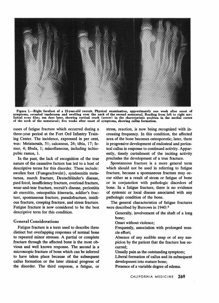
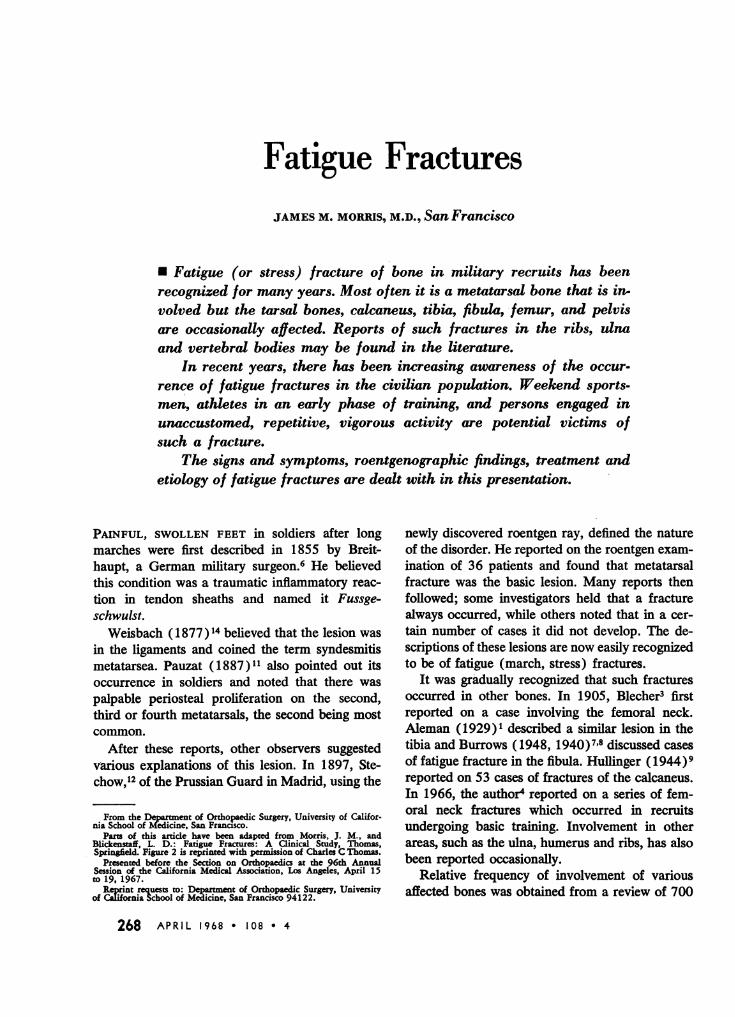
Figure 1.-Right forefoot of a 23-year-old recruit. Physical examination, approximately one week after onset ofsymptoms, revealed tenderness and swelling over the neck of the second metatarsal. Reading from left to right are:Initial x-ray film; ten days later, showing cortical crack (arrow) in the characteristic position in the medial cortexof the neck of the metatarsal; five weeks after onset of symptoms, showing callus formation.
cases of fatigue fracture which occurred during athree-year period at the Fort Ord Infantry Train-ing Center. The incidence, expressed in per cent,was: Metatarsals, 51; calcaneus, 26; tibia, 17; fe-mur, 4; fibula, 1; miscellaneous, including ischio-pubic ramus, 1.
In the past, the lack of recognition of the truenature of the causative factors has led to a host ofdescriptive terms for this disorder. These include:swollen foot (Fussgeschwulst), syndesmitis meta-tarsea, march fracture, Deutschliinder's disease,pied force, insufficiency fracture, overload fracture,wear-and-tear fracture, recruit's disease, periostitisab exercitio, osteopathia itineraria, soldier's frac-ture, spontaneous fracture, pseudofracture, insidi-ous fracture, creeping fracture, and stress fracture.Fatigue fracture is now considered to be the bestdescriptive term for this condition.
General ConsiderationsFatigue fracture is a term used to describe three
distinct but overlapping responses of normal boneto repeated minor stresses. A partial or completefracture through the affected bone is the most ob-vious and well known response. The second is amicroscopic fracture of bone which can be inferredto have taken place because of the subsequentcallus formation or the later clinical progress ofthe disorder. The third response, a fatigue, or
stress, reaction, is now being recognized with in-creasing frequency. In this condition, the affectedarea of the bone becomes osteoporotic; later, thereis progressive development of endosteal and perios-teal callus in response to continued activity. Appar-ently, timely curtailment of the inciting activityprecludes the development of a true fracture.
Spontaneous fracture is a more general termwhich should not be used in referring to fatiguefracture, because a spontaneous fracture may oc-cur either as a result of stress or fatigue of boneor in conjunction with pathologic disorders ofbone. In a fatigue fracture, there is no evidenceof systemic or local disease associated with anypathologic condition of the bone.The general characteristics of fatigue fractures
were described by Burrows in 1940:8Generally, involvement of the shaft of a longbone;Onset without violence;Frequently, association with prolonged mus-cle effort;Absence of any audible snap or of any sus-picion by the patient that the fracture has oc-curred;Usually pain as the outstanding symptom;Liberal formation of callus and its subsequentdevelopment into mature bone;Presence of a variable degree of edema.
CALIFORNIA MEDICINE 269
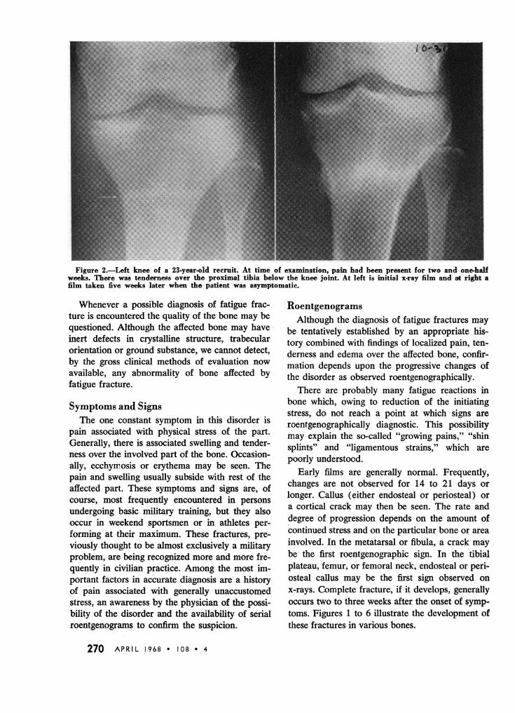
Figure 2.-Left knee of a 23-year-old recruit. At time of examination, pain had been present for two and one-halfweeks. There was tenderness over the proximal tibia below the knee joint. At left is initial x-ray film and at right afilm taken five weeks later when the patient was asymptomatic.
Whenever a possible diagnosis of fatigue frac-ture is encountered the quality of the bone may bequestioned. Although the affected bone may haveinert defects in crystalline structure, trabecularorientation or ground substance, we cannot detect,by the gross clinical methods of evaluation nowavailable, any abnormality of bone affected byfatigue fracture.
Symptoms and SignsThe one constant symptom in this disorder is
pain associated with physical stress of the part.Generally, there is associated swelling and tender-ness over the involved part of the bone. Occasion-ally, ecchyrnosis or erythema may be seen. Thepain and swelling usually subside with rest of theaffected part. These symptoms and signs are, ofcourse, most frequently encountered in personsundergoing basic military training, but they alsooccur in weekend sportsmen or in athletes per-forming at their maximum. These fractures, pre-viously thought to be almost exclusively a militaryproblem, are being recognized more and more fre-quently in civilian practice. Among the most im-portant factors in accurate diagnosis are a historyof pain associated with generally unaccustomedstress, an awareness by the physician of the possi-bility of the disorder and the availability of serialroentgenograms to confirm the suspicion.
RoentgenogramsAlthough the diagnosis of fatigue fractures may
be tentatively established by an appropriate his-tory combined with findings of localized pain, ten-derness and edema over the affected bone, confir-mation depends upon the progressive changes ofthe disorder as observed roentgenographically.
There are probably many fatigue reactions inbone which, owing to reduction of the initiatingstress, do not reach a point at which signs areroentgenographically diagnostic. This possibilitymay explain the so-called "growing pains," "shinsplints" and "ligamentous strains," which arepoorly understood.
Early films are generally normal. Frequently,changes are not observed for 14 to 21 days orlonger. Callus (either endosteal or periosteal) ora cortical crack may then be seen. The rate anddegree of progression depends on the amount ofcontinued stress and on the particular bone or areainvolved. In the metatarsal or fibula, a crack maybe the first roentgenographic sign. In the tibialplateau, femur, or femoral neck, endosteal or peri-osteal callus may be the first sign observed onx-rays. Complete fracture, if it develops, generallyoccurs two to three weeks after the onset of symp-toms. Figures 1 to 6 illustrate the development ofthese fractures in various bones.
270 APRIL 1968 * 108 * 4

It should be pointed out that, because of thelength of time between the onset of symptoms andthe development of findings that are visible onx-ray films, many cases of fatigue fracture arecompletely missed or misdiagnosed, especially incivilian practice (since the symptoms subside withdecrease of activity, serial x-ray films are not ob-tained).
Differential DiagnosisOccasionally the diagnosis of fatigue fracture
may be quite difficult and, at times, must be estab-lished largely by exclusion. Several conditions maybe confused with fatigue fracture and must bedistinguished. These include:
* Pathologic fractures seen in congenital de-fects, general systemic disease and meta-static bone tumors;
* Pseudofractures, or "Umbauzonen," suchas occur in Paget's disease, osteomalacia,rickets and Milkman's syndrome;
* Primary malignant disease of bone, suchas osteogenic sarcoma, Ewing's tumor orreticulum cell sarcoma;
* Osteitis due to tuberculosis or syphilis;
0Simple fracture due to violence;Osteomyelitis.
Pathologic fractures and, especially, so-calledpseudofractures are the cause of most of the prob-lems in differential diagnosis.
Figure 3.-Right ankle of a 68.year.old woman. Painhad been present for two and a half weeks and there wasedema and tenderness over the distal fibula above theankle joint. In initial x-ray film (left) fatigue fracture(arrow) is seen at upper border of syndesmosis. Treat.ment included a Gelocast wrapping and limited weight-hearing. Film taken five weeks later (right) shows pro.gression of healing.
In general, pathologic fractures can be distin-guished because they occur in obviously diseasedbone affected by such disorders as tumor, leuke-mia, osteoporosis and metabolic bone disease.
Pseudofractures may be described as transversezones of rarefaction varying in width from less thana millimeter to more than a centimeter, and usuallysymmetrical in distribution. In addition to theiroccurrence in rickets, osteomalacia and Milkman'ssyndrome, they have been described in associationwith renal rickets, coeliac disease, chronic idio-pathic steatorrhea and Paget's disease. They areusually of two types. The first type is associatedwith malacic diseases and appears as small sub-periosteal notches in the cortex when viewed tan-gentially, or as small, irregular, circular, punched-out zones of decalcification when viewed en face.This type slowly progresses across part or all ofthe bone as a band of decalcification. Periostealreaction, sometimes observed in curved bones, maybe apparent only on the concave side. The secondtype of pseudofracture is not associated with mal-acic disease and appears as cracks or fissures ex-tending through the cortex on one side, most oftenon the convex surface of pathologically curvedbones. Defects of this type are seen in Paget'sdisease.
These defects, or "pseudofractures," differ fromactual fractures in several respects: They developspontaneously without gross trauma and there isno crepitus or undue mobility except for a slightdegree of "elastic give." The similarity of "pseudo-fracture" to fatigue fracture and the source of con-fusion in diagnosis are apparent. However, as nowaccepted, they are two entirely separate entities.The importance of accurate differentiation for pur-poses of treatment and prognosis is obvious.
Fatigue Fracture Following Immobilization. Ihave recently seen a type of fracture that I feel de-serves mention because it might at some time alsoperplex others. This fracture is associated withincreased activity or weight-bearing after a periodof bed rest or immobilization. It has elements ofboth a true fatigue fracture and a pathologic frac-ture in that it is seen in bone which has becomeosteoporotic because of disuse.
TreatmentEarly recognition of a fatigue fracture is of pri-
mary importance in treatment. This is especiallytrue in the case of fractures of the femoral neck,
CALIFORNIA MEDICINE 271
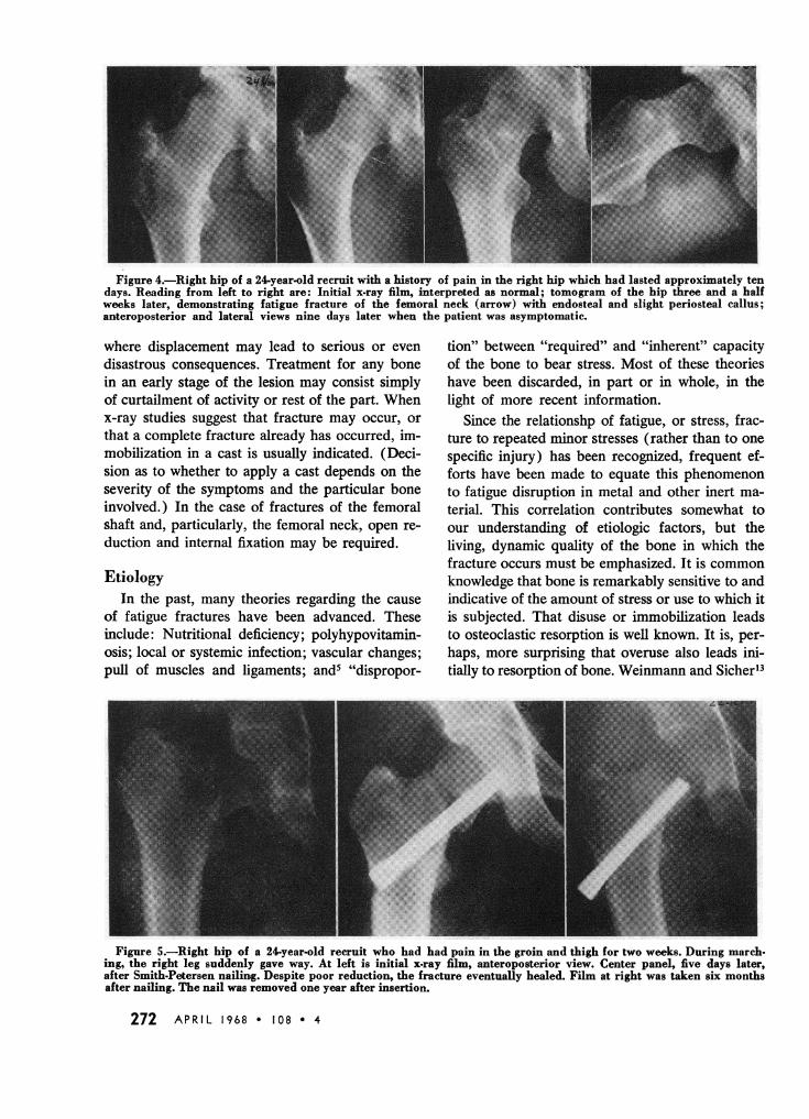
Figure 4.-Right hip of a 24-year-old recruit with a history of pain in the right hip which had lasted approximately tendays. Reading from left to right are: Initial x-ray film, interpreted as normal; tomogram of the hip three and a halfweeks later, demonstrating fatigue fracture of the femoral neck (arrow) with endosteal and slight periosteal callus;anteroposterior and lateral views nine days later when the patient was asymptomatic.
where displacement may lead to serious or evendisastrous consequences. Treatment for any bonein an early stage of the lesion may consist simplyof curtailment of activity or rest of the part. Whenx-ray studies suggest that fracture may occur, orthat a complete fracture already has occurred, im-mobilization in a cast is usually indicated. (Deci-sion as to whether to apply a cast depends on theseverity of the symptoms and the particular boneinvolved.) In the case of fractures of the femoralshaft and, particularly, the femoral neck, open re-duction and internal fixation may be required.
EtiologyIn the past, many theories regarding the cause
of fatigue fractures have been advanced. Theseinclude: Nutritional deficiency; polyhypovitamin-osis; local or systemic infection; vascular changes;pull of muscles and ligaments; and5 "dispropor-
tion" between "required" and "inherent" capacityof the bone to bear stress. Most of these theorieshave been discarded, in part or in whole, in thelight of more recent information.
Since the relationshp of fatigue, or stress, frac-ture to repeated minor stresses (rather than to onespecific injury) has been recognized, frequent ef-forts have been made to equate this phenomenonto fatigue disruption in metal and other inert ma-terial. This correlation contributes somewhat toour understanding of etiologic factors, but theliving, dynamic quality of the bone in which thefracture occurs must be emphasized. It is commonknowledge that bone is remarkably sensitive to andindicative of the amount of stress or use to which itis subjected. That disuse or immobilization leadsto osteoclastic resorption is well known. It is, per-haps, more surprising that overuse also leads ini-tially to resorption of bone. Weinmann and Sicher"
Figure 5.-Right hip of a 24-year-old recruit who had had pain in the groin and thigh for two weeks. During march-ing, the right leg suddenly gave way. At left is initial x-ray film, anteroposterior view. Center panel, five days later,after Smith-Petersen nailing. Despite poor reduction, the fracture eventually healed. Film at right was taken six monthsafter nailing. The nail was removed one year after insertion.
272 APRIL 1968 * 108 * 4

Figure 6.-Typical progression of a fatigue fracture of the ischiopubic ramus. X-ray films were taken one and a halfweeks, three and a half weeks and eight weeks after onset of symptoms. Initial film appears normal; progressive forma-tion of callus is seen in second and third.
said, "Increase of pressure or tension beyond thelimits of tolerance leads to destruction of bone byresorption...."
Johnson and coworkers0° studied, with use ofspecimens taken for biopsy, the development offatigue fracture of the tibia in 30 cases in whichthe duration of symptoms and roentgenographicsigns ranged from seven days to seven months.
During the first week, localized pain, tenderness,swelling and erythema were associated with veryactive osteoclastic resorption of the cortex asshown by local osteoporosis. Before resorptionbegan, lamellar birefringence in polarized light wasreduced. The normal sharply structured lamellarpattern became smudged or lost in the area of frac-ture. There appeared to be an "unraveling" of mar-ginal lamellar fibrils with consequent decrease inparallel orientation of the fibrils in the collagenbundles that constitute the lamellae. Resorptionfollowed the partial disorganization of the collagenlamellar structure. During the second week, peri-osteal (occasionally endosteal) callus formationoccurred. Resorption was complete at three weeksand callus formation was maximal at approximate-ly six weeks.
Remodeling, or transformation, from circumfer-ential lamellar bone to osteonal bone occurs be-tween 18 and 28 years of age. All biopsy speci-mens showed development of the lesion withinunosteonized lamellar bone. It appears, therefore,
that fatigue fracture may represent an accelerationof the normal internal remodeling of circumferen-tial lamellar bone to adult osteonized bone.
Knowledge of the mechanisms by which stressaffects osteogenesis and the replacement, or turn-over, of bone is, therefore, necessary before com-plete understanding of the causes of fatigue frac-tures is possible. Recent work by Bassett2 onelectrical effects in bone appears to suggest a linkbetween mechanical stress and biological activityin bone. Because bone is so highly crystalline,bending forces on bone generate electricity, rough-ly in proportion to the amount of its deformation.This is due to a piezoelectrical effect produced byone or all of three mechanisms: (1) Stress orbending of collagen fibers; (2) bending of muco-polysaccharide molecules; (3) stress on collagen-hydroxyapatite interface (a semiconductor of theP-n type). Areas under compression (concave)are negatively charged, while convex areas are pos-itively charged. Deposition of bone is influencedby weak, artificially induced currents and occursin the area of electronegativity, or concavity. Theconverse, that is, that electropositivity causes os-teoporosis of bone, has not been demonstrated.
Present etiologic knowledge of fatigue fracturescan be summarized as follows. Fatigue fracture be-gins with excessive elastic deformation and recoilof bone, causing an "unraveling" of the lamellarstructure of a segment of older circumferential
CALIFORNIA MEDICINE 273
bone. Subsequent local resorption of the circum-ferential bone is followed by replacement with os-teonal bone, a normal process accelerated by stressand, perhaps, mediated by electrical activity. Ifthis acceleration is too rapid, resorption occursmore rapidly than replacement, causing local os-teoporosis followed by callus formation. If me-chanical stress is too great, complete fractureoccurs.
REFERENCES1. Aleman, 0.: Tumors of foot from long marches
(syndesmitis metatarsea), T. Milit. Halsov., 54:191, 1929.2. Bassett, C. A. L.: Electrical effects in bone, Sci.
Amer., 213:18, Oct. 1965.3. Blecher, A.: Uber den Einfluss des Parademarsches
auf die Entstehung der Fussgeschwulst, Med. Klin., 1: 305,1905.
4. Blickenstaff, L. D., and Morris, J. M.: Fatigue frac-tures of the femoral neck, J. Bone Joint Surg., 48-A: 1031,Sept. 1966.
5. Brandt, G.: Schleichende Frakturen (Umbauzonen,Uberlastungsschiiden), Ergebn. Chir. Orthop., 33:1, 1941.
6. Breithaupt: Zur Pathologie des menschlichen Fusses,Med. Z., 24:169, 5 Sept. 1855; 24:175, 12 Sept. 1855.
7. Burrows, H. J.: Fatigue fractures of the fibula, J.Bone Joint Surg., 30-B:266, May 1948.
8. Burrows H. J.: Spontaneous fracture of the appar-ently normal fibula in its lowest third, Brit. J. Surg., 28:82, July 1940.
9. Hullinger, C. W.: Insufficiency fracture of the cal-caneus: Similar to march fracture of the metatarsal, J.Bone Joint Surg., 26:751, Oct. 1944.
10. Johnson, L. C., Stradford, H. T., Geis, R. W.,Dineen, J. R., and Kerley, E. R.: Histogenesis of stressfractures. Armed Forces Institute of Pathology AnnualLectures, 1963. (Abstract in J. Bone Joint Surg., 45-A:1452, 1963.)
11. Pauzat, J. E.: De la periostite osteoplasique desmetatarsiens a la suite des marches, Arch. Med. Pharm.Mil., 10:337, 1887.
12. Stechow: Fussoedem und Rontgenstrahlen,Deutsch. Mil.-arztl. Z., 26:465, 1897.
13. Weinmann, J. P., and Sicher, H.: Bones and Bones:Fundamentals of Bone Biology, 2nd ed., C. V. Mosby Co.,St. Louis, 1955, p. 138.
14. Weisbach: Die sogenannte "Fussgeschwulst," Syn-desmitis metatarsea, der Infanteristen in Folge von an-strengenden Miirschen, Deutsch. Mil.-arztl. Z., 6:551,1887.
.-t.-.., ;. -14,
..,'N ivA .
.4.me
.'-,,P-- .. .'-
W.-,*
274 APRIL 1968 * 108 * 4
Related Documents