Loma Linda University eScholarsRepository@LLU: Digital Archive of Research, Scholarship & Creative Works Loma Linda University Electronic eses, Dissertations & Projects 9-2017 Factors Associated with Orthodontically Induced Apical Root Resorption of Maxillary Incisors Brandon Malan Follow this and additional works at: hp://scholarsrepository.llu.edu/etd Part of the Orthodontics and Orthodontology Commons is esis is brought to you for free and open access by eScholarsRepository@LLU: Digital Archive of Research, Scholarship & Creative Works. It has been accepted for inclusion in Loma Linda University Electronic eses, Dissertations & Projects by an authorized administrator of eScholarsRepository@LLU: Digital Archive of Research, Scholarship & Creative Works. For more information, please contact [email protected]. Recommended Citation Malan, Brandon, "Factors Associated with Orthodontically Induced Apical Root Resorption of Maxillary Incisors" (2017). Loma Linda University Electronic eses, Dissertations & Projects. 428. hp://scholarsrepository.llu.edu/etd/428

Welcome message from author
This document is posted to help you gain knowledge. Please leave a comment to let me know what you think about it! Share it to your friends and learn new things together.
Transcript

Loma Linda UniversityTheScholarsRepository@LLU: Digital Archive of Research,Scholarship & Creative Works
Loma Linda University Electronic Theses, Dissertations & Projects
9-2017
Factors Associated with Orthodontically InducedApical Root Resorption of Maxillary IncisorsBrandon Malan
Follow this and additional works at: http://scholarsrepository.llu.edu/etd
Part of the Orthodontics and Orthodontology Commons
This Thesis is brought to you for free and open access by TheScholarsRepository@LLU: Digital Archive of Research, Scholarship & Creative Works. Ithas been accepted for inclusion in Loma Linda University Electronic Theses, Dissertations & Projects by an authorized administrator ofTheScholarsRepository@LLU: Digital Archive of Research, Scholarship & Creative Works. For more information, please [email protected].
Recommended CitationMalan, Brandon, "Factors Associated with Orthodontically Induced Apical Root Resorption of Maxillary Incisors" (2017). LomaLinda University Electronic Theses, Dissertations & Projects. 428.http://scholarsrepository.llu.edu/etd/428

LOMA LINDA UNIVERSITY
School of Dentistry
in conjunction with the
Faculty of Graduate Studies
____________________
Factors Associated with Orthodontically Induced Apical Root Resorption of Maxillary
Incisors
by
Brandon Malan
____________________
A Thesis submitted in partial satisfaction of
the requirements for the degree
Master of Science in Orthodontics and Dentofacial Orthopedics
____________________
September 2017

© 2017
Brandon B. Malan
All Rights Reserved
iii
Each person whose signature appears below certifies that this thesis in his opinion is
adequate, in scope and quality, as a thesis for the degree of Master of Science.
, Chairperson
Kitichai Rungcharassaeng, Professor of Orthodontics and Dentofacial Orthopedics
Joseph Caruso, Professor of Orthodontics and Dentofacial Orthopedics
Joseph Kan, Professor of Implant Dentistry

iv
ACKNOWLEDGEMENTS
I would like to express my deep gratitude and appreciation for all those who
helped me complete my thesis. I’d like to especially thank Dr. Kit for all the time he
worked with me. I would also like to thank my committee members, Dr. Caruso and Dr.
Kan, for reviewing my work and for their input. I’d like to thank Dr. Oyoyo who went
over the data multiple times with me. Most importantly I would like to thank my wife,
Ashley, for the support she has given me throughout my entire dental education.
v
CONTENT
Approval Page .................................................................................................................... iii
Acknowledgements ............................................................................................................ iv
Table of Contents .................................................................................................................v
List of Figures ................................................................................................................... vii
List of Tables ................................................................................................................... viii
List of Abbreviations ......................................................................................................... ix
Abstract ................................................................................................................................x
Chapter
1. Review of the Literature ..........................................................................................1
2. Factors Associated with Orthodontically Induced Apical Root Resorption
of Maxillary Incisors ................................................................................................9
Abstract ..............................................................................................................9
Introduction ......................................................................................................11
Statement of the problem ...........................................................................11
Hypothesis..................................................................................................12
Materials and methods .....................................................................................12
Patient selection .........................................................................................12
Data Collection ..........................................................................................13
Central Incisor Landmarks .........................................................................14
Volume Orientation ...................................................................................15
CBCT Tooth Position Measurements ........................................................15
Statistical Analysis .....................................................................................18
Results ..............................................................................................................19
Correlation Between RR and Tooth Movements .......................................20
Relationship of RR with Other Factors ......................................................27
Discussion ........................................................................................................30
Conclusions ......................................................................................................37

vi
3. Extended discussion ...............................................................................................38
Study Limitations .............................................................................................38
Future study directions .....................................................................................39
References ..........................................................................................................................41

vii
FIGURES
Figures Page
1. 0.15 mm Sagittal Slice Through Central Incisor at T1 and T2 ..............................14
2. Working Orientation in MPR.................................................................................15
3. Measurements of Horizontal, Vertical and Angulation Changes ..........................16
4. Measurements of Anterior-posterior and Rotational Changes ...............................17
5. Measurements of Inclination Changes ...................................................................17
viii
TABLES
Tables Page
1. Inclusion and exclusion criteria .......................................................................13
2. Root resorption severity distribution ...............................................................19
3. Intra-class correlation on repeated measurements ...........................................20
4. Correlation of amount of movement to root resorption ...................................21
5. Comparison of amount of RR according to amount of tooth movement
in the vertical dimension ..................................................................................22
6. Comparison of amount of RR according to amount of tooth movement
in the anterior-posterior dimension ..................................................................23
7. Comparison of amount of RR according to amount of mesio-distal tooth
movement .........................................................................................................24
8. Comparison of amount of RR according to amount of torque.........................25
9. Comparison of amount of RR according to amount of angulation ..................26
10. Comparison of amount of RR according to amount of rotation ......................26
11. Comparison of amount of RR according to amount of total tooth
movement .........................................................................................................27
12. Correlation of other variables to RR ................................................................28
13. Comparison of root resorption based on ethnicity ..........................................28
14. Comparison of root resorption based on gender ..............................................29
15. Comparison of root resorption based on the use of expanders ........................29
16. Comparison of root resorption based on extractions .......................................29
17. Comparison of root resorption based on asthmatic condition .........................30
ix
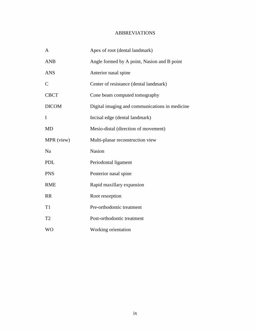
ABBREVIATIONS
A Apex of root (dental landmark)
ANB Angle formed by A point, Nasion and B point
ANS Anterior nasal spine
C Center of resistance (dental landmark)
CBCT Cone beam computed tomography
DICOM Digital imaging and communications in medicine
I Incisal edge (dental landmark)
MD Mesio-distal (direction of movement)
MPR (view) Multi-planar reconstruction view
Na Nasion
PDL Periodontal ligament
PNS Posterior nasal spine
RME Rapid maxillary expansion
RR Root resorption
T1 Pre-orthodontic treatment
T2 Post-orthodontic treatment
WO Working orientation

x
ABSTRACT OF THE THESIS
Factors Associated with Orthodontically Induced Apical Root Resorption of Maxillary
Incisors
by
Brandon Malan
Master of Science, Graduate Program in Orthodontics and Dentofacial Orthopedics
Loma Linda University, September 2017
Dr. Kitichai Rungcharassaeng, Chairperson
Introduction: Resorption of root apices is a ubiquitous occurrence in orthodontic
treatment. Although most occurrence of resorption during orthodontic treatment is
clinically inconsequential, a small percentage of patients have a severe amount of root
structure that is lost. There are factors that are widely accepted as responsible for apical
root resorption (RR), such as heavy compressive forces on the periodontal ligament
(PDL). Unfortunately, it is still largely unpredictable if one patient will experience more
root loss than what is considered normal. Thus, it is of clinical interest to further study
what factors play a role in RR.
Purpose: The purpose of this study was to utilize Cone Beam Computed
Tomography (CBCT) to evaluate whether certain treatment-related and patient-related
factors are associated with increased severity of orthodontically induced apical root
resorption.
Methods: Initial (T1) and final (T2) Digital Imaging and Communications in
Medicine (DICOM) CBCT images of patients orthodontically treated at Loma Linda
University were imported into OsiriX MD software (version 7.5.1, Pimeo, Bernex,
Switzerland) for measuring RR of right and left maxillary central incisors. Using
xi
fiduciary markers at the anterior nasal spine (ANS), posterior nasal spine (PNS), and
nasion (Na), movement of the incisors were assessed at three dental landmarks, the
incisal edge (I), the center of resistance (C), and the apex (A). Patient treatment records
were reviewed for information regarding patient age, gender, ethnicity, medical history,
expander appliances used, whether teeth were extracted, and time in treatment. Non-
parametric Spearman-Rho correlation tests were performed to determine whether
correlations existed between specific directions of tooth movement, time in treatment, or
age of the patient and the severity of RR. Kruskal-Wallis and Mann-Whitney U tests
were also used to determine differences in RR among groups of ethnicity, gender,
expansion treatment, extraction treatment and asthmatics.
Results: A total of 291 patients (582 teeth) were included in this study. Total
movement at A, intrusion at A and retraction at I were directions of movement that had
the highest correlations with RR at 0.344, 0.343, and 0.328 respectively. Time in
treatment had a significant but weak correlation with RR of 0.213. There was no
correlation with the patient age and amount of resorption. Males had statistically more
RR than females. However, males also had statistically more total movement of the root
apex. Incisors treated with extraction of two premolars also had more RR but also more
total movement at the apex compared to non-extraction treatment. Patients treated with a
rapid maxillary expansion appliance or a quad helix had more RR than those treated non-
expansion. There were no differences in RR among ethnicities or between asthmatics and
non-asthmatics.
Conclusions: In our sample, total movement at the apex, intrusion at the apex, and
retraction at the incisal edge had the highest correlation with root resorption. Treatment

xii
involving rapid palatal expansion and extractions had higher means of resorption.
Additionally, there were no differences in severity of resorption among ethnicities or
asthmatics and non-asthmatics.

1
CHAPTER ONE
LITERATURE REVIEW
Apical root resorption is a common iatrogenic effect from orthodontic tooth
movement.1 Histological studies have shown that 90% of teeth that have had orthodontic
forces applied to them have apical root resorption (RR).12 When evaluated from
radiographs, approximately 73% of teeth treated orthodontically show signs of RR.12
Fortunately, most resorption caused by orthodontic treatment will have little clinical
significance on the health of the tooth, however this may not be the case if resorption is
severe.4,5
The literature defines apical root resorption as severe once the root has lost more
than 4 mm or 1/3 of the original root length.12 This severity of RR has been reported to
occur to between 1-5% of treated teeth.12,15 Maxillary incisors have consistently been
identified as the teeth at highest risk of severe RR with 25% of them having greater than
2 mm of resorption.6
Prognosis of teeth that have undergone severe RR is questionable. Optimistic
results from long term studies have found that even when significant root loss has
occurred it is likely that the tooth will not be lost.4,5 Loss of a tooth as a direct outcome of
severe root resorption is considered uncommon.3 Stability may be maintained since root
structure lost at the apical portion of the root is more narrow and contributes less to the
overall periodontal ligament (PDL) surface area. An average root that is shortened 5 mm
will still retain 75% of its original periodontal attachment.12 Another comparison to
periodontal support is that 3 mm of RR is equivalent to 1 mm of crestal bone loss.12
2
The periodontal support that remains is more critical to the prognosis of the tooth
than simply how many millimeters of the apex was lost from RR. However, RR does
contribute to periodontal loss and leaves the tooth more vulnerable to the effects of
periodontal disease. Maxillary incisors are at considerable risk of hypermobility when
less than 9 mm of periodontal support is remaining.3,4
Apical resorption from orthodontic treatment is largely unavoidable. Orthodontic
forces placed on teeth produce local areas of hyalinization along the PDL space which are
attributed to the pathway of root resorption.35 The osteoclasts and macrophages that
remove the necrotic tissues in those areas also are capable of removing the protective
cementum; which leaves the tooth vulnerable to be further attacked in the resorptive
processes.17 Resorption craters on the lateral root surfaces are usually filled in and
repaired by cementoblasts, but when the apical surfaces are involved, cratering results in
permanent loss to root length.33
Although the biological process by which root resorption occurs is generally
understood, there are still many controversies surrounding what factors exacerbate this
process. Generally, these risk factors can be separated into two categories: patient-related
(biological factors) and treatment-related (mechanical factors).33
In terms of RR, some patients may have a heightened negative response to
orthodontic force. Evidence suggests that each patient’s risk for RR is dictated by
intrinsic patient characteristics or a genetic profile which the orthodontist has no control
over. Factors such as root shape, genetics, hormone deficiencies such as hypothyroidism
or hypopituitarism, asthma, bone density, allergies, chronic alcoholism, age, and gender

3
have been investigated.1,12 However, it is disputable whether certain of these factors
actually play a significant role in determining individual risk for RR.
Whether treatment at a younger age avoids more RR is debatable. Linge and
Linge36 reported less RR occurred if orthodontic treatment was performed before the age
of 11 years-old. By the time a child reaches 11 years-old the roots of the maxillary
incisors reach completion. At completion of the root apex, the apex is more prone to
resorption than a developing root.26 However, it is unclear how one can verify how much
root loss could be avoided if the original root length was not completely formed before
orthodontic treatment. Additionally, comprehensive orthodontic treatment before this
stage is not practical since it often precedes the permanent dentition stage. In other
studies, age has not been shown to have any significant impact on RR for patients
receiving comprehensive orthodontic treatment.9,16,27
Some studies have reported differences in severity of RR between males and
females. Braumind et al22 found that in their adult sample, males had an average of 1.2
mm more resorption than females. This is a significant difference compared to other
studies which found no difference between genders.14,16,26-28 Since most studies regarding
root resorption have a large adolescent sample size there could be differences between
male and female at later ages. However, there may also be confounding factors such as
the possibility that the adult male-seeking-orthodontic-patient may have greater
corrective needs than the adult female-seeking-patient. Other studies have reported a
difference in root resorption between male and female but those differences are not
statistically significant.6,9
4
Patients who have asthma or allergies have also been reported as being at greater
risk for RR. The systemic inflammatory processes in these patients could intensify the
inflammatory process involved in tooth movement.1,32 McNab et al31 found that
asthmatics had a higher overall incidence of root resorption to the maxillary posterior
dentition. However, it is important to note that there was no significant difference in the
incidence of moderate or severe root resorption between asthmatics and healthy patients.
Thus, clinical significance of asthmatics being more prone to root resorption may be
weak.
Dilacerated or pipette shaped roots have been reported to be at greater risk of
resorption.6,10,37 However, others have judged root shape to have no impact on the
amount of RR that occurs.26,38 In addition to the shape of the root, longer root lengths
have been found to have a weak but positive correlation to RR.6,10 A possible reason for
this is that longer roots require greater distances for the apex to travel in torqueing and
angulation correction. Additionally, it would be expected that orthodontists are generally
more cautious with their mechanics and forces when the roots start off stubby or short
compared to when the roots are long.
Genetic implications and the role it plays in RR are being further investigated.
Sameshima6 reported that Asians had significantly lower average root resorption than
Caucasians or Hispanics. Hispanics, with the highest mean root resorption among the
three ethnicities had a mean of 0.7 to 0.8 mm more resorption than the Asian sample.
Further evidence suggests a strong familial association with RR.32
Efforts have been made to determine specific genes that can be used as markers of
increased risk of severe RR. Patients who were homozygous with the IL-1beta allele 1
5
polymorphism were reported to have a 5.6 fold increased risk of having more than 2 mm
of resorption.8 Additionally, another study found that those who had the IL-6 SNP
rs1800796 GC genotype had greater resorption.9
Besides patient-related factors, many have investigated clinical variables that can
be controlled by the orthodontist to minimize the severity of resorption. Extraction
patterns, transverse expansion, surgical treatment, time in treatment, specific directions of
tooth movement, and proximity to the lingual cortical plate are typical factors explored in
such studies.12 Again, uncertainty exists on which of these factors have strong
associations with increased RR.11
Considerable evidence shows that heavy compressive forces on the PDL cause
more resorption than light forces or areas with tensile forces.17 Nevertheless, in clinical
practice a variety of force applications and conditions are introduced to the teeth in which
the clinician has limited knowledge of the magnitude of forces being applied. In an
attempt to evaluate whether there are directions of movement that pose greater risk of RR
than others, many investigators have tested for correlation between root resorption and
specific directions of tooth movement; such as buccal root torque versus lingual root
torque, intrusion versus extrusion, etc. Parker and Harris reported that incisor intrusion
and lingual root torque were the strongest predictors for resorption but that bodily
retraction, extrusion and buccal root torque had no significant relationship.14 Intrusion
had a correlation of 0.77 and resulted in 4x more root resorption than extrusive
movements.12 Intrusion in another study was not a significant factor in RR but rather
lingual root torque and retraction did have a significant relationship on RR.22,23
Sameshima found that anterior-posterior movement of the apex was significantly

6
associated with root resorption but not vertical movement.7 Braumind et al reported that
for every 1 mm of retraction there was on average approximately 0.3 mm of resorption.22
Kaley and Phillips23 reported that maxillary surgery, root torque and lingual cortical plate
approximation significantly increased the risk of resorption. Horiuchi13 found that
approximation of the lingual cortical plate explained 12% of the variation of the root
resorption they measured in their sample. However, lingual cortical plate approximation
may be more related to confounding variables such as retraction of incisors, extraction
treatment, and lingual root torque which as previously stated have been identified as
having a positive correlation with RR7,25,26,30.
In contrast to these findings, Simplicio et al reported that when incisors were
retracted with extraction of premolars, there were no significant correlations between
vertical, horizontal, or total apical movement of the maxillary incisors with severity of
RR.24 Mirabella et al37 found no correlation between RR and proximity to the lingual
cortical plate, rather they concluded that root resorption is more closely related to the
total root movement.
As evident in the literature, there remains a lack of consensus regarding which
treatment or patient related variables are most related to the severity of apical root
resorption that occurs. Thus, severity of RR as a consequence of treatment remains highly
unpredictable from patient to patient.
Lack of agreement between studies could be due to the use of conventional
radiographs to make measurements. Periapical and panoramic radiographs are vulnerable
to distortion and measurements of root changes may not be accurate.18 Lateral
cephalograms provide poor visualization of the location of root apices due to

7
superimposition of overlapping structures. The majority of past studies utilized periapical
or panoramic radiographs to determine incidence and severity of resorption. Many of
these studies could not objectively measure the root length before and after treatment
because of these limitations.39 Instead, studies sometimes classify RR into broad
categories of severity based on the general impression of RR shown in the radiograph.
Although this method aims to highlight the RR of clinical significance, this type of
classification and the radiographs used for it can be unreliable.39,40
Cone-beam computed tomography (CBCT) allows an examiner to avoid errors
related to projection geometry and structure superimposition.27,28 Dudic19 reported that
incidence of root resorption is underdiagnosed in panoramic radiographs (44% incidence)
compared to CBCT (69% incidence). Ponder20 showed that both high and low resolution
CBCT images had good agreement with microCT volumetric quantification of external
root resorption defects and can more accurately measure resorption defects than
periapical radiographs. Although their use for diagnosing root resorption is superior to
panoramic and periapical radiographs, there is not widespread prescription for CBCT
records since the effective dose for a CBCT image is much higher than that for
conventional radiographs.19,27 Utilizing CBCT technology in future RR studies however
will allow for more accurate measurements.
Limitations to previous clinical trials investigating RR risk factors include small
sample sizes and short evaluation periods which complicate the ability to come to any
certain conclusions.12 Retrospective studies allow for larger sample sizes and an
evaluation period that spans the entire treatment time, but they lack the ability to evaluate
precise forces applied and intermediate tooth movements that occurred. These limitations

8
and differences in study design lead to the lack of consensus on the role that risk factors
play in the severity of apical root resorption.12
Many controversies still exist regarding factors that increase the risk of RR.
However, as future research explores why some patients experience more RR than others,
orthodontists will be able to have a stronger evidenced-based approach to minimize RR
for those they treat.

9
CHAPTER TWO
FACTORS ASSOCIATED WITH ORTHODONTICALLY INDUCED APICAL
ROOT RESORPTION OF MAXILLARY INCISORS
Abstract
Introduction: Resorption of root apices is a ubiquitous occurrence in orthodontic
treatment. Although most occurrence of resorption during orthodontic treatment is
clinically inconsequential, a small percentage of patients have a severe amount of root
structure that is lost. There are factors that are widely accepted as responsible for root
resorption (RR), such as heavy compressive forces on the periodontal ligament (PDL).
Unfortunately, it is still largely unpredictable if one patient will experience more root loss
than what is considered normal. Thus, it is of clinical interest to further study what
factors play a role in RR.
Purpose: The purpose of this study was to utilize Cone Beam Computed
Tomography (CBCT) to evaluate whether certain treatment-related and patient-related
factors are associated with increased severity of orthodontically induced apical root
resorption.
Methods: Initial (T1) and final (T2) Digital Imaging and Communications in
Medicine (DICOM) CBCT images of patients orthodontically treated at Loma Linda
University were imported into OsiriX MD software (version 7.5.1, Pimeo, Bernex,
Switzerland) for measuring RR of right and left maxillary central incisors. Using
fiduciary markers at the anterior nasal spine (ANS), posterior nasal spine (PNS), and
nasion (Na), movement of the incisors were assessed at three dental landmarks, the
incisal edge (I), the center of resistance (C), and the apex (A). Patient treatment records

10
were reviewed for information regarding patient age, gender, ethnicity, medical history,
expander appliances used, whether teeth were extracted, and time in treatment. Non-
parametric Spearman-Rho correlation tests were performed to determine whether
correlations existed between specific directions of tooth movement, time in treatment, or
age of the patient and the severity of RR. Kruskal-Wallis and Mann-Whitney U tests
were also used to determine differences in RR among groups of ethnicity, gender,
expansion treatment, extraction treatment and asthmatics.
Results: A total of 291 patients (582 teeth) were included in the study. Total
movement at A, intrusion at A and retraction at I were directions of movement that had
the highest correlations with RR at 0.344, 0.343, and 0.328 respectively. Time in
treatment had a significant but weak correlation with RR of 0.213. There was no
correlation between the patient age and amount of resorption. Males had statistically
more RR than females. However, males also had statistically more total movement of the
root apex. Incisors treated with extraction of two premolars also had more RR but also
more total movement at the apex compared to non-extraction treatment. Patients treated
with a rapid maxillary expansion appliance or a quad helix had more RR than those
treated non-expansion. There were no differences in RR among ethnicities or between
asthmatics and non-asthmatics.
Conclusions: In our sample, total movement at the apex, intrusion at the apex, and
retraction at the incisal edge had the highest correlation with root resorption. Treatment
involving rapid palatal expansion and extractions did have a higher mean resorption.
Additionally, there were no differences in severity of resorption among ethnicities or
asthmatics and non-asthmatics.
11
Introduction
Statement of the Problem
Apical root resorption (RR) is an irreversible iatrogenic effect that can occur with
orthodontic treatment.1 Although most RR during orthodontic treatment is clinically
inconsequential, severe RR, although rare, is problematic. For those with severe RR,
avoidance of further root loss becomes a primary objective, leading to limitations of
treatment and possibly an esthetic compromise. After treatment the patient remains at risk
for tooth loss for those teeth affected.2-5
Maxillary incisors have consistently been reported as being most vulnerable to
severe RR.6 However, severity of resorption varies between individuals and it is difficult
to predict who is at highest risk.6,7 Factors that predispose a patient to severe RR may be
related to specific patient characteristics; such as genetics or root shape.8-12 Other factors
may be related to mechanical control or how the orthodontist moves the tooth during
treatment.1,7,10-15
It is widely accepted that heavy compressive forces on the periodontal ligament
(PDL) create hyalinized zones which lead to the destruction of the protective cementum
layer covering the root.15-17 However, magnitudes of force that the clinician delivers are
often unknown and certain directions of tooth movement may increase the incidence of
root resorption. However, there are areas of controversy regarding which directions of
tooth movement or orthodontic appliances are associated with more resorption.11
Most previous studies regarding RR have been limited to the use of periapical
films, panoramic radiographs, and lateral cephalograms to measure root length and tooth
position changes. Image distortion and image superimposition make these radiographic

12
images unreliable for measuring root resorption.18 Conversely, cone-beam computed
tomography (CBCT) has been shown to be superior for diagnosing and measuring
RR.19,20
The purpose of this study was to determine whether a relationship exists among
root resorption of the maxillary central incisors and treatment or patient related factors;
such as direction and magnitude of linear and angular tooth movement, time in treatment,
age of the patient, extraction of premolars, palatal expansion, gender, ethnicity and
history of asthma.
Hypothesis
The null hypothesis stated that there were no correlations between the amount of
root resorption and the direction and magnitude of linear and angular tooth movement.
Additionally, the null hypothesis stated that there were no differences in the amount of
root resorption when comparing different ethnicities, genders, presence and absence of
palatal expanders, extraction and non-extraction treatment, and presence and absence of
an asthmatic condition.
Materials and Methods
Patient Selection
This study was approved by the Institutional Review Board (IRB) of Loma Linda
University (LLU), Loma Linda, CA. Records were obtained of patients treated at Loma
Linda University Orthodontic Graduate clinic with pre-treatment (T1) and post-treatment

13
(T2) CBCT radiographs. One examiner (BM) performed all measurements and data
collection. Cases were selected based on the following inclusion/exclusion criteria:
Table 1. Inclusion and exclusion criteria used in patient selection
Inclusion Criteria
1. Full treatment case with T1 and T2 CBCT scans
2. Both T1 and T2 scans taken from NewTom 5G
Exclusion Criteria
3. Missing or not fully formed maxillary central incisor
1. Phase 1 treatment
2. Maxillary surgical cases
3. Changes in incisal contour to central incisor crowns
Data Collection
Records from patients who met the selection criteria were reviewed and the
following data recorded:
Chart number
Gender
Age at beginning of comprehensive of treatment
Ethnicity (Asian, Black, Caucasian, Hispanic)
Time in treatment
Orthodontic expanders used
Teeth that were extracted for treatment
Medical History
14
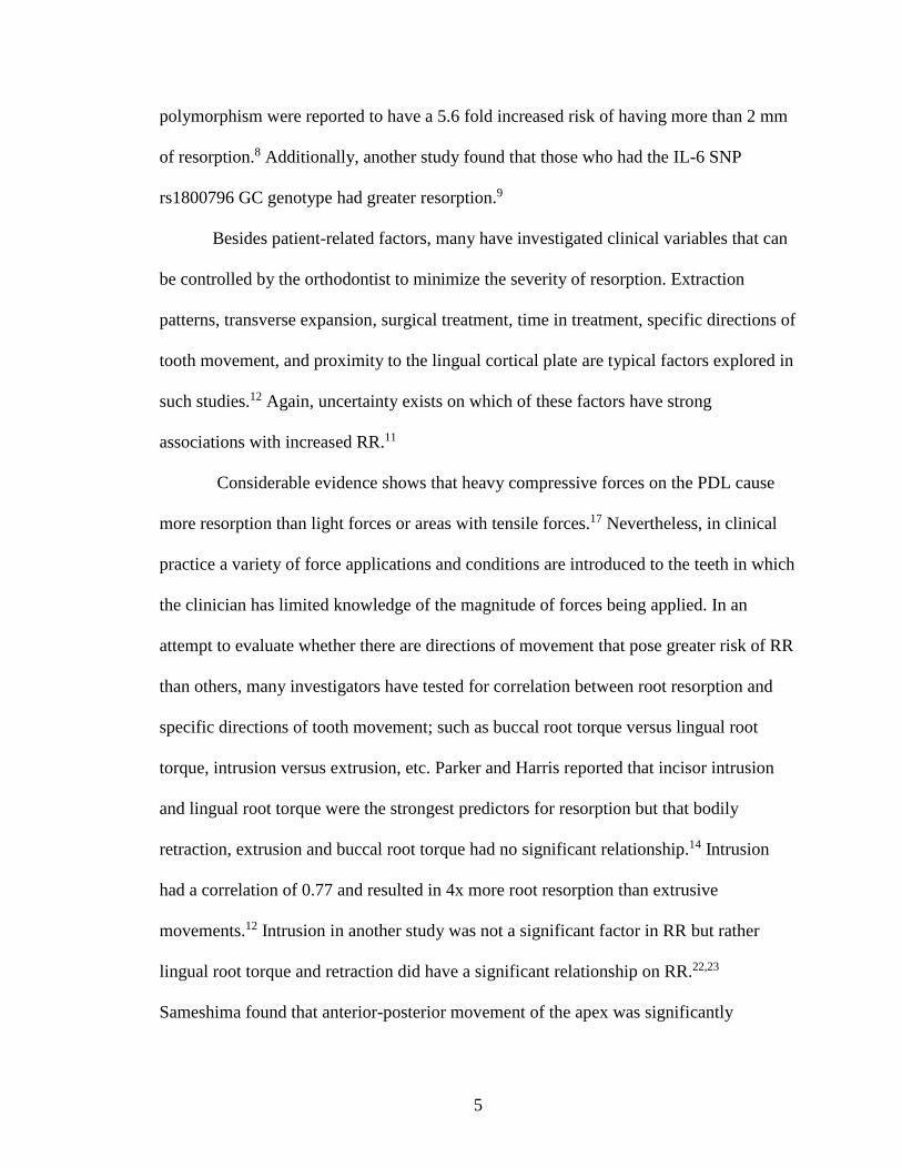
Central Incisor Landmarks
CBCT records of patients who met the criteria were anonymized and imported
into OsiriX MD (version 7.5.1, Pimeo, Bernex, Switzerland) as Digital Imaging and
Communication in Medicine (DICOM) files. In the multi-planar reconstruction (MPR)
view, a 0.15 mm sagittal slice was made through the middle of each maxillary central
incisor. In this sagittal view the incisal edge (I), center of resistance (C), and root apex
(A) were identified. The center of resistance was approximated as the bucco-lingual
center of the root at 1/3 of the length from the crestal bone to the root apex.21 The
measurement between I-A was recorded as tooth length. Additionally, the I-C length and
the CIA angle were recorded from T1 images in order to duplicate the I, C, and A points
on to the same tooth at T2 (Figure1).
Figure 1. 0.15 mm sagittal slice through the central incisor at T1 (a) and T2 (b).
Markers were placed at I, C, A on T1 (a). Distance and angle relationships between
those markers were duplicated on T2 (b).
b a
15
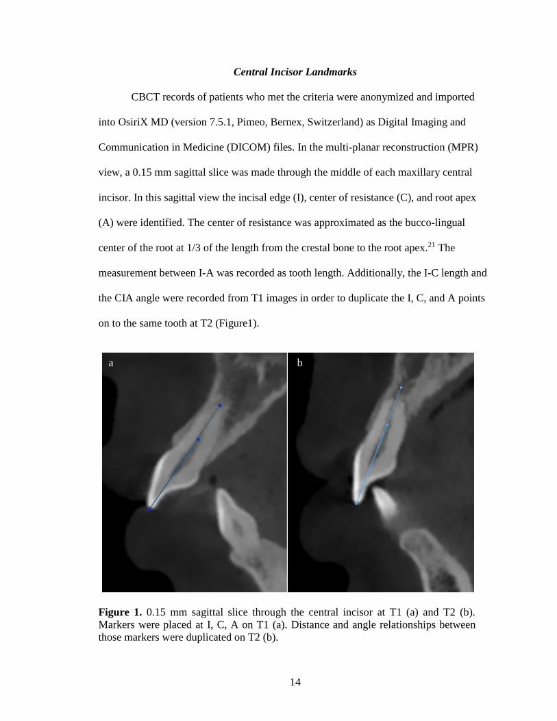
Volume Orientation
In MPR, a working orientation (WO) of the head was then constructed using
fiduciary markers at anterior nasal spine (ANS), posterior nasal spine (PNS), and nasion
(Na). The head was positioned so that the ANS-PNS line (palatal plane) aligned
completely horizontal in a sagittal view and vertical in the transverse view. In the coronal
view the Na-ANS line was orientated completely vertical. Once the head was in WO
(Figure 2), measurements of tooth positions were performed.
Figure 2. The head was orientated in all 3 planes in MPR utilizing cephalometric
landmarks, Na, ANS, and PNS. WO was established before measurements of tooth position
were made.
CBCT Tooth Position Measurements
Vertical, mesio-distal, and angulation measurements of tooth position were
performed in the coronal view (Figure 3). Mesio-distal positions of the landmarks were
determined using the horizontal distance away from the Na-ANS line. Vertical position
16
of the dental landmarks I, C, and A were measured as the vertical distance away from
ANS in WO. Angulation of the tooth was measured as the angle formed by I-A and the
vertical reference line.
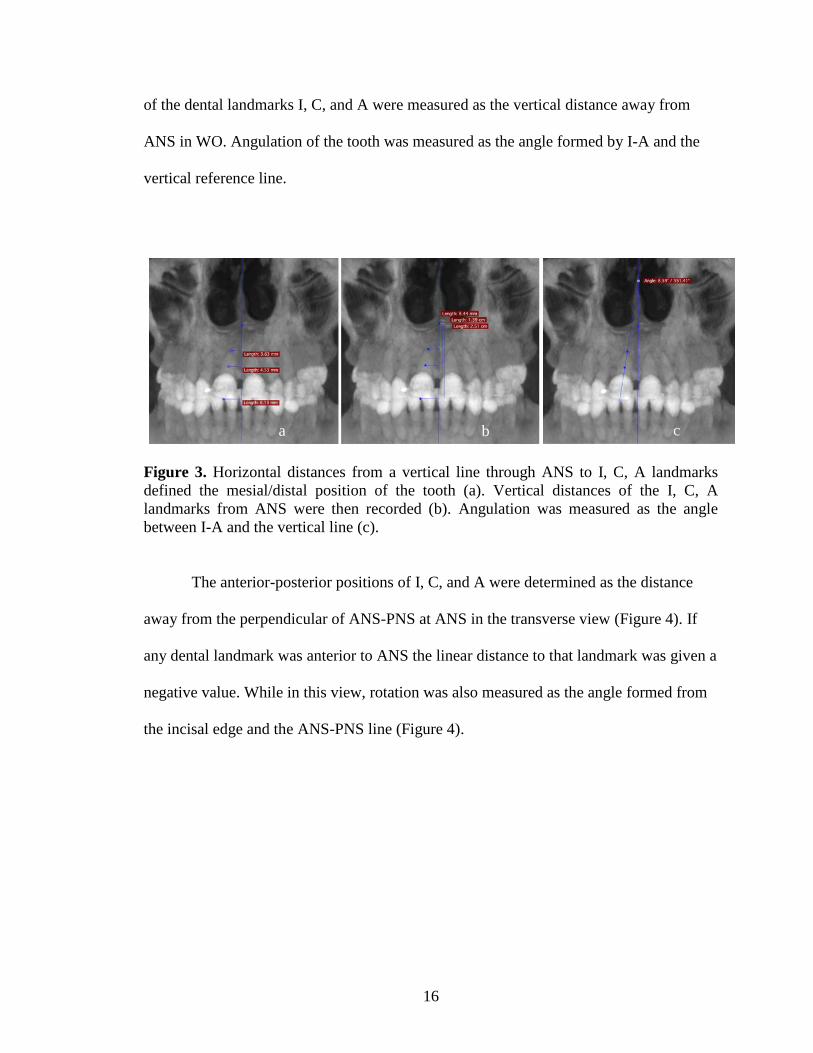
Figure 3. Horizontal distances from a vertical line through ANS to I, C, A landmarks
defined the mesial/distal position of the tooth (a). Vertical distances of the I, C, A
landmarks from ANS were then recorded (b). Angulation was measured as the angle
between I-A and the vertical line (c).
The anterior-posterior positions of I, C, and A were determined as the distance
away from the perpendicular of ANS-PNS at ANS in the transverse view (Figure 4). If
any dental landmark was anterior to ANS the linear distance to that landmark was given a
negative value. While in this view, rotation was also measured as the angle formed from
the incisal edge and the ANS-PNS line (Figure 4).
b
a b c
17
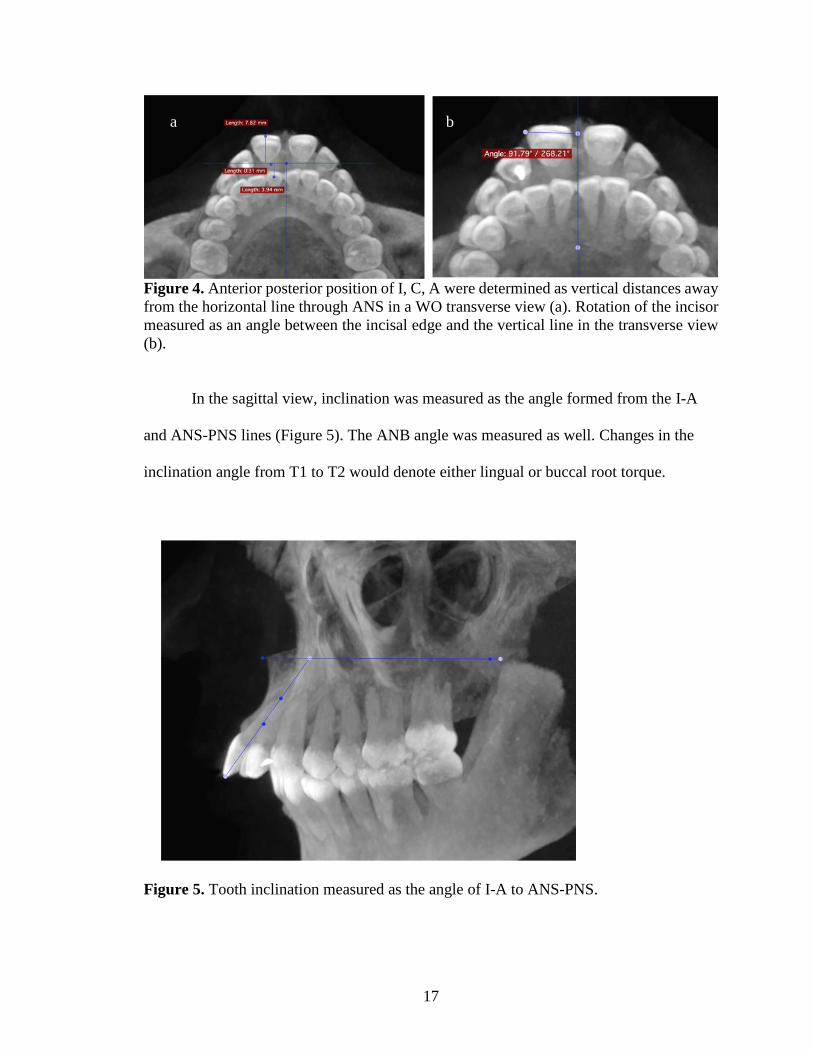
Figure 4. Anterior posterior position of I, C, A were determined as vertical distances away
from the horizontal line through ANS in a WO transverse view (a). Rotation of the incisor
measured as an angle between the incisal edge and the vertical line in the transverse view
(b).
In the sagittal view, inclination was measured as the angle formed from the I-A
and ANS-PNS lines (Figure 5). The ANB angle was measured as well. Changes in the
inclination angle from T1 to T2 would denote either lingual or buccal root torque.
Figure 5. Tooth inclination measured as the angle of I-A to ANS-PNS.
a b

18
Tooth length was measured from the incisal edge to the root apex on T2 CBCT
images. The difference in tooth length between T1 and T2 was recorded as the amount of
root resorption. However, the C and A landmarks were placed according to the I-C
length, I-A length and CIA angle measurements from T1 in order to duplicate the length
of the original root and position of the center of resistance. All measurements previously
explained were then performed on the T2 image. All linear and angular measurements
were recorded to the nearest 0.1 mm and 0.1 respectively.
Orthodontic movement of the dental landmarks were determined by calculating
the difference of the T1 and T2 positions. Negative numerical differences indicated that
the direction of movement was extrusion, retraction, or lingual root torque. Differences
with a positive value indicated the direction was intrusion, protraction, or buccal root
torque. Mesio-distal movements, rotation, and angulation changes were all treated as
absolute values with no respect to direction. The total absolute distances, irrespective of
direction, that each dental landmark moved from the T1 position was determined by
taking the square root of the sum of squares of the vertical, mesio-distal, and anterior-
posterior distances for each respective landmark.
Statistical Analysis
Each measured direction of tooth movement for each dental landmark was treated
as an independent variable. Non-parametric Spearman-Rho correlation analyses were
performed to determine the possible correlations with directions of tooth movement to
root resorption. Kruskal-Wallis and Mann-Whitney U tests were performed to determine
differences in root resorption between ethnicities, gender, asthmatics, extraction
19
treatment and use of expanders. The data regarding tooth movement was also stratified
according to the magnitude of movement in each direction and Kruskal-Wallis and post
hoc tests were used to determine differences of root resorption experienced at increasing
ranges of tooth movement. Reliability of the measurements was assessed using intra-class
correlation tests. For all statistical analyses the significance level was set at alpha ≤ 0.05.
Results
Two hundred ninety-one patients met the selection criteria for this study. One
hundred seventy-one were female and one hundred twenty were male. The mean age of
the patients was 17.0 ± 9.5 years with a range of 10 to 66 years. The average time in
treatment for the sample was 26.6 ± 8.3 months with a range of 9 to 60 months. The
mean root resorption in the entire sample was 1.08 ± 1.04 mm with a range of 0.0 to 7.0
mm. Fourteen teeth, or 2.4 %, of all incisors in the sample had 4 mm or more RR.
Both right and left incisors were grouped together for a total of 582 teeth for
statistical analysis. Most variables did not display normality; thus non-parametric
statistical testing was performed for all analyses.
Table 2. Root Resorption (RR) Severity Distribution
RR (mm) N % of total sample
0.0 - 0.4 183 31.4
0.5 - 0.9 141 24.2
1.0 - 1.4 110 18.9
1.5 - 1.9 55 9.5
2.0 - 2.4 38 6.5
2.5 - 2.9 22 3.8
3.0 - 3.4 10 1.7
3.5 - 3.9 9 1.5
4.0 14 2.4

20
All measurements were repeated on 30 patients and intra-class correlation tests
showed good agreement between original and repeated measurements with most
correlation coefficients above 0.85 (Table 3). Mesio-distal movement measurements at I
and C were the only measurements with lower coefficients at 0.78 and 0.77 respectively.
Table 3. Intra-Class Correlation on Repeated Measurements
Correlation Between the Amount of RR and Tooth Movement
Spearman-Rho correlation tests were performed for all directions of tooth
movement (Table 4). Weak but statistically significant correlations were found for all
linear directions of movement at landmarks A and C except for protraction. For linear
directions at landmark I, weak but statistically significant correlations were found only
for intrusion, retraction and total movement. For angular measurements, weak but
statistically significant correlations were found for all directions of movement.
Variable Intra-Class Correlation Variable Intra-Class Correlation
AP I 0.99 Angulation 0.87
AP C 0.91 Rotation 0.93
AP A 0.96 Inclination 0.99
Vertical A 0.94 ANB 0.99
Vertical C 0.92
Vertical I 0.92 Tooth length 0.97
MD I 0.78
MD C 0.77
MD A 0.89

21
Table 4. Correlation of Amount of Movement to RR
Direction Landmark Correlation p-value
Intrusion
A 0.343 < 0.001* C 0.274 0.001* I 0.147 0.037*
Extrusion
A 0.184 < 0.001* C 0.142 0.003* I -0.080 0.120
Retraction
A 0.268 < 0.001* C 0.283 < 0.001* I 0.328 < 0.001*
Protraction
A -0.103 0.176 C -0.054 0.483 I 0.064 0.264
Mesio-distal
A 0.165 < 0.001* C 0.112 0.007* I 0.050 0.230
Lingual Root Torque - 0.227 < 0.001*
Buccal Root Torque - 0.162 0.017*
Angulation - 0.092 0.026*
Rotation - 0.087 0.036*
Total Movement
A 0.344 < .001* C 0.303 < .001* I 0.181 < .001*
* Statistically significant
Comparisons of mean RR according to the magnitude of tooth movement was
made for each linear and angular direction using Kruskal-Wallis and post hoc tests.
Intrusion at A showed significantly more RR occurred after only 0.5 to 0.9 mm of
intrusion (Table 5). The means of RR associated with 0.5 to 0.9 mm of intrusion at A and
22
C were comparable to the RR associated with 2.0 to 2.9 mm of extrusion at A and C.
There were no significant differences among the RR associated with increasing
magnitudes of movement at I in either vertical dimension.
Table 5. Comparison of the amount of RR according to the amount of tooth movement
in the vertical dimension
Root Resorption (mm)
Movement (mm) N Mean SD Range p-value Intrusion A 0.002*
0.0 -0.4 51 0.79 0.83a 0.0 - 3.5 0.5- 0.9 34 1.33 1.00b 0.0 - 3.9 1.0- 1.4 20 1.44 1.15a,b 0.0 -4.0 >1.5 25 1.68 1.17b 0.0 - 4.1
Intrusion C < .001*
0.0 -0.4 74 0.88 0.79a 0.0 - 3.5 0.5- 0.9 52 1.33 0.99a,b 0.0 - 3.9 1.0- 1.4 19 2.06 1.18b 0.2 - 4.1 >1.5 8 1.11 0.94a,b 0.0 - 2.6
Intrusion I 0.292
0.0 -0.4 95 1.01 1.07 0.0 - 6.0 0.5- 0.9 48 1.02 0.94 0.0 - 3.9 1.0- 1.4 31 1.12 0.72 0.0 - 2.7 >1.5 26 1.14 0.82 0.2 - 3.3
Extrusion A < .001*
0.0 - 0.9 193 0.85 0.82a 0.0 - 4.6 1.0 - 1.9 140 0.88 0.89a 0.0 - 7.0 2.0 - 2.9 80 1.32 1.11b 0.0 - 5.3 3.0 39 1.88 1.64b 0.0 - 6.0
Extrusion C 0.005*
0.0 - 0.9 203 0.87 0.80a 0.0 - 4.6 1.0 - 1.9 145 1.01 1.11a,b 0.0 -7.0
2.0 - 2.9 55 1.32 1.18b 0.0 - 5.3 3.0 26 1.75 1.68a,b 0.0 - 6.0
Extrusion I 0.246
0.0 - 0.9 194 1.13 1.07 0.0 - 5.7 1.0 - 1.9 113 1.07 1.01 0.0 - 5.3 2.0 - 2.9 49 0.88 1.23 0.0 – 7.0 3.0 26 1.19 1.22 0.0 - 4.2
* Statistically significant

23
The mean RR increased with the magnitude of retraction at A and C but not with
protraction (Table 6). Each millimeter of retraction at C appeared to have greater impact
on the amount of resorption that occurred compared to retraction at A. The same
magnitudes of retraction at A and I were associated with similar resorption values.
Protraction at I showed significantly more RR once I was retracted 4 mm or more.
Table 6. Comparison of the amount of RR according to the amount of tooth movement in
the anterior-posterior dimension
Root Resorption (mm)
Movement (mm) N Mean SD Range p-value
Retraction A < .001*
0.0 - 0.9 128 0.86 0.86a 0.0 - 3.9 1.0 - 1.9 128 1.15 1.00a,b 0.0 - 6.0 2.0 - 2.9 83 1.24 1.04b,c 0.0 - 5.7 3.0 - 3.9 27 1.38 0.80b,c 0.3 - 3.0 4.0 43 1.89 1.33c 0.0 - 5.3
Retraction C < .001*
0.0 – 0.9 164 0.94 0.92a 0.0 – 7.0 1.0 – 1.9 146 1.26 1.01b 0.0 – 5.3 2.0 – 2.9 75 1.33 0.92b 0.0 – 4.3 3.0 – 3.9 20 2.06 1.33b,c 0.2 – 5.3 4.0 7 3.20 1.36c 1.6 – 5.3
Retraction I < .001*
0.0 – 0.9 69 0.78 0.79a 0.0 – 3.9 1.0 – 1.9 68 0.92 0.99a 0.0 – 5.3 2.0 – 2.9 65 1.20 1.14a,b 0.0 – 5.3 3.0 – 3.9 25 1.77 1.43b,c 0.0 – 4.3 4.0 47 1.78 1.25c 0.0 – 7.0
Protraction A 0.500
0.0 – 0.4 61 0.92 0.96 0.0 – 4.2 0.5 – 0.9 45 0.76 0.93 0.0 – 4.6 1.0 – 1.4 34 0.57 0.62 0.0 – 2.7 1.5 – 1.9 17 1.12 1.70 0.0 – 7.0 2.0 16 0.91 1.18 0.0 – 4.0
Protraction C 0.870
0.0 – 0.4 83 0.72 0.91 0.0 – 5.7 0.5 – 0.9 35 0.81 1.12 0.0 – 6.0
24
1.0 – 1.4 25 0.64 0.91 0.0 – 4.6 1.5 – 1.9 14 0.84 1.00 0.0 – 3.5 2.0 13 0.53 0.64 0.0 – 1.3
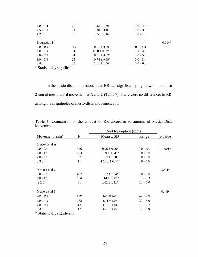
Protraction I 0.014*
0.0 – 0.9 116 0.91 0.89a 0.0 - 4.4 1.0 – 1.9 91 0.98 0.87a, b 0.0 – 4.6 2.0 – 2.9 51 0.81 0.62a 0.0 – 5.3 3.0 – 3.9 25 0.74 0.69a 0.0 – 3.4 4.0 25 1.81 1.56b 0.0 – 6.0
* Statistically significant
In the mesio-distal dimension, mean RR was significantly higher with more than
2 mm of mesio-distal movement at A and C (Table 7). There were no differences in RR
among the magnitudes of mesio-distal movement at I.
Table 7. Comparison of the amount of RR according to amount of Mesial-Distal
Movement
Root Resorption (mm)
Movement (mm) N Mean SD Range p-value
Mesio-distal A 0.0 - 0.9 346 0.96 0.96a 0.0 - 5.3 < 0.001*
1.0 - 1.9 173 1.09 1.04a,b 0.0 - 7.0 2.0 - 2.9 52 1.67 1.30c 0.0 - 6.0 3.0 11 1.50 1.03a,b,c 0.0 - 3.0
Mesio-distal C 0.004*
0.0 - 0.9 407 1.02 1.04a 0.0 - 7.0 1.0 - 1.9 154 1.14 0.98a,b 0.0 – 5.3 2.0 21 1.62 1.31b 0.0 – 6.0
Mesio-distal I 0.449
0.0 – 0.9 340 1.04 1.04 0.0 – 7.0
1.0 – 1.9 182 1.11 1.06 0.0 – 6.0 2.0 – 2.9 43 1.13 1.04 0.0 – 5.7 3.0 17 1.28 1.07 0.0 – 3.9
* Statistically significant

25
There appeared to be statistically significant differences in RR with increased
buccal root torque; however, no differences were actually found when the significant
values were adjusted by the Bonferroni correction for multiple comparisons in the post
hoc tests. Still there appears to be a clinical trend of increasing RR with increased buccal
root torque. A statistically significant increase in RR occurred with 10.0 to 14.9 of
lingual root torque and then another increase of RR with 20 or more (Table 8).
Table 8. Comparison of the amount of RR according to the amount torque
Root Resorption (mm)
Movement () N Mean SD Range p-value
Buccal Root Torque 0.014*
0.0 - 4.9 104 0.97 .98 0.0 - 4.6 5.0 - 9.9 64 0.99 1.13 0.0 - 4.3 10.0 - 14.9 21 1.11 0.94 0.0 - 3.6 15.0 - 19.9 17 1.60 1.24 0.0 - 4.1 20.0 11 2.20 1.89 0.0 - 7.0
Lingual Root Torque < .001*
0.0 - 4.9 148 0.81 0.65a 0.0 - 3.6 5.0 - 9.9 117 1.05 1.12a, b 0.0 - 5.3 10.0 - 14.9 58 1.30 0.95b, c 0.0 - 4.4 15.0 - 19.9 22 1.24 0.85a,b,c 0.0 – 3.5 20 20 2.00 1.58c 0.0 – 6.0
* Statistically significant
The Kruskal-Wallis test showed that a statistically significant difference in RR
existed with increased angulation changes (Table 9); however, again there were no
statistical differences found when adjusted by the Bonferroni correction for multiple
comparisons.
Rotation of 10 to 14.9 was associated with statistically more RR but no increase
of RR occurred with more rotation (Table 10).

26
Table 9. Comparison of the amount of RR according to the amount of angulation change
Root Resorption (mm)
Movement () N Mean SD Range p-value
Angulation 0.012*
0.0 - 4.9 425 0.98 0.95 0.0 - 5.3 5.0 - 9.9 134 1.25 1.17 0.0 - 7.0 10.0 23 1.70 1.41 0.0 -5.7
* Statistically significant
Table 10. Comparison of the amount of RR according to the amount of rotation
Root Resorption (mm)
Movement () N Mean SD Range p-value
Rotation 0.044*
0.0 - 4.9 208 0.99 1.03a 0.0 - 5.7 5.0 - 9.9 168 1.02 0.92a,b 0.0 -4.3 10.0 - 14.9 122 1.32 1.27b 0.0 - 7.0 15.0- 19.9 42 1.11 0.82a,b 0.0 - 3.4 20.0 42 0.95 0.94a,b 0.0 - 3.9
* Statistically significant
Root resorption significantly increased as total linear movement, irrespective of
direction, increased at all the dental landmarks (Table 11). It appeared that A and C had
more impact on root resorption for every millimeter of movement than at I.

27
Table 11. Comparison of the amount of RR according to the amount of total tooth
movement
Root Resorption (mm)
Movement (mm) N Mean SD Range p-value
Total A < .001*
0.0 - 1.4 119 0.66 0.69a 0.0 - 3.9 1.5 - 2.9 281 1.00 0.97b 0.0 - 7.0 3.0 - 4.4 124 1.21 1.00b 0.0 - 5.3 4.5 58 1.98 1.41c 0.0 - 6.0
Total C < .001*
0 - 0.9 76 0.58 0.57a 0.0 - 2.7 1 - 1.9 239 0.91 0.87b 0.0 - 7.0 2.0 - 2.9 160 1.19 1.08b 0.0 - 5.3 3.0 107 1.61 1.31c 0.0 - 6.0
Total I < .001*
0.0 - 1.9 208 0.94 0.98a 0.0 - 5.3 2.0- 3.9 268 0.94 0.87a 0.0 - 5.3
4.0 - 5.9 63 1.50 1.28b 0.0 - 7.0 6.0 43 1.92 1.38b 0.0 - 6.0
* Statistically significant
Relationship of RR with Other Factors
There was a statistically significant but weak correlation with root resorption and
time in treatment (r = 0.213, p < 0.001; Table 12).
No correlation was found between root resorption and the age of patient. There
was also no correlation with root resorption and the patient’s ANB angle when the patient
had a Class III skeletal relationship. However, there was a weak but significant
correlation (r = 0.200, p < 0.001; Table 12) with RR and positive ANB angles.
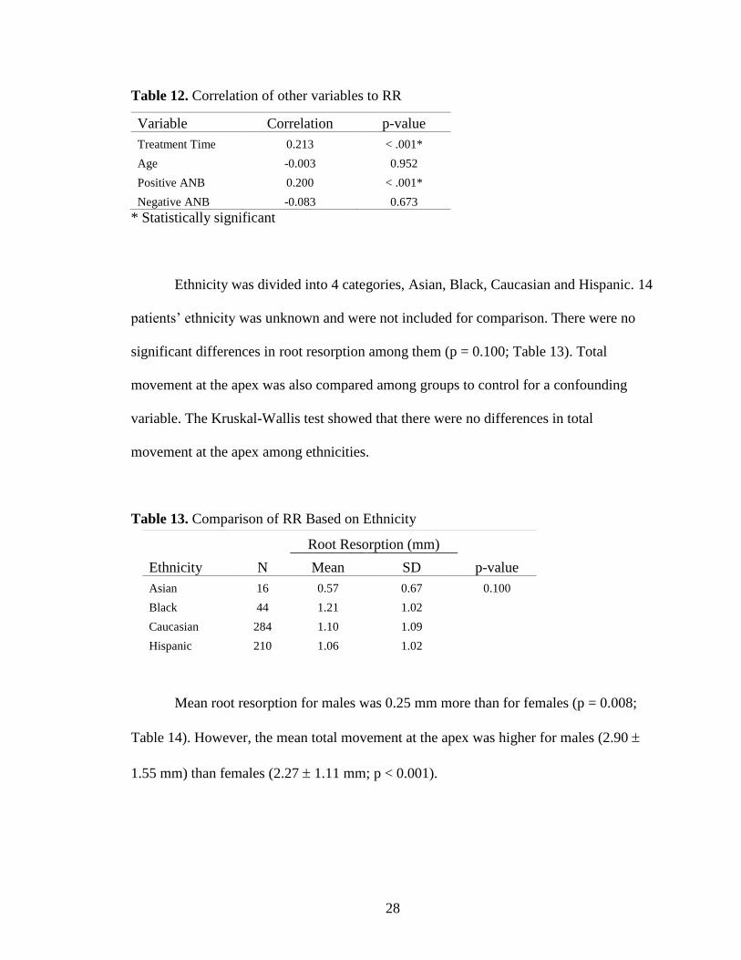
28
Table 12. Correlation of other variables to RR
Variable Correlation p-value
Treatment Time 0.213 < .001*
Age -0.003 0.952
Positive ANB 0.200 < .001*
Negative ANB -0.083 0.673
* Statistically significant
Ethnicity was divided into 4 categories, Asian, Black, Caucasian and Hispanic. 14
patients’ ethnicity was unknown and were not included for comparison. There were no
significant differences in root resorption among them (p = 0.100; Table 13). Total
movement at the apex was also compared among groups to control for a confounding
variable. The Kruskal-Wallis test showed that there were no differences in total
movement at the apex among ethnicities.
Table 13. Comparison of RR Based on Ethnicity
Mean root resorption for males was 0.25 mm more than for females (p = 0.008;
Table 14). However, the mean total movement at the apex was higher for males (2.90
1.55 mm) than females (2.27 1.11 mm; p < 0.001).
Root Resorption (mm)
Ethnicity N Mean SD p-value
Asian 16 0.57 0.67 0.100
Black 44 1.21 1.02
Caucasian 284 1.10 1.09
Hispanic 210 1.06 1.02

29
Table 14. Comparison of RR Based on Gender
Mean root resorption was significantly higher for patients who had rapid
maxillary expansion (RME) or expansion with a quad helix (p < 0.001; Table 15) while at
the same time there were no differences in the total movement at the apex among groups.
Treatment involving extraction of two upper premolars had higher mean RR than incisors
treated in non-extraction (p < 0.001; Table 16). Expectedly, the extraction group also had
more movement at the apex (2.75 1.45 mm) compared to the non-extraction group
(2.49 1.31 mm; p = 0.015).
Table 15. Comparison of RR Based on Use of RME appliance
Table 16. Comparison of RR Based on Extractions
Root Resorption (mm)
Gender N Mean SD p-value
Female 342 0.97 0.96 0.008*
Male 240 1.22 1.13
Root Resorption (mm)
Expander N Mean SD p-value
Non expansion 420 0.95a 0.96 <0.001*
RME 82 1.58b 1.34
Quad Helix 80 1.20b 0.96
Root Resorption (mm)
Extraction N Mean SD p-value
Non-EXT 462 0.99a 0.99 <0.001*
1 Tooth 14 0.94a, b 0.73
2 Teeth 106 1.43b 1.19
30
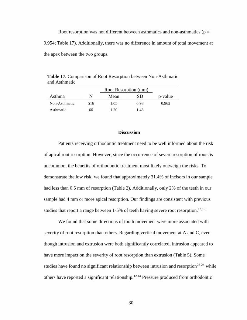
Root resorption was not different between asthmatics and non-asthmatics (p =
0.954; Table 17). Additionally, there was no difference in amount of total movement at
the apex between the two groups.
Table 17. Comparison of Root Resorption between Non-Asthmatic
and Asthmatic
Discussion
Patients receiving orthodontic treatment need to be well informed about the risk
of apical root resorption. However, since the occurrence of severe resorption of roots is
uncommon, the benefits of orthodontic treatment most likely outweigh the risks. To
demonstrate the low risk, we found that approximately 31.4% of incisors in our sample
had less than 0.5 mm of resorption (Table 2). Additionally, only 2% of the teeth in our
sample had 4 mm or more apical resorption. Our findings are consistent with previous
studies that report a range between 1-5% of teeth having severe root resorption.12,15
We found that some directions of tooth movement were more associated with
severity of root resorption than others. Regarding vertical movement at A and C, even
though intrusion and extrusion were both significantly correlated, intrusion appeared to
have more impact on the severity of root resorption than extrusion (Table 5). Some
studies have found no significant relationship between intrusion and resorption22-24 while
others have reported a significant relationship.12,14 Pressure produced from orthodontic
Root Resorption (mm)
Asthma N Mean SD p-value
Non-Asthmatic 516 1.05 0.98 0.962
Asthmatic 66 1.20 1.43
31
force during intrusion of an incisor can easily become excessive and lead to hyalinized
zones in the PDL. One has to be cautious of forces during intrusion since low forces
values can still produce high pressure when it is applied to a small root surface area.
Neither intrusion nor extrusion at I showed any trend of higher RR with
increasing movement (Table 5). This might be due to the fact that changes in vertical
position at the incisal edge of maxillary central incisors in this study could have been a
result of relative intrusion and extrusion from tipping of the tooth, rather than true
intrusion and extrusion.
In the anterior-posterior direction, there were significant correlations between
retraction and RR at all the dental landmarks whereas there were none between RR and
protraction (Table 6). Retraction of the incisor would likely bring the root against the
denser bone of the palatal cortical plate, which has been regarded as a risk factor of RR.23
Each millimeter of retraction at C appeared to have greater root resorption than retraction
at A and I. Retraction at C would indicate that lingual bodily movement (translation) of
the tooth occurred; and in the process the root apex could have cycled through many
redundant tipping and uprighting movements, thus exposing the root to more resorption.
Although protraction at A, C and I did not have any correlations with RR (p =
0.176, 0.483, 0.264; Table 4), when RR was compared according to the magnitude of
protraction at I there was significantly more RR once 4 mm or more of protraction
occurred (Table 6). Greater protraction at I at this magnitude could be related to
uncontrolled tipping which brings the apex of the root into close proximity to the palatal
cortical plate. All protraction values at A and C had low mean values of RR. The thin or
32
less dense buccal bone that the tooth moves against in this direction could be a factor as
to why values of RR were lower.
Unlike the other dimensions of linear movement, mesio-distal movement was not
subdivided into separate mesial and distal categories of movement. This is because unlike
the vertical and anterior-posterior movements, in mesial or distal movements the surface
area of the PDL being compressed and the density of the surrounding bone is generally
the same in either direction. Mesio-distal movements at A and C had significant
correlations with RR (p < 0.001, = 0.007) but not at I (p = 0.230; Table 4). This supports
the notion that orthodontic movement of the incisal edge alone likely has little association
with RR.
All angular measurements had statistically significant correlations although
angulation and rotation had very weak correlations with RR. Although buccal root torque
had a correlation of 0.162 (p = 0.017; Table 4) there were no statistically significant
differences of RR found among different magnitudes of buccal root torque (Table 8).
However, there still appeared to be a clinically relevant trend of increased RR in the
groups of higher buccal root torque, but the sample sizes in those groups were likely too
small to determine statistical difference.
Lingual root torque between 10 to 14.9 had significantly higher mean RR
compared to incisors with 0.0 to 4.9 lingual root inclination change (1.30 0.95 versus
0.81 0.65 mm, p <0.001; Table 8). An even higher and clinically significant mean RR
was associated with lingual root torque that exceeded 20 (2.00 1.58 mm). Lingual root
torque has previously been reported as an important factor related to root resorption.12,23
For instance, Parker and Harris reported lingual root torque as one of the strongest

33
predictors for resorption while buccal root torque had no significant relationship.14 While
we did find increases in lingual root torque to be associated with statistically significant
increases in mean resorption, we also found increasing buccal root torque to have similar
increasing trends of resorption although these were not statistically significant.
Total linear movement at all the dental landmarks had significant but weak
correlations with root resorption (Table 4). A meta-analysis performed on treatment
related factors found a high correlation of total movement at the apex with root
resorption.11 Total movement at the apex did have the highest correlation of all the
movement variables in our sample but the correlation was still weak. Significantly more
resorption occurred when the apex was moved 4.5 mm or more compared to when the
apex was moved between 0.0 to 1.49 mm (1.98 1.41 versus 0.66 0.69 mm, p< 0.001;
Table 11). When orthodontic forces are primarily concentrated on the apex it would be
expected that more resorption would take place.
Time in treatment also had a significant but weak correlation with root resorption
(p < 0.001; Table 12). The correlation we found was lower than what has previously been
reported.11 There is some ambiguity on how other studies measure time in treatment. This
variable may not be an accurate measurement of active treatment since it does not
necessarily indicate how long forces were applied to the incisors. Nevertheless, in our
study, time in treatment was initiated when upper incisors were bracketed or a palatal
expander was placed and ended when the brackets were removed. Motokowa et al25
found that RR was higher when treatment lasted longer than 30 months. Maues et al26
compared time in treatment of more than 3 years to less than 3 years and found that
significantly more resorption occurred in the former. Longer time in treatment could be

34
related to longer stimulation of resorptive processes. The accumulation of surface
resorption could lead to more severe resorption.
Negative ANB angles were not correlated with root resorption. Patients with
negative ANB angles have a Class III tendency and non-surgical orthodontics would
likely warrant protraction of the incisors. There was no correlation in protraction of the
upper incisors with root resorption which may be the reason that negative ANB angles
also did not have a significant correlation. Positive ANB angles did have a significant but
weak correlation (p < 0.001; Table 12) which is likely related to retraction of the upper
incisors, which is often necessary to resolve occlusal discrepancies due to mandibular
deficiencies.
There were no differences in the amount of root resorption among the ethnicities
identified. A previous study reported that Asians have less root resorption than
Caucasians and Hispanics.6 Asians in this sample did have less root resorption on
average, but it was not statistically significant. Our sample size from the Asian group was
also considerably smaller than that of Caucasians and Hispanics. However, it would have
been beneficial to have more Asian samples in our study for a better comparison. The
previously mentioned study also reported that Hispanics had the most resorption with a
mean of 0.7 to 0.8 mm more than Asians. Our results also conflict with this finding.
Our sample of male patients had a mean of 0.25 mm more resorption than female
patients. Another study reported their adult male patients had 1.2 mm more resorption
than their adult female patients.22 However, our sample of males also had more total
linear movement at A (2.90 1.55 mm) compared to females (2.27 1.11 mm, p <
0.001; Table 14). Thus the higher resorption in our sample of males may be related to the
35
magnitude of tooth movement more than the characteristic of gender. This may be likely
as many previous studies have found no difference of root resorption between genders.
1,6,9,14,16,26-28
There were two expansion groups in our study, patients treated with a rapid
maxillary expansion (RME) appliance, such as a hyrax or haas, and patients treated with
a dental expansion appliance, such as a quad helix. Both expansion groups had more root
resorption than the non-expansion group (p < 0.001; Table 15). The RME group had
more root resorption than the quad helix group but it was not statistically significant.
Other studies comparing expanders to root loss usually limit their investigation to the
premolars and molars since they are under direct force of the appliance. However, the
opening of the palatal suture during RME could introduce inflammatory mediators in
proximity of the incisor roots and increase the risk of resorption. Quad helixes can also
stretch the palatal suture for less dramatic skeletal changes.29 However, this is usually
possible only in younger patients. A previous study found no significant differences of
root resorption to the incisors for transverse treatments including rapid palatal expansion,
slow expansion or no expansion.7
Whether more resorption that occurred in the RME group was due to more tooth
movement or due to the rapid bone modeling occurring in close proximity to the roots is
difficult to determine. In our study, total movement at A was not statistically different
among non-expansion and the expansion groups. Yet, we know that between T1 and T2
the incisors are spaced apart from the expansion and more movement is needed to close
the space. The extra distance of root movement could not be recorded by our methods but
it may be partially responsible for why more root resorption occurred.

36
Patients who had two upper premolars extracted had more root resorption than
those who had no extraction (p < 0.001; Table 16). Usually treatment involving the
extraction of two upper premolars requires retraction of the incisors which is a direction
of movement that was found to be correlated with root resorption. Sameshima10 found no
differences in RR among different extraction patterns for those patients who had severe
resorption. McNab30 reported that the incidence of resorption of posterior teeth was
approximately 3.7 times higher for those who had extractions. Motokawa et al25 reported
their extraction group had a higher prevalence of severe resorption to the maxillary
central incisors as well, which supports our findings.
Our findings of root resorption in asthmatics conflicts with previous reports that
asthmatics are at greater risk for root resorption.12,31 It has been supposed that the
systemic inflammatory processes in asthmatics or patients with allergies could intensify
the inflammatory process involved in tooth movement.1,32 Our sample of asthmatics had
no significant difference of resorption compared to non-asthmatics. However, due to our
method of data collection, only the presence of an asthmatic condition, but not the
severity, was recorded and evaluated.
Other medical related factors such as medications taken, hormonal deficiencies
such as hypothyroidism or hypopituitarism, and chronic alcoholism have possible
relationships with root resorption.12 We attempted to gather as much information from
the medical history as possible but unfortunately sample sizes for these and other
conditions were too small to make any comparisons.
For all the variables that we examined for this study, no moderate or strong
correlations were found. However, based on our results, orthodontists should be more

37
cautious of root resorption in patients that require significant intrusion and retraction, yet
we acknowledge that there are likely other factors that have greater impact on the risk for
resorption such as force values12,15,22,33 and genetics.9,8
Conclusions
Based on the results from this study, we conclude that:
1. Weak but statistically significant correlations existed between RR and all
linear movements of A and C, except for protraction. At I, only retraction and
intrusion had significant but weak correlations with RR.
2. Total movement of A, intrusion of A, and retraction of I had the highest
correlations with RR (r= 0.344, 0.343, 0.328).
3. Weak but statistically significant correlations existed between all angular
measurements and RR.
4. Lingual root torque had the highest correlation of angular measurements with
RR (r=0.227).
5. Weak but statistically significant correlations with RR existed for both
treatment time and positive ANB angles (r= 0.213 and 0.200). No correlation
with RR existed for patient age or negative ANB angles.
6. Treatment with expansion or extraction of two premolars resulted in more RR
than non-expansion or non-extraction treatment respectively.
7. There were no differences in RR among different ethnicities nor between
asthmatics and non-asthmatics.

38
CHAPTER THREE
EXTENDED DISCUSSION
Study Limitations
It is difficult to be exact when determining the distance and direction of tooth
movement solely from orthodontic force. The direction of tooth movement in this study
was defined by the palatal plane as well as the other planes used in WO. The palatal plane
at ANS has long been established as a reference for cephalometric measurement of
maxillary dental changes during orthodontic treatment.34 When superimposing T1 and T2
cephalograms on these reference structures there would be minimal change in tooth
position in the absence of orthodontic treatment. The minimal change that would be
present would be due to post-eruption movement of the dentition if growth was present
between T1 and T2.
The upper incisors are expected to erupt downward and forward approximately
0.2 to 0.3 mm per year when superimposed over the palatal plane at ANS.34 This
movement forward and down would affect the ability to accurately measure extrusion and
intrusion or protraction and retraction that occurred solely from orthodontic force.
However, the effect is small. Additionally, 82% of the patients in the study were under 18
years old, making the effects of eruption present in most of the measurements performed.
Therefore, due to the difficultly to control for it and it being most likely inconsequential
to the results, natural post-eruption movement from growth was not accounted for.
Regarding the method of measuring root length changes, wear of incisal edges
during orthodontic treatment could have affected these measurements. Patients whose
treatment charts included notes of enameloplasty to the incisal edges of the maxillary
39
incisors were excluded from this study for this very reason. The measured changes in root
length could be more reliable if a more stable point was available for reference. The
cemento-enamel junction for example would have been stable. However, even though
anatomically the structure is stable, it was judged to be more difficult to replicate the
precise location of its position on the radiographic image compared to the incisal edge.
Lastly, while the distance that the tooth moved from T1 to T2 images can be
measured, it is uncertain how much the tooth moved between those time points. Often
with orthodontic treatment, “round tripping” occurs, in which the tooth is moved in one
direction but then is moved back to the other direction. The results in this study assumes
that “round tripping” is inconsequential to the movement that was measured.
Future Study Direction
The correlations in this study were determined based on the root resorption that
occurred in patients chosen in reverse chronological order from the T2 records date. Only
a very small percentage of the patients in the study had root resorption that was severe.
When enough data is available, a future study could include only data from patients with
severe resorption and analyze the treatment and patient related factors present.
Future studies in root resorption could also investigate genetics. These studies
could also be retrospective in design but patients would have to be recalled in order to
determine allele types of the specific genes being investigated.
Root shape and proximity to the cortical bone were not measured in this study,
but these are factors that should be followed up on with CBCT imaging. Additionally,
future studies that use CBCT data can analyze and measure root resorption as a volume

40
instead of a linear measurement. A volumetric measurement would be a more accurate
characterization of the destructive changes that occur to the root during treatment.

41
REFERENCES
1. Topkara A, Karaman AI, Kau CH. Apical root resorption caused by orthodontic
forces: A brief review and a long-term observation. Eur J Dent 2012;6:445-453.
2. Becker A, Chaushu S. Long-term follow-up of severely resorbed maxillary incisors
after resolution of an etiologically associated impacted canine. Am J Orthod
Dentofacial Orthop 2005;127:650-654; quiz 754.
3. Jonsson A, Malmgren O, Levander E. Long-term follow-up of tooth mobility in
maxillary incisors with orthodontically induced apical root resorption. Eur J Orthod
2007;29:482-487.
4. Levander E, Malmgren O. Long-term follow-up of maxillary incisors with severe
apical root resorption. Eur J Orthod 2000;22:85-92.
5. Remington DN, Joondeph DR, Artun J, Riedel RA, Chapko MK. Long-term
evaluation of root resorption occurring during orthodontic treatment. Am J Orthod
Dentofacial Orthop 1989;96:43-46.
6. Sameshima GT, Sinclair PM. Predicting and preventing root resorption: Part I.
Diagnostic factors. Am J Orthod Dentofacial Orthop 2001;119:505-510.
7. Sameshima GT, Sinclair PM. Predicting and preventing root resorption: Part II.
Treatment factors. Am J Orthod Dentofacial Orthop 2001;119:511-515.
8. Al-Qawasmi RA, Hartsfield JK, Jr., Everett ET, Flury L, Liu L, Foroud TM et al.
Genetic predisposition to external apical root resorption. Am J Orthod Dentofacial
Orthop 2003;123:242-252.
9. Guo Y, He S, Gu T, Liu Y, Chen S. Genetic and clinical risk factors of root
resorption associated with orthodontic treatment. Am J Orthod Dentofacial Orthop
2016;150:283-289.
10. Sameshima GT, Sinclair PM. Characteristics of patients with severe root resorption.
Orthod Craniofac Res 2004;7:108-114.
11. Segal GR, Schiffman PH, Tuncay OC. Meta analysis of the treatment-related factors
of external apical root resorption. Orthod Craniofac Res 2004;7:71-78.
12. Weltman B, Vig KW, Fields HW, Shanker S, Kaizar EE. Root resorption associated
with orthodontic tooth movement: a systematic review. Am J Orthod Dentofacial
Orthop 2010;137:462-476; discussion 412A.
13. Horiuchi A, Hotokezaka H, Kobayashi K. Correlation between cortical plate
proximity and apical root resorption. Am J Orthod Dentofacial Orthop
1998;114:311-318.

42
14. Parker RJ, Harris EF. Directions of orthodontic tooth movements associated with
external apical root resorption of the maxillary central incisor. Am J Orthod
Dentofacial Orthop 1998;114:677-683.
15. Roscoe MG, Meira JB, Cattaneo PM. Association of orthodontic force system and
root resorption: A systematic review. Am J Orthod Dentofacial Orthop
2015;147:610-626.
16. Bartley N, Turk T, Colak C, Elekdag-Turk S, Jones A, Petocz P et al. Physical
properties of root cementum: Part 17. Root resorption after the application of 2.5
degrees and 15 degrees of buccal root torque for 4 weeks: a microcomputed
tomography study. Am J Orthod Dentofacial Orthop 2011;139:e353-360.
17. Chan E, Darendeliler MA. Physical properties of root cementum: part 7. Extent of
root resorption under areas of compression and tension. Am J Orthod Dentofacial
Orthop 2006;129:504-510.
18. Tieu LD, Normando D, Toogood R, Flores-Mir C. Impact on perceived root
resorption based on the amount of incisal inclination as determined from
conventional panoramic radiography. Am J Orthod Dentofacial Orthop
2015;148:685-691.
19. Dudic A, Giannopoulou C, Leuzinger M, Kiliaridis S. Detection of apical root
resorption after orthodontic treatment by using panoramic radiography and cone-
beam computed tomography of super-high resolution. Am J Orthod Dentofacial
Orthop 2009;135:434-437.
20. Ponder SN, Benavides E, Kapila S, Hatch NE. Quantification of external root
resorption by low- vs high-resolution cone-beam computed tomography and
periapical radiography: A volumetric and linear analysis. Am J Orthod Dentofacial
Orthop 2013;143:77-91.
21. Burstone C. The biomechanics of tooth movement. BS Kraus, RA Riedel (Eds.)
Vistas in orthodontics. Lea & Febiger, Philadelphia 1962:197–213.
22. Baumrind S, Korn EL, Boyd RL. Apical root resorption in orthodontically treated
adults. Am J Orthod Dentofacial Orthop 1996;110:311-320.
23. Kaley J, Phillips C. Factors related to root resorption in edgewise practice. Angle
Orthod 1991;61:125-132.
24. Simplicio H, da Silva JS, Caldas SG, dos Santos-Pinto A. External apical root
resorption in retracted incisors. Orthodontics (Chic.) 2012;13:86-93.
25. Motokawa M, Sasamoto T, Kaku M, Kawata T, Matsuda Y, Terao A et al.
Association between root resorption incident to orthodontic treatment and treatment
factors. Eur J Orthod 2012;34:350-356.

43
26. Maues CP, do Nascimento RR, Vilella Ode V. Severe root resorption resulting from
orthodontic treatment: prevalence and risk factors. Dental Press J Orthod
2015;20:52-58.
27. Castro IO, Alencar AH, Valladares-Neto J, Estrela C. Apical root resorption due to
orthodontic treatment detected by cone beam computed tomography. Angle Orthod
2013;83:196-203.
28. Schwartz JP, Raveli TB, Almeida KC, Schwartz-Filho HO, Raveli DB. Cone beam
computed tomography study of apical root resorption induced by Herbst appliance. J
Appl Oral Sci 2015;23:479-485.
29. Bell RA, LeCompte EJ. The effects of maxillary expansion using a quad-helix
appliance during the deciduous and mixed dentitions. Am J Orthod 1981;79:152-
161.
30. McNab S, Battistutta D, Taverne A, Symons AL. External apical root resorption
following orthodontic treatment. Angle Orthod 2000;70:227-232.
31. McNab S, Battistutta D, Taverne A, Symons AL. External apical root resorption of
posterior teeth in asthmatics after orthodontic treatment. Am J Orthod Dentofacial
Orthop 1999;116:545-551.
32. Brezniak N, Wasserstein A. Orthodontically induced inflammatory root resorption.
Part II: The clinical aspects. Angle Orthod 2002;72:180-184.
33. Lopatiene K, Dumbravaite A. Risk factors of root resorption after orthodontic
treatment. Stomatologija 2008;10:89-95.
34. Ricketts RM. A four-step method to distinguish orthodontic changes from natural
growth. J Clin Orthod 1975;9:208-215, 218-228.
35. Kurol J, Owman-Moll P. Hyalinization and root resorption during early orthodontic
tooth movement in adolescents. Angle Orthod 1998;68:161-165.
36. Linge BO, Linge L. Apical root resorption in upper anterior teeth. Eur J Orthod
1983;5:173-183.
37. Mirabella AD, Artun J. Risk factors for apical root resorption of maxillary anterior
teeth in adult orthodontic patients. Am J Orthod Dentofacial Orthop 1995;108:48-55.
38. Brin I, Tulloch JF, Koroluk L, Philips C. External apical root resorption in Class II
malocclusion: a retrospective review of 1- versus 2-phase treatment. Am J Orthod
Dentofacial Orthop 2003;124:151-156.
39. Makedonas D, Lund H, Hansen K. Root resorption diagnosed with cone beam
computed tomography after 6 months and at the end of orthodontic treatment with
fixed appliances. Angle Orthod 2013;83:389-393.

44
40. Makedonas D, Lund H, Grondahl K, Hansen K. Root resorption diagnosed with cone
beam computed tomography after 6 months of orthodontic treatment with fixed
appliance and the relation to risk factors. Angle Orthod 2012;82:196-201.
Related Documents