Extracellular adenosine concentrations during in vitro ischaemia in rat hippocampal slices 1 Serena Latini, 1 Francesca Bordoni, 1 Felicita Pedata & * ,1 Renato Corradetti 1 Department of Preclinical and Clinical Pharmacology, University of Florence, Viale Pieraccini 6, 50139 Florence, Italy 1 The application of an ischaemic insult in hippocampal slices results in the depression of synaptic transmission, mainly attributed to the activation of A 1 adenosine receptors by adenosine released in the extracellular space. 2 To estimate the concentration of endogenous adenosine acting at the receptor level during an ischaemic episode, we recorded field e.p.s.ps (fe.p.s.ps) from hippocampal slices, and evaluated the ability of the selective A 1 receptor antagonist, 8-cyclopentyl-1,3-dipropylxanthine (DPCPX), to reverse the fe.p.s.p. depression induced by in vitro ischaemia. A relationship between the IC 50 of an antagonist and the endogenous concentration of a neurotransmitter has been used for pharmacological analysis. 3 The complete and reversible depression of fe.p.s.p. in the CA1 region induced by 5 min ischaemia was decreased in the presence of DPCPX (50 – 500 nM). 8-Phenyltheophylline (10 mM) abolished the depression of fe.p.s.ps during the ischaemic period, while a small (peak eect 12+4%) decrease in fe.p.s.ps was observed during the initial phase of reperfusion. 4 In the time-interval of maximal depression of fe.p.s.ps., IC 50 and adenosine concentration changed as function of time with a good degree of correlation. The maximal value of adenosine concentration was 30 mM. 5 Our data provide an estimation of the adenosine concentration reached at the receptor level during an ischaemic episode, with a higher time discrimination (15 s) than that achieved with any biochemical approach. This estimation may be useful in order to establish appropriate concentrations of purinergic compounds to be tested for their pharmacological eects during an ischaemic episode. Keywords: Adenosine; DPCPX; 8-PT; adenosine deaminase; synaptic responses; A 1 receptors; hippocampal slices; in vitro ischaemia; anoxia Abbreviations: aCSF, artificial cerebral spinal fluid; Ado, adenosine; ADA, adenosine deaminase; CNS, Central Nervous System; DPCPX, 8-cyclopentyl-1,3-dipropylxanthine; DPY, dipyridamole; EHNA, erythro-9-(2-hydroxy-3- nonyl)adenine hydrochloride; fe.p.s.p., field excitatory post synaptic potential; NBTI, S-(4-nitrobenzyl)-6- thioinosine; 8-PT, 8-phenyltheophylline Introduction In the Central Nervous System (CNS) adenosine is an important neuromodulator which exerts an inhibitory tonus on synaptic transmission, principally mediated by an inhibition of neurotransmitter release and by a reduction of postsynaptic excitability (Corradetti et al., 1984a; Dunwiddie, 1985; Proctor & Dunwiddie, 1987). This well established action of adenosine on neurotransmission is classically associated with the activation of the first (A 1 ) of four adenosine receptor types which have been cloned and identified in the CNS: A 1 ,A 2A , A 2B ,A 3 (Fredholm et al., 1994). In the brain region such as the hippocampus, under physiological conditions, adenosine A 1 receptors are tonically exposed to extracellular endogenous adenosine, as demonstrated by excitatory eects of adenosine receptor antagonists on electrophysiological responsiveness (Corradetti et al., 1984b; Dunwiddie & Hoer, 1980; Schubert, 1988; Wu & Saggau, 1994). On the other hand, an excitatory action of A 2A adenosine receptors on synaptic responses has been demonstrated in the hippocampus by use of the selective agonist CGS 21680 (Cunha et al., 1994; Sebastia˜o & Ribeiro, 1992), while A 3 adenosine receptors may be responsible for a desensitization of A 1 receptor-mediated responses (Dunwiddie et al., 1997). No information has been provided until now on the role of A 2B adenosine receptors on synaptic transmission. Brain extracellular concentration of adenosine can be increased by several stimuli, including electrical stimulation, hypoxia and ischaemia (Pedata et al., 1991; 1993; Phillis et al., 1987). It appears that in the hippocampus the application of ischaemic or hypoxic insults is associated with depressed synaptic transmission, mainly evoked by the activation of A 1 adenosine receptors by endogenous adenosine released in the extracellular space. Non-selective antagonists of adenosine receptors, like 8-phenyltheophilline (8-PT), have been found to delay or to prevent the hypoxia- or ischaemia-induced depression of synaptic transmission in the hippocampus (Fowler, 1989; 1990; Pedata et al., 1993), and the selective A 1 receptor antagonist, DPCPX, has been found to prevent the inhibition of synaptic responses evoked by hypoxia (Canhao et al., 1994; Lucchi et al., 1996). While an important neuroprotective role of A 1 adenosine receptor activation against ischaemic damage has been clearly demonstrated (Domenici et al., 1996; Heron et al., 1994), very few reports describe the role of A 2 and A 3 adenosine receptor during cerebral ischaemia. Since A 1 ,A 2 and A 3 adenosine receptors may be activated by dierent ranges of concentrations of endogenous adenosine (Fredholm et al., 1994), knowledge of adenosine concentrations reached in the extracellular space *Author for correspondence; E-mail: [email protected]fi.it British Journal of Pharmacology (1999) 127, 729 – 739 ª 1999 Stockton Press All rights reserved 0007 – 1188/99 $12.00 http://www.stockton-press.co.uk/bjp

Welcome message from author
This document is posted to help you gain knowledge. Please leave a comment to let me know what you think about it! Share it to your friends and learn new things together.
Transcript

Extracellular adenosine concentrations during in vitro ischaemia inrat hippocampal slices
1Serena Latini, 1Francesca Bordoni, 1Felicita Pedata & *,1Renato Corradetti
1Department of Preclinical and Clinical Pharmacology, University of Florence, Viale Pieraccini 6, 50139 Florence, Italy
1 The application of an ischaemic insult in hippocampal slices results in the depression of synaptictransmission, mainly attributed to the activation of A1 adenosine receptors by adenosine released inthe extracellular space.
2 To estimate the concentration of endogenous adenosine acting at the receptor level during anischaemic episode, we recorded ®eld e.p.s.ps (fe.p.s.ps) from hippocampal slices, and evaluated theability of the selective A1 receptor antagonist, 8-cyclopentyl-1,3-dipropylxanthine (DPCPX), toreverse the fe.p.s.p. depression induced by in vitro ischaemia. A relationship between the IC50 of anantagonist and the endogenous concentration of a neurotransmitter has been used forpharmacological analysis.
3 The complete and reversible depression of fe.p.s.p. in the CA1 region induced by 5 minischaemia was decreased in the presence of DPCPX (50 ± 500 nM). 8-Phenyltheophylline (10 mM)abolished the depression of fe.p.s.ps during the ischaemic period, while a small (peak e�ect 12+4%)decrease in fe.p.s.ps was observed during the initial phase of reperfusion.
4 In the time-interval of maximal depression of fe.p.s.ps., IC50 and adenosine concentrationchanged as function of time with a good degree of correlation. The maximal value of adenosineconcentration was 30 mM.
5 Our data provide an estimation of the adenosine concentration reached at the receptor levelduring an ischaemic episode, with a higher time discrimination (15 s) than that achieved with anybiochemical approach. This estimation may be useful in order to establish appropriateconcentrations of purinergic compounds to be tested for their pharmacological e�ects during anischaemic episode.
Keywords: Adenosine; DPCPX; 8-PT; adenosine deaminase; synaptic responses; A1 receptors; hippocampal slices; in vitroischaemia; anoxia
Abbreviations: aCSF, arti®cial cerebral spinal ¯uid; Ado, adenosine; ADA, adenosine deaminase; CNS, Central NervousSystem; DPCPX, 8-cyclopentyl-1,3-dipropylxanthine; DPY, dipyridamole; EHNA, erythro-9-(2-hydroxy-3-nonyl)adenine hydrochloride; fe.p.s.p., ®eld excitatory post synaptic potential; NBTI, S-(4-nitrobenzyl)-6-thioinosine; 8-PT, 8-phenyltheophylline
Introduction
In the Central Nervous System (CNS) adenosine is animportant neuromodulator which exerts an inhibitory tonuson synaptic transmission, principally mediated by an inhibition
of neurotransmitter release and by a reduction of postsynapticexcitability (Corradetti et al., 1984a; Dunwiddie, 1985; Proctor& Dunwiddie, 1987). This well established action of adenosine
on neurotransmission is classically associated with theactivation of the ®rst (A1) of four adenosine receptor typeswhich have been cloned and identi®ed in the CNS: A1, A2A,A2B, A3 (Fredholm et al., 1994). In the brain region such as the
hippocampus, under physiological conditions, adenosine A1
receptors are tonically exposed to extracellular endogenousadenosine, as demonstrated by excitatory e�ects of adenosine
receptor antagonists on electrophysiological responsiveness(Corradetti et al., 1984b; Dunwiddie & Ho�er, 1980; Schubert,1988; Wu & Saggau, 1994). On the other hand, an excitatory
action of A2A adenosine receptors on synaptic responses hasbeen demonstrated in the hippocampus by use of the selectiveagonist CGS 21680 (Cunha et al., 1994; SebastiaÄ o & Ribeiro,1992), while A3 adenosine receptors may be responsible for a
desensitization of A1 receptor-mediated responses (Dunwiddie
et al., 1997). No information has been provided until now onthe role of A2B adenosine receptors on synaptic transmission.
Brain extracellular concentration of adenosine can be
increased by several stimuli, including electrical stimulation,hypoxia and ischaemia (Pedata et al., 1991; 1993; Phillis et al.,1987). It appears that in the hippocampus the application of
ischaemic or hypoxic insults is associated with depressedsynaptic transmission, mainly evoked by the activation of A1
adenosine receptors by endogenous adenosine released in theextracellular space. Non-selective antagonists of adenosine
receptors, like 8-phenyltheophilline (8-PT), have been found todelay or to prevent the hypoxia- or ischaemia-induceddepression of synaptic transmission in the hippocampus
(Fowler, 1989; 1990; Pedata et al., 1993), and the selective A1
receptor antagonist, DPCPX, has been found to prevent theinhibition of synaptic responses evoked by hypoxia (Canhao et
al., 1994; Lucchi et al., 1996). While an importantneuroprotective role of A1 adenosine receptor activationagainst ischaemic damage has been clearly demonstrated(Domenici et al., 1996; Heron et al., 1994), very few reports
describe the role of A2 and A3 adenosine receptor duringcerebral ischaemia. Since A1, A2 and A3 adenosine receptorsmay be activated by di�erent ranges of concentrations of
endogenous adenosine (Fredholm et al., 1994), knowledge ofadenosine concentrations reached in the extracellular space*Author for correspondence; E-mail: [email protected]®.it
British Journal of Pharmacology (1999) 127, 729 ± 739 ã 1999 Stockton Press All rights reserved 0007 ± 1188/99 $12.00
http://www.stockton-press.co.uk/bjp

during an ischaemic event, is critical to evaluate theinvolvement of di�erent adenosine receptors.
In a recent work, Barlow (1995) explicitly set out the
relationship between the IC50 of an antagonist and theendogenous concentration of a neurotransmitter and suggestedthat by using an antagonist of known KB it is possible toestimate the concentration of an agonist of known KA and [A50]
acting at receptor level for producing the recorded pharmaco-logical e�ect. By using this relationship, in the present study weestimated the concentrations of adenosine acting at the
receptor level at various times during an ischaemia-like episodein vitro. To monitor the e�ect of endogenously releasedadenosine during ischaemia we recorded fe.p.s.ps and we
evaluated the ability of the selective A1 receptor antagonist,DPCPX, to reverse fe.p.s.p. depression induced by anischaemic episode on hippocampal slices.
Methods
Preparation of hippocampal slices
Experiments were carried out on rat hippocampal slices,
prepared as previously described (Corradetti et al., 1983).Charles River male Wistar rats, 150 ± 200 g body weight, werekilled by decapitation and their hippocampi rapidly removed
and placed on ice-cold oxygenated (95% O2-5% CO2) arti®cialcerebral spinal ¯uid (aCSF) of the following composition (mM):NaCl 124, KCl 3.33, KH2PO4 1.25,MgSO4 2, CaCl2 2, NaHCO3
25 and D-glucose 10. Slices (400 mm thick) were cut by aMcIlwain tissue chopper and kept in oxygenated aCSF for atleast 1 h at room temperature. A single slice was then placed ona nylon mesh, completely submerged in a small chamber
(0.5 ml) and superfused through a peristaltic pump withoxygenated aCSF (30 ± 328C) at a constant ¯ow rate of2 ml min71. The treated solutions reached the preparation in
90 s and this delay was taken into account in our calculations.In vitro ischaemia-like conditions were applied by superfusingthe slice for 5 min with aCSF without glucose and gassed with
nitrogen (95% N2-5% CO2) (Pedata et al., 1993). At the end ofthe ischaemic period, the slice was again superfusedwith normaloxygenated aCSF. Each slice was exposed to two periods ofischaemia-like conditions with a time interval of 50 min.
Extracellular recording
Test pulses (80 ms, 0.06 Hz) were delivered through a bipolarnichrome electrode positioned in the stratum radiatum.Evoked extracellular potentials were recorded with glass
microelectrodes (2 ± 10 MO) ®lled with 3 M NaCl, placed inthe CA1 region of the stratum radiatum. Responses wereampli®ed (Neurolog NL 104, Digitimer Ltd), digitized
(sample rate, 33.33 kHz), and stored on ¯oppy disks forlater analysis using pCLAMP 6 software facilities (AxonInstruments Inc.). Stimulus-response curves were obtainedby gradual increases in stimulus strength. The test stimulus
pulse was then adjusted to produce a ®eld excitatory postsynaptic potential (fe.p.s.p.) whose slope was 40 ± 50% of themaximum and was kept constant throughout the experiment.
The amplitude of fe.p.s.p. was routinely measured andexpressed as the percentage of the average amplitude of thepotentials measured during 10 min that preceded exposure
of the hippocampal slices to ischaemic conditions. In someexperiments both the amplitude and the initial slope offe.p.s.p. were quanti®ed, but since no appreciable di�erenceswere observed in the e�ect of drugs and of in vitro
ischaemia, usually only the measure of the amplitude wasexpressed in ®gures.
Pharmacological approaches
The ®rst step of our study was to estimate the concentration ofendogenous adenosine in our preparations using an approach
similar to that described by Dunwiddie & Diao (1994). Thisallowed us to compare our conditions with those of previouslypublished studies, and permitted us to implement our
calculations with the pharmacological parameters obtained inthese works. In parallel we tested whether DPCPX was able toantagonize a high concentration of adenosine (20 mM) under
conditions of substantial block of adenosine uptake anddeamination. This permitted the choice of antagonistconcentrations to be used to block the e�ects of adenosine
released by the ischaemic episode. Finally, to estimate theconcentrations of endogenous adenosine, data were analysedas suggested by Barlow (1995).
Collection of data and pharmacological analysis
To generate data for DPCPX concentration-response curves,
the amplitude of fe.p.s.ps evoked by test stimuli was measuredwhile slices were superfused with increasing concentrations ofthe antagonist using a cumulative protocol (unless otherwise
stated). The per cent changes in amplitude of recordedpotential were ®tted to a hyperbolic function (equation 1):
E � Emax=�1� ��B50�=�B��n� �1�where E is the per cent change in fe.p.s.p. amplitude produced
by the antagonist at the concentration [B], Emax is themaximum change in response, [B50] is the concentration ofthe antagonist producing a half-maximum e�ect and n is the
slope index. Non-linear regression ®tting was carried out withPrism 2.0 (GraphPad) software facilities.
The maximum response achievable in the preparation was
used to calculate the maximum fractional increase in theresponse. To estimate the concentration of endogenousadenosine acting on A1 receptors under control conditions,
this value was introduced in equation 2 (Dunwiddie & Diao,1994):
�Aend� � �FI�1=H � �A50� �2�where [Aend] is the concentration of endogenous adenosine, FIis the fractional increase in the response produced by an
antagonist in the presence of endogenous adenosine (e.g.: ifDPCPX increases the control response by 16% FI=0.16), andH is the Hill slope of the concentration-response curve of the
agonist. In our calculations we introduced a Hill slope of 1.52as obtained in the work by Dunwiddie & Diao (1994).
The estimation of adenosine concentration during the
ischaemic episode was based on the relationship among thedegree of agonist stimulation, the concentration of anantagonist producing 50% inhibition of agonist stimulation,and the dose ratio as de®ned by the Gaddum-Schild equation.
These relationships have been explicitly set out by Barlow(1995). In brief, the relationship between response, E, and theconcentration of the agonist [A] is described by equation 3:
E � Emax��A�n=��A�n � �A50�n�� �3�where Emax is the maximum response, [A50] is the agonistconcentration producing a half-maximum response, and n is
the slope index (Hill coe�cient). In the presence of anantagonist producing a dose ratio of r, the response isdescribed by equation 4:
Extracellular levels of adenosine during ischaemia730 S. Latini et al

E0 � Emaxf��A�=r�n=���A�=r�n � �A50�n�g �4�which, resolved for the percentage of reduction (Z) de®ned by[(E7E')/E]*100, can be reduced to equation 5:
Z � 100�rn ÿ 1�=�rn � ��A�=�A50��n� �5�
If the antagonist is competitive, the Gaddum-Schild
equation de®nes the dose ratio as (equation 6):
r � 1� ��B�=KB� �6�and allows the relation between percentage of inhibition, Z,and the antagonist concentration expressed as a fraction of thedissociation constant (KB), that is [B]/KB to be expressed by
using equation 7:
Z � 100��B�=KB�n=���B�=KB�n � ��A�=�A50��n� �7�
Therefore when KB of the antagonist and [A50] are known,
the equation can be resolved and the concentration of theagonist [A] calculated.
The assumptions and limitations in application of thisequation are fully developed in Barlow (1995). In the contextof our approach the use of equation 7 requires that the value of
[B]/KB is 41 which justi®es the use of the logistic expression.This can be assumed when the IC50 is 44KB. In ourexperiments the IC50 of the competitive antagonist DPCPXcan be calculated and the assumption veri®ed.
We calculated the IC50 of DPCPX using the logisticequation 1 adapted for inhibition curves.
Drugs
Adenosine, 8-cyclopentyl-1,3-dipropylxanthine (DPCPX), 8-
phenyltheophylline (8-PT), dipyridamole (DPY), erythro-9-(2-hydroxy-3-nonyl)adenine hydrochloride (EHNA) and S-(4-nitrobenzyl)-6-thioinosine (NBTI) were purchased from Re-
search Biochemicals International (Natick, MA, U.S.A.).Adenosine deaminase (ADA; 220 U mg71) was from Boeh-ringer Mannheim (Monza, Italy). DPCPX and NBTI weredissolved in DMSO and stock solutions were made to obtain
concentrations of DMSO of 0.05 and 0.01% in aCSF,
Figure 1 E�ects of DPCPX on fe.p.s.ps in normoxic slices and antagonism of adenosine-induced depression of fe.p.s.ps. (a) Timecourse of the changes in fe.p.s.p. amplitude elicited by application of DPCPX (10 ± 500 nM) in one typical of ®ve experiments.Increasing concentrations of the antagonist were applied with a cumulative protocol at times indicated by the staircase bar. (b)fe.p.s.ps recorded from the same experiment using the protocol shown in (a). Traces illustrate potentials evoked by stimulation ofthe stratum radiatum and recorded from the dendrite region of CA1 pyramidal cells. (c) DPCPX antagonized the e�ect of adenosine(Ado, 20 mM; open bar) applied in the presence (throughout the experiment starting from arrow) of uptake and deaminase inhibitors(DPY: 5 mM; NBTI: 100 nM; EHNA: 5 mM). Stepwise application of increasing concentrations of DPCPX (5 ± 500 nM) is indicatedby the staircase bar. Data shown are from one typical of four experiments. (d) Concentration-response curves for DPCPX were®tted to experimental points obtained from the protocols illustrated in (a) and (c), using nonlinear regression (see Methods).Symbols are means+s.e.mean of fe.p.s.p. amplitude values (expressed as per cent of predrug baseline) and show the e�ects ofDPCPX on fe.p.s.ps recorded from normoxic slices in control conditions (n=5) or in the presence of adenosine and uptake/deaminase inhibitors (n=4).
Extracellular levels of adenosine during ischaemia 731S. Latini et al

respectively. Experiments carried out in parallel for anunrelated project showed that this concentration of DMSOdid not a�ect the depression induced by the second ischaemic
episode. Dipyridamole was dissolved in ethanol and stocksolution were made to obtain a ®nal concentration of ethanolof 0.05% in aCSF. Stock solutions of 8-PT (10 mM) were madeup in alkalyne ethanol.
Statistics
All numerical data are expressed as the mean+s.e.mean.Statistical signi®cance was determined by paired Student's t-test or by analysis of variance followed by the Fisher post hoc
test.
Results
The major task of the present work was to produceexperimental data whose analysis could allow estimation
of endogenous adenosine concentrations acting at thereceptor level during an ischaemia-like episode in vitro.However, since we needed to introduce into our
calculations some pharmacological parameters alreadyavailable in the literature, we considered it necessary toevaluate whether our experimental conditions were similar
enough to include previously published values in ouranalysis. In addition, we investigated whether theadenosine A1 receptor antagonist was able to substantially
antagonize high concentrations of adenosine within arange of concentrations which insured maintenance of A1
receptor selectivity.
E�ects of DPCPX on fe.p.s.ps in normoxic conditions
In a ®rst series of experiments we tested the e�ects of DPCPX
(10 ± 500 nM) on the fe.p.s.p. evoked in normoxic conditions.DPCPX was allowed to equilibrate for at least 15 min, and asteady-state e�ect was considered to be reached when eight
consecutive fe.p.s.ps (2 min) showed a reproducible, constantamplitude. As shown in Figure 1a and b, in one typical of ®veexperiments, superfusion of slices with the antagonist elicited a
concentration-dependent increase in fe.p.s.p. amplitude.The maximum increase in fe.p.s.p. amplitude was
24.4+0.9% (P50.05, ANOVA) (Figure 1d), a value similar
to those found by Dunwiddie & Diao (1994) (19%) and byBrundege & Dunwiddie (1996) (22%) using theophylline or 8-cyclopentyltheophylline as antagonists.
DPCPX also antagonized the e�ects of 20 mM adenosine
applied in the presence of DPY (5 mM), NBTI (100 nM), andEHNA (5 mM), which substantially blocked the uptake anddeamination of adenosine (Dunwiddie & Diao 1994). Figure 1c
shows the time course of the changes in fe.p.s.p. amplitude inone typical experiment. The application of uptake anddeaminase inhibitors signi®cantly decreased fe.p.s.p. amplitude
(747.7+9.2% vs control, P50.04, Student t-test, n=4), andsubsequent application of adenosine 20 mM produced afurther, almost complete, inhibition of fe.p.s.ps
Figure 2 Decrease in the amplitude of synaptic potentials induced by 5 min in vitro ischaemia. (a) Traces are fe.p.s.ps recordedfrom the CA1 stratum radiatum in a typical experiment at the times indicated by corresponding numbers in (b). The secondischaemic period administered 50 min after the end of the ®rst period elicited a comparable depression of synaptic potentials. (b)Time-course of fe.p.s.p. amplitude before, during and after the application of two consecutive ischaemic insults of 5 min duration(®lled bars). Data are expressed as per cent of baseline values. Amplitudes of fe.p.s.p. in normoxic conditions (100%) were:1.3+0.1 mV before the ®rst period of ischaemia and 1.4+0.2 mV before the second (means+s.e.mean; n=5).
Extracellular levels of adenosine during ischaemia732 S. Latini et al
(777.2+3.1% vs uptake and deaminase inhibitors e�ect,P50.04; 790+1.9% vs control P50.005, Student t-test,n=4). In the absence of uptake and deaminase inhibitors,
20 mM adenosine induced a 55.1+6.5% depression of fe.p.s.p.amplitude (data not shown, P50.006, Student t-test, n=3).Superfusion of DPCPX (5 ± 500 nM), still in the presence of20 mM adenosine, elicited a concentration-dependent reappear-
ance of fe.p.s.ps, which in the presence of 500 nM wereincreased over control (pre-treatment) values by 15.5+0.1%(Figure 1d; P50.05; Student t-test n=4). Therefore, we
showed that DPCPX was able to antagonize the e�ect of ahigh concentration of adenosine.
E�ects of in vitro ischaemia on fe.p.s.ps
The application of ischaemia-like conditions for 5 min on
hippocampal slices resulted in a progressive depression ofsynaptic potentials recorded in the CA1 region, with acharacteristic time-course. As illustrated in Figure 2b, thedepression of synaptic potentials started about 45 s after the
beginning of the ischaemic period and reached completeinhibition of fe.p.s.ps after about 3 min. Immediately afterstarting reperfusion with the oxygenated aCSF solution, there
was a transient reappearance of synaptic potentials, followedby complete inhibition of fe.p.s.p. amplitude for about 3 min.After this period, fe.p.s.p. progressively reappeared and
reached a complete recovery of amplitude within 10 min fromthe beginning of reperfusion. The application of a second
period of 5 min in vitro ischaemia, 50 min after the end of the®rst period, resulted in a similar depression of fe.p.s.p.amplitude, followed by a faster recovery of fe.p.s.ps in
comparison with the ®rst period, i.e. after 7 ± 8 min ofreperfusion. This di�erence was consistently reproducible and,though small, was statistically signi®cant from the ®fth min ofreperfusion with normoxic aCSF (P50.05, Student t-test) until
complete recovery.In each experiment a ®rst ischaemic insult was applied to
evaluate the responsiveness of slice preparation to in vitro
ischaemia, and all the pharmacological evaluations werecarried out on the second ischaemic episode.
E�ects of DPCPX on fe.p.s.p. depression elicited by anischaemic episode
The application of the selective A1 receptor antagonist DPCPX(50 ± 500 nM) resulted in a concentration-dependent increase infe.p.s.p. amplitude and slope, and in a signi®cant reduction ofthe depression of fe.p.s.ps produced by ischaemia. Figure 3
summarizes the e�ects of 100 nM DPCPX on baseline fe.p.s.p.after recovery from the ®rst ischaemic period and on thedepression induced by the second ischaemic treatment. As
shown in Figure 3a, 5 min of ischaemia completely andreversibly (see Figure 3b) depressed the fe.p.s.p. Subsequentapplication of DPCPX increased the ®eld synaptic response
and partially prevented the e�ects of ischaemia. The time-course of fe.p.s.p. modi®cations produced by ischaemia-like
Figure 3 E�ects of the selective A1 receptor antagonist, DPCPX, on fe.p.s.p. depression induced by 5 min in vitro ischaemia. (a)Traces are fe.p.s.ps recorded from the CA1 stratum radiatum in a typical experiment at the times indicated by correspondingnumbers in (b). DPCPX (100 nM) was applied 20 min before the second ischaemic period and maintained thereafter. (b) Time-course of fe.p.s.p. amplitude changes elicited by 5 min ischaemia (®lled bars) in the absence and presence of DPCPX (100 nM; openbar). Data are expressed as per cent of baseline values. Amplitudes of fe.p.s.p. in normoxic conditions (100%) were: 1.1+0.1 mVbefore the ®rst period of ischaemia and 1.3+0.1 mV before the second (means+s.e.mean; n=5).
Extracellular levels of adenosine during ischaemia 733S. Latini et al

treatment in the absence and presence of 100 nM DPCPX isdepicted in Figure 3b. The concentration-dependent e�ects ofDPCPX on ischaemia-induced depression of fe.p.s.ps is shown
at an expanded time scale in Figure 4a. Statistical signi®canceof the antagonism of the ischaemia-induced depressionproduced by DPCPX at various times of the ischaemictreatment is shown in Figure 4b. DPCPX antagonized in a
concentration-dependent manner the ischaemia-induced de-pression of fe.p.s.p. In the interval between the second min ofischaemia and the fourth min of recovery the changes were
statistically signi®cant for all concentrations tested.
Evaluation of the contribution of adenosine receptors tothe fe.p.s.p. depression induced by in vitro ischaemia
A separate set of experiments was addressed to assess if, in
our experimental conditions, the depression of excitatory
neurotransmission observed during the ischaemic episode wasdue to the action of adenosine. Due to the poor solubility ofDPCPX, the concentration could not be raised above
500 nM. We therefore tested whether the presence ofadenosine deaminase (ADA) during the second ischaemicepisode, by decreasing the concentration of adenosine at thereceptor level, allowed for a full block of A1 receptors by
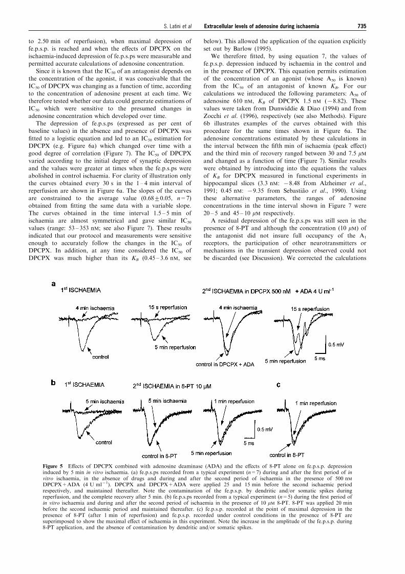
500 nM DPCPX. As shown in Figure 5a, the superfusion ofADA (4 U ml71+DPCPX 500 nM) elicited a reproducibleincrease in the amplitude of the fe.p.s.ps (15+4%; n=7).
When the second ischaemic episode was induced in thepresence of DPCPX+ADA, the depression was completelyantagonized for the ®rst 3 ± 4 min of ischaemia (Figure 5a).
In the following 2 ± 3 min (i.e. starting from the ®fth min ofischaemia through the third min of reperfusion), the fe.p.s.pswere contaminated by dendritic and/or somatic spikes. This
hampered precise measuring of fe.p.s.ps, but con®rmed thatthe depressant e�ects of ischaemia on excitatory neurotrans-mission are mediated by adenosine. To further con®rm thisconclusion, we tested the e�ects of 8-phenyltheophylline (8-
PT, 10 mM; n=5), a non-selective, potent adenosineantagonist on the fe.p.s.p. depression induced by ischaemia.As illustrated by Figure 5b, 8-PT increased the amplitude of
the fe.p.s.ps (22+3%) and delayed any decrease in fe.p.s.psfor at least 4 min. In three out of ®ve preparations, adecrease was found only after the ®fth min of ischaemia (i.e.
during the ®rst 2 min of reperfusion: Figure 5b and c). Thepeak of the depression was observed within the ®rst min ofreperfusion (12+4%, n=5), and the values of residual
depression observed in the interval between the fourth minof ischaemia and the second min of reperfusion were used forcorrecting our calculations of adenosine concentration (seebelow).
Pharmacological analysis and estimation of adenosineconcentrations in normoxic conditions
To estimate the concentration of adenosine under controlnormoxic conditions we calculated the maximum fractional
increase in fe.p.s.p. amplitude which was estimated by thee�ects of the A1 receptor antagonist DPCPX. As shown inFigure 1d the changes in fe.p.s.p. amplitude observed in thepresence of DPCPX in normoxic aCSF were ®tted using the
logistic equation (equation 1) and led to estimation of amaximum fractional increase of about 25%. By applyingequation 2, the calculated concentration of endogenous
adenosine in control conditions was 240 nM; 95% C.L.:225 ± 255 nM (n=5). This value was in agreement with those(120 ± 200 nM) found by Dunwiddie & Diao (1994) and that
calculated from the experiments devoted to studying the e�ectsof DPCPX during ischaemia (180 nM; 95% C.L.: 175 ±185 nM; see e.g. Figure 3b).
Pharmacological analysis and estimation of adenosineconcentrations in ischaemic conditions
The depression of fe.p.s.ps which developed during in vitroischaemia and reperfusion is temporally coincident with theincrease in adenosine ¯owing out of the cells (Latini et al.,
1998a). During the ®rst minutes of ischaemia the changes infe.p.s.p. amplitude produced by the ischaemic episode, andtheir modi®cation by DPCPX, were too small to allow
calculations of adenosine extracellular concentrations at anyexperimental times sampled by fe.p.s.p. recording. Thereforewe chose the time interval between the last minutes of ischaemiaand the beginning of reperfusion (from 3.25 min of ischaemia
Figure 4 E�ect of DPCPX on fe.p.s.p. amplitude during in vitroischaemia. (a) The graph shows the e�ects of DPCPX (50, 100 and500 nM) during the ®rst 2.5 min of in vitro ischaemia. In each curvethe fe.p.s.p. amplitude is expressed as the percentage of the averageamplitude of the potentials measured during 2 min that preceded thesecond ischaemic period. Each point represents the mean+s.e.meanof ®ve experiments for control, 100 and 500 nM DPCPX, and sixexperiments for 50 nM DPCPX. (b) Statistical analysis of resultsillustrated in (a). Each bar represents the average amplitude of fourconsecutive fe.p.s.ps recorded during the third and the ®fth min ofischaemia and the second and the fourth min of reperfusion, incontrol conditions or in the presence of several concentrations ofDPCPX: 50, 100 and 500 nM. Di�erences between data were analysedby two-way analysis of variance (P50.001) followed by post hocFisher's test: *P50.05 vs control, 8P50.05 vs DPCPX 50 nM.
Extracellular levels of adenosine during ischaemia734 S. Latini et al
to 2.50 min of reperfusion), when maximal depression offe.p.s.p. is reached and when the e�ects of DPCPX on theischaemia-induced depression of fe.p.s.ps were measurable and
permitted accurate calculations of adenosine concentration.Since it is known that the IC50 of an antagonist depends on
the concentration of the agonist, it was conceivable that theIC50 of DPCPX was changing as a function of time, according
to the concentration of adenosine present at each time. Wetherefore tested whether our data could generate estimations ofIC50 which were sensitive to the presumed changes in
adenosine concentration which developed over time.The depression of fe.p.s.ps (expressed as per cent of
baseline values) in the absence and presence of DPCPX was
®tted to a logistic equation and led to an IC50 estimation forDPCPX (e.g. Figure 6a) which changed over time with agood degree of correlation (Figure 7). The IC50 of DPCPX
varied according to the initial degree of synaptic depressionand the values were greater at times when the fe.p.s.ps wereabolished in control ischaemia. For clarity of illustration onlythe curves obtained every 30 s in the 1 ± 4 min interval of
reperfusion are shown in Figure 6a. The slopes of the curvesare constrained to the average value (0.68+0.05, n=7)obtained from ®tting the same data with a variable slope.
The curves obtained in the time interval 1.5 ± 5 min ofischaemia are almost symmetrical and gave similar IC50
values (range: 53 ± 353 nM; see also Figure 7). These results
indicated that our protocol and measurements were sensitiveenough to accurately follow the changes in the IC50 ofDPCPX. In addition, at any time considered the IC50 of
DPCPX was much higher than its KB (0.45 ± 3.6 nM, see
below). This allowed the application of the equation explicitlyset out by Barlow (1995).
We therefore ®tted, by using equation 7, the values of
fe.p.s.p. depression induced by ischaemia in the control andin the presence of DPCPX. This equation permits estimationof the concentration of an agonist (whose A50 is known)from the IC50 of an antagonist of known KB. For our
calculations we introduced the following parameters: A50 ofadenosine 610 nM, KB of DPCPX 1.5 nM (78.82). Thesevalues were taken from Dunwiddie & Diao (1994) and from
Zocchi et al. (1996), respectively (see also Methods). Figure6b illustrates examples of the curves obtained with thisprocedure for the same times shown in Figure 6a. The
adenosine concentrations estimated by these calculations inthe interval between the ®fth min of ischaemia (peak e�ect)and the third min of recovery ranged between 30 and 7.5 mMand changed as a function of time (Figure 7). Similar resultswere obtained by introducing into the equations the valuesof KB for DPCPX measured in functional experiments inhippocampal slices (3.3 nM: 78.48 from Alzheimer et al.,
1991; 0.45 nM: 79.35 from SebastiaÄ o et al., 1990). Usingthese alternative parameters, the ranges of adenosineconcentrations in the time interval shown in Figure 7 were
20 ± 5 and 45 ± 10 mM respectively.A residual depression of the fe.p.s.ps was still seen in the
presence of 8-PT and although the concentration (10 mM) of
the antagonist did not insure full occupancy of the A1
receptors, the participation of other neurotransmitters ormechanisms in the transient depression observed could not
be discarded (see Discussion). We corrected the calculations
Figure 5 E�ects of DPCPX combined with adenosine deaminase (ADA) and the e�ects of 8-PT alone on fe.p.s.p. depressioninduced by 5 min in vitro ischaemia. (a) fe.p.s.ps recorded from a typical experiment (n=7) during and after the ®rst period of invitro ischaemia, in the absence of drugs and during and after the second period of ischaemia in the presence of 500 nMDPCPX+ADA (4 U ml71). DPCPX and DPCPX+ADA were applied 25 and 15 min before the second ischaemic periodrespectively, and maintained thereafter. Note the contamination of the fe.p.s.p. by dendritic and/or somatic spikes duringreperfusion, and the complete recovery after 5 min. (b) fe.p.s.ps recorded from a typical experiment (n=5) during the ®rst period ofin vitro ischaemia and during and after the second period of ischaemia in the presence of 10 mM 8-PT. 8-PT was applied 20 minbefore the second ischaemic period and maintained thereafter. (c) fe.p.s.p. recorded at the point of maximal depression in thepresence of 8-PT (after 1 min of reperfusion) and fe.p.s.p. recorded under control conditions in the presence of 8-PT aresuperimposed to show the maximal e�ect of ischaemia in this experiment. Note the increase in the amplitude of the fe.p.s.p. during8-PT application, and the absence of contamination by dendritic and/or somatic spikes.
Extracellular levels of adenosine during ischaemia 735S. Latini et al

by subtracting the residual depression observed in 8-PT(during the interval between the fourth min of ischaemia and
the second min of reperfusion) from the values implementedin Barlow's equation. In the interval considered, theresulting concentrations of adenosine were accordingly lowerthan those in Figure 7 and peaked at 30 s reperfusion with
23 mM.
Discussion
Our results provide the ®rst estimation of the concentration ofadenosine acting at the receptor level during an ischaemia-like
episode in the CNS.We monitored the e�ects of an ischaemia-like episode on the
amplitude of extracellularly recorded fe.p.s.ps, which mainly
re¯ect the currents generated by the in¯ow of cations into CA1pyramidal cell dendrites due to activation of glutamatereceptors (Hubbard et al., 1969). The reduction of the fe.p.s.p.amplitude can be produced through several mechanisms whose
principles are: (i) competitive or non competitive antagonismof glutamate receptors; (ii) depolarization of the postsynapticcell elicited by excitatory receptor stimulation or by potassium
ion accumulation in the extracellular space and subsequentspreading depression; (iii) decrease in glutamate release byactions at presynaptic terminals. Among these possibilities the
most likely mechanism of fe.p.s.p. reduction during ischaemiais a decrease in glutamate release caused by activation ofpresynaptic A1 receptors (Corradetti et al., 1984a). In addition,
it has been shown that the postsynaptic action of adenosinedoes not a�ect the size of miniature postsynaptic excitatorycurrents recorded from CA1 pyramidal cells (Prince & Stevens,1992; Scholz & Miller, 1992), which suggests that the main
e�ect of A1 receptor stimulation on fe.p.s.p. is a decrease inglutamate release.
Other mechanisms not involving adenosine receptors might
participate in the decrease in fe.p.s.ps in our experiments. Forinstance, ATP-dependent potassium channels are known toopen during a hypoxic and/or hypoglycaemic episode (Ben-
Ari, 1989; Mourre et al., 1989; Tromba et al., 1992). Howeverit is generally accepted that synaptic depression duringischaemia is due to A1 adenosine receptor stimulation (Fowler,1990), and since the presence of the adenosine receptor
antagonist 8-phenyltheophylline (8-PT) fully prevents thedisappearance of fe.p.s.p. during an ischaemic episode (Pedataet al., 1993), it is conceivable that additional non adenosine-
mediated mechanisms represent a negligible source of error inour conditions.
This assumption was con®rmed by our experiments
performed in the presence of DPCPX+ADA or 8-PT alone
Figure 6 Estimation of the IC50 of DPCPX at 30 s intervals duringrecovery (min 1 ± 4) from an ischaemic episode of 5 min. (a) Eachcurve describes the concentration-dependent antagonism of fe.p.s.p.depression by DPCPX for a given time of recovery. The upper curveshows the e�ects of DPCPX at the ®rst min of recovery and thelowest is that obtained at the fourth. Note that with increasingrecovery time the initial degree of depression is progressively reduced.Each concentration of the antagonist was tested in a di�erent groupof preparations. DPCPX was applied at 50 nM (n=6), 100 nM (n=5),or 500 nM (n=5). In each preparation fe.p.s.ps were evoked every15 s and the per cent depressions elicited by the ischaemic episodewere averaged at corresponding times for each experimentalcondition (i.e. absence or presence of DPCPX). For clarity plotsare shown at 30 s intervals (values calculated every 15 s are shown inFigure 7). For each interval the fe.p.s.p. depression (expressed as percent decrease compared to baseline fe.p.s.p. amplitude) is plottedagainst the concentration of DPCPX present during the ischaemicepisode. Curves were obtained by nonlinear ®tting (for parameterssee text) of data in the logistic equation. (b) The ischaemia e�ect inthe absence or presence of the antagonist (at the same recovery timesshown in (a)) is plotted against the concentration of DPCPX. Curveswere obtained by nonlinear ®tting of data to the equation (7),keeping the slope index ®xed to the average value of 14.5+0.7obtained by ®tting the same data with unconstrained, variable slope.
Figure 7 Time-course of the changes in IC50 of DPCPX andadenosine concentrations during ischaemia and recovery. The valuesobtained from the analyses shown in Figure 6 are plotted againsttime to show the time-related changes of the IC50 of the antagonist,and the estimated concentrations of adenosine during the ischaemic-like episode and reperfusion with control aCSF. Note that 3 minafter the beginning of the ischaemic episode, adenosine concentrationis already 24 times higher than basal levels.
Extracellular levels of adenosine during ischaemia736 S. Latini et al

where the e�ect of the ischaemia was blocked for as long as5 min (i.e. the whole duration of the ischaemic episode).However, at the end of the 5 min ischaemic period and during
the ®rst 2 ± 3 min of reperfusion, a depression was stillobserved in the presence of any of the treatments. In addition,the treatment with DPCPX+ADA produced, mainly duringreperfusion, a contamination of the fe.p.s.p. by dendritic and/
or somatic spikes. It is known that the ischaemic insultincreases spontaneous release of glutamate and that A1
receptor stimulation can greatly limit the out¯ow of glutamate
(Heron et al., 1994). On the other hand, once the inhibitorye�ect of adenosine is completely blocked, glutamate can freely¯ow out of cells and, by stimulating ionotropic and
metabotropic receptors, depolarize pyramidal neurones andincrease their excitability. This produces a decrease in theamplitude of the fe.p.s.ps and the appearance of multiple
action potentials. Furthermore, ADA might enhance neuroneexcitability through unspeci®c e�ects, as suggested by themodest, if any, hyperexcitation observed during ischaemiawhen the adenosine receptors are blocked by 8-PT. On the
basis of this interpretation, we considered the experiments with8-PT more reliable for correcting our calculations for theparticipation of unidenti®ed mechanism(s) of fe.p.s.p. depres-
sion during reperfusion. However, an alternative explanationshould be taken into account based on the fact that 8-PT is acompetitive antagonist. From our calculations, the concentra-
tions of adenosine at the end of 5 min ischaemia approached20 mM and, considering the a�nity of 8-PT for A1 receptors(Ki=86 nM) relative to that of adenosine (EC50=610 nM) the
fraction of A1 receptors still occupied by the agonist(calculated with Gaddum's equation; see Kenakin (1993)) isnot negligible (about 20%). Under our conditions ofcompetitive antagonism, it is likely that the residual depression
(10 ± 15%) observed during reperfusion was still due to A1
receptor stimulation.Since A2A adenosine antagonists, (e.g. ZM 241385), do not
modify the depression of fe.p.s.p. induced by the ischaemicepisode (Latini et al., 1998b; Latini et al., manuscript inpreparation) and since 8-PT does not antagonize A3 receptors
(Fredholm et al., 1994), A2A and A3 receptors do not appeardirectly involved in the depression of fe.p.s.p. induced byischaemia. The role of A2B receptors, which are uniformlydistributed in the CNS (Dixon et al., 1996), remains to be
elucidated.In our experiments, we found that the recovery of the
fe.p.s.ps after the second ischaemic episode was slightly faster
than after the ®rst episode. This suggests a small butstatistically signi®cant `preconditioning e�ect' by the ®rstischaemic episode, possibly due to desensitization of the A1
receptors. It has been shown that prolonged (420 min)stimulation of A3 receptors by mM concentrations of adenosineblocks A1 receptor-mediated responses in the CA1 region of
the hippocampus (Dunwiddie et al., 1997). Such a mechanismis unlikely to occur in our conditions because the ®rstischaemic episode lasted only 5 min and it is di�cult toenvisage that adenosine reached mM concentrations. The
stimulation of A2A adenosine receptors also might desensitizeA1 receptors in the CA1 (Cunha et al., 1994; O'Kane & Stone,1998). However, O'Kane & Stone (1998) showed that
stimulation of A2A receptors does not modify A1 receptor-mediated inhibitory action on fe.p.s.ps, and convincinglydemonstrated that the possible interaction between A2A and
A1 receptors is selectively postsynaptic in the CA1 region. Wefavour an alternative interpretation based on measurements ofendogenous adenosine ¯owing out of hippocampal slicesduring two ischaemic episodes in similar (though not identical)
experimental conditions. We found a 36+8% (P50.05; n=7)reduction in the total amount of adenosine released over basallevels during the second ischaemic insult when compared to the
®rst. Consistent with the present ®ndings, the time-course ofthe out¯ow of adenosine after the second ischaemic insult wasfaster than after the ®rst (unpublished observations). Theseresults suggest a reduction in adenosine availability after the
®rst ischaemic episode.Our approach is based on the assumption that to decrease
fe.p.s.ps, adenosine acted on a single class of receptors with
identical properties, an assumption which has been supportedby previous work (Alzheimer et al., 1991; Dunwiddie et al.,1986; Dunwiddie & Diao, 1994; Dunwiddie & Fredholm, 1989;
Scholz & Miller, 1992; Wu & Saggau, 1994). For ourcalculations we introduced several parameters which wereobtained from previous work (Alzheimer et al., 1991;
Dunwiddie & Diao, 1994) conducted in hippocampal slicesby using methods similar to those applied in the presentresearch. Our estimation of the concentrations of adenosine isbased on the analysis proposed by Barlow (1995); (see
Methods). On the basis of this analysis the calculatedconcentrations of adenosine represent the degree of receptorstimulation. From our data, the most conservative estimation
of adenosine concentrations at the peak, after the ischaemicepisode, at the receptor level is between 20 and 30 mM,depending on the parameters used for estimation and/or on the
correction of raw data for the residual depressant e�ectsobserved in the presence of 8-PT. The rise in adenosineconcentration was time-locked to the ischaemic episode,
reaching its maximum within 45 s after reintroduction ofoxygenated control aCSF and rapidly declining thereafter.These results con®rm the observation by Latini et al. (1998a)that an increase in adenosine e�ux from hippocampal slices
closely parallels in time the depression of fe.p.s.ps produced byan ischaemic episode similar to that used in this study.
Under normoxic conditions the adenosine extracellular
concentration estimated in the brain by microdialysis 24 hafter ®bre implantation (40 ± 120 nM, values corrected forrecovery of the ®bre) (Ballarin et al., 1991; Pazzagli et al., 1993;
1994; 1995) or by the cortical cup technique (30 ± 50 nM)(Phillis, 1989) is in about the same concentration range as thatcalculated in the present work (180 ± 240 nM) and byDunwiddie & Diao (1994) (120 ± 200 nM) who used the same
preparation. This supports the interpretation that in normoxicconditions the released adenosine equilibrates in the extra-cellular ¯uid and therefore the degree of tonic stimulation at its
receptors can be inferred from the measure of adenosineconcentration in the CSF using the microdialysis technique.
After the in vitro ischaemic episode, adenosine increases
about 100 ± 150 fold, reaching an estimated value of 30 mM.This increase is much higher than that calculated in the ¯uidcollected from hippocampal slices (4 ± 6 fold) (Latini et al.,
1998a; Pedata et al., 1993) or from the hippocampus in vivo(19 ± 23 fold) (AndineÁ et al., 1990; Dux et al., 1990) afterinducing ischaemia. However, it should be noted that the timediscrimination of adenosine concentrations (15 s) allowed by
the present approach is greater than that achieved with anybiochemical approach, where the sampling periods are morelikely to represent the equilibrium reached in the CSF after
partial reuptake and degradation of released adenosine.Taking into consideration these observations, it is likely thatthe adenosine concentration of 30 mM estimated in our study
represents an adenosine concentration acting on receptorsbefore di�usion and equilibration in the CSF. Therefore thedegrees of receptor stimulation is greater than that extra-polated by the microdialysis adenosine measurement after
Extracellular levels of adenosine during ischaemia 737S. Latini et al

ischaemia. It deserves mention that adenosine concentrationsas high as 24 ± 40 mM have been measured by microdialysisafter induction of global ischaemia in the rat and gerbil (Dux et
al., 1990; Hagberg et al., 1987). However these values werereached on the ®rst day after microdialysis tube implantationwhen the estimated basal adenosine extracellular concentrationis 20 times higher than those found in experiments conducted
24 h after surgery (Pazzagli et al., 1993). The e�ect ofischaemia immediately after tube implantation of the probemay be therefore compounded by that of trauma due to the
microdialysis probe insertion.In agreement with the view that the presence of adenosine
on receptors, close to release sites, is functionally important,
Brundege & Dunwiddie (1996) have demonstrated, byapplying adenosine by the recording pipette, that the degreeof synaptic depression in the CA1 hippocampus is related to
the adenosine concentration in the electrode. Their datastrongly suggest that the principal source of adenosine, actingat the presynaptic level to decrease the excitatory input on therecorded CA1 pyramidal neurone, is the out¯ow of adenosine
from the cell from which the postsynaptic current wasrecorded. This implies that during the ischaemic episode theout¯ow of adenosine from CA1 pyramidal neurones is likely to
greatly a�ect excitatory neurotransmission.In the CNS adenosine may act through four receptor
subtypes to which the endogenous ligand shows varying
a�nity: a higher a�nity for A1 and A2A (1 ± 30 nM) than for
A2B and A3 (41 mM) receptors (Fredholm et al., 1994). Undernormoxic conditions the adenosine present at the receptor levelis therefore likely to preferentially activate A1 and A2A
receptors. On the other hand, if we assume that our estimationof adenosine concentration reached during an ischaemia-likeepisode re¯ects the order of magnitude of adenosine present atthe receptor level during an in vivo ischaemic episode, it is
conceivable that under these conditions all adenosine receptorssubtypes are activated during in vivo ischaemia. Stimulation ofat least A1, A2A and A3 adenosine receptors may modify the
outcome of an ischaemic episode. It appears that during in vivoischaemia A1 receptor stimulation protects cerebral tissue fromdamage (Rudolphi et al., 1992) whereas the role of A2A
(O'Regan et al., 1992; Phillis, 1989) and/or A3 adenosinereceptors remains to be clari®ed (Von Lubitz et al., 1994).
In conclusion our data provide a `dynamic' estimation of
the concentration of adenosine reached at the receptor levelduring a transient ischaemic episode. This information may beuseful in designing appropriate concentrations of purinergiccompounds to be tested for their pharmacological e�ects
during in vitro and/or in vivo ischaemic episodes.
This investigation was supported by grants from the University ofFlorence and MURST (60%).
References
ALZHEIMER, C., KARGL, L. & TEN-BRUGGENCATE, G. (1991).Adenosinergic inhibition in hippocampus is mediated byadenosine A1 receptors very similar to those of peripheral tissue.Eur. J. Pharmacol., 196, 313 ± 317.
ANDINEÂ , P., RUDOLPHI, K.A., FREDHOLM, B.B. & HAGBERG, H.
(1990). E�ect of propentofylline (HWA 285) on extracellularpurines and excitatory amino acids in CA1 of rat hippocampusduring transient ischaemia. Br. J. Pharmacol., 100, 814 ± 818.
BALLARIN, M., FREDHOLM, B.B., AMBROSIO, S. & MAHY, N.
(1991). Extracellular levels of adenosine and its metabolites inthe striatum of awake rats: inhibition of uptake and metabolism.Acta Physiol. Scand., 142, 97 ± 103.
BARLOW, R.B. (1995). Use of an antagonist for estimating the degreeof agonist stimulation during physiological release. TrendsPharmacol. Sci., 16, 262 ± 264.
BEN ARI, Y. (1989). E�ect of glibenclamide, a selective blocker of anATP-K+ channel, on the anoxic response of hippocampalneurones. P¯uÈgers Arch., 414 (Suppl 1), S111 ± S114.
BRUNDEGE, J.M. & DUNWIDDIE, T.V. (1996). Modulation ofexcitatory synaptic transmission by adenosine released fromsingle hippocampal pyramidal neurons. J. Neurosci., 16, 5603 ±5612.
CANHAÄ O, P., DE MENDONCË A, A. & RIBEIRO, J.A. (1994). 1,3-Dipropyl-8-cyclopentylxanthine attenuates the NMDA responseto hypoxia in the rat hippocampus. Brain Res., 661, 265 ± 273.
CORRADETTI, R., LO CONTE, G., MORONI, F., PASSANI, M.B. &
PEPEU, G. (1984a). Adenosine decreases aspartate and glutamaterelease from rat hippocampal slices. Eur. J. Pharmacol., 104, 19 ±26.
CORRADETTI, R., MONETI, G., MORONI, F., PEPEU, G. & WIER-
ASZKO, A. (1983). Electrical stimulation of the stratum radiatumincreases the release and neosynthesis of aspartate, glutamate,and gamma-aminobutyric acid in rat hippocampal slices. J.Neurochem., 41, 1518 ± 1525.
CORRADETTI, R., MORONI, F., PASSANI, M.B. & PEPEU, G. (1984b).8-Phenyltheophylline potentiates the electrical activity evoked inhippocampal slices. Eur. J. Pharmacol., 103, 177 ± 180.
CUNHA, R.A., JOHANSSON, B., VAN DER PLOEG, I., SEBASTIAÄ O,
A.M., RIBEIRO, J.A. & FREDHOLM, B.B. (1994). Evidence forfunctionally important adenosine A2a receptors in the rathippocampus. Brain Res., 649, 208 ± 216.
DIXON, A.K., GUBITZ, A.K., SIRINATHSINGHJI, D.J.S., RICHARD-
SON, P.J. & FREEMAN, T.C. (1996). Tissue distribution ofadenosine receptor mRNAs in the rat. Br. J. Pharmacol., 118,1461 ± 1468.
DOMENICI, M.R., SCOTTI DE CAROLIS, A. & SAGRATELLA, S.
(1996). Block by N6-L-phenylisopropyl-adenosine of the electro-physiological and morphological correlates of hippocampalischaemic injury in the gerbil. Br. J. Pharmacol., 118, 1551 ± 1557.
DUNWIDDIE, T.V. (1985). The physiological roles of adenosine in thecentral nervous system. In International Review of Neurobiology,eds. Smythies, J.R. & Bradley, R.J. pp. 63 ± 139. London:Academic Press.
DUNWIDDIE, T.V. & DIAO, L. (1994). Extracellular adenosineconcentration in hippocampal brain slices and the tonicinhibitory modulation of evoked excitatory responses. J.Pharmacol. Exp. Ther., 268, 537 ± 545.
DUNWIDDIE, T.V., DIAO, L., KIM, H.O., JIANG, J.-L. & JACOBSON,
K.A. (1997). Activation of hippocampal adenosine A3 receptorsproduces a desensitization of A1 receptor-mediated responses inrat hippocampus. J. Neurosci., 17, 607 ± 614.
DUNWIDDIE, T.V. & FREDHOLM, B.B. (1989). Adenosine A1
receptors inhibit adenylate cyclase activity and neurotransmitterrelease and hyperpolarize pyramidal neurons in rat hippocam-pus. J. Pharmacol. Exp. Ther., 249, 31 ± 37.
DUNWIDDIE, T.V. & HOFFER, B.J. (1980). Adenine nucleotide andsynaptic transmission in the in vitro rat hippocampus. Br. J.Pharmacol., 69, 59 ± 68.
DUNWIDDIE, T.V., WORTH, T.S. & OLSSON, R.A. (1986). Adenosineanalogs mediating depressant e�ects on synaptic transmission inrat hippocampus: structure-activity relationship for the N6
subregion. Naunyn-Schmiedeberg's Arch. Pharmacol., 334, 77 ±85.
DUX, E., FASTBOM, J., UNGERSTEDT, U., RUDOLPHI, K. &
FREDHOLM, B.B. (1990). Protective e�ect of adenosine and anovel xanthine derivative propentofylline on the cell damageafter bilateral carotid occlusion in the gerbil hippocampus. BrainRes., 516, 248 ± 256.
FOWLER, J.C. (1989). Adenosine antagonists delay hypoxia-induceddepression of neuronal activity in hippocampal brain slice. BrainRes., 490, 378 ± 384.
Extracellular levels of adenosine during ischaemia738 S. Latini et al

FOWLER, J.C. (1990). Adenosine antagonists alter the synapticresponse to in vitro ischaemia in the rat hippocampus. Brain Res.,509, 331 ± 334.
FREDHOLM, B.B., ABBRACCHIO, M.P., BURNSTOCK, G., DALY,
J.W., HARDEN, K., JACOBSON, K.A., LEFF, P. & WILLIAMS, M.
(1994). Nomenclature and classi®cation of purinoceptors.Pharmacol. Rev., 46, 143 ± 156.
HAGBERG, H., ANDERSSON, P., LACAREWICZ, J., JACOBSON, I.,
BUTCHER, S. & SANDBERG, M. (1987). Extracellular adenosine,inosine, hypoxanthine, and xanthine in relation to tissue nucleo-sides and purines in rat striatum during transient ischemia. J.Neurochem., 49, 227 ± 231.
HERON, A., LEKIEFFRE, D., LE PEILLET, E., FASBENNES, F.,
SEYLAZ, J., PLOTKINE, M. & BOULU, R.G. (1994). E�ects of anA1 adenosine receptor agonist on the neurochemical, behaviouraland histological consequences of ischemia. Brain Res., 641, 217 ±224.
HUBBARD, J.I., LLINAÂ S, R. & QUASTEL, D.M.J. (1969). Electro-physiological analysis of synaptic transmission. eds. Davson, H.,Green®eld, A.D.M., Whittam, R. & Brindley, G.S. pp. 1 ± 368.London: Edward Arnold.
KENAKIN, T. (1993). Pharmacologic analysis of drug-receptorinteraction. pp. 1 ± 484. New York: Raven Press.
LATINI, S., BORDONI, F., CORRADETTI, R., PEPEU, G. & PEDATA, F.
(1998a). Temporal correlation between adenosine out¯ow andsynaptic potential inhibition in rat hippocampal slices duringischemia-like conditions. Brain Res., 794, 325 ± 328.
LATINI, S., BORDONI, F., CORRADETTI, R., PEPEU, G. & PEDATA, F.
(1998b). The A2A adenosine receptor agonist CGS 21680 reducesthe synaptic depression induced by in vitro ischemia in rathippocampal slices. Drug. Dev. Res., 43, 213.
LUCCHI, R., LATINI, S., DE MENDONCË A, A., SEBASTIAÄ O, A.M. &
RIBEIRO, J.A. (1996). Adenosine by activating A1 receptorsprevents GABAA-mediated actions during hypoxia in the rathippocampus. Brain Res., 732, 261 ± 266.
MOURRE, C., BEN ARI, Y., BERNARDI, H., FOSSET, M. &
LAZDUNSKI, M. (1989). Antidiabetic sulfonylureas: localizationof binding sites in the brain and e�ect on the hyperpolarizationinduced by anoxia in hippocampal slices. Brain Res., 486, 159 ±164.
O'KANE, E.M. & STONE, T.W. (1998). Interaction between adenosineA1 and A2 receptor-mediated responses in the rat hippocampusin vitro. Eur. J. Pharmacol., 362, 17 ± 25.
O'REGAN, M.H., SIMPSON, R.E., PERKINS, L.M. & PHILLIS, J.W.
(1992). The selective A2 adenosine receptor agonist CGS 21680enhances excitatory transmitter amino acid release from theischemic rat cerebral cortex. Neurosci. Lett., 138, 169 ± 172.
PAZZAGLI, M., CORSI, C., FRATTI, S., PEDATA, F. & PEPEU, G.
(1995). Regulation of extracellular adenosine levels in thestriatum of aging rats. Brain Res., 684, 103 ± 106.
PAZZAGLI, M., CORSI, C., LATINI, S., PEDATA, F. & PEPEU, G.
(1994). In vivo regulation of extracellular adenosine levels in thecerebral cortex by NMDA and muscarinic receptors. Eur. J.Pharmacol., 254, 277 ± 282.
PAZZAGLI, M., PEDATA, F. & PEPEU, G. (1993). E�ect of K+
depolarisation, tetrodotoxin, and NMDA receptor inhibition onextracellular adenosine levels in rat striatum. Eur. J. Pharmacol.,234, 61 ± 65.
PEDATA, F., LATINI, S., PUGLIESE, A.M. & PEPEU, G. (1993).Investigation into the adenosine out¯ow from hippocampal slicesevoked by ischemia-like conditions. J. Neurochem., 61, 284 ± 289.
PEDATA, F., PAZZAGLI, M. & PEPEU, G. (1991). Endogenousadenosine release from hippocampal slices: excitatory aminoacid agonists stimulate release, antagonists reduce the electri-cally-evoked release. Naunyn-Schmiedeberg's Arch. Pharmacol.,344, 538 ± 543.
PHILLIS, J.W. (1989). Adenosine in the control of the cerebralcirculation. Cerebrovasc. Brain Metab. Rev., 1, 26 ± 54.
PHILLIS, J.W., WALTER, G.A., O'REGAN, M.H. & STAIR, R.E. (1987).Increases in cerebral cortical perfusate adenosine and inosineconcentrations during hypoxia and ischemia. J. Cereb. BloodFlow Metab., 7, 679 ± 686.
PRINCE, D.A. & STEVENS, C.F. (1992). Adenosine decreasesneurotransmitter release at central synapses. Proc. Natl. Acad.Sci. U.S.A., 89, 8586 ± 8590.
PROCTOR, W.R. & DUNWIDDIE, T.V. (1987). Pre- and postsynapticactions of adenosine in the in vitro rat hippocampus. Brain Res.,426, 187 ± 190.
RUDOLPHI, K.A., SCHUBERT, P., PARKINSON, F.E. & FREDHOLM,
B.B. (1992). Neuroprotective role of adenosine in cerebralischaemia. Trends Pharmacol. Sci., 13, 439 ± 445.
SCHOLZ, K.P. & MILLER, R.J. (1992). Inhibition of quantaltransmitter release in the absence of calcium in¯ux by a Gprotein-linked adenosine receptor at hippocampal synapses.Neuron, 8, 1139 ± 1150.
SCHUBERT, P. (1988). Physiological modulation by adenosine:selective blockade of A1-receptors with DPCPX enhancesstimulus train-evoked neuronal Ca++ in¯ux in rat hippocampalslices. Brain Res., 458, 162 ± 165.
SEBASTIAÄ O, A.M. & RIBEIRO, J.A. (1992). Evidence for the presenceof excitatory A2 adenosine receptors in the rat hippocampus.Neurosci. Lett., 138, 41 ± 44.
SEBASTIAÄ O, A.M., STONE, T.W. & RIBEIRO, J.A. (1990). Theinhibitory adenosine receptor at the neuromuscular junctionand hippocampus of the rat: antagonism by 1,3,8-substitutedxanthines. Br. J. Pharmacol., 101, 453 ± 459.
TROMBA, C., SALVAGGIO, A., RACAGNI, G. & VOLTERRA, A.
(1992). Hypoglycemia-activated K+ channels in hippocampalneurons. Neurosci. Lett., 143, 185 ± 189.
VON LUBITZ, D.K., LIN, R.C., POPIK, P., CARTER, M.F. &
JACOBSON, K.A. (1994). Adenosine A3 receptor stimulation andcerebral ischemia. Eur. J. Pharmacol., 263, 59 ± 67.
WU, L.-G. & SAGGAU, P. (1994). Adenosine inhibits evoked synaptictransmission primarily by reducing presynaptic calcium in¯ux inarea CA1 of hippocampus. Neuron, 12, 1139 ± 1148.
ZOCCHI, C., ONGINI, E., CONTI, A.M., MONOPOLI, A., NEGRETTI,
A., BARALDI, P.G. & DIONISOTTI, S. (1996). The non-xanthineheterocycling compound SCH 58261 is a new potent and selectiveA2a adenosine receptor antagonist. J. Pharmacol. Exp. Ther.,276, 398 ± 404.
(Received July 31, 1998Revised March 5, 1999
Accepted March 15, 1999)
Extracellular levels of adenosine during ischaemia 739S. Latini et al
Related Documents