Expression of estrogen receptor GPR30 in the rat spinal cord, autonomic and sensory ganglia Siok L. Dun 1 , G. Cristina Brailoiu 1 , Xin Gao 1 , Eugen Brailoiu 1 , Jeffrey B. Arterburn 2 , Eric R. Prossnitz 3 , Tudor I. Oprea 4 , and Nae J. Dun 1 1 Department of Pharmacology, Temple University School of Medicine, Philadelphia, PA 19140 USA 2 Department of Chemistry and Biochemistry, New Mexico State University, Las Cruces, NM 88003 USA 3 Department of Cell Biology and Physiology, University of New Mexico School of Medicine, Albuquerque, NM 87131 USA 4 Department of Biochemistry and Molecular Biology, University of New Mexico School of Medicine, Albuquerque, NM 87131 USA Abstract The G protein-coupled receptor GPR30 has recently been identified as a non nuclear estrogen receptor. RT-PCR revealed expression of GPR30 mRNA in varying quantities in the rat spinal cord, dorsal root ganglia, nodose ganglia, trigeminal ganglia, hippocampus, brain stem and hypothalamus. Immunohistochemical studies using a rabbit polyclonal antiserum against the human GPR30 C-terminus revealed a fine network of GPR30-immunoreactive (irGPR30) cell processes in the superficial layers of the spinal cord; some of which extended into deeper laminae. A population of neurons in the dorsal horn and ventral horn were irGPR30. Dorsal root, nodose and trigeminal ganglionic neurons displayed varying intensities of irGPR30. Positively labeled neurons were detected in the major pelvic ganglion, but not in the superior cervical ganglion. A population of chromaffin cells in the adrenal medulla was irGPR30, so were cells of the zona glomerulosa. Double-labeling the adrenal medulla with GPR30 antiserum and tyrosine hydroxylase (TH) antibody or phenylethanolamine-N-methyltransferase (PNMT) antiserum revealed that irGPR30 is expressed in the majority of TH-positive chromaffin cells. Lastly, some of the myenteric ganglion cells were irGPR30. Tissues processed with pre-immune serum resulted in no staining. Voltage-sensitive dye imaging studies showed that the selective GPR30 agonist G-1 (1, 10 and 100 nM) depolarized cultured spinal neurons in a concentration dependent manner. Collectively, our result provides the first evidence that GPR30 is expressed in neurons of the dorsal and ventral horn as well as in sensory and autonomic neurons, and activation of GPR30 by the selective agonist G-1 depolarizes cultured spinal neurons. Keywords adrenal gland; myenteric plexus; nodose ganglia; pelvic ganglia; trigeminal ganglia Corresponding author: Nae J. Dun, Department of Pharmacology, Temple University School of Medicine, 3420 N. Broad Street, Philadelphia, PA 19140 USA, Tel: 215-707-3207, Fax: 215-707-7068, Email: [email protected]. NIH Public Access Author Manuscript J Neurosci Res. Author manuscript; available in PMC 2009 June 8. Published in final edited form as: J Neurosci Res. 2009 May 15; 87(7): 1610–1619. doi:10.1002/jnr.21980. NIH-PA Author Manuscript NIH-PA Author Manuscript NIH-PA Author Manuscript

Welcome message from author
This document is posted to help you gain knowledge. Please leave a comment to let me know what you think about it! Share it to your friends and learn new things together.
Transcript
Expression of estrogen receptor GPR30 in the rat spinal cord,autonomic and sensory ganglia
Siok L. Dun1, G. Cristina Brailoiu1, Xin Gao1, Eugen Brailoiu1, Jeffrey B. Arterburn2, Eric R.Prossnitz3, Tudor I. Oprea4, and Nae J. Dun1
1Department of Pharmacology, Temple University School of Medicine, Philadelphia, PA 19140USA2Department of Chemistry and Biochemistry, New Mexico State University, Las Cruces, NM88003 USA3Department of Cell Biology and Physiology, University of New Mexico School of Medicine,Albuquerque, NM 87131 USA4Department of Biochemistry and Molecular Biology, University of New Mexico School ofMedicine, Albuquerque, NM 87131 USA
AbstractThe G protein-coupled receptor GPR30 has recently been identified as a non nuclear estrogenreceptor. RT-PCR revealed expression of GPR30 mRNA in varying quantities in the rat spinalcord, dorsal root ganglia, nodose ganglia, trigeminal ganglia, hippocampus, brain stem andhypothalamus. Immunohistochemical studies using a rabbit polyclonal antiserum against thehuman GPR30 C-terminus revealed a fine network of GPR30-immunoreactive (irGPR30) cellprocesses in the superficial layers of the spinal cord; some of which extended into deeper laminae.A population of neurons in the dorsal horn and ventral horn were irGPR30. Dorsal root, nodoseand trigeminal ganglionic neurons displayed varying intensities of irGPR30. Positively labeledneurons were detected in the major pelvic ganglion, but not in the superior cervical ganglion. Apopulation of chromaffin cells in the adrenal medulla was irGPR30, so were cells of the zonaglomerulosa. Double-labeling the adrenal medulla with GPR30 antiserum and tyrosinehydroxylase (TH) antibody or phenylethanolamine-N-methyltransferase (PNMT) antiserumrevealed that irGPR30 is expressed in the majority of TH-positive chromaffin cells. Lastly, someof the myenteric ganglion cells were irGPR30. Tissues processed with pre-immune serum resultedin no staining. Voltage-sensitive dye imaging studies showed that the selective GPR30 agonistG-1 (1, 10 and 100 nM) depolarized cultured spinal neurons in a concentration dependent manner.Collectively, our result provides the first evidence that GPR30 is expressed in neurons of thedorsal and ventral horn as well as in sensory and autonomic neurons, and activation of GPR30 bythe selective agonist G-1 depolarizes cultured spinal neurons.
Keywordsadrenal gland; myenteric plexus; nodose ganglia; pelvic ganglia; trigeminal ganglia
Corresponding author: Nae J. Dun, Department of Pharmacology, Temple University School of Medicine, 3420 N. Broad Street,Philadelphia, PA 19140 USA, Tel: 215-707-3207, Fax: 215-707-7068, Email: [email protected].
NIH Public AccessAuthor ManuscriptJ Neurosci Res. Author manuscript; available in PMC 2009 June 8.
Published in final edited form as:J Neurosci Res. 2009 May 15; 87(7): 1610–1619. doi:10.1002/jnr.21980.
NIH
-PA Author Manuscript
NIH
-PA Author Manuscript
NIH
-PA Author Manuscript
IntroductionMuch of our current knowledge regarding the physiology and pharmacology of estrogens invarious tissues is derived from studies related to the interaction of estrogens with the nuclearreceptors ERα and ERβ, which serve as transcription factors modifying the activity of targetgenes (Jensen and DeSombre, 1973; Kuiper et al., 1996; Mosselman et al., 1996). ERα, thefirst to be identified, is highly expressed in the female reproductive tract, mammary gland,endothelial cells, vascular smooth muscle cells and the hypothalamus; whereas, ERβ is moreabundantly expressed in the prostate, ovary and lungs, but less in the brain and vasculature(Nilsson et al., 2001).
Recently, two groups independently reported that, in addition to the genomic receptors, 17β-estradiol (E2) binds to a G protein-coupled receptor, GPR30 (Revankar et al.,2005;Thomas et al., 2005). Accordingly, a high-affinity, displaceable, single binding sitespecific for E2 was detected, respectively, in membranes of SKBR3 breast cancer cellsexpressing GPR30 but lacking genomic ERs, and in COS7 cells transfected with GPR30(Revankar et al., 2005; Thomas et al., 2005). The identification of GPR30 as a putativeestrogen receptor (IUPHAR classification: GPER[http://www.iuphar-db.org/GPCR/ReceptorDisplayForward?receptorID=3055]) raises thepossibility that steroid hormones may signal through receptors other than classical nuclearreceptors.
Earlier immunohistochemical studies showed that GPR30-immunoreactive (irGPR30) cellsare present in several regions of the rat brain including the olfactory tubercle, hypothalamicparaventricular and supraoptic nucleus, hippocampus, nucleus of the solitary tract andnucleus ambiguus (Funakoshi et al., 2006; Brailoiu et al., 2007, Sakamoto et al., 2007).Though overlapping with ERα and ERβ in several regions of the rat brain (Laflamme et al.,1998), the topographic distribution of irGPR30 is uniquely distinct from that of ERα andERβ (Brailoiu et al., 2007). Expression of ERα and ERβ is not limited to the central nervoussystem; as the rat spinal cord, peripheral autonomic and sensory ganglia are endowed withgenomic estrogen receptors (Papka et al., 1997; Zoubina and Smith, 2002; Bennett et al.,2003; Fan et al., 2007). We hypothesized that GPR30, in parallel with the genomic estrogenreceptor, is expressed in the spinal cord and peripheral nervous tissues. The present studywas undertaken to demonstrate the presence of GPR30 mRNA in the rat spinal cord,autonomic and sensory ganglia by RT-PCR, to detect immunohistochemically thedistribution of irGPR30 in these tissues using a previously characterized GPR30 antiserum(Revankar et al., 2005; Brailoiu et al., 2007), and to investigate the functionality of GPR30in cultured spinal neurons by monitoring membrane potential changes induced by theselective GPR30 agonist G-1 (Bologa et al., 2006; Brailoiu et al., 2007).
Materials and MethodsImmunohistochemistry
Adult male and female Sprague-Dawley rats, weighing 250–300 g (Ace Animals Inc.,Boyertown, PA, USA) were used. Animal protocols were reviewed and approved by theInstitutional Animal Care and Use Committee. Rats were anesthetized with urethane (1.2 g/kg, i.p.) and intracardially perfused with 0.1 M phosphate buffered saline (PBS) followed by4% paraformaldehyde/0.2% picric acid in PBS. Spinal cord, dorsal root ganglia, nodoseganglia, trigeminal ganglia, superior cervical ganglia, major pelvic ganglia, adrenal glands,and proximal segments of the ileum were removed, post-fixed for 2 h, and stored in 30%sucrose/PBS solution overnight. Spinal cord and ganglia were sectioned to 40 µm using acryostat. In preparing the myenteric plexus, layers of smooth muscles were removed and theplexus was processed as a whole mount (Dun et al., 1997).
Dun et al. Page 2
J Neurosci Res. Author manuscript; available in PMC 2009 June 8.
NIH
-PA Author Manuscript
NIH
-PA Author Manuscript
NIH
-PA Author Manuscript
Tissues were processed for GPR30-immunoreactivity (irGPR30) by the avidin-biotincomplex procedure or fluorescent method (Brailoiu et al., 2005b; 2007). Tissues were firsttreated with 3% H2O2 to quench endogenous peroxidase, washed several times, blockedwith 10% normal goat serum, and incubated in GPR30 antiserum (1:1000 dilution). GPR30antiserum was a rabbit polyclonal antibody directed against the human C-terminus GPR30(CAVIPDSTEQSDVRFSSAV; Revankar et al., 2005).The specificity of GPR30 antiserumhas been previously confirmed (Revankar et al., 2005; Brailoiu et al., 2007). After thoroughrinsing, sections were incubated in biotinylated anti-rabbit IgG (1:150 dilution, VectorLaboratories, Burlingame, CA, USA) for 2 h, rinsed with PBS, and incubated in avidin-biotin complex solution for 1 h (1:100 dilution, Vector Laboratories). Following washes inTris-buffered saline, sections were incubated in 0.05% diaminobenzidine/0.001% H2O2solution and washed for at least 2 h with Tris buffered saline. Sections were mounted onslides with 0.25% gel alcohol, air-dried, dehydrated with absolute alcohol followed byxylene, and coverslipped with Permount. For control experiments, sections were processedwith pre-immune serum in place of GPR30 antiserum.
In double-labeling and some single staining experiments, the fluorescent method was used.Tissue sections were first blocked with normal goat serum (1:10 in PBS, 0.5% BSA, 0.4%Triton X-100) and then incubated with GPR30 antiserum (1:500 dilution). Following severalwashes with PBS, sections were incubated with biotinylated anti-rabbit IgG (1:50, VectorLaboratories), washed with PBS, and incubated with avidin-fluorescein isothiocyanate(FITC). After rinsing with PBS, adrenal gland sections were incubated with either tyrosinehydroxylase antibody (TH; 1:700, a monoclonal antibody from Chemicon, Temecula, CA,USA) or phenylethanolamine-N-methyltransferase antiserum (PNMT; 1:300, a guinea pigpolyclonal from Chemicon). Tissues were incubated in Texas Red conjugated to appropriatesecond primary antibodies, washed with PBS, and mounted in Citifluor and coverslipped.Sections of major pelvic ganglion or spinal cord were double-labeled with GPR30 antiserumand choline acetyltransferase antiserum (ChAT; 1:100, a goat polyclonal from Chemicon).Sections were examined under a confocal scanning laser microscope (Leica TCS SL) withexcitation/emission wavelengths set to 488/520 nm for FITC and 543/620 nm for Texas Redin the sequential mode.
RT-PCRTotal RNA from various tissues and brain regions of the rats was isolated using Trizol®reagent according to the manufacturer’s protocol (Invitrogen, Carlsbad, CA). The purity andconcentration of RNA were assessed by spectrophotometric analysis. First-strand cDNA wassynthesized from 3 µg of total RNA with random hexamer primers and the SuperScript IIIreverse transcriptase according to the manufacturer’s protocol (Invitrogen). 2 µl of 20µlcDNA product was amplified with the following specific primers based on GPR30(NM_133573) and β-actin (NM_031144) sequences: GPR30: 5’ TCTGTG AC CC TTTCAGCAAGTCCT 3’(upstream), 5’AGAAGGCTG CCAG GT TG ACTATGT3’(downstream); β-actin:5’ CCATTGAACACGGCAT TG TCACCA 3’ (up stream),5’ACTCCTGCTTGCTGATCCACATCT3’(downstream). Amplifications were performedusing Platinum Taq DNA polymerase (Invitrogen) as follows: GPR30: 38 cycles of 94°C for60 s, 58°C for 60 s, 72°C for 60 s; β-actin: 29 cycles of 94°C for 30 s, 55°C for 45 s and72°C for 60 s. PCR products were visualized by an ethidium bromide staining under UVlight following electrophoresis on 1 % agarose gel.
Spinal cord cultureSpinal cord neurons were dissociated from neonatal (1-3-days old) Sprague Dawley rats aspreviously described (Brailoiu et al., 2005a, 2008). Spinal cord was removed and quicklyimmersed in ice-cold Hank’s balanced salt solution without calcium and magnesium (HBSS,
Dun et al. Page 3
J Neurosci Res. Author manuscript; available in PMC 2009 June 8.
NIH
-PA Author Manuscript
NIH
-PA Author Manuscript
NIH
-PA Author Manuscript
Mediatech, Herndon, VA). Tissues were minced and subject to enzymatic digestion withpapain for 20 min at 37°C followed by mechanical trituration. After centrifugation at 500xg, fractions enriched in neurons were collected and re-suspended in Neurobasal-A medium(Invitrogen, Carlsbad, CA) containing 2 mM glutamine, 100 units/ml penicillin G, 100 µg/ml streptomycin and 10% fetal bovine serum. Cells were plated on round glass coverslips in24-well plates. Cultures were maintained at 37°C in a humidified atmosphere with 5% CO2.The mitotic inhibitor cytosine β-arabino furanoside (1 μM) (Sigma-Aldrich, St. Louis, MO)was added to the culture to inhibit glial cell proliferation (Schoniger et al., 2001). Cultureswere maintained by changing the media every 2–3 days.
Optical imaging using DiSBAC4(3)DiSBAC4(3), bis-[1,3-dibutylbarbituric acid] trimethineoxonol, a slow-responding voltage-sensitive fluorescent dye, was used to detect relative changes in membrane potential ofsingle cells in response to G-1 (PubChem ID 5322399[http://pubchem.ncbi.nlm.nih.gov/summary/summary.cgi?cid=5322399]). Optical imagingusing voltage sensor probes is a reliable approach in monitoring membrane potentialchanges in neurons (Ebner and Chen, 1995; Kunkler et al., 2005; Brailoiu et al., 2008).Briefly, the cells were incubated for 30 min in HBSS containing 0.5 mM DiSBAC4(3). Thefluorescence (excitation wavelength = 480 nm, emission wavelength = 540 nm) wascontinuously recorded at a rate of 10 points min−1. Background values (windows ofidentical area placed beside the cells) were always subtracted. The dye partition between thecell membrane and the cytosol is a function of membrane potential. Depolarization of themembrane leads to a sequestration of the dye into cytosol and is associated with an increasein the fluorescence intensity (Brauner et al., 1984); whereas, the dye concentrates in the cellmembrane during membrane hyperpolarization, leading to a decrease of cytoplasmicfluorescence intensity.
Calibration of DiSBAC4(3)Calibration of DiSBAC4(3) fluorescence was performed using the Na+-K+ ionophoregramicidin in Na+-free physiological solution (Brauner et al., 1984). The osmolarity wasmaintained constant by addition of N-methylglucamine. In the presence of gramicidin (1μM), the Na+ concentration gradient is zero, and the membrane potential is approximatelyequal to K+ equilibrium potential which is determined by the Nernst equation. Theintracellular K+ and Na+ concentrations were assumed to be 130 mM and 10 mM,respectively. The addition of gramicidin with various concentrations of K+ to the culturedneurons alters the cell membrane potential, thereby, altering fluorescence. According to thecalibration measurement, a change in DiSBAC4(3) fluorescence intensity by 1.092 areequivalent to a change in membrane potential of 1 mV.
StatisticsPaired t-test followed by one way ANOVA was used to evaluate significant differencesbetween controls and G-1 treated neurons in optical imaging experiments.
ResultsRT-PCR
RT-PCR results show that GPR30 mRNA was abundantly expressed in the rat spinal cord,brain stem, and trigeminal ganglia (Fig.1). The level appeared to be lower in nodose anddorsal root ganglia, and barely detectable in superior cervical ganglia (Fig. 1). Consistentwith an earlier report that irGPR30 is strongly expressed in the rat hippocampus and
Dun et al. Page 4
J Neurosci Res. Author manuscript; available in PMC 2009 June 8.
NIH
-PA Author Manuscript
NIH
-PA Author Manuscript
NIH
-PA Author Manuscript
hypothalamus (Brailoiu et al., 2007), GPR30 mRNA was abundantly expressed in thesetissues (Fig. 1).
GPR30-immunoreactivity in the spinal cordPreliminary results showed that the distribution profile of irGPR30 in the spinal cord,adrenal glands, autonomic and sensory ganglia was similar in the adult male (n=4) andfemale (n=3) rats. Immunohistochemical results described below were from adult male rats.
Immunoreactivity was conspicuously present in two regions of the cervical, thoracic, lumbarand sacral segments of the spinal cord; namely, the superficial and deeper layers of thedorsal horn and ventral horn (Fig. 2A, C–D). Fine irGPR30 cell processes or terminals weredetected in the superficial layers; with fewer cell processes in the deeper laminae (Fig. 2A).In addition, small diameter irGPR30 cells occurred in the superficial as well as deeper layers(Fig. 2A). With respect to the ventral horn, few strongly labeled and many moderatelylabeled neurons were seen in each spinal section (Fig. 2C & D). Unlike the genomicestrogen receptor, irGPR30 was detected in the cytoplasm (Fig. 2D). Double-labeling thespinal cord sections with GPR30 antiserum and choline acetyltransferase (ChAT)-antiserumrevealed that irGPR30 is present in ChAT-positive cells (Fig.5. J–L). For negative controls,spinal sections processed with pre-immune serum resulted in no labeling (Fig. 2B).
Sensory and sympathetic gangliaImmunoreactivity of varying intensities was detected in neurons of the trigeminal ganglia(Fig. 3A), nodose ganglia (Fig. 3B) and dorsal root ganglia (Fig. 3C). In all three ganglia,immunoreactivity was detected in large as well as small diameter cells (Fig. 3A–C). On theother hand, superior cervical ganglia, which are taken as representatives of sympatheticganglia, expressed little or no irGPR30 (Fig. 3D).
Major pelvic ganglia, myenteric plexus and adrenal glandsWithin the major pelvic ganglia, irGRP30 was detected in numerous ganglion cells (Fig.4A). With respect to the myenteric plexus, irGPR30 was present in clusters of ganglion cellsand cell processes (Fig. 4B). In the case of adrenal glands, cells of the zona glomerulosa(arrows), which lie immediately beneath the capsule, expressed irGPR30 (Fig. 4C). Withinthe medulla, groups of irGPR30 cells intermixed with clusters of non labeled or lightlylabeled chromaffin cells (Fig. 4D).
Adrenal chromaffin cells are either epinephrine- or norepinephrine-containing. Double-labeling the sections with GPR30 antiserum and tyrosine hydroxylase (TH) antibody orphenylethanolamine-N-methyltransferase (PNMT) antiserum revealed that irGPR30 isexpressed in the majority of TH-positive cells (Fig. 5A–C), and is not expressed in PNMT-positive cells (Fig. 5D–F). Rat major pelvic ganglion cells express synthetic enzymes foreither norepinephrine or acetylcholine (Keast et al., 1995). Double-labeling the major pelvicganglion sections with GPR30- and choline acetyltransferase (ChAT)-antiserum revealedthat irGPR30 is present in ChAT-positive cells (Fig. 5G–I, solid arrows). By inference,irGPR30 ganglion cells that do not express ChAT are presumably noradrenergic (Fig. 5G–I;open arrows); the latter were often encircled by a network of ChAT-positive fibers.
G-1 and resting membrane potentialThe mean resting membrane potential of cultured spinal motoneurons as monitored bychanges in DiSBAC4(3) fluorescence was −63 ± 5 mV, which was comparable to thatrecorded by intracellular microelectrodes from motoneurons in neonatal rat spinal cordslices (Jiang et al., 1990; Wang and Dun, 1990). G-1 is a selective agonist of GPR30,lacking affinity for the genomic ERα and ERβ estrogen receptors (Bologa et al., 2006).
Dun et al. Page 5
J Neurosci Res. Author manuscript; available in PMC 2009 June 8.
NIH
-PA Author Manuscript
NIH
-PA Author Manuscript
NIH
-PA Author Manuscript
Administration of G-1 (100 nM) produced a depolarization of spinal motoneurons with anamplitude of 8.6 ± 0.64 mV (n=23); a representative example is shown in Fig. 6A. Theamplitude of depolarizations was dose-dependent, as G-1 (1 nM, 10 nM and 100 nM)depolarized spinal motoneurons by 0.84 ± 0.76 mV (n=14), 3.7 ± 0.48 mV (n=19) and 8.6 ±0.64 mV (n=23), respectively (Fig. 6B).
DiscussionResults from RT-PCR and immunohistochemical studies show that the recently identifiednon nuclear estrogen receptor GPR30 is expressed in the rat spinal cord, adrenal gland,autonomic and sensory ganglia. In the spinal cord, irGPR30 is detected in cells as well ascell processes of the dorsal horn, and in a population of ventral horn neurons. With respectto the peripheral nervous tissues, irGPR30 is expressed in neurons of the trigeminal, nodoseand dorsal root ganglia, major pelvic ganglia and myenteric ganglia; and it is undetectable insympathetic ganglia. In the adrenal gland, irGPR30 is associated with cells of the zonaglomerulosa and TH-containing chromaffin cells. The GPR30 antiserum used in this studywas the same used in our previous study where the expression of irGPR30 in the rat centralnervous system was investigated (Brailoiu et al., 2007). As in the previous study, tissuesprocessed with pre-immune serum were devoid of irGPR30, supporting the specificity of theantiserum. RT-PCR results corroborate the immunohistochemical observations in thatGPR30 transcripts can be detected in the spinal cord, and sensory ganglia; whereas the levelappears to be very low in sympathetic ganglia. Expression of GPR30 mRNA in the ratdorsal root ganglia has recently been reported (Kuhn et al., 2008).
There is evidence that estrogen can induce rapid signaling via activation of non-nuclear ERαand ERβ receptors. For example, estrogen mediates rapid actions on gonadotropin-releasinghormone neurons (Abraham et al., 2003), reduces calcium currents in rat neostriatal neurons(Mermelstein et al., 2003), modulates inositol phospholipid hydrolysis by norepinephrine(Favit et al., 1991) and inhibits L-glutamate uptake activity of astrocytes (Sato et al., 2003).Moreover, in mouse midbrain neurons and astrocytes, estrogen elicits fast and transitoryCa2+ responses (Beyer and Raab, 1998; Chaban et al., 2004) from thapsigargin-sensitivecalcium stores. In addition to the rapid effects mediated by non-nuclear ERα and ERβ andGPR30, another estrogen receptor, which mediates fast events, has been described. ER-X ispresent in developing brain but not in adult and is located intracellularly in caveolar-likemicrodomains. This location allowed ER-X to rapidly interact with the kinases of theMAPK cascade (Toran-Allerand et al., 2002).
In our previous study, the pattern of irGPR30 expression in the female and male brain isfound to be similar (Brailoiu et al., 2007). A similar observation is noted relative to thedistribution pattern of irGPR30 in the spinal cord, adrenal gland, autonomic and sensoryganglion of male and female rats. Similarly, the pattern of distribution of genomic estrogenreceptors in the central nervous system of male rats is comparable to that of female rats(Laflamme et al., 1998) as well as the brainstem and spinal cord of male and female mice(VanderHorst et al., 2005). While a gender difference is not apparent in the pattern ofdistribution of genomic and non genomic estrogen receptors in the nervous system, thedensity or pharmacological property of estrogen receptors may be substantially different infemales as compared to males. The observation that the pattern of distribution of estrogenreceptors is similar in male and female rat central and peripheral nervous systems lendsfurther support to the notion that estrogens modulate a range of biological activities in malesand females, and may not be restricted to reproductive or gender-related behaviors (Zoubinaand Smith, 2002).
Dun et al. Page 6
J Neurosci Res. Author manuscript; available in PMC 2009 June 8.
NIH
-PA Author Manuscript
NIH
-PA Author Manuscript
NIH
-PA Author Manuscript
While there is a general agreement that GPR30 is a non nuclear estrogen receptor (Revankaret al., 2005; Thomas et al., 2005), the site where GPR30 is concentrated; i.e., plasmamembrane versus cytoplasm, in neuronal or non neuronal cells, has yet to be clearlyresolved (Revankar et al., 2005; Funakoshi et al., 2006; Filardo et al., 2007; Revankar et al.,2007). In the case of rat spinal neurons, sensory and autonomic ganglion cells examinedhere, irGPR30 seems to be distributed throughout the cytoplasm, excluding the nucleus.Similarly, irGRP30 is found to be present in the cytoplasm of central neurons of the rats(Brailoiu et al., 2007). Unlike the ERα-and ERβ-immunoreactivity that is present in thenucleus (Papka et al., 1997; VanderHorst et al., 2005), irGPR30 is non nuclear in the centraland peripheral neurons.
When comparing the expression of GPR30 and ERα or ERβ in the rat brain, several areas,such as the olfactory tubercule and Islands of Calleja, express ERα and ERβ. (Laflamme etal., 1998) as well as GPR30 (Brailoiu et al., 2007). Overall, the pattern of GPR30 expressionappears to be distinct from that of ERα and ERβ. in the rat brain (Brailoiu et al., 2007). Withrespect to the rat spinal cord, ERα- and ERβ-immunoreactive neurons are highly expressedin the superficial layers of the dorsal horn, with fewer cells in the ventral horn and, thispattern of distribution is similar in male and female rats (Papka et al., 1997; VanderHorst etal., 2005). In the case of irGPR30, the superficial layers of the dorsal horn and ventral horncontain positively labeled cells. The function of GPR30 and genomic estrogen receptors inthe spinal cord is largely unknown. The presence of irGPR30 in the superficial layers of thedorsal horn suggests that activation of GPR30 may be involved in the processing of sensoryand/or nociceptive information. There is evidence supporting a modulatory role forestrogens in pain processing at the spinal level (McCarthy et al., 1990; Hucho et al., 2006;Fan et al., 2007; Spooner et al., 2007). More recently, GPR30 agonists have been shown toinduce mechanical hyperalgesia in the rat (Kuhn et al., 2008). Further, aromatase, theenzyme responsible for the synthesis of estradiol from testosterone, is also present in thesuperficial layers of the dorsal horn of different species, suggesting a local synthesis ofestrogen (Evrard, 2006). Similarly, rat cultured motoneurons express genomic estrogenreceptors as well as aromatase (Rakotoarivelo et al., 2004). Similar to testosterone, estradiolis shown to be neuroprotective on motoneurons in vivo and in vitro and promotesregeneration following injury (Tanzer and Jones, 1997; Nakamizo et al., 2000; Huppenbaueret al., 2005; Ahmed et al., 2006). The question whether estradiol exerts its neuroprotectiveeffect via genomic (ERα and ERβ) estrogen receptors or GPR30 or both remains to beinvestigated.
Several studies showed that the genomic estrogen receptors ERα and ERβ proteins andmRNAs are expressed in the rat autonomic (major pelvic) and sensory (nodose and dorsalroot) ganglia (Papka et al., 1997, 2001; Bennett et al., 2003; Purves-Tyson et al., 2007). Thepresent result shows that GPR30 is expressed in a population of neurons of the autonomic(major pelvic and myenteric) and sensory ganglia (dorsal root, nodose and trigeminal)examined here. Rapid actions of estrogen via cyclic AMP response element binding protein(CREB) were reported in dorsal root ganglion neurons (Purves-Tyson and Keast, 2004).Sympathetic (superior cervical) ganglia seem to contain very low levels of GPR30transcripts or irGPR30 as shown here. In contrast, sympathetic postganglionic neuronsexpress ERα and ERβ (Zoubina and Smith, 2002). In the sensory ganglia, irGPR30 appearsto be present in ganglion cells of different sizes, suggesting that GPR30 expressing ganglioncells participate in nociceptive as well as non-nociceptive transmission. Whether or notnuclear estrogen receptors and GPR30 are expressed in the same subset of autonomic andsensory ganglion cells is not known. Similarly, the functional role of activation of nuclearestrogen receptors and GPR30 in autonomic and sensory pathways remains to be studied.Estrogen has been shown to increase sensory nociceptor innervation of arterioles (Blacklocket al., 2004), promote neuritogenesis in dorsal root ganglion neurons via either an NGF-
Dun et al. Page 7
J Neurosci Res. Author manuscript; available in PMC 2009 June 8.
NIH
-PA Author Manuscript
NIH
-PA Author Manuscript
NIH
-PA Author Manuscript
independent mechanism (Blacklock et al., 2005) or the renin-angiotensin system(Chakrabarty et al., 2008)
In the adrenal gland, irGPR30 is noted in a population of cells of the zona glomerulosa aswell as adrenal chromaffin cells. GPR30 and aromatase, but not ERα or ERβ, are expressedin the zona glomerulosa in human adrenal tissue from subjects older than 6–20 years(Baquedano et al., 2007). Zona glomerulosa cells have a high proliferation index from earlyinfancy to late puberty (Baquedano et al., 2005). It has been suggested that estrogens, in anautocrine and/or paracrine manner, may stimulate adrenal progenitor cell proliferation andmigration via a GPR30-dependent mechanism (Baquedano et al., 2007).
Chromaffin cells have two known phenotypes: epinephrine- and norepinephrine-containing.Double-labeling studies show that irGPR30 is expressed in a population of TH positive,presumably norepinephrine-containing, chromaffin cells. The functional significance ofdifferential expression of GPR30 in norepinephrine-containing chromaffin cells is not clear.Environmental estrogenic pollutants, such as p-nonylphenol and bisphenol A, arecompounds that are shown to have estrogenic effects on fetal development and thereproductive system (Bechi et al., 2006). A recent study shows that in bovineadrenomedullary cells environmental estrogenic pollutants stimulated catecholaminesynthesis by activating tyrosine hydroxylase; the latter was not antagonized by the nuclearestrogen receptor antagonist ICI182,780 (Yanagihara et al., 2005). This observation raisesthe possibility that the stimulatory effect of environmental estrogenic pollutants oncatecholamine synthesis may be related to the activation of GPR30.
The observations that the selective GPR30 agonist G-1 (Bologa et al., 2006) depolarizescultured spinal neurons support the contention that GPR30 expressed in spinal neurons isfunctional. Our finding that activation of GPR30 by G-1 is excitatory in spinal neurons mayexplain the mechanical hyperalgesia induced by G-1 injected into the hind paw (Kuhn et al.,2008).
AcknowledgmentsThis study was supported by NIH Grants NS18710 and HL51314 (N.J.D.), CA116662 and CA118743 (E.R.P.)from the Department of Health and Human Services, and by the New Mexico Tobacco Settlement funds (T.I.O.).We thank Dr. Patrick Piggot for access to the confocal microscope.
ReferencesAbraham I, Han HJ, Todman MG, Korach KS, Herbison AE. Estrogen receptor-β mediates rapid
estrogen actions on gonadotropin-releasing hormone neurons in vitro. J Neurosci. 2003; 23:5771–5777. [PubMed: 12843281]
Ahmed Y, Lin DL, Ferguson C, Esparza N, Damaser MS. Effect of estrogen on urethral function andnerve regeneration following pudendal nerve crush in the female rat. J Urol. 2006; 175:1948–1952.[PubMed: 16600804]
Baquedano MS, Berensztein E, Saraco N, Dorn GV, de Davila MT, Rivarola MA, Belgorosky A.Expression of the IGF system in human adrenal tissues from early infancy to late puberty:implications for the development of adrenarche. Pediatr Res. 2005; 58:451–458. [PubMed:16148056]
Baquedano MS, Saraco N, Berensztein E, Pepe C, Bianchini M, Levy E, Goñi J, Rivarola MA,Belgorosky A. Identification and developmental changes of aromatase and estrogen receptorexpression in prepubertal and pubertal human adrenal tissues. J Clin Endocrinol Metab. 2007;92:2215–2222. [PubMed: 17405842]
Bechi N, Ietta F, Romagnoli R, Focardi S, Corsi I, Buffi C, Paulesu L. Estrogen-like response to p-Nonyphenol in human first trimester placenta and BeWo Choriocarcinoma cells. Toxicol Sci. 2006;93:75–81. [PubMed: 16790488]
Dun et al. Page 8
J Neurosci Res. Author manuscript; available in PMC 2009 June 8.
NIH
-PA Author Manuscript
NIH
-PA Author Manuscript
NIH
-PA Author Manuscript

Bennett HL, Gustafsson JA, Keast JR. Estrogen receptor expression in lumbosacral dorsal rootganglion cells innervating the female rat urinary bladder. Auton Neurosci. 2003; 105:90–100.[PubMed: 12798205]
Beyer C, Raab H. Nongenomic effects of oestrogen: embryonic mouse midbrain neurones respondwith a rapid release of calcium from intracellular stores. Eur J Neurosci. 1998; 10:255–262.[PubMed: 9753134]
Blacklock AD, Cauveren JA, Smith PG. Estrogen selectively increases sensory nociceptor innervationof arterioles in the female rat. Brain Res. 2004; 1018:55–65. [PubMed: 15262205]
Blacklock AD, Johnson MS, Krizsan-Agbas D, Smith PG. Estrogen increases sensory nociceptorneuritogenesis in vitro by a direct, nerve growth factor-independent mechanism. Eur J Neurosci.2005; 21:2320–2328. [PubMed: 15932591]
Bologa CG, Revankar CM, Young SM, Edwards BS, Arterburn JB, Kiselyov AS, Parker MA,Tkachenko SE, Savchuck NP, Sklar LA, Oprea TI, Prossnitz ER. Virtual and biomolecularscreening converge on a selective agonist for GPR30. Nat Chem Biol. 2006; 4:207–212. [PubMed:16520733]
Brailoiu E, Dun SL, Brailoiu GC, Mizuo K, Sklar LA, Oprea TI, Prossnitz ER, Dun NJ. Distributionand characterization of estrogen receptor G protein-coupled receptor 30 in the rat central nervoussystem. J Endocrinol. 2007; 193:311–321. [PubMed: 17470522]
Brailoiu E, Hoard JL, Filipeanu CM, Brailoiu GC, Dun SL, Patel S, Dun NJ. Nicotinic acid adeninedinucleotide phosphate potentiates neurite outgrowth. J Biol Chem. 2005a; 280:5646–5650.[PubMed: 15528210]
Brailoiu GC, Dun SL, Ohsawa M, Yin D, Yang J, Chang JK, Brailoiu E, Dun NJ. KiSS-1 expressionand metastin-like immunoreactivity in the rat brain. J Comp Neurol. 2005b; 481:314–329.[PubMed: 15593369]
Brailoiu GC, Brailoiu E, Chang JK, Dun NJ. Excitatory effects of HIV-1 Tat on cultured rat cerebralcortical neurons. Neurosci. 2008; 151:701–710.
Brauner T, Hulser DF, Strasse RJ. Comparative measurements of membrane potentials withmicroelectrodes and voltage-sensitive dyes. Biochm Biophys Acta. 1984; 771:208–216.
Chaban VV, Lakhter AJ, Micevych P. A membrane estrogen receptor mediates intracellular calciumrelease in astrocytes. Endocrinology. 2004; 145:3788–3795. [PubMed: 15131017]
Chakrabarty A, Blacklock A, Svojanovsky S, Smith PG. Estrogen elicits dorsal root ganglion axonsprouting via a renin-angiotensin system. Endocrinol. 2008; 149:3452–3460.
Dun NJ, Dun SL, Lin HH, Hwang LL, Saria A, Fischer-Colbrie R. Secretoneurin-likeimmunoreactivity in rat sympathetic, enteric and sensory ganglia. Brain Res. 1997; 760:8–16.[PubMed: 9237512]
Ebner TJ, Chen G. Use of voltage-sensitive dyes and optical recordings in the central nervous system.Prog Neurobiol. 1995; 46:463–506. [PubMed: 8532849]
Evrard HC. Estrogen synthesis in the spinal dorsal horn: a new central mechanism for the hormonalregulation of pain. Am J Physiol Regul Integr Comp Physiol. 2006; 291:R291–R299. [PubMed:16914420]
Fan X, Kim HJ, Warner M, Gustafsson JA. Estrogen receptor beta is essential for sprouting ofnociceptive primary afferents and for morphogenesis and maintenance of the dorsal horninterneurons. Proc Natl Acad Sci U S A. 2007; 104:13696–13701. [PubMed: 17693550]
Favit A, Fiore L, Nicoletti F, Canonico PL. Estrogen modulates stimulation of inositol phospholipidhydrolysis by norepinephrine in rat brain slices. Brain Res. 1991; 555:65–69. [PubMed: 1657297]
Filardo E, Quinn J, Pang Y, Graeber C, Shaw S, Dong J, Thomas P. Activation of the novel estrogenreceptor G protein-coupled receptor 30 (GPR30) at the plasma membrane. Endocrinol. 2007;148:3236–3245.
Funakoshi T, Yanai A, Shinoda K, Kawano MM, Mizukami Y. G protein-coupled receptor 30 is anestrogen receptor in the plasma membrane. Biochem Biophys Res Commun. 2006; 346:904–910.[PubMed: 16780796]
Hucho TB, Dina OA, Kuhn J, Levine JD. Estrogen controls PKC epsilon-dependent mechanicalhyperalgesia through direct action on nociceptive neurons. Eur J Neurosci. 2006; 24:527–534.[PubMed: 16836642]
Dun et al. Page 9
J Neurosci Res. Author manuscript; available in PMC 2009 June 8.
NIH
-PA Author Manuscript
NIH
-PA Author Manuscript
NIH
-PA Author Manuscript
Huppenbauer CB, Tanzer L, DonCarlos LL, Jones KJ. Gonadal steroid attenuation of developinghamster facial motoneuron loss by axotomy: equal efficacy of testosterone, dihydrotestosterone,and 17-beta estradiol. J Neurosci. 2005; 25:4004–4013. [PubMed: 15843602]
Jensen EV, DeSombre ER. Estrogen-receptor interaction. Sci. 1973; 182:126–134.Jiang ZG, Shen E, Dun NJ. Excitatory and inhibitory transmission from dorsal root afferents to
neonate rat motoneurons in vitro. Brain Res. 1990; 535:110–118. [PubMed: 1963341]Keast JR, Luckensmeyer GB, Schemann M. All pelvic neurons in male rats contain immunoreactivity
for the synthetic enzymes of either noradrenaline or acetylcholine. Neurosci Lett. 1995; 196:209–212. [PubMed: 7501285]
Kuhn J, Dina OA, Goswami C, Suckow V, Levine JD, Hucho T. GPR30 estrogen receptor agonistsinduce mechanical hyperalgesia in the rat. Eur J Neurosci. 2008; 27:1700–1709. [PubMed:18371086]
Kuiper GG, Enmark E, Pelto-Huikko M, Nilsson S, Gustafsson JA. Cloning of a novel receptorexpressed in rat prostate and ovary. Proc Natl Acad Sci USA. 1996; 93:5925–5930. [PubMed:8650195]
Kunkler PE, Hulse RE, Schmitt MW, Nicholson C, Kraig RP. Optical current source density analysisin hippocampal organotypic culture shows that spreading depression occurs with uniquelyreversing currents. J Neurosci. 2005; 25:3952–3961. [PubMed: 15829647]
Laflamme N, Nappi RE, Drolet G, Labrie C, Rivest S. Expression and neuropepetidergiccharacterization of estrogen receptors (ERα and ERβ) throughout the rat brain: anatomicalevidence of distinct roles of each subtype. J Neurobiol. 1998; 36:357–378. [PubMed: 9733072]
McCarthy MM, Caba M, Komisaruk BR, Beyer C. Modulation by estrogen and progesterone of theeffect of muscimol on nociception in the spinal cord. Pharmacol Biochem Behav. 1990; 37:123–128. [PubMed: 2263654]
Mermelstein PG, Becker JB, Surmeier DJ. Estradiol reduces calcium currents in rat neostriatal neuronsvia a membrane receptor. J Neurosci. 1996; 16:595–604. [PubMed: 8551343]
Mosselman S, Polman J, Dijkema R. ER beta: Identification and characterization of a novel humanestrogen receptor. FEBS Lett. 1996; 392:49–53. [PubMed: 8769313]
Nakamizo T, Urushitani M, Inoue R, Shinohara A, Sawada H, Honda K, Kihara T, Akaike A,Shimohama S. Protection of cultured spinal motor neurons by estradiol. Neuroreport. 2000;11:3493–3497. [PubMed: 11095506]
Nilsson S, Mäkelä S, Treuter E, Tujague M, Thomsen J, Andersson G, Enmark E, Pettersson K,Warner M, Gustafsson JA. Mechanisms of estrogen action. Physiol Rev. 2001; 81:1535–1565.[PubMed: 11581496]
Papka RE, Srinivasan B, Miller KE, Hayashi S. Localization of estrogen receptor protein and estrogenrecptor messager RNA in peripheral autonomic and sensory neurons. Neurosci. 1997; 79:1153–1163.
Papka RE, Storey-Workley M, Shughrue PJ, Merchenthaler I, Collins JJ, Usip S, Saunders PT,Shupnik M. Estrogen receptor-alpha and beta-immunoreactivity and mRNA in neurons of senoryand autonomic ganglia and spinal cord. Cell Tissue Res. 2001; 304:193–214. [PubMed: 11396714]
Purves-Tyson TD, Arshi MS, Handelsman DJ, Cheng Y, Keast JR. Androgen and estrogen receptor-mediated mechanisms of testosterone action in male rat pelvic autonomic ganglia. Neurosci. 2007;148:92–104.
Purves-Tyson TD, Keast JR. Rapid actions of estradiol on cyclic amp response-element bindingprotein phosphorylation in dorsal root ganglion neurons. Neurosci. 2004; 129:629–637.
Rakotoarivelo C, Petite D, Lambard S, Fabre C, Rouleau C, Lumbroso S, de Weille J, Privat A,Carreau S, Mersel M. Receptors to steroid hormones and aromatase are expressed by culturedmotoneurons but not by glial cells derived from rat embryo spinal cord. Neuroendocrinol. 2004;80:284–297.
Revankar CM, Cimino DF, Sklar LA, Arterburn JB, Prossnitz ER. A transmembrane intracellularestrogen receptor mediates rapid cell signaling. Science. 2005; 307:1625–1630. [PubMed:15705806]
Dun et al. Page 10
J Neurosci Res. Author manuscript; available in PMC 2009 June 8.
NIH
-PA Author Manuscript
NIH
-PA Author Manuscript
NIH
-PA Author Manuscript
Revankar CM, Mitchell HD, Field AS, Burai R, Corona C, Ramesh C, Sklar LA, Arterburn JB,Prossnitz ER. Synthetic estrogen derivatives demonstrate the functionality of intracellular GPR30.ACS Chem Biol. 2007; 2:536–544. [PubMed: 17655271]
Sakamoto H, Matsuda K, Hosokawa K, Nishi M, Morris JF, Prossnitz ER, Kawata M. Expression of Gprotein-coupled receptor-30, a G protein-coupled membrane estrogen receptor, in oxytocinneurons of the rat paraventricular and supraoptic nuclei. Endocrinol. 2007; 148:5842–5850.
Sato K, Matsuki N, Ohno Y, Nakazawa K. Estrogens inhibit l-glutamate uptake activity of astrocytesvia membrane estrogen receptor α. J Neurochem. 2003; 86:1498–1505. [PubMed: 12950458]
Schoniger S, Wehming S, Gonzalez C, Schobitz K, Rodriguez E, Oksche A, Yulis CR, Nurnberger F.The dispersed cell culture as model for functional studies of the subcommissural organ:preparation and characterization of the culture system. J Neurosci Methods. 2001; 107:47–61.[PubMed: 11389941]
Spooner MF, Robichaud P, Carrier JC, Marchand S. Endogenous pain modulation during the formalintest in estrogen receptor beta knockout mice. Neurosci. 2007; 150:675–680.
Tanzer L, Jones KJ. Gonadal steroid regulation of hamster facial nerve regeneration: effects ofdihydrotestosterone and estradiol. Exp Neurol. 1997; 146:258–264. [PubMed: 9225759]
Thomas P, Pang Y, Filardo EJ, Dong J. Identity of an estrogen membrane receptor coupled to a Gprotein in human breast cancer cells. Endocrinol. 2005; 146:624–632.
Toran-Allerand CD, Guan X, MacLusky NJ, Horvath TL, Diano S, Singh M, Connolly ES Jr,Nethrapalli IS, Tinnikov AA. ER-X: a novel, plasma membrane-associated, putative estrogenreceptor that is regulated during development and after ischemic brain injury. J Neurosci. 2002;22:8391–8401. [PubMed: 12351713]
VanderHorst VGJM, Gustafsson JA, Ulfhake B. Estrogen receptor-α and -β immunoreactive neuronsin the brainstem and spinal cord of the male and female mice: relationships to monoaminergic,cholinergic, and spinal projection systems. J Comp Neurol. 2005; 488:152–179. [PubMed:15924341]
Wang MY, Dun NJ. 5-Hydroxytryptamine responses in neonate rat motoneurones in vitro. J Physiol(Lond). 1990; 430:87–103. [PubMed: 2150862]
Yanagihara N, Toyohira Y, Ueno S, Tsutsui M, Utsunomiya K, Liu M, Tanaka K. Stimulation ofcatecholamine synthesis by environmental estrogenic pollutants. Endocrinol. 2005; 146:265–272.
Zoubina EV, Smith PG. Distributions of estrogen receptors alpha and beta in sympathetic neurons offemale rats: enriched expression by uterine innervation. J Neurobiol. 2002; 52:14–23. [PubMed:12115890]
Dun et al. Page 11
J Neurosci Res. Author manuscript; available in PMC 2009 June 8.
NIH
-PA Author Manuscript
NIH
-PA Author Manuscript
NIH
-PA Author Manuscript
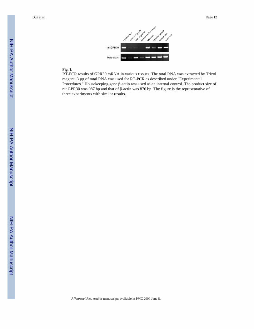
Fig. 1.RT-PCR results of GPR30 mRNA in various tissues. The total RNA was extracted by Trizolreagent. 3 µg of total RNA was used for RT-PCR as described under "ExperimentalProcedures." Housekeeping gene β-actin was used as an internal control. The product size ofrat GPR30 was 987 bp and that of β-actin was 876 bp. The figure is the representative ofthree experiments with similar results.
Dun et al. Page 12
J Neurosci Res. Author manuscript; available in PMC 2009 June 8.
NIH
-PA Author Manuscript
NIH
-PA Author Manuscript
NIH
-PA Author Manuscript
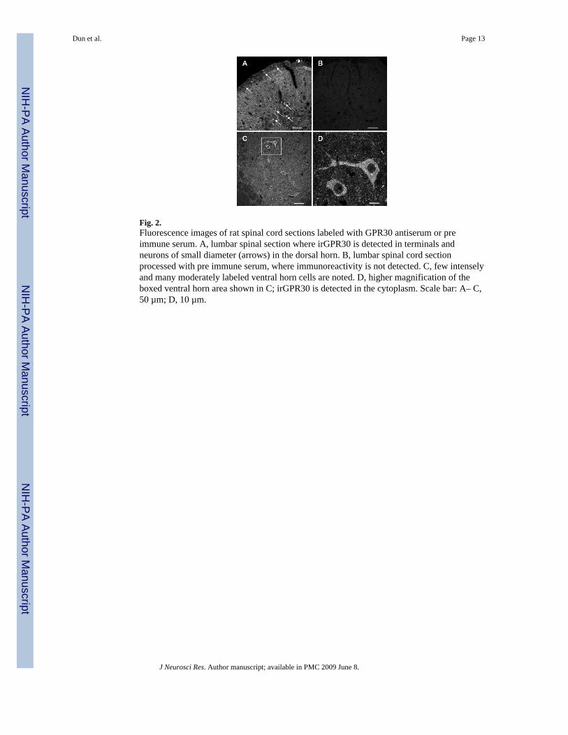
Fig. 2.Fluorescence images of rat spinal cord sections labeled with GPR30 antiserum or preimmune serum. A, lumbar spinal section where irGPR30 is detected in terminals andneurons of small diameter (arrows) in the dorsal horn. B, lumbar spinal cord sectionprocessed with pre immune serum, where immunoreactivity is not detected. C, few intenselyand many moderately labeled ventral horn cells are noted. D, higher magnification of theboxed ventral horn area shown in C; irGPR30 is detected in the cytoplasm. Scale bar: A– C,50 µm; D, 10 µm.
Dun et al. Page 13
J Neurosci Res. Author manuscript; available in PMC 2009 June 8.
NIH
-PA Author Manuscript
NIH
-PA Author Manuscript
NIH
-PA Author Manuscript
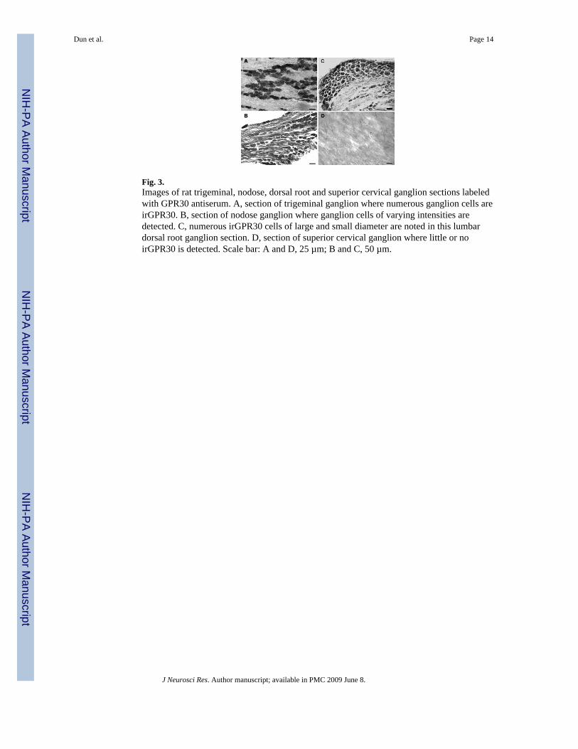
Fig. 3.Images of rat trigeminal, nodose, dorsal root and superior cervical ganglion sections labeledwith GPR30 antiserum. A, section of trigeminal ganglion where numerous ganglion cells areirGPR30. B, section of nodose ganglion where ganglion cells of varying intensities aredetected. C, numerous irGPR30 cells of large and small diameter are noted in this lumbardorsal root ganglion section. D, section of superior cervical ganglion where little or noirGPR30 is detected. Scale bar: A and D, 25 µm; B and C, 50 µm.
Dun et al. Page 14
J Neurosci Res. Author manuscript; available in PMC 2009 June 8.
NIH
-PA Author Manuscript
NIH
-PA Author Manuscript
NIH
-PA Author Manuscript
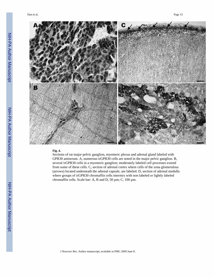
Fig. 4.Sections of rat major pelvic ganglion, myenteric plexus and adrenal gland labeled withGPR30 antiserum. A, numerous irGPR30 cells are noted in the major pelvic ganglion. B,several irGPR30 cells in a myenteric ganglion; moderately labeled cell processes extendfrom some of these cells. C, section of adrenal cortex where cells of the zona glomerulosa(arrows) located underneath the adrenal capsule, are labeled. D, section of adrenal medullawhere groups of irGPR30 chromaffin cells intermix with non labeled or lightly labeledchromaffin cells. Scale bar: A, B and D, 50 µm; C, 100 µm.
Dun et al. Page 15
J Neurosci Res. Author manuscript; available in PMC 2009 June 8.
NIH
-PA Author Manuscript
NIH
-PA Author Manuscript
NIH
-PA Author Manuscript
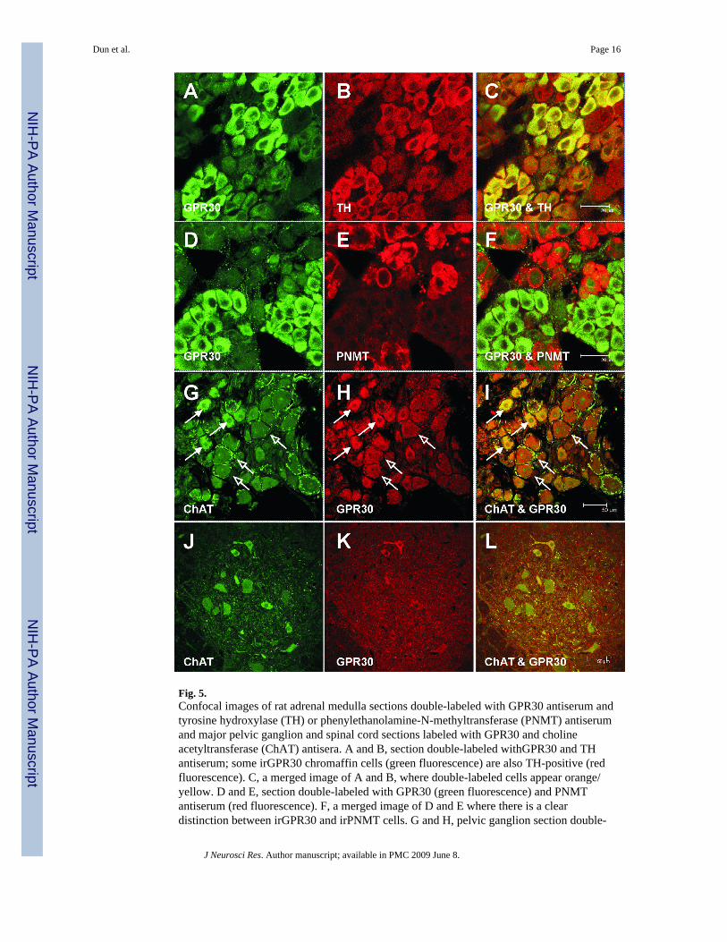
Fig. 5.Confocal images of rat adrenal medulla sections double-labeled with GPR30 antiserum andtyrosine hydroxylase (TH) or phenylethanolamine-N-methyltransferase (PNMT) antiserumand major pelvic ganglion and spinal cord sections labeled with GPR30 and cholineacetyltransferase (ChAT) antisera. A and B, section double-labeled withGPR30 and THantiserum; some irGPR30 chromaffin cells (green fluorescence) are also TH-positive (redfluorescence). C, a merged image of A and B, where double-labeled cells appear orange/yellow. D and E, section double-labeled with GPR30 (green fluorescence) and PNMTantiserum (red fluorescence). F, a merged image of D and E where there is a cleardistinction between irGPR30 and irPNMT cells. G and H, pelvic ganglion section double-
Dun et al. Page 16
J Neurosci Res. Author manuscript; available in PMC 2009 June 8.
NIH
-PA Author Manuscript
NIH
-PA Author Manuscript
NIH
-PA Author Manuscript
labeled with ChAT (green fluorescence) and GPR30 (red fluorescence) antiserum. irGPR30was present in some cholinergic cells (solid arrows); while other irGPR30 cells (openarrows) do not express ChAT, but are surrounded by ChAT immunoreactive fibers. I, amerged image of G and H; several double-labeled cells (solid arrows) are irGPR30 andChAT positive, and several irGPR30 cells are ChAT negative (open arrows). J and K, spinalcord sections double-labeled with ChAT (green fluorescence) and GPR30 (redfluorescence). L, merged image of J and K; few double-labeled cells are irGPR30 and ChATpositive. Scale bar: A–F, 20 µm, G–L, 50 µm
Dun et al. Page 17
J Neurosci Res. Author manuscript; available in PMC 2009 June 8.
NIH
-PA Author Manuscript
NIH
-PA Author Manuscript
NIH
-PA Author Manuscript
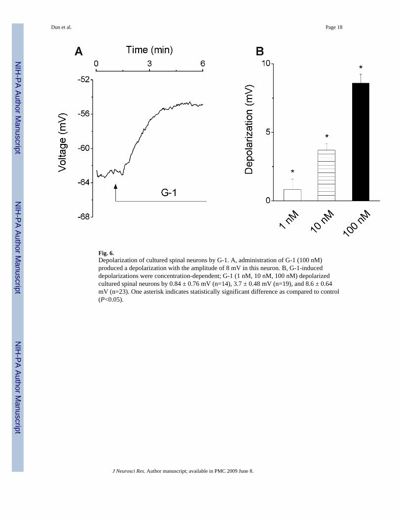
Fig. 6.Depolarization of cultured spinal neurons by G-1. A, administration of G-1 (100 nM)produced a depolarization with the amplitude of 8 mV in this neuron. B, G-1-induceddepolarizations were concentration-dependent; G-1 (1 nM, 10 nM, 100 nM) depolarizedcultured spinal neurons by 0.84 ± 0.76 mV (n=14), 3.7 ± 0.48 mV (n=19), and 8.6 ± 0.64mV (n=23). One asterisk indicates statistically significant difference as compared to control(P<0.05).
Dun et al. Page 18
J Neurosci Res. Author manuscript; available in PMC 2009 June 8.
NIH
-PA Author Manuscript
NIH
-PA Author Manuscript
NIH
-PA Author Manuscript
Related Documents