In vivo Effects of a GPR30 Antagonist Megan K. Dennis 1 , Ritwik Burai 2 , Chinnasamy Ramesh 2 , Whitney K. Petrie 1 , Sara N. Alcon 1 , Tapan K. Nayak 1 , Cristian G. Bologa 3 , Andrei Leitao 3 , Eugen Brailoiu 6 , Elena Deliu 6 , Nae J. Dun 6 , Larry A. Sklar 4,5 , Helen J. Hathaway 1,4 , Jeffrey B. Arterburn 2,4,* , Tudor I. Oprea 3,4,* , and Eric R. Prossnitz 1,4,* 1 Department of Cell Biology & Physiology, University of New Mexico Health Sciences Center, Albuquerque, NM 87131 2 Department of Chemistry and Biochemistry, New Mexico State University, Las Cruces, NM 88003 3 Division of Biocomputing, Department of Biochemistry & Molecular Biology, University of New Mexico Health Sciences Center, Albuquerque, NM 87131 4 UNM Cancer Center, University of New Mexico Health Sciences Center, Albuquerque, NM 87131 5 Department of Pathology, University of New Mexico Health Sciences Center, Albuquerque, NM 87131 6 Department of Pharmacology, Temple University School of Medicine, Philadelphia, PA 19140 Abstract Estrogen is central to many physiological processes throughout the human body. We have previously shown that the G protein-coupled receptor GPR30/GPER, in addition to classical nuclear estrogen receptors (ERα/β), activates cellular signaling pathways in response to estrogen. In order to distinguish between the actions of classical estrogen receptors and GPR30, we have previously characterized a selective agonist of GPR30, G-1 (1). To complement the pharmacological properties of G-1, we sought to identify an antagonist of GPR30 that displays similar selectivity against the classical estrogen receptors. Here we describe the identification and characterization of a G-1 analog, G15 (2) that binds to GPR30 with high affinity and acts as an antagonist of estrogen signaling through GPR30. In vivo administration of G15 reveals that GPR30 contributes to both uterine and neurological responses initiated by estrogen. The identification of this antagonist will accelerate the evaluation of the roles of GPR30 in human physiology. Introduction Estrogens play an important role in many areas of human physiology (including reproduction and the immune, vascular and nervous systems) as well as disease states such as cancer, depression and reproductive disorders 1,2 . Estrogen has long been known to act through soluble nuclear receptors that function as ligand-activated transcription factors. However, in addition to gene regulation, estrogen also mediates rapid signaling events, more commonly associated with growth factor and G protein-coupled receptors 3 . Recent studies reveal that GPR30 (International Union of Basic and Clinical Pharmacology designation: * Correspondence should be addressed to E.R.P. ([email protected]), T.I.O. ([email protected]) or J.B.A. ([email protected]). NIH Public Access Author Manuscript Nat Chem Biol. Author manuscript; available in PMC 2010 May 4. Published in final edited form as: Nat Chem Biol. 2009 June ; 5(6): 421–427. doi:10.1038/nchembio.168. NIH-PA Author Manuscript NIH-PA Author Manuscript NIH-PA Author Manuscript

Welcome message from author
This document is posted to help you gain knowledge. Please leave a comment to let me know what you think about it! Share it to your friends and learn new things together.
Transcript
In vivo Effects of a GPR30 Antagonist
Megan K. Dennis1, Ritwik Burai2, Chinnasamy Ramesh2, Whitney K. Petrie1, Sara N.Alcon1, Tapan K. Nayak1, Cristian G. Bologa3, Andrei Leitao3, Eugen Brailoiu6, ElenaDeliu6, Nae J. Dun6, Larry A. Sklar4,5, Helen J. Hathaway1,4, Jeffrey B. Arterburn2,4,*, TudorI. Oprea3,4,*, and Eric R. Prossnitz1,4,*
1Department of Cell Biology & Physiology, University of New Mexico Health Sciences Center,Albuquerque, NM 871312Department of Chemistry and Biochemistry, New Mexico State University, Las Cruces, NM880033Division of Biocomputing, Department of Biochemistry & Molecular Biology, University of NewMexico Health Sciences Center, Albuquerque, NM 871314UNM Cancer Center, University of New Mexico Health Sciences Center, Albuquerque, NM871315Department of Pathology, University of New Mexico Health Sciences Center, Albuquerque, NM871316Department of Pharmacology, Temple University School of Medicine, Philadelphia, PA 19140
AbstractEstrogen is central to many physiological processes throughout the human body. We havepreviously shown that the G protein-coupled receptor GPR30/GPER, in addition to classicalnuclear estrogen receptors (ERα/β), activates cellular signaling pathways in response to estrogen.In order to distinguish between the actions of classical estrogen receptors and GPR30, we havepreviously characterized a selective agonist of GPR30, G-1 (1). To complement thepharmacological properties of G-1, we sought to identify an antagonist of GPR30 that displayssimilar selectivity against the classical estrogen receptors. Here we describe the identification andcharacterization of a G-1 analog, G15 (2) that binds to GPR30 with high affinity and acts as anantagonist of estrogen signaling through GPR30. In vivo administration of G15 reveals thatGPR30 contributes to both uterine and neurological responses initiated by estrogen. Theidentification of this antagonist will accelerate the evaluation of the roles of GPR30 in humanphysiology.
IntroductionEstrogens play an important role in many areas of human physiology (includingreproduction and the immune, vascular and nervous systems) as well as disease states suchas cancer, depression and reproductive disorders1,2. Estrogen has long been known to actthrough soluble nuclear receptors that function as ligand-activated transcription factors.However, in addition to gene regulation, estrogen also mediates rapid signaling events, morecommonly associated with growth factor and G protein-coupled receptors3. Recent studiesreveal that GPR30 (International Union of Basic and Clinical Pharmacology designation:
*Correspondence should be addressed to E.R.P. ([email protected]), T.I.O. ([email protected]) or J.B.A.([email protected]).
NIH Public AccessAuthor ManuscriptNat Chem Biol. Author manuscript; available in PMC 2010 May 4.
Published in final edited form as:Nat Chem Biol. 2009 June ; 5(6): 421–427. doi:10.1038/nchembio.168.
NIH
-PA Author Manuscript
NIH
-PA Author Manuscript
NIH
-PA Author Manuscript
GPER), an intracellular transmembrane G protein-coupled estrogen receptor, mediatesnumerous aspects of cellular signaling ranging from calcium mobilization to EGFRtransactivation to gene regulation4. The classical nuclear estrogen receptors (ERα/β) appearto overlap with GPR30 not only in many of their cellular and physiological responses4 butalso in their ligand specificity5, making pharmacologic resolution of individual receptorfunctions challenging. For example, 17β-estradiol (3), 4-hydroxytamoxifen (4) andICI182,780 (5) each bind to GPR30 in addition to classical estrogen receptors, though withdifferent outcomes with respect to agonism and antagonism6-8. Whereas 17β-estradiol, 4-hydroxytamoxifen and ICI182,780 all activate GPR30, 17β-estradiol is an ERα agonist, 4-hydroxytamoxifen is a selective estrogen receptor modulator (SERM) and ICI182,780 is apure ERα antagonist9. Interestingly, until recently, GPR30-specific ligands were unknown.
In 2006, we described a highly selective GPR30 agonist named G-1 that shows no detectableactivity towards the classical estrogen receptors10. This compound activates multiplecellular signaling pathways via GPR30 and has been used to examine the cellular andphysiological actions of GPR30. Cellular effects include activation of calcium mobilizationin cancer cells10, LHRH neurons11 and hypothalamic neurons12, spinal neurondepolarization13, protein kinase Cε activation14 and phosphatidyl inositol-3-kinase (PI3K)activation10, gene expression15,16, proliferation15,17, oocyte meitotic arrest18 and primordialfollicle formation19. G-1 has also been used to probe the role of GPR30 in vivo withreported effects including estrogen-induced thymic atrophy20, experimental autoimmuneencephalomyelitis21 and vascular regulation22. In each of these animal models, the G-1-mediated effects were absent in GPR30 knockout mice, establishing the selectivity of thiscompound for GPR30. Thus, the availability of a selective GPR30 agonist has, in a verybrief time, greatly advanced our understanding of the biological functions of GPR30.
Unfortunately, to date, antagonists of GPR30 have not been identified. To better understandthe actions of GPR30, we identified a selective GPR30 antagonist using a combination ofvirtual and biomolecular screening. The compound is related in structure to the agonist G-1and binds to GPR30 but not ERα or ERβ. Cellular assays demonstrate that this antagonistprevents both estrogen- and G-1-mediated mobilization of intracellular calcium in ER-negative breast cancer cells. Furthermore, estrogen-mediated GPR30-dependent PI3Kactivation is blocked, whereas no effect on either ERα- or ERβ-mediated PI3K activation inresponse to estrogen is observed. In vivo studies utilizing both the agonist and antagonistreveal that GPR30 contributes to estrogen-mediated proliferation of the uterine epitheliumand plays an important role in the anti-depressive effects of estrogen. The introduction ofthis first GPR30-selective antagonist should provide additional avenues for characterizingthe physiological functions of GPR30.
RESULTSVirtual & biomolecular screening and chemical synthesis
We recently employed a combination of virtual and biomolecular screening to identify thefirst GPR30-specific ligand, a substituted dihydroquinoline, named G-110 (Fig. 1a). Toidentify potentially novel GPR30-specific ligands, we again employed virtual screening toidentify G-1-like structures of interest from the NIH Molecular Libraries Small MoleculeRepository (MLSMR). We performed a SMARTS substructure search (Daylight TheoryManual, Daylight Chemical Informations Systems Inc., http://www.daylight.com/dayhtml/doc/theory/theory.smarts.html) of the MLSMR (consisting of 144,457 molecules at the timeof the search, March 2007) for compounds containing the core scaffold of G-1 (Fig. 1b)using a custom JAVA program built using the OpenEye OEJava toolkit (OEChem - JavaTheory Manual, OpenEye Scientific Software Inc., http://www.eyesopen.com/docs/html/
Dennis et al. Page 2
Nat Chem Biol. Author manuscript; available in PMC 2010 May 4.
NIH
-PA Author Manuscript
NIH
-PA Author Manuscript
NIH
-PA Author Manuscript
javaprog/). The search identified 64 molecules, of which 57 were obtained from theMLSMR.
To accomplish the primary biomolecular screen for GPR30 antagonism, we utilized calciummobilization in GPR30-expressing SKBr3 cells and tested for the ability of the compound toblock cellular activation by estrogen. Primary screening of the 57 G-scaffold containingcompounds from the MLSMR yielded 8 that showed some inhibition of estrogen-mediatedcalcium mobilization in cells expressing GPR30. Of particular interest was one compound(designated G15) that closely resembled G-1 but lacked the ethanone moiety of the molecule(Fig. 1c). Based on the structural overlap of G-1 with estrogen and in particular the similar,though not identical spacing of oxygen atoms at the extremes of the molecules, wespeculated that the ketone functionality of G-1 might play an important role as a hydrogenbond acceptor by inducing conformational changes that activate GPR30. For this reason, inparallel to the virtual screening efforts, G15 was also chemically synthesized as a possibleantagonist candidate.
The tetrahydro-3H-cyclopenta[c]quinoline scaffold of G-1 is synthetically accessible via theversatile three-component Povarov cyclization. We evaluated a series of reaction conditionsemploying protic and Lewis acid catalysts to optimize reaction rate, yield, anddiastereoselectivity for the construction of G-1-like derivatives. Our optimized one-stepprocedure employing the catalyst Sc(OTf)3 in acetonitrile23 resulted in rapid reaction times,high product yield and enhanced selectivity favoring the syn diastereomeric products. Thesynthesis of G15 from aniline, 6-bromopiperonal and cyclopentadiene is illustrated in Fig.1d. Precipitation from dichloromethane/methanol gave analytically pure G15 in yieldsexceeding 85% as a racemic mixture of syn diastereomers. The syn diastereomer isdistinguished by the 1H-NMR coupling pattern of H-4 (4.65 ppm) with a coupling constantof 3.25 Hz that is characteristic for the syn orientation of the cyclopentene ring and phenylgroup. The product was fully characterized by NMR spectroscopy, and HPLC-MS withpositive ion detection (showing the correct molecular ion (MH+)) as well as UV detection(PDA/λmax= 294 nm), yielding a single peak.
G15 inhibits cellular signaling through GPR30Chemically synthesized G15 was subjected to multiple cellular and physiological assays inorder to characterize its biological effects. Competitive binding assays using endogenousGPR30 and a novel iodinated GPR30-selective G-1 analog (manuscript in preparation),demonstrated that G15 binds to GPR30 with an affinity of approximately 20 nM (Fig. 2a).This compares to an affinity for G-1, utilizing the same assay, of approximately 7 nM,similar to our previously reported affinity of G-1 for recombinant GPR30 of 11 nM 10 andreported affinities for 17β-estradiol between 3-6 nM 6,7. Thus removal of the ethanonemoiety resulted in a decrease in relative binding affinity of approximately 3 fold. Additionalcompetitive binding studies to assess interactions with ERα and ERβ revealed that similar toG-1, G15 displays little binding to ERα or ERβ at concentrations up to 10 μM, whereestrogen competes with a Ki of approximately 0.3-0.5 nM (Figs. 2b and c). These resultsreveal that G15, like G-1, displays high affinity for GPR30 with minimal binding to ERαand ERβ (Ki > 10 μM).
Evaluation of the functional capabilities of G15 with respect to the rapid mobilization ofintracellular calcium demonstrated that G15 alone was incapable of inducing a response inSKBr3 breast cancer cells, which are ERα and ERβ negative but express GPR30, whereasstimulation by either estrogen or G-1 induced a response (Fig. 3a). In contrast, stimulation ofthe cells with G-1 or estrogen subsequent to G15 exposure substantially reduced theresponse to G-1 or estrogen (Fig. 3a). There was however no inhibition of the calciumresponse mediated by ATP through endogenous purinergic receptors, indicating the
Dennis et al. Page 3
Nat Chem Biol. Author manuscript; available in PMC 2010 May 4.
NIH
-PA Author Manuscript
NIH
-PA Author Manuscript
NIH
-PA Author Manuscript
antagonistic effect is specific to GPR30. Inhibition of G-1-mediated calcium mobilization inSKBr3 cells by G15 was dose-dependent, yielding an IC50 of approximately 185 nM (Fig.3b), whereas inhibition of E2-mediated calcium mobilization yielded a similar IC50 ofapproximately 190 nM (Fig. 3c).
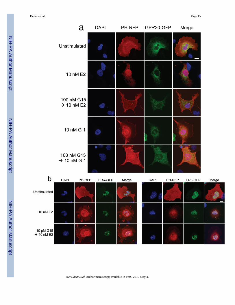
In addition to intracellular calcium mobilization, we have demonstrated that estrogenstimulation of ERα, ERβ or GPR30 results in the nuclear accumulation of PIP3 as a result ofPI3K activation6, revealed by the translocation of an Akt PH domain-fluorescent proteinfusion protein reporter24. To determine whether G15 similarly inhibits GPR30-mediatedPI3K activation, we examined the activation of PI3K in receptor-transfected COS7 cells,where estrogen stimulates the nuclear accumulation of PIP3 through all three receptors andG-1 selectively activates GPR30 but not ERα or ERβ. Not only was G15 capable ofinhibiting the G-1-mediated activation of PI3K in GPR30-transfected cells, it alsoeffectively blocked the estrogen-mediated response in GPR30-transfected cells (Fig. 4a) buthad no effect on the estrogen-mediated response in ERα or ERβ-transfected cells, even atconcentrations 100-fold greater than that required to inhibit GPR30 (Fig. 4b). To determinewhether G15 inhibits PI3K activation in cells endogenously expressing GPR30, weexamined PI3K activation in SKBr3 breast cancer cells. As in GPR30-transfected cells, G15was able to inhibit both estrogen and G-1 stimulation of PI3K (Fig. 4c). In total, these resultsdemonstrate that G15 can selectively inhibit GPR30.
G15 inhibits GPR30-mediated function in vivoOne goal of developing GPR30-specific agonists and antagonists is the elucidation of theroles of GPR30 in normal and disease physiology. One of the best characterized assays forestrogenic activity is the uterine response in the mouse, where uterine water content (i.e.imbibition) and epithelial cell proliferation are highly responsive to estrogen treatment,particularly following ovariectomy25. In the ovariectomized mouse model, a single injectionof estrogen (E2) led to a 17-fold increase in the proliferative index of uterine epitheliarelative to control, as measured by immunodetection of Ki-67 protein (Fig. 5a). Here weshow that the GPR30 agonist G-1 also increases proliferation, by 3-4 fold over sham (Fig.5b), and that there is little difference in proliferation rates across a 25-fold dose range,suggesting a maximal response was achieved. In contrast, treatment with the GPR30antagonist G15 alone did not alter proliferation relative to sham injections (Fig. 5b). Whenmice were treated with G15 plus E2 (Fig. 5a), proliferation was reduced by approximately50% in a dose-dependent manner, being maximal at a 10-fold molar excess of G15. G15treatment also blocked G-1-induced proliferation in a dose-dependent manner (Fig. 5b),being maximal at a 15-fold molar excess of G15. These results suggest that GPR30contributes to a specific estrogenic response, proliferation. Neither G-1 nor G15 had anyeffect on uterine wet weight or imbibition, evaluated by measuring uterine weight and bymicroscopic evaluation of histologic sections (not shown). In conclusion, GPR30 appears tocontribute to the proliferative response in the uterus, while another response, imbibition,appears to be solely mediated by ERα26.
Clinical observations suggest that vulnerability to depression in the female population isassociated with hormonal fluctuations, in which estrogens may play an important role. Forexample, chronic treatment of women with E2 or conjugated equine estrogens attenuateddepressive symptoms during peri-menopausal and postpartum periods27,28. Several animalmodels have been developed to evaluate putative anti-depressants29,30 and havedemonstrated the anti-depressive effects of estrogenic compounds31. Among these, the tailsuspension test32,33 is a convenient model in which many antidepressants reduce theduration of immobility, suggesting this parameter is an index of antidepressant activity34.Since GPR30 expression has been demonstrated in the male (as well as female) brain12,male mice were used to evaluate the potential neurological effects of G-1 and G15, given
Dennis et al. Page 4
Nat Chem Biol. Author manuscript; available in PMC 2010 May 4.
NIH
-PA Author Manuscript
NIH
-PA Author Manuscript
NIH
-PA Author Manuscript
that behavioral and neurochemical depression studies are carried out almost exclusively inmale mice34. The antidepressant action of G-1 was compared to that of E2 and the tricyclicantidepressant desipramine, which markedly reduced immobility time, when compared tocontrol vehicle-injected animals (Fig. 5c). G-1 dose-dependently decreased immobility time,whereas pretreatment of the mice with G15, which alone had no significant effect onimmobility time, significantly attenuated the effects of both G-1 and E2. Pretreatment of themice with G15 did not influence the immobility time of subsequent desipramine treatment.Together, these results suggest a neurological role for GPR30 in the regulation ofdepression.
DISCUSSIONIn this paper, we described the synthesis and characterization of the first GPR30 antagonist,G15. Binding studies demonstrated that G15 exhibited only moderately reduced binding toGPR30 (about 3-fold) compared to G-1, and yet no significant binding to either ERα or ERβat concentrations as high as 1-10 μM. Functional assays revealed that G15 blocked bothestrogen- and G-1-mediated mobilization of intracellular calcium in ER-negative SKBr3breast cancer cells. In addition, GPR30-dependent PI3K activation by either estrogen or G-1was blocked by prior incubation with G15. However, G15 was unable to prevent estrogen-mediated PI3K activation through either ERα or ERβ. In vivo studies demonstrated thatG15 completely blocked uterine epithelial cell proliferation mediated by GPR30 in responseto G-1 but only partially inhibited the estrogen-mediated response (presumably occurringthrough activation of all estrogen receptors). Finally, we established that the anti-depressiveeffects of estrogen appear to be mediated through GPR30, in that G-1 recapitulated theeffects of estrogen and that G15 inhibited the anti-depressive effects of both G-1 andestrogen.
The selectivity of G15 towards GPR30 in cellular assays is consistent with the selectivity ofG-1 for GPR30 in cells expressing both GPR30 and classical estrogen receptors as well asthe stimulatory effects of estrogen in cells expressing only GPR30. Given the similarity instructure between G15 and G-1, with the difference being the lack of an ethanone moiety inG15, we suggest that G-1 activates GPR30 in a similar manner to the way in which estrogenactivates classical estrogen receptors and presumably GPR30. Crystal structures of estrogen-bound ERα reveal extensive hydrogen bonding networks between the hydroxyl groups ofthe estrogen and receptor hydrogen bond donors and acceptors35. Assuming that G-1activates GPR30 through similar networks of hydrogen bonds at the distal ends of themolecule, removal of one hydrogen bond acceptor could allow binding to occur without theagonist-induced receptor conformational changes required for activation.
Although estrogen mediates effects on most physiological systems, including the nervous,immune and vascular systems, its most appreciated role is in reproduction. In the uterus, avariety of cellular and molecular responses, including imbibition, proliferation, andinduction of gene expression, are mediated by estrogen. Whereas ERα plays a major role inthese responses26, ERβ appears to play no role in imbitition, although it plays a role in thesuppression of uterine epithelial proliferation36. Here, we have demonstrated that theGPR30-specific antagonist G15 is capable of partially inhibiting estrogen-dependent uterineepithelial proliferation, but not imbibition (wet weight increase), suggesting that GPR30, inaddition to ERα, plays a role in promoting uterine epithelial proliferation. Our results are incontrast to a recent paper that reported no effect of G-1 on proliferation in the uterus37;however in that report, G-1 effects on uterine epithelial proliferation were not quantitated. Itis possible that a 3-fold difference in proliferation was not detected by visual inspectionalone, particularly when compared to the massive response to estrogen. In contrast to therestricted role of GPR30 in uterine responses to estrogen, GPR30 appears to contribute in a
Dennis et al. Page 5
Nat Chem Biol. Author manuscript; available in PMC 2010 May 4.
NIH
-PA Author Manuscript
NIH
-PA Author Manuscript
NIH
-PA Author Manuscript
significant way to the anti-depressive effects of estrogen with G-1 fully recapitulating theestrogen-mediated effects, and G15 equally inhibiting both estrogen- and G-1-mediatedresponses.
Although estrogen mediates the full range of uterine responses, including proliferation,imbibition, immune responses and gene expression, other estrogenic compounds have beenobserved to regulate these responses differentially. For example, DES (6) is weaker thanestrogen in inducing uterine eosinophilia, imbibition and proliferation, equal to estrogen inmediating epithelial hypertrophy and stronger than estrogen in inducing the reduction ofepithelial cell height and myometrial cell hypertrophy38. In addition, genistein (7) has only alimited ability to induce proliferation whereas estrogen-regulated genes are fully induced39.Finally, the ERα-selective compound PPT (8) is less effective in stimulating imbibition andthe expression of complement component 3 and glucose-6-phosphate dehydrogenase but isas effective as estrogen in regulating lactoferrin, androgen receptor, and progesteronereceptor expression40. Since genistein has been shown to bind and activate GPR3041, it isunclear whether the varying effects of genistein and other compounds on distinct aspects ofuterine physiology are due to either differential activation of classical estrogen receptor(s) orcomplex combinatorial effects on multiple estrogen receptors, including GPR30.
In conclusion, we report the identification and preliminary characterization of the firstselective GPR30 antagonist. The discovery of this high affinity GPR30-selective antagonistthat does not bind significantly to classical nuclear estrogen receptors has yielded novelinsights into the physiological roles of GPR30 in the reproductive and nervous systems.Future studies utilizing GPR30-selective agonists and antagonists will further define the roleof GPR30 in vivo and open the door to the generation of diagnostics and therapeuticsdirected at individual estrogen receptors.
METHODSChemical synthesis and characterization of G15
The compound G15 (4-(6-Bromo-benzo[1,3]dioxol-5-yl)-3a,4,5,9b-tetrahydro-3H-cyclopenta[c]quinoline) was synthesized using an optimized one-step procedure similar toKobayashi et. al.23 A catalytic amount of Sc(OTf)3 (0.049 g, 0.1mmol, 10 mol%) inanhydrous acetonitrile (1 mL) was added to a mixture of 6-bromopiperanal (0.229 g, 1.00mmol), aniline (0.093 g, 1.0 mmol) and freshly distilled cyclopentadiene (0.33 g, 5.0 mmol)in acetonitrile (3 mL). The reaction was stirred at ambient temperature (~23 °C) for 3 h withmonitoring of product formation by thin layer chromatography using 80% hexane/ethylacetate eluent (Rf 0.6). The volatiles were removed in vacuo. The residue was dissolved inmethylene chloride (10 mL) and then precipitated by drop-wise addition of methanol (5mL), and the product was isolated by filtration and washed with additional methanol to givethe product as a colorless solid (321mg, 87%): mp 178-180° C.
Spectroscopic characterization of G15 yielded the following: 1H NMR (300 MHz, DMSO-d6): δ 7.23 (s, 1H), 7.13 (s, 1H), 6.97 (d, J = 7.9 Hz, 1H), 6.87 (ddd, J = 7.3, 7.3, 1.3 Hz,1H), 6.68 (dd, J = 7.9, 1.3 Hz, 1H), 6.60 (ddd, J = 7.3, 7.3, 1.3 Hz, 1H), 6.1 (d, J = 0.9 Hz,1H), 6.06 (d, J = 0.9 Hz, 1H), 5.88-5.82 (m, 1H), 5.60-5.53 (m, 2H), 4.65 (d, J = 3.2 Hz,1H), 3.98 (d, J = 9.8 Hz, 1H), 3.07-2.95 (m, 1H), 2.48-2.38 (m, 1H), 1.69-1.59 (m, 1H). 13CNMR (75 MHz, CDCl3): δ 147.5, 147.2, 145.3, 134.6, 134.0, 130.3, 129.0, 126.3, 126.2,119.4, 116.1, 113.0, 112.9, 108.1, 101.7, 56.7, 46.1, 42.2, 31.4. FT-IR (KBr, cm-1): 3338(w),2887(w), 1605(w), 1587(s), 1473(s). HRMS (m/z): calcd. C19H16BrNO2 requires MH+,370.0443, found MH+, 370.0435. Anal (C19H15BrNO2) C, H, N.
Dennis et al. Page 6
Nat Chem Biol. Author manuscript; available in PMC 2010 May 4.
NIH
-PA Author Manuscript
NIH
-PA Author Manuscript
NIH
-PA Author Manuscript
G15 was analyzed by standard LC-MS methods with electrospray positive ion detection.The LC-MS chromatogram showed the correct molecular (MH+) ion as well as a single peakwith UV-Vis λmax 294 nm. Only one diastereomer was obtained; examination of the 1H-NMR data shows H-4 (4.65 ppm) with a coupling constant of 3.2 Hz, indicating a cisorientation of the cyclopentene and phenyl group, in agreement with the all-cisstereochemistry of similar reaction products of cyclopentadiene established previously. ThusG15 is a racemic but diastereomerically pure compound.
Ligand binding assaysBinding assays for ERα and ERβ were performed as previously described6. Briefly, COS7cells were transiently transfected with either ERα-GFP or ERβ-GFP). Following serumstarvation for 24 h, cells (~5×10⁴) were incubated with G-15 for 20 min. in a final volume of10 μL prior to addition of 10 μL 20 nM E2-Alexa633 in saponin-based permeabilizationbuffer. Following 10 min at RT, cells were washed once with 200 μL PBS/2%BSA,resuspended in 20 μL and 2 μL samples were analyzed on a DAKO Cyan flow cytometersusing HyperCyt™as described42. For GPR30 binding, a radioiodinated derivative of G-1, 1-{2-[4-(6-Bromo-benzo[1,3]dioxol-5-yl)-3a,4,5,9b-tetrahydro-3H-cyclopenta[c]quinolin-8-yl]-ethyl}-3-(3-iodo-phenyl)-urea, was used (see Supplementary Information, manuscript inpreparation). Briefly, Hec50 cells were cultured in phenol-red free DMEM/F-12 containing10% charcoal-stripped FBS, plated in 24-well tissue culture plates and grown to 80%confluence. Wells were rinsed with PBS and cells were incubated with competitor (G-1 orG15) for 30 minutes prior to addition of approximately 0.5-1 μCi of radioligand. The 125Iradiolabeled ligand was prepared from the corresponding tributylstannane using Iodo-genbeads (Pierce) following the manufacturer’s recommended protocol. Complete details of thesynthesis and radiolabeling will be described elsewhere (manuscript in preparation). Wellswere incubated at 37°C for 1 hour, rinsed with PBS and radioactivity collected by ethanolextraction and counted in a Wallac Wizard 1480 gamma counter (Perkin Elmer,Gaithersburg, MD).
Intracellular calcium mobilizationSKBr3 cells (1 × 10⁷) were incubated in HBSS containing 3 μM indo1-AM (Invitrogen) and0.05% pluronic acid for 1 h at RT. Cells were then washed twice with HBSS, incubated atRT for 20 min, washed again with HBSS, resuspended in HBSS at a density of 108 cells/mLand kept on ice until assay, performed at a density of 2 × 10⁶ cells/mL. Ca++ mobilizationwas determined ratiometrically using λex 340 nm and λem 400/490 nm at 37°C in aspectrofluorometer (QM-2000-2, Photon Technology International) equipped with amagnetic stirrer. The relative 490nm/400nm ratio was plotted as a function of time.
PI3K activationThe PIP3 binding domain of Akt fused to mRFP1 (PH-mRFP1) was used to localize cellularPIP3. COS7 cells (cotransfected with GPR30-GFP or ERα/β-GFP and PH-mRFP1) orSKBr3 (transfected with PH-mRFP1) cells were plated on coverslips and serum starved for24 h followed by stimulation with ligands as indicated. The cells were fixed with 2% PFA inPBS, washed, mounted in Vectashield and analyzed by confocal microscopy using a ZeissLSM510 confocal fluorescent microscope.
Mouse uterine estrogenicity assayC57Bl6 female mice (Harlan) were ovariectomized at 10 weeks of age. E2, G-1, and G15were dissolved in absolute ethanol at 1 mg/mL (E2 and G-1 were diluted to 10 μg/mL inethanol, G15 was diluted to 50 μg/mL in ethanol). For treatment with all three compounds,10 μL was added to 90 μL aqueous vehicle (0.9% NaCl with 0.1% albumin and 0.1%
Dennis et al. Page 7
Nat Chem Biol. Author manuscript; available in PMC 2010 May 4.
NIH
-PA Author Manuscript
NIH
-PA Author Manuscript
NIH
-PA Author Manuscript
Tween-20). Ethanol alone (10 μL) was added to 90 μL aqueous vehicle as control (sham).At 12 days post-ovariectomy, mice were injected subcutaneously at 5:00 pm with 100 μLconsisting of 1) sham; 2) 200 ng E2 (0.74 nmol); 3) 40, 200, or 1000 ng G-1 (0.1, 0.5, or 2.4nmol, respectively); 4) 272, 900, 2725, or 10000 ng G15 (2.4, 7.4, or 27 nmol, respectively)or 5) G15 combined with E2 or G-1 (at the same concentrations as used individually: G-1was used at 200 ng (0.5 nmol) in all G-1 + G15 combination experiments). The doses ofG15 were chosen to represent an approximately 1:1, 1:3.3, 1:10, and 1:35-fold molar excessrelative to E2. Eighteen hr after injection, mice were sacrificed and uteri were dissected,fixed in 4% paraformaldehyde, and embedded in paraffin. Five-micron sections were placedon slides, and proliferation in uterine epithelia was quantitated by immunofluorescenceusing anti-Ki-67 antibody (LabVision) followed by goat anti-mouse IgG conjugated toAlexa488 (Invitrogen). Nuclei were counterstained with 4’,6-diamidino-2-phenylindole(DAPI). At least 4 animals per treatment were analyzed, and the Ki-67 immunodetectionwas repeated three times per mouse.
Mouse Depression assayAdult male ICR mice, weighing 20-25 g, were used. Animals were maintained at roomtemperature, with free access to tap water and standard diet, under a 10:14 light/dark cycle(lights on 8:00h) and were housed 5/cage. Mice were acclimated to the laboratory for at leastone hour before testing and used only once. All experiments were conducted during the lightphase, between 8:30 and 14:30 h. Procedures used in this study were performed inaccordance with the NIH Guide for the Care and Use of Laboratory Animals and approvedby the Institutional Animal Care and Use Committee. The procedure was similar to thatdescribed by Steru et al. 33. Mice were isolated and suspended 35 cm above the floor by anadhesive tape placed 1 cm from the tip of the tail. The mouse was 15 cm away from thenearest object. The total amount of time each animal remained immobile (mice wereconsidered immobile only when they hung passively and completely motionless) during a 6-min period was recorded (in seconds) as immobility time. Each animal received twosuccessive injections (0.1 mL/mouse) in order to eliminate any possible bias comparingsingle-compound treatments to dual-compound treatments. G1 and G15 were first dissolvedin DMSO and diluted with saline; the final concentration in DMSO was 1 mM. Desipramineand E2 (cyclodextrin-encapsulated, 4-5.5% E2) were dissolved in saline solution and DMSOwas added to a final concentration of 1 mM. An appropriate vehicle-treated group (salinewith 1 mM DMSO) was included as a control (sham). All solutions were freshly preparedbefore each experimental series. Independent groups of mice (n=12-16) were treated withtwo consecutive intraperitoneal injections as follows: vehicle solution + vehicle solution(sham group); vehicle + G-1 (indicated amount in nmol); vehicle + desipramine (10mg/kg);G15 (10nmol/mouse) + desipramine (10mg/kg); G15 (10nmol/mouse) + G-1 (1nmol/mouse); vehicle + G15 (10nmol/mouse); vehicle + soluble E2 (5 mg/kg); G15 (25nmol/mouse) + soluble E2 (5 mg/kg). The second compound was injected 15 min (7 min for E2)after the first injection and the tail suspension test performed 30 min after the secondinjection.
Supplementary MaterialRefer to Web version on PubMed Central for supplementary material.
AcknowledgmentsThis work was supported by NIH grants CA118743 and CA127731, and grants from the Oxnard and StranahanFoundations to ERP, by the New Mexico Molecular Libraries Screening Center (NIH MH074425) to LAS, the NewMexico Tobacco Settlement fund to TIO, the New Mexico Cowboys for Cancer Research to JBA and NIH grantsR37 NS18710 to NJD and HL90804 to EB. Flow cytometry data and confocal images in this study were generated
Dennis et al. Page 8
Nat Chem Biol. Author manuscript; available in PMC 2010 May 4.
NIH
-PA Author Manuscript
NIH
-PA Author Manuscript
NIH
-PA Author Manuscript
in the Flow Cytometry and Fluorescence Microscopy Facilities, which received support from the University of NewMexico Health Sciences Center and the University of New Mexico Cancer Center as detailed: http://hsc.unm.edu/crtc/microscopy/Facility.html. In vivo data were generated with the support by the UNM Cancer Center AnimalModels & Imaging Core.
References1. Edwards DP. Regulation of signal transduction pathways by estrogen and progesterone. Annu Rev
Physiol. 2005; 67:335–376. [PubMed: 15709962]
2. Lange CA, Gioeli D, Hammes SR, Marker PC. Integration of rapid signaling events with steroidhormone receptor action in breast and prostate cancer. Annu Rev Physiol. 2007; 69:171–199.[PubMed: 17037979]
3. Fu XD, Simoncini T. Extra-nuclear signaling of estrogen receptors. IUBMB Life. 2008; 60:502–510. [PubMed: 18618586]
4. Prossnitz ER, et al. Estrogen signaling through the transmembrane G protein-coupled receptorGPR30. Annu Rev Physiol. 2008; 70:165–190. [PubMed: 18271749]
5. Prossnitz ER, Sklar LA, Oprea TI, Arterburn JB. GPR30: a novel therapeutic target in estrogen-related disease. Trends Pharmacol Sci. 2008; 29:116–123. [PubMed: 18262661]
6. Revankar CM, Cimino DF, Sklar LA, Arterburn JB, Prossnitz ER. A transmembrane intracellularestrogen receptor mediates rapid cell signaling. Science. 2005; 307:1625–1630. [PubMed:15705806]
7. Thomas P, Pang Y, Filardo EJ, Dong J. Identity of an estrogen membrane receptor coupled to a G-protein in human breast cancer cells. Endocrinology. 2005; 146:624–632. [PubMed: 15539556]
8. Filardo EJ, Quinn JA, Bland KI, Frackelton AR Jr. Estrogen-induced activation of Erk-1 and Erk-2requires the G protein-coupled receptor homolog, GPR30, and occurs via trans-activation of theepidermal growth factor receptor through release of HB-EGF. Mol Endocrinol. 2000; 14:1649–1660. [PubMed: 11043579]
9. Ariazi EA, Ariazi JL, Cordera F, Jordan VC. Estrogen receptors as therapeutic targets in breastcancer. Curr Top Med Chem. 2006; 6:181–202. [PubMed: 16515478]
10. Bologa CG, et al. Virtual and biomolecular screening converge on a selective agonist for GPR30.Nat Chem Biol. 2006; 2:207–212. [PubMed: 16520733]
11. Noel SD, Keen KL, Baumann DI, Filardo EJ, Terasawa E. Involvement of G-Protein CoupledReceptor 30 (GPR30) in Rapid Action of Estrogen in Primate LHRH Neurons. Mol Endocrinol.2009
12. Brailoiu E, et al. Distribution and characterization of estrogen receptor G protein-coupled receptor30 in the rat central nervous system. J Endocrinol. 2007; 193:311–321. [PubMed: 17470522]
13. Dun SL, et al. Expression of estrogen receptor GPR30 in the rat spinal cord and in autonomic andsensory ganglia. J Neurosci Res. 2009
14. Kuhn J, et al. GPR30 estrogen receptor agonists induce mechanical hyperalgesia in the rat. Eur JNeurosci. 2008; 27:1700–1709. [PubMed: 18371086]
15. Albanito L, et al. G protein-coupled receptor 30 (GPR30) mediates gene expression changes andgrowth response to 17beta-estradiol and selective GPR30 ligand G-1 in ovarian cancer cells.Cancer Res. 2007; 67:1859–1866. [PubMed: 17308128]
16. Prakash Pandey D, et al. Estrogenic GPR30 signalling induces proliferation and migration of breastcancer cells through CTGF. EMBO J. 2009
17. Teng J, Wang ZY, Prossnitz ER, Bjorling DE. The G protein-coupled receptor GPR30 inhibitshuman urothelial cell proliferation. Endocrinology. 2008; 149:4024–4034. [PubMed: 18467434]
18. Pang Y, Dong J, Thomas P. Estrogen signaling characteristics of Atlantic croaker G protein-coupled receptor 30 (GPR30) and evidence it is involved in maintenance of oocyte meiotic arrest.Endocrinology. 2008; 149:3410–3426. [PubMed: 18420744]
19. Wang C, Prossnitz ER, Roy SK. G protein-coupled receptor 30 expression is required for estrogenstimulation of primordial follicle formation in the hamster ovary. Endocrinology. 2008; 149:4452–4461. [PubMed: 18499747]
Dennis et al. Page 9
Nat Chem Biol. Author manuscript; available in PMC 2010 May 4.
NIH
-PA Author Manuscript
NIH
-PA Author Manuscript
NIH
-PA Author Manuscript
20. Wang C, et al. GPR30 contributes to estrogen-induced thymic atrophy. Mol Endocrinol. 2008;22:636–648. [PubMed: 18063692]
21. Wang C, et al. Membrane estrogen receptor regulates experimental autoimmune encephalomyelitisthrough up-regulation of programmed death 1. J Immunol. 2009; 182:3294–3303. [PubMed:19234228]
22. Haas E, et al. Regulatory Role of G Protein-Coupled Estrogen Receptor for Vascular Function andObesity. Circ Res. 2009
23. Kobayashi S, Ishitani H, Nagayama S. Lanthanide triflate catalyzed imino Diels-Alder reactions;convenient synthesis of pyridine and quinoline derivatives. Synthesis. 1995:1195–1202.
24. Balla T, Varnai P. Visualizing cellular phosphoinositide pools with GFP-fused protein-modules.Sci STKE. 2002; 2002:PL3. [PubMed: 11917154]
25. Owens JW, Ashby J. Critical review and evaluation of the uterotrophic bioassay for theidentification of possible estrogen agonists and antagonists: in support of the validation of theOECD uterotrophic protocols for the laboratory rodent. Organisation for Economic Co-operationand Development. Crit Rev Toxicol. 2002; 32:445–520. [PubMed: 12487363]
26. Hewitt SC, Harrell JC, Korach KS. Lessons in estrogen biology from knockout and transgenicanimals. Annu Rev Physiol. 2005; 67:285–308. [PubMed: 15709960]
27. Epperson CN, Wisner KL, Yamamoto B. Gonadal steroids in the treatment of mood disorders.Psychosom Med. 1999; 61:676–697. [PubMed: 10511016]
28. Genazzani AR, Spinetti A, Gallo R, Bernardi F. Menopause and the central nervous system:intervention options. Maturitas. 1999; 31:103–110. [PubMed: 10227002]
29. Porsolt RD, Anton G, Blavet N, Jalfre M. Behavioural despair in rats: a new model sensitive toantidepressant treatments. Eur J Pharmacol. 1978; 47:379–391. [PubMed: 204499]
30. Willner P. Animal models of depression: an overview. Pharmacol Ther. 1990; 45:425–455.[PubMed: 2405444]
31. Estrada-Camarena E, Fernandez-Guasti A, Lopez-Rubalcava C. Antidepressant-like effect ofdifferent estrogenic compounds in the forced swimming test. Neuropsychopharmacology. 2003;28:830–838. [PubMed: 12637949]
32. Steru L, et al. The automated Tail Suspension Test: a computerized device which differentiatespsychotropic drugs. Prog Neuropsychopharmacol Biol Psychiatry. 1987; 11:659–671. [PubMed:2894041]
33. Steru L, Chermat R, Thierry B, Simon P. The tail suspension test: a new method for screeningantidepressants in mice. Psychopharmacology (Berl). 1985; 85:367–370. [PubMed: 3923523]
34. Cryan JF, Mombereau C, Vassout A. The tail suspension test as a model for assessingantidepressant activity: review of pharmacological and genetic studies in mice. Neurosci BiobehavRev. 2005; 29:571–625. [PubMed: 15890404]
35. Tanenbaum DM, Wang Y, Williams SP, Sigler PB. Crystallographic comparison of the estrogenand progesterone receptor’s ligand binding domains. Proc Natl Acad Sci U S A. 1998; 95:5998–6003. [PubMed: 9600906]
36. Wada-Hiraike O, et al. Role of estrogen receptor beta in uterine stroma and epithelium: Insightsfrom estrogen receptor beta-/- mice. Proc Natl Acad Sci U S A. 2006; 103:18350–18355.[PubMed: 17110437]
37. Otto C, et al. GPR30 localizes to the endoplasmic reticulum and is not activated by estradiol.Endocrinology. 2008
38. Grunert G, Porcia M, Tchernitchin AN. Differential potency of oestradiol-17 beta anddiethylstilboestrol on separate groups of responses in the rat uterus. J Endocrinol. 1986; 110:103–114. [PubMed: 3734672]
39. Diel P, et al. The differential ability of the phytoestrogen genistein and of estradiol to induceuterine weight and proliferation in the rat is associated with a substance specific modulation ofuterine gene expression. Mol Cell Endocrinol. 2004; 221:21–32. [PubMed: 15223129]
40. Frasor J, et al. Response-specific and ligand dose-dependent modulation of estrogen receptor (ER)alpha activity by ERbeta in the uterus. Endocrinology. 2003; 144:3159–3166. [PubMed:12810572]
Dennis et al. Page 10
Nat Chem Biol. Author manuscript; available in PMC 2010 May 4.
NIH
-PA Author Manuscript
NIH
-PA Author Manuscript
NIH
-PA Author Manuscript
41. Thomas P, Dong J. Binding and activation of the seven-transmembrane estrogen receptor GPR30by environmental estrogens: a potential novel mechanism of endocrine disruption. J SteroidBiochem Mol Biol. 2006; 102:175–179. [PubMed: 17088055]
42. Ramirez S, Aiken CT, Andrzejewski B, Sklar LA, Edwards BS. High-throughput flow cytometry:validation in microvolume bioassays. Cytometry A. 2003; 53:55–65. [PubMed: 12701132]
Dennis et al. Page 11
Nat Chem Biol. Author manuscript; available in PMC 2010 May 4.
NIH
-PA Author Manuscript
NIH
-PA Author Manuscript
NIH
-PA Author Manuscript
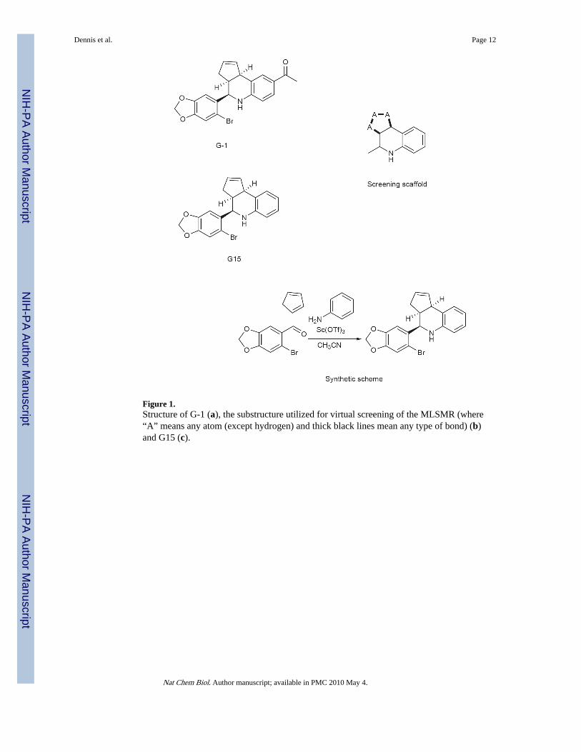
Figure 1.Structure of G-1 (a), the substructure utilized for virtual screening of the MLSMR (where“A” means any atom (except hydrogen) and thick black lines mean any type of bond) (b)and G15 (c).
Dennis et al. Page 12
Nat Chem Biol. Author manuscript; available in PMC 2010 May 4.
NIH
-PA Author Manuscript
NIH
-PA Author Manuscript
NIH
-PA Author Manuscript
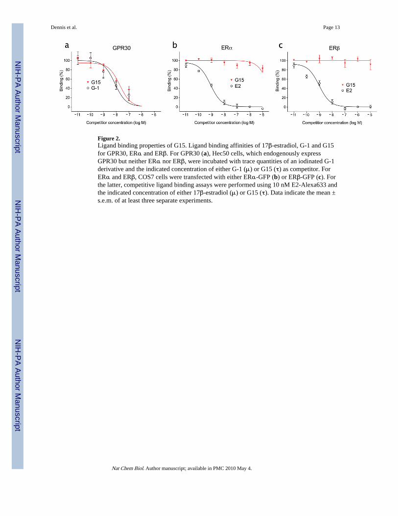
Figure 2.Ligand binding properties of G15. Ligand binding affinities of 17β-estradiol, G-1 and G15for GPR30, ERα and ERβ. For GPR30 (a), Hec50 cells, which endogenously expressGPR30 but neither ERα nor ERβ, were incubated with trace quantities of an iodinated G-1derivative and the indicated concentration of either G-1 (μ) or G15 (τ) as competitor. ForERα and ERβ, COS7 cells were transfected with either ERα-GFP (b) or ERβ-GFP (c). Forthe latter, competitive ligand binding assays were performed using 10 nM E2-Alexa633 andthe indicated concentration of either 17β-estradiol (μ) or G15 (τ). Data indicate the mean ±s.e.m. of at least three separate experiments.
Dennis et al. Page 13
Nat Chem Biol. Author manuscript; available in PMC 2010 May 4.
NIH
-PA Author Manuscript
NIH
-PA Author Manuscript
NIH
-PA Author Manuscript
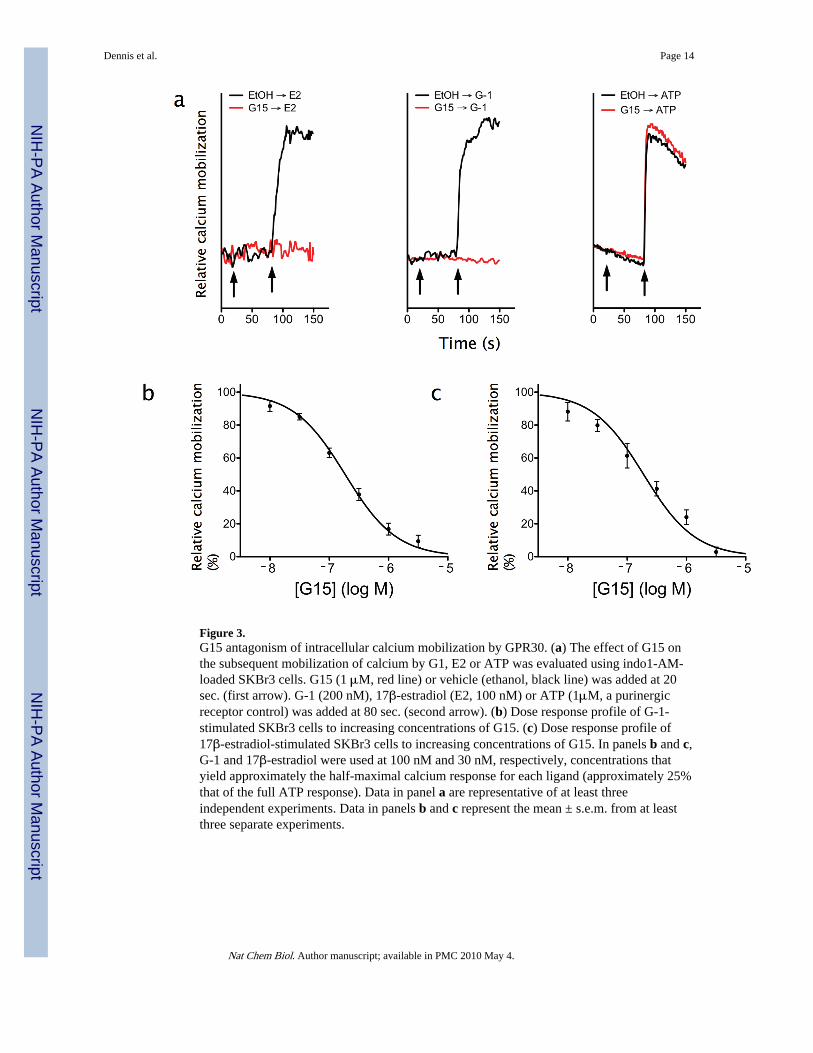
Figure 3.G15 antagonism of intracellular calcium mobilization by GPR30. (a) The effect of G15 onthe subsequent mobilization of calcium by G1, E2 or ATP was evaluated using indo1-AM-loaded SKBr3 cells. G15 (1 μM, red line) or vehicle (ethanol, black line) was added at 20sec. (first arrow). G-1 (200 nM), 17β-estradiol (E2, 100 nM) or ATP (1μM, a purinergicreceptor control) was added at 80 sec. (second arrow). (b) Dose response profile of G-1-stimulated SKBr3 cells to increasing concentrations of G15. (c) Dose response profile of17β-estradiol-stimulated SKBr3 cells to increasing concentrations of G15. In panels b and c,G-1 and 17β-estradiol were used at 100 nM and 30 nM, respectively, concentrations thatyield approximately the half-maximal calcium response for each ligand (approximately 25%that of the full ATP response). Data in panel a are representative of at least threeindependent experiments. Data in panels b and c represent the mean ± s.e.m. from at leastthree separate experiments.
Dennis et al. Page 14
Nat Chem Biol. Author manuscript; available in PMC 2010 May 4.
NIH
-PA Author Manuscript
NIH
-PA Author Manuscript
NIH
-PA Author Manuscript
Dennis et al. Page 15
Nat Chem Biol. Author manuscript; available in PMC 2010 May 4.
NIH
-PA Author Manuscript
NIH
-PA Author Manuscript
NIH
-PA Author Manuscript
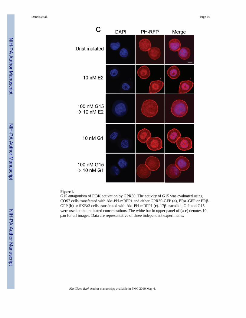
Figure 4.G15 antagonism of PI3K activation by GPR30. The activity of G15 was evaluated usingCOS7 cells transfected with Akt-PH-mRFP1 and either GPR30-GFP (a), ERα-GFP or ERβ-GFP (b) or SKBr3 cells transfected with Akt-PH-mRFP1 (c). 17β-estradiol, G-1 and G15were used at the indicated concentrations. The white bar in upper panel of (a-c) denotes 10μm for all images. Data are representative of three independent experiments.
Dennis et al. Page 16
Nat Chem Biol. Author manuscript; available in PMC 2010 May 4.
NIH
-PA Author Manuscript
NIH
-PA Author Manuscript
NIH
-PA Author Manuscript
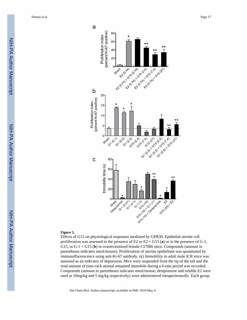
Figure 5.Effects of G15 on physiological responses mediated by GPR30. Epithelial uterine cellproliferation was assessed in the presence of E2 or E2 + G15 (a) or in the presence of G-1,G15, or G-1 + G15 (b) in ovariectomized female C57Bl6 mice. Compounds (amount inparentheses indicates nmol/mouse). Proliferation of uterine epithelium was quantitated byimmunofluorescence using anti-Ki-67 antibody. (c) Immobility in adult male ICR mice wasassessed as an indicator of depression. Mice were suspended from the tip of the tail and thetotal amount of time each animal remained immobile during a 6-min period was recorded.Compounds (amount in parentheses indicates nmol/mouse; desipramine and soluble E2 wereused at 10mg/kg and 5 mg/kg respectively) were administered intraperitoneally. Each group
Dennis et al. Page 17
Nat Chem Biol. Author manuscript; available in PMC 2010 May 4.
NIH
-PA Author Manuscript
NIH
-PA Author Manuscript
NIH
-PA Author Manuscript
consisted of 10-12 animals. For all panels, results are expressed as mean ± s.e.m., andstatistical significance (P<0.05) was assessed by student’s t test: *, significantly differentthan sham; **, significantly different than E2 or G-1, respectively.
Dennis et al. Page 18
Nat Chem Biol. Author manuscript; available in PMC 2010 May 4.
NIH
-PA Author Manuscript
NIH
-PA Author Manuscript
NIH
-PA Author Manuscript
Related Documents