ORIGINAL RESEARCH ARTICLE published: 06 March 2014 doi: 10.3389/fmicb.2014.00077 Exploring the role of CheA3 in Desulfovibrio vulgaris Hildenborough motility Jayashree Ray 1 , Kimberly L. Keller 2† , Michela Catena 1 , Thomas R. Juba 2 , Marcin Zemla 3 , Lara Rajeev 1 , Bernhard Knierim 3 , Grant M. Zane 2 , Jarrod J. Robertson 2 , Manfred Auer 3 , Judy D. Wall 2 and Aindrila Mukhopadhyay 1 * 1 Physical Biosciences Division, Lawrence Berkeley National Laboratory, Berkeley, CA, USA 2 Biochemistry Division, University of Missouri, Columbia, MO, USA 3 Life Sciences Division, Lawrence Berkeley National Laboratory, Berkeley, CA, USA Sulfate-reducing bacteria such as Desulfovibrio vulgaris Hildenborough are often found in environments with limiting growth nutrients. Using lactate as the electron donor and carbon source, and sulfate as the electron acceptor, wild type D. vulgaris shows motility on soft agar plates. We evaluated this phenotype with mutants resulting from insertional inactivation of genes potentially related to motility. Our study revealed that the cheA3 (DVU2072) kinase mutant was impaired in the ability to form motility halos. Insertions in two other cheA loci did not exhibit a loss in this phenotype. The cheA3 mutant was also non-motile in capillary assays. Complementation with a plasmid-borne copy of cheA3 restores wild type phenotypes. The cheA3 mutant displayed a flagellum as observed by electron microscopy, grew normally in liquid medium, and was motile in wet mounts. In the growth conditions used, the D. vulgaris fliA mutant (DVU3229) for FliA, predicted to regulate flagella-related genes including cheA3, was defective both in flagellum formation and in forming the motility halos. In contrast, a deletion of the flp gene (DVU2116) encoding a pilin-related protein was similar to wild type. We conclude that wild type D. vulgaris forms motility halos on solid media that are mediated by flagella-related mechanisms via the CheA3 kinase. The conditions under which the CheA1 (DVU1594) and CheA2 (DVU1960) kinase function remain to be explored. Keywords: sensor histidine kinase, cheA, soft agar plate assay, Palleroni chamber assay, electron acceptor, motility INTRODUCTION Desulfovibrio vulgaris Hildenborough is an anaerobic model sulfate-reducing bacterium (SRB), representing the broad class of SRB that play an essential role in biogeochemical processes such as sulfur- and metal-cycling (Zhou et al., 2011). Motility, its rela- tion to core physiology such as electron transfer (Tai et al., 2010), and the global nature of its regulation (Ueki et al., 2012) are key topics of research in both model and newly discovered anaerobic metal- and sulfate-reducing organisms (Takaki et al., 2010). The genomes of many organisms that occupy such ecological niches are now sequenced and reveal that some microbes have more than one putative chemotaxis-related gene cluster. Shewanella oneiden- sis MR1 encodes three chemotaxis gene clusters, one of which was shown to respond to electron acceptor concentrations (Bencharit and Ward, 2005; Li et al., 2007). Geobacter spp., are also anaero- bic metal-reducing bacteria and encode six chemotaxis clusters, the functions of which are yet to be specifically elucidated (Tran et al., 2008). D. vulgaris displays a single polar flagellum (Postgate and Campbell, 1966) and is documented to have motility on soft agar plates prepared with 0.7% (wt/vol) agarose and defined lactate/sulfate medium (Clark et al., 2007), with concentrations not considered limiting for either lactate or sulfate (Postgate, 1963; Mukhopadhyay et al., 2006). Aside from flagellar and pilin protein encoding genes, the genome of D. vulgaris encodes three separate chemotaxis clusters, each of which includes a putative cheA (Figures 1, S1). Here, we examine the observed motility in D. vulgaris as a function of lactate and sulfate in the medium and examine the role of several motility related genes in this phenotype. MATERIALS AND METHODS BACTERIAL GROWTH AND CULTURE MAINTENANCE All strains and plasmids used in this study are listed in Table 1. D. vulgaris Hildenborough strain ATCC 29579 was obtained from the American Type Culture Collection (Manassas, VA, USA). Bacterial strains were grown and maintained as described previously (Mukhopadhyay et al., 2006). Unless noted other- wise, D. vulgaris was grown in defined LS4D medium with sodium lactate (60 mM) as the electron donor and sodium sul- fate (30mM) as the electron acceptor. Modified LS4D media, the MOYLS4 and MOY media reported previously (Zane et al., 2010), were used during construction of the cheA knock-out mutants. D. vulgaris strain JW801, lacking the native plasmid pDV1 (Clark et al., 2007), was used as a non-motile con- trol and was grown similarly to the wild type. For growth of D. vulgaris mutant strains CA023, mutated in cheA1 (DVU1594); CA007, mutated in cheA2 (DVU1960); and CA022, mutated in www.frontiersin.org March 2014 | Volume 5 | Article 77 | 1 Edited by: Biswarup Mukhopadhyay, Virginia Polytechnic Institute and State University, USA Reviewed by: Heribert Cypionka, University of Oldenburg, Germany Birgit E. Scharf, Virginia Polytechnic Institute and State University, USA *Correspondence: Aindrila Mukhopadhyay, Physical Biosciences Division, Lawrence Berkeley National Laboratory, 1 Cyclotron Rd., Berkeley, CA 94720, USA e-mail: [email protected] † Present address: Kimberly L. Keller, Biology Department, William Woods University, Fulton, USA

Welcome message from author
This document is posted to help you gain knowledge. Please leave a comment to let me know what you think about it! Share it to your friends and learn new things together.
Transcript
ORIGINAL RESEARCH ARTICLEpublished: 06 March 2014
doi: 10.3389/fmicb.2014.00077
Exploring the role of CheA3 in Desulfovibrio vulgarisHildenborough motilityJayashree Ray1, Kimberly L. Keller2†, Michela Catena1, Thomas R. Juba2, Marcin Zemla3,
Lara Rajeev1, Bernhard Knierim3, Grant M. Zane2, Jarrod J. Robertson2, Manfred Auer3, Judy D. Wall2
and Aindrila Mukhopadhyay1*
1 Physical Biosciences Division, Lawrence Berkeley National Laboratory, Berkeley, CA, USA2 Biochemistry Division, University of Missouri, Columbia, MO, USA3 Life Sciences Division, Lawrence Berkeley National Laboratory, Berkeley, CA, USA
Sulfate-reducing bacteria such as Desulfovibrio vulgaris Hildenborough are often foundin environments with limiting growth nutrients. Using lactate as the electron donor andcarbon source, and sulfate as the electron acceptor, wild type D. vulgaris shows motilityon soft agar plates. We evaluated this phenotype with mutants resulting from insertionalinactivation of genes potentially related to motility. Our study revealed that the cheA3(DVU2072) kinase mutant was impaired in the ability to form motility halos. Insertionsin two other cheA loci did not exhibit a loss in this phenotype. The cheA3 mutant wasalso non-motile in capillary assays. Complementation with a plasmid-borne copy of cheA3restores wild type phenotypes. The cheA3 mutant displayed a flagellum as observed byelectron microscopy, grew normally in liquid medium, and was motile in wet mounts. Inthe growth conditions used, the D. vulgaris �fliA mutant (DVU3229) for FliA, predicted toregulate flagella-related genes including cheA3, was defective both in flagellum formationand in forming the motility halos. In contrast, a deletion of the flp gene (DVU2116) encodinga pilin-related protein was similar to wild type. We conclude that wild type D. vulgarisforms motility halos on solid media that are mediated by flagella-related mechanismsvia the CheA3 kinase. The conditions under which the CheA1 (DVU1594) and CheA2(DVU1960) kinase function remain to be explored.
Keywords: sensor histidine kinase, cheA, soft agar plate assay, Palleroni chamber assay, electron acceptor, motility
INTRODUCTIONDesulfovibrio vulgaris Hildenborough is an anaerobic modelsulfate-reducing bacterium (SRB), representing the broad class ofSRB that play an essential role in biogeochemical processes suchas sulfur- and metal-cycling (Zhou et al., 2011). Motility, its rela-tion to core physiology such as electron transfer (Tai et al., 2010),and the global nature of its regulation (Ueki et al., 2012) are keytopics of research in both model and newly discovered anaerobicmetal- and sulfate-reducing organisms (Takaki et al., 2010). Thegenomes of many organisms that occupy such ecological nichesare now sequenced and reveal that some microbes have more thanone putative chemotaxis-related gene cluster. Shewanella oneiden-sis MR1 encodes three chemotaxis gene clusters, one of which wasshown to respond to electron acceptor concentrations (Bencharitand Ward, 2005; Li et al., 2007). Geobacter spp., are also anaero-bic metal-reducing bacteria and encode six chemotaxis clusters,the functions of which are yet to be specifically elucidated (Tranet al., 2008).
D. vulgaris displays a single polar flagellum (Postgate andCampbell, 1966) and is documented to have motility on softagar plates prepared with 0.7% (wt/vol) agarose and definedlactate/sulfate medium (Clark et al., 2007), with concentrationsnot considered limiting for either lactate or sulfate (Postgate,1963; Mukhopadhyay et al., 2006). Aside from flagellar and pilin
protein encoding genes, the genome of D. vulgaris encodes threeseparate chemotaxis clusters, each of which includes a putativecheA (Figures 1, S1). Here, we examine the observed motility inD. vulgaris as a function of lactate and sulfate in the mediumand examine the role of several motility related genes in thisphenotype.
MATERIALS AND METHODSBACTERIAL GROWTH AND CULTURE MAINTENANCEAll strains and plasmids used in this study are listed in Table 1.D. vulgaris Hildenborough strain ATCC 29579 was obtainedfrom the American Type Culture Collection (Manassas, VA,USA). Bacterial strains were grown and maintained as describedpreviously (Mukhopadhyay et al., 2006). Unless noted other-wise, D. vulgaris was grown in defined LS4D medium withsodium lactate (60 mM) as the electron donor and sodium sul-fate (30 mM) as the electron acceptor. Modified LS4D media,the MOYLS4 and MOY media reported previously (Zane et al.,2010), were used during construction of the cheA knock-outmutants. D. vulgaris strain JW801, lacking the native plasmidpDV1 (Clark et al., 2007), was used as a non-motile con-trol and was grown similarly to the wild type. For growth ofD. vulgaris mutant strains CA023, mutated in cheA1 (DVU1594);CA007, mutated in cheA2 (DVU1960); and CA022, mutated in
www.frontiersin.org March 2014 | Volume 5 | Article 77 | 1
Edited by:
Biswarup Mukhopadhyay, VirginiaPolytechnic Institute and StateUniversity, USA
Reviewed by:
Heribert Cypionka, University ofOldenburg, GermanyBirgit E. Scharf, Virginia PolytechnicInstitute and State University, USA
*Correspondence:
Aindrila Mukhopadhyay, PhysicalBiosciences Division, LawrenceBerkeley National Laboratory,1 Cyclotron Rd., Berkeley,CA 94720, USAe-mail: [email protected]†Present address:
Kimberly L. Keller, BiologyDepartment, William WoodsUniversity, Fulton, USA
Ray et al. D. vulgaris motility
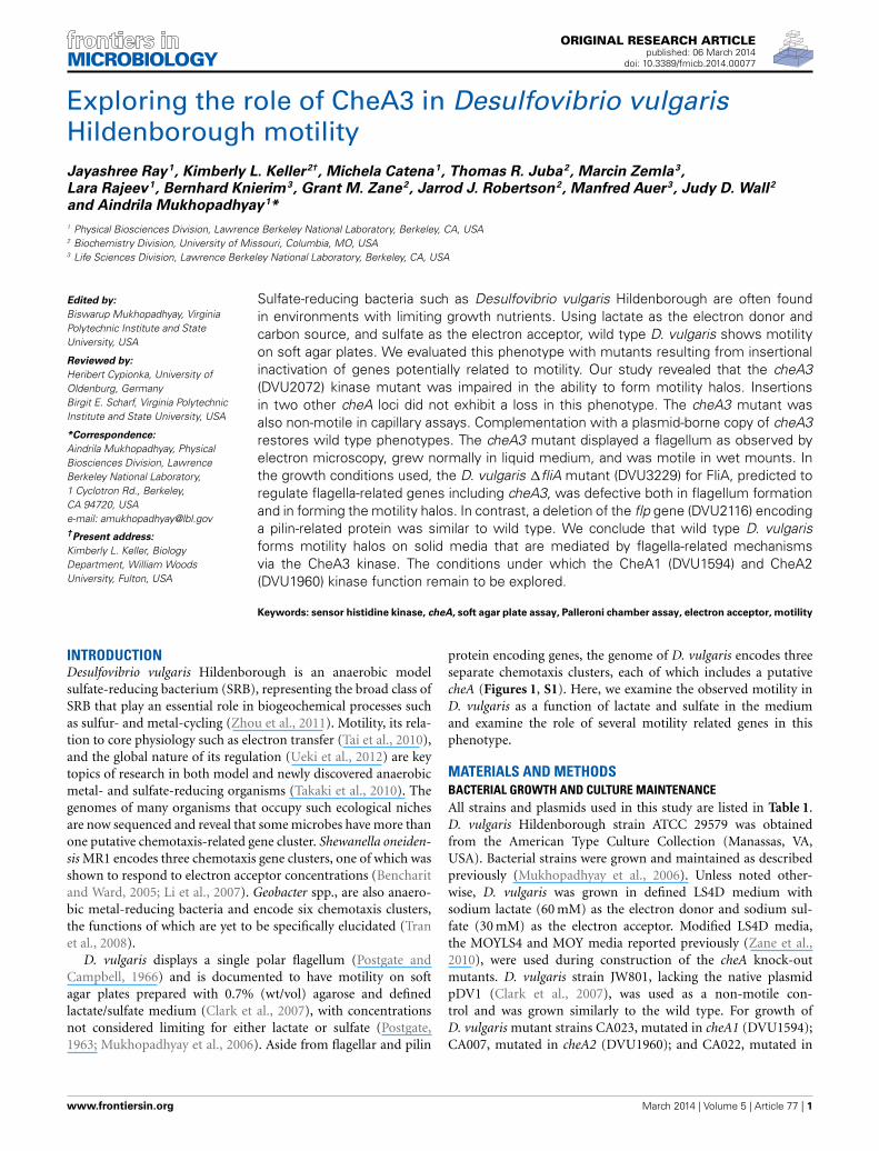
FIGURE 1 | (A) Soft agar plate assays for D. vulgaris wild type, JW801,CA023 (cheA1 mutant), CA007 (cheA2 mutant), and CA022 (cheA3 mutant)strains in modified LS4D medium as described in methods with 0.4% (wt/vol)agar. Motility halos were imaged after 4 days of incubation in an anaerobicchamber at 30◦C. No growth was observed for either the wild type or JW801strain in the control plate that contains no sulfate (left panel). In LS4D plates,the wild type forms a motility halo, whereas, JW801 was impaired in forminga halo (center panel); the cheA3 mutant shows a defect in motility (rightpanel) relative to the cheA1 and cheA2 mutants and the wild type. (B)
Growth assays of D. vulgaris wild type, JW801, CA023 (cheA1 mutant),CA007 (cheA2 mutant), CA022 (cheA3 mutant), and the cheA3complemented strain, cheA3::pTOPO-cheA3int(pMO2027). Assays weredone in LS4D medium at 30–32◦C. Cultures were started at an approximateOD600 of 0.1 and grown until the late stationary phase. Data points are theaverages of triplicate measurements. (C) Operons encoding the three cheAchemotaxis genes in D. vulgaris as predicted in www.microbesonline.org(Dehal et al., 2009). Top: cheA1; Middle: cheA2; and Bottom: cheA3.Arrowheads indicate the direction of transcription.
cheA3 (DVU2072). The antibiotic G418 (Sigma Aldrich, St Louis,MO) was added to a final concentration of 400 μg/ml (Kelleret al., 2009). For the complementation strain, cheA3::pTOPO-cheA3int(pMO2027), an additional antibiotic, spectinomycin(100 μg/ml), was added during growth. All D. vulgaris stocks werestored in 10% (vol/vol) glycerol at −80◦C and were used as 10%(vol/vol) inocula into 10–30 ml of fresh medium and the cellswere grown to mid-log phase (optical density at 600 nm (OD600)of 0.3 to 0.4).
CONSTRUCTION OF CheA INSERTIONAL MUTANTSGene disruption mutants in the cheA genes were created by singlecrossover homologous recombination with suicide vectors con-taining 750-base pair internal gene regions. The internal genefragments were produced by PCR amplification with primerslisted in Table A1 and cloned into the pENTR/D-TOPO plasmid(Life Technologies, Grant Island, NY, USA). The suicide vectorswere confirmed by sequencing and electroporated into wild-type D. vulgaris prepared as described previously (Keller et al.,2009). Transformants were recovered and colonies confirmed as
described (Zane et al., 2010). Southern blot analysis was per-formed on all the mutant strains as described previously (Kelleret al., 2009) to verify that the gene disruption occurred at the cor-rect locus. The transposon mutant in cheA3 used for the Palleronichamber assays (cheA3::TnRL27) was obtained from the D. vul-garis transposon mutant collection (Zane and Wall, 2013) cited inearlier reports (Fels et al., 2013; Figueiredo et al., 2013; Kazakovet al., 2013).
COMPLEMENTATION OF CheA::PTOPO-Chea3int MUTANTThe cheA3 gene was amplified by Herculase II (AgilentTechnologies, Santa Clara, CA, USA) with primers listed inTable A1 and cloned into pMO9075 for expression from theaph(3′)-II promoter. After selection of the recombinant plasmidand verification of the insert sequence, one isolate was namedpMO2027. To obtain a complemented cheA3 mutant, cheA3 cells(CA022) were transformed with pMO2027 by electroporationas described (Keller et al., 2009), with the following exceptions:MOYLS4 (60/30, lactate/sulfate) medium was used throughoutgrowth, electroporation, recovery and selective plating of the
Frontiers in Microbiology | Microbial Physiology and Metabolism March 2014 | Volume 5 | Article 77 | 2
Ray et al. D. vulgaris motility
Table 1 | Strains and plasmids used.
Strain Description Source
D. vulgaris Hildenborough Wild type D. vulgaris Hildenborough containing the 202 kbplasmid pDV1
ATCC29579
JW801 D. vulgaris Hildenborough �pDV1 Clark et al., 2007
JW9003 The JW9003 deletion mutant is a deletion of DVU2116 (flp)and DORF39640
This study
JW9017 �fliA Kmr This study
CA007 cheA2::pTOPO-cheA2int Kmr This study
CA022 cheA3:: pTOPO-cheA3int Kmr This study
CA023 cheA1:: pTOPO-cheA1int Kmr This study
GZ10278 cheA3 Transposon mutant (at bp 2299/3270), cheA3::TnRL27 Figueiredo et al., 2013;Zane and Wall, 2013
PLASMIDS
pENTR/D-TOPO TOPO cloning vector, Kmr Invitrogen
pCR2.1-TOPO TOPO cloning vector, Ampr Kmr Invitrogen
pCR8/GW/TOPO TOPO cloning vector, Specr Invitrogen
pSC27 Desulfovibrio shuttle vector containing the SRB repliconpBG1; source of aph(3′)-II; Kmr
Rousset et al., 1998
pTOPO-cheA3int Internal 750 bp fragment of cheA3 cloned into pCR2.1-TOPOAmpr Kmr
This study
pTOPO-cheA2int Internal 750 bp fragment of cheA2 cloned into pCR2.1-TOPOAmpr Kmr
This study
pTOPO-cheA2int Internal 750 bp fragment of cheA1 cloned intopENTR/D-TOPO Kmr
This study
pMO9002 pCR8/GW/TOPO with 684 bp upstream and 861 bpdownstream of aph(3 ′)-II cassette to deleteflp; Spr Kmr
This study
pMO9016 pCR8/GW/TOPO with 960 bp upstream and 942 bpdownstream of aph(3 ′)-II cassette to delete fliA; Spr Kmr
This study
pMO9075 Desulfovibrio shuttle vector containing SRB replicon (pBG1)and aph(3′)-IIp; Spr; for complementation constructs
Keller et al., 2009
pMO2027 pMO9075 with aph(3′)-IIp::cheA3; Spr This study
complemented mutant and the electroporation parameters wereset at 1500 V, 250 �, and 25 μF. Following sequence verificationof the plasmid recovered from the cheA3 mutant, one isolatewas chosen as the complemented strain, cheA3::pTOPO-cheA3int(pMO2027), for comparison of phenotypes.
CONSTRUCTION OF JW9017 (fliA) AND JW9003 (flp) DELETIONMUTANTSThe pMO9016 and pMO9002 plasmids for the marker-exchangedeletion of fliA (DVU3229) and flp (DVU2116), respectively, wereconstructed by splicing by overlap extension (SOE) PCR (Hortonet al., 1990) of three PCR amplimers as previously described(Zane et al., 2010). Transformation of the fliA and flp deletionplasmids into D. vulgaris was performed as previously described(Zane et al., 2010), with the exception that the G418-resistanttransformants were selected from electroporated cells mixed intomolten MOYLS4 medium with 400 μg G418/ml and poured intoempty petri dishes for solidification.
GROWTH ASSAYSCells were recovered overnight in 10 ml liquid MOYLS4 mediumand used to inoculate 20–25 ml volume of fresh LS4D at a start-ing OD600 of 0.05–0.1. Growth assays were conducted in triplicateunder anaerobic conditions at a temperature of 30–32◦C. OD600
was monitored with a spectrophotometer (Agilent HP DiodeArray Model 8452A, Agilent Technologies, Santa Clara, CA, USA)periodically as a function of time until the late stationary phase.
SOFT AGAR PLATE ASSAYSSoft agar plate assays were used to study the motility as describedin other reports (Li et al., 2007) with a few modifications. Amodified formulation for LS4D medium was solidified with 0.4%(wt/vol) agar for motility assays. D. vulgaris cells were grownto an OD600 of 0.3–0.4 and 2 μl of cells were stabbed intothe middle of the soft agar bed. For the sulfate disc assays,0.4% (wt/vol) soft agar medium contained 12 mM sodium sul-fate. A nylon membrane disc was pre-soaked in 30 mM sul-fate or water and placed 0.5 cm from the center of inocula-tion immediately prior to inoculation. Plates were incubatedat 30–32◦C in the anaerobic chamber for 4–5 days to obtaina reasonable amount of motility. Photographs in Figures 1, 2were taken under white light by a Biospectrum AC ImagingSystem (UVP, Upland, CA, USA) with the following constantinstrumental parameters: exposure time: 634 μs; filter: SyBr Gold(485–655 nm); aperture: 1.2; zoom: 20%; focus: 80%; trans illu-mination: white. Figure 3A was imaged with a Nikon D5000camera at the Veterinary Biomedical Communications at theUniversity of Missouri-College of Veterinary Medicine. For the
www.frontiersin.org March 2014 | Volume 5 | Article 77 | 3
Ray et al. D. vulgaris motility
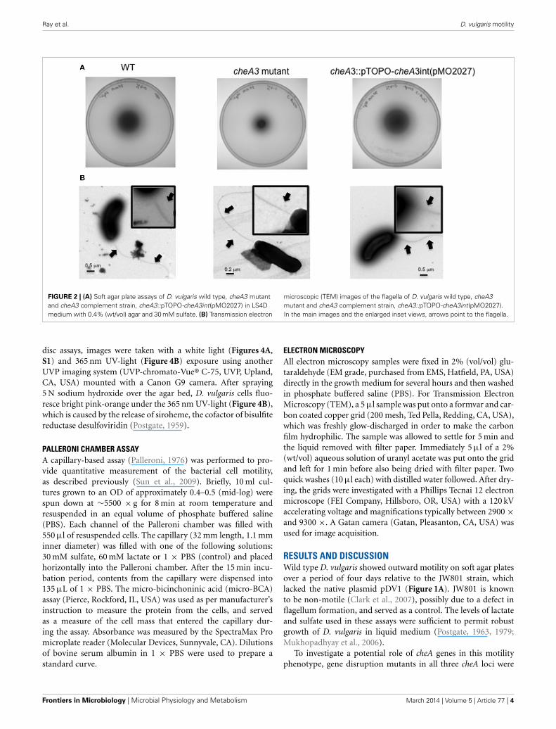
FIGURE 2 | (A) Soft agar plate assays of D. vulgaris wild type, cheA3 mutantand cheA3 complement strain, cheA3::pTOPO-cheA3int(pMO2027) in LS4Dmedium with 0.4% (wt/vol) agar and 30 mM sulfate. (B) Transmission electron
microscopic (TEM) images of the flagella of D. vulgaris wild type, cheA3mutant and cheA3 complement strain, cheA3::pTOPO-cheA3int(pMO2027).In the main images and the enlarged inset views, arrows point to the flagella.
disc assays, images were taken with a white light (Figures 4A,S1) and 365 nm UV-light (Figure 4B) exposure using anotherUVP imaging system (UVP-chromato-Vue® C-75, UVP, Upland,CA, USA) mounted with a Canon G9 camera. After spraying5 N sodium hydroxide over the agar bed, D. vulgaris cells fluo-resce bright pink-orange under the 365 nm UV-light (Figure 4B),which is caused by the release of siroheme, the cofactor of bisulfitereductase desulfoviridin (Postgate, 1959).
PALLERONI CHAMBER ASSAYA capillary-based assay (Palleroni, 1976) was performed to pro-vide quantitative measurement of the bacterial cell motility,as described previously (Sun et al., 2009). Briefly, 10 ml cul-tures grown to an OD of approximately 0.4–0.5 (mid-log) werespun down at ∼5500 × g for 8 min at room temperature andresuspended in an equal volume of phosphate buffered saline(PBS). Each channel of the Palleroni chamber was filled with550 μl of resuspended cells. The capillary (32 mm length, 1.1 mminner diameter) was filled with one of the following solutions:30 mM sulfate, 60 mM lactate or 1 × PBS (control) and placedhorizontally into the Palleroni chamber. After the 15 min incu-bation period, contents from the capillary were dispensed into135 μL of 1 × PBS. The micro-bicinchoninic acid (micro-BCA)assay (Pierce, Rockford, IL, USA) was used as per manufacturer’sinstruction to measure the protein from the cells, and servedas a measure of the cell mass that entered the capillary dur-ing the assay. Absorbance was measured by the SpectraMax Promicroplate reader (Molecular Devices, Sunnyvale, CA). Dilutionsof bovine serum albumin in 1 × PBS were used to prepare astandard curve.
ELECTRON MICROSCOPYAll electron microscopy samples were fixed in 2% (vol/vol) glu-taraldehyde (EM grade, purchased from EMS, Hatfield, PA, USA)directly in the growth medium for several hours and then washedin phosphate buffered saline (PBS). For Transmission ElectronMicroscopy (TEM), a 5 μl sample was put onto a formvar and car-bon coated copper grid (200 mesh, Ted Pella, Redding, CA, USA),which was freshly glow-discharged in order to make the carbonfilm hydrophilic. The sample was allowed to settle for 5 min andthe liquid removed with filter paper. Immediately 5 μl of a 2%(wt/vol) aqueous solution of uranyl acetate was put onto the gridand left for 1 min before also being dried with filter paper. Twoquick washes (10 μl each) with distilled water followed. After dry-ing, the grids were investigated with a Phillips Tecnai 12 electronmicroscope (FEI Company, Hillsboro, OR, USA) with a 120 kVaccelerating voltage and magnifications typically between 2900 ×and 9300 ×. A Gatan camera (Gatan, Pleasanton, CA, USA) wasused for image acquisition.
RESULTS AND DISCUSSIONWild type D. vulgaris showed outward motility on soft agar platesover a period of four days relative to the JW801 strain, whichlacked the native plasmid pDV1 (Figure 1A). JW801 is knownto be non-motile (Clark et al., 2007), possibly due to a defect inflagellum formation, and served as a control. The levels of lactateand sulfate used in these assays were sufficient to permit robustgrowth of D. vulgaris in liquid medium (Postgate, 1963, 1979;Mukhopadhyay et al., 2006).
To investigate a potential role of cheA genes in this motilityphenotype, gene disruption mutants in all three cheA loci were
Frontiers in Microbiology | Microbial Physiology and Metabolism March 2014 | Volume 5 | Article 77 | 4
Ray et al. D. vulgaris motility
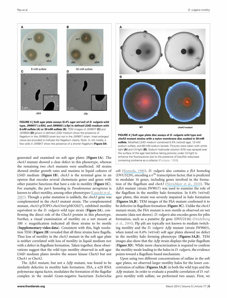
FIGURE 3 | Soft agar plate assays (0.4% agar wt/vol) of D. vulgaris wild
type, JW9017 (�fliA), and JW9003 (�flp) in defined LS4D medium with
6 mM sulfate (A) or 30 mM sulfate (B). TEM images of JW9017 (C) andJW9003 (D) grown in defined LS4D medium show the presence offlagellum in the JW9003 strain but not in the JW9017 strain. Inset enlargedviews are provided to indicate the flagellum clearly. Note: In rich media, afew cells in JW9017 show the presence of a shorter flagellum (Figure S4).
generated and examined on soft agar plates (Figure 1A). ThecheA3 mutant showed a clear defect in this phenotype, whereasthe remaining two cheA mutants were unaffected. All strainsshowed similar growth rates and maxima in liquid cultures ofLS4D medium (Figure 1B). cheA3 is the terminal gene in anoperon that encodes several chemotaxis genes and genes withother putative functions that have a role in motility (Figure 1C).For example, the parA homolog in Pseudomonas aeruginosa isknown to affect motility, among other phenotypes (Lasocki et al.,2007). Though a polar mutation is unlikely, the cheA3 gene wascomplemented in the cheA3 mutant strain. The complementedmutant, cheA3::pTOPO-cheA3int(pMO2027), exhibited motilityequivalent to the D. vulgaris wild type strain (Figure 2A), con-firming the direct role of the CheA3 protein in this phenotype.Further, a visual examination of motility on a wet mount at100 × magnification indicated all three strains to be motile(Supplementary video data). Consistent with this, high resolu-tion TEM (Figure 2B) revealed that all three strains have flagella.Thus loss of motility in the cheA3 mutant in the soft agar plateis neither correlated with loss of motility in liquid medium norwith a defect in flagellum formation. Taken together, these obser-vations suggest that the wild type motility observed in soft agarLS4D medium plates involve the sensor kinase CheA3 but notCheA1 or CheA2.
The �fliA mutant, but not a �flp mutant, was found to besimilarly defective in motility halo formation. FliA, a α28 RNApolymerase sigma factor, modulates the formation of the flagellarcomplex in the model Gram-negative bacterium Escherichia
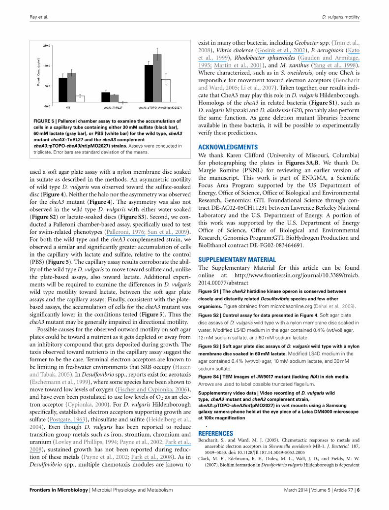
FIGURE 4 | Soft agar plate disc assays of D. vulgaris wild type and
cheA3 mutant strains with a nylon membrane disc soaked in 30 mM
sulfate. Modified LS4D medium contained 0.4% (wt/vol) agar, 12 mMsodium sulfate, and 60 mM sodium lactate. Pictures were taken with whitelight (A) and UV-light (B). Sodium hydroxide solution (5 N) was sprayed overthe surface of the agar bed before taking pictures under UV-light toenhance the fluorescence due to the presence of bisulfite reductasecontaining siroheme as a cofactor (Postgate, 1959).
coli (Komeda, 1986). D. vulgaris also contains a fliA homolog(DVU3229), encoding a σ70 transcription factor, that is predictedto modulate 16 genes, including genes involved in the forma-tion of the flagellum and cheA3 (Novichkov et al., 2010). The�fliA mutant (strain JW9017) was used to examine the role ofthe flagellum in the motility halo formation. In 0.4% (wt/vol)agar plates, this strain was severely impaired in halo formation(Figures 3A,B). TEM images of the FliA mutant confirmed it tobe defective in flagellum formation (Figure 3C). Unlike the cheA3mutant strain, the FliA mutant is non-motile as observed on wetmounts (data not shown). D. vulgaris also encodes genes for pilinformation, such as a putative flp gene (DVU2116) (Heidelberget al., 2004). Flp pili are typically not known to mediate twitch-ing motility and the D. vulgaris �flp mutant (strain JW9003),when tested on 0.4% (wt/vol) soft agar plates showed no defectin the motility halo forming phenotype (Figures 3A,B). TEMimages also show that the �flp strain displays the polar flagellum(Figure 3D). While more characterization is required to confirmthe motility mode leading to the halos in D. vulgaris, the evidencepoints toward a flagellum-based mechanism.
Upon using two different concentrations of sulfate in the softagar plates, we observed larger motility halos for the lower con-centration of sulfate (Figures 3A,B) in both the wild type and the�flp mutant. In order to evaluate a possible correlation of D. vul-garis motility with sulfate, we performed two assays. First, we
www.frontiersin.org March 2014 | Volume 5 | Article 77 | 5
Ray et al. D. vulgaris motility
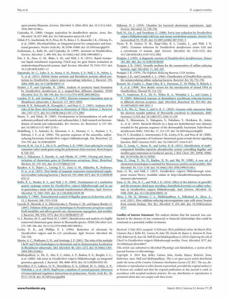
FIGURE 5 | Palleroni chamber assay to examine the accumulation of
cells in a capillary tube containing either 30 mM sulfate (black bar),
60 mM lactate (gray bar), or PBS (white bar) for the wild type, cheA3
mutant cheA3::TnRL27 and the cheA3 complement
cheA3::pTOPO-cheA3int(pMO2027) strains. Assays were conducted intriplicate. Error bars are standard deviation of the means.
used a soft agar plate assay with a nylon membrane disc soakedin sulfate as described in the methods. An asymmetric motilityof wild type D. vulgaris was observed toward the sulfate-soakeddisc (Figure 4). Neither the halo nor the asymmetry was observedfor the cheA3 mutant (Figure 4). The asymmetry was also notobserved in the wild type D. vulgaris with either water-soaked(Figure S2) or lactate-soaked discs (Figure S3). Second, we con-ducted a Palleroni chamber-based assay, specifically used to testfor swim-related phenotypes (Palleroni, 1976; Sun et al., 2009).For both the wild type and the cheA3 complemented strain, weobserved a similar and significantly greater accumulation of cellsin the capillary with lactate and sulfate, relative to the control(PBS) (Figure 5). The capillary assay results corroborate the abil-ity of the wild type D. vulgaris to move toward sulfate and, unlikethe plate-based assays, also toward lactate. Additional experi-ments will be required to examine the differences in D. vulgariswild type motility toward lactate, between the soft agar plateassays and the capillary assays. Finally, consistent with the plate-based assays, the accumulation of cells for the cheA3 mutant wassignificantly lower in the conditions tested (Figure 5). Thus thecheA3 mutant may be generally impaired in directional motility.
Possible causes for the observed outward motility on soft agarplates could be toward a nutrient as it gets depleted or away froman inhibitory compound that gets deposited during growth. Thetaxis observed toward nutrients in the capillary assay suggest theformer to be the case. Terminal electron acceptors are known tobe limiting in freshwater environments that SRB occupy (Hazenand Tabak, 2005). In Desulfovibrio spp., reports exist for aerotaxis(Eschemann et al., 1999), where some species have been shown tomove toward low levels of oxygen (Fischer and Cypionka, 2006),and have even been postulated to use low levels of O2 as an elec-tron acceptor (Cypionka, 2000). For D. vulgaris Hildenboroughspecifically, established electron acceptors supporting growth aresulfate (Postgate, 1963), thiosulfate and sulfite (Heidelberg et al.,2004). Even though D. vulgaris has been reported to reducetransition group metals such as iron, strontium, chromium anduranium (Lovley and Phillips, 1994; Payne et al., 2002; Park et al.,2008), sustained growth has not been reported during reduc-tion of these metals (Payne et al., 2002; Park et al., 2008). As inDesulfovibrio spp., multiple chemotaxis modules are known to
exist in many other bacteria, including Geobacter spp. (Tran et al.,2008), Vibrio cholerae (Gosink et al., 2002), P. aeruginosa (Katoet al., 1999), Rhodobacter sphaeroides (Gauden and Armitage,1995; Martin et al., 2001), and M. xanthus (Yang et al., 1998).Where characterized, such as in S. oneidensis, only one CheA isresponsible for movement toward electron acceptors (Bencharitand Ward, 2005; Li et al., 2007). Taken together, our results indi-cate that CheA3 may play this role in D. vulgaris Hildenborough.Homologs of the cheA3 in related bacteria (Figure S1), such asD. vulgaris Miyazaki and D. alaskensis G20, probably also performthe same function. As gene deletion mutant libraries becomeavailable in these bacteria, it will be possible to experimentallyverify these predictions.
ACKNOWLEDGMENTSWe thank Karen Clifford (University of Missouri, Columbia)for photographing the plates in Figures 3A,B. We thank Dr.Margie Romine (PNNL) for reviewing an earlier version ofthe manuscript. This work is part of ENIGMA, a ScientificFocus Area Program supported by the US Department ofEnergy, Office of Science, Office of Biological and EnvironmentalResearch, Genomics: GTL Foundational Science through con-tract DE-AC02-05CH11231 between Lawrence Berkeley NationalLaboratory and the U.S. Department of Energy. A portion ofthis work was supported by the U.S. Department of EnergyOffice of Science, Office of Biological and EnvironmentalResearch, Genomics Program:GTL BioHydrogen Production andBioEthanol contract DE-FG02-083464691.
SUPPLEMENTARY MATERIALThe Supplementary Material for this article can be foundonline at: http://www.frontiersin.org/journal/10.3389/fmicb.2014.00077/abstractFigure S1 | The cheA3 histidine kinase operon is conserved between
closely and distantly related Desulfovibrio species and few other
organisms. Figure obtained from microbesonline.org (Dehal et al., 2009).
Figure S2 | Control assay for data presented in Figure 4. Soft agar plate
disc assays of D. vulgaris wild type with a nylon membrane disc soaked in
water. Modified LS4D medium in the agar contained 0.4% (wt/vol) agar,
12 mM sodium sulfate, and 60 mM sodium lactate.
Figure S3 | Soft agar plate disc assays of D. vulgaris wild type with a nylon
membrane disc soaked in 60 mM lactate. Modified LS4D medium in the
agar contained 0.4% (wt/vol) agar, 10 mM sodium lactate, and 30 mM
sodium sulfate.
Figure S4 | TEM images of JW9017 mutant (lacking fliA) in rich media.
Arrows are used to label possible truncated flagellum.
Supplementary video data | Video recording of D. vulgaris wild
type, cheA3 mutant and cheA3 complement strain,
cheA3::pTOPO-cheA3int(pMO2027) in wet mounts using a Samsung
galaxy camera-phone held at the eye piece of a Leica DM4000 microscope
at 100x magnification
.
REFERENCESBencharit, S., and Ward, M. J. (2005). Chemotactic responses to metals and
anaerobic electron acceptors in Shewanella oneidensis MR-1. J. Bacteriol. 187,5049–5053. doi: 10.1128/JB.187.14.5049-5053.2005
Clark, M. E., Edelmann, R. E., Duley, M. L., Wall, J. D., and Fields, M. W.(2007). Biofilm formation in Desulfovibrio vulgaris Hildenborough is dependent
Frontiers in Microbiology | Microbial Physiology and Metabolism March 2014 | Volume 5 | Article 77 | 6
Ray et al. D. vulgaris motility
upon protein filaments. Environ. Microbiol. 9, 2844–2854. doi: 10.1111/j.1462-2920.2007.01398.x
Cypionka, H. (2000). Oxygen respiration by desulfovibrio species. Annu. Rev.Microbiol. 54, 827–848. doi: 10.1146/annurev.micro.54.1.827
Dehal, P. S., Joachimiak, M. P., Price, M. N., Bates, J. T., Baumohl, J. K., Chivian, D.,et al. (2009). MicrobesOnline: an integrated portal for comparative and func-tional genomics. Nucleic Acids Res. 38, D396–D400. doi: 10.1093/nar/gkp919
Eschemann, A., Kuhl, M., and Cypionka, H. (1999). Aerotaxis in Desulfovibrio.Environ. Microbiol. 1, 489–494. doi: 10.1046/j.1462-2920.1999.00057.x
Fels, S. R., Zane, G. M., Blake, S. M., and Wall, J. D. (2013). Rapid transpo-son liquid enrichment sequencing (TnLE-seq) for gene fitness evaluation inunderdeveloped bacterial systems. Appl. Environ. Microbiol. 79, 7510–7517. doi:10.1128/AEM.02051-13
Figueiredo, M. C., Lobo, S. A., Sousa, S. H., Pereira, F. P., Wall, J. D., Nobre, L.S., et al. (2013). Hybrid cluster proteins and flavodiiron proteins afford pro-tection to Desulfovibrio vulgaris upon macrophage infection. J. Bacteriol. 195,2684–2690. doi: 10.1128/JB.00074-13
Fischer, J. P., and Cypionka, H. (2006). Analysis of aerotactic band formationby Desulfovibrio desulfuricans in a stopped-flow diffusion chamber. FEMSMicrobiol. Ecol. 55, 186–194. doi: 10.1111/j.1574-695X.2005.00024.x
Gauden, D. E., and Armitage, J. P. (1995). Electron transport-dependent taxis inRhodobacter sphaeroides. J. Bacteriol. 177, 5853–5859.
Gosink, K. K., Kobayashi, R., Kawagishi, I., and Hase, C. C. (2002). Analyses of theroles of the three cheA homologs in chemotaxis of Vibrio cholerae. J. Bacteriol.184, 1767–1771. doi: 10.1128/JB.184.6.1767-1771.2002
Hazen, T., and Tabak, H. (2005). Developments in bioremediation of soils andsediments polluted with metals and radionuclides: 2. field research on bioreme-diation of metals and radionuclides. Rev. Environ. Sci. Biotechnol. 4, 157–183.doi: 10.1007/s11157-005-2170-y
Heidelberg, J. F., Seshadri, R., Haveman, S. A., Hemme, C. L., Paulsen, I. T.,Kolonay, J. F., et al. (2004). The genome sequence of the anaerobic, sulfate-reducing bacterium Desulfovibrio vulgaris Hildenborough. Nat. Biotechnol. 22,554–559. doi: 10.1038/nbt959
Horton, R. M., Cai, Z. L., Ho, S. N., and Pease, L. R. (1990). Gene splicing by overlapextension: tailor-made genes using the polymerase chain reaction. Biotechniques8, 528–535.
Kato, J., Nakamura, T., Kuroda, A., and Ohtake, H. (1999). Cloning and charac-terization of chemotaxis genes in Pseudomonas aeruginosa. Biosci. Biotechnol.Biochem. 63, 155–161. doi: 10.1271/bbb.63.155
Kazakov, A. E., Rajeev, L., Luning, E. G., Zane, G. M., Siddartha, K., Rodionov,D. A., et al. (2013). New family of tungstate-responsive transcriptional regula-tors in sulfate-reducing bacteria. J. Bacteriol. 195, 4466–4475. doi: 10.1128/JB.00679-13
Keller, K. L., Bender, K. S., and Wall, J. D. (2009). Development of a markerlessgenetic exchange system for Desulfovibrio vulgaris hildenborough and its usein generating a strain with increased transformation efficiency. Appl. Environ.Microbiol. 75, 7682–7691. doi: 10.1128/AEM.01839-09
Komeda, Y. (1986). Transcriptional control of flagellar genes in Escherichia coli K-12. J. Bacteriol. 168, 1315–1318.
Lasocki, K., Bartosik, A. A., Mierzejewska, J., Thomas, C. M., and Jagura-Burdzy, G.(2007). Deletion of the parA (soj) homologue in Pseudomonas aeruginosa causesParB instability and affects growth rate, chromosome segregation, and motility.J. Bacteriol. 189, 5762–5772. doi: 10.1128/JB.00371-07
Li, J., Romine, M. F., and Ward, M. J. (2007). Identification and analysis of a highlyconserved chemotaxis gene cluster in Shewanella species. FEMS Microbiol. Lett.273, 180–186. doi: 10.1111/j.1574-6968.2007.00810.x
Lovley, D. R., and Phillips, E. J. (1994). Reduction of chromate byDesulfovibrio vulgaris and Its c(3) cytochrome. Appl. Environ. Microbiol. 60,726–728.
Martin, A. C., Wadhams, G. H., and Armitage, J. P. (2001). The roles of the multipleCheW and CheA homologues in chemotaxis and in chemoreceptor localizationin Rhodobacter sphaeroides. Mol. Microbiol. 40, 1261–1272. doi: 10.1046/j.1365-2958.2001.02468.x
Mukhopadhyay, A., He, Z., Alm, E. J., Arkin, A. P., Baidoo, E. E., Borglin, S. C.,et al. (2006). Salt stress in Desulfovibrio vulgaris Hildenborough: an integratedgenomics approach. J. Bacteriol. 188, 4068–4078. doi: 10.1128/JB.01921-05
Novichkov, P. S., Laikova, O. N., Novichkova, E. S., Gelfand, M. S., Arkin, A. P.,Dubchak, I., et al. (2010). RegPrecise: a database of curated genomic inferencesof transcriptional regulatory interactions in prokaryotes. Nucleic Acids Res. 38,D111–D118. doi: 10.1093/nar/gkp894
Palleroni, N. J. (1976). Chamber for bacterial chemotaxis experiments. Appl.Environ. Microbiol. 32, 729–730.
Park, H., Lin, S., and Voordouw, G. (2008). Ferric iron reduction by Desulfovibriovulgaris Hildenborough wild type and energy metabolism mutants. Antonie VanLeeuwenhoek 93, 79–85. doi: 10.1007/s10482-007-9181-3
Payne, R. B., Gentry, D. M., Rapp-Giles, B. J., Casalot, L., and Wall, J. D.(2002). Uranium reduction by Desulfovibrio desulfuricans strain G20 anda cytochrome c3 mutant. Appl. Environ. Microbiol. 68, 3129–3132. doi:10.1128/AEM.68.6.3129-3132.2002
Postgate, J. (1959). A diagnostic reaction of Desulphovibrio desulphuricans. Nature183, 481–482. doi: 10.1038/183481b0
Postgate, J. R. (1963). Versatile medium for the enumeration of sulfate-reducingbacteria. Appl. Microbiol. 11, 265–267.
Postgate, J. R. (1979). The Sulphate-Reducing Bacteria. CUP Archive.Postgate, J. R., and Campbell, L. L. (1966). Classification of Desulfovibrio species,
the nonsporulating sulfate-reducing bacteria. Bacteriol. Rev. 30, 732–738.Rousset, M., Casalot, L., Rapp-Giles, B. J., Dermoun, Z., De Philip, P., Belaich, J.
P., et al. (1998). New shuttle vectors for the introduction of cloned DNA inDesulfovibrio. Plasmid 39, 114–122.
Sun, Y., Gustavson, R. L., Ali, N., Weber, K. A., Westphal, L. L., and Coates, J.D. (2009). Behavioral response of dissimilatory perchlorate-reducing bacteriato different electron acceptors. Appl. Microbiol. Biotechnol. 84, 955–963. doi:10.1007/s00253-009-2051-3
Tai, S. K., Wu, G., Yuan, S., and Li, K. C. (2010). Genome-wide expression linksthe electron transfer pathway of Shewanella oneidensis to chemotaxis. BMCGenomics 11:319. doi: 10.1186/1471-2164-11-319
Takaki, Y., Shimamura, S., Nakagawa, S., Fukuhara, Y., Horikawa, H., Ankai,A., et al. (2010). Bacterial lifestyle in a deep-sea hydrothermal vent chimneyrevealed by the genome sequence of the thermophilic bacterium Deferribacterdesulfuricans SSM1. DNA Res. 17, 123–137. doi: 10.1093/dnares/dsq005
Tran, H. T., Krushkal, J., Antommattei, F. M., Lovley, D. R., and Weis, R. M. (2008).Comparative genomics of Geobacter chemotaxis genes reveals diverse signalingfunction. BMC Genomics 9:471. doi: 10.1186/1471-2164-9-471
Ueki, T., Leang, C., Inoue, K., and Lovley, D. R. (2012). Identification of multi-component histidine-aspartate phosphorelay system controlling flagellar andmotility gene expression in Geobacter species. J. Biol. Chem. 287, 10958–10966.doi: 10.1074/jbc.M112.345041
Yang, Z., Geng, Y., Xu, D., Kaplan, H. B., and Shi, W. (1998). A new set ofchemotaxis homologues is essential for Myxococcus xanthus social motility. Mol.Microbiol. 30, 1123–1130. doi: 10.1046/j.1365-2958.1998.01160.x
Zane, G. M., and Wall, J. (2013). Desulfovibrio vulgaris Hildenborough trans-poson mutant library: Available online at: http://desulfovibriomaps.biochem.
missouri.edu/mutants/Zane, G. M., Yen, H. C., and Wall, J. D. (2010). Effect of the deletion of qmoABC
and the promoter-distal gene encoding a hypothetical protein on sulfate reduc-tion in Desulfovibrio vulgaris Hildenborough. Appl. Environ. Microbiol. 76,5500–5509. doi: 10.1128/AEM.00691-10
Zhou, J., He, Q., Hemme, C. L., Mukhopadhyay, A., Hillesland, K., Zhou, A.,et al. (2011). How sulphate-reducing microorganisms cope with stress: lessonsfrom systems biology. Nat. Rev. Microbiol. 9, 452–466. doi: 10.1038/nrmicro2575
Conflict of Interest Statement: The authors declare that the research was con-ducted in the absence of any commercial or financial relationships that could beconstrued as a potential conflict of interest.
Received: 13 July 2013; accepted: 12 February 2014; published online: March 2014.Citation: Ray J, Keller KL, Catena M, Juba TR, Zemla M, Rajeev L, Knierim B, ZaneGM, Robertson JJ, Auer M, Wall JD and Mukhopadhyay A (2014) Exploring the role ofCheA3 in Desulfovibrio vulgaris Hildenborough motility. Front. Microbiol. 5:77. doi:10.3389/fmicb.2014.00077This article was submitted to Microbial Physiology and Metabolism, a section of thejournal Frontiers in Microbiology.Copyright © 2014 Ray, Keller, Catena, Juba, Zemla, Rajeev, Knierim, Zane,Robertson, Auer, Wall and Mukhopadhyay. This is an open-access article distributedunder the terms of the Creative Commons Attribution License (CC BY). The use, dis-tribution or reproduction in other forums is permitted, provided the original author(s)or licensor are credited and that the original publication in this journal is cited, inaccordance with accepted academic practice. No use, distribution or reproduction ispermitted which does not comply with these terms.
www.frontiersin.org March 2014 | Volume 5 | Article 77 | 7
06
Ray et al. D. vulgaris motility
APPENDIX
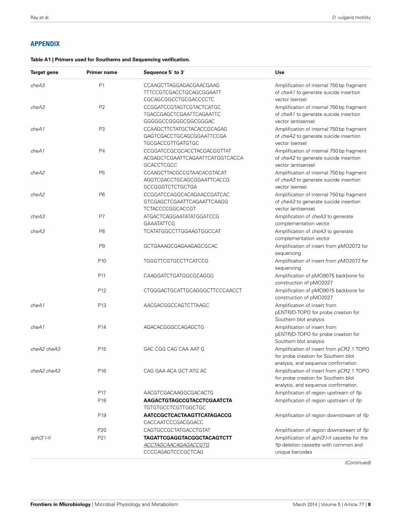
Table A1 | Primers used for Southerns and Sequencing verification.
Target gene Primer name Sequence 5′ to 3′ Use
cheA3 P1 CCAAGCTTAGGAGACGAACGAAGTTTCCGTCGACCTGCAGCGGAATTCGCAGCGGCCTGCGACCCCTC
Amplification of internal 750 bp fragmentof cheA1 to generate suicide insertionvector (sense)
cheA3 P2 CCGGATCCGTAGTCGTACTCATGCTGACCGAGCTCGAATTCAGAATTCGGGGGCCGGGGCGGCGGGAC
Amplification of internal 750 bp fragmentof cheA1 to generate suicide insertionvector (antisense)
cheA1 P3 CCAAGCTTCTATGCTACACCGCAGAGGAGTCGACCTGCAGCGGAATTCCGATGCGACCGTTGATGTGC
Amplification of internal 750 bp fragmentof cheA2 to generate suicide insertionvector (sense)
cheA1 P4 CCGGATCCGCGCACCTACGACGGTTATACGAGCTCGAATTCAGAATTCATGGTCACCAGCACCTCGCC
Amplification of internal 750 bp fragmentof cheA2 to generate suicide insertionvector (antisense)
cheA2 P5 CCAAGCTTACGCCGTAACACGTACATAGGTCGACCTGCAGCGGAATTCACCGGCCGGGTCTCTGCTGA
Amplification of internal 750 bp fragmentof cheA3 to generate suicide insertionvector (sense)
cheA2 P6 CCGGATCCAGGCACAGAACCGATCACGTCGAGCTCGAATTCAGAATTCAAGGTCTACCCCGGCACCGT
Amplification of internal 750 bp fragmentof cheA3 to generate suicide insertionvector (antisense)
cheA3 P7 ATGACTCAGGAATATATGGATCCGGAAATATTCG
Amplification of cheA3 to generatecomplementation vector
cheA3 P8 TCATATGGCCTTGGAAGTGGCCAT Amplification of cheA3 to generatecomplementation vector
P9 GCTGAAAGCGAGAAGAGCGCAC Amplification of insert from pMO2072 forsequencing
P10 TGGGTTCGTGCCTTCATCCG Amplification of insert from pMO2072 forsequencing
P11 CAAGGATCTGATGGCGCAGGG Amplification of pMO9075 backbone forconstruction of pMO2027
P12 CTGGGACTGCATTGCAGGGCTTCCCAACCT Amplification of pMO9075 backbone forconstruction of pMO2027
cheA1 P13 AACGACGGCCAGTCTTAAGC Amplification of insert frompENTR/D-TOPO for probe creation forSouthern blot analysis
cheA1 P14 AGACACGGGCCAGAGCTG Amplification of insert frompENTR/D-TOPO for probe creation forSouthern blot analysis
cheA2 cheA3 P15 GAC CGG CAG CAA AAT G Amplification of insert from pCR2.1 TOPOfor probe creation for Southern blotanalysis, and sequence confirmation.
cheA2 cheA3 P16 CAG GAA ACA GCT ATG AC Amplification of insert from pCR2.1 TOPOfor probe creation for Southern blotanalysis, and sequence confirmation.
P17 AACGTCGACAAGGCGACACTG Amplification of region upstream of flp
P18 AAGACTGTAGCCGTACCTCGAATCTA
TGTGTGCCTCGTTGGCTGCAmplification of region upstream of flp
P19 AATCCGCTCACTAAGTTCATAGACCG
CACCAATCCCGACGGACCAmplification of region downstream of flp
P20 CAGTGCCGCTATGACCTGTAT Amplification of region downstream of flp
aph(3 ′)-II P21 TAGATTCGAGGTACGGCTACAGTCTT
ACCTAGCAACAGAGACCGTGCCCCAGAGTCCCGCTCAG
Amplification of aph(3′)-II cassette for theflp deletion cassette with common andunique barcodes
(Continued)
Frontiers in Microbiology | Microbial Physiology and Metabolism March 2014 | Volume 5 | Article 77 | 8
Ray et al. D. vulgaris motility
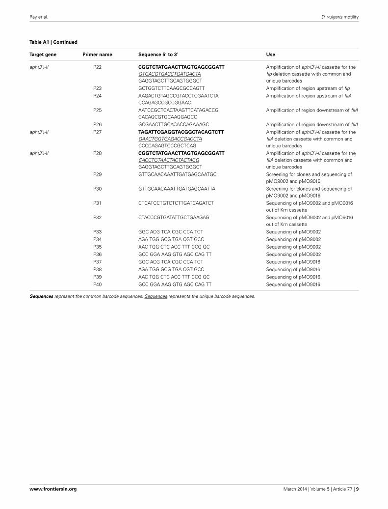
Table A1 | Continued
Target gene Primer name Sequence 5′ to 3′ Use
aph(3′)-II P22 CGGTCTATGAACTTAGTGAGCGGATT
GTGACGTGACCTGATGACTAGAGGTAGCTTGCAGTGGGCT
Amplification of aph(3′)-II cassette for theflp deletion cassette with common andunique barcodes
P23 GCTGGTCTTCAAGCGCCAGTT Amplification of region upstream of flp
P24 AAGACTGTAGCCGTACCTCGAATCTACCAGAGCCGCCGGAAC
Amplification of region upstream of fliA
P25 AATCCGCTCACTAAGTTCATAGACCGCACAGCGTGCAAGGAGCC
Amplification of region downstream of fliA
P26 GCGAACTTGCACACCAGAAAGC Amplification of region downstream of fliA
aph(3′)-II P27 TAGATTCGAGGTACGGCTACAGTCTT
GAACTGGTGAGACCGACCTACCCCAGAGTCCCGCTCAG
Amplification of aph(3′)-II cassette for thefliA deletion cassette with common andunique barcodes
aph(3′)-II P28 CGGTCTATGAACTTAGTGAGCGGATT
CACCTGTAACTACTACTAGGGAGGTAGCTTGCAGTGGGCT
Amplification of aph(3′)-II cassette for thefliA deletion cassette with common andunique barcodes
P29 GTTGCAACAAATTGATGAGCAATGC Screening for clones and sequencing ofpMO9002 and pMO9016
P30 GTTGCAACAAATTGATGAGCAATTA Screening for clones and sequencing ofpMO9002 and pMO9016
P31 CTCATCCTGTCTCTTGATCAGATCT Sequencing of pMO9002 and pMO9016out of Km cassette
P32 CTACCCGTGATATTGCTGAAGAG Sequencing of pMO9002 and pMO9016out of Km cassette
P33 GGC ACG TCA CGC CCA TCT Sequencing of pMO9002
P34 AGA TGG GCG TGA CGT GCC Sequencing of pMO9002
P35 AAC TGG CTC ACC TTT CCG GC Sequencing of pMO9002
P36 GCC GGA AAG GTG AGC CAG TT Sequencing of pMO9002
P37 GGC ACG TCA CGC CCA TCT Sequencing of pMO9016
P38 AGA TGG GCG TGA CGT GCC Sequencing of pMO9016
P39 AAC TGG CTC ACC TTT CCG GC Sequencing of pMO9016
P40 GCC GGA AAG GTG AGC CAG TT Sequencing of pMO9016
Sequences represent the common barcode sequences. Sequences represents the unique barcode sequences.
www.frontiersin.org March 2014 | Volume 5 | Article 77 | 9
Related Documents