Experimentally Approaching the Solvent-Accessible Surface Area of a Protein: Insights into the Acid Molten Globule of Bovine α-Lactalbumin Patricio O. Craig†, Gabriela E. Gómez†, Daniela B. Ureta†, Julio J. Caramelo† and José M. Delfino⁎† Departamento de Química Biológica, Facultad de Farmacia y Bioquímica, Universidad de Buenos Aires e Instituto de Química y Fisicoquímica Biológica (Consejo Nacional de Investigaciones Científicas y Técnicas), Junín 956, C1113AAD Buenos Aires, Argentina Received 18 June 2009; received in revised form 21 August 2009; accepted 25 September 2009 Available online 1 October 2009 Each conformational state of a protein is inextricably related to a defined extent of solvent exposure that plays a key role in protein folding and protein interactions. However, accurate measurement of the solvent-accessible surface area (ASA) is difficult for any state other than the native (N) state. We address this fundamental physicochemical parameter through a new experimental approach based on the reaction of the photochemical reagent diazirine (DZN) with the polypeptide chain. By virtue of its size, DZN is a reasonable molecular mimic of aqueous solvent. Here, we structurally characterize nonnative states of the paradigmatic protein α-lactalbumin. Covalent tagging resulting from unspecific methylene (:CH 2 ) reaction allows one to obtain a global estimate of ASA and to map out solvent accessibility along the amino acid sequence. By its mild apolar nature, DZN also reveals a hydrophobic phase in the acid-stabilized state of α-lactalbumin, in which there is clustering of core residues accessible to the solvent. In a fashion reminiscent of the N state, this acid-stabilized state also exhibits local regions where increased :CH 2 labeling indicates its nonhomogenous nature, likely pointing to the existence of packing defects. By contrast, the virtual absence of a defined long-range organization brings about a featureless labeling pattern for the unfolded state. Overall, :CH 2 labeling emerges as a fruitful technique that is able to quantify the ASA of the polypeptide chain, thus probing conformational features such as the outer exposed surface and inner cavities, as well as revealing the existence of noncompact apolar phases in nonnative states. © 2009 Elsevier Ltd. All rights reserved. Edited by K. Kuwajima Keywords: diazirine; protein folding; nonnative states; solvent-accessible surface area; α-lactalbumin *Corresponding author. E-mail address: [email protected]. †P.O.C., J.J.C. and J.M.D. are career investigators of the Consejo Nacional de Investigaciones Científicas y Técnicas (CONICET). G.E.G. was a recipient of a graduate student fellowship from the University of Buenos Aires (UBA) and is currently an Estenssoro fellow from the YPF Foundation. D.B.U. is professional technician of CONICET. Present addresses: P. O. Craig and J. J. Carmelo, Fundación Instituto Leloir e Instituto de Investigaciones Bioquímicas de Buenos Aires (Consejo Nacional de Investigaciones Científicas y Técnicas), Av. Patricias Argentinas 435, C1405BWE, Buenos Aires, Argentina. Abbreviations used: ASA, accessible surface area; N, native; DZN, diazirine; :CH 2 , methylene; U, unfolded; α-LA, α-lactalbumin; A, acid-stabilized; I, intermediate; [ 3 H]DZN, tritiated derivative of diazirine; CM α-LA, carbamidomethylated α-LA; ANS, 1-anilino-8-naphthalenesulfonic acid; HEWL, hen egg white lysozyme; GdnHCl, guanidine hydrochloride; TFA, trifluoroacetic acid; RP, reversed-phase; PDB, Protein Data Bank; CONICET, Consejo Nacional de Investigaciones Científicas y Técnicas. doi:10.1016/j.jmb.2009.09.058 J. Mol. Biol. (2009) 394, 982–993 Available online at www.sciencedirect.com 0022-2836/$ - see front matter © 2009 Elsevier Ltd. All rights reserved.

Welcome message from author
This document is posted to help you gain knowledge. Please leave a comment to let me know what you think about it! Share it to your friends and learn new things together.
Transcript
doi:10.1016/j.jmb.2009.09.058 J. Mol. Biol. (2009) 394, 982–993
Available online at www.sciencedirect.com
Experimentally Approaching the Solvent-AccessibleSurface Area of a Protein: Insights into the Acid MoltenGlobule of Bovine α-Lactalbumin
Patricio O. Craig†, Gabriela E. Gómez†, Daniela B. Ureta†,Julio J. Caramelo† and José M. Delfino⁎†
Departamento de QuímicaBiológica, Facultad de Farmaciay Bioquímica, Universidad deBuenos Aires e Instituto deQuímica y FisicoquímicaBiológica (Consejo Nacional deInvestigaciones Científicas yTécnicas), Junín 956,C1113AAD Buenos Aires,Argentina
Received 18 June 2009;received in revised form21 August 2009;accepted 25 September 2009Available online1 October 2009
*Corresponding author. E-mail add†P.O.C., J.J.C. and J.M.D. are care
(CONICET). G.E.G. was a recipientcurrently an Estenssoro fellow from
Present addresses: P. O. Craig andde Buenos Aires (Consejo NacionalBuenos Aires, Argentina.
Abbreviations used: ASA, accessiα-LA, α-lactalbumin; A, acid-stabilicarbamidomethylated α-LA; ANS, 1guanidine hydrochloride; TFA, trifluNacional de Investigaciones Científi
0022-2836/$ - see front matter © 2009 E
Each conformational state of a protein is inextricably related to a definedextent of solvent exposure that plays a key role in protein folding and proteininteractions. However, accurate measurement of the solvent-accessiblesurface area (ASA) is difficult for any state other than the native (N) state.We address this fundamental physicochemical parameter through a newexperimental approach based on the reaction of the photochemical reagentdiazirine (DZN) with the polypeptide chain. By virtue of its size, DZN is areasonable molecular mimic of aqueous solvent. Here, we structurallycharacterize nonnative states of the paradigmatic protein α-lactalbumin.Covalent tagging resulting from unspecific methylene (:CH2) reaction allowsone to obtain a global estimate of ASA and to map out solvent accessibilityalong the amino acid sequence. By its mild apolar nature, DZN also reveals ahydrophobic phase in the acid-stabilized state of α-lactalbumin, in whichthere is clustering of core residues accessible to the solvent. In a fashionreminiscent of the N state, this acid-stabilized state also exhibits local regionswhere increased :CH2 labeling indicates its nonhomogenous nature, likelypointing to the existence of packing defects. By contrast, the virtual absence ofa defined long-range organization brings about a featureless labeling patternfor the unfolded state. Overall, :CH2 labeling emerges as a fruitful techniquethat is able to quantify the ASA of the polypeptide chain, thus probingconformational features such as the outer exposed surface and inner cavities,as well as revealing the existence of noncompact apolar phases in nonnativestates.
© 2009 Elsevier Ltd. All rights reserved.
Keywords: diazirine; protein folding; nonnative states; solvent-accessiblesurface area; α-lactalbumin
Edited by K. Kuwajimaress: [email protected] investigators of the Consejo Nacional de Investigaciones Científicas y Técnicasof a graduate student fellowship from the University of Buenos Aires (UBA) and isthe YPF Foundation. D.B.U. is professional technician of CONICET.J. J. Carmelo, Fundación Instituto Leloir e Instituto de Investigaciones Bioquímicas
de Investigaciones Científicas y Técnicas), Av. Patricias Argentinas 435, C1405BWE,
ble surface area; N, native; DZN, diazirine; :CH2, methylene; U, unfolded;zed; I, intermediate; [3H]DZN, tritiated derivative of diazirine; CM α-LA,-anilino-8-naphthalenesulfonic acid; HEWL, hen egg white lysozyme; GdnHCl,oroacetic acid; RP, reversed-phase; PDB, Protein Data Bank; CONICET, Consejocas y Técnicas.
lsevier Ltd. All rights reserved.
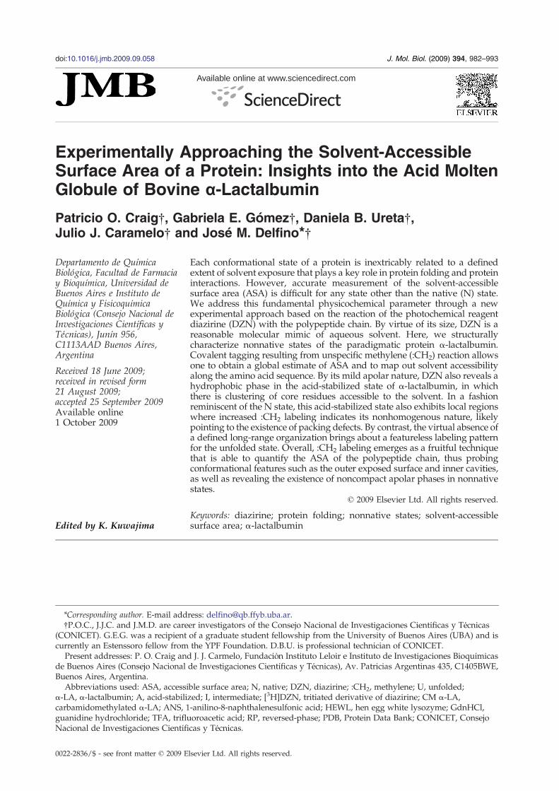
Scheme 1. Photolysis of DZN.
983The Acid Molten Globule of Bovine α-Lactalbumin
Introduction
The polypeptide chain adopts various conforma-tions across a funneled energy landscape en route tothe native (N) state.1 However, not every conforma-tion is likely to be significantly populated; therefore,nonnative states represent a limited repertoire offolded forms that can sometimes be stabilized underappropriate conditions.2,3 There is much to belearned about the folding process by a detailedcharacterization of these partially folded forms forwhich scant information is currently available.Inextricably associated with the events underlying
protein folding or oligomerization is the fact that theaccessible surface area (ASA) of the polypeptidechain becomes minimized. Nevertheless, despitebeing of great theoretical interest,4,5 there is hardlyany experimental method suitable for approaching adirect measurement of ASA. In this regard, para-meters such as ΔCp,
6–9 m value,10 and ΔRg,11 mea-
sured along conformational transitions, are thoughtto be correlated to changes in the accessibility of thewhole polypeptide chain, but do not directly yieldinformation along the protein sequence. In a differ-ent vein, 1H/2H exchange of amide protons12–14 is apowerful technique used to experimentally assesssolvent exposure, but data are intrinsically limited toamide protons belonging to the backbone chain,therefore becoming strongly dependent on second-ary-structure integrity. Besides, one main drawbackarises from the labile nature of the label, a factor thatgreatly restricts further analytical processing of thesample.An alternative approach aims at a promiscuous
modification of the protein surface by chemicalmeans. Such an approach would expand the set oftargets to all solvent-accessible sites, while concur-rently obviating chemical selectivity as much aspossible, thus generating a map of the topographyof the folded chain. Known ‘footprinting’ reagentsbelong to this class. A prominent example is thehydroxyl radical (·OH), which has been successfullyused to explore the structure and interactionsinvolving nucleic acids15–17 and proteins.18–21 How-ever, the ·OH species shows some chemical selec-tivity in its reaction with peptide targets, thuspotentially biasing the analysis to particular spotsalong the structure. The disruption of the polypep-tide chain gives rise to products that are generallystable; however, most often, radical-triggered reac-tions generate ill-characterized derivatives that com-plicate ensuing analytical procedures. A related—butessentially different—approach relies on the applica-tion of electron-deficient species, such as nitrenes orcarbenes, for the modification reaction.22,23 For theachievement of as close amimic of thewatermoleculeas geometrically possible, methylene (:CH2), thesmallest and most reactive member of this family,emerges as a natural candidate.24,25 :CH2 insertsreadily into any X–H bond (X: C, O, N, or S),generating methylated products that are minimallydifferent in their physicochemical character from theunreacted counterparts (Scheme 1). The best source
of :CH2 is diazirine (DZN; CH2N2) because of theready solubility of this gas in aqueous solutionsand its chemical inertness (unless irradiated atλ∼320 nm26). Due to its extremely short half-life,:CH2will react with the target groups belonging to itsimmediate molecular cage. Therefore, the pattern oflabeled sites represents the distribution of DZN priorto the photolysis event. In regard to geometry (sizeand shape), DZN surveys essentially the sameaccessible surface probed by the water molecule.In our laboratory, we have advanced the :CH2
labeling technique to address the unfolding transi-tions of single-domain proteins25,27 and the contactsurface of an antigen–antibody complex.28 Otherresearchers have also applied this method to theconformational study of a heterotrimeric multido-main protein.29
In this work, we show how we can exploit thecapabilities of :CH2 labeling to explore the structureof a partially folded form of a model protein and todraw comparisons with the N state and the unfolded(U) state. The molten globule state of bovine α-lactalbumin (α-LA) obtained at acidic pH [acid-stabilized (A) state] represents a paradigmaticsystem that has been characterized by severalbiochemical and biophysical methods.2,3,30 Accuratequantification of the :CH2 labeling yield along theprotein sequence and its correlation with the extentand nature of the ASA allow us to derive insightsinto the fine structure of this nonnative state.
Results and Discussion
Conformational-dependent :CH2 labeling of α-LA
α-LA was labeled in the N, U, or intermediate (I)state by reaction with [3H]methylene produced byphotolysis of the tritiated derivative of DZN ([3H]DZN) (Table 1). The use of the 3H label allows themeasurement of the extent of the reaction even atvery low modification levels. Under this condition,the modified protein is mostly monolabeled, mini-mizing any possible conformational perturbationand limiting the heterogeneity of the products.Under all conditions known to populate the Ustate, the :CH2 labeling yield is uniformly higherthan that observed for the N state, in agreement withthe expected larger solvent exposure of the poly-peptide chain. The increment is the same, regardlessof the nature of the denaturant used or the pH value,a corollary of the indiscriminate chemical reactivityof the :CH2 species. On the other hand, reduction of
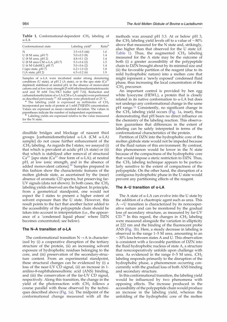
Table 1. Conformational-dependent :CH2 labeling ofα-LA
Conformational state Labeling yielda Ratiob
N 3.5±0.5 (44) 1.0U (8 M urea, pH 7) 5.0±0.3 (8) 1.4U (8 M urea, pH 2) 4.8±0.1 (2) 1.4U (8 M urea CM α-LA, pH 7) 5.3±0.4 (2) 1.5U (6 M GdnHCl, pH 7) 5.0±0.6 (2) 1.4I (apo state, pH 7) 6.2±1.0 (2) 1.8I (A state, pH 2) 6.5±0.2 (6) 1.9
Samples of α-LA were incubated under strong denaturingconditions (U state), at pH 2 (A state), or in the apo state (Ca2+
depleted) stabilized at neutral pH, in the absence of monovalentcations and at low ionic strength [5mMethylenediaminetetraaceticacid and 50 mM Tris/HCl buffer (pH 7.0)]. Reduction andcarbamidomethylation ofα-LA (CMα-LA sample)wereperformedas described previously.31 All samples were photolysed at 25 °C.
a The labeling yield is expressed as millimoles of CH2incorporated per mole of protein at 1 mM [3H]DZN concentration.Values are expressed as mean±standard deviation. The values inparentheses indicate the number of independent experiments.
b Labeling yields are expressed relative to the value measuredfor the N state.
984 The Acid Molten Globule of Bovine α-Lactalbumin
disulfide bridges and blockage of nascent thiolgroups [carbamidomethylated α-LA (CM α-LA)sample] do not cause further enhancement of the:CH2 labeling. As regards the I states, we assayed (i)that which is prevalent at acidic pH (A state) or (ii)that which is stabilized after removal of structuralCa2+ [apo state (Ca2+-free form of α-LA); at neutralpH, at low ionic strength, and in the absence ofadded monovalent cations].32 Samples prepared inthis fashion show the characteristic features of themolten globule state, as ascertained by the (near)absence of aromatic CD spectra, but preserving far-UV signals (data not shown). In both cases, the :CH2labeling yields observed are the highest. In principle,from a geometrical standpoint, one would notexpect the I states to present a higher extent ofsolvent exposure than the U state. However, thisresult points to the fact that another factor added tothe accessibility of the polypeptide chain should betaken into account in interpretation (i.e., the appear-ance of a ‘condensed liquid phase’ where DZNbecomes concentrated; see below).
The N–A transition of α-LA
The conformational transition N→A is character-ized by (i) a cooperative disruption of the tertiarystructure of the protein, (ii) an increasing solventexposure of hydrophobic residues belonging to thecore, and (iii) preservation of the secondary-struc-ture content. From an experimental standpoint,these structural changes can be evidenced by (i) aloss of the near-UV CD signal, (ii) an increase in 1-anilino-8-naphthalenesulfonic acid (ANS) binding,and (iii) the conservation of the far-UV CD signal,respectively. Along this transition, the change in theyield of the photoreaction with :CH2 follows acourse parallel with those observed by the techni-ques described above (Fig. 1a). The midpoint of theconformational change measured with all the
methods was around pH 3.3. At or below pH 3,the :CH2 labeling yield levels off to a value of ∼80%above that measured for the N state and, strikingly,also higher than that observed for the U state (cf.Table 1). Thus, the augmented :CH2 labelingmeasured for the A state may be the outcome ofboth (i) a greater accessibility of the polypeptidechain to DZN brought about by its minimal size and(ii) the favorable partition of the reagent (due to itsmild hydrophobic nature) into a molten core thatmight represent a ‘newly exposed’ condensed fluidphase, thus increasing the local concentration of the:CH2 precursor.An important control is provided by hen egg
white lysozyme (HEWL), a protein that is closelyrelated in its native conformation to α-LA but doesnot undergo any conformational change in the samepH range.33 Consistently, no significant change inthe :CH2 labeling yield occurs (Fig. 1a, inset), thusdemonstrating that pH bears no direct influence onthe chemistry of the labeling reaction. This observa-tion guarantees that differences in the extent oflabeling can be safely interpreted in terms of theconformational characteristics of the protein.Partition of DZN into the hydrophobic core of the
molten globule state would not be impeded becauseof the fluid nature of this environment. By contrast,this phenomenon would be lower in the N statebecause of the compactness of the hydrophobic corethat would impose a steric restriction to DZN. Thus,the :CH2 labeling technique appears to be particu-larly sensitive to the extent of compactness of thepolypeptide. On the other hand, the disruption of acontiguous hydrophobic phase in the U state wouldprevent any partitioning effects of the reagent.
The A–U transition of α-LA
The A state of α-LA can evolve into the U state bythe addition of a chaotropic agent such as urea. ThisA→U transition is characterized by its noncooper-ative nature and can be monitored by the gradualloss of secondary structure, as measured by far-UVCD.34 In this regard, the changes in :CH2 labelingwere measured alongside the variation in ellipticityat 222 nm and the binding of the fluorescent probeANS (Fig. 1b). Here, a steady decrease in labeling isobserved in the range 1–5 M urea, amounting to an∼30% loss between states A and U. This observationis consistent with a favorable partition of DZN intothe fluid hydrophobic nucleus of state A, a structurethat noncooperatively unfolds upon challenge withurea. As evidenced in the range 0–5 M urea, :CH2labeling responds primarily to the disruption of thehydrophobic phase, a phenomenon occurring con-currently with the gradual loss of both ANS bindingand secondary structure.In this conformational transition, the labeling yield
would be influenced by two phenomena withopposing effects. The increase produced in theaccessibility of the polypeptide chainwould producean increase in the labeling yield, whereas theunfolding of the hydrophobic core of the molten
Fig. 1. Conformational transi-tions of α-LA. α-LA (0.14 mM)was photolabeled with [3H]DZN(0.35 mM [3H]DZN, 0.5 mCi/mmol) in buffer A adjusted todifferent pH values [pH 2–7; (a)the N→A transition] or adjusted topH 2 and added with urea up todifferent final concentrations [0–8 M; (b) the A→U transition] andfurther processed as indicated inMaterials and Methods. The extentof reaction was expressed as theaverage number of moles of CH2per mole of protein at 1 mM [3H]DZN (black filled circles). Themolar ellipticity measured at222 nm (gray filled square) and270 nm (black open circles) and thefluorescence intensity at 470 nmassociated with ANS binding tothe protein (black open triangles)are also plotted. The inset in (a)shows the :CH2 labeling yield ofHEWL (open circles) side by sidewith that measured for α-LA (filledcircles) as a function of pH.
985The Acid Molten Globule of Bovine α-Lactalbumin
globule would decrease [3H]DZN binding to theprotein and its contribution to the labeling yield. Weobserved a decrease in the labeling yield of theprotein as a function of urea concentration. This doesnot rule out the existence of an accessibilityincrement but indicates that this phenomenon iscoupled to the unfolding of the hydrophobic core35,36and that this last effect dominates the change in thelabeling yield observed during this transition.In the range 5–7 M urea, the invariance of the
labeling yield (i) parallels the virtual disappearanceof any fluorescence signal due to bound ANS, sup-porting the absence of any hydrophobic phase in thiscondition, and (ii) does not accompany the observedfurther decrease in ellipticity, indicating that loss ofsecondary structure by itself does not influencemuch the solvent accessibility of the polypeptidechain. This goes in line with the fact that the overallexposure of the polypeptide chain is dominated bythe exposure of the side chains of amino acids,regardless of the kind of organization imposed onthe backbone (ASA calculations not shown).
Peptide analysis of α-LA labeled in the N, A, orU state
To extract site-specific structural information, wemeasured the radioactivity associated with tryptic
peptides derived from α-LA modified under exper-imental conditions where the N, A, or U statebecomes populated (Fig. 2). This protease is anadvantageous choice because the mean peptidelength (∼10 amino acids) allows for adequateaveraging over the chemical composition, providinga useful window for discriminating variations dueto conformational features rather than variationsdue to specific chemistry (i.e., avoiding the slightpreference of :CH2 for nucleophilic groups).24,37Thus, we obtained a collection of 11 nonoverlappingpeptides that span ∼93% of the protein sequence.In a scenario dominated by the unspecific reaction
of the :CH2 species with the polypeptide chain, fordifferences in the labeling yield of each peptide to beevaluated, results should be expressed as the labelincorporated per mass of peptide (mol CH2/gpeptide; alternatively, one could have chosen toexpress results per mole of amino acid). In thisfashion, labeling data will become independent ofpeptide length. After this normalization, differencesin the extent of reaction at a given spot due to aparticular microenvironment will become evident.Proof of the unspecific behavior of the labelingevent is that all peptides become labeled for anyconformational state being considered. For peptidesderived from the U state, an almost featureless pat-tern, showing a constant level of methylation (3.4±
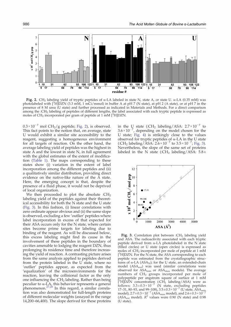
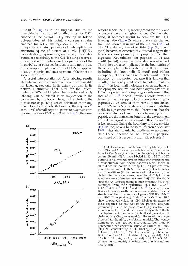
Fig. 3. Correlation plot between :CH2 labeling yieldand ASA. The radioactivity associated with each trypticpeptide derived from α-LA photolabeled in the N state(filled circles) or U state (open circles) is expressed asmoles of :CH2 incorporated per mole of peptide at 1 mM[3H]DZN. For the N state, the ASA corresponding to eachpeptide was estimated from the crystallographic struc-tures of α-LA (ASAN); for the U state, an extended-chainmodel (ASAext) was used (similar correlations wereobserved for ASAmin or ASAmax models). The averagenumbers of CH2 groups incorporated per mole ofpolypeptide per angstrom square of surface at 1 mM[3H]DZN concentration (:CH2 labeling/ASA) were asfollows: 3.3 ±0.3×10− 7 (N state, excluding peptides17–31, 80–93, and 99–108), 3.5±0.3×10⁻⁷ (U state, ASAminmodel), 2.7±0.3×10⁻⁷ (ASAmax model), and 2.6±0.3×10⁻⁷(ASAext model). R2 values were 0.90 (N state) and 0.98(U state).
Fig. 2. CH2 labeling yield of tryptic peptides of α-LA labeled in state N, state A, or state U. α-LA (0.35 mM) wasphotolabeled with [3H]DZN (3.3 mM, 1 mCi/mmol) in buffer A at pH 7 (N state), at pH 2 (A state), or at pH 7 in thepresence of 8 M urea (U state) and further processed as indicated in Materials and Methods. For a direct comparisonamong the :CH2 labeling of peptides of different lengths, the label associated with each tryptic peptide is expressed asmoles of CH2 incorporated per gram of peptide at 1 mM [3H]DZN.
986 The Acid Molten Globule of Bovine α-Lactalbumin
0.3×10⁻⁷ mol CH2/g peptide; Fig. 2), is observed.This fact points to the notion that, on average, stateU would exhibit a similar site accessibility to thereagent, suggesting a homogeneous environmentfor all targets of reaction. On the other hand, theaverage labeling yield of peptides was the highest instate A and the lowest in state N, in full agreementwith the global estimates of the extent of modifica-tion (Table 1). The maps corresponding to thesestates show (i) variation in the extent of labelincorporation among the different peptides and (ii)a qualitatively similar distribution, providing directevidence on the native-like nature of the A state.Here, the emerging concept is that, despite thepresence of a fluid phase, it would not be deprivedof local organization.We then proceeded to plot the absolute :CH2
labeling yield of the peptides against their theoret-ical accessibility for both the N state and the U state(Fig. 3). In this fashion, (i) linear correlations withzero ordinate appear obvious and (ii) the same slopeis observed, excluding a few ‘outlier’ peptides wherelabel incorporation in excess of that expected fortheir ASA occurs only for the N state, where certainsites become prime targets for labeling due tobinding of the reagent. As will be discussed below,this excess labeling might find its cause in theinvolvement of these peptides in the boundary ofcavities amenable to lodging the reagent DZN, thusprolonging its residence time and therefore increas-ing the yield of reaction. A contrasting picture arisesfrom the same analysis applied to peptides derivedfrom the protein labeled in the U state, where no‘outlier’ peptides appear, as expected from the‘equalization’ of the microenvironments for thereaction, leaving the collisional factor as the onlyone influencing the :CH2 labeling. Rather than beingpeculiar to α-LA, this behavior represents a generalphenomenon.27,28 In this regard, a similar correla-tion was also demonstrated for full-length proteinsof different molecular weights (assayed in the range14,200–66,400). The slope derived for these proteins
in the U state (:CH2 labeling/ASA: 2.7×10⁻⁷ to3.6×10−7, depending on the model chosen for theU state; Fig. 4) is strikingly close to the valuesobserved for tryptic peptides of α-LA in the U state(:CH2 labeling/ASA: 2.6×10⁻⁷ to 3.5×10⁻⁷; Fig. 3).Nevertheless, the slope of the same set of proteinslabeled in the N state (:CH2 labeling/ASA: 5.8±
987The Acid Molten Globule of Bovine α-Lactalbumin
0.7 × 10− 7; Fig. 4) is the highest, due to theunavoidable inclusion of binding sites for DZNenhancing the overall :CH2 labeling in foldedpolypeptides. At this point, a consensus valueemerges for :CH2 labeling/ASA (∼3×10⁻⁷ CH2groups incorporated per mole of polypeptide perangstrom square of surface at 1 mM [3H]DZNconcentration), representing exclusively the contri-bution of accessibility to the :CH2 labeling observed.It is important to underscore the significance of thelinear behavior observed because it validates the useof the unspecific photoreaction of DZN to approx-imate an experimental measurement of the extent ofsolvent exposure.A useful interpretation of :CH2 labeling results
stems from the consideration of the surface availablefor labeling, not only in its extent but also in itsnature. Distinctive ‘host’ sites for the ‘guest’molecule DZN, which give rise to enhanced :CH2labeling, can be related to its implication in thecondensed hydrophobic phase, not excluding thepersistence of packing defects (cavities). A predic-tion of local hydrophobicity based on the sequence45
at the level of small peptides points to twomain sites(around residues 17–31 and 93–108; Fig. 5), the same
Fig. 4. Correlation plot between :CH2 labeling yieldand ASA. α-LA, bovine growth hormone, β-lactamasefrom Bacillus licheniformis, ovalbumin (OVA), and bovineserum albumin (BSA) were labeled in 40 mM Tris/HClbuffer (pH 7.4), whereas trypsin from bovine pancreas andα-chymotrypsin from bovine pancreas were labeled in40 mM sodium acetate buffer (pH 4). All proteins werephotolabeled under both N conditions (a; black circles)and U conditions (in the presence of 8 M urea) (b; graycircles). Results are expressed as moles of :CH2 incorpo-rated per mole of protein at 1 mM [3H]DZN. For the Nstate, the ASA corresponding to each protein (ASAN) wasestimated from their structures: PDB IDs 1OVA,38
4BLM,39 4CHA,40 1TLD,41 and 1F6S.42 The structures ofBSA and bovine growth hormone were modeled from thestructure of their human homologues (PDB IDs 1GNI43
and 1HGU,44 respectively). In the N state, OVA and BSAshow anomalous values of :CH2 labeling (in excess ofthose reported for the rest of the proteins assayed),presumably due to the presence of highly reactive thiolgroups in the former and the known ability of the latter tobind hydrophobic molecules. For the U state, an extended-chain model (ASAext) was used (similar correlations wereobserved for the ASAmin or ASAmax models). The averagenumbers of CH2 groups incorporated per mole ofpolypeptide per angstrom square of surface at 1 mM[3H]DZN concentration (:CH2 labeling/ASA) were asfollows: 5.8±0.7×10⁻⁷ (N state, excluding OVA andBSA), 3.6±0.4×10− 7 (U state, ASAmin model), 2.7±0.3×10⁻⁷ (U state, ASAmax model), and 2.7±0.3×10⁻⁷
(U state, ASAext model). R2 values were 0.79 (N state) and0.98 (U state).
regions where the :CH2 labeling yield for the N andA states shows the highest values. On the otherhand, it becomes useful to compare the U/Nlabeling ratio (Table 2) with surface data derivedfrom the known structure of the protein (Fig. 6a).The :CH2 labeling of most peptides (Fig. 6b, blue orcyan) behaves as expected of a general reagent thatlabels surfaces primarily in proportion to theirexposure. Nevertheless, for peptides 17–31 and99–108 (in red), a very low correlation was observed.These sites are also implicated in the boundaries ofthe only empty cavities located in the flexible regionincluding the loop/helix D motif (red blobs).Occupancy of these voids with DZN would not beimpeded by the protein because it is known thatbreathing motions permit access to molecules of thissize.49,50 In fact, small molecules such as methane orcyclopropane occupy two homologous cavities inHEWL, a protein with a topology closely resemblingthat of α-LA.51 Indeed, previous work from ourlaboratory lends support to this contention: trypticpeptides 74–96 derived from HEWL photolabeledwith DZN in its N state show an enhanced labelingyield, in agreement with the observation that thebackbone atoms of amino acids belonging to thispeptide are themain contributors to the environmentaround the largest cavity present in this protein.28 Inα-LA, residues lining the boundary of these cavities(Fig. 6b, red) belong to the so-called aromatic clusterI42,46—sites that would be predicted to accommo-date DZN—because of the favorable partitioncoefficient of this reagent in aromatic solvents.24
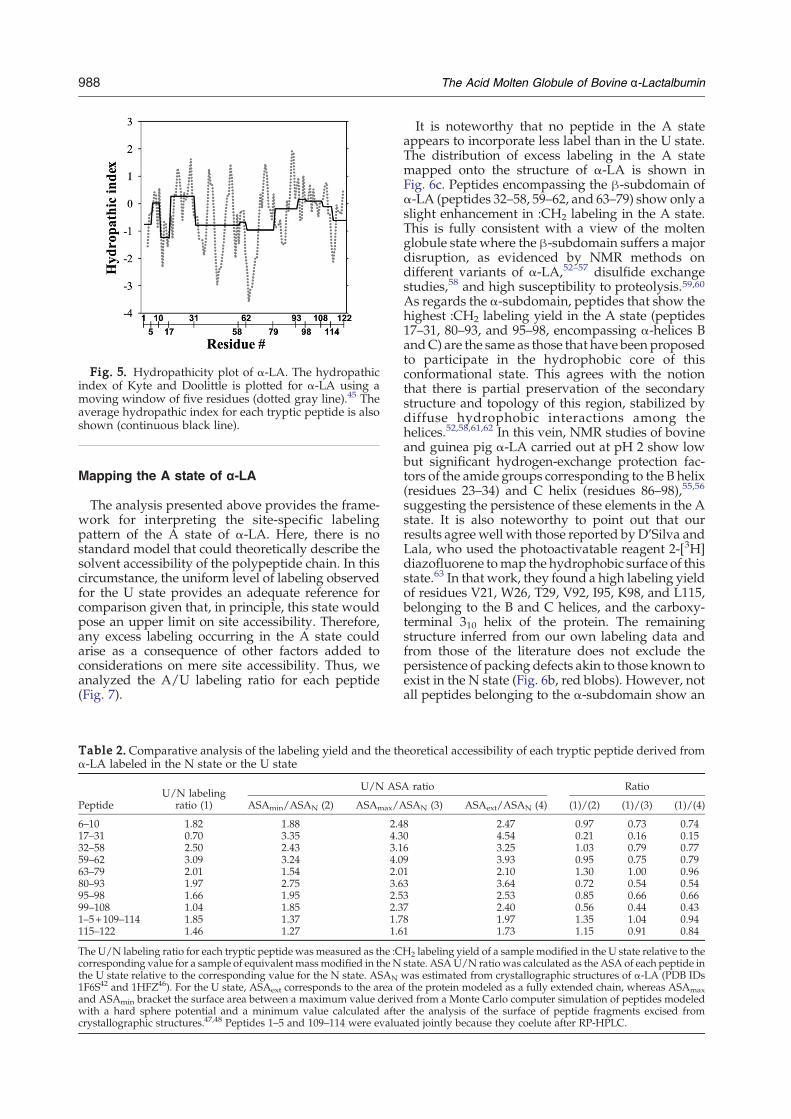
Fig. 5. Hydropathicity plot of α-LA. The hydropathicindex of Kyte and Doolittle is plotted for α-LA using amoving window of five residues (dotted gray line).45 Theaverage hydropathic index for each tryptic peptide is alsoshown (continuous black line).
988 The Acid Molten Globule of Bovine α-Lactalbumin
Mapping the A state of α-LA
The analysis presented above provides the frame-work for interpreting the site-specific labelingpattern of the A state of α-LA. Here, there is nostandard model that could theoretically describe thesolvent accessibility of the polypeptide chain. In thiscircumstance, the uniform level of labeling observedfor the U state provides an adequate reference forcomparison given that, in principle, this state wouldpose an upper limit on site accessibility. Therefore,any excess labeling occurring in the A state couldarise as a consequence of other factors added toconsiderations on mere site accessibility. Thus, weanalyzed the A/U labeling ratio for each peptide(Fig. 7).
Table 2. Comparative analysis of the labeling yield and the thα-LA labeled in the N state or the U state
PeptideU/N labeling
ratio (1)
U/N AS
ASAmin/ASAN (2) ASAmax/A
6–10 1.82 1.88 2.417–31 0.70 3.35 4.332–58 2.50 2.43 3.159–62 3.09 3.24 4.063–79 2.01 1.54 2.080–93 1.97 2.75 3.695–98 1.66 1.95 2.599–108 1.04 1.85 2.31–5+109–114 1.85 1.37 1.7115–122 1.46 1.27 1.6
The U/N labeling ratio for each tryptic peptide was measured as the :Ccorresponding value for a sample of equivalent massmodified in the Nthe U state relative to the corresponding value for the N state. ASAN1F6S42 and 1HFZ46). For the U state, ASAext corresponds to the area oand ASAmin bracket the surface area between a maximum value derivwith a hard sphere potential and a minimum value calculated aftecrystallographic structures.47,48 Peptides 1–5 and 109–114 were evalua
It is noteworthy that no peptide in the A stateappears to incorporate less label than in the U state.The distribution of excess labeling in the A statemapped onto the structure of α-LA is shown inFig. 6c. Peptides encompassing the β-subdomain ofα-LA (peptides 32–58, 59–62, and 63–79) show only aslight enhancement in :CH2 labeling in the A state.This is fully consistent with a view of the moltenglobule state where the β-subdomain suffers a majordisruption, as evidenced by NMR methods ondifferent variants of α-LA,52–57 disulfide exchangestudies,58 and high susceptibility to proteolysis.59,60As regards the α-subdomain, peptides that show thehighest :CH2 labeling yield in the A state (peptides17–31, 80–93, and 95–98, encompassing α-helices BandC) are the same as those that have been proposedto participate in the hydrophobic core of thisconformational state. This agrees with the notionthat there is partial preservation of the secondarystructure and topology of this region, stabilized bydiffuse hydrophobic interactions among thehelices.52,58,61,62 In this vein, NMR studies of bovineand guinea pig α-LA carried out at pH 2 show lowbut significant hydrogen-exchange protection fac-tors of the amide groups corresponding to the B helix(residues 23–34) and C helix (residues 86–98),55,56
suggesting the persistence of these elements in the Astate. It is also noteworthy to point out that ourresults agreewell with those reported byD'Silva andLala, who used the photoactivatable reagent 2-[3H]diazofluorene tomap the hydrophobic surface of thisstate.63 In that work, they found a high labeling yieldof residues V21, W26, T29, V92, I95, K98, and L115,belonging to the B and C helices, and the carboxy-terminal 310 helix of the protein. The remainingstructure inferred from our own labeling data andfrom those of the literature does not exclude thepersistence of packing defects akin to those known toexist in the N state (Fig. 6b, red blobs). However, notall peptides belonging to the α-subdomain show an
eoretical accessibility of each tryptic peptide derived from
A ratio Ratio
SAN (3) ASAext/ASAN (4) (1)/(2) (1)/(3) (1)/(4)
8 2.47 0.97 0.73 0.740 4.54 0.21 0.16 0.156 3.25 1.03 0.79 0.779 3.93 0.95 0.75 0.791 2.10 1.30 1.00 0.963 3.64 0.72 0.54 0.543 2.53 0.85 0.66 0.667 2.40 0.56 0.44 0.438 1.97 1.35 1.04 0.941 1.73 1.15 0.91 0.84
H2 labeling yield of a sample modified in the U state relative to thestate. ASAU/N ratio was calculated as the ASA of each peptide inwas estimated from crystallographic structures of α-LA (PDB IDsf the protein modeled as a fully extended chain, whereas ASAmaxed from a Monte Carlo computer simulation of peptides modeledr the analysis of the surface of peptide fragments excised fromted jointly because they coelute after RP-HPLC.
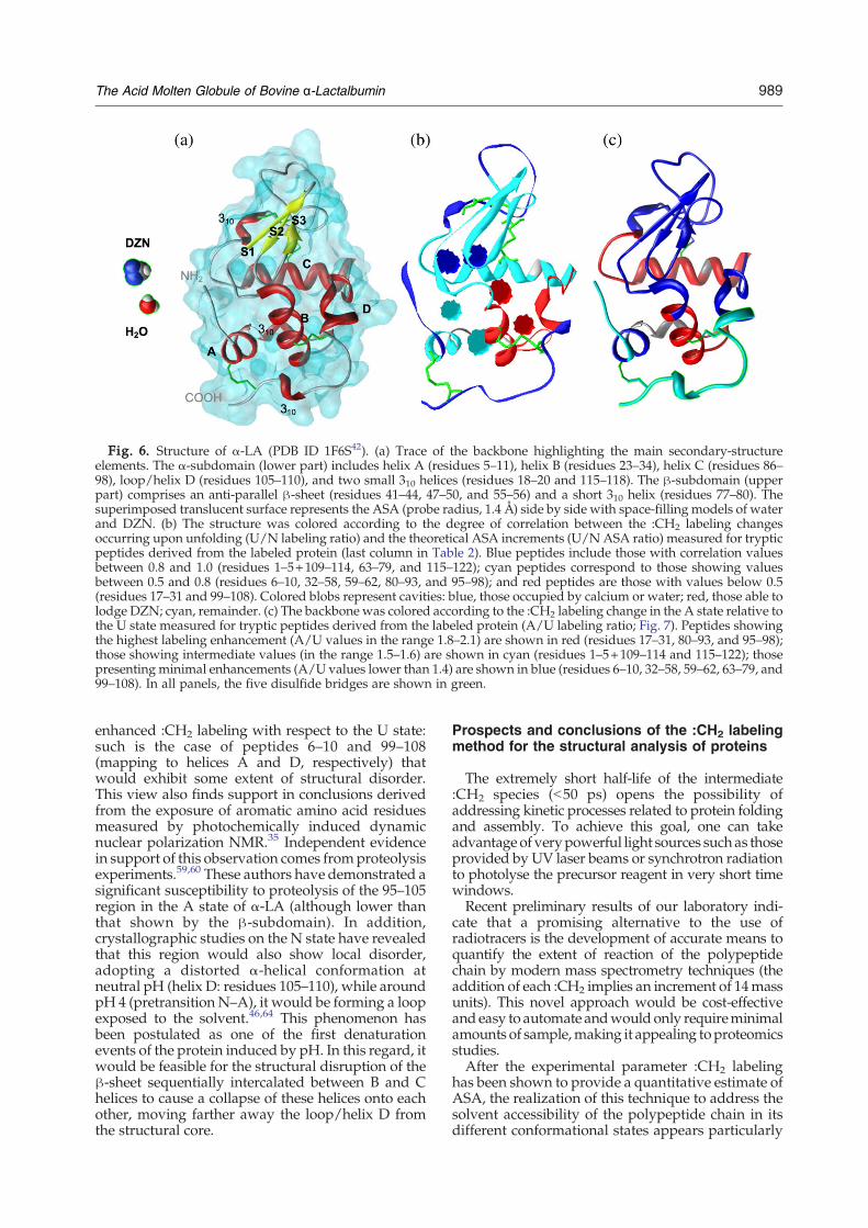
Fig. 6. Structure of α-LA (PDB ID 1F6S42). (a) Trace of the backbone highlighting the main secondary-structureelements. The α-subdomain (lower part) includes helix A (residues 5–11), helix B (residues 23–34), helix C (residues 86–98), loop/helix D (residues 105–110), and two small 310 helices (residues 18–20 and 115–118). The β-subdomain (upperpart) comprises an anti-parallel β-sheet (residues 41–44, 47–50, and 55–56) and a short 310 helix (residues 77–80). Thesuperimposed translucent surface represents the ASA (probe radius, 1.4 Å) side by side with space-filling models of waterand DZN. (b) The structure was colored according to the degree of correlation between the :CH2 labeling changesoccurring upon unfolding (U/N labeling ratio) and the theoretical ASA increments (U/N ASA ratio) measured for trypticpeptides derived from the labeled protein (last column in Table 2). Blue peptides include those with correlation valuesbetween 0.8 and 1.0 (residues 1–5+109–114, 63–79, and 115–122); cyan peptides correspond to those showing valuesbetween 0.5 and 0.8 (residues 6–10, 32–58, 59–62, 80–93, and 95–98); and red peptides are those with values below 0.5(residues 17–31 and 99–108). Colored blobs represent cavities: blue, those occupied by calcium or water; red, those able tolodge DZN; cyan, remainder. (c) The backbone was colored according to the :CH2 labeling change in the A state relative tothe U state measured for tryptic peptides derived from the labeled protein (A/U labeling ratio; Fig. 7). Peptides showingthe highest labeling enhancement (A/U values in the range 1.8–2.1) are shown in red (residues 17–31, 80–93, and 95–98);those showing intermediate values (in the range 1.5–1.6) are shown in cyan (residues 1–5+109–114 and 115–122); thosepresenting minimal enhancements (A/U values lower than 1.4) are shown in blue (residues 6–10, 32–58, 59–62, 63–79, and99–108). In all panels, the five disulfide bridges are shown in green.
989The Acid Molten Globule of Bovine α-Lactalbumin
enhanced :CH2 labeling with respect to the U state:such is the case of peptides 6–10 and 99–108(mapping to helices A and D, respectively) thatwould exhibit some extent of structural disorder.This view also finds support in conclusions derivedfrom the exposure of aromatic amino acid residuesmeasured by photochemically induced dynamicnuclear polarization NMR.35 Independent evidencein support of this observation comes fromproteolysisexperiments.59,60 These authors have demonstrated asignificant susceptibility to proteolysis of the 95–105region in the A state of α-LA (although lower thanthat shown by the β-subdomain). In addition,crystallographic studies on the N state have revealedthat this region would also show local disorder,adopting a distorted α-helical conformation atneutral pH (helix D: residues 105–110), while aroundpH 4 (pretransition N–A), it would be forming a loopexposed to the solvent.46,64 This phenomenon hasbeen postulated as one of the first denaturationevents of the protein induced by pH. In this regard, itwould be feasible for the structural disruption of theβ-sheet sequentially intercalated between B and Chelices to cause a collapse of these helices onto eachother, moving farther away the loop/helix D fromthe structural core.
Prospects and conclusions of the :CH2 labelingmethod for the structural analysis of proteins
The extremely short half-life of the intermediate:CH2 species (b50 ps) opens the possibility ofaddressing kinetic processes related to protein foldingand assembly. To achieve this goal, one can takeadvantage of verypowerful light sources suchas thoseprovided by UV laser beams or synchrotron radiationto photolyse the precursor reagent in very short timewindows.Recent preliminary results of our laboratory indi-
cate that a promising alternative to the use ofradiotracers is the development of accurate means toquantify the extent of reaction of the polypeptidechain by modern mass spectrometry techniques (theaddition of each :CH2 implies an increment of 14massunits). This novel approach would be cost-effectiveand easy to automate andwouldonly requireminimalamounts of sample,making it appealing toproteomicsstudies.After the experimental parameter :CH2 labeling
has been shown to provide a quantitative estimate ofASA, the realization of this technique to address thesolvent accessibility of the polypeptide chain in itsdifferent conformational states appears particularly
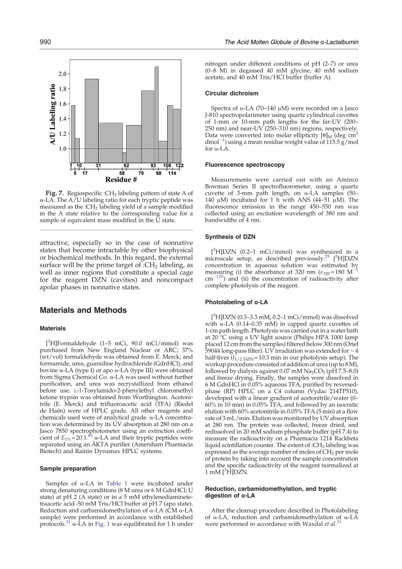
Fig. 7. Regiospecific :CH2 labeling pattern of state A ofα-LA. The A/U labeling ratio for each tryptic peptide wasmeasured as the :CH2 labeling yield of a sample modifiedin the A state relative to the corresponding value for asample of equivalent mass modified in the U state.
990 The Acid Molten Globule of Bovine α-Lactalbumin
attractive, especially so in the case of nonnativestates that become intractable by other biophysicalor biochemical methods. In this regard, the externalsurface will be the prime target of :CH2 labeling, aswell as inner regions that constitute a special cagefor the reagent DZN (cavities) and noncompactapolar phases in nonnative states.
Materials and Methods
Materials
[3H]Formaldehyde (1–5 mCi, 90.0 mCi/mmol) waspurchased from New England Nuclear or ARC; 37%(wt/vol) formaldehyde was obtained from E. Merck; andformamide, urea, guanidine hydrochloride (GdnHCl), andbovine α-LA (type I) or apo α-LA (type III) were obtainedfrom Sigma Chemical Co. α-LA was used without furtherpurification, and urea was recrystallized from ethanolbefore use. L-1-Tosylamido-2-phenylethyl chloromethylketone trypsin was obtained from Worthington. Acetoni-trile (E. Merck) and trifluoroacetic acid (TFA) (Riedelde Haën) were of HPLC grade. All other reagents andchemicals used were of analytical grade. α-LA concentra-tion was determined by its UV absorption at 280 nm on aJasco 7850 spectrophotometer using an extinction coeffi-cient of E1%=20.1.65 α-LA and their tryptic peptides wereseparated using an ÄKTA purifier (Amersham PharmaciaBiotech) and Rainin Dynamax HPLC systems.
Sample preparation
Samples of α-LA in Table 1 were incubated understrong denaturing conditions (8 M urea or 6 M GdnHCl; Ustate) at pH 2 (A state) or in a 5 mM ethylenediaminete-traacetic acid–50 mM Tris/HCl buffer at pH 7 (apo state).Reduction and carbamidomethylation of α-LA (CM α-LAsample) were performed in accordance with establishedprotocols.31 α-LA in Fig. 1 was equilibrated for 1 h under
nitrogen under different conditions of pH (2–7) or urea(0–8 M) in degassed 40 mM glycine, 40 mM sodiumacetate, and 40 mM Tris/HCl buffer (buffer A).
Circular dichroism
Spectra of α-LA (70–140 μM) were recorded on a JascoJ-810 spectropolarimeter using quartz cylindrical cuvettesof 1-mm or 10-mm path lengths for the far-UV (200–250 nm) and near-UV (250–310 nm) regions, respectively.Data were converted into molar ellipticity [θ]M (deg cm2
dmol−1) using amean residueweight value of 115.5 g/molfor α-LA.
Fluorescence spectroscopy
Measurements were carried out with an AmincoBowman Series II spectrofluorometer, using a quartzcuvette of 3-mm path length, on α-LA samples (50–140 μM) incubated for 1 h with ANS (44–51 μM). Thefluorescence emission in the range 450–550 nm wascollected using an excitation wavelength of 380 nm andbandwidths of 4 nm.
Synthesis of DZN
[3H]DZN (0.2–1 mCi/mmol) was synthesized in amicroscale setup, as described previously.25 [3H]DZNconcentration in aqueous solution was estimated bymeasuring (i) the absorbance at 320 nm (ɛ320=180 M−1
cm−125) and (ii) the concentration of radioactivity aftercomplete photolysis of the reagent.
Photolabeling of α-LA
[3H]DZN (0.3–3.3 mM, 0.2–1 mCi/mmol) was dissolvedwith α-LA (0.14–0.35 mM) in capped quartz cuvettes of1-cmpath length. Photolysiswas carried out in awater bathat 20 °C using a UV light source (Philips HPA 1000 lampplaced 12 cm from the samples) filteredbelow300 nm (Oriel59044 long-pass filter). UV irradiation was extended for∼4half-lives (t1/2 DZN=10.3 min in our photolysis setup). Theworkupprocedure consisted of addition of urea (up to 8M),followed by dialysis against 0.07 mMNa2CO3 (pH 7.5–8.0)and freeze drying. Finally, the samples were dissolved in6 M GdnHCl in 0.05% aqueous TFA, purified by reversed-phase (RP) HPLC on a C4 column (Vydac 214TP510),developed with a linear gradient of acetonitrile/water (0–60% in 10 min) in 0.05% TFA, and followed by an isocraticelutionwith 60% acetonitrile in 0.05% TFA (5min) at a flowrate of 3mL/min. Elutionwasmonitored byUVabsorptionat 280 nm. The protein was collected, freeze dried, andredissolved in 20 mM sodium phosphate buffer (pH 7.4) tomeasure the radioactivity on a Pharmacia 1214 Rackbetaliquid scintillation counter. The extent of :CH2 labeling wasexpressed as the average number of moles of CH2 per moleof protein by taking into account the sample concentrationand the specific radioactivity of the reagent normalized at1 mM [3H]DZN.
Reduction, carbamidomethylation, and trypticdigestion of α-LA
After the cleanup procedure described in Photolabelingof α-LA, reduction and carbamidomethylation of α-LAwere performed in accordance with Waxdal et al.31
991The Acid Molten Globule of Bovine α-Lactalbumin
Peptide mapping
Tryptic peptides derived from α-LA samples labeled inthe N, A, or U state were separated by size-exclusionchromatography on a Superdex Peptide HR 10/30 column(GE Healthcare) using a 4 M urea–50 mM Tris/HCl(pH 7.0) buffer at a flow rate of 0.5 mL/min. Elution wasmonitored by both UV absorption at 215 nm andradioactivity associated with fractions collected at 30-stime intervals. Collected peaks were further separated byRP-HPLC on a C18 column (Vydac 218TP54) using alinear gradient of acetonitrile/water (0–60% in 60 min) in0.05% TFA at a flow rate of 1 mL/min. Elution wasmonitored by UV absorption at 215 and 280 nm and by theradioactivity associated with fractions collected at 1-mintime intervals. Peptide identification was achieved byamino acid analysis on an Applied Biosystems 420A. The:CH2 labeling yield of peptides was calculated by the ratioof the radioactivity associated with each peak and theamount of peptide eluted.
Molecular modeling
The ASAs of α-LA and its peptides in the N and U stateswere calculated with MSP66 using a probe radius of 1.4 Å.The analysis of the N state was based on crystallographicstructures available for the wild-type protein [ProteinData Bank (PDB) ID 1F6S42] and for an M90V mutantvariant (PDB ID 1HFZ46), whereas that of the U state wasbased on an extended-chain model (ASAext) constructedwith MacroModel.67 Additional ASA estimates of the Ustate (ASAmax and ASAmin) were obtained through a Webresource‡.47,48 Cavities were calculated with MSP on thestructure PDB ID 1F6S (devoid of Ca2+ and water) using aprobe radius of 1.3 Å. Figure 6b and c were built withSwiss-PdbViewer68 and Chimera,69 respectively, runningon a personal computer under Windows XP. MacroModeland MSP were run on SGI workstations (Indigo XS24ZR4000 and O2 R10000 under Irix 5.3 or 6.5, respectively).
Acknowledgements
P.O.C., J.J.C., and J.M.D. are career investigators ofthe Consejo Nacional de Investigaciones Científicasy Técnicas (CONICET). G.E.G. was a recipient of agraduate student fellowship from the University ofBuenos Aires and is currently an Estenssoro fellowat the YPF Foundation. D.B.U. is a professionaltechnician at CONICET.This research has been supported by grants to J.M.
D. from the University of Buenos Aires, CONICET,and the Agencia Nacional de Promoción Científica yTecnológica.
References
1. Bryngelson, J. D., Onuchic, J. N., Socci, N. D. &Wolynes, P. G. (1995). Funnels, pathways, and theenergy landscape of protein folding: a synthesis.Proteins, 21, 167–195.
‡http://roselab.jhu.edu/utils/unfolded.html
2. Ptitsyn, O. B. (1995). Molten globule and proteinfolding. Adv. Protein Chem. 47, 83–229.
3. Arai, M. & Kuwajima, K. (2000). Role of the moltenglobule state in protein folding. Adv. Protein Chem. 53,209–282.
4. Lee, B. & Richards, F. M. (1971). The interpretation ofprotein structures: estimation of static accessibility.J. Mol. Biol. 55, 379–400.
5. Richards, F. M. (1977). Areas, volumes, packing andprotein structure.Annu. Rev. Biophys. Bioeng. 6, 151–176.
6. Makhatadze, G. I. & Privalov, P. L. (1990). Heatcapacity of proteins: I. Partial molar heat capacity ofindividual amino acid residues in aqueous solution:hydration effect. J. Mol. Biol. 213, 375–384.
7. Privalov, P. L. & Makhatadze, G. I. (1990). Heatcapacity of proteins: II. Partial molar heat capacity ofthe unfolded polypeptide chain of proteins: proteinunfolding effects. J. Mol. Biol. 213, 291–385.
8. Livingstone, J. R., Spolar, R. S. & Record, M. T., Jr(1991). Contribution to the thermodynamics of proteinfolding from the reduction in water-accessible nonpo-lar surface area. Biochemistry, 30, 4237–4244.
9. Loladze, V. V., Ermolenko, D. N. & Makhatadze, G. I.(2001). Heat capacity changes upon burial of polar andnonpolar groups in proteins. Protein Sci. 10, 1343–1352.
10. Myers, J. K., Pace, C. N. & Scholtz, J. M. (1995).Denaturant m values and heat capacity changes:relation to changes in accessible surface areas ofprotein unfolding. Protein Sci. 4, 2138–2148.
11. Geierhaas, C. D., Nickson, A. A., Lindorff-Larsen, K.,Clarke, J. & Vendruscolo, M. (2007). BPPred: a Web-based computational tool for predicting biophysicalparameters of proteins. Protein Sci. 16, 125–134.
12. Englander, S. W. & Krishna, M. M. G. (2001).Hydrogen exchange. Nat. Struct. Biol. 8, 741–742.
13. Truhlar, S. M., Croy, C. H., Torpey, J. W., Koeppe, J. R.& Komives, E. A. (2006). Solvent accessibility ofprotein surfaces by amide H/2H exchange MALDI-TOF mass spectrometry. J. Am. Soc. Mass Spectrom. 17,1490–1497.
14. Englander, S. W. (2006). Hydrogen exchange andmass spectrometry: a historical perspective. J. Am. Soc.Mass Spectrom. 17, 1481–1489.
15. Tullius, T. D. & Dombroski, B. A. (1985). Iron (II)EDTA used to measure the helical twist along anyDNA molecule. Science, 230, 679–681.
16. Tullius, T. D., Dombroski, B. A., Churchill, M. E. A. &Kam, L. (1989). Hydroxyl radical footprinting: a highresolution method for mapping protein–DNA con-tacts. In Recombinant DNA Methodology (Wu, R.,Grossman, L. & Moldave, K., eds), Academic Press,San Diego, CA.
17. Jain, S. S. & Tullius, T. D. (2008). Footprinting protein–DNA complexes using the hydroxyl radical. Nat.Protoc. 3, 1092–1100.
18. Maleknia, S. D., Brenowitz, M. & Chance, M. R. (1999).Millisecond radiolytic modification of peptides bysynchrotron X-rays identified by mass spectrometry.Anal. Chem. 71, 3965–3973.
19. Maleknia, S. D., Ralston, C. Y., Brenowitz, M. D.,Downard, K. M. & Chance, M. R. (2001). Determina-tion of macromolecular folding and structure by syn-chrotron X-ray radiolysis techniques. Anal. Biochem.289, 103–115.
20. Maleknia, S. D. &Downard, K.M. (2001). Unfolding ofapomyoglobin helices by synchrotron radiolysis andmass spectrometry. Eur. J. Biochem. 268, 5578–5588.
21. Takamoto, K. & Chance, M. R. (2006). Radiolyticprotein footprinting with mass spectrometry to probe
992 The Acid Molten Globule of Bovine α-Lactalbumin
the structure of macromolecular complexes. Annu.Rev. Biophys. Biomol. Struct. 35, 251–276.
22. Brunner, J. (1993). New photolabeling and cross-linking methods. Annu. Rev. Biochem. 62, 483–514.
23. Delfino, J. M., Schreiber, S. L. & Richards, F. M. (1993).Design, synthesis and properties of a photoactivatablemembrane-spanning phospholipidic probe. J. Am.Chem. Soc. 115, 3458–3474.
24. Richards, F. M., Lamed, R., Wynn, R., Patel, D. &Olack, G. (2000). Methylene as a possible universalfootprinting reagent that will include hydrophobicsurface areas: overview and feasibility: properties ofdiazirine as a precursor. Protein Sci. 9, 2506–2517.
25. Craig, P. O., Ureta, D. B. & Delfino, J. M. (2002).Probing protein conformation with a minimal photo-chemical reagent. Protein Sci. 11, 1353–1366.
26. Liu, M. T. H. (1982). The thermolysis and photolysis ofdiazirines. Chem. Soc. Rev. 11, 127–140.
27. Ureta, D. B., Craig, P. O., Gómez, G. E. & Delfino, J. M.(2007). Assessing native and non-native conforma-tional states of a protein by methylene carbenelabeling: the case of Bacillus licheniformis beta-lacta-mase. Biochemistry, 46, 14567–14577.
28. Gómez, G. E., Cauerhff, A., Craig, P. O., Goldbaum,F.A. & Delfino, J. M. (2006). Exploring proteininterfaces with a general photochemical reagent.Protein Sci. 15, 744–752.
29. Nuss, J. E. & Alter, G. M. (2004). Denaturation ofreplication protein A reveals an alternative conforma-tion with intact domain structure and oligonucleotidebinding activity. Protein Sci. 13, 1365–1378.
30. Kuwajima, K. (1996). The molten globule state ofα-lactalbumin. FASEB J. 10, 102–109.
31. Waxdal, M. J., Konigsberg, W. H., Henley, W. L. &Edelman, G. M. (1968). The covalent structure of ahuman gamma G-immunoglobulin: II. Isolation andcharacterization of the cyanogen bromide fragments.Biochemistry, 7, 1959–1966.
32. Yutani, K., Ogasahara, K. & Kuwajima, K. (1992).Absence of the thermal transition in apo-alpha-lactalbumin in the molten globule state. A study bydifferential scanning microcalorimetry. J. Mol. Biol.228, 347–350.
33. Imoto, T., Johnson, L. N., North, A. C. T., Phillips, D. C.& Rupley, J. A. (1972). In The Enzymes (Boyer, P. D.,ed.), The Enzymes, vol. 7, pp. 665–868. Academic Press,New York, NY.
34. Shimizu, A., Ikeguchi,M. & Sugai, S. (1993). Unfoldingof themolten globule state of α-lactalbumin studied by1H-NMR. Biochemistry, 32, 13198–13203.
35. Mok, K. H., Nagashima, T., Day, I. J., Hore, P. J. &Dobson, C. M. (2005). Multiple subsets of side-chainpacking in partially folded states of alpha-lactalbumins.Proc. Natl Acad. Sci. USA, 102, 8899–8904.
36. Chakraborty, S., Ittah, V., Bai, P., Luo, L., Haas, E. &Peng, Z. (2001). Structure and dynamics of the α-lactalbumin molten globule: fluorescence studiesusing proteins containing a single tryptophan residue.Biochemistry, 40, 7228–7238.
37. Turro, N. J., Cha, Y. & Gould, I. R. (1987). Reactivityand intersystem crossing of singlet methylene insolution. J. Am. Chem. Soc. 109, 2101–2107.
38. Stein, P. E., Leslie, A. G.W., Finch, J. T. & Carrell, R.W.(1991). Crystal structure of uncleaved ovalbumin at1.95 Å resolution. J. Mol. Biol. 221, 941–959.
39. Knox, J. R. & Moews, P. C. (1991). Beta-lactamase ofBacillus licheniformis 749/C. Refinement at 2 Å resolu-tion and analysis of hydration. J. Mol. Biol. 220,435–455.
40. Tsukada, D. M. & Blow, D. M. (1985). Structure ofalpha-chymotrypsin refined at 1.68 Å resolution.J. Mol. Biol. 184, 703–711.
41. Bartunik, L. J., Summers, L. J. & Bartsch, H. H. (1989).Crystal structure of bovine beta-trypsin at 1.5 Åresolution in a crystal form with low molecularpacking density. Active site geometry, ion pairs andsolvent structure. J. Mol. Biol. 210, 813–828.
42. Chrysina,E.D.,Brew,K.&Acharya,K.R.(2000).Crystalstructuresofapo-andholo-bovineα-lactalbuminat2.2-Å resolution reveal an effect of calcium on inter-lobeinteractions. J.Biol.Chem.275, 37021–37029.
43. Petitpas, I., Gruene, T., Bhattacharya, A. A. & Curry, S.(2001). Crystal structures of human serum albumincomplexed with monounsaturated and polyunsatu-rated fatty acids. J. Mol. Biol. 314, 955–960.
44. Chantalat, L., Jones, N. D., Korber, F., Navaza, J. &Pavlovsky, A. G. (1995). The crystal structure of wildtype growth-hormone at 2.5 Å resolution. Protein Pept.Lett. 2, 333.
45. Kyte, J. & Doolittle, R. F. (1982). A simple methodfor displaying the hydropathic character of a protein.J. Mol. Biol. 157, 105–132.
46. Pike, A. C. W., Brew, K. & Acharya, K. R. (1996).Crystal structures of guinea-pig, goat and bovineα-lactalbumin highlight the enhanced conformationalflexibility of regions that are significant for its action inlactose synthase. Structure, 4, 691–703.
47. Creamer, T. P., Srinivasan, R. & Rose, G. D. (1995).Modeling unfolded states of peptides and proteins.Biochemistry, 34, 16245–16250.
48. Creamer, T. P., Srinivasan, R. & Rose, G. D. (1997).Modeling unfolded states of proteins and peptides:II. Backbone solvent accessibility. Biochemistry, 36,2832–2835.
49. Karplus, M. (2003). Molecular dynamics of biologicalmacromolecules: a brief history and perspective.Biopolymers, 68, 350–358.
50. Lakowicz, J. R. & Weber, G. (1973). Quenching ofprotein fluorescence by oxygen. Detection of structuralfluctuations in proteins on the nanosecond time scale.Biochemistry, 12, 4171–4179.
51. Otting, G., Liepinsh, E., Halle, B. & Frey, U. (1997).NMR identification of hydrophobic cavities with lowwater occupancies in protein structures using smallgas molecules. Nat. Struct. Biol. 4, 396–404.
52. Wijesinha-Bettoni, R., Dobson, C. M. & Redfield, C.(2001). Comparison of the denaturant-induced unfold-ing of the bovine and human α-lactalbumin moltenglobule. J. Mol. Biol. 312, 261–273.
53. Baum, J., Dobson, C. M., Evans, P. A. & Hanley, C.(1989). Characterization of a partly folded protein byNMR methods: studies on the molten globule state ofguinea pig α-lactalbumin. Biochemistry, 28, 7–13.
54. Schulman, B. A., Kim, P. S., Dobson, C. M. & Redfield,C. (1997). A residue-specific NMR view of the non-cooperative unfolding of a molten globule.Nat. Struct.Biol. 4, 630–634.
55. Forge, V., Wijesinha, R. T., Balbach, J., Brew, K.,Robinson, C. V., Redfield, C. & Dobson, C. M. (1999).Rapid collapse and slow structural reorganisationduring the refolding of bovine alpha-lactalbumin.J. Mol. Biol. 288, 673–688.
56. Chyan, C., Wormald, C., Dobson, C. M., Evans, P. A.& Baum, J. (1993). Structure and stability of the moltenglobule state of guinea-pig α-lactalbumin: a hydrogenexchange study. Biochemistry, 32, 5681–5691.
57. Schulman, B. A., Redfield, C., Peng, A., Dobson, C. M.& Kim, P. S. (1995). Different subdomains are most
993The Acid Molten Globule of Bovine α-Lactalbumin
protected from hydrogen exchange in the moltenglobule and native states of human α-lactalbumin.J. Mol. Biol. 253, 651–657.
58. Wu, L., Peng, Z. & Kim, P. S. (1995). Bipartite structureof α-lactalbumin molten globule. Nat. Struct. Biol. 2,281–285.
59. Polverino de Laureto, P., De Filippis, V., Di Bello, M.,Zambonin, M. & Fontana, A. (1995). Probing themolten globule state of α-lactalbumin by limitedproteolysis. Biochemistry, 34, 12596–12604.
60. Polverino de Laureto, P., Scaramella, E., Frigo, M.,Wondrich, F. G., de Filippis, V., Zambonin, M. &Fontana, A. (1999). Limited proteolysis of bovineα-lactalbumin and characterization of protein domains.Protein Sci. 8, 2290–2303.
61. Peng, Z. & Kim, P. S. (1994). A protein dissectionstudy of a molten globule. Biochemistry, 33, 2136–2141.
62. Peng, Z., Wu, L. C. & Kim, P. S. (1995). Local structuralpreferences in the α-lactalbumin molten globule.Biochemistry, 34, 3248–3252.
63. D'Silva, P. R. & Lala, A. K. (1999). Hydrophobicphotolabeling as a new method for structural charac-
terization of molten globule and related proteinfolding intermediates. Protein Sci. 8, 1099–1103.
64. Harata, K. & Muraki, M. (1992). X-ray structuralevidence for a local helix–loop transition in α-lactalbumin. J. Biol. Chem. 267, 1419–1421.
65. Kronman, M. J. & Andreotti, R. E. (1964). Inter andintramolecular interactions of α-lactalbumin: I. The appa-rent heterogeneity at acid pH. Biochemistry, 3, 1145–1151.
66. Connolly, M. L. (1993). The molecular surface pack-age. J. Mol. Graphics, 11, 139–141.
67. Mohamadi, F., Richards, N. G. J., Guida, W. C.,Liskamp, R., Lipton, M., Caufield, C. et al. (1990).MacroModel—an integrated software system formodeling organic and bioorganic molecules usingmolecular mechanics. J. Comput. Chem. 11, 440–467.
68. Guex, N. & Peitsch, M. C. (1997). SWISS-MODEL andthe Swiss-PdbViewer: an environment for compara-tive protein modeling. Electrophoresis, 18, 2714–2723.
69. Huang, C. C., Couch, G. S., Pettersen, E. F. & Ferrin,T. E. (1996). Chimera: an extensible molecular model-ing application constructed using standard compo-nents. Pac. Symp. Biocomput. 1, 724.
Related Documents