DISEASES OF AQUATIC ORGANISMS Dis Aquat Org Vol. 56: 25–30, 2003 Published August 15 INTRODUCTION Piscirickettsiosis is a septicaemic disease that has been reported from seawater salmon farms around the world (Fryer et al. 1990, Cvitanich et al. 1991, Grant et al. 1996, Olsen et al. 1997, Palmer et al. 1997, Jones et al. 1998). In Chile, the disease has caused serious eco- nomic losses due to high mortality rates and the ele- vated cost of treatment. It is caused by the bacterium Piscirickettsia salmonis (Fryer et al. 1992), which was first isolated in 1989 (Fryer et al. 1990). Mechanisms for the transmission of Piscirickettsia salmonis are not well established. Smith et al. (1999) demonstrated experimental penetration of the bac- terium through macroscopically intact skin and gills, producing mortality as well as macroscopic and histopathological lesions. Nonetheless, the mode of entry of P. salmonis through these routes is not known. In reference to the vertical transmission of the agent, Larenas et al. (1996b) demonstrated that P. salmonis can be transmitted experimentally from either male or female rainbow trout Oncorhynchus mykiss brood- stock to their progeny. The agent was detected in ‘eye stage’ ova, but the persistence of the bacterium to later stages of development was not established. As P. salmonis was observed in large numbers in both coelomic and seminal fluids, we postulated that it would penetrate the ova during fertilisation. That hypothesis was tested in this study by experimentally infecting rainbow trout broodstock and examining the progeny of these fish as they progressed from ova to fry and alevin stages. © Inter-Research 2003 · www.int-res.com *Email: [email protected] Experimental vertical transmission of Piscirickettsia salmonis and in vitro study of attachment and mode of entrance into the fish ovum J. J. Larenas 1, *, J. Bartholomew 2 , O. Troncoso 1 , S. Fernández 1 , H. Ledezma 1 , N. Sandoval 1 , P. Vera 1 , J. Contreras 1 , P. Smith 1 1 Departamento de Patología Animal, Facultad de Ciencias Veterinarias, Universidad de Chile, Casilla 2 Correo 15, Chile 2 Department of Microbiology, Oregon State University, Corvallis, Oregon 97331, USA ABSTRACT: Piscirickettsia salmonis is a pathogenic bacterial agent causing septicaemic disease in salmon. Since its isolation in Chile in 1989, P. salmonis has continually produced high mortality rates in salmon farms. Little information exists regarding the mechanisms of vertical transmission of this pathogen. Experimental vertical transmission was established in the present study by inoculation of male and female rainbow trout broodstock with P. salmonis. The bacterium was subsequently detected using indirect immunofluorescence in milt and coelomic fluid of the majority of inoculated broodstock (14/15). Bacteria were detected in the fry when 1 or both parents were inoculated, although none of the infected fry presented signs of the disease. P. salmonis was also detected in progeny obtained through fertilisation ova from non-inoculated females incubated in a medium con- taining a bacterial suspension, demonstrating transmission during the process of fertilisation. Ova infected in vitro were examined at sample periods from 30 s to 60 min using scanning electron microscopy. This demonstrated that the bacterium attaches to the ova by means of membrane exten- sions, structures which we have called ‘piscirickettsial attachment complex’ (PAC) and which would allow later penetration into the ovum. KEY WORDS: Bacteria · Piscirickettsia salmonis · Piscirickettsiosis · Vertical transmission · Scanning electron microscopy · Virulence factor Resale or republication not permitted without written consent of the publisher

Welcome message from author
This document is posted to help you gain knowledge. Please leave a comment to let me know what you think about it! Share it to your friends and learn new things together.
Transcript
DISEASES OF AQUATIC ORGANISMSDis Aquat Org
Vol. 56: 25–30, 2003 Published August 15
INTRODUCTION
Piscirickettsiosis is a septicaemic disease that hasbeen reported from seawater salmon farms around theworld (Fryer et al. 1990, Cvitanich et al. 1991, Grant etal. 1996, Olsen et al. 1997, Palmer et al. 1997, Jones etal. 1998). In Chile, the disease has caused serious eco-nomic losses due to high mortality rates and the ele-vated cost of treatment. It is caused by the bacteriumPiscirickettsia salmonis (Fryer et al. 1992), which wasfirst isolated in 1989 (Fryer et al. 1990).
Mechanisms for the transmission of Piscirickettsiasalmonis are not well established. Smith et al. (1999)demonstrated experimental penetration of the bac-terium through macroscopically intact skin and gills,producing mortality as well as macroscopic and
histopathological lesions. Nonetheless, the mode ofentry of P. salmonis through these routes is not known.In reference to the vertical transmission of the agent,Larenas et al. (1996b) demonstrated that P. salmoniscan be transmitted experimentally from either male orfemale rainbow trout Oncorhynchus mykiss brood-stock to their progeny. The agent was detected in ‘eyestage’ ova, but the persistence of the bacterium to laterstages of development was not established. As P.salmonis was observed in large numbers in bothcoelomic and seminal fluids, we postulated that itwould penetrate the ova during fertilisation. Thathypothesis was tested in this study by experimentallyinfecting rainbow trout broodstock and examining theprogeny of these fish as they progressed from ova tofry and alevin stages.
© Inter-Research 2003 · www.int-res.com*Email: [email protected]
Experimental vertical transmission of Piscirickettsiasalmonis and in vitro study of attachment and mode
of entrance into the fish ovum
J. J. Larenas1,*, J. Bartholomew2, O. Troncoso1, S. Fernández1, H. Ledezma1,N. Sandoval1, P. Vera1, J. Contreras1, P. Smith1
1Departamento de Patología Animal, Facultad de Ciencias Veterinarias, Universidad de Chile, Casilla 2 Correo 15, Chile2Department of Microbiology, Oregon State University, Corvallis, Oregon 97331, USA
ABSTRACT: Piscirickettsia salmonis is a pathogenic bacterial agent causing septicaemic disease insalmon. Since its isolation in Chile in 1989, P. salmonis has continually produced high mortality ratesin salmon farms. Little information exists regarding the mechanisms of vertical transmission of thispathogen. Experimental vertical transmission was established in the present study by inoculation ofmale and female rainbow trout broodstock with P. salmonis. The bacterium was subsequentlydetected using indirect immunofluorescence in milt and coelomic fluid of the majority of inoculatedbroodstock (14/15). Bacteria were detected in the fry when 1 or both parents were inoculated,although none of the infected fry presented signs of the disease. P. salmonis was also detected inprogeny obtained through fertilisation ova from non-inoculated females incubated in a medium con-taining a bacterial suspension, demonstrating transmission during the process of fertilisation. Ovainfected in vitro were examined at sample periods from 30 s to 60 min using scanning electronmicroscopy. This demonstrated that the bacterium attaches to the ova by means of membrane exten-sions, structures which we have called ‘piscirickettsial attachment complex’ (PAC) and which wouldallow later penetration into the ovum.
KEY WORDS: Bacteria · Piscirickettsia salmonis · Piscirickettsiosis · Vertical transmission · Scanningelectron microscopy · Virulence factor
Resale or republication not permitted without written consent of the publisher
Dis Aquat Org 56: 25–30, 2003
MATERIALS AND METHODS
Fish. Rainbow trout Oncorhynchus mykiss brood-stock (ca. 2000 g) (n = 30) were obtained from a fishfarm located in an area where piscirickettsiosis hasnever been described (Fifth Region, Chile). Five fishfrom the farm were sampled to determine the initial in-fection status of the group. These fish were euthanizedby anaesthetic overdose (benzocaina, Veterquímica) toperform necropsies, during which renal tissue-smearswere evaluated using indirect immunofluorescencetechniques (IFAT) to detect Renibacterium salmoni-narum (BKD-Fluoro Test, BiosChile) and Piscirickettsiasalmonis, the latter according to a method described byLannan et al. (1991) and modified by Larenas et al.(1996a). Samples for bacteriology and virology werealso obtained using standard techniques. Anti-P. salmonis antibodies were kindly provided by Dr.John Fryer of Oregon State University, USA. The re-maining fish were maintained in 2500 l plastic tanks inan experimental unit belonging to the Departamentode Patología Animal of the Facultad de Ciencias Veteri-narias y Pecuarias, University of Chile (Santiago).Filtered and chlorine-free potable water flowed con-stantly into the tanks (1 l min–1). Outflow of water waschlorinated (5 ppm) and treated with UV light. Eachtank was continuously supplied with air using a systemof independent pumps (ca. 8 mg l–1). The fish were fed acommercially available pelleted food twice daily.
Piscirickettsia salmonis. Strain LF-89 (ATCC[R] VR1361) (Fryer et al. 1992) was used to inoculate allexperimental animals and ova. The bacteria was main-tained and multiplied in the cell line CHSE-214 (ATCCCRL 1681) without antibiotics (Lannan et al. 1984),according to the methodology established by Fryer etal. (1990). The microorganism was utilised once a cyto-pathic effect close to 100% was observed. The super-natant was titered by endpoint dilution on microplates.Final titers were calculated using the technique ofReed & Muench (1938).
Experimental design. Eight males and 7 femaleswere inoculated intraperitoneally (IP) with 0.2 ml of asuspension containing 105.5 TCID50 ml–1 of Piscirick-ettsia salmonis Strain LF-89. Another group of 5 malesand 5 females were not inoculated to obtain gametesfree of P. salmonis. Gametes were collected for in vitrofertilisation by manual stripping at approximately 60 dpost-inoculation (PI). After spawning the fish wereeuthanized for detection of P. salmonis.
Ova from all inoculated females (F+) were combinedin a plastic container to create a gamete pool. Ova fromnon-inoculated females (F–), and milt from both inocu-lated and non-inoculated males (M+ and M–) werepooled in the same way. Each gamete pool was dividedinto equal parts to carry out cross fertilisation (Table 1).
Ova were artificially fertilised by the dry method, inwhich ova were mixed with milt and allowed to rest fora period of 15 min before placing them in an incubatortray with running water.
A third group of ova from the pool of F– broodstockwas fertilised with milt from the M– pool. Immediatelyafter the mixing, 30 ml of a cell culture suspension ofPiscirickettsia salmonis (106.2 TCID50 ml–1) were addedto the mixture and incubated for 15 min (M–F–[+]).These crosses created 5 different groups of fertilisedova (Table 1). The 5 groups were incubated separatelyon mesh baskets in 150 l rectangular plastic tanks, sup-plied with air and a continuous and independent flowof water at 9°C and a rate of 1 l min–1 tank–1.
Detection of Piscirickettsia salmonis in adults. Adultfish were euthanized and the following samples weretaken: renal tissue and coelomic and seminal fluids.Smears of tissue and fluids were evaluated using IFATto detect Renibacterium salmoninarum and P. salmo-nis. Samples for bacteriology and virology were alsoobtained using standard techniques.
Detection of Piscirickettsia salmonis in fry. Afterincubating until hatching at 321 Accumulated ThermicUnits, 25 sac fry (ca. 0.1 g) were selected randomlyfrom each group and processed individually. Each waseuthanized with benzocaine and then ground, filtered(5 µm Millipore filter) and centrifuged. Two smearswere obtained from each resulting pellet, and werefixed with methanol for 3 min. Another sample of 25 frywas taken from each group when they reached anapproximate weight of 1 g, from which renal tissuesmears were obtained. All samples obtained wereanalysed using immunofluorescent techniques as pre-viously described.
Detection of Piscirickettsia salmonis in ova. Ovawere obtained from a female not inoculated with P.
26
Ova groupa BrooderMale (M) Female (F)
M–F– (–) (–)M+F– (+) (–)M–F+ (–) (+)M+F+ (+) (+)M–F–(+)b (–) (–)aFertilisation was carried out at approximately 60 d afterintraperitoneal inoculation of P. salmonis
bThis group of ova was obtained by experimental inocula-tion of P. salmonis during fertilisation. 30 ml of a suspen-sion of P. salmonis with a titer of 106.2 TCID50 ml–1 wereadded to a group of 600 ova
Table 1. Piscirickettsia salmonis infecting Oncorhynchusmykiss. Experimental groups of fertile ova obtained fromrainbow trout broodstock inoculated with P. salmonis (+) and
from non-inoculated adults (–).
Larenas et al.: Vertical transmission of piscirickettsiosis
salmonis. Two ova were placed in a 2.5 ml Eppendorftube containing 200 µl of a suspension of P. salmonis(105.2 TCID50 ml–1) for 30 s. The medium containing theagent was previously filtered through 5 µm Milliporefilters. This procedure was repeated for periods of 45 sand for 1, 5, 10, 30 and 60 min. Another sample of ova(n = 2) was placed in the bacterial suspension with300 µl of milt from a non-inoculated male for the sameperiods described above. Immediately after incuba-tion, all ova samples were fixed in 3% glutaraldehyde(Sigma) in 0.1 M sodium cacodylate for 24 h, dehy-drated in acetone, dried to critical point, and coveredwith 20 nm of gold. Two ova were available for eachtime period studied: 1 intact ovum with chorion orexternal membrane, and 1 in which the external mem-brane was surgically excised immediately after fixa-tion. All samples were done in duplicate.
The ova were examined under a Tesla 343 scanningelectron microscope operated at 20 kV. The imagesobtained were analysed using an image processingsoftware (WinTip, v. 3.1a). Ova from the same femalebut not incubated in the Piscirickettsia salmonis sus-pension were used as negative controls.
RESULTS
Vertical transmission
No mortality, clinical signs of disease, or macroscopiclesions of piscirickettsiosis were observed in eitherinoculated broodstock or non-inoculated controls. Pis-cirickettsia salmonis was detected from renal tissuesmears in 6 of 8 males (75%), and 6 of 7 females(85.7%) by IFAT (Table 2). In these same fish, miltsmears were positive in 7 of 8 males (87.5%), andsmears from coelomic fluid were positive in all inocu-lated females (7/7).
In the 3 groups of fry created from crosses where 1 ormore parents were inoculated, detection of Piscirick-ettsia salmonis varied from 16 to 24% (Table 3). Thebacterium was also detected in 28% of the fry createdby incubating gametes from negative parents with P.salmonis (M–F–[+]). When progeny of these crosseswere tested later, when fry were approximately 1 g,the percent of fish positive in each group ranged from12 to 16% (Table 3).
In vitro attachment study
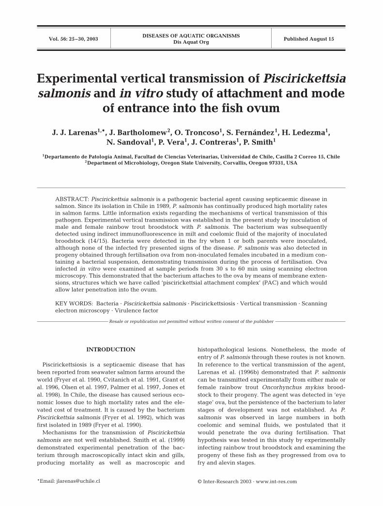
Scanning electron microscope images show that at45 s after initial contact of ova with the bacteria, Pisci-rickettsia salmonis was attached to the surface of theova by means of pseudopod-like extensions that
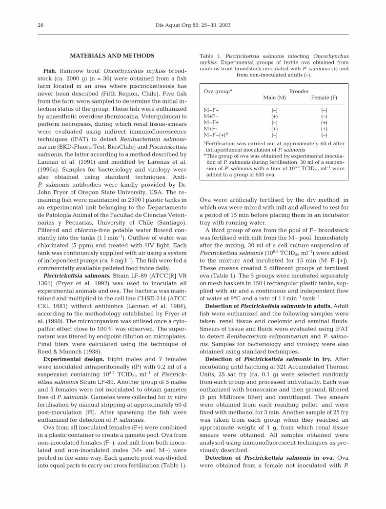
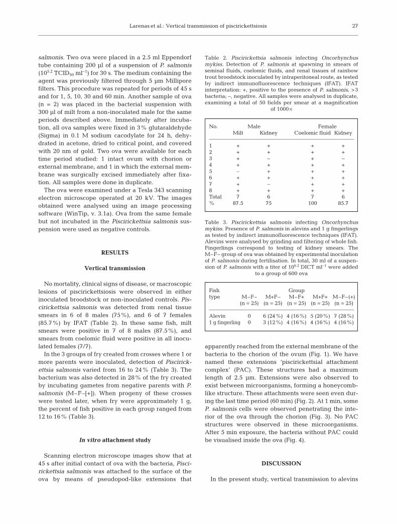
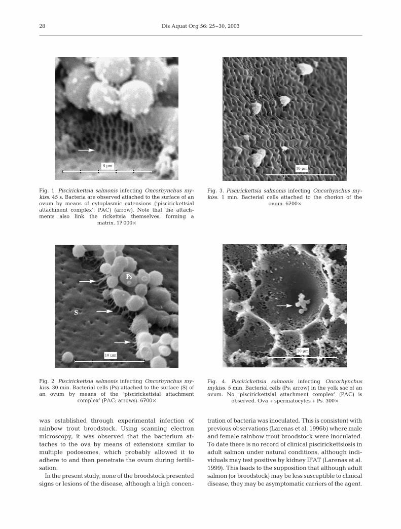
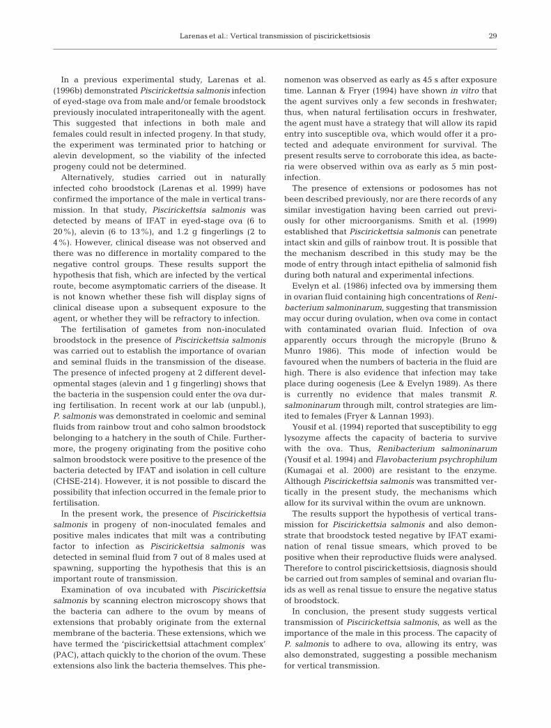
apparently reached from the external membrane of thebacteria to the chorion of the ovum (Fig. 1). We havenamed these extensions ‘piscirickettsial attachmentcomplex’ (PAC). These structures had a maximumlength of 2.5 µm. Extensions were also observed toexist between microorganisms, forming a honeycomb-like structure. These attachments were seen even dur-ing the last time period (60 min) (Fig. 2). At 1 min, someP. salmonis cells were observed penetrating the inte-rior of the ova through the chorion (Fig. 3). No PACstructures were observed in these microorganisms.After 5 min exposure, the bacteria without PAC couldbe visualised inside the ova (Fig. 4).
DISCUSSION
In the present study, vertical transmission to alevins
27
No. Male FemaleMilt Kidney Coelomic fluid Kidney
1 + + + +2 + + + +3 + – + –4 + + + +5 – + + +6 + + + +7 + – + +8 + + + +Total 7 6 7 6% 87.5 75 100 85.7
Table 2. Piscirickettsia salmonis infecting Oncorhynchusmykiss. Detection of P. salmonis at spawning in smears ofseminal fluids, coelomic fluids, and renal tissues of rainbowtrout broodstock inoculated by intraperitoneal route, as testedby indirect immunofluorescence techniques (IFAT). IFATinterpretation: +, positive to the presence of P. salmonis, >3bacteria; –, negative. All samples were analysed in duplicate,examining a total of 50 fields per smear at a magnification
of 1000 ¥
Fish Grouptype M–F– M+F– M–F+ M+F+ M–F–(+)
(n = 25) (n = 25) (n = 25) (n = 25) (n = 25)
Alevin 0 6 (24%) 4 (16%) 5 (20%) 7 (28%)1 g fingerling 0 3 (12%) 4 (16%) 4 (16%) 4 (16%)
Table 3. Piscirickettsia salmonis infecting Oncorhynchusmykiss. Presence of P. salmonis in alevins and 1 g fingerlingsas tested by indirect immunofluorescence techniques (IFAT).Alevins were analysed by grinding and filtering of whole fish.Fingerlings correspond to testing of kidney smears. TheM–F– group of ova was obtained by experimental inoculationof P. salmonis during fertilisation. In total, 30 ml of a suspen-sion of P. salmonis with a titer of 106.2 DICT ml–1 were added
to a group of 600 ova
Dis Aquat Org 56: 25–30, 2003
was established through experimental infection ofrainbow trout broodstock. Using scanning electronmicroscopy, it was observed that the bacterium at-taches to the ova by means of extensions similar tomultiple podosomes, which probably allowed it toadhere to and then penetrate the ovum during fertili-sation.
In the present study, none of the broodstock presentedsigns or lesions of the disease, although a high concen-
tration of bacteria was inoculated. This is consistent withprevious observations (Larenas et al. 1996b) where maleand female rainbow trout broodstock were inoculated.To date there is no record of clinical piscirickettsiosis inadult salmon under natural conditions, although indi-viduals may test positive by kidney IFAT (Larenas et al.1999). This leads to the supposition that although adultsalmon (or broodstock) may be less susceptible to clinicaldisease, they may be asymptomatic carriers of the agent.
28
Fig. 1. Piscirickettsia salmonis infecting Oncorhynchus my-kiss. 45 s. Bacteria are observed attached to the surface of anovum by means of cytoplasmic extensions (‘piscirickettsialattachment complex’; PAC) (arrow). Note that the attach-ments also link the rickettsia themselves, forming a
matrix. 17 000 ¥
Fig. 3. Piscirickettsia salmonis infecting Oncorhynchus my-kiss. 1 min. Bacterial cells attached to the chorion of the
ovum. 6700 ¥
Fig. 4. Piscirickettsia salmonis infecting Oncorhynchusmykiss. 5 min. Bacterial cells (Ps; arrow) in the yolk sac of anovum. No ‘piscirickettsial attachment complex’ (PAC) is
observed. Ova + spermatocytes + Ps. 300 ¥
Fig. 2. Piscirickettsia salmonis infecting Oncorhynchus my-kiss. 30 min. Bacterial cells (Ps) attached to the surface (S) ofan ovum by means of the ‘piscirickettsial attachment
complex’ (PAC; arrows). 6700 ¥
5 µm10 µm
20 µm10 µm
Larenas et al.: Vertical transmission of piscirickettsiosis
In a previous experimental study, Larenas et al.(1996b) demonstrated Piscirickettsia salmonis infectionof eyed-stage ova from male and/or female broodstockpreviously inoculated intraperitoneally with the agent.This suggested that infections in both male andfemales could result in infected progeny. In that study,the experiment was terminated prior to hatching oralevin development, so the viability of the infectedprogeny could not be determined.
Alternatively, studies carried out in naturallyinfected coho broodstock (Larenas et al. 1999) haveconfirmed the importance of the male in vertical trans-mission. In that study, Piscirickettsia salmonis wasdetected by means of IFAT in eyed-stage ova (6 to20%), alevin (6 to 13%), and 1.2 g fingerlings (2 to4%). However, clinical disease was not observed andthere was no difference in mortality compared to thenegative control groups. These results support thehypothesis that fish, which are infected by the verticalroute, become asymptomatic carriers of the disease. Itis not known whether these fish will display signs ofclinical disease upon a subsequent exposure to theagent, or whether they will be refractory to infection.
The fertilisation of gametes from non-inoculatedbroodstock in the presence of Piscirickettsia salmoniswas carried out to establish the importance of ovarianand seminal fluids in the transmission of the disease.The presence of infected progeny at 2 different devel-opmental stages (alevin and 1 g fingerling) shows thatthe bacteria in the suspension could enter the ova dur-ing fertilisation. In recent work at our lab (unpubl.),P. salmonis was demonstrated in coelomic and seminalfluids from rainbow trout and coho salmon broodstockbelonging to a hatchery in the south of Chile. Further-more, the progeny originating from the positive cohosalmon broodstock were positive to the presence of thebacteria detected by IFAT and isolation in cell culture(CHSE-214). However, it is not possible to discard thepossibility that infection occurred in the female prior tofertilisation.
In the present work, the presence of Piscirickettsiasalmonis in progeny of non-inoculated females andpositive males indicates that milt was a contributingfactor to infection as Piscirickettsia salmonis wasdetected in seminal fluid from 7 out of 8 males used atspawning, supporting the hypothesis that this is animportant route of transmission.
Examination of ova incubated with Piscirickettsiasalmonis by scanning electron microscopy shows thatthe bacteria can adhere to the ovum by means ofextensions that probably originate from the externalmembrane of the bacteria. These extensions, which wehave termed the ‘piscirickettsial attachment complex’(PAC), attach quickly to the chorion of the ovum. Theseextensions also link the bacteria themselves. This phe-
nomenon was observed as early as 45 s after exposuretime. Lannan & Fryer (1994) have shown in vitro thatthe agent survives only a few seconds in freshwater;thus, when natural fertilisation occurs in freshwater,the agent must have a strategy that will allow its rapidentry into susceptible ova, which would offer it a pro-tected and adequate environment for survival. Thepresent results serve to corroborate this idea, as bacte-ria were observed within ova as early as 5 min post-infection.
The presence of extensions or podosomes has notbeen described previously, nor are there records of anysimilar investigation having been carried out previ-ously for other microorganisms. Smith et al. (1999)established that Piscirickettsia salmonis can penetrateintact skin and gills of rainbow trout. It is possible thatthe mechanism described in this study may be themode of entry through intact epithelia of salmonid fishduring both natural and experimental infections.
Evelyn et al. (1986) infected ova by immersing themin ovarian fluid containing high concentrations of Reni-bacterium salmoninarum, suggesting that transmissionmay occur during ovulation, when ova come in contactwith contaminated ovarian fluid. Infection of ovaapparently occurs through the micropyle (Bruno &Munro 1986). This mode of infection would befavoured when the numbers of bacteria in the fluid arehigh. There is also evidence that infection may takeplace during oogenesis (Lee & Evelyn 1989). As thereis currently no evidence that males transmit R.salmoninarum through milt, control strategies are lim-ited to females (Fryer & Lannan 1993).
Yousif et al. (1994) reported that susceptibility to egglysozyme affects the capacity of bacteria to survivewith the ova. Thus, Renibacterium salmoninarum(Yousif et al. 1994) and Flavobacterium psychrophilum(Kumagai et al. 2000) are resistant to the enzyme.Although Piscirickettsia salmonis was transmitted ver-tically in the present study, the mechanisms whichallow for its survival within the ovum are unknown.
The results support the hypothesis of vertical trans-mission for Piscirickettsia salmonis and also demon-strate that broodstock tested negative by IFAT exami-nation of renal tissue smears, which proved to bepositive when their reproductive fluids were analysed.Therefore to control piscirickettsiosis, diagnosis shouldbe carried out from samples of seminal and ovarian flu-ids as well as renal tissue to ensure the negative statusof broodstock.
In conclusion, the present study suggests verticaltransmission of Piscirickettsia salmonis, as well as theimportance of the male in this process. The capacity ofP. salmonis to adhere to ova, allowing its entry, wasalso demonstrated, suggesting a possible mechanismfor vertical transmission.
29
Dis Aquat Org 56: 25–30, 2003
Acknowledgements. We thank Drs. E. Arellano and A. Gua-jardo for their help with the experimental trials, and Drs. J.Arias and C. Rose for valuable contributions to this paper.This work was supported by Grant 1000788 of the ChileanFund for Science and Technology (FONDECYT).
LITERATURE CITED
Bruno DW, Munro ALS (1986) Observations on Renibac-terium salmoninarum and the salmonid egg. Dis AquatOrg 1:83–87
Cvitanich J, Gárate O, Smith CE (1991) The isolation of arickettsia-like organism causing disease and mortality inChilean salmonids and its confirmation by Koch’s postu-late. J Fish Dis 14:121–145
Evelyn TPT, Prosperi-Porta L, Ketcheson JE (1986) Experi-mental intra-ovum infection of salmonid eggs with Reni-bacterium salmoninarum and vertical transmission of thepathogen with such eggs despite their treatment with ery-thromycin. Dis Aquat Org 1:197–202
Fryer JL, Lannan CN (1993) The history and current statusof Renibacterium salmoninarum, the causative agent ofbacterial kidney disease in Pacific salmon. Fish Res 17:15–33
Fryer JL, Lannan C, Garcés L, Larenas J, Smith P (1990) Iso-lation of a rickettsiales-like organism from diseased cohosalmon (Oncorhynchus kisutch) in Chile. Fish Pathol 25:107–114
Fryer JL, Lannan CN, Giovannoni SJ, Wood ND (1992) Piscir-ickettsia salmonis gen. nov., sp. nov., the causative agentof an epizootic disease in salmonid fishes. Int J Syst Bacte-riol 42:120–126
Grant AN, Brown AG, Cox DJ, Birkbeck TH, Griffen AA(1996) Rickettsia-like organism in farmed salmon. Vet Rec138:423–424
Jones SRM, Markham RJF, Groman DB, Cusack RR (1998)Virulence and antigenic characteristics of a culturedRickettsiales-like organism isolated from farmed Atlanticsalmon Salmo salar in eastern Canada. Dis Aquat Org 33:25–31
Kumagai A, Yamaoka Sh, Takahashi K, Fukuda H, Wak-abayashi H (2000) Waterborne transmission of Flavobac-
terium psychrophilum in coho salmon eggs. Fish Pathol35:25–28
Lannan C, Fryer J (1994) Extracellular survival of Piscirick-ettsia salmonis. J Fish Dis 17:545–548
Lannan CN, Winton JR, Fryer JL (1984) Fish cell lines: estab-lishment and characterization of nine cell lines fromsalmonids. In Vitro 20:671–676
Lannan C, Ewing SA, Fryer JL (1991) A fluorescent antibodytest for detection of rickettsia causing disease in Chileansalmonids. J Aquat Anim Health 3:229–234
Larenas J, Astorga C, Contreras J, Garcés H, Fryer J, Smith P(1996a) Rapid detection of Piscirickettsia salmonis usingmicrowave irradiation. Fish Pathol 31:231–232
Larenas J, Astorga C, Contreras J, Smith P (1996b) Detecciónde Piscirickettsia salmonis en ovas fertilizadas prove-nientes de truchas arco iris (Oncorhynchus mykiss) exper-imentalmente infectadas. Arch Med Vet 28:161–166
Larenas J, Pérez B, Hidalgo L, Contreras J, Smith PA (1999)Vertical transmission of Piscirickettsia salmonis in cohosalmon (Oncorhynchus kisutch) under farming conditions.IXth International Conference ‘Diseases of Fish and Shell-fish’, Abstracts Book p–069, Rhodes
Lee EG, Evelyn TP (1989) Effect of Renibacterium salmoni-narum levels in the ovarian fluid of spawning chinook onthe prevalence of the pathogen in their eggs and progeny.Dis Aquat Org 7:179–184
Olsen AB, Melby HP, Speilberg L, Evensen ∆, Hastein T(1997) Piscirickettsia salmonis infection in Atlantic salmonSalmo salar in Norway—epidemiological, pathologicaland microbiological findings. Dis Aquat Org 31:35–48
Palmer R, Ruttledge M, Callanan K, Drinan E (1997) A pisci-rickettsiosis-like disease in farmed Atlantic salmon inIreland—isolation of the agent. Bull Eur Assoc Fish Pathol17:68–72
Reed LJ, Muench H (1938) A simple method of estimatingfifty percent endpoints. Am J Hyg 27:497–499
Smith PA, Pizarro P, Ojeda P, Contreras J, Oyanedel S, Lare-nas J (1999) Routes of entry of Piscirickettsia salmonis inrainbow trout Oncorhynchus mykiss. Dis Aquat Org 37:165–172
Yousif AN, Albright LJ, Evelyn TPT (1994) In vitro evidencefor the antibacterial role of lysozyme in salmonid eggs. DisAquat Org 19:15–19
30
Editorial responsibility: David Bruno,Aberdeen, Scotland, UK
Submitted: January 23, 2002; Accepted: April 23, 2003Proofs received from author(s): August 7, 2003
Related Documents