Piscirickettsia salmonis SCAHLS ANZSDP Piscirickettsia salmonis Jan 2009 1 of 20 Piscirickettsia salmonis S Corbeil AAHL Fish Diseases Laboratory Australian Animal Health Laboratory CSIRO Livestock Industries Private Bag 24 Geelong VIC 3220 [email protected] MS Crane AAHL Fish Diseases Laboratory Australian Animal Health Laboratory CSIRO Livestock Industries Private Bag 24 Geelong VIC 3220 [email protected] SUMMARY Piscirickettsia salmonis is the first of the previously unrecognised rickettsial pathogens of fish to be isolated, characterised, and demonstrated to be the aetiological agent of an epizootic disease. P salmonis was first isolated in coho salmon in Chile, subsequently, piscirickettsiosis was observed in other salmonid species (chinook salmon, Atlantic salmon, rainbow trout and masou salmon) and also in other parts of the world. It is now known that rickettsia-like organisms affect fish over broad host and geographic ranges. In Chile the onset of the disease most commonly occurs following transfer of fish from freshwater to seawater holding facilities. Signs of the disease include lethargy and darkening of the skin, swollen kidneys and enlarged spleen and anaemia. Identification of the agent: Diagnosis of piscirickettsiosis is based on a range of procedures. Presumptive diagnosis is made following clinical and pathological observations. P salmonis is confirmed following histopathological examination, isolation in tissue culture combined with identification by either immunofluorescence or immunoperoxidase staining as well as dot blot DNA hybridisation. In addition, polymerase chain reaction techniques are available for the rapid identification of P salmonis in clinically affected animals. Status of Australia and New Zealand: P salmonis is exotic to Australia and New Zealand. In 2001 rickettsia-like organism (RLO) was identified in Atlantic salmon farmed in Tasmania. However, the Tasmanian RLO differs at the genetic and antigenic level from P salmonis isolates found overseas. 3 The RLO has not been detected in any species in New Zealand.

Welcome message from author
This document is posted to help you gain knowledge. Please leave a comment to let me know what you think about it! Share it to your friends and learn new things together.
Transcript

Piscirickettsia salmonis
SCAHLS ANZSDP Piscirickettsia salmonis Jan 2009 1 of 20
Piscirickettsia salmonis
S Corbeil AAHL Fish Diseases LaboratoryAustralian Animal Health LaboratoryCSIRO Livestock IndustriesPrivate Bag 24Geelong VIC [email protected]
MS Crane AAHL Fish Diseases LaboratoryAustralian Animal Health LaboratoryCSIRO Livestock IndustriesPrivate Bag 24Geelong VIC [email protected]
SUMMARY
Piscirickettsia salmonis is the first of the previously unrecognised rickettsial pathogens of fish to be isolated, characterised, and demonstrated to be the aetiological agent of an epizootic disease. P salmonis was first isolated in coho salmon in Chile, subsequently, piscirickettsiosis was observed in other salmonid species (chinook salmon, Atlantic salmon, rainbow trout and masou salmon) and also in other parts of the world. It is now known that rickettsia-like organisms affect fish over broad host and geographic ranges. In Chile the onset of the disease most commonly occurs following transfer of fish from freshwater to seawater holding facilities. Signs of the disease include lethargy and darkening of the skin, swollen kidneys and enlarged spleen and anaemia.
Identification of the agent: Diagnosis of piscirickettsiosis is based on a range of procedures. Presumptive diagnosis is made following clinical and pathological observations. P salmonis is confirmed following histopathological examination, isolation in tissue culture combined with identification by either immunofluorescence or immunoperoxidase staining as well as dot blot DNA hybridisation. In addition, polymerase chain reaction techniques are available for the rapid identification of P salmonis in clinically affected animals.
Status of Australia and New Zealand: P salmonis is exotic to Australia and New Zealand. In 2001 rickettsia-like organism (RLO) was identified in Atlantic salmon farmed in Tasmania. However, the Tasmanian RLO differs at the genetic and antigenic level from P salmonis isolates found overseas. 3 The RLO has not been detected in any species in New Zealand.

Piscirickettsia salmonis
SCAHLS ANZSDP Piscirickettsia salmonis Jan 2009 2 of 20
Part 1. Diagnostic Overview
Introduction
Piscirickettsia salmonis is an obligate, intracellular, bacterial pathogen of salmonids, first identified in Chile and later identified in Canada and several European countries.1This Gram-negative, pleomorphic, coccoid bacterium replicates within membrane-bound cytoplasmic vacuoles in the cells of infected fish.2 Molecular phylogenetic analysis based on sequencing of the 16S rRNA gene placed P salmonis in a new family of Piscirickettsiae within the class of γ-proteobacteria.
Aetiology
Piscirickettsia salmonis epizootics in Chile have been responsible for significant economic losses to the salmonid aquaculture industry; mortality rates were reported to be 30-90% among coho salmon.4 Piscirickettsia-salmonis-like organisms have recently been isolated from non-salmonid fish in various countries.5, 6, 7, 8, 9 The relationships of most of these organisms to P salmonis have not been fully elucidated, but the organism isolated from white seabass (Atractoscion nobilis) is genetically and serologically indistinguishable from P salmonis.6
Epidemiology
Transmission of Piscirickettsia salmonis to coho salmon can occur via skin, gills and intestine10. The disease has been primarily reported in marine fish farms, and has also been observed in freshwater facilities11, 12. Horizontal transmission occurs in saltwater and freshwater.13, 14 It is not known whether transmission can occur via vectors or via vertical transmission. The onset of the disease most commonly occurs following transfer of fish from freshwater to seawater holding facilities. Fish of all ages, from smolts to market size14, are susceptible to the disease. The disease begins approximately 1 month after fish are introduced into the seawater net pens.
Antibacterial treatment provides some benefit, it does not control the disease. Currently, oxolinic acid appears to be the drug of choice. Eggs may be disinfected as part of good hatchery practice. Management of the disease is based on a range of husbandry practices including the application of immunostimulants of unproven efficacy and the control of vertical transmission by an expensive selection procedure during reproduction.15 Vaccines consisting of inactivated P salmonis bacterins have not yielded significant protection against piscirickettsiosis.16, 17 However, vaccines developed using recombinant DNA technology have shown high levels of efficacy in the laboratory15, 17 and are a promising avenue for the control of piscirickettsiosis.
Clinical Signs and Pathology
A range of gross signs of disease may be present in salmonids infected with P salmonis. Severely affected fish are dark, anorexic and lethargic. They often swim near the water surface or at the edges of the cages. Fish with milder infections may appear normal. Infections of the brain may cause erratic swimming behaviour.18 Skin
Piscirickettsia salmonis
SCAHLS ANZSDP Piscirickettsia salmonis Jan 2009 3 of 20
lesions, appearing as small white patches that can progress to shallow ulcers, may also be present on some fish. The most consistent external sign seen during P salmonisinfections is pale gills indicating anaemia but this is not pathognomonic for the disease.
In common with many systemic, chronic inflammatory diseases of salmonids, the internal signs include a swollen and discoloured kidney and an enlarged spleen. Ascites in the peritoneal cavity and haemorrhages on the visceral fat, stomach, swim bladder and body musculature can also occur.14, 19 Hallmark internal lesions of the disease are found in the liver, which may exhibit large, whitish or yellow, multifocal, coalescing, pyogranulomatous nodules. These lesions often rupture, resulting in shallow crater-like cavities in the liver The most marked histological changes are found in the liver, kidney, spleen and intestine with lesser changes in the brain, heart, ovary and gills. 14, 19, 20, 21 Multifocal necrosis of hepatocytes, accompanied by a chronic inflammatory infiltrate of mononuclear cells, is seen in the liver. Vascular and perivascular necrosis are also evident in the liver, and intravascular coagulation resulting in fibrin thrombi within major vessels is a common finding. The focal areas of necrosis underlie the pale circular lesions seen grossly in more chronically infected fish. In more acute infections, the coalescence of areas of necrosis results in a more mottled appearance of the liver rather than discrete nodules. Granulomatous inflammation also occurs in the interstitium of the kidney and parenchyma of the spleen. Vascular changes similar to those in the liver may also be seen in the kidney and spleen. Meningitis, endocarditis, peritonitis, pancreatitis, and branchitis may be seen with accompanying chronic inflammatory and vascular changes similar to those in the liver and haematopoietic organs. Using high magnification, examination of lesions revealsaggregates of the organism in the cytoplasm of degenerated hepatocytes and in macrophages. Infected macrophages are usually hypertrophied and replete with cellular debris. In tissue sections stained with haematoxylin and eosin (H&E) the organism appears as basophilic or amphophilic spheres, about 1 µm in diameter.
References
1. Fryer JL, Mauel MJ. The rickettsia: an emerging group of pathogens in fish. Emer Infect Dis 1997;3:137-144.
2. Fryer JL, Lannan CN, Garces LH, Larenas JJ, Smith PA. Isolation of a rickettsiales-like organism from diseased coho salmon Oncorhynchus kisutch in Chile. Fish Pathol 1990;25(2):107-114.
3. Corbeil S, Hyatt AD, Crane MSJ. Characterisation of an emerging rickettsia-like organism in Tasmanian farmed Atlantic salmon Salmo salar. Dis Aquat Org2005;64(1):37-44.
4. Bravo S, Campos M. Coho salmon syndrome in Chile. AFS/FHS Newsletter1989;17(3):3.

Piscirickettsia salmonis
SCAHLS ANZSDP Piscirickettsia salmonis Jan 2009 4 of 20
5. Chen SC, Tung MC, Chen SP, Tsai JF, Wang PC, Chen RS, Lin SC, Adams A. Systemic granulomas caused by a rickettsia-like organism in Nile tilapia, Oreochronuis niloticus (L.), from southern Taiwan. J Fish Dis 1994;17:591-599.
6. Chen MF, Yun S, Marty GD, McDowell TS, House ML, Appersen JA, Guenther TA, Arkush KD, Hedrick RP. A Piscirickettsia salmonis-like bacterium associated with mortality of white seabass Atractoscion nobilis. Dis Aquat Org 2000;43:117-126.
7. Chen SC, Wang PC, Tung MC, Thompson KD, Adams A. A Pisicirickettsia salmonis-like organism in grouper, Epinephelus melanostigma, in Taiwan. J Fish Dis2000;23:415-418.
8. Arkush KD, McBride AM, Mendonca HL, Okihiro MS, Andree KB, Marshall S, Henriquez V, Hedrick RP. Genetic characterization and experimental pathogenesis of Piscirickettsia salmonis isolated from white seabass Atractoscion nobilis. Dis Aquat Org 2005;63(2-3):139-149.
9. McCarthy U, Steiropoulos NA, Thompson KD, Adams A, Ellis AE, Ferguson HW. Confirmation of Piscirickettsia salmonis as a pathogen in European seabass Dicentrarchus labrax and phylogenetic comparison with salmonid strains. Dis Aquat Org 2005;64(2):107-119.
10. Smith PA, Rojas ME, Guajardo A, Contreras J, Morales MA, Larenas J. Experimental infection of coho salmon Oncorhynchus kisutch by exposure of skin, gills and intestine with Piscirickettsia salmonis. Dis Aquat Org 2004;61(1-2):53-57.
11. Bravo S. Piscirickettsiosis in freshwater. Bull Eur Assoc Fish Pathol 1994;14:137-138.
12. Gaggero A, Castro H, Sandino AM. First isolation of Piscirickettsia salmonis from coho salmon, Oncorhynchus kisutch (Walbaum), and rainbow trout, Oncorhynchusmykiss (Walbaum), during the freshwater stage of their life cycle. J Fish Dis1995;18:277-279.
13. Almendras FE, Fuentealba IC, Jones SRM, Markham F, Spangler E. Experimental infection and horizontal transmission of Piscirickettsia salmonis in freshwater-raised Atlantic salmon, salmo salar L. J Fish Dis 20:409-418.
14. Cvitanich JD, Garate NO, Smith CE. Isolation of a rickettsial-like organism causing diseases and mortality in Chilean salmonids and its confirmation by Koch’s postulate. J Fish Dis 1991;14:121-145.
15. Wilhelm V, Alvaro M, Burzio LO, Rosemblatt M, Engel E, Valenzuela, Parada G, Valenzuela PDT. A vaccine against the salmonid pathogen Piscirickettsia salmonisbased on recombinant proteins. Vaccine 2006;24:5083-5091.
16. Smith PA, Contreras JR, Larenas JJ. Immunization with bacterial antigens: piscirickettsiosis. Dev Biol Stand 1997;90:161-166.
Piscirickettsia salmonis
SCAHLS ANZSDP Piscirickettsia salmonis Jan 2009 5 of 20
17. Kuzyk MA, Burian J, Machander D, Dolhaine D, Cameron S, Thornton JC, Kay WW. An efficacious recombinant subunit vaccine against the salmonid rickettsial pathogen Piscirickettsia salmonis. Vaccine 2001;19:2337-2344.
18. Skarmeta AM, Henriquez V, Zahr M, Orrego C, Marshall SH. Isolation of a virulent Piscirickettsia salmonis from the brain of naturally infected coho salmon. Bull Eur Assoc Fish Pathol 2000;20:261-264.
19. Schafer JW, Alvarado V, Enriquez R, Monras M. The 'coho salmon syndrome' (CSS): a new disease in Chilean salmon, reared in sea water. Bull Eur Assoc Fish Pathol 1990;10:130.
20. Branson EJ and Nieto Diaz-Munoz D. Description of a new disease condition occurring in farmed coho salmon, Oncorhynchus kisutch (Walbaum), in South America. J Fish Dis 1991;14:147-156.
21. Palmer R, Ruttledge M, Callanan K, Drinan E. A Piscirickettsiosis-like disease in farmed Atlantic salmon in Ireland - isolation of the agent. Bull Eur Assoc Fish Pathol1997;17:68-72.
22. Olsen AB, Melby HP, Speilberb L, Evensen O, Hastein T. Piscirickettsia salmonis infection in Atlantic salmon Salmo salar in Norway – epidemiological, pathological and microbiological findings. Dis Aquat Org 1997;31(1):35-48.

Piscirickettsia salmonis
SCAHLS ANZSDP Piscirickettsia salmonis Jan 2009 6 of 20
Part 2. Diagnostic Tests
Case definition
This standard diagnostic procedure documents the methods for the identification of P salmonis following submission of samples from fish presenting clinical signs. Thehistology, immunohistochemistry and conventional PCR methods are based on those outlined in the OIE Manual of Diagnostic tests for Aquatic Animals.23
Range of tests available and appropriate applications
All methods established to date have been developed for detection and identification of P salmonis in diseased fish. None of these tests have been validated for the detection of sub-clinical carriers of P salmonis and therefore should NOT be used for surveillance and monitoring purposes. Thus the tests described in this ANZSDP, for identification by immunohistochemical and PCR tests, are appropriate for diagnosis and/or P salmonis exclusion in diseased fish.
Storage of samples
Samples must be maintained between 4-10OC (shipping on ice packs in a styrofoam shipping container is appropriate) if isolation of the rickettsia is to be attempted. Freezing fish tissues will not affect the performance of PCR assays.
Tissues to be examined
Tissues suitable for examination include kidney and liver.
Tests available
This ANZSDP describes methods for the identification of P salmonis using histology, immunohistochemical tests, molecular diagnostics (PCR) and pathogen isolation in cell culture.
References
23. OIE. Piscirickettsiosis. In: Manual of Diagnostic Tests for Aquatic Animals Chapter 2.1.13. Office International des Epizooties 5th Edition, 2006, Paris.
Piscirickettsia salmonis
SCAHLS ANZSDP Piscirickettsia salmonis Jan 2009 7 of 20
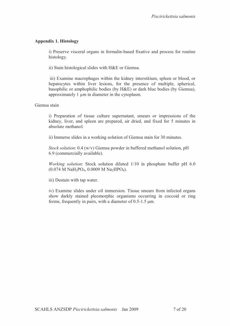
Appendix 1. Histology
i) Preserve visceral organs in formalin-based fixative and process for routine histology.
ii) Stain histological slides with H&E or Giemsa.
iii) Examine macrophages within the kidney interstitium, spleen or blood, or hepatocytes within liver lesions, for the presence of multiple, spherical, basophilic or amphophilic bodies (by H&E) or dark blue bodies (by Giemsa), approximately 1 µm in diameter in the cytoplasm.
Giemsa stain
i) Preparation of tissue culture supernatant, smears or impressions of the kidney, liver, and spleen are prepared, air dried, and fixed for 5 minutes in absolute methanol.
ii) Immerse slides in a working solution of Giemsa stain for 30 minutes.
Stock solution: 0.4 (w/v) Giemsa powder in buffered methanol solution, pH6.9 (commercially available).
Working solution: Stock solution diluted 1/10 in phosphate buffer pH 6.0 (0.074 M NaH2PO4, 0.0009 M Na2HPO4).
iii) Destain with tap water.
iv) Examine slides under oil immersion. Tissue smears from infected organs show darkly stained pleomorphic organisms occurring in coccoid or ring forms, frequently in pairs, with a diameter of 0.5-1.5 μm.
Piscirickettsia salmonis
SCAHLS ANZSDP Piscirickettsia salmonis Jan 2009 8 of 20
Appendix 2. Identification by Immunohistochemical (Immunoperoxidase) Test
Introduction
Bacterial identification by immunoperoxidase test has become a standard procedure where specific antibodies are available. Briefly, P salmonis-infected tissues are fixed and can be stored until use. The fixed preparations are incubated with a primary antibody preparation (polyclonal) that will bind to specific epitopes, if present. Excess antibody is removed by washing, and a secondary peroxidase-conjugated antibody (e.g. peroxidase anti-sheep Ig if the primary antibody was raised in sheep) is added. After an incubation period, excess conjugate is removed by washing, peroxidase substrate 3-amino-9-ethyl carboxyzole (AEC) is added and colour is allowed to develop. Finally, following rinsing in water, cells are counterstained with Mayer’s haematoxylin, rinsed in water and developed with Scott’s solution. Thus, if any bacteria recognised by the primary antibody are present, a positive colour reaction will occur.
List of equipment
-Incubator 37oC-Refrigerator-Microscope slides (frosted ends)-Coverslips-Immunostaining chambers-Light microscope fitted with 4X and 10X objectives
Reagents
-TRIS-HCL 0.005M buffer pH7.6 Sodium Chloride 8.1 gTRIS (TRIS hydroxymethylmethylamine) 0.6 gIN HCL 3.8 mLDistilled water to 1 L
-80% (v/v) acetone in water-Tween 20 0.05% (v/v) in 0.005M TRIS-HCL buffer pH7.6-Skim milk powder solution 1% (w/v) in 0.005M TRIS-HCL buffer pH7.6-Anti-P salmonis (LF-89 isolate) sheep polyclonal antibodies (Microtek International Inc. cat # SPS01)-Peroxidase-labelled donkey anti-sheep IgG -3-amino-9-ethyl carboxyzole -Dimethylformamide -Acetate buffer 0.05 M (pH 5.0)
Sodium acetate Na2C 2H3O2.3H2O 6.8 gDistilled water 1 LUse glacial acetic acid (approx. 1.0 mL) to alter pH to 5.0
-Hydrogen peroxide 30% (v/v)-Deionised water-Mayer’s haematoxylin -Scott’s solution
NaHCO3 7.0 gMgSO4 40.0 g

Piscirickettsia salmonis
SCAHLS ANZSDP Piscirickettsia salmonis Jan 2009 9 of 20
Distilled water 2.0 L
- Mounting Medium
Quality control
For immunoperoxidase testing, separate control tissues are set up in parallel with the tissues of the test samples. Positive controls are tissues from fish infected with a known P salmonis isolate (e.g. LF-89). These tissues are no longer available in Australia and would need to be produce by experimental infection at AAHL when funding is made available. Positive control tissues and a known uninfected tissue (negative control) are processed on the same day as the test samples. Antibody preparations for diagnostic tests are stored at –20OC until use and check-tested on a regular basis.
The working dilutions of each diagnostic reagent (antibodies, conjugates, substrates) need to be determined prior to use and check-tested on a regular basis.
Procedures
Immunoperoxidase test procedure
Prepare 1% (w/v) and 0.1% (w/v) skim milk powder solution in PBSA for antibody dilution
Dilute the anti-P salmonis primary sheep polyclonal antibody (1/100) and normal sheep serum (1/100) in 1% skim milk powder solution in sterile PBSA. After washing in PBSA, the sections are incubated with the primary antibody solution at 37oC for 1 h. Sections are washed with PBSA then incubated with a peroxidase labelled donkey anti-sheep IgG diluted 1:200 in 0.1% skim milk in PBSA at 37oC for 1 h. Following a further wash in PBSA, the sections are incubated with freshly prepared substrate solution (2mg AEC, 200 μL dimethylformamide, 10mL 0.05 M acetate buffer pH 5.0, 5 μL 30% (v/v) hydrogen peroxide) at room temperature for 20 min. The sections are rinsed in tap water, counterstained with Mayer’s haematoxylin, rinsed in Scott’s solution and mounted for light microscopic examination.
Interpretation
Positive reaction: Grainy, focal, brick-red staining of cells indicates presence of P salmonis LF-89 isolate (Figure 1a) and Tasmanian RLO (Figure 1b) identified by the diagnostic polyclonal antiserum. Note that the staining is much weaker on the Tasmanian RLO-infected tissue due to its different antigens.
Negative reaction: No red staining apparent (Figure 1c).
Background staining: Non-grainy, non-focal, pale, pinkish staining may occur and could be due to any of a number of reasons including non-specific binding.

Piscirickettsia salmonis
SCAHLS ANZSDP Piscirickettsia salmonis Jan 2009 10 of 20
Figure 1a. Immunohistochemistry of P salmonis (LF-89 isolate)-infected fish tissue, using sheep anti-LF-89 P salmonis polyclonal antibodies.
Figure 1b. Immunohistochemistry of Tasmanian RLO-infected fish tissue, using sheep anti-LF-89 P salmonis polyclonal antibodies. Note that the staining is weaker due to antigenic determinant differences between P salmonis and the RLO.

Piscirickettsia salmonis
SCAHLS ANZSDP Piscirickettsia salmonis Jan 2009 11 of 20
Figure 1c. Immunohistochemistry of Tasmanian RLO-infected fish tissue, using non-immune sheep polyclonal antibodies.

Piscirickettsia salmonis
SCAHLS ANZSDP Piscirickettsia salmonis Jan 2009 12 of 20
Appendix 3. Identification by PCR
Introduction
Both nested24 and single step25 PCR assays were developed during the 1990s to facilitate the detection and characterisation of P salmonis. The nested PCR is the current method recommended by OIE for detection and identification of P salmonis. Single step PCR methods are also used for detection an identification of P salmonis, but the workers who developed these methods suggest that the targeted ITS region of the rRNA operon is more variable than the16S region exploited in the nested PCR therefore allowing finer discrimination in the description of new P salmonis isolates. More recently a quantitative real-time PCR assay for P salmonis was developed26 and was shown to be as sensitive as the nested PCR assay. This assay offers the advantages of being faster to perform as well as not presenting the risk of cross-contamination inherent to nested PCR assays. In addition the real-time PCR allows quantification of P salmonis in samples. Laboratories undertaking these assays will need to verify performance to take into account differences in thermal cyclers.
The following section presents the methodologies for the three assays:
Reagents
Reagents stored at –20C-Taq polymerase -dNTPs (1.25mM) -Mg free buffer 10X -MgCl2 (25mM) -100% Ethanol AR grade-70% Ethanol-Primers (18μM)-23S FAM probe-HotStarTaq Mastermix -100bp DNA ladder & loading dye - Universal Master Mix (qPCR) -18S endogenous control primers and VIC probe (Applied Biosystems cat # 4308329 or equivalent)
Reagents stored at room temperatureQIAamp DNA Mini Kit (QIAGEN Cat # 51304):-DNA columns-Buffer ATL-Buffer AL-Buffer AW1-Buffer AW2-Buffer AE-Proteinase K-Agarose -Ethidium bromide -40 x Tris-Acetate EDTA Buffer
Piscirickettsia salmonis
SCAHLS ANZSDP Piscirickettsia salmonis Jan 2009 13 of 20
List of equipment
Apart from the normal range of equipment required in the standard diagnostic laboratory (e.g. refrigerators, freezers, mixers, micropipettes, biological safety cabinets, centrifuges, balances, microwave oven, thermometers), specialised equipment required to undertake diagnostic PCR may include dry heat blocks, thermocycler, gel electrophoresis equipment, UV transilluminator, and a camera system. The DNA amplicon will need to be sequenced for confirmation of P salmonisgene sequence, however, sequencing services are available at various locations around Australia if sequencing equipment is not available on site.
Quality control
Molecular diagnosis should be operated under an ISO 17025 accredited and audited quality assurance program. Thus, such a program would include initial evaluation of kits and validation of performance; ongoing internal evaluation through mandatory use of appropriate quality control samples where available; and performance monitoring through quality assessment or proficiency programs.
External quality control samples over the appropriate range of testing must be obtained or manufactured wherever possible. Wherever possible, quality control samples should be included in every assay run and the data presented so that run-to-run performance can be monitored. Positive, negative and reagent controls should be conducted as specified in the protocol. As a norm, formalin-fixed controls would be conducted with formalin-fixed test samples and appropriate unfixed controls would be conducted with fresh tissue, culture supernatants or other test samples. Stocks of controls should be established. These controls should be evaluated prior to storage and used in a check-testing regimen and as controls for the conduct of disease investigations. In addition, amplification of the 18S rDNA gene can be performed on the DNA extracted from fish tissues to validate the extraction procedure as well as to rule out the presence of PCR inhibitors.
Procedures
Sample preparation
Due to the sensitivity of PCR tests, care at every step of sample preparation must be taken to ensure that cross-contamination of diagnostic samples does not occur. Thus all instruments and sample containers must be clean and uncontaminated i.e. not pre-exposed to aquatic pathogens. Wherever possible, it is recommended that disposable containers are used.
Samples would be handled and processed using sterile disposable single-use containers, instruments and reagents to minimise the risks of contamination of the samples. As a general principle, samples to be used in the PCR suite for molecular diagnosis will be inactivated by an approved method prior to movement to the PCR suite.
Piscirickettsia salmonis
SCAHLS ANZSDP Piscirickettsia salmonis Jan 2009 14 of 20
For each fish sample, approximately 20 mg of inner liver tissues are harvested using sterile scissors and tweezers and put in a sterile 1.5 mL vial (conical bottom). Tissues are mashed using a disposable plastic green pestle and each sample receives 180 L of ATL buffer.
Nucleic acids are extracted from submitted samples in a Biological Safety Cabinet Class II working within a dedicated PCR suite.
Nucleic acid extraction
P salmonis nucleic acid is obtained from cell culture supernatant or tissue samples using the QIAamp DNA extraction kit following manufacturer’s instructions.
P salmonis-specific nested PCR 24
Primary amplification
A) The PCR mixture for a single sample consists of the following reagents: 9.5 μL of deionised sterile water; 12.5 μL of HotStar Taq Master mix, 0.5 μL of the universal nested (Eubacterial 16S rDNA) forward primer Eub-B AGAGTTTGATCMTGGCTCAG (18 μM); 0.5 μL of the reverse primer Eub-A: AAGGAGGTGATCCANCCRCA (18 μM); and 2 μL of extracted DNA. For multiple samples, the volumes are increased by the appropriate multiple. The mixture is incubated in an automatic thermal cycler (Perkin Elmer GeneAmp 2400) that is programmed for: one cycle at 94C for 15 minutes (activation of the Hotstar Taq polymerase); 35 cycles at 94C for 1 minute (denaturation), 50C for 2 minutes (annealing) and 72C for 3 minutes (extension); and, finally, one cycle at 72C for 7 minutes (final extension).
B) Alternatively, the following reagents can be used (per sample) for the primary amplification: 12.5 μL of deionised sterile water, 4 μL dNTPs, 2.5 μL MgCl2-free buffer10X, 1.5 μL MgCl2, 1 μL Eub-B primer (18 μM), 1 μL Eub-A primer (18 μM), 0.5 μL Taq polymerase, 2 μL of sample DNA. The mixture is incubated in an automatic thermal cycler (Perkin Elmer GeneAmp 2400) that is programmed for: one cycle at 94C for 2 minutes; 35 cycles at 94C for 1 minute, 50C for 2 minutes and 72C for 3 minutes; and, finally, one cycle at 72C for 7 minutes.
To validate the DNA extraction process and rule out the presence of PCR inhibitors in samples the amplification of the 18S rDNA is performed using the following primers and cycling conditions:18S Forward Primer 5'- CGG CTA CCA CAT CCA AGG AA -3'18S Reverse Primer 5'- GCT GGA ATT ACC GCG GCT -3'
One cycle at 94C for 2 minutes (non-HotStar Taq polymerase) followed by 35 cycles of 94C for 30 seconds (denaturation), 60C for 30 seconds (annealing) and 72C for 30 seconds (extension); and, finally, one cycle at 72C for 5 minutes (final extension).

Piscirickettsia salmonis
SCAHLS ANZSDP Piscirickettsia salmonis Jan 2009 15 of 20
Amplified DNA (size: ~190 bp) is detected by electrophoresis on a 2% (w/v) agarose gel containing 0.5 μg/mL ethidium bromide.
Nested PCR amplification
The second amplification is performed by adding 2 μL of the first PCR products to the reaction mixtures described above (methods A or B) except for the presence of the P salmonis specific primers PS2S (forward, CTAGGAGATGAGCCCGCTTG) and PS2AS (reverse, GCTACACCTGCGAAACCACTT) instead of Eub-A and Eub-B, under the following reaction conditions: 1 cycle at 94C for 15 minutes (if using HotStar Taq, otherwise 1 cycle at 94C for 2 minutes for non-HotStar Taq); 35 cycles at 94C for 1 minute, 61C for 2 minutes and 72C for 3 minutes; and, finally, one cycle at 72C for 7 minutes.
Amplified DNA (size: 469 bp) is detected by electrophoresis on a 2% (w/v) agarose gel containing 0.5 μg/mL ethidium bromide (Figure 2).
Figure 2. The P salmonis-specific nested PCRAmplicons (bright bands) of 469 base pairs in size are seen in lanes d, e, f and g. Wells e, f, and g contained DNA samples extracted from supernatants of cell cultures infected with P salmonis isolates LF-89, ATL-4-91, and NOR-92, respectively. Lane d contained a DNA sample extracted from Tasmanian RLO-infected fish tissue. Lanes a, b, and c contained DNA samples from unrelated fish bacteria used as negative controls. The last lane on the right-hand-side of the gel contained the 100 bp ladder. In addition to the specific bands in lanes d, e, f and g, there are some non-specific bands in each lane, including lanes d, e, f and g. Note that the Tasmanian RLO is detected by the PCR assay but sequencing of the amplicon reveals sequence differences.3
Interpretation
At the completion of the PCR, specific amplicons of the correct size are identified by agarose gel electrophoresis:
The negative control sample must have no evidence of specific amplicon.
A positive control sample must yield a specific P salmonis amplicon of 469 bp.
a b c d e f g
500bp

Piscirickettsia salmonis
SCAHLS ANZSDP Piscirickettsia salmonis Jan 2009 16 of 20
Amplicons of the correct size are then eluted from the gel, and the DNA sequence is determined (by using the PCR primers PS2S and PS2AS as sequencing primers).
Sequence identity and genotype are determined by a BLASTn search of the GenBank database.
An assay is valid only when all controls yield the expected results.
Single-step PCR25
A single-step PCR can be used to amplify the internal transcribed spacer (ITS) of the rDNA operon of exotic isolates of P salmonis. The specific P salmonis primers ITS-1 (5'-TGATTTTATTGTTTAGTGAGAATGA-3') and ITS-4 (5'-ATGCACTTATTCACTTGATCATA-3') are used at a final concentration of 1 μM. Volumes (2 μL) of DNA sample are added to 23 μL of reaction mixture for the amplification. Thermo cycling conditions for the PCR are: 95oC for 15 min (1 cycle), followed by 94oC for 30 sec, 50oC for 30 sec and 72oC for 30 sec (35 cycles), followed by 72oC for 5 min (1 cycle). PCR products are detected by electrophoresis on a 2% (w/v) agarose gel containing 0.5 μg/mL ethidium bromide (Figure 3).
Sequence identity and genotype are determined by a BLASTn search of the GenBank database (accession number AY578985).
This PCR will also amplify the Tasmanian RLO DNA, but the amplicon has a 19 bp deletion towards the 3’-end of the amplicon.3 A BLASTn search will also confirm the identity of the amplicon. Running both P salmonis and the Tasmanian RLO DNA, on the 2% agarose gel as positive controls should facilitate interpretation of the results as it is usually possible to visually differentiate the amplicons based on size (migration in the gel). Sequence analysis must be undertaken for confirmation.
fdcba
300bp
e

Piscirickettsia salmonis
SCAHLS ANZSDP Piscirickettsia salmonis Jan 2009 17 of 20
Figure 3. Detection of P salmonis using a single-step PCR methodThe primers used for the rDNA amplification were RTS-1 and RTS-425 producing a 283 bp amplicon in P. salmonis. Several other species of bacteria were used as specificity controls. Lanes; a- Hafnia alvei,b- Aeromonas salmonicida, c- Yersinia ruckeri, d- Vibrio anguillarum, e- Tasmanian RLO, f- P. salmonis (ATL-4-91), g- 100 bp ladder. Vibrio anguillarum shows a weak non-specific band just below the expected P. salmonis amplicon. Note that the Tasmanian RLO is detected by the PCR assay but has a band slightly lower than P salmonis (ATL-4-91). In addition, sequencing revealed that the amplicon from the Tasmanian RLO is shorter than the amplicon from P salmonis by 19 base pairs.3
Real-time qPCR (TaqMan assay specific) 26
The quantitative PCR (qPCR) can be used to evaluate the titre of P salmonis in fish tissues (Figure 4). In order to do so, a dilution series of DNA extracted from P salmonis titrated in cell culture must be used to establish a reference curve.26
Alternatively, the real-time PCR assay, which was shown to be as sensitive as the nested PCR assay26, can also be used simply as a qualitative assay. The qPCR assay has the advantages that it is rapid, quantitative, and less prone to cross-contamination, which can occur with conventional nested-PCR assays.
Primers and probe for the TaqMan assay were designed using Primer Express Software version 1.5 (PE Applied Biosystems). The P salmonis primers and probe are based on the 23S rDNA gene, a relatively conserved genomic region among several isolates of P salmonis. Primer and probe sequences are as follows: Forward primer (F-760): 5’-tctgggaagtgtggcgataga-3’; reverse primer (R-836): 5’-tcccgacctactcttgtttcatc-3’; The 6-carboxyfluorescein (FAM) and 6-carboxytetramethylrhodamine (TAMRA), labelled probe (PS23S): 6FAM-tgatagccccgtacacgaaacggcata-TAMRA.
The P salmonis primers are used at a final concentration of 900 nM. The 23S FAM probe is used at a final concentration of 250 nM. The 18S rRNA endogenous control primers and probe are used to validate the DNA extraction procedure from fish tissues, confirm the integrity of the extracted DNA and determine the absence of PCR inhibitor. The primers are used at a limiting conditions at a final concentration of 113 nM each. The VIC probe is used at a final concentration of 31 nM. The reactions are carried out in a 96-well plate in a 25 μL reaction volume containing 12.5 μL Universal Master Mix (PE Applied Biosystems). Volumes (2 μL) of each DNA sample are added. Standard thermal cycling conditions are used (50oC for 2 min, 95oC for 10 min, followed by 40 cycles of 95oC for 15 sec and 60oC for 1 min). A sample is considered positive when the change in fluorescence (ΔRn) of FAM or VIC, relative to that of ROX (internal reference signal), exceeds the set threshold values of 0.055 for FAM and 0.04 for VIC in the linear range of the amplification plots at a cycle threshold (CT) value below 40. CT is defined as the cycle at which a statistically significant increase in fluorescence output above background is detected.
It should be noted that the TaqMan assay detects both P salmonis and the Tasmanian RLO. To confirm the identity of the aetiological agent, one of the conventional PCR tests and sequencing of the amplicon must be performed.

Piscirickettsia salmonis
SCAHLS ANZSDP Piscirickettsia salmonis Jan 2009 18 of 20
Results obtained so far suggest that the P salmonis TaqMan assay would be complementary to the standard screening methods for Piscirickettsiosis recommended by the O.I.E.
Figure 4. P salmonis qPCR assay.The graph shows the log of the change in fluorescence (y-axis) intensity versus the number of threshold cycles (x-axis) for each of several different target concentrations equivalent to the following Tissue Culture Infectious Doses50/mL. Blue curve: 100 TCID50/mL; yellow curve: 10 TCID50/mL; red curve: 1 TCID50/mL; green curve: 0.5 TCID50/mL; pink curve: 0.1 TCID50/mL. Note that for each sample preparation, the TaqMan assay is performed in triplicate (there are three curves for each target concentration). As the relative amount of target nucleic acid decreases, there is greater variation between the triplicates due to experimental error such as pipetting error. For example, one of green curves and one of the red curves are below the detection limit as would be expected for nucleic acid levels at the lower threshold of detection.
References
24. Mauel MJ, Giovannoni SJ, Fryer JL. Development of polymerase chain reaction assays for detection, identification, and differentiation of Piscirickettsia salmonis. Dis Aquat Org 1996;26:189-195.
25. Marshall S, Heath S, Henriquez V, Orrego C. Minimally invasive detection of Piscirickettsia salmonis in cultivated salmonids via the PCR. App Env Microbiol 1998;64(8):3066-3069.
26. Corbeil S, McColl KA, Crane MSJ. Development of a TaqMan quantitative PCR assay for the identification of Piscirickettsia salmonis. Bull Eur Assoc Fish Pathol2003;23(3):95-101.
Piscirickettsia salmonis
SCAHLS ANZSDP Piscirickettsia salmonis Jan 2009 19 of 20
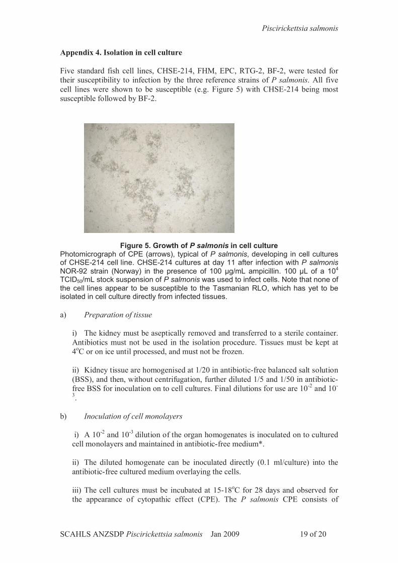
Appendix 4. Isolation in cell culture
Five standard fish cell lines, CHSE-214, FHM, EPC, RTG-2, BF-2, were tested for their susceptibility to infection by the three reference strains of P salmonis. All five cell lines were shown to be susceptible (e.g. Figure 5) with CHSE-214 being most susceptible followed by BF-2.
Figure 5. Growth of P salmonis in cell culturePhotomicrograph of CPE (arrows), typical of P salmonis, developing in cell cultures of CHSE-214 cell line. CHSE-214 cultures at day 11 after infection with P salmonisNOR-92 strain (Norway) in the presence of 100 μg/mL ampicillin. 100 μL of a 104
TCID50/mL stock suspension of P salmonis was used to infect cells. Note that none of the cell lines appear to be susceptible to the Tasmanian RLO, which has yet to be isolated in cell culture directly from infected tissues.
a) Preparation of tissue
i) The kidney must be aseptically removed and transferred to a sterile container. Antibiotics must not be used in the isolation procedure. Tissues must be kept at 4oC or on ice until processed, and must not be frozen.
ii) Kidney tissue are homogenised at 1/20 in antibiotic-free balanced salt solution (BSS), and then, without centrifugation, further diluted 1/5 and 1/50 in antibiotic-free BSS for inoculation on to cell cultures. Final dilutions for use are 10-2 and 10-
3.
b) Inoculation of cell monolayers
i) A 10-2 and 10-3 dilution of the organ homogenates is inoculated on to cultured cell monolayers and maintained in antibiotic-free medium*.
ii) The diluted homogenate can be inoculated directly (0.1 ml/culture) into the antibiotic-free cultured medium overlaying the cells.
iii) The cell cultures must be incubated at 15-18oC for 28 days and observed for the appearance of cytopathic effect (CPE). The P salmonis CPE consists of

Piscirickettsia salmonis
SCAHLS ANZSDP Piscirickettsia salmonis Jan 2009 20 of 20
plaque-like clusters (c.f. Figure 5). With time, the CPE progresses until the entire cell sheet is destroyed.
iv) If CPE does not occur (except in positive controls), cultures should be incubated for an additional 14 days.
*Experiments carried out at AAHL showed that ampicillin used at a concentration of 100 μg/mL of culture medium did not inhibit the growth of P salmonis.
Related Documents