1 Exome Sequencing Links Loss-of-Function and Missense Mutations in PARN and RTEL1 with Familial Pulmonary Fibrosis and Telomere Shortening Bridget D. Stuart 1,2 , Jungmin Choi 3,4 , Samir Zaidi 3,4 , Chao Xing 1 , Brody Holohan 5 , Rui Chen 6 , Mihwa Choi 1 , Pooja Dharwadkar 6 , Fernando Torres 6 , Carlos E. Girod 6 , Jonathan Weissler 6 , John Fitzgerald 6 , Corey Kershaw 6 , Julia Klesney-Tait 7 , Yulonda Mageto 8 , Jerry Shay 5 , Weizhen Ji 3,4 , Kaya Bilguvar 3,9 , Shrikant Mane 3,9 , Richard Lifton 3,4,9,10 and Christine Kim Garcia 1,6 From the 1 Eugene McDermott Center for Human Growth and Development, the 2 Department of Pediatrics, University of Texas Southwestern Medical Center; the 3 Department of Genetics and the 4 Howard Hughes Medical Institute, Yale University School of Medicine; the 5 Department of Cell Biology, 6 Department of Internal Medicine, University of Texas Southwestern Medical Center; the 7 Department of Internal Medicine, University of Iowa Medical Center; the 8 Department of Internal Medicine, University of Vermont; and the 9 Yale Center for Genome Analysis and the 10 Department of Internal Medicine, Yale University. Corresponding author: Christine Kim Garcia, MD, PhD University of Texas Southwestern Medical Center 5323 Harry Hines Blvd. Dallas, TX 75390-8591 Telephone: 214-648-1600 Fax: 214-648-1666 Email: [email protected] Funding: U54 HG006504 01 (Yale Center for Mendelian Genomics) ; NIH K12 HD068269 (B.D.S.); NIH UL1TR000451 and KL2TR000453 from the National Center for Advancing Translational Sciences (NCATS) and NIH R01HL093096 (C.K.G.) Manuscript style: Formatted as a Letter for Nature Genetics

Welcome message from author
This document is posted to help you gain knowledge. Please leave a comment to let me know what you think about it! Share it to your friends and learn new things together.
Transcript

1
Exome Sequencing Links Loss-of-Function and Missense Mutations in PARN and RTEL1
with Familial Pulmonary Fibrosis and Telomere Shortening
Bridget D. Stuart1,2, Jungmin Choi3,4, Samir Zaidi3,4, Chao Xing1, Brody Holohan5, Rui Chen6,
Mihwa Choi1, Pooja Dharwadkar6, Fernando Torres6, Carlos E. Girod6, Jonathan Weissler6,
John Fitzgerald6, Corey Kershaw6, Julia Klesney-Tait7, Yulonda Mageto8, Jerry Shay5, Weizhen
Ji3,4, Kaya Bilguvar3,9, Shrikant Mane3,9, Richard Lifton3,4,9,10 and Christine Kim Garcia1,6
From the 1Eugene McDermott Center for Human Growth and Development, the 2Department of
Pediatrics, University of Texas Southwestern Medical Center; the 3Department of Genetics and
the 4Howard Hughes Medical Institute, Yale University School of Medicine; the 5Department of
Cell Biology, 6Department of Internal Medicine, University of Texas Southwestern Medical
Center; the 7Department of Internal Medicine, University of Iowa Medical Center; the
8Department of Internal Medicine, University of Vermont; and the 9Yale Center for Genome
Analysis and the 10Department of Internal Medicine, Yale University.
Corresponding author: Christine Kim Garcia, MD, PhD University of Texas Southwestern Medical Center 5323 Harry Hines Blvd. Dallas, TX 75390-8591 Telephone: 214-648-1600 Fax: 214-648-1666 Email: [email protected]
Funding: U54 HG006504 01 (Yale Center for Mendelian Genomics); NIH K12 HD068269
(B.D.S.); NIH UL1TR000451 and KL2TR000453 from the National Center for Advancing
Translational Sciences (NCATS) and NIH R01HL093096 (C.K.G.)
Manuscript style: Formatted as a Letter for Nature Genetics

2
Idiopathic pulmonary fibrosis (IPF) is an age-related disease featuring progressive lung
scarring. To elucidate the molecular basis of IPF, we performed exome sequencing of
familial pulmonary fibrosis kindreds. Gene burden analysis comparing 78 European
cases and 2816 controls implicated PARN, an exoribonuclease with no prior connection
to telomere biology or disease, with five novel heterozygous damaging mutations in
unrelated cases and none in controls (P-value = 1.3 x 10⁻⁸); mutations were shared by all
affected relatives (backwards odds in favor of linkage = 4096:1). RTEL1, an established
locus for dyskeratosis congenita, harbored significantly more novel damaging and
conserved missense variants in cases than controls (P = 1.6 x 10⁻⁶). PARN and RTEL1
mutation carriers had shortened leukocyte telomere lengths and epigenetic inheritance
of short telomeres was seen in family members. Together these genes explain ~7% of
familial pulmonary fibrosis and strengthen the link between lung fibrosis and telomere
dysfunction.
Idiopathic pulmonary fibrosis (IPF) is the prototype of adult-onset interstitial lung disease
that preferentially affects males and smokers1,2. The disease is progressive, with a life
expectancy of 2-3 years after diagnosis. The genetic basis of IPF is incompletely understood.
Common variants explain a small fraction of disease risk, including loci near MUC5B and TERT,
the protein component of telomerase3. Rare coding mutations in TERT are found in ~15% of
familial pulmonary fibrosis kindreds and show autosomal dominant transmission with incomplete
penetrance4-6. Less frequently, rare mutations with large effect are found in genes encoding the
human telomerase RNA (TERC)4,5, dyskerin (DKC1)7,8, and surfactant proteins (SFTPC,
SFTPA2)9-11. Some probands of familial pulmonary fibrosis kindreds have short telomere
lengths that are unexplained by telomerase mutations12, suggesting a role for other genes
involved in telomere maintenance.
Whole exome sequencing and analysis was performed on genomic DNA samples from

3
99 probands with familial pulmonary fibrosis of unknown genetic cause (see Methods). Principal
component analysis of genotypes from exome data revealed that 79 probands clustered with
HapMap subjects of European ancestry while 19 and 1 clustered with subjects of Mexican and
African American Ancestry, respectively. Control subjects sequenced and analyzed on the same
platforms led to identification of 2816 controls of European ancestry (Figure S1). With a
population disease frequency of familial pulmonary fibrosis of <1 per 100,00013, we expected
dominant alleles of large effect to be individually very rare in the population. To enrich for
variants that are likely to alter the function of encoded proteins, we identified damaging variants
(premature termination, frameshift, splice site) and variants that altered positions that were
highly conserved across phylogeny. We compared the burden of damaging, and damaging plus
missense variants, that were found once among cases and controls and not in the NHLBI
Exome Server (ESP) or 1000 Genomes databases (Table S1). Since subjects in the NHLBI
ESP database included subjects with various cardiopulmonary disease, as a check to ensure
that we did not exclude disease-related variants, we also performed these analyses considering
all alleles with MAF < 0.1% (Table S2). Q-Q plots of damaging and missense mutations
demonstrated excellent matching of expected and observed P-values, while observed P-values
for damaging singletons were generally below the expected values owing to the paucity of such
variants (Figure 1).
Two genes surpassed thresholds for genome-wide significance in these analyses (P <
2.4 x 10⁻⁶ after accounting for examination of 21,000 genes), while no other gene had P-value <
10-4 (Figure 1, Table 1, Table S1 and Table S2). Poly(A)-specific Ribonuclease Deadenylation
Nuclease (PARN), a 3’ exoribonuclease that has not previously been implicated in disease or
telomere maintenance, had 6 damaging variants in probands and 0 in controls (P = 3.8 x 10⁻¹⁰);
all of these were confirmed by Sanger sequencing and all were absent in dbSNP, 1000
Genomes, and NHLBI cohorts (Table 2). Two PARN variants were identical and subsequently

4
found to be identical by descent from a recent common ancestor (see below). Retrospectively,
considering only 5 independent novel damaging mutations in PARN, the result remained highly
significant (P = 1.3 x 10⁻⁸). The significance of this result is further supported by the absence of
damaging mutations in PARN among 6500 subjects (including 4300 subjects of European
ancestry) in the NHLBI controls (P = 1.7 x 10⁻⁹ in European cases vs. controls). An additional
novel missense variant (K421R) was found in a proband of European ancestry. PARN is among
the 20% of genes with the lowest prevalence of rare variants that are likely to disrupt normal
function (mutation-intolerant genes)14, consistent with these variants causing haploinsufficiency.
Two probands not known to be related (F349 and F373) shared the same rare PARN
variant, altering the canonical splice acceptor of the fourth intron (AG>GG; IVS4 -2a>g). Tracing
birth and death records of both kindreds established that these two individuals had a common
great grandmother (individual II.2, Figure 2). Kinship analysis using the Beagle program
revealed that the probands share an estimated 6.4% of their genomes. The IVS4 -2a>g variant
lies on a segment of identity by descent that is ~18 Mb in length, supporting the relatedness of
these two individuals.
As an independent test of the significance of these PARN variants, their segregation was
compared to the segregation of pulmonary fibrosis in the extended kindreds of each index case.
Among 7 relatives with pulmonary fibrosis in whom mutation status was assessed either by
direct sequencing or by imputation of obligate carriers, all inherited the novel PARN variants
identified in their respective probands, an event that was highly unlikely to occur by chance (lod
score of 3.6, backwards odds of 4096:1 in favor of linkage of rare PARN variants in affected-
only analysis). The mutations were also shared by 5 relatives identified as having significant
lung disease who did not meet current criteria for a diagnosis of interstitial lung disease (Table
S3). There were also 9 clinically unaffected subjects who harbored the rare variants found in
probands, indicating incomplete penetrance of pulmonary fibrosis.

5
All five loss-of-function PARN variants involve residues within the CAF1 ribonuclease
domain, which is conserved through yeast and encode a critical component of a cytoplasmic
deadenylase (Figure 2G). A lymphoblastic cell line (LCL) derived from the proband with the
Q177X mutation demonstrated greater expression of the wildtype than the mutant allele (Figure
S2A), and PARN protein expression was reduced in six independent LCLs representing three
different loss of function mutations (Q177X, IVS4 and IVS6 splice site) (Figure S2B). There was
no apparent decrease in PARN expression in the LCLs derived from subjects heterozygous for
the K421R variant.
The other gene with a significant mutation burden was Regulator of Telomere Elongation
Helicase 1 (RTEL1), which is known to play a role in telomere maintenance. Mutations in
RTEL1 have recently been shown to cause Hoyeraal-Hreidarsson syndrome, a severe variant of
dyskeratosis congenita presenting in childhood and associated with telomere shortening15-19.
Affected subjects typically have biallelic mutations, however heterozygotes have sometimes
been noted to display disease manifestations.
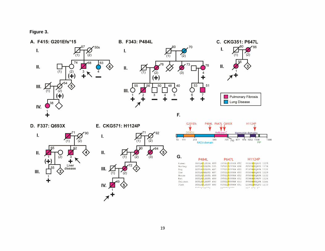
We found five novel heterozygous variants in RTEL1 in pulmonary fibrosis probands
(two damaging and three missense variants at highly conserved positions), whereas four
singletons were observed among 2816 control subjects (Figure 3, Table 2, P = 1.6 x 10⁻⁶).
Similarly, in the NHLBI cohort, there were six singleton variants in RTEL1 among 4300 subjects
of European ancestry (2 damaging and 4 conserved missense; P = 7.1 x 10⁻⁷ vs. cases). RTEL1
ranks among the 2% most mutation-intolerant genes in the genome28, consistent with
phenotypic effects from heterozygous mutations. RTEL1 contains an amino-terminal helicase
domain which preserves telomere length during replication by unwinding the repetitive telomere
TTAGGG sequences, which are organized in G-quartet secondary structures, and by
disassembling the lasso-like T-loops at the end of telomeres20. Two rare RTEL1 variants are
predicted to be loss-of-function alleles; the frameshift mutation (G201Efs) and the premature
6
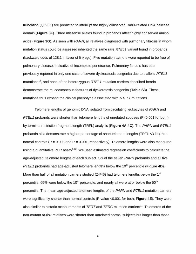
truncation (Q693X) are predicted to interrupt the highly conserved Rad3-related DNA helicase
domain (Figure 3F). Three missense alleles found in probands affect highly conserved amino
acids (Figure 3G). As seen with PARN, all relatives diagnosed with pulmonary fibrosis in whom
mutation status could be assessed inherited the same rare RTEL1 variant found in probands
(backward odds of 128:1 in favor of linkage). Five mutation carriers were reported to be free of
pulmonary disease, indicative of incomplete penetrance. Pulmonary fibrosis has been
previously reported in only one case of severe dyskeratosis congenita due to biallelic RTEL1
mutations16, and none of the heterozygous RTEL1 mutation carriers described herein
demonstrate the mucocutaneous features of dyskeratosis congenita (Table S3). These
mutations thus expand the clinical phenotype associated with RTEL1 mutations.
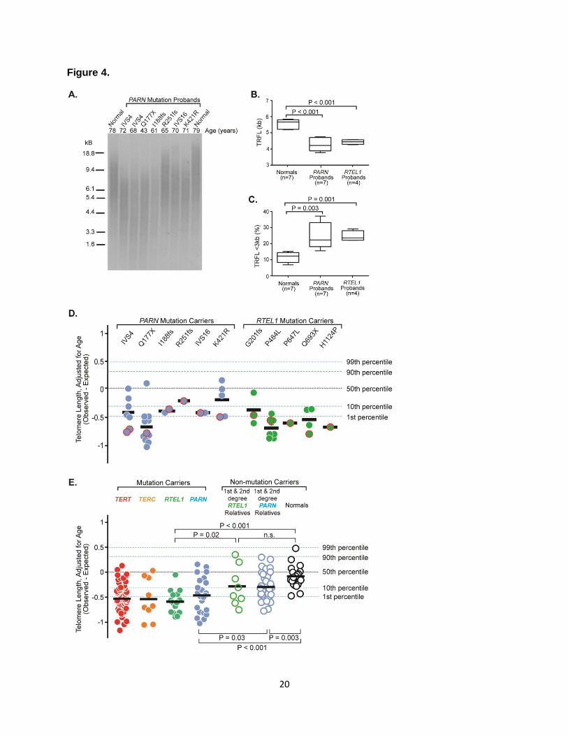
Telomere lengths of genomic DNA isolated from circulating leukocytes of PARN and
RTEL1 probands were shorter than telomere lengths of unrelated spouses (P<0.001 for both)
by terminal restriction fragment length (TRFL) analysis (Figure 4A-4C). The PARN and RTEL1
probands also demonstrate a higher percentage of short telomere lengths (TRFL <3 kb) than
normal controls (P = 0.003 and P = 0.001, respectively). Telomere lengths were also measured
using a quantitative PCR assay6,12. We used estimated regression coefficients to calculate the
age-adjusted, telomere lengths of each subject. Six of the seven PARN probands and all five
RTEL1 probands had age-adjusted telomere lengths below the 10th percentile (Figure 4D).
More than half of all mutation carriers studied (24/46) had telomere lengths below the 1st
percentile, 65% were below the 10th percentile, and nearly all were at or below the 50th
percentile. The mean age-adjusted telomere lengths of the PARN and RTEL1 mutation carriers
were significantly shorter than normal controls (P-value <0.001 for both; Figure 4E). They were
also similar to historic measurements of TERT and TERC mutation carriers21. Telomeres of the
non-mutant at-risk relatives were shorter than unrelated normal subjects but longer than those

7
of PARN or RTEL1 mutation carriers (Figure 4E). This finding is consistent with epigenetic
inheritance of telomere lengths, as has been observed in TERT kindreds6.
These studies implicate rare heterozygous loss of function variants in PARN and RTEL1
in pulmonary fibrosis with shortened telomeres. The results show a significantly increased
burden of these variants in cases compared to two independent control cohorts. In addition,
these mutations have large effects on disease risk as indicated by the high odds ratios for
association of these rare variants with disease. While no other genes in this study were
significant at genome-wide thresholds, we can’t exclude effects of variants in other genes in
small numbers of kindreds. This study demonstrates the utility of exome sequencing of
probands to discover novel disease loci with large effect despite complexities such as locus
heterogeneity, reduced penetrance, and late onset of disease.
Novel damaging or conserved PARN and RTEL1 variants were found in 12% of
probands studied. By extrapolation to our complete cohort of familial pulmonary fibrosis
kindreds that includes subjects with mutations in known genes, PARN and RTEL1 mutations are
found in at least 4 and 3%, respectively. These frequencies are intermediate between those for
TERT mutations (16%), and SFTPC (2%) or SFTPA2 (1%) mutations.
To our knowledge, this is the first report of a clinical phenotype associated with
heterozyous PARN mutations. PARN plays a major role in translational silencing of maternal
gene expression through 5'-cap-dependent deadenylation of mRNAs during oocyte maturation
and early development22,23. In addition, PARN has been shown to be involved in the maturation
of argonaute2-cleaved precursor microRNAs24 and H/ACA box snoRNAs25. The mechanism
linking PARN mutation to telomere shortening is currently unclear. However, H/ACA snoRNAs
are known to associate with four evolutionarily conserved and essential proteins: dyskerin,
GAR1, NHP2 and NOP10, and mutation of three of these cause dyskeratosis congenita26-28. We
8
speculate that PARN haploinsufficiency may lead to telomere shortening through dysregulation
of H/ACA box snoRNAs or the RNA component of human telomerase (TERC), which itself
contains an H/ACA domain.
Short telomere lengths are disproportionately represented in patients with sporadic
IPF12,29 and are associated with worse survival of IPF patients21. The identification of two new
genes contributing to this trait and telomere shortening strengthens the link between telomere
attrition and lung fibrosis and identifies a new gene required for telomere maintenance.
Methods
Methods and any associated references are available in the online version of the paper.
Accession codes. The position of the DNA and protein variants are described using PARN
NM_2582.3 (variant 1) and RTEL1 NM_1283009.1 (variant 3). Whole exome sequencing data
has been deposited in dbGAP under accession ___ (this is currently pending).
Acknowledgements
We are grateful to the probands and their families for their participation, to Ashley Young for
technical excellence and to Gentry Wools, Sam Nolasco and Kim Stephens for their help with
blood sample collections and to the staff of the Yale Center for Genome Analysis for production
of exome sequence data.
Author Contributions

9
C.K.G, C.X, and R. L. conceived, designed and directed the study; J.C., S.Z., C.X., W.J., K.B.
and S.M. performed genetic analyses, B.D.S, B.H., R.C., M.C. and J.S performed and directed
experiments, P.D., F.T., C.E.G., J.W., J.F., C.K., J.K.-T., and Y.M. contributed clinical
evaluations. All authors approved the final manuscript and contributed critical revisions to its
intellectual content.

10
References
1. Travis, W.D., et al. An official American Thoracic Society/European Respiratory Society
statement: Update of the international multidisciplinary classification of the idiopathic interstitial pneumonias. Am J Respir Crit Care Med 188, 733-748 (2013).
2. Raghu, G., et al. An official ATS/ERS/JRS/ALAT statement: idiopathic pulmonary fibrosis: evidence-based guidelines for diagnosis and management. Am J Respir Crit Care Med 183, 788-824 (2011).
3. Fingerlin, T.E., et al. Genome-wide association study identifies multiple susceptibility loci for pulmonary fibrosis. Nat Genet 45, 613-620 (2013).
4. Armanios, M.Y., et al. Telomerase mutations in families with idiopathic pulmonary fibrosis. N Engl J Med 356, 1317-1326 (2007).
5. Tsakiri, K.D., et al. Adult-onset pulmonary fibrosis caused by mutations in telomerase. Proc Natl Acad Sci U S A 104, 7552-7557 (2007).
6. Diaz de Leon, A., et al. Telomere lengths, pulmonary fibrosis and telomerase (TERT) mutations. PLoS ONE 5, e10680 (2010).
7. Kropski, J.A., et al. A novel dyskerin (DKC1) mutation is associated with familial interstitial pneumonia. Chest 146, e1-7 (2014).
8. Parry, E.M., et al. Decreased dyskerin levels as a mechanism of telomere shortening in X-linked dyskeratosis congenita. J Med Genet 48, 327-333 (2011).
9. Nogee, L.M., et al. A mutation in the surfactant protein C gene associated with familial interstitial lung disease. N Engl J Med 344, 573-579 (2001).
10. Thomas, A.Q., et al. Heterozygosity for a surfactant protein C gene mutation associated
with usual interstitial pneumonitis and cellular nonspecific interstitial pneumonitis in one kindred. Am J Respir Crit Care Med 165, 1322-1328 (2002).
11. Wang, Y., et al. Genetic defects in surfactant protein A2 are associated with pulmonary fibrosis and lung cancer. Am J Hum Genet 84, 52-59 (2009).
12. Cronkhite, J.T., et al. Telomere shortening in familial and sporadic pulmonary fibrosis. Am J Respir Crit Care Med 178, 729-737 (2008).
13. Marshall, R.P., Puddicombe, A., Cookson, W.O. & Laurent, G.J. Adult familial cryptogenic fibrosing alveolitis in the United Kingdom. Thorax 55, 143-146 (2000).
14. Petrovski, S., Wang, Q., Heinzen, E.L., Allen, A.S. & Goldstein, D.B. Genic intolerance to functional variation and the interpretation of personal genomes. PLoS Genet 9,
e1003709 (2013). 15. Ballew, B.J., et al. A recessive founder mutation in regulator of telomere elongation
helicase 1, RTEL1, underlies severe immunodeficiency and features of Hoyeraal Hreidarsson syndrome. PLoS Genet 9, e1003695 (2013).
16. Deng, Z., et al. Inherited mutations in the helicase RTEL1 cause telomere dysfunction and Hoyeraal-Hreidarsson syndrome. Proc Natl Acad Sci U S A 110, E3408-3416
(2013). 17. Le Guen, T., et al. Human RTEL1 deficiency causes Hoyeraal-Hreidarsson syndrome
with short telomeres and genome instability. Hum Mol Genet 22, 3239-3249 (2013). 18. Walne, A.J., Vulliamy, T., Kirwan, M., Plagnol, V. & Dokal, I. Constitutional mutations in
RTEL1 cause severe dyskeratosis congenita. Am J Hum Genet 92, 448-453 (2013). 19. Ballew, B.J., et al. Germline mutations of regulator of telomere elongation helicase 1,
RTEL1, in Dyskeratosis congenita. Hum Genet 132, 473-480 (2013).
20. Vannier, J.B., Pavicic-Kaltenbrunner, V., Petalcorin, M.I., Ding, H. & Boulton, S.J. RTEL1 dismantles T loops and counteracts telomeric G4-DNA to maintain telomere integrity. Cell 149, 795-806 (2012).

11
21. Stuart, B.D., et al. Effect of telomere length on survival in patients with idiopathic pulmonary fibrosis: an observational cohort study with independent validation. The lancet. Respiratory medicine 2, 557-565 (2014).
22. Korner, C.G., et al. The deadenylating nuclease (DAN) is involved in poly(A) tail removal during the meiotic maturation of Xenopus oocytes. The EMBO journal 17, 5427-5437
(1998). 23. Dehlin, E., Wormington, M., Korner, C.G. & Wahle, E. Cap-dependent deadenylation of
mRNA. The EMBO journal 19, 1079-1086 (2000). 24. Yoda, M., et al. Poly(A)-specific ribonuclease mediates 3'-end trimming of Argonaute2-
cleaved precursor microRNAs. Cell reports 5, 715-726 (2013). 25. Berndt, H., et al. Maturation of mammalian H/ACA box snoRNAs: PAPD5-dependent
adenylation and PARN-dependent trimming. RNA (New York, N.Y 18, 958-972 (2012). 26. Heiss, N.S., et al. X-linked dyskeratosis congenita is caused by mutations in a highly
conserved gene with putative nucleolar functions. Nat Genet 19, 32-38 (1998). 27. Vulliamy, T., et al. Mutations in the telomerase component NHP2 cause the premature
ageing syndrome dyskeratosis congenita. Proc Natl Acad Sci U S A 105, 8073-8078 (2008).
28. Walne, A.J., et al. Genetic heterogeneity in autosomal recessive dyskeratosis congenita with one subtype due to mutations in the telomerase-associated protein NOP10. Hum Mol Genet (2007).
29. Alder, J.K., et al. Short telomeres are a risk factor for idiopathic pulmonary fibrosis. Proc Natl Acad Sci U S A 105, 13051-13056 (2008).
12
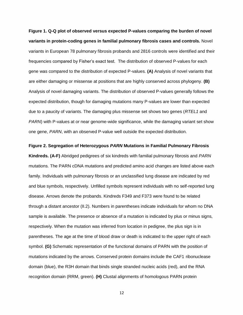
Figure 1. Q-Q plot of observed versus expected P-values comparing the burden of novel
variants in protein-coding genes in familial pulmonary fibrosis cases and controls. Novel
variants in European 78 pulmonary fibrosis probands and 2816 controls were identified and their
frequencies compared by Fisher’s exact test. The distribution of observed P-values for each
gene was compared to the distribution of expected P-values. (A) Analysis of novel variants that
are either damaging or missense at positions that are highly conserved across phylogeny. (B)
Analysis of novel damaging variants. The distribution of observed P-values generally follows the
expected distribution, though for damaging mutations many P-values are lower than expected
due to a paucity of variants. The damaging plus missense set shows two genes (RTEL1 and
PARN) with P-values at or near genome-wide significance, while the damaging variant set show
one gene, PARN, with an observed P-value well outside the expected distribution.
Figure 2. Segregation of Heterozygous PARN Mutations in Familial Pulmonary Fibrosis
Kindreds. (A-F) Abridged pedigrees of six kindreds with familial pulmonary fibrosis and PARN
mutations. The PARN cDNA mutations and predicted amino acid changes are listed above each
family. Individuals with pulmonary fibrosis or an unclassified lung disease are indicated by red
and blue symbols, respectively. Unfilled symbols represent individuals with no self-reported lung
disease. Arrows denote the probands. Kindreds F349 and F373 were found to be related
through a distant ancestor (II.2). Numbers in parentheses indicate individuals for whom no DNA
sample is available. The presence or absence of a mutation is indicated by plus or minus signs,
respectively. When the mutation was inferred from location in pedigree, the plus sign is in
parentheses. The age at the time of blood draw or death is indicated to the upper right of each
symbol. (G) Schematic representation of the functional domains of PARN with the position of
mutations indicated by the arrows. Conserved protein domains include the CAF1 ribonuclease
domain (blue), the R3H domain that binds single stranded nucleic acids (red), and the RNA
recognition domain (RRM, green). (H) Clustal alignments of homologous PARN protein

13
sequences from Homo sapiens (human), Macaca mulatta (monkey), Canis familiaris (dog), Bos
taurus (cow), Mus musculus (mouse), Rattus norvegicus (rat), Gallus gallus (chicken), Danio
rerio (fish).
Figure 3. Segregation of Heterozygous RTEL1 Mutations in Familial Pulmonary Fibrosis
Kindreds and the Location of RTEL1 Alternations in the Different Protein Domains. (A-E)
Abridged pedigrees of six kindreds with familial pulmonary fibrosis and RTEL1 mutations. The
RTEL1 cDNA mutations and predicted amino acid changes are listed above each family.
Pedigree information is presented in a similar manner as in Figure 1. (F) Schematic
representation of the functional domains of RTEL1 protein with the position of the mutations
indicated by the arrows. Conserved protein domains: DEAD2 domain (orange bar), helicase C
domain (red), RAD3-related DNA helicase domain (blue), harmonin N-like domian (purple) and
the proliferating cell nuclear antigen (PCNA)-interacting protein (PIP) domain (green). (G)
Clustal alignments of homologous RTEL1 protein sequences from species listed in Figure 2.
Figure 4. PARN and RTEL1 Mutations Are Associated with Short Leukocyte Telomere
Lengths. (A) Telomere lengths determined by Southern blot terminal restriction fragment length
(TRFL) analysis of nonrelated normal (spouse) controls and PARN probands. The age of each
individual is indicated above the blot. Average TRFL (B) and percent short TRFL (<3 kb) (C) are
indicated for the normals and PARN and RTEL1 probands, who have an average age of 64, 64
and 60 years, respectively. (D) Telomere length of genomic DNA isolated from circulating
leukocytes from heterozygous PARN and RTEL1 mutation carriers as measured by a
multiplexed qPCR assay. Telomere lengths are age-adjusted and are expressed as the
observed minus the expected length. Each symbol represents a unique individual; the proband
for each family is indicated by the red outline. The mean telomere length is indicated by the
black bar. The black dotted line shows the median telomere length of an independent control
group; approximate age-adjusted prediction bands (percentiles, blue dotted lines) were

14
previously calculated from a linear regression model using the controls. (E) Genomic DNA
telomere lengths as measured by the multiplexed qPCR assay of individuals heterozygous for a
mutation in TERT, TERC, RTEL1 and PARN are shown as red, orange, green and blue colored
circles, respectively. Telomere lengths of 1st and 2nd degree relatives of the PARN or RTEL1
mutation carriers who did not inherit the variants are shown as open green and blue circles,
respectively. Open black circles represent unrelated normal controls.

15
Table 1. Increased burden of novel variants in PARN and RTEL1 in familial pulmonary fibrosis probands vs. controls of European descent
YCGA controls
Number of Alleles, Damaging plus Missense at Conserved Positions
Number of Alleles, Damaging
Controls Cases p-value Controls Cases p-value
Gene # variant #
reference # variant
# reference
# variant # reference # variant #
reference
PARN 11 5621 6 150 3.37E-06 0 5632 5 151 1.33E-08
RTEL1 4 5598 5 151 1.58E-06 0 5602 2 154 7.29E-04
NHLBI
controls
Number of Alleles, Damaging plus Missense at Conserved Positions
Number of Alleles, Damaging
Controls Cases p-value Controls Cases p-value
Gene # variant #
reference # variant
# reference
# variant # reference # variant #
reference
PARN 8 8592 6 150 7.76E-08 0 8600 5 151 1.69E-09
RTEL1 6 8594 5 151 7.14E-07 2 8598 2 154 1.85E-03
YCGA controls, variants analyzed from Yale Center for Genome Analysis; NHLBI, variants from NHLBI ESP database
16
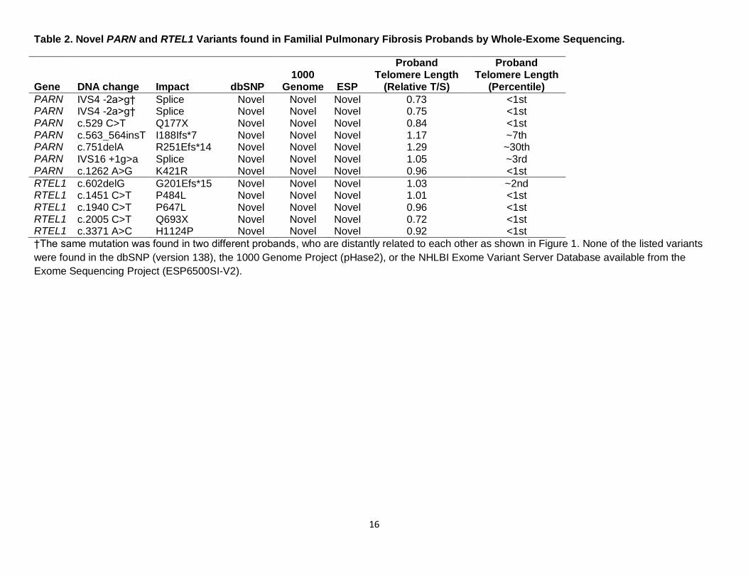
Table 2. Novel PARN and RTEL1 Variants found in Familial Pulmonary Fibrosis Probands by Whole-Exome Sequencing.
Gene DNA change Impact dbSNP 1000
Genome ESP
Proband Telomere Length
(Relative T/S)
Proband Telomere Length
(Percentile)
PARN IVS4 -2a>g† Splice Novel Novel Novel 0.73 <1st PARN IVS4 -2a>g† Splice Novel Novel Novel 0.75 <1st PARN c.529 C>T Q177X Novel Novel Novel 0.84 <1st PARN c.563_564insT I188Ifs*7 Novel Novel Novel 1.17 ~7th PARN c.751delA R251Efs*14 Novel Novel Novel 1.29 ~30th PARN IVS16 +1g>a Splice Novel Novel Novel 1.05 ~3rd PARN c.1262 A>G K421R Novel Novel Novel 0.96 <1st
RTEL1 c.602delG G201Efs*15 Novel Novel Novel 1.03 ~2nd RTEL1 c.1451 C>T P484L Novel Novel Novel 1.01 <1st RTEL1 c.1940 C>T P647L Novel Novel Novel 0.96 <1st RTEL1 c.2005 C>T Q693X Novel Novel Novel 0.72 <1st RTEL1 c.3371 A>C H1124P Novel Novel Novel 0.92 <1st
†The same mutation was found in two different probands, who are distantly related to each other as shown in Figure 1. None of the listed variants
were found in the dbSNP (version 138), the 1000 Genome Project (pHase2), or the NHLBI Exome Variant Server Database available from the
Exome Sequencing Project (ESP6500SI-V2).

17
Figure 1.

18
Figure 2.
19
Figure 3.
20
Figure 4.
Related Documents
![Research Paper MiR-185 targets POT1 to induce telomere ... · and induce telomere fragility, replication fork stalling, and telomere elongation [5, 6]. POT1 is a key protein linking](https://static.cupdf.com/doc/110x72/603d50e8cb3cfc37ff77b2c6/research-paper-mir-185-targets-pot1-to-induce-telomere-and-induce-telomere-fragility.jpg)