Journal of NeuroVirology, 11: 319–328, 2005 c 2005 Journal of NeuroVirology ISSN: 1355-0284 print / 1538-2443 online DOI: 10.1080/13550280500187005 Exogenous IL-7 induces Fas-mediated human neuronal apoptosis: potential effects during human immunodeficiency virus type 1 infection Giuseppe Nunnari, Yan Xu, Edward A Acheampong, Jianhua Fang, Rene Daniel, Chune Zhang, Hui Zhang, Muhammad Mukhtar, and Roger J. Pomerantz Center for Human Virology and Biodefense, Division of Infectious Diseases and Environmental Medicine, Department of Medicine, Thomas Jefferson University, Philadelphia, Pennsylvania, USA The use of exogenous cytokines is part of translational immune-antiretroviral approaches to induce immune reconstitution and possibly eliminate the per- sistence of human immunodeficiency virus type 1 (HIV-1) in virally suppressed infected individuals on highly active antiretroviral therapy (HAART). Re- cently, our laboratories demonstrated that interleukin-7 (IL-7) has significant efficiency in stimulating HIV-1 replication from proviral latency in CD4+ T lymphocytes of infected patients. The authors now investigated the possible role of IL-7 in HIV-1–associated dementia (HAD). The authors demonstrated that the IL-7 receptor is expressed on both human neurons (i.e., differenti- ated NT2 cells) and human astrocytes, with relatively higher mRNA levels in neurons. The translational protein levels of IL-7 receptor α were not pro- portional to those of the mRNA levels in these central nervous system (CNS)- based cell types. Exogenous IL-7 was observed to only slightly down-regulate IL-7 receptor α expression on both neurons and astrocytes, as assayed by Western blotting. Instead of promoting survival, surprisingly, exogenous IL- 7 induced neuronal apoptosis, as detected by TUNEL assays. Furthermore, IL-7 augmented neuronal apoptosis induced by HIV-1 gp120. Human apop- tosis genomic microarray analyses of IL-7–treated human neurons showed up-regulated expression of proapoptotic genes: protein kinases, caspase-10, FAST kinase, tumor necrosis factor (TNF) receptor, and BCL2-antagonist of cell death. These data suggest that IL-7 leads to neuronal apoptosis by a molecu- lar mechanism(s) that occurs via Fas-mediated activation-induced cell death. These studies may therefore not only be key in evaluating the potential use of IL-7 in vivo as a therapeutic modality, but also suggest that IL-7, which is increased endogenously in HIV-1–infected individuals late in disease, may be involved in the neuronal apoptosis demonstrated during HAD. Journal of NeuroVirology (2005) 11, 319–328. Keywords: apoptosis; CNS; HIV-1; IL-7; neurons Address correspondence to Giuseppe Nunnari or Roger J. Pomerantz, Center for Human Virology and Biodefense, Division of Infectious Diseases and Environmental Medicine, Department of Medicine, Thomas Jefferson University, Jefferson Alumni Hall, 1020 Locust Street, Suite 329, Philadelphia, PA 19107, USA. E-mail: [email protected] or RogerJ.Pomerantz@ jefferson.edu The authors wish to thank Dr. Avindra Nath of Johns Hopkins University for producing the human primary astrocytes, and Brenda O. Gordon and Rita M. Victor for excellent secretarial assis- tance. This work was supported in part by USPHS grants NS41864, NS44513, AA13849, and AI43289 to R. J. P. Received 20 February 2005; revised 25 April 2005; accepted 7 May 2005. Introduction Despite the use of highly active antiretroviral ther- apy (HAART), eliminating latent human immunode- ficiency virus (HIV)-1 reservoirs remains a significant challenge. The use of exogenous cytokine treatment to purge latently infected cells and improve immune function is a potential immune-adjunctive therapy to HAART. Interleukin-6 (IL-6), IL-2, and tumor necro- sis factor alpha (TNF-α) treatment have been shown to activate latently infected CD4+ T cells, increase the turnover rate of latent virus in vitro and also

Welcome message from author
This document is posted to help you gain knowledge. Please leave a comment to let me know what you think about it! Share it to your friends and learn new things together.
Transcript
Journal of NeuroVirology, 11: 319–328, 2005c© 2005 Journal of NeuroVirologyISSN: 1355-0284 print / 1538-2443 onlineDOI: 10.1080/13550280500187005
Exogenous IL-7 induces Fas-mediated humanneuronal apoptosis: potential effects during humanimmunodeficiency virus type 1 infection
Giuseppe Nunnari, Yan Xu, Edward A Acheampong, Jianhua Fang, Rene Daniel, Chune Zhang, Hui Zhang,Muhammad Mukhtar, and Roger J. Pomerantz
Center for Human Virology and Biodefense, Division of Infectious Diseases and Environmental Medicine, Departmentof Medicine, Thomas Jefferson University, Philadelphia, Pennsylvania, USA
The use of exogenous cytokines is part of translational immune-antiretroviralapproaches to induce immune reconstitution and possibly eliminate the per-sistence of human immunodeficiency virus type 1 (HIV-1) in virally suppressedinfected individuals on highly active antiretroviral therapy (HAART). Re-cently, our laboratories demonstrated that interleukin-7 (IL-7) has significantefficiency in stimulating HIV-1 replication from proviral latency in CD4+T lymphocytes of infected patients. The authors now investigated the possiblerole of IL-7 in HIV-1–associated dementia (HAD). The authors demonstratedthat the IL-7 receptor is expressed on both human neurons (i.e., differenti-ated NT2 cells) and human astrocytes, with relatively higher mRNA levelsin neurons. The translational protein levels of IL-7 receptor α were not pro-portional to those of the mRNA levels in these central nervous system (CNS)-based cell types. Exogenous IL-7 was observed to only slightly down-regulateIL-7 receptor α expression on both neurons and astrocytes, as assayed byWestern blotting. Instead of promoting survival, surprisingly, exogenous IL-7 induced neuronal apoptosis, as detected by TUNEL assays. Furthermore,IL-7 augmented neuronal apoptosis induced by HIV-1 gp120. Human apop-tosis genomic microarray analyses of IL-7–treated human neurons showedup-regulated expression of proapoptotic genes: protein kinases, caspase-10,FAST kinase, tumor necrosis factor (TNF) receptor, and BCL2-antagonist of celldeath. These data suggest that IL-7 leads to neuronal apoptosis by a molecu-lar mechanism(s) that occurs via Fas-mediated activation-induced cell death.These studies may therefore not only be key in evaluating the potential useof IL-7 in vivo as a therapeutic modality, but also suggest that IL-7, whichis increased endogenously in HIV-1–infected individuals late in disease, maybe involved in the neuronal apoptosis demonstrated during HAD. Journal ofNeuroVirology (2005) 11, 319–328.
Keywords: apoptosis; CNS; HIV-1; IL-7; neurons
Address correspondence to Giuseppe Nunnari or Roger J.Pomerantz, Center for Human Virology and Biodefense, Divisionof Infectious Diseases and Environmental Medicine, Departmentof Medicine, Thomas Jefferson University, Jefferson Alumni Hall,1020 Locust Street, Suite 329, Philadelphia, PA 19107, USA.E-mail: [email protected] or [email protected]
The authors wish to thank Dr. Avindra Nath of Johns HopkinsUniversity for producing the human primary astrocytes, andBrenda O. Gordon and Rita M. Victor for excellent secretarial assis-tance. This work was supported in part by USPHS grants NS41864,NS44513, AA13849, and AI43289 to R. J. P.
Received 20 February 2005; revised 25 April 2005; accepted7 May 2005.
Introduction
Despite the use of highly active antiretroviral ther-apy (HAART), eliminating latent human immunode-ficiency virus (HIV)-1 reservoirs remains a significantchallenge. The use of exogenous cytokine treatmentto purge latently infected cells and improve immunefunction is a potential immune-adjunctive therapy toHAART. Interleukin-6 (IL-6), IL-2, and tumor necro-sis factor alpha (TNF-α) treatment have been shownto activate latently infected CD4+ T cells, increasethe turnover rate of latent virus in vitro and also
IL-7 neuronal apoptosis with HIV-1320 G Nunnari et al
elevate the CD4+ T-cell count in a well-establishedSCIDhu mouse model (Brooks et al, 2001; Chun et al,1998). Recently, IL-7 was reported to induce the ex-pression of latent HIV-1 with minimal effects on T-cell phenotype (Llano et al, 2001; Scripture-Adamset al, 2002). Moreover, we recently demonstrated thatIL-7 is a potent and proviral strain-specific inducer oflatent, in vivo HIV-1 cellular reservoirs of infected in-dividuals on virally suppressive HAART (Wang et al,2005).
Immunologically, IL-7 is a nonredundant cytokineproduced by stromal cells of the thymus and thebone marrow and by keratinocytes (Sudo et al, 1989;Wolf et al, 1992; Heufler et al, 1993). It plays an im-portant role in B-cell lymphopoiesis and differenti-ation of thymocytes into mature T cells (Fry et al,2001; Grabstein et al, 1990). The high-affinity IL-7receptor (IL-7R) is responsible for mediating the ef-fects of IL-7 (Goodwin et al, 1990). The IL-7 receptorcomplex is composed of α and ϒc subunits, whichare shared by IL-2, IL-4, IL-9, and IL-15 (Leonardet al, 2001). ϒc serves primarily to activate signaltransduction through the complex, whereas IL-7Rαdetermines specific signaling events through its as-sociation with cytoplasmic signaling molecules (Laiet al, 1997). IL-7Rα has three cytoplasmic domains,A (acidic), S (serine-containing), and T (tyrosine-containing), serving as potential docking regions forthese kinases (Lin et al, 1995). The IL-7 receptorcomplex signals through phosphorylation of tyro-sine, serine, threonine protein kinases, activates sig-nal transducer and activator of transcription (STAT),plus it also increases transcription of early responsegenes (Miyazaki et al, 1994; Lin et al, 1995; Wilkset al, 1994; Cosenza et al, 2002). Recent evidencesuggests that the actions of survival/death factorsare affected by modulation of the phosphorylationstatus of important elements of the apoptotic pro-cess (Cross et al, 2000). Signal transduction path-ways also could activate the death receptors, suchas Fas, TNF, and TNF-related ligand (TRAIL), lead-ing to cell death (Cross et al, 2000; Dudley et al,2004).
Of note, there is an inverse correlation betweenCD4+ T-cell depletion and the serum levels of IL-7in patients with HIV-1 infection (Llano et al, 2001;Napolitano et al, 2001). These data suggest that IL-7is not only an indicator of CD4+ T-cell depletion,but also consequently a marker of disease progres-sion towards acquired immunodeficiency syndrome(AIDS). Taken together, these studies suggest that IL-7could possibly be a candidate for future study as anadjunctive therapy to HAART.
Jaleco et al (2003) demonstrated that IL-7 regu-lates T-cell homeostasis by modulating the balancebetween proliferation and apoptosis in recent thymicemigrants (RTEs), and mature naı̈ve and memoryT cells. They found that RTEs and mature naı̈veand memory CD4+ T cells were sensitive to Fas-mediated cell death following exposure to either IL-2
or IL-7 alone. Furthermore, Fas engagement in thepresence of both IL-2 and IL-7 resulted in high levelsof caspase-dependent apoptosis.
Cytokines could originate from the brain or the pe-riphery (Raber et al, 1994), and penetrate the blood-brain barrier (BBB) (Ellison et al, 1987). During HIV-1infection, elevated cytokines from infected peripherycells and brain cells could induce neuronal apoptosis(Kaul et al, 2001), leading to HIV-1–associated dimen-tia (HAD). Interestingly, HAD occurs during the lateststages of AIDS, in which the highest levels of IL-7 arereached. It is reasonable to speculate the IL-7 mightplay either a direct or indirect role in the pathogene-sis of HAD.
IL-7R has been reported to be widely present in dif-ferent human organs (Cosenza et al, 2002; Dus et al,2003). Nonetheless, little is known about the role ofIL-7 in the central nervous system (CNS) in general,and during HAD in particular.
In this study, we demonstrated that IL-7R mRNAand protein are expressed in human mature neuronsfrom differentiated NT2 cells and human astrocytes.We found that IL-7 only slightly down-regulated IL-7Rα expression in these CNS cell types. Finally,we observed that IL-7 induced neuronal apoptosis,but did not induce programmed cell death in earlypassage primary astrocytes. Furthermore, IL-7 aug-mented neuronal apoptosis induced by the HIV-1envelope protein gp120. Gene microarray analysesshowed that up-modulated proapoptotic genes wererelated to Fas-mediated activation-induced cell deathin human neurons.
Results
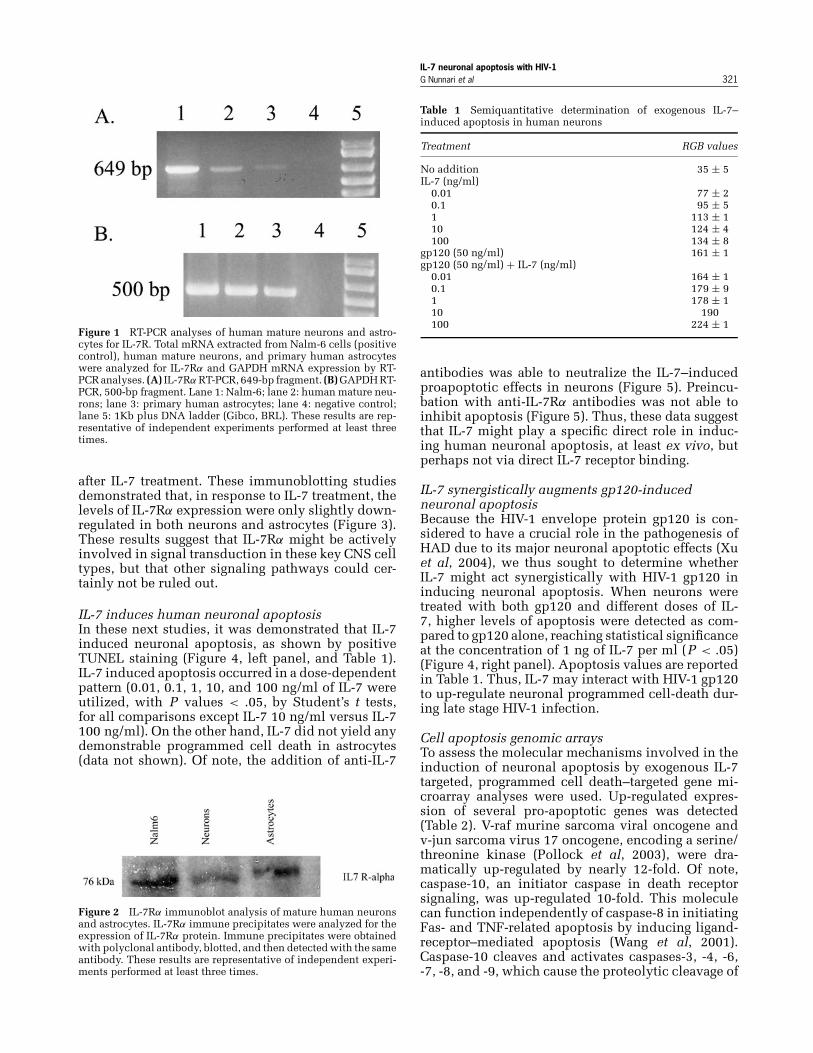
Expression of IL7R mRNA and protein on humanneurons and astrocytesIn the present study, we investigated whether theIL-7 receptor α (IL-7Rα) is expressed on human neu-rons and astrocytes. First, we determined the expres-sion of IL-7 mRNA by these cells in vitro. Nalm-6B-cells were used as positive controls for the detec-tion of IL7Rα mRNA. As shown in Figure 1, IL7RαmRNA was detected on both human differentiatedneurons and astrocytes, but with repeatedly higherlevels demonstrated in neurons.
The expression of human IL-7 receptor protein, anintegral strongly glycosylated membrane protein of76 kDa, designated as CD127 (Cosenza et al, 2002),was then measured. The p76 IL-7Rα protein was de-tected by immunoblotting on both human neuronsand astrocytes, as illustrated in Figure 2.
IL-7 and IL-7Rα expression in both humanneurons and astrocytesThe biological effects of IL-7 are mediated througha receptor complex containing the IL-7Rα and theϒc subunits (Lai et al, 1997). As such, we comparedthe baseline levels of IL-7Rα expression with those
IL-7 neuronal apoptosis with HIV-1G Nunnari et al 321
Figure 1 RT-PCR analyses of human mature neurons and astro-cytes for IL-7R. Total mRNA extracted from Nalm-6 cells (positivecontrol), human mature neurons, and primary human astrocyteswere analyzed for IL-7Rα and GAPDH mRNA expression by RT-PCR analyses. (A) IL-7Rα RT-PCR, 649-bp fragment. (B) GAPDH RT-PCR, 500-bp fragment. Lane 1: Nalm-6; lane 2: human mature neu-rons; lane 3: primary human astrocytes; lane 4: negative control;lane 5: 1Kb plus DNA ladder (Gibco, BRL). These results are rep-resentative of independent experiments performed at least threetimes.
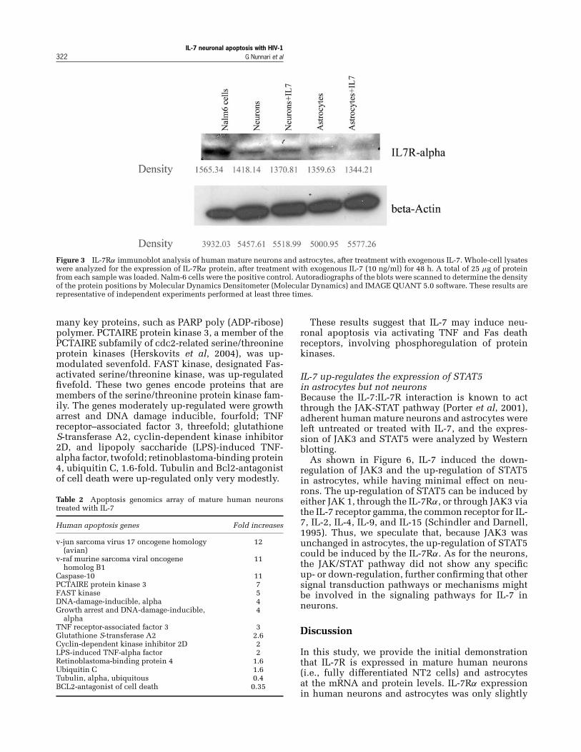
after IL-7 treatment. These immunoblotting studiesdemonstrated that, in response to IL-7 treatment, thelevels of IL-7Rα expression were only slightly down-regulated in both neurons and astrocytes (Figure 3).These results suggest that IL-7Rα might be activelyinvolved in signal transduction in these key CNS celltypes, but that other signaling pathways could cer-tainly not be ruled out.
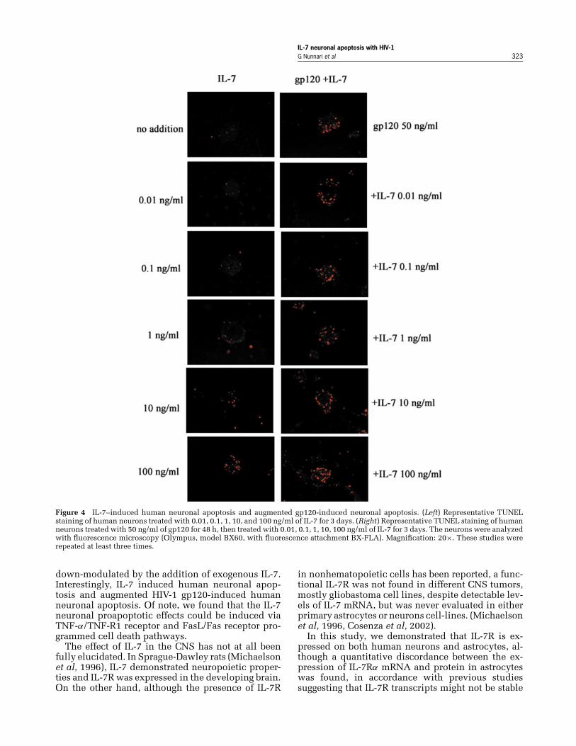
IL-7 induces human neuronal apoptosisIn these next studies, it was demonstrated that IL-7induced neuronal apoptosis, as shown by positiveTUNEL staining (Figure 4, left panel, and Table 1).IL-7 induced apoptosis occurred in a dose-dependentpattern (0.01, 0.1, 1, 10, and 100 ng/ml of IL-7 wereutilized, with P values < .05, by Student’s t tests,for all comparisons except IL-7 10 ng/ml versus IL-7100 ng/ml). On the other hand, IL-7 did not yield anydemonstrable programmed cell death in astrocytes(data not shown). Of note, the addition of anti-IL-7
Figure 2 IL-7Rα immunoblot analysis of mature human neuronsand astrocytes. IL-7Rα immune precipitates were analyzed for theexpression of IL-7Rα protein. Immune precipitates were obtainedwith polyclonal antibody, blotted, and then detected with the sameantibody. These results are representative of independent experi-ments performed at least three times.
Table 1 Semiquantitative determination of exogenous IL-7–induced apoptosis in human neurons
Treatment RGB values
No addition 35 ± 5IL-7 (ng/ml)
0.01 77 ± 20.1 95 ± 51 113 ± 110 124 ± 4100 134 ± 8
gp120 (50 ng/ml) 161 ± 1gp120 (50 ng/ml) + IL-7 (ng/ml)
0.01 164 ± 10.1 179 ± 91 178 ± 110 190100 224 ± 1
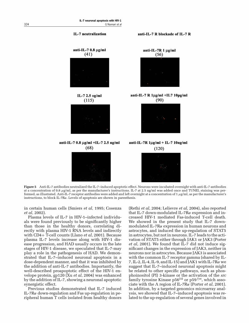
antibodies was able to neutralize the IL-7–inducedproapoptotic effects in neurons (Figure 5). Preincu-bation with anti-IL-7Rα antibodies was not able toinhibit apoptosis (Figure 5). Thus, these data suggestthat IL-7 might play a specific direct role in induc-ing human neuronal apoptosis, at least ex vivo, butperhaps not via direct IL-7 receptor binding.
IL-7 synergistically augments gp120-inducedneuronal apoptosisBecause the HIV-1 envelope protein gp120 is con-sidered to have a crucial role in the pathogenesis ofHAD due to its major neuronal apoptotic effects (Xuet al, 2004), we thus sought to determine whetherIL-7 might act synergistically with HIV-1 gp120 ininducing neuronal apoptosis. When neurons weretreated with both gp120 and different doses of IL-7, higher levels of apoptosis were detected as com-pared to gp120 alone, reaching statistical significanceat the concentration of 1 ng of IL-7 per ml (P < .05)(Figure 4, right panel). Apoptosis values are reportedin Table 1. Thus, IL-7 may interact with HIV-1 gp120to up-regulate neuronal programmed cell-death dur-ing late stage HIV-1 infection.
Cell apoptosis genomic arraysTo assess the molecular mechanisms involved in theinduction of neuronal apoptosis by exogenous IL-7targeted, programmed cell death–targeted gene mi-croarray analyses were used. Up-regulated expres-sion of several pro-apoptotic genes was detected(Table 2). V-raf murine sarcoma viral oncogene andv-jun sarcoma virus 17 oncogene, encoding a serine/threonine kinase (Pollock et al, 2003), were dra-matically up-regulated by nearly 12-fold. Of note,caspase-10, an initiator caspase in death receptorsignaling, was up-regulated 10-fold. This moleculecan function independently of caspase-8 in initiatingFas- and TNF-related apoptosis by inducing ligand-receptor–mediated apoptosis (Wang et al, 2001).Caspase-10 cleaves and activates caspases-3, -4, -6,-7, -8, and -9, which cause the proteolytic cleavage of
IL-7 neuronal apoptosis with HIV-1322 G Nunnari et al
Figure 3 IL-7Rα immunoblot analysis of human mature neurons and astrocytes, after treatment with exogenous IL-7. Whole-cell lysateswere analyzed for the expression of IL-7Rα protein, after treatment with exogenous IL-7 (10 ng/ml) for 48 h. A total of 25 μg of proteinfrom each sample was loaded. Nalm-6 cells were the positive control. Autoradiographs of the blots were scanned to determine the densityof the protein positions by Molecular Dynamics Densitometer (Molecular Dynamics) and IMAGE QUANT 5.0 software. These results arerepresentative of independent experiments performed at least three times.
many key proteins, such as PARP poly (ADP-ribose)polymer. PCTAIRE protein kinase 3, a member of thePCTAIRE subfamily of cdc2-related serine/threonineprotein kinases (Herskovits et al, 2004), was up-modulated sevenfold. FAST kinase, designated Fas-activated serine/threonine kinase, was up-regulatedfivefold. These two genes encode proteins that aremembers of the serine/threonine protein kinase fam-ily. The genes moderately up-regulated were growtharrest and DNA damage inducible, fourfold; TNFreceptor–associated factor 3, threefold; glutathioneS-transferase A2, cyclin-dependent kinase inhibitor2D, and lipopoly saccharide (LPS)-induced TNF-alpha factor, twofold; retinoblastoma-binding protein4, ubiquitin C, 1.6-fold. Tubulin and Bcl2-antagonistof cell death were up-regulated only very modestly.
Table 2 Apoptosis genomics array of mature human neuronstreated with IL-7
Human apoptosis genes Fold increases
v-jun sarcoma virus 17 oncogene homology(avian)
12
v-raf murine sarcoma viral oncogenehomolog B1
11
Caspase-10 11PCTAIRE protein kinase 3 7FAST kinase 5DNA-damage-inducible, alpha 4Growth arrest and DNA-damage-inducible,
alpha4
TNF receptor-associated factor 3 3Glutathione S-transferase A2 2.6Cyclin-dependent kinase inhibitor 2D 2LPS-induced TNF-alpha factor 2Retinoblastoma-binding protein 4 1.6Ubiquitin C 1.6Tubulin, alpha, ubiquitous 0.4BCL2-antagonist of cell death 0.35
These results suggest that IL-7 may induce neu-ronal apoptosis via activating TNF and Fas deathreceptors, involving phosphoregulation of proteinkinases.
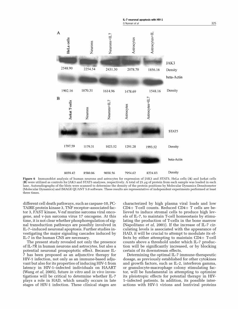
IL-7 up-regulates the expression of STAT5in astrocytes but not neuronsBecause the IL-7:IL-7R interaction is known to actthrough the JAK-STAT pathway (Porter et al, 2001),adherent human mature neurons and astrocytes wereleft untreated or treated with IL-7, and the expres-sion of JAK3 and STAT5 were analyzed by Westernblotting.
As shown in Figure 6, IL-7 induced the down-regulation of JAK3 and the up-regulation of STAT5in astrocytes, while having minimal effect on neu-rons. The up-regulation of STAT5 can be induced byeither JAK 1, through the IL-7Rα, or through JAK3 viathe IL-7 receptor gamma, the common receptor for IL-7, IL-2, IL-4, IL-9, and IL-15 (Schindler and Darnell,1995). Thus, we speculate that, because JAK3 wasunchanged in astrocytes, the up-regulation of STAT5could be induced by the IL-7Rα. As for the neurons,the JAK/STAT pathway did not show any specificup- or down-regulation, further confirming that othersignal transduction pathways or mechanisms mightbe involved in the signaling pathways for IL-7 inneurons.
Discussion
In this study, we provide the initial demonstrationthat IL-7R is expressed in mature human neurons(i.e., fully differentiated NT2 cells) and astrocytesat the mRNA and protein levels. IL-7Rα expressionin human neurons and astrocytes was only slightly
IL-7 neuronal apoptosis with HIV-1G Nunnari et al 323
Figure 4 IL-7–induced human neuronal apoptosis and augmented gp120-induced neuronal apoptosis. (Left) Representative TUNELstaining of human neurons treated with 0.01, 0.1, 1, 10, and 100 ng/ml of IL-7 for 3 days. (Right) Representative TUNEL staining of humanneurons treated with 50 ng/ml of gp120 for 48 h, then treated with 0.01, 0.1, 1, 10, 100 ng/ml of IL-7 for 3 days. The neurons were analyzedwith fluorescence microscopy (Olympus, model BX60, with fluorescence attachment BX-FLA). Magnification: 20×. These studies wererepeated at least three times.
down-modulated by the addition of exogenous IL-7.Interestingly, IL-7 induced human neuronal apop-tosis and augmented HIV-1 gp120-induced humanneuronal apoptosis. Of note, we found that the IL-7neuronal proapoptotic effects could be induced viaTNF-α/TNF-R1 receptor and FasL/Fas receptor pro-grammed cell death pathways.
The effect of IL-7 in the CNS has not at all beenfully elucidated. In Sprague-Dawley rats (Michaelsonet al, 1996), IL-7 demonstrated neuropoietic proper-ties and IL-7R was expressed in the developing brain.On the other hand, although the presence of IL-7R
in nonhematopoietic cells has been reported, a func-tional IL-7R was not found in different CNS tumors,mostly gliobastoma cell lines, despite detectable lev-els of IL-7 mRNA, but was never evaluated in eitherprimary astrocytes or neurons cell-lines. (Michaelsonet al, 1996, Cosenza et al, 2002).
In this study, we demonstrated that IL-7R is ex-pressed on both human neurons and astrocytes, al-though a quantitative discordance between the ex-pression of IL-7Rα mRNA and protein in astrocyteswas found, in accordance with previous studiessuggesting that IL-7R transcripts might not be stable
IL-7 neuronal apoptosis with HIV-1324 G Nunnari et al
Figure 5 Anti-IL-7 antibodies neutralized the IL-7–induced apoptotic effect. Neurons were incubated overnight with anti-IL-7 antibodiesat a concentration of 0.8 μg/ml, as per the manufacturer’s instructions. IL-7 at 2.5 ng/ml was added once and TUNEL staining was per-formed, as illustrated. Anti-IL-7 receptor antibodies were added and left overnight at a concentration of 1 μg/ml, as per the manufacturer’sinstructions, to block IL-7Rα. Levels of apoptosis are shown in parenthesis.
in certain human cells (Smiers et al, 1995; Cosenzaet al, 2002).
Plasma levels of IL-7 in HIV-1–infected individu-als were found previously to be significantly higherthan those in the healthy donors, correlating di-rectly with plasma HIV-1 RNA levels and indirectlywith CD4+ T-cell counts (Llano et al, 2001). Becauseplasma IL-7 levels increase along with HIV-1 dis-ease progression, and HAD usually occurs in the latestages of HIV-1 disease, we speculated that IL-7-mayplay a role in the pathogenesis of HAD. We demon-strated that IL-7–induced neuronal apoptosis in adose-dependent manner, and that it was inhibited bythe addition of anti-IL-7 antibodies. Importantly, thewell-described proapoptotic effect of the HIV-1 en-velope protein, gp120 (Xu et al, 2004) was enhancedby the addition of IL-7, showing a neuronal apoptoticsynergistic effect.
Previous studies demonstrated that IL-7 inducedIL-7Rα down-regulation and Fas up-regulation in pe-ripheral human T cells isolated from healthy donors
(Rethi et al, 2004; Lelievre et al, 2004), also reportedthat IL-7 down-modulated IL-7Rα expression and in-creased HIV-1 mediated Fas-induced T-cell death.We showed in the present study that IL-7 down-modulated IL-7Rα expression in human neurons andastrocytes, and induced the up-regulation of STAT5in astrocytes, but not in neurons. IL-7 leads to the acti-vation of STAT5 either through JAK1 or JAK3 (Porteret al, 2001). We found that IL-7 did not induce sig-nificant changes in the expression of JAK3, neither inneurons nor in astrocytes. Because JAK3 is associatedwith the common IL-7 receptor gamma (shared by IL-7, IL-2, IL-4, IL-9, and IL-15) and JAK1 with IL-7Rα wesuggest that IL-7–induced neuronal apoptosis mightbe related to other specific pathways, such as phos-phoinositol (PI) 3-kinase or the activation of the srcfamily tyrosine Kinase p56lck or p59 f yn, which asso-ciate with the A region of IL-7Rα (Porter et al, 2001).In addition, by a targeted genomics microarray anal-ysis, we showed that IL-7–induced apoptosis was re-lated to the up-regulation of several genes involved in
IL-7 neuronal apoptosis with HIV-1G Nunnari et al 325
Figure 6 Immunoblot analysis of human neurons and astrocytes for expression of JAK3 and STAT5. HeLa cells (A) and Jurkat cells(B) were utilized as controls for JAK3 and STAT5 analyses, respectively. A total of 25 μg of protein from each sample was loaded in eachlane. Autoradiographs of the blots were scanned to determine the density of the protein positions by Molecular Dynamics Densitometer(Molecular Dynamics) and IMAGE QUANT 5.0 software. These results are representative of independent experiments performed at leastthree times.
different cell death pathways, such as caspase-10, PC-TAIRE protein kinase 3, TNF receptor–associated fac-tor 3, FAST kinase, V-raf murine sarcoma viral onco-gene, and v-jun sarcoma virus 17 oncogene. At thistime, it is not clear whether phosphoregulation of sig-nal transduction pathways are possibly involved inIL-7–induced neuronal apoptosis. Further studies in-vestigating the major signaling cascades induced byIL-7 in the human CNS are necessary.
The present study revealed not only the presenceof IL-7R in human neurons and astrocytes, but also apotential neuronal proapoptotic effect. Because IL-7 has been proposed as an adjunctive therapy forHIV-1 infection, not only as an immune-based adju-vant but also for its properties of inducing HIV-1 fromlatency in HIV-1–infected individuals on HAART(Wang et al, 2005), future in vitro and in vivo inves-tigations will be critical to determine whether IL-7plays a role in HAD, which usually occurs in latestages of HIV-1 infection. These clinical stages are
characterized by high plasma viral loads and lowCD4+ T-cell counts. Reduced CD4+ T cells are be-lieved to induce stromal cells to produce high lev-els of IL-7, to maintain T-cell homeostasis by stimu-lating the production of T-cells in the bone marrow(Napolitano et al, 2001). If the increase of IL-7 cir-culating levels is associated with the appearance ofHAD, it will be crucial to attempt to modulate its ef-fects by either attempting to maintain CD4+ T-cellcounts above a threshold under which IL-7 produc-tion will be significantly increased, or by blockingcertain of its downstream effects.
Determining the optimal IL-7 immune-therapeuticdosage, as previously established for other cytokinesand growth factors, such as IL-2, interferon gamma,or granulocyte-macrophage colony stimulating fac-tor, will be fundamental in attempting to optimizeits pleiotropic effects for potential therapy in HIV-1–infected patients. In addition, its possible inter-actions with HIV-1 virions and lentiviral proteins
IL-7 neuronal apoptosis with HIV-1326 G Nunnari et al
in inducing neuronal programmed cell death mayalso represent a rational target towards interdictingin HAD.
Materials and methods
Cell cultures and IL-7 treatmenthNT-2 neuronal precursor cells were purchased fromStratagene Cloning System (Stratagene, La Jolla,CA). A total of 2.6 × 10⁶ cells were seeded in T75flasks in the complete Dulbecco’s modified Eagle’smedium DMEM/F12 medium (containing 0.05 Upenicillin–0.05 U streptomycin and 10% fetal bovineserum) and treated with 10 μm retinoic acid (Sigma,St Louis, MO) three times per week for 6 weeks.The differentiated cells were detached by 0.05%trypsin-EDTA and replated in T175 flask in completeDMEM/F12 medium containing 5% fetal bovineserum, and further treated with mitotic inhibitors(1 μm cytosine arabinoside, 10 μm fluorodeoxyuri-dine, and 10 μm uridine; Sigma) three times perweek for 10 days (Xu et al, 2004). Mature humanneurons were replated on poly-D-lysine and laminincoated (both were 10 μg/ml, Sigma) four-well glasschamber slides in the conditioned medium from thefirst replating for TUNEL assays, and T25 flasks forhuman apoptosis gene arrays.
Primary human astrocytes (passage 2), obtainedfrom Dr. Avindra Nath of Johns Hopkins Univer-sity, were cultured in DMEM medium contain-ing 0.05 U penicillin–0.05 U streptomycin and10% fetal bovine. Nalm-6 B-cells were cultured inRPMI-1640 medium containing 0.05 U penicillin–0.05 U streptomycin, 10% fetal bovine serum, and2-mercaptoethanol (1000,000×). All cultures weremaintained at 37◦C in an incubator with 5% CO2.Recombinant human IL-7 (R&D Systems) at 0.01, 0.1,1, 10, and 100 ng/ml was added once at the initia-tion of the experiments to human neurons and humanastrocytes.
To attempt to inhibit IL-7–induced apoptotic ef-fects, anti-IL-7 antibodies (R&D Systems) were usedin parallel experiments. Anti-IL-7R antibodies wereutilized in competition assays to further determinethe role played by the IL-7R (R&D Systems). Humanneurons were treated overnight with anti-IL-7R priorto IL-7 exposure.
IL-7 receptor alpha mRNA determinationsHuman neurons and astrocytes untreated and treatedwith IL-7 were lysed in 1 ml of Trizol reagent (GibcoBRL, Baltimore, MD) and 0.2 ml of chloroform wasadded afterwards. The samples were centrifuged at13,000 rpm for 30 min at 4◦C. Total RNA was pre-cipitated from the aqueous phase by adding 0.5 mlof isopropanol. After incubation at room temperaturefor 10 min, the precipitates were pelleted by centrifu-gation at 13,000 rpm for 10 min at 4◦C. The RNApellets were washed with 75% ethanol, air dried,and then dissolved in Diethyl polycarbonate (DEPC)-treated water.
The SuperScript First-Strand Synthesis kit waspurchased from Gibco BRL (Baltimore, MD). TotalRNA (1 to 5 μg) was hybridized with 1 μl Oligo (dT)in a total volume of 10 μl at 65◦C for 5 min, then onice for 1 min. The RNA/primer mixture was first in-cubated with reverse transcriptase (RT) reaction mix-ture (2 μl 10× RT buffer, 4 μl 25 mM MgCl2,2 μl 0.1 MDithiothreithol (MDTT), and 1 μl RNase Out recom-binant RAase inhibitor) at 42◦C for 2 min, and thenincubated with 1 μl of SuperScript II RT for 50 min.The reaction was terminated by incubation at 70◦C for15 min. RNase H, 1 μl, was added to the reaction toremove any remaining RNA. Oligonucleotides usedfor the polymerase chain, reactions, (PCR) amplifica-tion were (IL7R1) 5′ TGGAGACTTGGAAGATGCA 3′;(IL7R2) 5′ TTAGTAAGATAGGATCCAT 3′ for IL7Rαand (G1) 5′ CAAAGTTGTCATGGATGAC 3′; (G2) 5′CAAAGTTGTCATGGACC 3′ for GAPDH. PCR wasperformed using a 9700 PCR thermal cycler (Perkin-Elmer), under the following conditions: denature at95◦C for 45 s, primer annealing 57◦C for 45 s, andDNA extension at 72◦C for 1 min for 40 cycles.
The gel photographs were scanned and further ana-lyzed for density comparison by using Molecular Dy-namics Densitometer and IMAGE QUANT 5.0 soft-ware. The density ratio of IL-7Rα RNA bands versusthe GAPDH bands served as a standard, according toknown fold dilution of cDNA from Nalm-6 cells. Thelinearity between the density ratio and fold dilutionwas confirmed.
ImmunoprecipitationAdherent human mature neurons and astrocytes werecultured in T75 flasks until confluent, prior to theexperiments. A total of 1 × 10⁷ nonadherent Nalm-6B cells, neurons, and astrocytes, were lysed in 1 mlcold lysis buffer (Novagen, Madison, WI) contain-ing 1× protease inhibitor cocktail (Pierce, Rockford,IL) on ice for 30 min. The cell lysates were spun at10,000 g for 15 min at 4◦C. The supernatants were col-lected, 10 μg of polyclonal anti-IL-7Rα (R&D Systems,Minneapolis, MN) were added to each sample, andincubated at 4◦C overnight. Fifty microliters of a pro-tein G slurry (Calbiochem, Darmstadt, Germany) wasadded to each sample and incubated at 4◦C for 1 h ona rocking platform. Samples were spun at 10,000 gfor 30 s at 4◦C. After supernatants were completelyremoved, the beads were washed three times with500 μl of lyses buffer. After the last washing, 50 μl of1× Laemmli sample buffer was added to each samplebead pellet. Samples were heated at 95◦C for 10 minand centrifuged again. The supernatant of each sam-ple was collected and loaded on to gels.
Determination of IL-7Rα by Western blottingHuman neurons and astrocytes untreated and treatedwith IL-7 were lysed with Cytobuster Protein Extrac-tion Reagent (Novagen, Madison, WI). Protein con-centrations were determined with the BCA Proteinassay kit (BioRad, Herculas, CA). Approximately
IL-7 neuronal apoptosis with HIV-1G Nunnari et al 327
25 μg of each protein preparation was resolved on7.5% sodium dodecyl sulfate (SDS)-polyacrylamidegels (BioRad, Herculas, CA) and transferred to PVDFmembranes (Amersham Biosciences, Piscathaway,NJ) by electroblotting. The membranes were incu-bated in blocking buffer (10 mM phosphate-bufferedsaline (PBS), pH 7.2, 150 mM NaCl, 0.1% Tween 20,5% nonfat dry milk) at 4◦C for overnight. The mem-branes were then incubated with primary antibody(polyclonal goat anti-h IL-7Rα antibody, 1:1000, R&DSystems, Minneapolis, MN), rabbit polyclonal anti-JAK3 and anti-STAT5 antibodies at concentrations of1:500 (Santa Cruz Biotechnology, CA), for 1 h at roomtemperature in PBST (10 mM Phosphate-Buffered Di-fluoride PBS, pH 7.2, 150 mM NaCl, 0.1% Tween20). The secondary antibody, horseradish peroxidaselabeled anti-goat immunoglobulin G (IgG) antibody(1:500, for anti-IL-7R and 1:5000 for JAK3 and STAT5;Santa Cruz Biotechnology) was applied at room tem-perature for 1 h in PBST. The membranes were rinsedin PBST, and proteins were visualized using an en-hanced chemoluminscent (ECL) Western Blotting De-tection Reagent (Amersham Biosciences, Piscataway,NJ), and exposure to Kodak BioMax MS autoradio-graphic film (Kodak, Rochester, NY).
Immunoblot analyses of immune precipitationcomplexes prepared from human neurons and astro-cytes were performed for the expression of IL-7Rαprotein, JAK3, and STAT5. The Nalm-6 B cells wereused as positive controls for the expression of IL-7Rαprotein, plus HeLa cells and Jurkat cells were utilizedfor JAK3 and STAT5 analyses, respectively. We uti-lized Restore Western blot stripping buffer (Pierce,Rockford, IL) to strip the blots, using normal goat IgG(R&D Systems) as an isotype control.
HIV-1 viral envelope protein gp120and IL-7 treatmentRecombinant viral Env protein of the HIV-1 straingp120 from BaL (R5-tropic) (endotoxin levels lessthan 10 EU/ml) was purchased from Intracel Cor-poration (Rockville, MD). The protein was added ataconcentration of 50 ng/ml to the neuronal cell cul-ture medium, as a relatively low concentration whichwe previously demonstrated would induce apopto-sis in human neurons (Xu et al, 2004). After 48 h,the medium was removed and cells were washed
References
Alper O, Hacker NF, et al (1999). Protein kinase A-Ialphasubunit-directed antisense inhibition of ovarian can-cer cell growth: crosstalk with tyrosine kinase signalingpathway. Oncogene 18: 4999–5004.
Becker EB, Howell J, et al (2004). Characterization ofthe c-Jun N-terminal kinase-BimEL signaling path-way in neuronal apoptosis. J Neurosci 24: 8762–8770.
Bellacosa A, Testa JR, et al (1991). A retroviral oncogene,akt, encoding a serine-threonine kinase containing anSH2-like region. Science 254: 274–277.
twice with complete DMEM/F12 medium. Thegp120-treated neurons were then treated once withIL-7 at concentrations of 0.01 ng/ml, 0.1 ng/ml,1 ng/ml, 10 ng/ml, and 100 ng/ml, and then culturedfor 3 days before DNA fragmentation and terminaldeoxynucleotidyl transferase (Tdt)-mediated dUTPnick end-labeling (TUNEL) assays.
TUNEL assays and semiquantitative apoptosisdeterminationsThe in situ cell death detection kit, TMR red, waspurchased from Roche Diagnostics (Indianapolis, IN).Human neurons and astrocytes seeded in four cham-ber slides were washed twice with PBS, pH 7.4,fixed with acetone for 10 min at room temperature,and assayed for apoptosis according to the manufac-ture’s instructions. The cells were analyzed by fluo-rescence microscopy (Olympus System MicroscopeModel BX60 with fluorescence attachment BX-FLA).
For semiquantitative measurements of apoptosis,images generated with a CCD array camera (RT Color,Diagnostic Instruments, Sterling Heights, MI) weresubjected to fluorescence brightness value determina-tions on a monochromatic scale in light of red, blue,green (RGB) values (0–255 ASCII numbers). Apopto-sis was defined based on a red monochromatic scalein the range of 0 to 255. Blank values were subtractedfrom the average of seven random values from differ-ent cells (Xu et al, 2004).
Human apoptosis gene microarrayThe CLONTECH’s Atlas TM cDNA expression hu-man apoptosis array was purchased from BD Bio-sciences Clontech (Palo Alto, CA), and used accord-ing to the instructions. The total RNA was isolatedfrom untreated or treated neurons using the RNA-gents kit from Promega (Madison, WI). cDNA probemixtures were synthesized by reverse transcribingrespective RNA using cDNA synthesis primer mixprovided in a kit and α-32P dATP (Perkin ElmerSciences Inc, Boston, MA). Each radioactively la-beled probe mix was then hybridized to separateAtlas arrays overnight at 68◦C. After washing, the hy-bridization patterns were analyzed by autoradiogra-phy and quantified with a phosphor imager (Molecu-lar Dynamics). Differences in gene expression levelswere quantitated by the BD AtlasImage 2.7 Software.
Brooks DG, Kitchen SG, et al (2001). Generation of HIVlatency during thymopoiesis. Nat Med 7: 459–464.
Chun TW, Engel D, et al (1998). Induction of HIV-1replication in latently infected CD4+ T cells using acombination of cytokines. J Exp Med 188: 83–91.
Cosenza L, Gorgun G, et al (2002). Interleukin-7 receptor ex-pression and activation in nonhaematopoietic neoplas-tic cell lines. Cell Signal 14: 317–325.
Cross TG, Scheel-Toellner D, et al (2000). Serine/threonine protein kinases and apoptosis. Exp Cell Res256: 34–41.
IL-7 neuronal apoptosis with HIV-1328 G Nunnari et al
Deacon EM, Pongracz J, et al (1997). PKC isoenzymes: dif-ferential involvement in apoptosis and pathogenesis.J Clin Pathol Mol Pathol 50: 124–131.
Dudley AC, Thomas D, et al (2004). The STATs in cellstress-type responses. Cell Communi Signal 2: 1478–1481.
Dus D, Krawczenko A, et al (2003). IL-7 receptor is presenton human microvascular endothelial cells. ImmunolLett 86: 163–168.
Ellison M, Povlishock JN, et al (1987). Blood-brain barrierdysfunction in cats following recombinant interleukin-2infusion. Cancer Res 47: 5765–5770.
Fry AS, Connick E, et al (2001). A potential role forinterleukin-7 in T-cell homeostasis. Blood 97: 2983–2990.
Goodwin RG, Friend DJ, et al (1990). Cloning of the humanand murine interleukin-7 receptors: demonstration of asoluble form and homology to a new receptor superfam-ily. Cell 60: 941–951.
Grabstein KH, Namen AE, et al (1990). Regulation of T cellproliferation by IL-7. J Immunol 144: 3015–3020.
Herskovits AZ, Davies P, et al (2004). Cloning and expres-sion analysis of two novel PCTAIRE 3 transcripts fromhuman brain. Gene 328: 59–67.
Heufler C, Tppar G, et al (1993). Interleukin 7 is producedby murine and human keratinocytes. J Exp Med 178:1109–1114.
Ichijo H (1999). From receptor to stress-activated MAP ki-nases. Oncogene 18: 6087–6093.
Jaleco S, Swainson L, et al (2003). Homeostasis of naı̈ve andmemory CD4+ T-cell: IL-2 and IL-7 differentially regu-late the balance between proliferation and Fas-mediatedapoptosis. J Immunol 171: 61–68.
Kaul M, Garden GA, et al (2001). Pathways to neuronal in-jury and apoptosis in HIV-associated dementia. Nature410: 988–994.
Lai SY, Molden J, et al (1997). Shared ϒc subunit withinthe human interleukin-7 receptor complex. J Clin Invest99: 169–177.
Lelievre JD, Petit F (2005). Interleukin 7 increases humanimmunodeficiency HIV-1 LAI-mediated Fas-induced Tcell death. J Virol In press.
Le-Niculescu H, Bonfoco E, et al (1999). Withdrawal of sur-vival factors results in activation of the JNK pathway inneuronal cells leading to Fas ligand induction and celldeath. Mol. Cell Biol 19: 751–763.
Leonard WJ (2001). Cytokines and immunodeficiency dis-ease. Nat Rev Immunol 1: 200–208.
Lin JX, Migone TS, et al (1995). The role of shared receptormotifs and common stat proteins in the generation ofcytokine pleiotropy and redundancy by IL-2, IL-4, IL-7,IL-13, and IL-15. Immunity 2: 331–339.
Llano A, Barretina J, et al (2001). Interleukin-7 in plasmacorrelates with CD4 T-cell depletion and may be asso-ciated with emergence of syncytium-inducing variantshuman immunodeficiency virus type 1-positive individ-uals. J Virol 75: 10319–10325.
Marte BM, Downward J (1997). PKB/Akt: connecting phos-phoinositide 3-kinase to cell survival and beyond.Trends Biochem Sci 22: 355–358.
Macaluso M, Russo G, et al (2002). Ras family genes: an in-teresting link between cell cycle and cancer. J Cell Phys-iol 192: 125–130.
Michaelson MD, Mehler MF, et al (1996). Interleukin-7 istrophic for embryonic neurons and is expressed in de-veloping brain. Dev Biol 179: 251–263.
Miyazaki T, Kawahara A, et al (1994). Functional activa-tion of Jak1 and Jak3 by selective association with IL-2receptor subunits. Science 266: 1045–1047.
Napolitano LA, Grant RM, et al (2001). Increased produc-tion of IL-7 accompanies HIV-mediated T-cell deple-tion: implications for T-cell homeostasis. Nat Med 7:73–79.
Pimentel-Muinos FX, Seed B (1999). Regulated commit-ment of TNF receptor signaling: a molecular switch fordeath or activation. Immunity 11: 783–793.
Pollock PM, Harper UL, et al (2003). High frequency ofBRAF mutations in nevi. Nat Genet 33: 19–20.
Porter BO, Scibelli P, et al (2001). Control of T cell devel-opment in vivo by subdomains within the IL-7 recep-tor alpha-chain cytoplasmic tail. J Immunol 166: 262–269.
Raber J, Merlo PE, et al (1994). IL-1β potentiates theacetycholine-induced release of vasopressin from thehypothalamus in vitro, but not from the amygdala. Neu-roendocrinology 59: 208–217.
Rethi B, Fluur C, et al (2004). Decreased IL-7Rα expressionof T cell is associated with CD4+ T cell depletion andhigh levels of serum IL-7 during HIV infection. Immuno2004 online abstracts 840.
Sasson S, Zanetti G, et al IL-7 Down-regulates Surface Ex-pression of IL-7Ralpha in vitro and in vivo. The 11thConference on Retroviruses and Opportunistic Infec-tions, San Francisco, CA, USA. Abstract N 249.
Schmitz I, Krueger A, et al (2003). An IL-2-dependentswitch between CD95 signaling pathways sensitize pri-mary human T cells toward CD95-mediated activation-induced cell death. J Immunol 171: 2930–2936.
Scripture-Adams DD, Brooks DG, et al (2002). Interleukin-7induces expression of latent human immunodeficiencyvirus type 1 with minimal effects on T-cell phenotype.J Virol 76: 13077–13082.
Smiers FJ, van Paassen M, et al (1995). Heterogeneity ofproliferative responses of human B cell precursor acutelymphoblastic leukemia (BCP-ALL) cells to interleukin7 (IL-7): no correlation with immunoglobulin gene sta-tus and expression of IL-7 receptor or IL-2/IL-4/IL-7 re-ceptor common gamma chain genes. Leukemia 9: 1039–1045.
Sudo T, Ito M, et al (1989). Interleukin 7 production andfunction in stromal cell-dependent B cell development.J Exp Med 170: 333–338.
Wang J, Chun HJ, et al (2001). Caspase-10 is an initiatorcaspase in death receptor signaling. Proc Natl Acad SciU S A 98: 13884–13888.
Wang FX, Xu Y, et al (2005). IL-7 is a potent and proviralstrain-specific inducer of latent, in vivo HIV-1 cellularreservoirs of infected individuals on virally-suppressiveHAART. J Clin Inves 115: 128–137.
Wilks AF, Harpur AG, et al (1994). Cytokine signal trans-duction and the JAK family of protein tyrosine kinase.Bioassays 16: 313–320.
Wolf SS, Cohen A, et al (1992). Expression of cytokinesand their receptors by human thymocytes and thymicstromal cells. Immunology 77: 362–368.
Xia Z, Dickens M, et al (1995). Opposing effects of ERKand JNK-p38 MAP kinases on apoptosis. Science 270:1326–1331.
Xu Y, Kulkosky J, et al (2004). HIV-1 mediated apoptosis ofneuronal cells: proximal molecular mechanisms of HIV-1-induced encephalopathy. Proc Natl Acad Sci U S A101: 7070–7075.
Related Documents