Exercise Science Exercise Science Section 4: Bone and Muscle Injuries Section 4: Bone and Muscle Injuries and and Joint Mechanics and Joint Injuries Joint Mechanics and Joint Injuries

Welcome message from author
This document is posted to help you gain knowledge. Please leave a comment to let me know what you think about it! Share it to your friends and learn new things together.
Transcript

Exercise ScienceExercise ScienceSection 4: Bone and Muscle Injuries andSection 4: Bone and Muscle Injuries and
Joint Mechanics and Joint InjuriesJoint Mechanics and Joint Injuries

Stress fracture – most difficult to detectSimple fracture – no separation (hairline fracture)Compound fracture – bone breaks into separate
piecesComminuted fracture – bone shatters into many
pieces
Simple fracture
Compound fracture
Comminuted fracture

*Stress Fracture*Exact MOI is unknown
*Possible MOIs:*Overload caused by muscle contractions
*Altered stress distribution in bone due to muscle fatigue
*Change in ground reaction forces
*Performing rhythmic, repetitive movements

*Comminuted Fracture*3 or more fragments
*MOI: *Hard, direct blow
*Fall in awkward position

*Impacted Fracture*Bone is compressed
*MOI:*Fall from a height
*Immediate splinting and traction are required
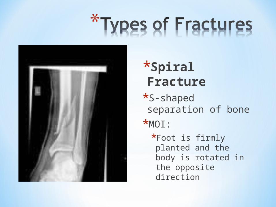
*Spiral Fracture*S-shaped separation of bone
*MOI:*Foot is firmly planted and the body is rotated in the opposite direction

*Contrecoup Fracture*Fracture that occurs on the side opposite of the trauma site
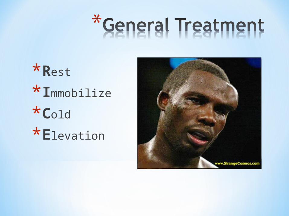
*Rest
*Immobilize
*Cold
*Elevation

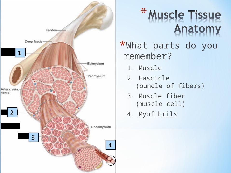
*Muscles are made up of bundlesof muscle fibers, called fascicles
*Fascicle is a bundle of muscle fibers
*A muscle fiber is a muscle cell….made up of many small myofibrils
*Myofibrils contain filaments
*Two types ofprotein filaments
TTIIssssuuee
AAnnaattoommyy
MuscleMuscle
FilamentsFilaments
MyofibrilsMyofibrils
Muscle FibersMuscle Fibers
FascicleFascicle
DD
CC
BBAA
*What parts do you remember?1. Muscle
2. Fascicle (bundle of fibers)
3. Muscle fiber (muscle cell)
4. Myofibrils
3344
22
11

*A sprain is a wrenching, twisting or stretching injury to a ligament.
Sprains often affect theankles, knees, or wrists.
SprainSprain
Result in pain, swelling, redness, bruising, and difficulty Result in pain, swelling, redness, bruising, and difficulty using injured joint.using injured joint.

*A strain is an injury to a muscle or tendon, and is often caused by overuse, force, or stretching.
*Injured area experiences: *pain and soreness*swelling *warmth, bruising,
or redness *difficulty using or
moving the injured area in a normal manner
StrainStrain
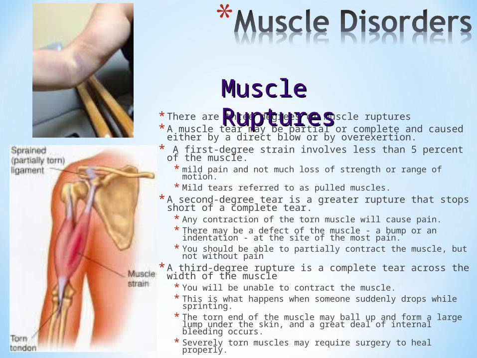
* There are three degrees of muscle ruptures * A muscle tear may be partial or complete and caused
either by a direct blow or by overexertion.* A first-degree strain involves less than 5 percent of the
muscle. * mild pain and not much loss of strength or range of motion.* Mild tears referred to as pulled muscles.
* A second-degree tear is a greater rupture that stops short of a complete tear.* Any contraction of the torn muscle will cause pain. * There may be a defect of the muscle - a bump or an
indentation - at the site of the most pain. * You should be able to partially contract the muscle, but not
without pain* A third-degree rupture is a complete tear across the
width of the muscle* You will be unable to contract the muscle. * This is what happens when someone suddenly drops while
sprinting. * The torn end of the muscle may ball up and form a large
lump under the skin, and a great deal of internal bleeding occurs.
* Severely torn muscles may require surgery to heal properly.
Muscle RupturesMuscle Ruptures
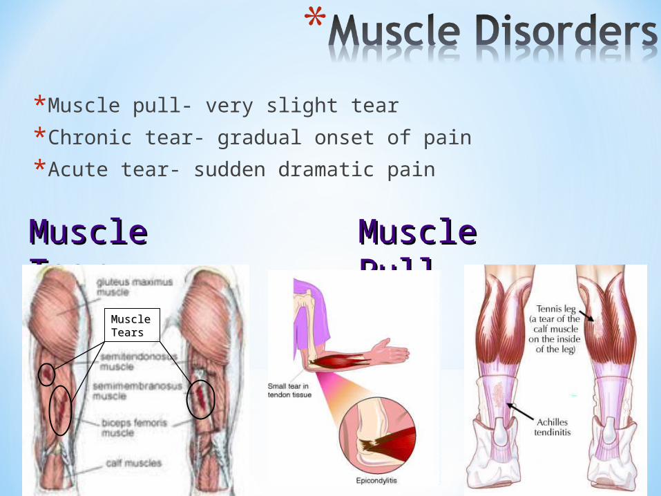
*Muscle pull- very slight tear
*Chronic tear- gradual onset of pain
*Acute tear- sudden dramatic pain
Muscle PullMuscle PullMuscle TearMuscle Tear
Muscle Muscle TearsTears

*Shin splints is pain resulting from damage to the muscles along the shin.
Pain is felt in different areas, depending on which muscles are affected.
Shin splintsShin splints
Shin splints represent an "overuse injury" and occur most commonly in runners.

*R.I.C.E.*Rest: Stop all activities whichcause pain. *Ice: Helps reduce swelling. Never ice more than 10-15 min. at a time. Protect the skin. *Compression: Wrap the strained area to reduce swelling. *Elevation: Keep the strained area as close to the level of the heart as is conveniently possible to keep blood from pooling in the injured area.
Treatment for Muscle InjuriesTreatment for Muscle Injuries
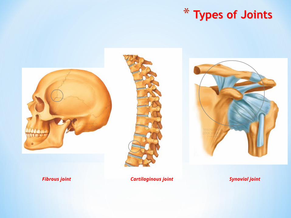
Cartilaginous jointFibrous joint Synovial joint

Ball-and-socket joint
Hinge joint
Saddle joint
Gliding joint
Pivot joint
Ellipsoid joint

Bone
Blood vessels
Nerve
Synovial membrane
Joint cavity (filled with synovial fluid)
Fibrous capsule
Joint capsule
Bursa
Tendon sheath
Tendon
Articular cartilage
Fibrous layer
Membranous layer Periosteum

Tendons:Composed of collagen
(bundles of white, fibrous protein)Attach muscle to boneVascular
Ligaments:Tough bands of white, fibrous tissueAttach bone to boneAvascular

Strains, pulls, and tearsTerms used to describe injuries
to all joint tissue typesTendinitis
Inflammation of a tendonDislocations
Bone displaced from its original location
SeparationsFibrous ligaments that bind the
bones tear and separateCartilage
Torn cartilageShin splints
Tearing of the interosseous membrane or the periosteum
Tendinitis

*Dislocation*Result due to forces that cause the joint to
go beyond its normal anatomical limits
*Two classes:
*Subluxations
*Luxations

*Subluxation*Partial dislocation*Incomplete separation between 2 articulating bones

*Luxation*Complete dislocations*Total separation between 2 articulating bones

Clavicle
Coracoclavicular ligament
Coracoid process
Scapula
Acromioclavicular ligament
Acromion
Coracoacromial ligament
Glenohumeral ligaments and joint capsule
Tendon of biceps brachii (long head)
Humerus

Biceps tendinitisCaused by overuse of the
biceps brachii muscleShoulder separation
Tearing of the acromioclavicular ligament
Shoulder dislocationOccurs when the humerus
“pops out” of the glenoid fossaRotator cuff tears
An injury to one of the rotator cuff tendons
Shoulder separation

Patella
Medial (Tibial) collateral ligament
Patellar ligament
Tibial tuberosity
Tibia
Quadriceps tendon
Fibula

Femur
Lateral (Fibular) collateral ligament removed
Medial (Tibial) collateral ligament removed
Lateral Meniscus
Tibial Tuberosity
Fibula
Lateral Condyle Medial Condyle
Medial Meniscus
Tibia
Posterior cruciate ligament
Anterior cruciate ligament

Femur
Adductor magnus tendon
Medial head of gastrocnemius tendon
Semimembranosus tendon
Medial (Tibial) collateral ligament
Lateral (Fibular) collateral ligament
Fibular head
Lateral head of gastrocnemius tendon
Oblique popliteal ligament
Fibula
Tibia
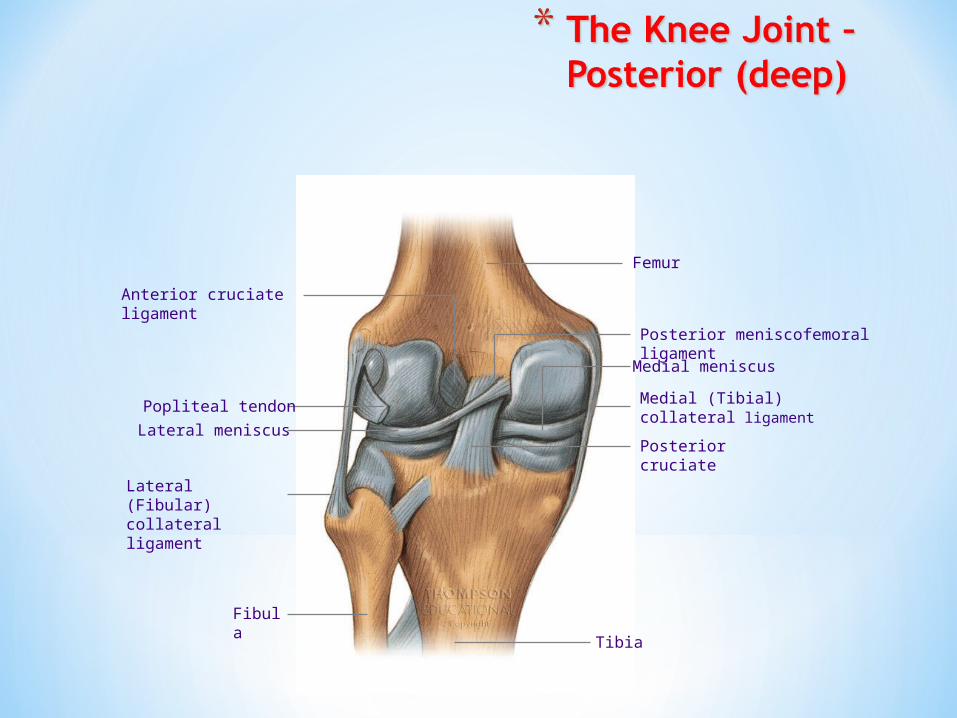
Anterior cruciate ligament
Popliteal tendon
Lateral meniscus
Lateral (Fibular) collateral ligament
Medial (Tibial) collateral ligament
Medial meniscus
Posterior cruciate
Femur
Fibula
Tibia
Posterior meniscofemoral ligament
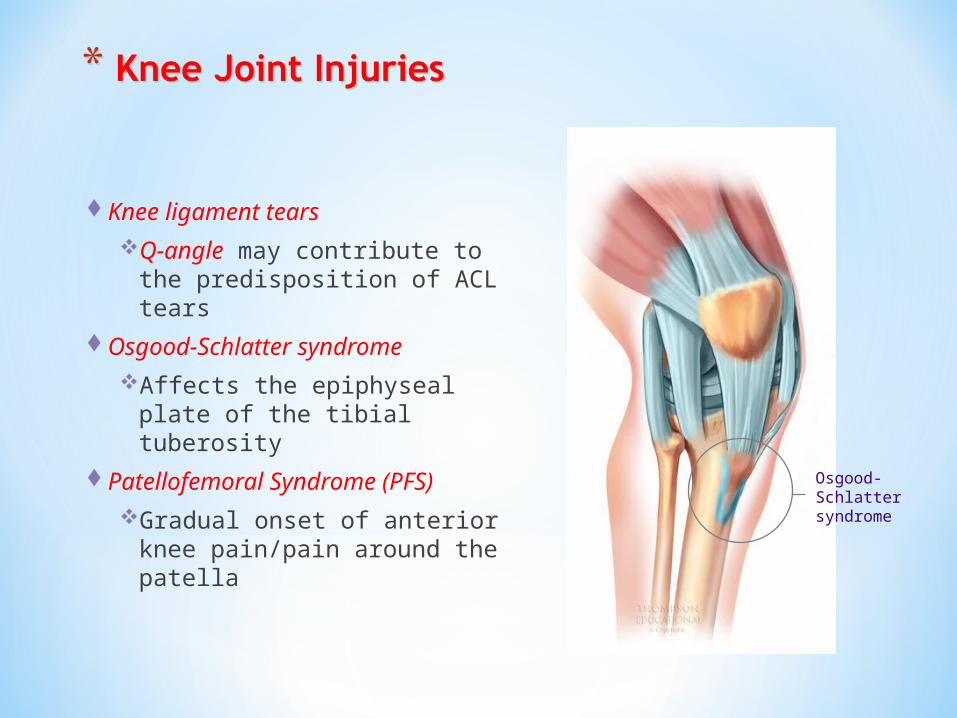
Knee ligament tearsQ-angle may contribute to the
predisposition of ACL tearsOsgood-Schlatter syndrome
Affects the epiphyseal plate of the tibial tuberosity
Patellofemoral Syndrome (PFS)Gradual onset of anterior knee
pain/pain around the patellaOsgood-Schlatter syndrome

Tibia
Medial malleolus
Calcaneal (Achilles) tendon
Long plantar ligament
Deltoid ligament

Tibia
Fibula
Posterior tibiofibular ligament
Lateral malleolus
Anterior tibiofibular ligament
Anterior talofibular ligament
Calcaneus
Posterior talofibular ligament
Anterior talofibular ligament

Inversion sprains“twisted ankle”
Eversion sprainsOccurs to the deltoid ligamentPott’s Fracture
A force on the medial side of ankle causing the deltoid ligament to rip off the tip of the medial malleolus; and a break of the fibula
Inversion sprain

S.H.A.R.P P.I.E.R. Principle
Swelling: instantly or over time Pressure: tensor wrap
Heat: increased temperature in the area
Ice: placed on affected area
Altered: tissue will not function properly
Elevate: to reduce swelling
Red: in colour Restrict: tensors, slings, or crutches
Painful: to touch or move
Related Documents