EXCRETION AND HOMEOSTASIS Common terms 1. Excretion is the removal of waste products produced as a result of metabolic processes in an organism. E.g. urea and carbon dioxide. 2. Osmoregulation: is the process by which osmotic pressure of blood and tissue fluid is kept constant. 3. Secretion: is the active or passive flow of substances from living cells. Such substances as enzymes, hormones, mucus etc. 4. Homeostasis: This is the maintenance of a relatively constant internal environment of the body. The internal environment of the body is composed of blood and tissue fluid which surround cells. Factors of the internal environment that are kept constant Body temperature pH Water content Glucose concentration Salt content Levels of carbon dioxide etc Note Egestion, the removal of undigested food materials should not be confused with excretion because most of the contents of the feaces, apart from the bile pigments, have not taken part in reactions in the cells of the body. SIGNIFICANCE OF EXCRETION Eliminates waste products formed during body metabolism Removal of waste products which would be toxic to body cells It allows for the removal of unwanted materials taken into the body along with useful nutrients It involves the removal of materials synthesized in excess of the current body demands 1

Welcome message from author
This document is posted to help you gain knowledge. Please leave a comment to let me know what you think about it! Share it to your friends and learn new things together.
Transcript
EXCRETION AND HOMEOSTASIS
Common terms
1. Excretion is the removal of waste products produced as a result of metabolic
processes in an organism. E.g. urea and carbon dioxide.
2. Osmoregulation: is the process by which osmotic pressure of blood and
tissue fluid is kept constant.
3. Secretion: is the active or passive flow of substances from living cells. Such
substances as enzymes, hormones, mucus etc.
4. Homeostasis: This is the maintenance of a relatively constant internal
environment of the body.
The internal environment of the body is composed of blood and tissue fluid which
surround cells.
Factors of the internal environment that are kept constant
Body temperature
pH
Water content
Glucose concentration
Salt content
Levels of carbon dioxide etc
Note
Egestion, the removal of undigested food materials should not be confused with
excretion because most of the contents of the feaces, apart from the bile
pigments, have not taken part in reactions in the cells of the body.
SIGNIFICANCE OF EXCRETION
Eliminates waste products formed during body metabolism
Removal of waste products which would be toxic to body cells
It allows for the removal of unwanted materials taken into the body along
with useful nutrients
It involves the removal of materials synthesized in excess of the current
body demands
1
Core m5
Typewritten text
S3 BIOLOGY
Continuous removal of waste products allows for maintenance of a constant
internal environment of the body.
Excretory products arise in a number of ways and these include, from:-
Unwanted substances absorbed with food
Absorption of excess nutrients
Osmoregulatory processes
Breakdown of protoplasmic constituents
From body metabolism
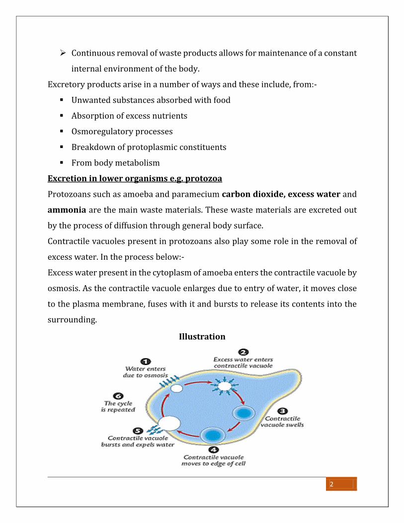
Excretion in lower organisms e.g. protozoa
Protozoans such as amoeba and paramecium carbon dioxide, excess water and
ammonia are the main waste materials. These waste materials are excreted out
by the process of diffusion through general body surface.
Contractile vacuoles present in protozoans also play some role in the removal of
excess water. In the process below:-
Excess water present in the cytoplasm of amoeba enters the contractile vacuole by
osmosis. As the contractile vacuole enlarges due to entry of water, it moves close
to the plasma membrane, fuses with it and bursts to release its contents into the
surrounding.
Illustration
2
EXCRETION IN PLANTS
Plants lack specialized excretory organs and have few excretion related problems
because of the following reasons:
Plants are producers and synthesise all their organic nutrients in sufficient
amounts, rarely do excesses occur
They usually reuse most of the waste products in other metabolic processes.
Carbon dioxide is used up in photosynthesis
Most of their waste molecules are simple molecules such as oxygen, carbon
dioxide and water which can easily diffuse through stomata
Plants store some of their wastes in organs such as fruits, bark, petals and
leaves which are periodically shed off thus eliminating the waste products.
These include iron, tannins, nicotinic acid etc.
Some of the waste products can be stored in dead tissues of the plant such
as the cortex without any harmful effects
Plants produce waste products at a slower rate since due to lower metabolic
rates. At a slow rate, waste products can’t affect plant metabolism.
Plants deal with their waste products through the following ways
Diffusion of gaseous wastes like oxygen and carbon dioxide through
stomata and lenticels
Transpiration; plants can lose excess water as vapour through the stomata
into the atmosphere
Recycling, most of the plant waste products are used as raw materials in
other processes. E.g. CO2 in photosynthesis
Deposition; Some toxic wastes like alkaloids, nicotine, quinine and cocaine
are deposited in dead tissues and peripheral structures like leaves and fruits
which are later lost from the plant.
3

Useful plant waste products to man
Waste products Source Importance
Gums Barks of plants e.g.
Acacia.
Necessary in food processing
industries such as making of chewing
gum
Resins Conifers For manufacture of paint
Latex rubber tree for making rubber used to make tyres
and other rubber products
Nicotine Found in leaves of
tobacco plant,
for making insecticides
Cocaine Leaves of cocoa
plant
Pain relieving drug
Local anaesthesia
Tannin Wood or bark of
trees e.g. Acacia,
wattle
For treatment of leather
Quinine Bark of cinchona
tree
Making drugs for treating malaria
Oxygen Released through
stomata
For aerobic respiration
Other plant waste products include carbon dioxide given off during respiration
and excess water given off during transpiration.
4
EXCRETION AND OSMOREGULATION IN ANIMALS
EXCRETORY PRODUCTS
Excretory products are categorized as nitrogenous waste products and non-
nitrogenous waste products. The table below summarises the source and fate of
the major waste products among organisms
1. Non-nitrogenous wastes: These waste products do not contain nitrogen.
The table of non-nitrogenous wastes from different animals
Excretory How formed How & where excreted
Carbon
dioxide
During respiration of
most living things
Diffusion through stomata in plants,
diffusion from respiratory surface in
animals.
Water In Aerobic respiration By osmosis or evaporation at the
cell surface
Mineral Ions Metabolism of
mineral nutrients
Excreted in urine and sweat
Bile
pigments
In breakdown of
haemoglobin
In bile stored in the gall bladder and
egested in faeces out of body.
2. NITROGENOUS WASTES: These are waste products that contain nitrogen.
They are formed as products of metabolism of protein (amino acids).
These include
Ammonia
Urea
Trimethylamine oxide
Uric acid
5
A table summarising the chief nitrogenous waste products of different
animals and their habitats
Animal Habitat Chief nitrogenous
waste
Excretory organ
Fresh water
invertebrates
Fresh water
Ammonia Surface
membrane
Insects and
other
arthropods
Land
(terrestrial)
Uric acid Malpighian
tubules
Cartilaginous
fish
Marine water
(sea/ocean)
Urea Kidney
Fresh water fish
(bony)
Fresh waters Ammonia Kidney
Marine water
fish (bony)
Marine waters Trimethylamine
oxide
Kidney
Larval
amphibians
Fresh water Ammonia Gills
Adult
amphibians
Terrestrial/fre
sh water
Urea Kidney
Reptiles and
birds
Terrestrial Uric acid Kidney
Mammals like
humans
Terrestrial Urea Kidney
6
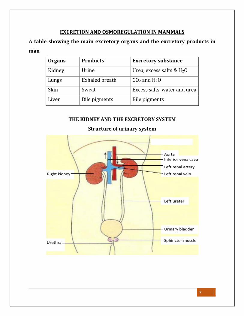
EXCRETION AND OSMOREGULATION IN MAMMALS
A table showing the main excretory organs and the excretory products in
man
Organs Products Excretory substance
Kidney Urine Urea, excess salts & H2O
Lungs Exhaled breath CO2 and H2O
Skin Sweat Excess salts, water and urea
Liver Bile pigments Bile pigments
THE KIDNEY AND THE EXCRETORY SYSTEM
Structure of urinary system
7
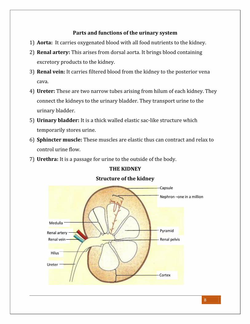
Parts and functions of the urinary system
1) Aorta: It carries oxygenated blood with all food nutrients to the kidney.
2) Renal artery: This arises from dorsal aorta. It brings blood containing
excretory products to the kidney.
3) Renal vein: It carries filtered blood from the kidney to the posterior vena
cava.
4) Ureter: These are two narrow tubes arising from hilum of each kidney. They
connect the kidneys to the urinary bladder. They transport urine to the
urinary bladder.
5) Urinary bladder: It is a thick walled elastic sac-like structure which
temporarily stores urine.
6) Sphincter muscle: These muscles are elastic thus can contract and relax to
control urine flow.
7) Urethra: It is a passage for urine to the outside of the body.
THE KIDNEY
Structure of the kidney
8
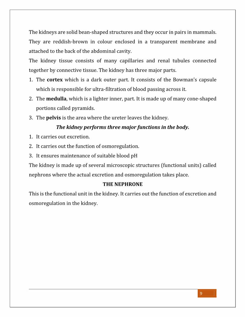
The kidneys are solid bean-shaped structures and they occur in pairs in mammals.
They are reddish-brown in colour enclosed in a transparent membrane and
attached to the back of the abdominal cavity.
The kidney tissue consists of many capillaries and renal tubules connected
together by connective tissue. The kidney has three major parts.
1. The cortex which is a dark outer part. It consists of the Bowman’s capsule
which is responsible for ultra-filtration of blood passing across it.
2. The medulla, which is a lighter inner, part. It is made up of many cone-shaped
portions called pyramids.
3. The pelvis is the area where the ureter leaves the kidney.
The kidney performs three major functions in the body.
1. It carries out excretion.
2. It carries out the function of osmoregulation.
3. It ensures maintenance of suitable blood pH
The kidney is made up of several microscopic structures (functional units) called
nephrons where the actual excretion and osmoregulation takes place.
THE NEPHRONE
This is the functional unit in the kidney. It carries out the function of excretion and
osmoregulation in the kidney.
9
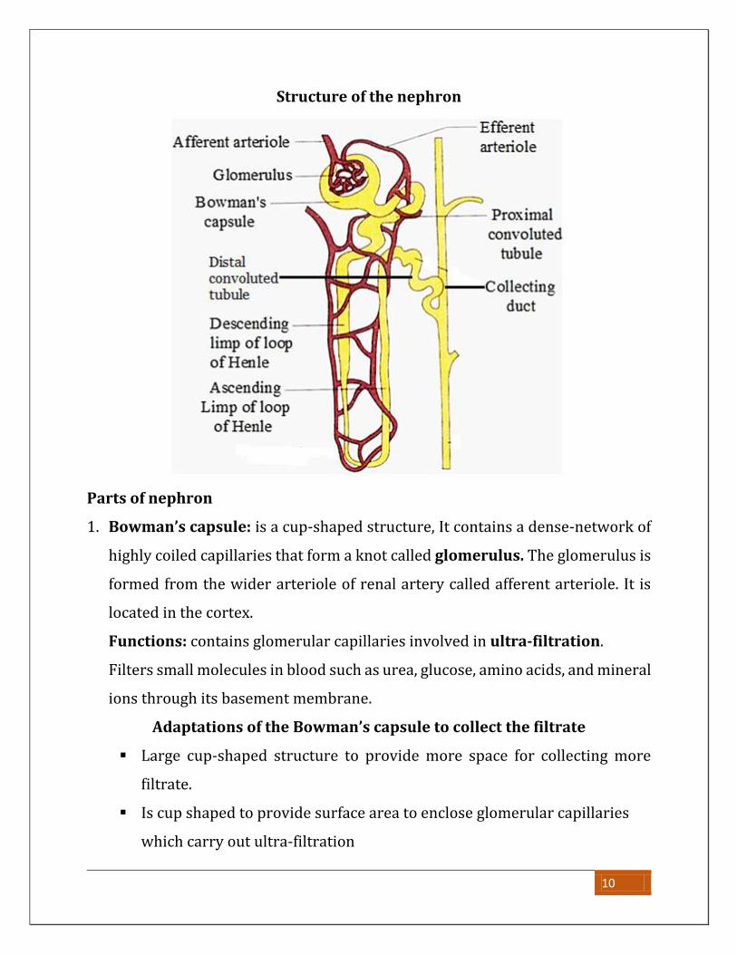
Structure of the nephron
Parts of nephron
1. Bowman’s capsule: is a cup-shaped structure, It contains a dense-network of
highly coiled capillaries that form a knot called glomerulus. The glomerulus is
formed from the wider arteriole of renal artery called afferent arteriole. It is
located in the cortex.
Functions: contains glomerular capillaries involved in ultra-filtration.
Filters small molecules in blood such as urea, glucose, amino acids, and mineral
ions through its basement membrane.
Adaptations of the Bowman’s capsule to collect the filtrate
Large cup-shaped structure to provide more space for collecting more
filtrate.
Is cup shaped to provide surface area to enclose glomerular capillaries
which carry out ultra-filtration
10
Porous basement membrane to allow filtration of materials.
2. Glomeruli; these are numerous capillaries found in the Bowman’s capsule
formed due to branching of the afferent arteriole
Adaptations of the glomerulus to ultra-filtration
Afferent arteriole wider than the efferent arteriole creating a high blood
pressure that forces small molecules out of the glomerulus.
Many blood capillaries to provide a large surface area for ultra-filtration.
Porous membrane to can allow small sized molecules to pass through.
3. Proximal convoluted tubule: is a highly coiled structure leading from the
Bowman’s capsule.
Function: It is a site where re-absorption of useful materials such as glucose
and some small amino acids and water from glomerular filtrate back to blood
takes place.
Adaptations of proximal convoluted tubule to its function
Long providing a large surface area over which absorption of materials can
occur
Tubule one cell thick to reduce the diffusion distance for faster diffusion of
materials
Cells in the walls have microvilli to increase the surface area over which
absorption can occur
Surrounded by numerous blood capillaries for rapid transportation of
absorbed materials
Cells in the walls contain large quantities of mitochondria to produce a large
quantity of ATP for rapid active transport of materials.
4. Loop of Henle: is a U shaped tubule, made up of a descending (going down)
limb and an ascending (going up) limb.
11
Function:
Re-absorption of water.
Re-absorption of mineral ions.
Adaptation of loop of Henle to its function
Long to increase surface area for reabsorption of water and mineral ions.
Blood capillaries running along its length to transport away water and salts
to maintain a concentration gradient.
5. Distal convoluted tubule: this is the second coiled tube found after the
ascending limb of loop of Henle.
Function: For re-absorption of ions like chloride ions, sodium ions together with
water.
Adaptation of distal convoluted tubule to its function
Coiled to slow down movement of renal fluid so as to increase the
reabsorption of mineral ions.
Has cells sensitive to antidiuretic hormone to allow water reabsorption
6. Collecting duct: a tubule to which numerous distal tubules attach to deliver
renal fluid.
Functions
Carries urine from the distal tubule to the pelvis of kidney.
Allows reabsorption of water thus conserving it.
URINE FORMATION
The process of urine formation takes place in the nephrone. It occurs in two
phases.
1. Ultra-filtration: which is filtration of small molecules through tiny pores at
high pressures.
12
2. Selective re-absorption: is the process of taking back into blood of some of
the essential substances still required by the body from the glomerular filtrate.
It is called re-absorption because initial absorption took place in the ileum.
Ultra filtration
Much blood comes from the afferent arteriole into the glomerulus at high
pressure due to pumping action of the heart.
More pressure is generated in the blood capillaries of the glomerulus since the
afferent arteriole which brings in blood has a wider lumen compared to the
efferent arteriole which takes blood out of the glomeruli.
The pressure forces small molecules such as glucose, urea, water, salts and
vitamins to filter out of the walls of blood capillaries to form the glomerular
filtrate.
Proteins and blood cells do not filter out because they have larger molecules,
which cannot pass through the tiny pores in the walls of the glomerulus.
The filtrate formed moves from the Bowman’s capsule through the capsular
space and pores in basement membrane to proximal convoluted tubule
where selective reabsorption starts to occur.
The pressure required for ultra-filtration is derive from; pressure due to pumping
action of heart, pressure developed due to difference in size of afferent arteriole
and efferent arteriole, hydrostatic pressure of blood.
NOTE: Ultrafiltration is a passive, mechanical process that occurs due to
pressure.
Selection of substances passing through from the blood into the filtrate
within the nephrone is achieved purely on the basis of size.
13
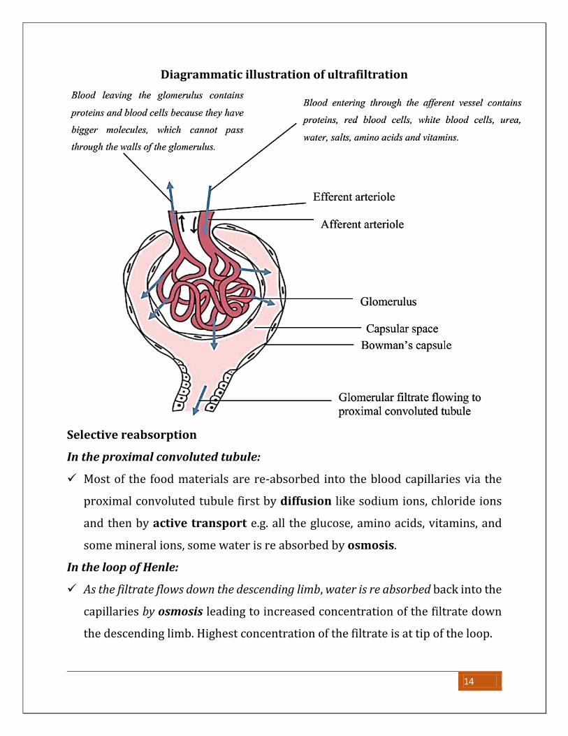
Diagrammatic illustration of ultrafiltration
Selective reabsorption
In the proximal convoluted tubule:
Most of the food materials are re-absorbed into the blood capillaries via the
proximal convoluted tubule first by diffusion like sodium ions, chloride ions
and then by active transport e.g. all the glucose, amino acids, vitamins, and
some mineral ions, some water is re absorbed by osmosis.
In the loop of Henle:
As the filtrate flows down the descending limb, water is re absorbed back into the
capillaries by osmosis leading to increased concentration of the filtrate down
the descending limb. Highest concentration of the filtrate is at tip of the loop.
14
As the filtrate ascends, first in the thin section, sodium ions and chloride ions are
reabsorbed by diffusion and in the thick section of the ascending limb of loop of
Henle, salts like Na+ and K+ are reabsorbed by active transport. This leads to a
decrease in concentration of the glomerular filtrate in the ascending limb.
In the distal convoluted tubule:
Selective re-absorption of salts by diffusion occurs.
Substances such as Urea are actively transported (secreted) from blood into
the filtrate.
Water is reabsorbed only due to stimulation by Anti diuretic hormone (ADH)
In the collecting duct:
Water is lost to the highly concentrated medulla tissues by osmosis from which
later the remaining filtrate is urine which goes via the ureter and temporarily
stored in the urinary bladder.
Water is reabsorbed only due to stimulation by Anti diuretic hormone (ADH)
Summary of the steps involved in formation of urine in the kidneys
Region of nephron Function
Bowman’s capsule Ultra filtration of the blood in glomerulus; high
hydrostatic pressure produces an ultra-filtrate free of
plasma proteins and red blood cells
Proximal
convoluted tubules
Reabsorption of useful substances such as Na+, K+, Cl-,
HCO3- , amino acids and glucose by a mixture of
diffusion and active transport
Reabsorption of water by osmosis
Reabsorption of urea by diffusion
Loop of Henle Reabsorption of water from the collecting duct
15
Distal convoluted
tubule
Reabsorption of Na+
Reabsorption of water under the control of ADH
Secretion of H+, NH4+ and reabsorption of HCO3- to
control blood pH
Secretion of some drugs
Collecting duct Reabsorption of water under control of ADH
Comparison of composition of substances in blood and urine
Explanation of observations
There are proteins in blood plasma but none in glomerular filtrate and urine
because proteins are not filtered out of the blood vessels into the glomerulus
due to the large size of their molecules.
Urea is more in urine than in both blood plasma and glomerular filtrate, this
increase is because of two reasons;
Extensive re-absorption of water from the glomerular filtrate back into
blood causing an increase in amount of urea per unit volume of urine
Secretion of excess urea in blood into the distal convoluted tubules
Glucose is present in glomerular filtrate and completely absent in urine. This is
because glucose molecules are small in size and pass through the glomerular
.
16
pores into the filtrate. However, all the glucose molecules are reabsorbed back
in the proximal convoluted tubules hence absent in urine
Salts like chlorides and sodium ions are more in urine than in blood. This is
because they are in excess in blood and few are reabsorbed back into the blood.
Because of this they tend to concentrate in urine.
Water is more in urine than in blood because it is used to dissolve urea.
NOTE: the relative amounts of water in urine and in blood varies depending on
the amount of water in the body, amount of solutes in the body, temperature and
body activity.
OSMOREGULATION
This is the process by which osmotic pressure of body fluids is kept constant.
Significance of osmoregulation
The osmotic pressure of blood must be regulated within narrow limits in order to
prevent the body cells from either losing or gaining too much water.
If too much water is gained the cells would burst and if the body has less water,
the body fluids will become more concentrated and cells will crenate, this would
result in the damage of body tissues thus greatly affecting the functioning of the
body.
The osmotic pressure of the body fluids depends on the balance between the
concentration of water and salts in them. Decreasing the amount of water in blood
or increasing the amount of dissolved salts results into increase in the osmotic
pressure of blood and vice versa
Sources and losses of water and salts from the body.
The body gains water from:- The body lose water through:-
17
Food eaten,
Drinking water
Body metabolic processes like
respiration
Absorption from surrounding
for fresh water organisms
Passing it out with faeces and
urine, Sweating
As vapour in exhaled air
Osmosis into surrounding water
for marine organisms
Salts are gained from Salts are lost through
Food
drinks
sweat
urine
diffusion into surrounding water
NOTE:
The overall effect of osmoregulation is to strike a balance between water and
mineral salt loss and gain
Role of the kidney in osmoregulation
The osmotic pressure of blood is under the control of the hypothalamus which
stimulates the pituitary gland to release a hormone called Anti-diuretic
hormone (ADH) also known as Vasopressin
Regulation of the osmotic pressure of the blood when it high
A decrease in the amount of water in blood due to taking of salty meals, this
leads to increase in osmotic pressure of blood.
Increased osmotic pressure is detected by the osmoreceptors in the
hypothalamus, this stimulates the posterior pituitary gland to release more
Anti-Diuretic hormone (ADH) into blood.
ADH reaches the kidney tubules and increases their permeability to water
hence more water is reabsorbed back to blood.
18
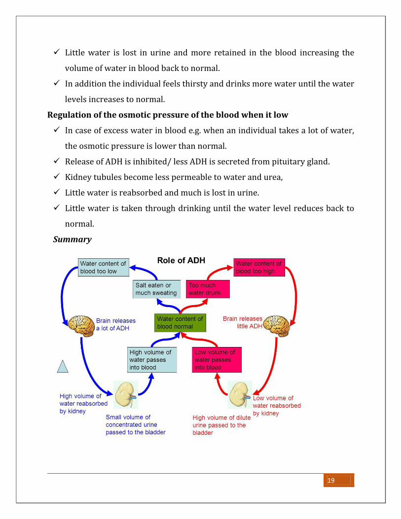
Little water is lost in urine and more retained in the blood increasing the
volume of water in blood back to normal.
In addition the individual feels thirsty and drinks more water until the water
levels increases to normal.
Regulation of the osmotic pressure of the blood when it low
In case of excess water in blood e.g. when an individual takes a lot of water,
the osmotic pressure is lower than normal.
Release of ADH is inhibited/ less ADH is secreted from pituitary gland.
Kidney tubules become less permeable to water and urea,
Little water is reabsorbed and much is lost in urine.
Little water is taken through drinking until the water level reduces back to
normal.
Summary
19
Note:
On a cold day one the metabolic rate increases so as to generate more heat in
the body to maintain the body temperature constant. This result into
generation of much water yet little is lost in sweat. This explains why one
needs to urinate several times in a short period of time.
In the same way; because of the high metabolic rate, one tends to feel hungry
faster on a cold day as food is quickly broken down to generate heat
A person whose pituitary gland is faulty can’t produce adequate amount of
ADH produces large volumes of dilute urine (Diuresis) and urinates
frequently. This disorder is known as diabetes inspidus and may lead to
dehydration
Alcohol tends to inhibit release of adrenaline leading to increase in volume
and frequency of urine formation.
Role of the liver in formation and elimination of urea
End products of protein digestion i.e. amino acids are absorbed into blood
capillaries in the villi in the ileum, blood capillaries join up to form hepatic portal
vein, which transports the amino acids through the blood stream to the liver.
In the liver excess amino acids cannot be store in the body. Excess amino acids
are split by enzymes during the process of deamination by liver cells producing
ammonia and an acid, the acid is converted into carbohydrates or fat and stored,
ammonia combines with carbon dioxide to form urea and water, urea dissolves
in the blood plasma, and is transported to the kidneys where it is eliminated in
the urine, a small amount also excreted in sweat.
20
Role of the mammalian liver in glucose regulation
Significance of blood sugar regulation
o Prevents the cells running short of glucose in case its level drops, since
glucose (blood sugar) is the main source of energy decrease in its
concentration below normal would result in less energy being produced,
slowing down metabolic reactions in the body that need energy.
o Too much glucose would increase the osmotic pressure of blood, causing the
cells to take in more water by osmosis causing their bursting, slowing down
metabolic reactions.
Control of blood glucose levels
The normal blood glucose level is 90mg/100cm3 of blood, though it may
increase greatly after a meal of carbohydrates or decrease during starvation.
Blood glucose concentration is controlled by the pancreas. The pancreas has
glucose receptor cells which monitor the concentration of glucose in the blood,
and it also has endocrine cells (called the islets of Langerhans), which secrete
two hormones. The alpha cells (α cells) secrete a hormone called glucagon,
while the beta cells (β cells) secrete a hormone called insulin. These two
hormones are antagonistic, and have opposite effects on blood glucose.
Mechanism of blood sugar regulation
After a meal of carbohydrates, glucose is absorbed from the gut into the hepatic
portal vein, increasing the blood glucose concentration. This is detected by the
pancreas, which secretes insulin from its beta cells which moves through the
blood stream to the liver where it promotes.
Conversion of glucose to glycogen for storage in the liver
Conversion of glucose to fats for storage in fat adipose tissues
Increase in breakdown of glucose by respiration to provide energy.
21
This reduces to glucose level back to normal (set point) level, the pancreas stops
secreting insulin.
If the glucose level falls too far for example during starvation or fasting, the
pancreas detects this, the alpha cells of the pancreas release glucagon
hormone into blood. This is moves through the blood stream to the liver where
it promotes:
Breakdown of glycogen to glucose
Breakdown of fats to glucose
Reduction glucose breakdown by respiration.
This allows the glucose levels to increase back to normal, the pancreas stops
producing glucagon.
Failure to produce insulin causes the presence of much glucose in urine a
condition known as diabetes mellitus.
NOTE:
An individual whose pancreas cannot secrete sufficient amounts of insulin
suffers an abnormality called diabetes mellitus. The blood glucose level
increases such that some glucose may not be reabsorbed from the proximal
convoluted tubules thus appearing in urine. The condition is treated by
injecting a patient with insulin.
The treatment cannot be administered orally being that it’s a protein that
would be broken down by enzymes into amino acids.
Diabetic individuals are advised to eat frequently to replace the glucose lost
in urine
Functions of the liver
Regulation of blood glucose levels. The liver converts excess glucose to
glycogen which can be broken down to glucose thus maintaining a constant
amount of glucose in blood
22
Formation and distribution of fats. The liver converts excess glucose to fats
for storage in fat tissues in the body
Production of red blood cells in infants whose bones are not fully developed.
NB: In adults, the role of red blood cell production is taken up by bone
marrows and liver takes on an opposite role of breaking down old red blood
cells.
Formation of bile; bile contains pigments from breakdown of haemoglobin in
the liver and bile salts. It is stored in the gall bladder and added to food in the
duodenum during digestion
Storage of iron, iron from haemoglobin breakdown is stored in the liver for
later use
Storage of vitamins; fat-soluble vitamins A, D, E and K are commonly stored in
the liver. This explains why a liver is a good source of vitamins
Deamination; excess amino acids are deaminated in the liver, forming
ammonia which is converted to urea and eliminated in urine. The remaining
portion of the amino acid is converted to a carbohydrate which can be
respired.
Detoxification; the liver converts any toxic substances into harmless
substances which can be excreted in urine. E.g. nicotine, drugs, alcohol
Manufacture of plasma proteins like fibrinogen which is needed for blood
clotting.
Storage of blood; the liver has a vast network of blood vessels which can store
a large volume of blood.
Heat production; given the wide range of metabolic reactions occurring in the
liver, significant amount of heat is produced thus maintaining a constant body
temperature.
23
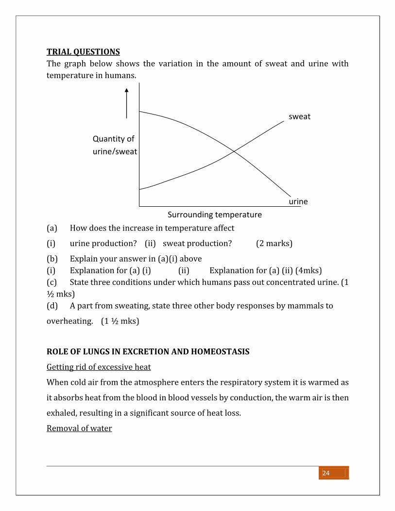
TRIAL QUESTIONS
The graph below shows the variation in the amount of sweat and urine with
temperature in humans.
(a) How does the increase in temperature affect
(i) urine production? (ii) sweat production? (2 marks)
(b) Explain your answer in (a)(i) above
(i) Explanation for (a) (i) (ii) Explanation for (a) (ii) (4mks)
(c) State three conditions under which humans pass out concentrated urine. (1
½ mks)
(d) A part from sweating, state three other body responses by mammals to
overheating. (1 ½ mks)
ROLE OF LUNGS IN EXCRETION AND HOMEOSTASIS
Getting rid of excessive heat
When cold air from the atmosphere enters the respiratory system it is warmed as
it absorbs heat from the blood in blood vessels by conduction, the warm air is then
exhaled, resulting in a significant source of heat loss.
Removal of water
sweat
urine
Surrounding temperature
Quantity of
urine/sweat
24
In coming dry air from the environment dissolves in the mucus, this is warmed
until it form water vapour also water vapour formed during metabolism diffuses
into alveolar sac and expelled during exhalation.
Removal of carbon dioxide
Carbon dioxide produced during metabolism diffuses along its concentration
gradient from the blood to the alveolar air at the lungs, this is expelled out of the
body during exhalation.
SAMPLE QUESTIONS
1. (a) Describe the process of urine formation in man
(b)Describe the difference between glomerular filtration rates between an
animal living in fresh water and one living in a desert though they may be
of the same size
(c)State with a reason the major nitrogenous waste excreted by each of the
animals above
2. (a) State the conditions under which a human being can produce
(i) Concentrated urine
(ii) Dilute urine
(iii) Urine containing glucose
(b)Briefly describe the experiment you would carry out to test for presence of
glucose in urine of a patient
3. How the structures of the mammalian nephron are related to the functions
they perform / adaptations of the mammalian nephron to its functions.
THE SKIN
This is the most extensively distributed tissue found all over the body of mammals.
It is a continuous protective layer over the body.
25

Structure of the skin
The skin consists of two main layers.
1. The epidermis (outer layer)
2. The dermis (inner layer)
THE EPIDERMIS:
This is made up of three sub layers.
a) The Malpighian layer.
b) The granular layer.
c) The cornified layer.
1) The Malpighian layer
This is the inner most sub layer in the epidermis. It consists of dividing cells which
give rise to cells of the granular layer. It secretes a pigment called melanin, which
gives the skin its colour and protects the skin from ultraviolent rays. Albinos do
not produce melanin in their skins.
26
2) The granular layer
This contains living cells arising from the malphigian layer. It is the biggest layer
of the epidermis. It gives rise to cells of the cornified layer.
3) The cornified layer.
This is the outermost layer of the skin. It is made up of dead cells, which are
keratinized. Cells of this layer continuously ware away and are replaced by cells
from the granular layer. Its function is to protect the inner parts of the body from
mechanical injury and entry of bacteria and other germs. It also offers water
proofing to the skin.
THE DERMIS:
This is the inner layer of the skin. It is below the malpighian layer. It is thicker than
the epidermis. It contains the sweat glands, nerve fibres, fat cells and blood
capillaries.
Other parts of the skin
1) Hairs.
The hairs extend from the dermis through the epidermis. They arise from hair
follicles in the dermis. They protect the body and trap a layer of air on the skin,
which insulates the body against heat loss.
2) Sebaceous gland
This secretes an oily substance called sebum. This oil softens the cornified layer
and prevents it from cracking. The oil also provides water proofing to the skin.
3) Nerve endings.
These perceive external stimuli and transport impulses to the central nervous
system.
4) Sweat glands.
These are coiled tubular glands located in the dermis. They excrete sweat, which
is released out of the skin through the sweat duct.
27
Functions of the skin
i) To protect the tissue below it from mechanical damage, bacterial and viral
infections.
ii) It also prevents excess loss of water from the body.
iii) It is a sense organ and it is sensitive to pain, touch and heat.
iv) It keeps the body temperature of endothermic organisms constant.
v) It synthesizes vitamin D in presence of sunlight.
vi) It is an excretory organ. It excretes sweat, which contains urea, water and
excess salts.
TEMPERATURE REGULATION IN MAMMALS
Temperature regulation is the process of maintaining a fairly constant body
temperature despite changes in the environmental temperature.
This is important because it ensures an ideal temperature for body metabolic
reactions which are controlled by enzymes to take place faster.
NB: To maintain body temperature constant, there must be a balance between
heat loss and gain. Mammals gain heat through the following ways:
Radiation directly from the sun and warm objects in the environment
Convection from warm air of water molecules in contact with the body
Conduction from hot objects in direct contact with the body
Metabolic reactions in the body (for endotherms)
Ways by which organisms lose heat include;
Evaporation of water .e.g. during sweating
Conduction to cold objects in contact with the body.
Convection to cold air or water molecules in contact with the body
Radiation of heat to cold objects in the environment
28
The rate of heat loss and gain depends on;
Surface area to volume ratio i.e.
Small organisms having a large surface area to volume ration tend to lose
more heat than the large ones with small surface area to volume ratio.
Temperature of surrounding environment: Organisms tend to lose more heat
in cold environment and gain more in hot environment.
Rate of respiration: The higher the rate of respiration, the more heat energy
gained by the body.
Humidity of the environment: Heat loss increases in humid conditions because
high humidity makes the environment colder.
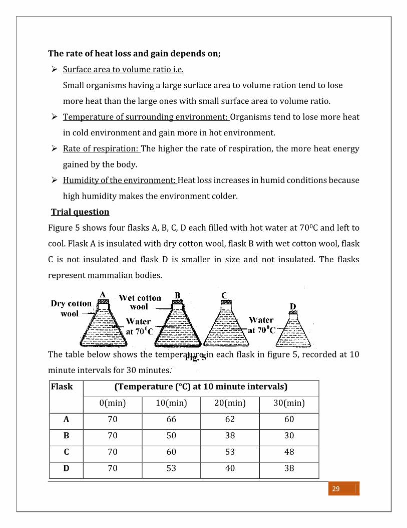
Trial question
Figure 5 shows four flasks A, B, C, D each filled with hot water at 700C and left to
cool. Flask A is insulated with dry cotton wool, flask B with wet cotton wool, flask
C is not insulated and flask D is smaller in size and not insulated. The flasks
represent mammalian bodies.
The table below shows the temperature in each flask in figure 5, recorded at 10
minute intervals for 30 minutes.
Flask (Temperature (°C) at 10 minute intervals)
0(min) 10(min) 20(min) 30(min)
A 70 66 62 60
B 70 50 38 30
C 70 60 53 48
D 70 53 40 38
29
Study the information and answer the questions that follow:
(a) For each flask draw a graph to show the changes in temperature with time
in the space on next page. Use the same X and Y axes for all the graphs. (06
marks)
(b) Calculate the average rate of cooling in each flask. (04 marks)
(c) Explain the rate of cooling in
(i) flask A (02mks) (ii) flask B (03mks)
(iii) flask C (02mks) (iv) flask D(02mks)
(d) From the information, state two factors that affect the rate of cooling from a
body. (01 mark)
MEANS OF TEMPERATURE REGULATION
Mammals are grouped into two according to their ability or inability to maintain
a constant body temperature; i.e. Homoiothermic and poikilothermic animals
POIKILOTHERMIC ANIMALS (Ectothermic animals)
These are animals whose body temperature changes with change in
environmental temperature (Greek; Poikilos means various). Such Animals are
called ectothermic because their bodies mainly obtain heat from the
environment. Their body temperature therefore is always lower than
environmental temperature hence the term cold blooded animals
Examples include:-
all invertebrates,
fish,
amphibians
reptiles
Homoiothermic animals (Endothermic animals)
These are animals that maintain a constant body temperature despite changes in
environmental temperatures. (Greek; Homoios means constant). Such animal are
also called endothermic because heat is mainly generated by metabolic reactions
occurring within the body.
30
Examples include: - birds and mammals only.
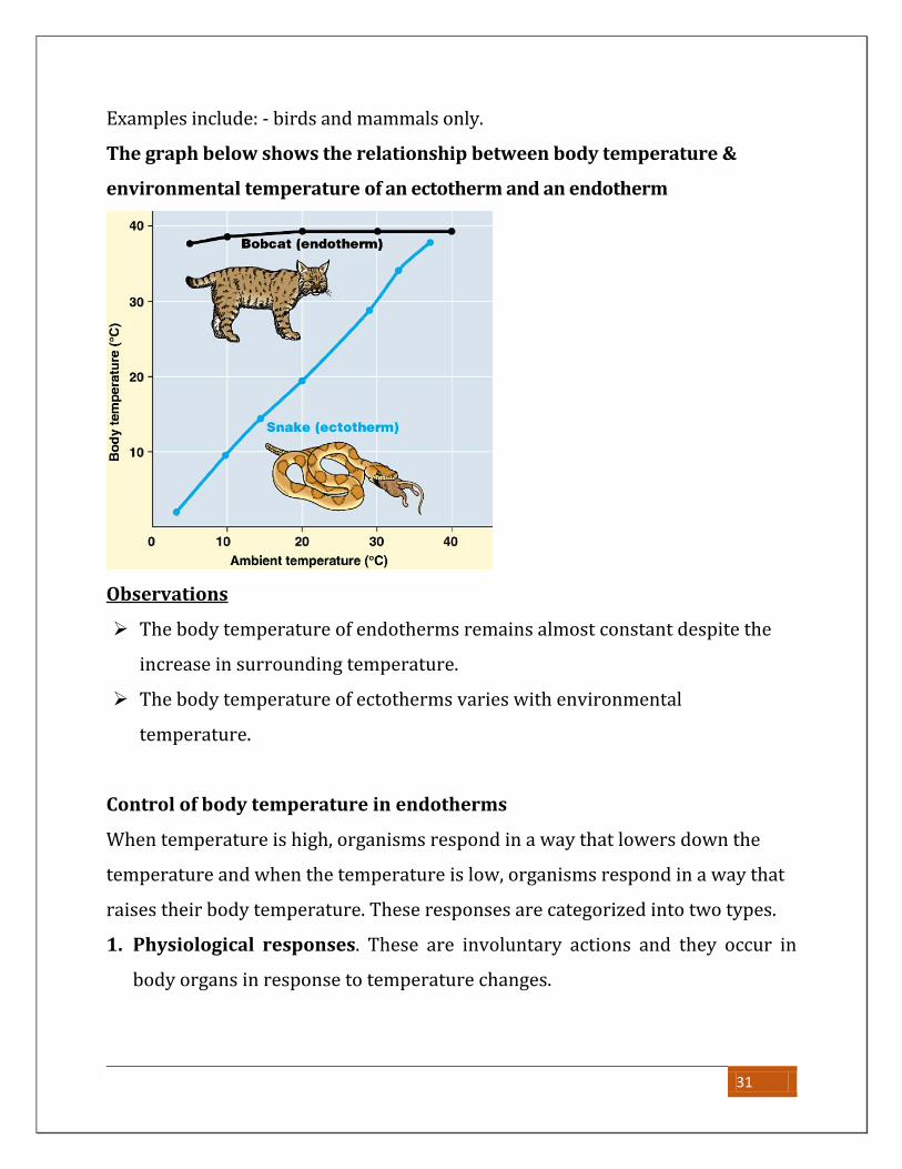
The graph below shows the relationship between body temperature &
environmental temperature of an ectotherm and an endotherm
Observations
The body temperature of endotherms remains almost constant despite the
increase in surrounding temperature.
The body temperature of ectotherms varies with environmental
temperature.
Control of body temperature in endotherms
When temperature is high, organisms respond in a way that lowers down the
temperature and when the temperature is low, organisms respond in a way that
raises their body temperature. These responses are categorized into two types.
1. Physiological responses. These are involuntary actions and they occur in
body organs in response to temperature changes.
31
2. Behavioral responses. These are voluntary responses from the organism. The
organism consciously decides what to do when external and internal
temperatures change.
Response to cold weather in endothermic animals
Physiological means.
The erector pill muscles of the hair contract to make the hairs stand upright to
the skin. The hairs trap a layer of air, which insulates the skin.
The rate of sweating reduces in order to reduce on the amount of heat lost
through it.
The metabolic activity of the liver increases to produce energy in form of heat.
Blood vessels near the skin constrict in the process called vasoconstriction to
reduce on the blood reaching the skin. This reduces heat loss through radiation.
Small animals like the mouse undergo hibernation where they dig holes and
live deep in them to reduce heat loss
Shivering. This is the rhythmic contractions of the skeletal muscles. It results
into production of heat energy.
Some have a thick fat layer to act as an insulator.
Some are very big and thus have a small surface area to volume ratio. This
reduces the rate of heat loss.
Some have few sweat glands to reduce of the heat lost during sweating
Behavioral means.
Endotherms may raise their body temperature behaviourally through;
Sitting near hot bodied to raise their body temperature by conduction or
radiation.
Humans take hot drinks.
They do physical exercises to raise the metabolic activity of the body.
They can take a hot bath
32
Hibernation. This is a state of long rest by burrowing into crevices and holes
during extreme coldness.
They put on thick clothes, which insulate their bodies.
Sun bathing
Response to hot weather in endothermic animals
In hot environment, animals control the body temperature by increasing heat
loss and lowering heat production through the following ways:
Physiological means.
The erecter pilli muscle of the skin relaxes making the hairs to fall on the skin.
This allows heat loss by radiation.
The metabolic rate of the body reduces to reduce on the amount of heat
produced.
Sweating increases. In this process excessive heat is lost as latent heat of
vaporization to evaporate the sweat from the body hence losing heat.
Vasodilatation. Vessels dilate and allow more blood to reach the skin surface in
order to lose heat to the surroundings by radiation.
Animals living in hot environments have a thin fat layer to reduce on the
insulation.
Having little hairs on the body to allow easy loss of heat.
Having less fat to reduce on the insulation effect of fats.
Having a large surface area to volume ratio. To allow a faster rate of heat loss.
Some have a lot of sweat glands to increase heat loss through sweat.
Having many blood vessels near the skin for easy loss of heat by radiation.
Behavioural means.
Some rest on cold bodies like rocks to lose heat by conduction.
Humans sit near fans.
33
Some take cold drinks.
They put on light clothes
Panting. This involves hanging out of the tongue for example in dogs. This
results into evaporation from the mouth, which eventually cools the animal.
Swimming.
Resting under shade.
Bathing cold water.
Aestivation. This is a state of long rest by burrowing in crevices and holes
during extreme hotness.
Putting on lighter clothes in man.
TEMPERATURE CONTROL IN ECTOTHERMIC ANIMALS
Their body temperature is controlled by only behavioral means.
During hot conditions, they lose heat by.
They rest on cold rocks to lose heat by conduction.
They rest on cold stones and in shades to lose heat.
They burrow in cracks and lose heat by radiation.
Aestivation. This is a state of long rest by burrowing underground or under
rocks during high temperatures.
Thermal gaping. This is the opening of the mouth to lose water by evaporation.
This results into cooling. Thermal gaping occurs in crocodiles and a few other
reptiles.
During cold conditions, they gain heat by;
Resting on hot rocks to gain heat by conduction.
They rest under the sun to gain heat by radiation.
They rest near hot bodies to gain heat by radiation.
They burrow in hot sand to gain heat by conduction.
34
Basking in the sun to gain heat.
Hibernation. This is a state of long rest by burrowing into crevices and holes
during extreme coldness.
Advantages of endothermy
They are able to live in a wide range of environment irrespective of the
prevailing temperature; because they are able to regulate their body
temperature.
They remain active at all times of the day as they regulate their body
temperature
Body processes occur efficiently because the body temperature is maintained
near to optimum temperatures for body enzymes.
Endotherms show faster responses to changes in the environment which
increases chances of survival.
Disadvantages of endothermy
Much food must be consumed to maintain a high rate of meatabolism to
generate heat.
Much heat is lost from the body as the body temperature is higher than that of
the environment.
Advantages of ectothermy
Ectotherms consume less food as heat is mainly obtained from the environment.
They produce fewer wastes due to low metabolism.
Disadvantages of ectothermy
Slow responses to stimuli due to low metabolic when the temperature is low
They can occupy a limited range of habitats where temperatures are favourable
They are not active at some times of the day when temperatures are too high or
low
35
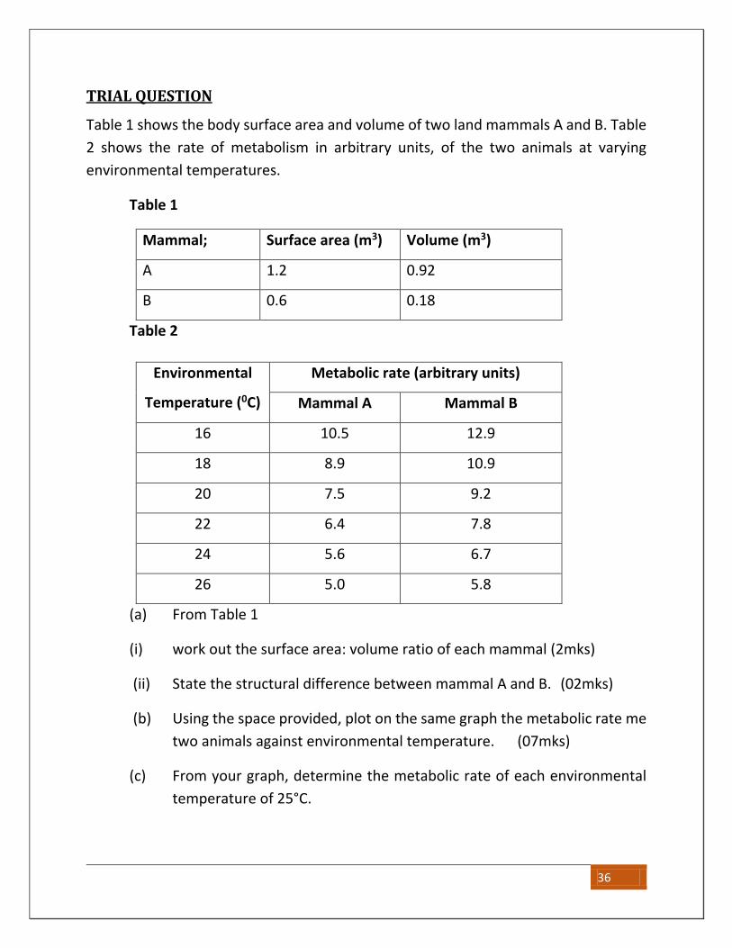
TRIAL QUESTION
Table 1 shows the body surface area and volume of two land mammals A and B. Table
2 shows the rate of metabolism in arbitrary units, of the two animals at varying
environmental temperatures.
Table 1
Mammal; Surface area (m3) Volume (m3)
A 1.2 0.92
B 0.6 0.18
Table 2
Environmental
Temperature (0C)
Metabolic rate (arbitrary units)
Mammal A Mammal B
16 10.5 12.9
18 8.9 10.9
20 7.5 9.2
22 6.4 7.8
24 5.6 6.7
26 5.0 5.8
(a) From Table 1
(i) work out the surface area: volume ratio of each mammal (2mks)
(ii) State the structural difference between mammal A and B. (02mks)
(b) Using the space provided, plot on the same graph the metabolic rate me
two animals against environmental temperature. (07mks)
(c) From your graph, determine the metabolic rate of each environmental
temperature of 25°C.
36
(d) (i) How does environmental temperature affect the metabolic rate of
the mammals? (02 marks)
(ii) Explain why variation of temperature affects the metabolic rate of
the mammals as stated in (c) (i), (02 marks)
e) From the information provided explain why at any environmental
temperature, me metabolic rate of mammal B is higher than that of
mammal A. (03mks)
37
Related Documents


![Excretion [2015]](https://static.cupdf.com/doc/110x72/55d39c87bb61eb05278b46dd/excretion-2015-55d47f0693bf7.jpg)