RESEARCH Open Access Evolution of the Pax-Six-Eya-Dach network: the calcisponge case study Sofia AV Fortunato 1,2 , Sven Leininger 1,3 and Maja Adamska 1* Abstract Background: The Pax-Six-Eya-Dach network (PSEDN) is involved in a variety of developmental processes, including well documented roles in determination of sensory organs and morphogenesis in bilaterian animals. Expression of PSEDN components in cnidarians is consistent with function in sensory organ development. Recent work in demosponges demonstrated the presence of single homologs of Pax and Six genes, and their possible involvement in morphogenesis, but the absence of the remaining network components. Calcisponges are evolutionarily distant from demosponges, and the developmental toolkits of these two lineages differ significantly. We used an emerging model system, Sycon ciliatum, to identify components of the PSEDN and study their expression during embryonic and postembryonic development. Results: We identified two Pax, three Six and one Eya genes in calcisponges, a situation strikingly different than in the previously studied demosponges. One of the calcisponge Pax genes can be identified as PaxB, while the second Pax gene has no clear affiliation. The three calcisponge Six genes could not be confidently classified within any known family of Six genes. Expression analysis in adult S. ciliatum demonstrated that representatives of Pax, Six and Eya are expressed in patterns consistent with roles in morphogenesis of the choanocyte chambers. Distinct paralogues of Pax and Six genes were expressed early in the development of the putative larval sensory cells, the cruciform cells. While lack of known photo pigments in calcisponge genomes precludes formal assignment of function to the cruciform cells, we also show that they express additional eumetazoan genes involved in specification of sensory and neuronal cells: Elav and Msi. Conclusions: Our results indicate that the role of a Pax-Six-Eya network in morphogenesis likely predates the animal divergence. In addition, Pax and Six, as well as Elav and Msi are expressed during differentiation of cruciform cells, which are good candidates for being sensory cells of the calcaronean sponge larvae. Keywords: Calcisponges, Sycon, Eyes absent, Pax, Six, Sensory cells Background In insect and vertebrate model systems, Pax, Six, Eyes ab- sent (Eya) and Dachshund (Dach) form a network (PSEDN) interconnected by a series of protein-protein and protein- DNA interactions [1,2]. This network is often referred to as Retinal Determination Gene Network (RDGN), although it is involved in a variety of developmental processes in addition to eye development, including roles in morpho- genesis of other sensory organs, kidneys and the branchial arches [3-5]. It has been suggested that the insect and verte- brate PSEDN/RDGN are not homologous [6], although studies in cnidarians indicate deep evolutionary roots of this network. In particular, it has been demonstrated that members of the Pax, Six and Eya families are expressed during neural development and sensory organ formation in a wide range of cnidarians [7-15]. In demosponges, two components of the PSEDN have been identified, PaxB and Six1/2 [16,17]. A recent study in the freshwater demosponge Ephydatia muelleri shows that these genes are co-expressed, potentially interact and are likely involved in juvenile/adult morphogenesis [16,17]. While sponges lack a nervous system, larvae of some species have well defined sensory cells, organized into simple organ-like structures [18,19]. For example, the parenchymella-type larvae of Amphimedon queenslandica have a pigmented ring equipped with long “steering” cilia at their posterior pole known as the sensory organ of the larva. Although opsin is not found in the A. queenslandica * Correspondence: [email protected] 1 Sars International Centre for Marine Molecular Biology, University of Bergen, Thormøhlensgt. 55, Bergen 5008, Norway Full list of author information is available at the end of the article © 2014 Fortunato et al.; licensee BioMed Central Ltd. This is an Open Access article distributed under the terms of the Creative Commons Attribution License (http://creativecommons.org/licenses/by/4.0), which permits unrestricted use, distribution, and reproduction in any medium, provided the original work is properly credited. The Creative Commons Public Domain Dedication waiver (http://creativecommons.org/publicdomain/zero/1.0/) applies to the data made available in this article, unless otherwise stated. Fortunato et al. EvoDevo 2014, 5:23 http://www.evodevojournal.com/content/5/1/23

Welcome message from author
This document is posted to help you gain knowledge. Please leave a comment to let me know what you think about it! Share it to your friends and learn new things together.
Transcript
Fortunato et al. EvoDevo 2014, 5:23http://www.evodevojournal.com/content/5/1/23
RESEARCH Open Access
Evolution of the Pax-Six-Eya-Dach network: thecalcisponge case studySofia AV Fortunato1,2, Sven Leininger1,3 and Maja Adamska1*
Abstract
Background: The Pax-Six-Eya-Dach network (PSEDN) is involved in a variety of developmental processes, includingwell documented roles in determination of sensory organs and morphogenesis in bilaterian animals. Expression of PSEDNcomponents in cnidarians is consistent with function in sensory organ development. Recent work in demospongesdemonstrated the presence of single homologs of Pax and Six genes, and their possible involvement in morphogenesis,but the absence of the remaining network components. Calcisponges are evolutionarily distant from demosponges, andthe developmental toolkits of these two lineages differ significantly. We used an emerging model system, Sycon ciliatum,to identify components of the PSEDN and study their expression during embryonic and postembryonic development.
Results: We identified two Pax, three Six and one Eya genes in calcisponges, a situation strikingly different than in thepreviously studied demosponges. One of the calcisponge Pax genes can be identified as PaxB, while the secondPax gene has no clear affiliation. The three calcisponge Six genes could not be confidently classified within anyknown family of Six genes. Expression analysis in adult S. ciliatum demonstrated that representatives of Pax, Sixand Eya are expressed in patterns consistent with roles in morphogenesis of the choanocyte chambers. Distinctparalogues of Pax and Six genes were expressed early in the development of the putative larval sensory cells, thecruciform cells. While lack of known photo pigments in calcisponge genomes precludes formal assignment of functionto the cruciform cells, we also show that they express additional eumetazoan genes involved in specification of sensoryand neuronal cells: Elav and Msi.
Conclusions: Our results indicate that the role of a Pax-Six-Eya network in morphogenesis likely predates theanimal divergence. In addition, Pax and Six, as well as Elav and Msi are expressed during differentiation ofcruciform cells, which are good candidates for being sensory cells of the calcaronean sponge larvae.
Keywords: Calcisponges, Sycon, Eyes absent, Pax, Six, Sensory cells
BackgroundIn insect and vertebrate model systems, Pax, Six, Eyes ab-sent (Eya) and Dachshund (Dach) form a network (PSEDN)interconnected by a series of protein-protein and protein-DNA interactions [1,2]. This network is often referred to asRetinal Determination Gene Network (RDGN), although itis involved in a variety of developmental processes inaddition to eye development, including roles in morpho-genesis of other sensory organs, kidneys and the branchialarches [3-5]. It has been suggested that the insect and verte-brate PSEDN/RDGN are not homologous [6], althoughstudies in cnidarians indicate deep evolutionary roots of
* Correspondence: [email protected] International Centre for Marine Molecular Biology, University of Bergen,Thormøhlensgt. 55, Bergen 5008, NorwayFull list of author information is available at the end of the article
© 2014 Fortunato et al.; licensee BioMed CentCommons Attribution License (http://creativecreproduction in any medium, provided the orDedication waiver (http://creativecommons.orunless otherwise stated.
this network. In particular, it has been demonstratedthat members of the Pax, Six and Eya families areexpressed during neural development and sensoryorgan formation in a wide range of cnidarians [7-15]. Indemosponges, two components of the PSEDN have beenidentified, PaxB and Six1/2 [16,17]. A recent study in thefreshwater demosponge Ephydatia muelleri shows thatthese genes are co-expressed, potentially interact and arelikely involved in juvenile/adult morphogenesis [16,17].While sponges lack a nervous system, larvae of some
species have well defined sensory cells, organized intosimple organ-like structures [18,19]. For example, theparenchymella-type larvae of Amphimedon queenslandicahave a pigmented ring equipped with long “steering” ciliaat their posterior pole known as the sensory organ of thelarva. Although opsin is not found in the A. queenslandica
ral Ltd. This is an Open Access article distributed under the terms of the Creativeommons.org/licenses/by/4.0), which permits unrestricted use, distribution, andiginal work is properly credited. The Creative Commons Public Domaing/publicdomain/zero/1.0/) applies to the data made available in this article,
Fortunato et al. EvoDevo 2014, 5:23 Page 2 of 12http://www.evodevojournal.com/content/5/1/23
genome, the larval phototactic behavior [20] is likely me-diated by cryptochrome [21], which has also been sug-gested to participate in light reception in adult tissue ofanother demosponge, Suberites domuncula [22]. Signifi-cantly, AmqCry2 expression is associated with the pig-ment ring [23]. Unfortunately, no information regardingexpression of Pax or Six genes during development of thepigment ring is published, making it impossible to predictwhether the ancestral PSEDN function was related tomorphogenesis only or both morphogenesis and sensoryorgan formation.We have recently began developing Sycon ciliatum as
a model representing calcisponges (subclass Calcaro-nea), a lineage evolutionarily distant from demospongesand appearing to significantly differ from demospongesin its gene content [24-27]. Embryonic and postembryo-nic development of syconoid calcaronean species is wellstudied, allowing us to relate gene expression patterns todevelopmental events. Importantly, different stages of ra-dial (choanocyte) chamber morphogenesis can be com-pared in a single specimen fixed during the growth phase:when the asconoid body plan of the juvenile gives rise tothe syconoid body plan of the adult, radial chambers formaround the original central choanocyte chamber andcontinue to develop sequentially from bottom to top,with the region just under the osculum remaining inasconoid organization [28]. The amphiblastulae larvaeof calcisponges from the subclass Calcaronea are strikinglydifferent from the parenchymellae [25,29,30]. Amphiblas-tulae are composed of only three cell types of embryonicorigin: macromeres, micromeres, and four cruciform cellsdistributed around the “equator” and conveying uniquetetra-radial symmetry to the larva [24,31]. While the mac-romeres and micromeres participate in formation of thejuvenile body upon metamorphosis, the cruciform cellsdegenerate upon settlement [32]. The function of cruci-form cells has not been studied experimentally, but basedon ultrastructure examination it has been suggested thatthey might act as photoreceptors [33]. Intriguingly, differ-entiating cruciform cells of S. ciliatum express SoxB [24],a transcription factor involved in bilaterian neurogenesis[34,35] and expressed in cnidarian neurosensory cells [36].In addition, they express several genes which, while clearlyhaving multitude of roles in animal development, are alsoimplicated in specification of neuronal cell types in eume-tazoans: components of the Wnt pathway (dvl, tcf andbeta-catenin), Smad1/5 and nanos, lending support to thenotion that they could be sensory cells [25,37-39].In thisstudy we chose to address the evolution of the PSEDN bystudying expression of potential components of this net-work in S. ciliatum, focusing on the cruciform cells as thelikely sensory cells of the larvae, and on the adult morpho-genesis represented by formation of the radial (choano-cyte) chambers.
We searched the genomic and transcriptomic datasetsof S. ciliatum and a second calcaronean species, Leuco-solenia complicata, for genes encoding the componentsof the PSED network. To gain additional insight intoidentity of the cruciform cells, we also searched forgenes encoding known proteins involved in photorecep-tion (opsin and cryptochrome), and the RNA bindingproteins Elav and Musashi, which are involved in specifi-cation of neurosensory cells in eumetazoans. In thispaper, we report that calciponge genomes contain anortholog of the Eya gene, which has not been previouslyreported in demosponges. We have not identified opsinand dachshund in calcisponges, which is consistent withthe absence of these genes in demosponges. On theother hand, cryptochrome, which is present in demos-ponges, and likely responsible for light perception in thedemosponge larvae, is absent from the calcisponge ge-nomes. Expression of Pax, Six, Eya, Msi and Elav genesin Sycon ciliatum was studied by in situ hybridization.Here we show that Pax, Six and Eya genes are co-expressed during morphogenesis of the radial chambers,and that Pax and Six, as well as Elav and Msi are co-expressed during formation of cruciform cells.
MethodsSequence retrieval, alignment and phylogenetic analysesSycon ciliatum and Leucosolenia complicata Pax, Six andEya genes were identified by BLAST searches of Sycon andLeucosolenia draft genomes (a preliminary draft of Leucoso-lenia) and transcriptomes as previously described [24] usingspecific domains from the following taxa: Bilateria, Bran-chiostoma floridae and Mus musculus; Porifera, Amphime-don queenslandica; Cnidaria, Nematostella vectensis.For Eya, alignments were performed using the con-
served ED domain. For Pax genes two alignments wereperformed: in the first alignment, the complete paireddomain (PD) was included and in the second alignmentthe truncated RED-PD domain was used. Lack of home-odomains in the calcisponge Pax sequences precludeshomeodomain-based phylogenetic analyses. For Six genes,the homeodomain along with the extended sine oculis do-main was used. A combination of ClustalX and MUSCLEwas used for the alignments, which were manually cor-rected where necessary.The amino acid substitution model of protein evolution
was determined by ProtTest 3.0 [40]. For all analyses thebest model of protein evolution was LG +G, except forthe analysis of the complete PD domain of Pax geneswhere invariant site gamma LG +G + I was optimal.Bayesian and maximum likelihood (ML) analyses wereundertaken on conserved regions. For the MrBayes 3.1analyses [41] (with LG model incorporated by in-housemodification), a set of four independent Metropolis-coupled Markov Chain Monte Carlo (MCMC) were
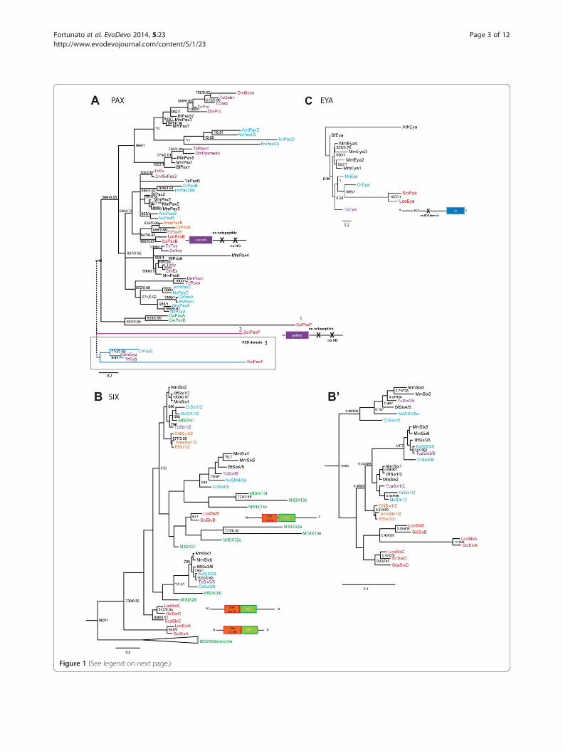
Figure 1 (See legend on next page.)
Fortunato et al. EvoDevo 2014, 5:23 Page 3 of 12http://www.evodevojournal.com/content/5/1/23
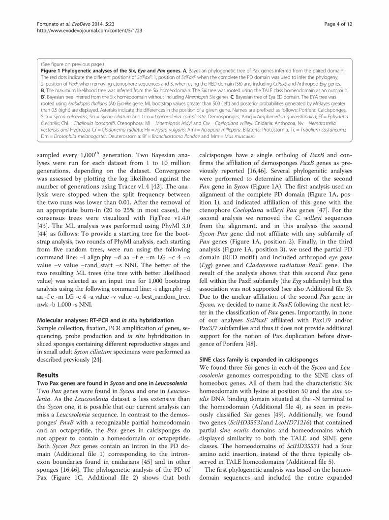
(See figure on previous page.)Figure 1 Phylogenetic analyses of the Six, Eya and Pax genes. A, Bayesian phylogenetic tree of Pax genes inferred from the paired domain.The red dots indicate the different positions of SciPaxF: 1, position of SciPaxF when the complete the PD domain was used to infer the phylogeny;2, position of PaxF when removing ctenophore sequences and 3, when using the RED domain (S6) and including CrPaxE and Arthropod Eyg genes.B, The maximum likelihood tree was inferred from the Six homeodomain. The Six tree was rooted using the TALE class homeodomain as an outgroup.B’, Bayesian tree inferred from the Six homeodomain without including Mnemiopsis Six genes. C, Bayesian tree of Eya ED domain. The EYA tree wasrooted using Arabidopsis thaliana (At) Eya-like gene. ML bootstrap values greater than 500 (left) and posterior probabilities generated by MrBayes greaterthan 0.5 (right) are displayed. Asterisks indicate the differences in the position of a given gene. Names are prefixed as follows: Porifera: Calcisponges,Sca = Sycon calcavaris; Sci = Sycon ciliatum and Lco = Leucosolenia complicata. Demosponges, Amq = Amphimedon queenslandica; Ef = Ephydatiafluviatilis; Chl = Chalinula loosanoffi. Ctenophora: Ml =Mnemiopsis leidyi and Cw = Coeloplana willeyi. Cnidaria: Anthozoa, Nv = Nematostellavectensis and Hydrozoa Cr = Cladonema radiatu; Hv = Hydra vulgaris; Ami = Acropora millepora. Bilateria: Protostomia, Tc = Tribolium castaneum.;Dm = Drosophila melanogaster. Deuterostomia: Bf = Branchiostoma floridae and Mm =Mus musculus.
Fortunato et al. EvoDevo 2014, 5:23 Page 4 of 12http://www.evodevojournal.com/content/5/1/23
sampled every 1,000th generation. Two Bayesian ana-lyses were run for each dataset from 1 to 10 milliongenerations, depending on the dataset. Convergencewas assessed by plotting the log likelihood against thenumber of generations using Tracer v1.4 [42]. The ana-lysis were stopped when the split frequency betweenthe two runs was lower than 0.01. After the removal ofan appropriate burn-in (20 to 25% in most cases), theconsensus trees were visualized with FigTree v1.4.0[43]. The ML analysis was performed using PhyMl 3.0[44] as follows: To provide a starting tree for the boot-strap analysis, two rounds of PhyMl analysis, each startingfrom five random trees, were run using the followingcommand line: −i align.phy –d aa –f e –m LG –c 4 –avalue –v value –rand_start –s NNI. The better of thetwo resulting ML trees (the tree with better likelihoodvalue) was selected as an input tree for 1,000 bootstrapanalysis using the following command line: -i align.phy -daa -f e -m LG -c 4 -a value -v value -u best_random_tree.nwk -b 1,000 -s NNI.
Molecular analyses: RT-PCR and in situ hybridizationSample collection, fixation, PCR amplification of genes, se-quencing, probe production and in situ hybridization insliced sponges containing different reproductive stages andin small adult Sycon ciliatum specimens were performed asdescribed previously [24].
ResultsTwo Pax genes are found in Sycon and one in LeucosoleniaTwo Pax genes were found in Sycon and one in Leucoso-lenia. As the Leucosolenia dataset is less extensive thanthe Sycon one, it is possible that our current analysis canmiss a Leucosolenia sequence. In contrast to the demos-ponges’ PaxB with a recognizable partial homeodomainand an octapeptide, the Pax genes in calcisponges donot appear to contain a homeodomain or octapeptide.Both Sycon Pax genes contain an intron in the PD do-main (Additional file 1) corresponding to the intron-exon boundaries found in cnidarians [45] and in othersponges [16,46]. The phylogenetic analysis of the PD ofPax (Figure 1C, Additional file 2) shows that both
calcisponges have a single ortholog of PaxB and con-firms the affiliation of demosponges PaxB genes as pre-viously reported [16,46]. Several phylogenetic analyseswere performed to determine affiliation of the secondPax gene in Sycon (Figure 1A). The first analysis used analignment of the complete PD domain (Figure 1A, pos-ition 1), and indicated affiliation of this gene with thectenophore Coeloplana willeyi Pax genes [47]. For thesecond analysis we removed the C. willeyi sequencesfrom the alignment, and in this analysis the secondSycon Pax gene did not affiliate with any subfamily ofPax genes (Figure 1A, position 2). Finally, in the thirdanalysis (Figure 1A, position 3), we used the partial PDdomain (RED motif ) and included arthropod eye gone(Eyg) genes and Cladonema radiatum PaxE gene. Theresult of the analysis shows that this second Pax genefell within the PaxE subfamily (the Eyg subfamily) but thisassociation was not supported (see also Additional file 3).Due to the unclear affiliation of the second Pax gene inSycon, we decided to name it PaxF, following the next let-ter in the classification of Pax genes. Importantly, in noneof our analyses SciPaxF affiliated with Pax1/9 and/orPax3/7 subfamilies and thus it does not provide additionalsupport for the notion of Pax duplication before diver-gence of Porifera [48].
SINE class family is expanded in calcispongesWe found three Six genes in each of the Sycon and Leu-cosolenia genomes corresponding to the SINE class ofhomeobox genes. All of them had the characteristic Sixhomeodomain with lysine at position 50 and the sine oc-ulis DNA binding domain situated at the -N terminal tothe homeodomain (Additional file 4), as seen in previ-ously classified Six genes [49]. Additionally, we foundtwo genes (SciHD35531and LcoHD71216) that containedpartial sine oculis domains and homeodomains whichdisplayed similarity to both the TALE and SINE geneclasses. The homeodomains of SciHD35531 had a fouramino acid insertion, instead of the three typically ob-served in TALE homeodomains (Additional file 5).The first phylogenetic analysis was based on the homeo-
domain sequences and included the entire expanded
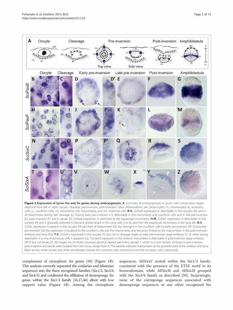
Figure 2 Expression of Sycon Pax and Six genes during embryogenesis. A, Summary of embryogenesis in Sycon, with consecutive stagesdepicted from left to right: oocyte, cleavage, pre-inversion, post-inversion, larva. Abbreviations: pin, pinacocytes; ch, choanocytes; ac, accessorycells; cc, cruciform cells; mi, micromeres ma, macromeres; and mc, maternal cells. B-G, SciPaxB expression is detectable in the oocytes (B), and inall blastomeres during late cleavage (C). During early pre-inversion it is detectable in the micromeres and cruciform cells and in late pre-inversion(E), post-inversion (F) and in larvae (G) SciPaxB expression is restricted to the equatorial micromeres. H-M, SciPaxF expression is detectable in theoocytes (H) and is gradually restricted to become predominant in the cross cells (J to K), and then the equatorial micromeres in the larva (M). N-S,SciSixC expression is present in the oocytes (N) and then all blastomeres (O), but strongest in the cruciform cells by early pre-inversion (P). During latepre-inversion (Q) the expression is localized to the cruciform cells and the macromeres, and becomes limited to the macromeres in the post-inversionembryos and larva (R,S). T-Z, SciSixA is expressed in the oocytes (T), but not in cleavage stages or early pre-inversion stage embryos (U, V), when strongexpression in a ring of accessory cells is apparent (U). Transient expression in the anterior micromeres is detectable in post-inversion stage embryos(W-Y) but not larvae (Z). All images are of whole mounted, glycerol-cleared specimens, except Y, which is a resin section. Embryos in pre-inversion,post-inversion and larvae were isolated from the tissue, except from K. The asterisk indicates macromeres at the posterior pole of the embryo and larva.Black arrows, white arrows and white arrowheads indicate the cruciform cells, micromeres and the accessory cells, respectively.
Fortunato et al. EvoDevo 2014, 5:23 Page 5 of 12http://www.evodevojournal.com/content/5/1/23
complement of ctenophore Six genes [50] (Figure 1B).This analysis correctly separated the cnidarian and bilateriansequences into the three recognized families (Six1/2, Six3/6and Six4/5) and confirmed the affiliation of demosponge Sixgenes within the Six1/2 family [16,17,46] albeit with lowsupport value (Figure 1B). Among the ctenophore
sequences, MlSix41 nested within the Six1/2 family,consistent with the presence of the ETSY motif in itshomeodomain, while MlSix36 and MlSix28 groupedwith the Six3/6 family as described [50]. Surprisingly,none of the calcisponge sequences associated withdemosponge sequences or any other recognized Six
Fortunato et al. EvoDevo 2014, 5:23 Page 6 of 12http://www.evodevojournal.com/content/5/1/23
families. Because of unclear affiliation, calcisponge Sixgenes are referred to as SixA, SixB and SixC (with theSixC sequences being most similar to the SixC se-quence from Sycon calcaravis, ScaSixC [47]). In the treepresented on Figure 1B, Sci/LcoSixB genes associatedwith several of the ctenophore sequences, and thisgrouping receives moderate support in the ML analysis;while SciSixA and SciSixC fell completely outside of therecognized Six families. Given the difficulty in assigningthe ctenophore Six genes and the fact that many ofthem are on long branches, we suspected that affilia-tions of calcisponge and Mnemiopsis Six genes mightrepresent an artefact of long-branch attraction. We havethus carried out additional analyses with limited comple-ments or completely without the ctenophore sequences,utilizing either only the homeodomain (Figure 1B’ andAdditional file 6), or the SINE domain together with thehomeodomain (Additional file 7). In particular, we werehoping to differentiate between scenarios in which thethree calcisponge Six genes are all descended from a Six1/2 ancestral sequence, and are a result of family expansionin the calcisponge lineage, or are remnants of ances-tral sequences, which are preserved in ctenophore and
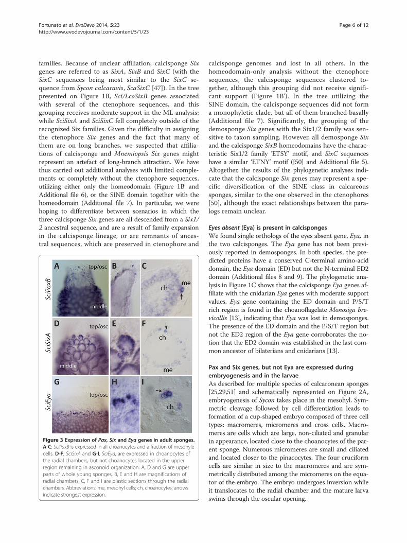
Figure 3 Expression of Pax, Six and Eya genes in adult sponges.A-C; SciPaxB is expressed in all choanocytes and a fraction of mesohylecells. D-F, SciSixA and G-I, SciEya, are expressed in choanocytes ofthe radial chambers, but not choanocytes located in the upperregion remaining in asconoid organization. A, D and G are upperparts of whole young sponges, B, E and H are magnifications ofradial chambers, C, F and I are plastic sections through the radialchambers. Abbreviations: me, mesohyl cells; ch, choanocytes; arrowsindicate strongest expression.
calcisponge genomes and lost in all others. In thehomeodomain-only analysis without the ctenophoresequences, the calcisponge sequences clustered to-gether, although this grouping did not receive signifi-cant support (Figure 1B’). In the tree utilizing theSINE domain, the calcisponge sequences did not forma monophyletic clade, but all of them branched basally(Additional file 7). Significantly, the grouping of thedemosponge Six genes with the Six1/2 family was sen-sitive to taxon sampling. However, all demosponge Sixand the calcisponge SixB homeodomains have the charac-teristic Six1/2 family 'ETSY' motif, and SixC sequenceshave a similar ‘ETNY’ motif ([50] and Additional file 5).Altogether, the results of the phylogenetic analyses indi-cate that the calcisponge Six genes may represent a spe-cific diversification of the SINE class in calcareoussponges, similar to the one observed in the ctenophores[50], although the exact relationships between the para-logs remain unclear.
Eyes absent (Eya) is present in calcispongesWe found single orthologs of the eyes absent gene, Eya, inthe two calcisponges. The Eya gene has not been previ-ously reported in demosponges. In both species, the pre-dicted proteins have a conserved C-terminal amino-aciddomain, the Eya domain (ED) but not the N-terminal ED2domain (Additional files 8 and 9). The phylogenetic ana-lysis in Figure 1C shows that the calcisponge Eya genes af-filiate with the cnidarian Eya genes with moderate supportvalues. Eya gene containing the ED domain and P/S/Trich region is found in the choanoflagelate Monosiga bre-vicollis [13], indicating that Eya was lost in demosponges.The presence of the ED domain and the P/S/T region butnot the ED2 region of the Eya gene corroborates the no-tion that the ED2 domain was established in the last com-mon ancestor of bilaterians and cnidarians [13].
Pax and Six genes, but not Eya are expressed duringembryogenesis and in the larvaeAs described for multiple species of calcaronean sponges[25,29,51] and schematically represented on Figure 2A,embryogenesis of Sycon takes place in the mesohyl. Sym-metric cleavage followed by cell differentiation leads toformation of a cup-shaped embryo composed of three celltypes: macromeres, micromeres and cross cells. Macro-meres are cells which are large, non-ciliated and granularin appearance, located close to the choanocytes of the par-ent sponge. Numerous micromeres are small and ciliatedand located closer to the pinacocytes. The four cruciformcells are similar in size to the macromeres and are sym-metrically distributed among the micromeres on the equa-tor of the embryo. The embryo undergoes inversion whileit translocates to the radial chamber and the mature larvaswims through the oscular opening.
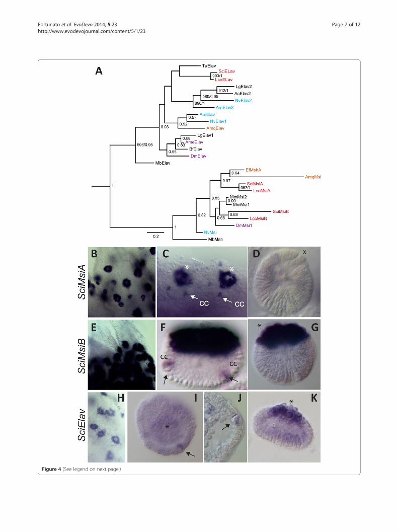
Figure 4 (See legend on next page.)
Fortunato et al. EvoDevo 2014, 5:23 Page 7 of 12http://www.evodevojournal.com/content/5/1/23
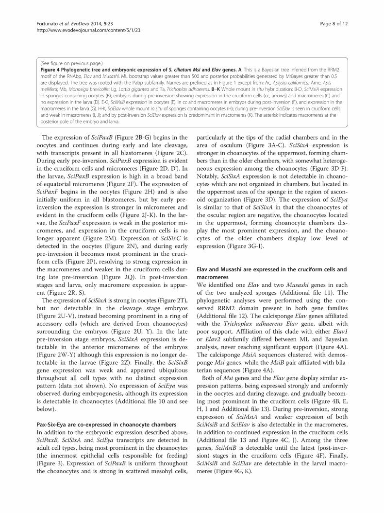
(See figure on previous page.)Figure 4 Phylogenetic tree and embryonic expression of S. ciliatum Msi and Elav genes. A, This is a Bayesian tree inferred from the RRM2motif of the RNAbp, Elav and Musashi. ML bootstrap values greater than 500 and posterior probabilities generated by MrBayes greater than 0.5are displayed. The tree was rooted with the Pabp subfamily. Names are prefixed as in Figure 1 except from: Ac, Aplysia californica; Ame, Apismellifera; Mb, Monosiga brevicollis; Lg, Lottia gigantea and Ta, Trichoplax adhaerens. B–K Whole mount in situ hybridization: B-D, SciMsiA expressionin sponges containing oocytes (B); embryos during pre-inversion showing expression in the cruciform cells (cc, arrows) and macromeres (C) andno expression in the larva (D). E-G, SciMsiB expression in oocytes (E), in cc and macromeres in embryos during post-inversion (F), and expression in themacromeres in the larva (G). H-K, SciElav whole mount in situ of sponges containing oocytes (H); during pre-inversion SciElav is seen in cruciform cellsand weak in macromeres (I, J); and by post-inversion SciElav expression is predominant in macromeres (K). The asterisk indicates macromeres at theposterior pole of the embryo and larva.
Fortunato et al. EvoDevo 2014, 5:23 Page 8 of 12http://www.evodevojournal.com/content/5/1/23
The expression of SciPaxB (Figure 2B-G) begins in theoocytes and continues during early and late cleavage,with transcripts present in all blastomeres (Figure 2C).During early pre-inversion, SciPaxB expression is evidentin the cruciform cells and micromeres (Figure 2D, D’). Inthe larvae, SciPaxB expression is high in a broad bandof equatorial micromeres (Figure 2F). The expression ofSciPaxF begins in the oocytes (Figure 2H) and is alsoinitially uniform in all blastomeres, but by early pre-inversion the expression is stronger in micromeres andevident in the cruciform cells (Figure 2J-K). In the lar-vae, the SciPaxF expression is weak in the posterior mi-cromeres, and expression in the cruciform cells is nolonger apparent (Figure 2M). Expression of SciSixC isdetected in the oocytes (Figure 2N), and during earlypre-inversion it becomes most prominent in the cruci-form cells (Figure 2P), resolving to strong expression inthe macromeres and weaker in the cruciform cells dur-ing late pre-inversion (Figure 2Q). In post-inversionstages and larva, only macromere expression is appar-ent (Figure 2R, S).The expression of SciSixA is strong in oocytes (Figure 2T),
but not detectable in the cleavage stage embryos(Figure 2U-V), instead becoming prominent in a ring ofaccessory cells (which are derived from choanocytes)surrounding the embryos (Figure 2U, Y). In the latepre-inversion stage embryos, SciSixA expression is de-tectable in the anterior micromeres of the embryos(Figure 2W-Y) although this expression is no longer de-tectable in the larvae (Figure 2Z). Finally, the SciSixBgene expression was weak and appeared ubiquitousthroughout all cell types with no distinct expressionpattern (data not shown). No expression of SciEya wasobserved during embryogenesis, although its expressionis detectable in choanocytes (Additional file 10 and seebelow).
Pax-Six-Eya are co-expressed in choanocyte chambersIn addition to the embryonic expression described above,SciPaxB, SciSixA and SciEya transcripts are detected inadult cell types, being most prominent in the choanocytes(the innermost epithelial cells responsible for feeding)(Figure 3). Expression of SciPaxB is uniform throughoutthe choanocytes and is strong in scattered mesohyl cells,
particularly at the tips of the radial chambers and in thearea of osculum (Figure 3A-C). SciSixA expression isstronger in choanocytes of the uppermost, forming cham-bers than in the older chambers, with somewhat heteroge-neous expression among the choanocytes (Figure 3D-F).Notably, SciSixA expression is not detectable in choano-cytes which are not organized in chambers, but located inthe uppermost area of the sponge in the region of ascon-oid organization (Figure 3D). The expression of SciEyais similar to that of SciSixA in that the choanocytes ofthe oscular region are negative, the choanocytes locatedin the uppermost, forming choanocyte chambers dis-play the most prominent expression, and the choano-cytes of the older chambers display low level ofexpression (Figure 3G-I).
Elav and Musashi are expressed in the cruciform cells andmacromeresWe identified one Elav and two Musashi genes in eachof the two analyzed sponges (Additional file 11). Thephylogenetic analyses were performed using the con-served RRM2 domain present in both gene families(Additional file 12). The calcisponge Elav genes affiliatedwith the Trichoplax adhaerens Elav gene, albeit withpoor support. Affiliation of this clade with either Elav1or Elav2 subfamily differed between ML and Bayesiananalysis, never reaching significant support (Figure 4A).The calcisponge MsiA sequences clustered with demos-ponge Msi genes, while the MsiB pair affiliated with bila-terian sequences (Figure 4A).Both of Msi genes and the Elav gene display similar ex-
pression patterns, being expressed strongly and uniformlyin the oocytes and during cleavage, and gradually becom-ing most prominent in the cruciform cells (Figure 4B, E,H, I and Additional file 13). During pre-inversion, strongexpression of SciMsiA and weaker expression of bothSciMsiB and SciElav is also detectable in the macromeres,in addition to continued expression in the cruciform cells(Additional file 13 and Figure 4C, J). Among the threegenes, SciMsiB is detectable until the latest (post-inver-sion) stages in the cruciform cells (Figure 4F). Finally,SciMsiB and SciElav are detectable in the larval macro-meres (Figure 4G, K).
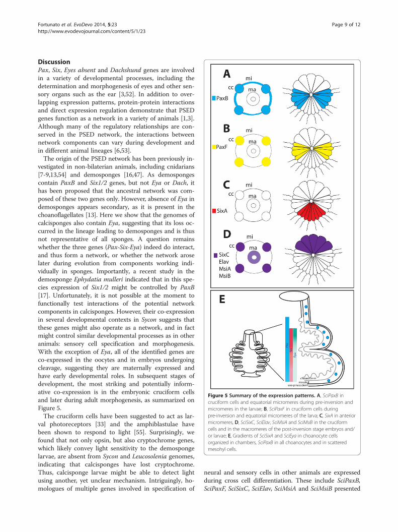
Figure 5 Summary of the expression patterns. A, SciPaxB incruciform cells and equatorial micromeres during pre-inversion andmicromeres in the larvae; B, SciPaxF in cruciform cells duringpre-inversion and equatorial micromeres of the larva; C, SixA in anteriormicromeres, D, SciSixC, SciElav, SciMsiA and SciMsiB in the cruciformcells and in the macromeres of the post-inversion stage embryos and/or larvae; E, Gradients of SciSixA and SciEya in choanocyte cellsorganized in chambers, SciPaxB in all choanocytes and in scatteredmesohyl cells.
Fortunato et al. EvoDevo 2014, 5:23 Page 9 of 12http://www.evodevojournal.com/content/5/1/23
DiscussionPax, Six, Eyes absent and Dachshund genes are involvedin a variety of developmental processes, including thedetermination and morphogenesis of eyes and other sen-sory organs such as the ear [3,52]. In addition to over-lapping expression patterns, protein-protein interactionsand direct expression regulation demonstrate that PSEDgenes function as a network in a variety of animals [1,3].Although many of the regulatory relationships are con-served in the PSED network, the interactions betweennetwork components can vary during development andin different animal lineages [6,53].The origin of the PSED network has been previously in-
vestigated in non-bilaterian animals, including cnidarians[7-9,13,54] and demosponges [16,47]. As demospongescontain PaxB and Six1/2 genes, but not Eya or Dach, ithas been proposed that the ancestral network was com-posed of these two genes only. However, absence of Eya indemosponges appears secondary, as it is present in thechoanoflagellates [13]. Here we show that the genomes ofcalcisponges also contain Eya, suggesting that its loss oc-curred in the lineage leading to demosponges and is thusnot representative of all sponges. A question remainswhether the three genes (Pax-Six-Eya) indeed do interact,and thus form a network, or whether the network aroselater during evolution from components working indi-vidually in sponges. Importantly, a recent study in thedemosponge Ephydatia mulleri indicated that in this spe-cies expression of Six1/2 might be controlled by PaxB[17]. Unfortunately, it is not possible at the moment tofunctionally test interactions of the potential networkcomponents in calcisponges. However, their co-expressionin several developmental contexts in Sycon suggests thatthese genes might also operate as a network, and in factmight control similar developmental processes as in otheranimals: sensory cell specification and morphogenesis.With the exception of Eya, all of the identified genes areco-expressed in the oocytes and in embryos undergoingcleavage, suggesting they are maternally expressed andhave early developmental roles. In subsequent stages ofdevelopment, the most striking and potentially inform-ative co-expression is in the embryonic cruciform cellsand later during adult morphogenesis, as summarized onFigure 5.The cruciform cells have been suggested to act as lar-
val photoreceptors [33] and the amphiblastulae havebeen shown to respond to light [55]. Surprisingly, wefound that not only opsin, but also cryptochrome genes,which likely convey light sensitivity to the demospongelarvae, are absent from Sycon and Leucosolenia genomes,indicating that calcisponges have lost cryptochrome.Thus, calcisponge larvae might be able to detect lightusing another, yet unclear mechanism. Intriguingly, ho-mologues of multiple genes involved in specification of
neural and sensory cells in other animals are expressedduring cross cell differentiation. These include SciPaxB,SciPaxF, SciSixC, SciElav, SciMsiA and SciMsiB presented
Fortunato et al. EvoDevo 2014, 5:23 Page 10 of 12http://www.evodevojournal.com/content/5/1/23
in this study (see Figure 5A), SciSoxB [24], components ofthe Wnt pathway (Dvl, Tcf and Beta-catenin), Smad1/5and Nanos [25].The observed co-expression indicates that SciSixC, Sci-
PaxB and SciPaxF may potentially interact during embryo-genesis, while SciPaxB, SciSixA and SciEya may potentiallyinteract during adult morphogenesis (Figure 5). Amongthe three potential components of the PSEN in Syconexpressed in the choanocytes, SciPaxB has the broadest ex-pression, with transcripts uniformly detected in all cho-anocytes. In contrast, SciSixA and SciEya transcriptsare conspicuously absent from the uppermost choano-cytes remaining in asconoid organization, but are par-ticularly strongly expressed in the choanocytes of theuppermost chambers which are undergoing morpho-genesis, with SciEya expression diminishing along asomewhat steeper gradient than SciSixA, so that bothare expressed at low levels in the already formed cham-bers (Figure 5b). SciPaxB, SciSixA and SciEya expres-sion patterns are thus consistent with interaction ofthese three genes during formation and maintenance ofthe organization of the radial chambers, and thus witha concerted role in morphogenesis. Despite lack of Eyaof the demosponge Ephydatia, the morphogenetic roleof the potential network might be a conserved feature,as knock down of EmPaxB and EmSix1/2 results in ap-parent dysmorphogenesis of the juveniles [17].
ConclusionsOverall, the presence of the sponge Eya gene and co-expression of Pax, Six and Eya genes in calcisponges indi-cate that the Pax-Six-Eya network may have already beenestablished in the last common ancestor of sponges andeumetazoans, with Eya subsequently lost in demosponges.Based on gene expression during adult body plan forma-tion in Sycon, we propose that this network had an ancientrole in morphogenesis. Additionally, co-expression of Paxand Six with conserved eumetazoan neural genes Elav andMsi in candidate larval sensory cells, suggests these genescould be ancestrally involved in the determination of sen-sory cell types. We envisage a scenario in which a simplePSE network was active in early metazoans, and that add-itional genes, such as Dachshund, were then later co-opted into the network to expand its regulatory capacityin more complex animals.
Additional files
Additional file 1: The predicted Pax protein sequences andintron-exon boundaries. Exon-intron boundaries are highlighted inyellow, the paired domain is red.
Additional file 2: Alignment of the PD domain. This alignment wasused for the phylogenetic analyses displayed in Figure 1C. The red box
indicates the location of the RED motif used for the phylogeneticanalyses in Additional file 3. Abbreviations are as in Figure 1.
Additional file 3: Bayesian phylogenetic tree of the Pax gene familyinferred from the RED motif of the PRD domain. Support values onnodes are as follows: left, bootstrap (BT) values obtained from ML analysis;right, posterior probability from the Bayesian analysis. For abbreviations ofspecies names see Figure 1.
Additional file 4: Six genes protein sequences in Sycon andLeucosolenia. Exon-intron boundaries are indicated by highlighting. Sineoculis domain is red, the homeodomain is underlined.
Additional file 5: Alignment of the homeodomain of the SINE andTALE classes including all of Six genes and selected TALE genesidentified in calcisponges. Abbreviations are as in Figure 1.
Additional file 6: Maximum likelihood tree of the SINE class.Phylogenetic tree inferred from the homeodomain of Six and TALEgenes. Bootstrap values are displayed on each node. Names are prefixedas in Figure 1. The tree was rooted with a selection of TALE class ofhomeobox genes. Mnemiopsis Six genes found in long branches on thetree from Figure 1B were not included in this analysis.
Additional file 7: Maximum likelihood phylogenetic analyses ofsine oculis domain and homeodomain of the Six class. ML bootstrapvalues greater than 500 are displayed. Names are prefixed as in Figure 1.
Additional file 8: EYA protein sequences. Red indicates the locationof the ED domain. Exon-intron boundaries are highlighted.
Additional file 9: Alignment of the ED domain. This alignment wasused for the phylogenetic analyses displayed in Figure 1B. Abbreviationsare as in Figure 1.
Additional file 10: The SciEya gene is not expressed duringembryogenesis. A, oocytes; B, embryos during pre-inversion and C,post-inversion.
Additional file 11: Elav and Msi protein sequences. Exon-intronboundaries are highlighted. Red indicates the location of the RMM2 domain.
Additional file 12: RRM2 motif alignment for Msi and Elavsequences. This alignment, without gaps, was used for the phylogeneticanalyses displayed in Figure 4.
Additional file 13: Predominant expression of SciMsiA and SciElavin the cruciform cells. Late cleavage and pre-inversion stage embryosare shown for SciMsiA and SciElav, respectively.
AbbreviationsBLAST: Basic Local Alignment Search Tool; ED: Eya domain; ML: MaximumLikelihood; PD: Paired domain; PSEDN: Pax-Six-Eya-Dach network; RDGN: RetinalDetermination Gene Network.
Competing interestsThe authors declare that they have no competing interests.
Authors’ contributionsSF and MA conceived and designed the study. MA and SL carried out fieldcollection. SF performed phylogenetic analyses and analyzed expression of allgenes presented in this manuscript except Musashi, which were studied by SL.SF drafted and MA edited the manuscript with input from SL. All authors readand approved the final manuscript.
AcknowledgementsWe thank M. Adamski for sharing unpublished sequence assemblies andparticipation in field collections and G. Richards for insightful comments onthe manuscript. This work was supported by the core budget of the SarsCentre to MA.
Author details1Sars International Centre for Marine Molecular Biology, University of Bergen,Thormøhlensgt. 55, Bergen 5008, Norway. 2Department of Biology, Universityof Bergen, Thormøhlensgt. 55, Bergen 5008, Norway. 3Current address:Institute of Marine Research, Nordnesgaten 50, Bergen 5005, Norway.
Fortunato et al. EvoDevo 2014, 5:23 Page 11 of 12http://www.evodevojournal.com/content/5/1/23
Received: 10 March 2014 Accepted: 15 May 2014Published: 23 June 2014
References1. Donner A, Maas R: Conservation and non-conservation of genetic
pathways in eye specification. Int J Dev Biol 2004, 48:743–753.2. Friedrich M: Ancient mechanisms of visual sense organ development
based on comparison of the gene networks controlling larval eye,ocellus, and compound eye specification in drosophila. Arthropod StructDev 2006, 35:357–378.
3. Silver S, Rebay I: Signaling circuitries in development: insights from theretinal determination gene network. Dev Suppl 2005, 132:3–13.
4. Kozmik Z, Holland N, Kreslova J, Oliveri D, Schubert M, Jonasova K, HollandL, Pestarino M, Benes V, Candiani S: Pax-Six-Eya-Dach network duringamphioxus development: conservation in vitro but context specificityin vivo. Dev Biol 2007, 306:143–159.
5. Bassham S, Postlethwait JH: The evolutionary history of placodes: amolecular genetic investigation of the larvacean urochordate Oikopleuradioica. Development 2005, 132:4259–4272.
6. Wagner G: The developmental genetics of homology. Nat Rev Genet 2007,8:473–479.
7. Matus D, Pang K, Daly M, Martindale M: Expression of Pax gene familymembers in the anthozoan cnidarian, Nematostella vectensis. Evol Dev2007, 9:25–38.
8. Suga H, Tschopp P, Graziussi DF, Stierwald M, Schmid V, Gehring WJ:Flexibly deployed Pax genes in eye development at the early evolutionof animals demonstrated by studies on a hydrozoan jellyfish. Proc NatlAcad Sci U S A 2010, 107:14263–14268.
9. Kozmik Z, Daube M, Frei E, Norman B, Kos L, Dishaw L, Noll M, Piatigorsky J:Role of Pax genes in eye evolution: a cnidarian PaxB gene uniting Pax2and Pax6 functions. Dev Cell 2003, 5:773–785.
10. Kumar J: The sine oculis homeobox (SIX) family of transcription factors asregulators of development and disease. Cell Mol Life Sci 2009, 66:565–583.
11. Kozmik Z: The role of Pax genes in eye evolution. Brain Res Bull 2008,75:335–339.
12. Kozmik Z: Pax genes in eye development and evolution. Curr Opin GenetDev 2005, 15:430–438.
13. Graziussi DF, Suga H, Schmid V, Gehring WJ: The “Eyes absent” (eya) genein the eye-bearing hydrozoan jellyfish Cladonema radiatum:conservation of the retinal determination network. J Exp Zool B Mol DevEvol 2012, 318:257–267.
14. Galliot B, Quiquand M, Ghila L, De Rosa R, Miljkovic-Licina M, Chera S:Origins of neurogenesis, a cnidarian view. Dev Biol 2009, 332:2–24.
15. Sinigaglia C, Busengdal H, Leclère L, Technau U, Rentzsch F: The bilaterianhead patterning gene six3/6 controls aboral domain development in acnidarian. PLoS Biol 2013, 11(2):e1001488.
16. Hill A, Boll W, Ries C, Warner L, Osswalt M, Hill M, Noll M: Origin of Pax andSix gene families in sponges: Single PaxB and Six1/2 orthologs inChalinula loosanoffi. Dev Biol 2010, 343:106–123.
17. Rivera A, Winters I, Rued A, Ding S, Posfai D, Cieniewicz B, Cameron K,Gentile L, Hill A: The evolution and function of the Pax/Six regulatorynetwork in sponges. Evol Dev 2013, 15:186–196.
18. Maldonado M: The ecology of the sponge larva. Can J Zool 2006,84:175–194.
19. Ludeman D, Farrar N, Riesgo A, Paps J, Leys S: Evolutionary origins ofsensation in metazoans: functional evidence for a new sensory organ insponges. BMC Evol Biol 2014, 14:3.
20. Leys S, Cronin T, Degnan B, Marshall J: Spectral sensitivity in a spongelarva. J Comp Physiol Neuroethol Sens Neural Behav Physiol 2002,188:199–202.
21. Feuda R, Hamilton S, McInerney J, Pisani D: Metazoan opsin evolutionreveals a simple route to animal vision. Proc Natl Acad Sci U S A 2012,109:18868–18872.
22. Müller WE, Wang X, Schröder HC, Korzhev M, Grebenjuk VA, Markl JS,Jochum KP, Pisignano D, Wiens M: A cryptochrome-based photosensorysystem in the siliceous sponge Suberites domuncula (Demospongiae).FEBS J 2010, 277:1182–1201.
23. Rivera A, Ozturk N, Fahey B, Plachetzki D, Degnan B, Sancar A, Oakley T:Blue-light-receptive cryptochrome is expressed in a sponge eye lackingneurons and opsin. J Exp Biol 2012, 215:1278–1286.
24. Fortunato S, Adamski M, Bergum B, Guder C, Jordal S, Leininger S, ZwafinkC, Rapp HT, Adamska M: Genome-wide analysis of the sox family in thecalcareous sponge Sycon ciliatum: multiple genes with uniqueexpression patterns. EvoDevo 2012, 3:14.
25. Leininger S, Adamski M, Bergum B, Guder C, Liu J, Laplante M, Bråte J,Hoffman NF, Fortunato S, Jordal S, Rapp HT, Adamska M: Developmentalgene expression provides clues to relationships between sponge andeumetazoan body plans. Nat Commun 2014, 5:3905.
26. Riesgo A, Farrar N, Windsor PJ, Giribet G, Leys SP: The analysis of eighttranscriptomes from all poriferan classes reveals surprising geneticcomplexity in sponges. Mol Biol Evol 2014, 31:1102–1120.
27. Sebé-Pedrós A, Ariza-Cosano A, Weirauch M, Leininger S, Yang A, TorruellaG, Adamski M, Adamska M, Hughes T, Gómez-Skarmeta J, Ruiz-Trillo I: Earlyevolution of the T-box transcription factor family. Proc Natl Acad Sci U S A2013, 110:16050–16055.
28. Maas O: Die Weiterentwicklung der Syconen nach der metamorphose.Zeutsch wiss Zool 1900, 67:215–240.
29. Ereskovsky AV: The Comparative Embryology of Sponges. DordrechtHeidelberg London New York: Springer; 2010.
30. Maldonado M, Bergquist P: Atlas of Marine Invertebrate Larvae. San Diego,CA: Academic Press; 2002.
31. Manuel M: Early evolution of symmetry and polarity in metazoan bodyplans. C R Biol 2009, 332:184–209.
32. Amano S, Hori I: Metamorphosis of calcareous sponges. 2. Cellrearrangement and differentiation in metamorphosis. Invert Reprod Dev1993, 24:13–26.
33. Tuzet O: Éponges calcaires. In Traité de Zoologie Anatomie, Systématique,Biologie Spongiaires. Edited by Grassé P-P. Paris: Masson et Cie; 1973:27–132.
34. Phochanukul N, Russell S: No backbone but lots of Sox: invertebrate Soxgenes. Int J Biochem Cell Biol 2010, 42:453–464.
35. Watanabe H, Fujisawa T, Holstein TW: Cnidarians and the evolutionaryorigin of the nervous system. Dev Growth Differ 2009, 51:167–183.
36. Jager M, Queinnec E, Le Guyader H, Manuel M: Multiple Sox genes areexpressed in stem cells or in differentiating neuro-sensory cells in thehydrozoan Clytia hemisphaerica. EvoDevo 2011, 2:12.
37. Kanska J, Frank U: New roles for Nanos in neural cell fate determinationrevealed by studies in a cnidarian. J Cell Sci 2013, 126:3192–3203.
38. Grigoryan T, Wend P, Klaus A, Birchmeier W: Deciphering the function ofcanonical Wnt signals in development and disease: conditional loss- andgain-of-function mutations of beta-catenin in mice. Genes Dev 2008,22:2308–2341.
39. Hegarty S, O'Keeffe G, Sullivan A: BMP-Smad 1/5/8 signalling in thedevelopment of the nervous system. Prog Neurobiol 2013, 109:28–41.
40. Abascal F, Zardoya R, Posada D: ProtTest: selection of best-fit models ofprotein evolution. Bioinformatics 2005, 21:2104–2105.
41. Ronquist F, Huelsenbeck JP: MrBayes 3: Bayesian phylogenetic inferenceunder mixed models. Bioinformatics 2003, 19:1572–1574.
42. Rambaut A, Drummond AJ: Tracer v1.5.; 2009. http://beast.bio.ed.ac.uk/software/tracer/.
43. Rambaut A: Figtree v1.4.0.; 2012. http://tree.bio.ed.ac.uk/software/figtree/.44. Guindon S, Dufayard J-F, Lefort V, Anisimova M, Hordijk W, Gascuel O: New
algorithms and methods to estimate maximum-likelihood phylogenies:assessing the performance of PhyML 3.0. Syst Biol 2010, 59:307–321.
45. Ryan J, Burton P, Mazza M, Kwong G, Mullikin J, Finnerty J: The cnidarian-bilaterian ancestor possessed at least 56 homeoboxes: evidence fromthe starlet sea anemone. Nematostella vectensis. Genome Biol 2006, 7:R64.
46. Larroux C, Luke G, Koopman P, Rokhsar D, Shimeld S, Degnan B: Genesisand expansion of metazoan transcription factor gene classes. Mol BiolEvol 2008, 25:980–996.
47. Hoshiyama D, Iwabe N, Miyata T: Evolution of the gene families formingthe Pax/Six regulatory network: isolation of genes from primitive animalsand molecular phylogenetic analyses. FEBS Lett 2007, 581:1639–1643.
48. Breitling R, Gerber JK: Origin of the paired domain. Dev Genes Evol 2000,210:644–650.
49. Takatori N, Butts T, Candiani S, Pestarino M, Ferrier D, Saiga H, Holland P:Comprehensive survey and classification of homeobox genes in the genomeof amphioxus, Branchiostoma floridae. Dev Genes Evol 2008, 218:579–590.
50. Ryan J, Pang K, Program NCS, Mullikin J, Martindale M, Baxevanis A: Thehomeodomain complement of the ctenophore Mnemiopsis leidyisuggests that Ctenophora and Porifera diverged prior to theParaHoxozoa. EvoDevo 2010, 1:9.
Fortunato et al. EvoDevo 2014, 5:23 Page 12 of 12http://www.evodevojournal.com/content/5/1/23
51. Franzen W: Oogenesis and larval development of Scypha Ciliata (Porifera,Calcarea). Zoomorphology 1988, 107:349–357.
52. Rebay I, Silver S, Tootle T: New vision from Eyes absent: transcriptionfactors as enzymes. Trends Genet 2005, 21:163–171.
53. Schlosser G, Wagner GP: Modularity in development and evolution. Chicago:University of Chicago Press; 2004.
54. Stierwald M, Yanze N, Bamert R, Kammermeier L, Schmid V: The Sineoculis/Six class family of homeobox genes in jellyfish with and withouteyes: development and eye regeneration. Dev Biol 2004, 274:70–81.
55. Elliot GR, Macdonald TA, Leys SP: Sponge larval phototaxis: a comparativestudy. Bollettino dei Musei e degli Istituti Biologici dell’Universita di Genova2004, 68:291–300.
doi:10.1186/2041-9139-5-23Cite this article as: Fortunato et al.: Evolution of the Pax-Six-Eya-Dachnetwork: the calcisponge case study. EvoDevo 2014 5:23.
Submit your next manuscript to BioMed Centraland take full advantage of:
• Convenient online submission
• Thorough peer review
• No space constraints or color figure charges
• Immediate publication on acceptance
• Inclusion in PubMed, CAS, Scopus and Google Scholar
• Research which is freely available for redistribution
Submit your manuscript at www.biomedcentral.com/submit
Related Documents