Downloaded from www.microbiologyresearch.org by IP: 54.224.121.223 On: Wed, 20 Apr 2016 23:50:02 Evidence that avian reovirus sA protein is an inhibitor of the double-stranded RNA-dependent protein kinase Claudia Gonza ´ lez-Lo ´ pez, 1 3 Jose ´ Martı ´nez-Costas, 1 Mariano Esteban 2 and Javier Benavente 1 Correspondence Javier Benavente [email protected] 1 Departamento de Bioquı ´mica y Biologı ´a Molecular, Facultad de Farmacia, Universidad de Santiago de Compostela, 15782 Santiago de Compostela, Spain 2 Centro Nacional de Biotecnologı ´a, CSIC, Campus Universidad Auto ´ noma, 28049 Madrid, Spain Received 25 November 2002 Accepted 6 February 2003 The results of a previous study demonstrated that avian reovirus is highly resistant to the antiviral effects of interferon and suggested that the double-stranded RNA (dsRNA)-binding sA protein might play an important role in that resistance. To gather more evidence on the interferon-inhibitory activity of sA protein, its gene was cloned into the prokaryotic maltose-binding protein (MBP) gene fusion vector pMalC and into the recombinant vaccinia virus WRS2. The two recombinant sA proteins displayed a dsRNA-binding affinity similar to that of sA protein synthesized in avian reovirus-infected cells. Interestingly, MBP–sA, but not MBP, was able to relieve the translation- inhibitory activity of dsRNA in reticulocyte lysates by blocking the activation of endogenous dsRNA- dependent enzymes. In addition, transient expression of sA protein in HeLa cells rescued gene expression of a vaccinia virus mutant lacking the E3L gene, and insertion of the sA-encoding gene into vaccinia virus conferred protection for the virus against interferon in chicken cells. Further studies demonstrated that expression of recombinant sA in mammalian cells interfered with dsRNA-dependent protein kinase (PKR) function. From these results we conclude that sA is capable of reversing the interferon-induced antiviral state by down-regulating PKR activity in a manner similar to other virus-encoded dsRNA-binding proteins. INTRODUCTION The interaction of extracellular interferon (IFN) with species-specific cell-surface receptors initiates a signal transduction pathway that leads to increased expression of more than 30 different proteins, some of which are thought to play an important role in fighting viral infections (for reviews, see Foster, 1997; Goodbourn et al., 2000; Stark et al., 1998). Among the best characterized are the 29,59- oligoadenylate synthetase system (2-5A synthetase) and the double-stranded RNA (dsRNA)-activated protein kinase (PKR), which are activated by dsRNA and play key roles in regulating intracellular protein synthesis (reviewed in Clemens, 1997; Player & Torrence, 1998). IFN induces several different forms of 2-5A synthetase, which, on interaction with dsRNA, catalyse the conversion of ATP into short oligonucleotides of the general structure ppp(A29p59)nA. These oligonucleotides bind and activate a latent endoribonuclease L (RNase L), which catalyses the indiscriminate degradation of RNAs, thereby leading to a general inhibition of intracellular protein synthesis (reviewed in Rebouillat & Hovanessian, 1999). RNase L has also been shown to cleave ribosomal RNAs (rRNAs) in a site-specific manner (Iordanov et al., 2000; Rivas et al., 1998; Wreschner et al., 1981). On the other hand, activation of PKR by dsRNA interaction leads to phosphorylation of the alpha subunit of eukaryotic translation initiation factor 2, giving rise to inhibition of protein synthesis by preventing the recycling of initiation factors (reviewed in Clemens, 1997; Clemens & Elia, 1997). Activation of these two IFN- induced antiviral systems has also been shown to trigger apoptosis in a way that is independent of the particular means of achieving translational inhibition (Iordanov et al., 2001; Castelli et al., 1997; Diaz-Guerra et al., 1997; Lee & Esteban, 1994). Avian reoviruses are members of the Orthoreovirus genus, one of nine genera of the Reoviridae family. They are non- enveloped viruses that replicate in the cytoplasm of infected cells and contain ten dsRNA genome segments enclosed in a double protein capsid shell 70–80 nm in diameter (reviewed in Robertson & Wilcox, 1986). The avian reovirus genome encodes at least ten structural proteins and four non-structural proteins, although very little is known about 3Present address: Instituto de Biologı ´a Molecular ‘Severo Ochoa’, Campus Universidad Auto ´ noma, 28049 Madrid, Spain. 0001-9004 G 2003 SGM Printed in Great Britain 1629 Journal of General Virology (2003), 84, 1629–1639 DOI 10.1099/vir.0.19004-0

Welcome message from author
This document is posted to help you gain knowledge. Please leave a comment to let me know what you think about it! Share it to your friends and learn new things together.
Transcript

Downloaded from www.microbiologyresearch.org by
IP: 54.224.121.223
On: Wed, 20 Apr 2016 23:50:02
Evidence that avian reovirus sA protein is aninhibitor of the double-stranded RNA-dependentprotein kinase
Claudia Gonzalez-Lopez,13 Jose Martınez-Costas,1 Mariano Esteban2
and Javier Benavente1
Correspondence
Javier Benavente
1Departamento de Bioquımica y Biologıa Molecular, Facultad de Farmacia, Universidad deSantiago de Compostela, 15782 Santiago de Compostela, Spain
2Centro Nacional de Biotecnologıa, CSIC, Campus Universidad Autonoma, 28049 Madrid,Spain
Received 25 November 2002
Accepted 6 February 2003
The results of a previous study demonstrated that avian reovirus is highly resistant to the antiviral
effects of interferon and suggested that the double-stranded RNA (dsRNA)-binding sA protein
might play an important role in that resistance. To gather more evidence on the interferon-inhibitory
activity of sA protein, its gene was cloned into the prokaryotic maltose-binding protein (MBP) gene
fusion vector pMalC and into the recombinant vaccinia virus WRS2. The two recombinant sA
proteins displayed a dsRNA-binding affinity similar to that of sA protein synthesized in avian
reovirus-infected cells. Interestingly, MBP–sA, but not MBP, was able to relieve the translation-
inhibitory activity of dsRNA in reticulocyte lysates by blocking the activation of endogenous dsRNA-
dependent enzymes. In addition, transient expression of sA protein in HeLa cells rescued gene
expression of a vaccinia virus mutant lacking the E3L gene, and insertion of the sA-encoding gene
into vaccinia virus conferred protection for the virus against interferon in chicken cells. Further
studies demonstrated that expression of recombinant sA in mammalian cells interfered with
dsRNA-dependent protein kinase (PKR) function. From these results we conclude that sA is
capable of reversing the interferon-induced antiviral state by down-regulating PKR activity in a
manner similar to other virus-encoded dsRNA-binding proteins.
INTRODUCTION
The interaction of extracellular interferon (IFN) withspecies-specific cell-surface receptors initiates a signaltransduction pathway that leads to increased expression ofmore than 30 different proteins, some of which are thoughtto play an important role in fighting viral infections (forreviews, see Foster, 1997; Goodbourn et al., 2000; Starket al., 1998). Among the best characterized are the 29,59-oligoadenylate synthetase system (2-5A synthetase) and thedouble-stranded RNA (dsRNA)-activated protein kinase(PKR), which are activated by dsRNA and play key rolesin regulating intracellular protein synthesis (reviewed inClemens, 1997; Player & Torrence, 1998).
IFN induces several different forms of 2-5A synthetase,which, on interaction with dsRNA, catalyse the conversionof ATP into short oligonucleotides of the general structureppp(A29p59)nA. These oligonucleotides bind and activate alatent endoribonuclease L (RNase L), which catalyses theindiscriminate degradation of RNAs, thereby leading to
a general inhibition of intracellular protein synthesis(reviewed in Rebouillat & Hovanessian, 1999). RNase Lhas also been shown to cleave ribosomal RNAs (rRNAs) ina site-specific manner (Iordanov et al., 2000; Rivas et al.,1998; Wreschner et al., 1981). On the other hand, activationof PKR by dsRNA interaction leads to phosphorylation ofthe alpha subunit of eukaryotic translation initiation factor2, giving rise to inhibition of protein synthesis by preventingthe recycling of initiation factors (reviewed in Clemens,1997; Clemens & Elia, 1997). Activation of these two IFN-induced antiviral systems has also been shown to triggerapoptosis in a way that is independent of the particularmeans of achieving translational inhibition (Iordanov et al.,2001; Castelli et al., 1997; Diaz-Guerra et al., 1997; Lee &Esteban, 1994).
Avian reoviruses are members of the Orthoreovirus genus,one of nine genera of the Reoviridae family. They are non-enveloped viruses that replicate in the cytoplasm of infectedcells and contain ten dsRNA genome segments enclosedin a double protein capsid shell 70–80 nm in diameter(reviewed in Robertson &Wilcox, 1986). The avian reovirusgenome encodes at least ten structural proteins and fournon-structural proteins, although very little is known about
3Present address: Instituto de Biologıa Molecular ‘Severo Ochoa’,Campus Universidad Autonoma, 28049 Madrid, Spain.
0001-9004 G 2003 SGM Printed in Great Britain 1629
Journal of General Virology (2003), 84, 1629–1639 DOI 10.1099/vir.0.19004-0

Downloaded from www.microbiologyresearch.org by
IP: 54.224.121.223
On: Wed, 20 Apr 2016 23:50:02
the functions of most of these proteins (Bodelon et al.,2001; Varela et al., 1996).
A previous study showed that four avian reovirus strains,including S1133, are resistant to the antiviral action of anatural chicken IFN produced in embryonated eggs (Elliset al., 1983). A recent study, performed in our laboratory,revealed that exposure of chicken embryo fibroblasts (CEFs)to a recombinant chicken interferon (rcIFN) induces astrong intracellular antiviral state sufficient to inhibit thereplication of vesicular stomatitis virus and vaccinia virusbut not the replication of avian reovirus S1133. In thesame study we also showed that the translation-inhibitoryactivity of dsRNA in reticulocyte lysates can be relieved byextracts of avian reovirus-infected cells, suggesting thepresence of a protranslational factor in these extracts.Further in vitro translation experiments indirectly suggestedthat this factor is avian reovirus sA protein (Martınez-Costas et al., 2000). Protein sA is a minor component ofthe virus inner capsid that binds dsRNA very tightly in asequence-independent manner (Martinez-Costas et al., 1997,2000; Yin et al., 2000). However, no consensus dsRNA-binding motifs have been found in the amino acid sequenceof sA protein, as found in the sequences of the dsRNA-binding proteins NS1 of influenza virus and VP6 of blue-tongue virus (Hatada & Fukuda, 1992; Stauber et al., 1997).
In this study we have performed experiments to characterizethe IFN-inhibitory activity of avian reovirus sAprotein. Ourresults suggest that sA is a key factor in the IFN-resistantphenotype displayed by avian reovirus because of its abilityto down-regulate PKR function.
METHODS
Cells and viruses. Primary cultures of CEFs were prepared from9- to 10-day-old chicken embryos and grown in monolayers inmedium 199 supplemented with 10% tryptose phosphate broth and5% calf serum. African green monkey kidney BSC-40 and humanHeLa cell lines were grown in monolayers in medium 199 supple-mented with 10% foetal bovine serum (FBS). Strain S1133 of avianreovirus was grown in semiconfluent monolayers of primary CEFs,as previously described (Grande & Benavente, 2000). Vacciniaviruses were propagated in BSC-40 cells, as previously described(Lee & Esteban, 1994). In this study we used wild-type vacciniavirus WR strain and the following recombinant vaccinia viruses:WR68K, which expresses a human recombinant IPTG-induciblePKR (Lee & Esteban, 1994); WRLuc, which constitutively expressesa recombinant firefly luciferase protein (Gherardi et al., 1999);WRE3L2, which lacks the E3L gene (Beattie et al., 1995); andWRS2, which was generated in our laboratory as follows. Semi-confluent monolayer cultures of BSC-40 cells were infected with0?01 p.f.u. per cell of WR and after 1 h adsorption at 37 C, theinoculum was removed and the cells were lipofected with 2?5 mg per106 cells of the vaccinia virus insertion plasmid pHLZ/S2 (seebelow). The cells were subsequently incubated for 5 h at 37 ˚C, thenthe transfection medium was removed and replaced by DMEM sup-plemented with 3% FBS. At 48 h post-transfection, the cells wereharvested, subjected to three cycles of freezing and thawing andsonicated. Serial dilutions of the cell extracts were used to infectfresh BSC-40 cell monolayers and after 1 h adsorption at 37 ˚C, theinoculum was removed and replaced by DMEM supplemented with
2% FBS and 1% agar. Three days later, the agar was covered withDMEM supplemented with 1% agar and 0?03% X-Gal and the cellswere incubated for 12–24 h. Blue plaques were selected and homo-geneous recombinant virus was isolated by three more rounds ofplaque purification. WRS2 contains the sA-encoding gene under thecontrol of the synthetic early/late PE/L promoter. The correct orien-tation of the S2 insert was confirmed by nucleotide sequencing.Virus titres were determined by plaque assay, as previously described(Martınez-Costas et al., 2000).
Plasmids. To generate a plasmid expressing a maltose-bindingprotein (MBP)-tagged sA fusion protein, total RNA from avianreovirus-infected cells was amplified by RT-PCR with the forwardprimer 59-GCGGGATCCACGATGGCGCGTG-39 (BamHI site under-lined) and the reverse primer 59-GCGAAGCTTGCGTACGACC-CTACGC-39 (HindIII site underlined). The resulting cDNA wasdigested and cloned into the BamHI and HindIII sites of pMalC(New England Biolabs) and the resulting recombinant plasmid,pMalCS2, was introduced into E. coli strain BL21. The correct orien-tation of the insert was confirmed by nucleotide sequencing.
Plasmid pPR15 (containing the luciferase reporter gene under thecontrol of the vaccinia virus p4b late promoter), plasmid pPR35(designed for IPTG-inducible expression of genes) and recombinantplasmid pPR35E3L have all been described previously (Diaz-Guerraet al., 1997; Rodriguez & Smith, 1990). For the generation of thepPR35S2 plasmid and the recombinant vaccinia virus insertionplasmid pHLZ/S2, the S2 avian reovirus gene was excised frompMALCS2 plasmid by HindIII digestion and after filling the endswith Klenow DNA polymerase, the insert was digested again withBamHI. The resulting DNA fragment was then inserted into theBamHI and SmaI sites of the vaccinia virus insertion plasmid pPR35and into the BglII and SmaI sites of the vaccinia virus insertion plas-mid pHLZ (Rodriguez & Smith, 1990).
Bacterial expression, protein purification and antibody genera-tion. For expression of MBP and MBP–sA, cultures of pMalC-and pMalCS2-transformed BL21 bacteria were grown in LB mediumsupplemented with 0?2% glucose and 100 mg ampicillin ml21 up toan optical density of 0?6 at 600 nm. The cells were then inducedwith 1 mM IPTG, incubated for 2 h at 37 C and finally lysed bysonication in a buffer containing 0?25% Tween 20, 1 mM DTT,200 mM NaCl, 20 mM Tris/HCl, pH 7?5 and 1 mg lysozyme ml21.The resulting extracts were clarified by centrifugation and MBPand MBP–sA were purified from the supernatants using amylose–agarose columns, as described by the manufacturer (New EnglandBiolabs). When indicated, protease factor Xa (New EnglandBiolabs) was added to the MBP–sA-containing sample at a w/wratio of 1% in column buffer (20 mM Tris/HCl, pH 7?4, 200 mMNaCl, 1 mM EDTA, 1 mM DTT) and incubated overnight at4 C. Protein sA was subsequently isolated from the mixture byQ-Sepharose chromatography performed in a buffer containing20 mM Tris/HCl, pH 7?5, and 50 mM NaCl. Alternatively, super-natant extracts were subjected to poly(I : C)–agarose chromatography(Amersham Bioscience), as previously described (Martınez-Costaset al., 2000).
Preparation of rabbit polyclonal antibodies against both gel-purifiedand native sA was carried out as described previously (Bodelon et al.,2001).
Interferon treatment, infections, in vitro translation, proteinanalysis, detection of apoptosis and analysis of rRNA integ-rity. Treatment of CEFs with rcIFN, infection of cell monolayers,in vitro translation, protein radiolabelling and SDS-PAGE analysishave all been described previously (Martınez-Costas et al., 2000).Densitometric analysis of viral protein bands was performed using aFluorS Multilmager system and Quantity One software (Bio-Rad).
1630 Journal of General Virology 84
C. Gonzalez-Lopez and others

Downloaded from www.microbiologyresearch.org by
IP: 54.224.121.223
On: Wed, 20 Apr 2016 23:50:02
Immunoprecipitation, immunoblotting and affinity chromato-
graphy methods have all been described previously (Bodelon et al.,2001; Martınez-Costas et al., 2000). Detection of oligonucleosomalDNA fragments, estimation of the amount of cytoplasmic histone-associated DNA and analysis of the integrity of 18S and 28S rRNAshave also been described (Labrada et al., 2002).
Transient transfection of HeLa cells. Semiconfluent HeLa cellmonolayers grown in 12-well plates were infected with 2?5 p.f.u.WRE3L2 per cell and transfected 1 h later with 0?2 mg pPR15 perwell plus the indicated amounts of pPR35-derived plasmids, usingLipofectamine (Invitrogen) according to the manufacturer’s instruc-tions. Cells were then induced or not with 1?5 mM IPTG and lysed24 h later. Luciferase activity in cell extracts was determined with aluminometer as previously described (Brasier et al., 1989).
RESULTS AND DISCUSSION
Bacterial expression of sA protein, purificationand generation of polyclonal antibodies
As a first step to raise polyclonal antibodies against avianreovirus sA protein, the avian reovirus S2 gene was clonedinto the MBP gene fusion vector pMalC and the resultingrecombinant plasmid, pMalCS2, was used to transformBL21 E. coli cells. A comparative electrophoretic analysis ofthe proteins present in extracts of bacteria that had beentransformed with plasmids pMalC or pMalCS2 (Fig. 1A)showed that both MBP (Fig. 1A, lane 3) and MBP–sA(Fig. 1A, lane 4) were expressed in IPTG-induced cells, but
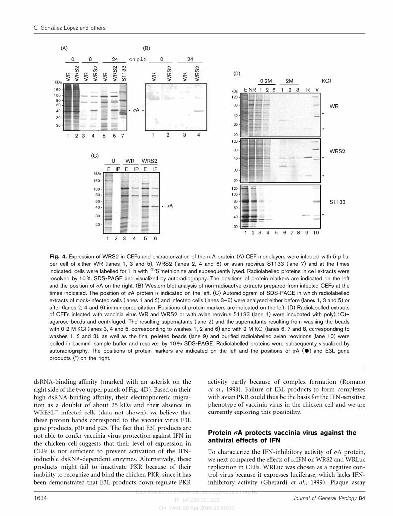
Fig. 1. Expression of sA protein in bacterial cells and characterization of anti-sA antibodies. (A) Cell extracts prepared fromuninduced (lanes 1 and 2) or IPTG-induced bacteria transformed with plasmids pMalC (lane 3) or pMalCS2 (lane 4) wereanalysed by 12% SDS-PAGE and stained with Coomassie blue. In the same gel, supernatant (lanes 5 and 6) and pelletedfractions (lanes 7 and 8) resulting from centrifugation of these extracts were also analysed, as well as proteins of thesupernatant fractions that were retained on amylose–agarose (lanes 9 and 10) or on poly(I : C)–agarose beads (lanes 11 and12). Positions of protein markers are indicated on the left and positions of MBP and MBP–sA on the right. (B) Electrophoreticanalysis of reovirions (lane 1), amylose–agarose-purified MBP–sA (lane 2), factor Xa-digested MBP–sA (lane 3) and thefraction of Xa-digested MBP–sA that was not retained on a Q-Sepharose column (lane 4). Positions of the three size classesof avian reovirus polypeptides are indicated on the left and positions of sA, MBP and MBP–sA on the right. (C) Immunoblotanalysis, with polyclonal antibodies against gel-purified sA, of extracts of MBP–sA-transformed bacteria either uninduced(lane 1) or after IPTG induction (lane 2) and of extracts of mock-infected (lane 3) and S1133-infected (lane 4) CEFs.Positions of protein markers are indicated on the left and positions of sA and MBP–sA on the right. (D) 35S-labelled extractsof IPTG-induced, MBP–sA-transformed bacteria (lanes 1 and 2) and S1133-infected CEFs (lanes 3 and 4) were analysed bySDS-PAGE and autoradiography, either before (lanes 1 and 3) or after (lanes 2 and 4) immunoprecipitation with antiserumraised against native sA. Positions of the three size classes of avian reovirus polypeptides are indicated on the right andpositions of sA and MBP–sA on the left.
http://vir.sgmjournals.org 1631
Avian reovirus sA down-regulates PKR activity

Downloaded from www.microbiologyresearch.org by
IP: 54.224.121.223
On: Wed, 20 Apr 2016 23:50:02
not in the corresponding uninduced bacteria (Fig. 1A,lanes 1 and 2). Furthermore, when the extracts of inducedbacteria were centrifuged, larger amounts of both MBPand MBP–sA appeared in the supernatant fractions(Fig. 1A, lanes 5 and 6) than in the pellet fractions(Fig. 1A, lanes 7 and 8), revealing that the two proteinswere expressed mainly in soluble form. The identity ofMBP–sA was initially assessed by affinity chromato-graphy; protein MBP–sA was specifically retained on bothamylose– and poly(I : C)–agarose columns (Fig. 1A, lanes10 and 12), whereas MBP was retained on the former butnot on the latter column (Fig. 1A, lanes 9 and 11). Theidentity of MBP–sA was further confirmed by immuno-logical assays (see below).
Digestion of MBP–sA (Fig. 1B, lane 2) with proteasefactor Xa yielded two polypeptides (lane 3); the retardedpolypeptide was identified as recombinant sA (rsA) becauseit comigrated with the sA protein of avian reovirions(Fig. 1B, lane 1) and because of its ability to bind dsRNA,but not amylose (data not shown). The rsA protein wassubsequently isolated by ion exchange chromatography,using a Q-Sepharose column (Fig. 1B, lane 4).
Both native and gel-purified rsA were used as immuno-gens for polyclonal antibody production. The proteinswere subcutaneously injected into rabbits and 2 weeksafter the second boost antisera were collected and tested.A subsequent Western blot analysis (Fig. 1C) showed thatwhile antiserum raised against gel-purified sA did notrecognize any protein in extracts of either uninducedbacteria (Fig. 1C, lane 1) or uninfectedCEFs (Fig. 1C, lane 3),it recognized both MBP–sA in extracts of IPTG-inducedpMalCS2-transformed bacteria (Fig. 1C, lane 2) andnaturally occurring sA protein present in extracts of avianreovirus-infected cells (Fig. 1C, lane 4). On the other hand,while the antiserum raised against native rsA was able toimmunoprecipitate MBP–sA (Fig. 1D, lane 2) and S1133sA (Fig. 1D, lane 4) from 35S-labelled cell extracts andpurified virions (Fig. 1D, lanes 1 and 3), it did notimmunoprecipitate any protein from extracts of eitheruninduced bacteria or uninfected CEFs (data not shown).
Protein sA abolishes the inhibition oftranslation by dsRNA in vitro
The results of a previous study indirectly suggested that sAprotein is able to prevent the activation of the dsRNA-dependent enzymes in reticulocyte lysates (Martınez-Costaset al., 2000). To obtain direct evidence for the protransla-tional activity of this protein, we compared the capabilityof MBP and MBP–sA to relieve the translation-inhibitoryactivity of dsRNA in reticulocyte lysates (Fig. 2). The resultsshowed that, while MBP and MBP–sA did not inhibitexogenous tobacco mosaic virus (TMV) mRNA transla-tion (Fig. 2, compare lanes 3 and 4 with lane 2), dsRNAinduced a drastic inhibition of TMV protein synthesis(Fig. 2, compare lane 5 with lane 2). Interestingly, whereasthe inhibitory activity of dsRNA remained intact after
preincubation with MBP (Fig. 2, lane 6), it was completelyabolished after preincubation with MBP–sA (lane 7). Thisresult indicates that sA is able to reverse the translation-inhibitory activity of dsRNA, probably because of its abilityto sequester dsRNA from the dsRNA-dependent enzymes.To confirm this hypothesis, we next investigated howchanges in the order of addition of dsRNA and MBP–sA toreticulocyte lysates affected the translational efficiency ofthe reticulocyte lysate. Compared with the standard condi-tion (Fig. 2, lane 7), inhibition of translation was observedwhen the two compounds were added together withoutpreincubation (Fig. 2, lane 8) and this was even morepronounced when MBP–sA was added 5 min later thandsRNA (Fig. 2, lane 9). A similar translational rescue wasalso observed when dsRNA was preincubated with Xa-excised sA instead of MBP–sA (results not shown).
Together, these findings demonstrate that MBP–sA exertsits protranslational activity by blocking the activation ofendogenous dsRNA-dependent enzymes, rather than byinhibiting their activities. Our results also provide directevidence that the previously proposed protranslationalfactor present in extracts of avian reovirus-infected cells isindeed sA protein (Martınez-Costas et al., 2000).
Transient expression of rsA rescues WRE3L22
gene expression in HeLa cells
As a first approach to document the capacity of rsA toreverse the antiviral activity of IFN in vivo, we investigated
Fig. 2. MBP–sA reverses the translation inhibition capacity ofdsRNA in reticulocyte extracts. The effect of different additiveson the translational capacity of exogenous TMV RNA was evalu-ated. Lane 1, no mRNA added; lane 2, TMV RNA translation;lane 3, TMV RNA translation in the presence of MBP; lane 4,TMV RNA translation in the presence of MBP–sA; lane 5, TMVRNA translation in the presence of dsRNA; lane 6, as in lane5, but dsRNA was preincubated with MBP, then added to thelysate; lane 7, as in lane 5, but dsRNA was preincubated withMBP–sA; lane 8, as in lane 5, but dsRNA and MBP–sA wereadded together to the lysate without preincubation; lane 9, asin lane 5, but MBP–sA was added to the lysate 5 min laterthan dsRNA. Positions of protein molecular mass markers areindicated on the left.
1632 Journal of General Virology 84
C. Gonzalez-Lopez and others

Downloaded from www.microbiologyresearch.org by
IP: 54.224.121.223
On: Wed, 20 Apr 2016 23:50:02
the potential of the avian reovirus protein rsA to rescuethe replication of the IFN-sensitive recombinant vacciniavirus WRE3L2 in HeLa cells. This mutant virus lacks theE3L gene and in contrast to the wild-type vaccinia virus, itsreplication in HeLa cells is restricted and sensitive to IFN(Beattie et al., 1995) and late in infection the mutantvirus triggers apoptosis (Rivas et al., 1998). To measurethe ability of rsA to reverse blockage of WRE3L2 ininfected HeLa cells, we used a previously described transienttransfection–infection assay, which analyses the capabilityof proteins expressed from pPR35-derived plasmids topromote WRE3L2 gene expression in HeLa cells (Rivaset al., 1998). In this assay, promotion of WRE3L2 geneexpression is easily monitored by measuring the activityof luciferase expressed from plasmid pPR15, which con-tains the luciferase reporter gene under the control of thevaccinia virus late promoter p4b (Rodriguez & Smith, 1990).The plasmid vectors used for cotransfection were the emptyvaccinia virus insertional vector pPR35 and its derivedvectors pPR35S2 and pPR35E3L, which contain the avianreovirus S2 gene and the vaccinia virus E3L gene, respec-tively, expressed from the late p4b promoter and controlledby two lac operator sequences. pPR35 plasmids also expressthe lac repressor from the constitutively active p7?5Kvaccinia virus promoter.
HeLa cell monolayers were infected with 2?5 p.f.u. WRE3L2
per cell and 1 h later cells were transfected with the lucifer-ase reporter plasmid pPR15 plus one of the three vectorsmentioned above. Infections were allowed to proceed for24 h in the presence or absence of 1?5 mM IPTG, then cellextracts were prepared and luciferase activity was deter-mined with a luminometer (Fig. 3). As expected, the levelsof luciferase activity were low in uninduced cells and inIPTG-induced cells that had been transfected with the
empty plasmid pPR35. However, luciferase levels augmen-ted considerably after IPTG induction of cells transfectedwith the positive control plasmid pPR35E3L or the sA-encoding plasmid pPR35S2. These results strongly suggestthat both avian reovirus sA protein and vaccinia virus E3Lgene products are able to rescueWRE3L2 gene expression inHeLa cells, probably because of their ability to bind dsRNA.Our finding that this increase is slightly higher in cellsexpressing sA than in those expressing E3L, together withthe fact that E3L products have been shown to preventactivation of both PKR and 2-5A synthetase (Ho & Shuman,1996; Rivas et al., 1998; Romano et al., 1998), suggest that sAplays a critical role in modulation of the IFN-inducibleantiviral enzymes.
sA protein expressed by a recombinant vacciniavirus retains strong dsRNA-binding activity
Since vaccinia virus replication in CEFs, but not in mostother cell types, is highly sensitive to IFN (Grun et al., 1987;Martınez-Costas et al., 2000; Youngner et al., 1972), we nextsought to assess whether sA protein can confer IFNresistance to vaccinia virus by comparing the IFN suscept-ibility of wild-type WR with that of a recombinant vacciniavirus expressing sA protein. To accomplish this, we firstgenerated the recombinant vaccinia virus WRS2, whichcontains the sA-encoding gene inserted into the vacciniavirus genome under the control of the synthetic early/latePE/L promoter and then examined the capacity of this virusto express a functional rsA protein in CEFs.
A comparative electrophoretic analysis of the 35S-labelledproteins synthesized in WR- and WRS2-infected CEFs(Fig. 4A) revealed that while there were no detectabledifferences in the protein pattern at the onset of the infection(Fig. 4A, compare lanes 1 and 2), a prominent 40 kDaradioactive protein band was detected at 8 and 24 h post-infection (p.i.) in extracts of WRS2-infected cells (Fig. 4A,lanes 4 and 6), but not in extracts of WR-infected cells(Fig. 4A, lanes 3 and 5). The 40 kDa protein comigratedwith the sA protein synthesized in avian reovirus-infectedcells (Fig. 4A, compare lanes 6 and 7). Immunoblot andimmunoprecipitation analysis of cell extracts with poly-clonal anti-sA antibodies confirmed the sA identity of the40 kDa band present in WRS2-infected CEFs (Fig. 4B, C).Affinity chromatographic assays on poly(I : C)–agaroserevealed that the rsA protein expressed by WRS2 displayeda dsRNA-binding affinity similar to that of the sA proteinsynthesized in avian reovirus-infected CEFs; the twoproteins showed a very strong dsRNA-binding affinity,since they remained attached to the matrix after washing thedsRNA–agarose beads with a buffer containing 2 M KCl(Fig. 4D, lane 9 in panelsWRS2 and S1133). Taken together,our results demonstrate that high levels of a functional sAprotein are expressed by WRS2 in CEFs.
The chromatographic assay also revealed the presence inWR- and WRS2-infected cells, but not in S1133-infectedcells, of low molecular weight polypeptides with high
Fig. 3. Stimulation of luciferase expression. HeLa cells grownin 12-well plates were infected with 2?5 p.f.u. WRE3L2 per celland 1 h later transfected with 0?2 mg per well of the plasmidpPR15 together with 2?0 mg per well of the plasmids indicated.The cells were incubated in the absence (2) or presence (+)of 1?5 mM IPTG for 24 h, then lysed and luciferase activity inthe resulting extracts measured with a luminometer. The resultsshown are means of four independent experiments and errorbars indicate standard deviations of the mean.
http://vir.sgmjournals.org 1633
Avian reovirus sA down-regulates PKR activity
Downloaded from www.microbiologyresearch.org by
IP: 54.224.121.223
On: Wed, 20 Apr 2016 23:50:02
dsRNA-binding affinity (marked with an asterisk on theright side of the two upper panels of Fig. 4D). Based on theirhigh dsRNA-binding affinity, their electrophoretic migra-tion as a doublet of about 25 kDa and their absence inWRE3L2-infected cells (data not shown), we believe thatthese protein bands correspond to the vaccinia virus E3Lgene products, p20 and p25. The fact that E3L products arenot able to confer vaccinia virus protection against IFN inthe chicken cell suggests that their level of expression inCEFs is not sufficient to prevent activation of the IFN-inducible dsRNA-dependent enzymes. Alternatively, theseproducts might fail to inactivate PKR because of theirinability to recognize and bind the chicken PKR, since it hasbeen demonstrated that E3L products down-regulate PKR
activity partly because of complex formation (Romanoet al., 1998). Failure of E3L products to form complexeswith avian PKR could thus be the basis for the IFN-sensitivephenotype of vaccinia virus in the chicken cell and we arecurrently exploring this possibility.
Protein sA protects vaccinia virus against theantiviral effects of IFN
To characterize the IFN-inhibitory activity of sA protein,we next compared the effects of rcIFN on WRS2 and WRLucreplication in CEFs. WRLuc was chosen as a negative con-trol virus because it expresses luciferase, which lacks IFN-inhibitory activity (Gherardi et al., 1999). Plaque assay
Fig. 4. Expression of WRS2 in CEFs and characterization of the rsA protein. (A) CEF monolayers were infected with 5 p.f.u.per cell of either WR (lanes 1, 3 and 5), WRS2 (lanes 2, 4 and 6) or avian reovirus S1133 (lane 7) and at the timesindicated, cells were labelled for 1 h with [35S]methionine and subsequently lysed. Radiolabelled proteins in cell extracts wereresolved by 10% SDS-PAGE and visualized by autoradiography. The positions of protein markers are indicated on the leftand the position of sA on the right. (B) Western blot analysis of non-radioactive extracts prepared from infected CEFs at thetimes indicated. The position of sA protein is indicated on the left. (C) Autoradiogram of SDS-PAGE in which radiolabelledextracts of mock-infected cells (lanes 1 and 2) and infected cells (lanes 3–6) were analysed either before (lanes 1, 3 and 5) orafter (lanes 2, 4 and 6) immunoprecipitation. Positions of protein markers are indicated on the left. (D) Radiolabelled extractsof CEFs infected with vaccinia virus WR and WRS2 or with avian reovirus S1133 (lane 1) were incubated with poly(I : C)–agarose beads and centrifuged. The resulting supernatants (lane 2) and the supernatants resulting from washing the beadswith 0?2 M KCl (lanes 3, 4 and 5, corresponding to washes 1, 2 and 6) and with 2 M KCl (lanes 6, 7 and 8, corresponding towashes 1, 2 and 3), as well as the final pelleted beads (lane 9) and purified radiolabelled avian reovirions (lane 10) wereboiled in Laemmli sample buffer and resolved by 10% SDS-PAGE. Radiolabelled proteins were subsequently visualized byautoradiography. The positions of protein markers are indicated on the left and the positions of sA ($) and E3L geneproducts (*) on the right.
1634 Journal of General Virology 84
C. Gonzalez-Lopez and others

Downloaded from www.microbiologyresearch.org by
IP: 54.224.121.223
On: Wed, 20 Apr 2016 23:50:02
analysis of infectious progeny virus production (Fig. 5A)revealed that WRS2 replication in CEFs is much moreresistant to rcIFN than WRLuc replication, suggesting thatsA protein confers vaccinia virus protection against IFN. Asubsequent SDS-PAGE analysis of protein synthesis inrcIFN-treated cells (Fig. 5B) showed that while the IFNtreatment did not affect protein synthesis in uninfected cells(Fig. 5B, compare lanes 1 and 2) and only induced a slightreduction of viral protein synthesis in WRS2-infected cells(Fig. 5B, compare lanes 6–8), it caused a drastic inhibitionof viral protein synthesis in WRLuc-infected cells (Fig. 5B,compare lanes 3–5). A densitometric analysis of the twovaccinia virus protein bands marked with asterisks on theright of the autoradiogram shown in Fig. 5(B) confirmedthat viral protein synthesis in WRS2-infected CEFs wasmuch more resistant to rcIFN than that in WRLuc-infectedcells (Fig. 5C). Taken together, these results suggest that IFNinhibits replication of vaccinia virus at a translational or apretranslational step and that expression of avian reovirussA protein relieves such inhibition.
Since intracellular activation of the dsRNA-dependentIFN-inducible enzymes PKR and 2-5A synthetase hasbeen shown to trigger apoptosis (Iordanov et al., 2001;
Castelli et al., 1997; Diaz-Guerra et al., 1997; Lee et al., 1994),we next evaluated the capacity of rsA to interfere withactivation of these enzymes by examining its capacity toprevent apoptosis induction in IFN-treated vaccinia virus-infected CEFs. The apoptotic state of these cells was asses-sed by examining internucleosomal DNA fragmentationinto an oligonucleosomal-length DNA ladder, which isconsidered a reliable biochemical hallmark of apoptosis(McCarthy & Evan, 1998). The electrophoretic analysisshown in Fig. 6(A) revealed that whereas pretreatment ofCEFs with IFN did not lead to DNA laddering in eitheruninfected (Fig. 6A, lanes 1 and 2) or WRS2-infected(Fig. 6A, lanes 6–8) cells, DNA laddering was evident inIFN-treated, WRLuc-infected cells (Fig. 6A, compare lane 3with lanes 4 and 5). These findings suggest that the rsAprotein expressed byWRS2 prevents apoptosis induction bydown-regulating the activity of dsRNA-dependent enzymes.
Taken together, our findings indicate that sA protein is ableto abrogate the antiviral effects of IFN, presumably throughits ability to bind and sequester dsRNA from the IFN-inducible antiviral pathways, as has been reported for otherdsRNA-binding proteins of viral origin. Thus, influenzavirus NS1 and herpex simplex virus type 1 Us11 protein have
Fig. 5. Effect of rcIFN on virus replication. CEF monolayers were treated with the indicated doses of IFN and 20 h later weremock-infected (U) or infected with 0?1 p.f.u. per cell of the recombinant vaccinia viruses WRLuc or WRS2. (A) At 20 h p.i.cells were lysed and the concentration of infectious vaccinia virus particles in cell extracts was determined by plaque assayon CEF monolayers. The results shown are the means of three independent experiments. (B) At 20 h p.i. cells were labelledfor 1 h with [35S]methionine and lysed. The resulting extracts were analysed by SDS-PAGE and the proteins visualizedby autoradiography. (C) Densitometric analysis of the two viral protein bands marked with asterisks on the right of (B). Theresults shown are the means of scanning autoradiograms from three separate experiments. Values are expressed as arbitrarydensitometric units.
http://vir.sgmjournals.org 1635
Avian reovirus sA down-regulates PKR activity
Downloaded from www.microbiologyresearch.org by
IP: 54.224.121.223
On: Wed, 20 Apr 2016 23:50:02
been shown to inhibit PKR activation (Hatada et al., 1999;Lu et al., 1995; Poppers et al., 2000), whereas reovirus s3,rotavirus NSP3 and vaccinia virus E3L have been reportedto interfere with activation of both PKR and 2-5A synthe-tase (Beattie et al., 1995; Langland et al., 1994; Lloyd &Shatkin, 1992).
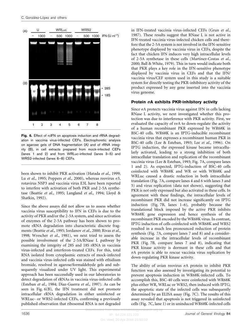
Since the above assays did not allow us to assess whethervaccinia virus susceptibility to IFN in CEFs is due to theactivity of PKR and/or the 2-5A system, and since activationof enzymes of the 2-5A pathway has been shown to pro-mote rRNA degradation into characteristic discrete frag-ments (Beattie et al., 1995; Iordanov et al., 2000; Rivas et al.,1998; Wrescher et al., 1981), we next tried to assess thepossible involvement of the 2-5A/RNase L pathway byexamining the integrity of 28S and 18S rRNA in vacciniavirus-infected and interferon-treated CEFs. For this, totalRNA isolated from cytoplasmic extracts of mock-infectedand vaccinia virus-infected cells was stained with ethidiumbromide, resolved in agarose/formaldehyde gels and sub-sequently visualized under UV light. This experimentalapproach has been successfully used in our laboratories todetect degradation of rRNAs in vaccinia virus-infected cells(Esteban et al., 1984; Diaz-Guerra et al., 1997). As can beseen in Fig. 6(B), the IFN treatment did not promoteintracellular rRNA degradation in either uninfected orWRLuc- or WRS2-infected CEFs, confirming a previouslypublished observation that ribosomal RNA is not degraded
in IFN-treated vaccinia virus-infected CEFs (Grun et al.,1987). These results suggest that RNase L is not active inIFN-treated vaccinia virus-infected chicken cells and there-fore that the 2-5A system is not involved in the IFN-sensitivephenotype displayed by vaccinia virus in CEFs, despite thefact that chicken IFN induces very high intracellular levelsof 2-5A synthetase in these cells (Martınez-Costas et al.,2000; Ball & White, 1979). This in turn would indicate boththat PKR plays a key role in the IFN-sensitive phenotypedisplayed by vaccinia virus in CEFs and that the IFN/vaccinia virus/CEF system used in this study is a suitablesystem for directly testing the PKR-inhibitory activity of theproduct expressed by any gene inserted into the vacciniavirus genome.
Protein sA exhibits PKR-inhibitory activity
Since sA protects vaccinia virus against IFN in cells lackingRNase L activity, we next investigated whether this pro-tection was due to interference with PKR activity. First, weevaluated the capacity of rsA to down-regulate the activityof a human recombinant PKR expressed by WR68K inBSC-40 cells. WR68K is an IPTG-inducible recombinantvaccinia virus that expresses a recombinant human PKR inBSC-40 cells (Lee & Esteban, 1993; Lee et al., 1996). OnIPTG induction, the expressed kinase became intracellu-larly activated, leading to a strong inhibition of bothintracellular translation and replication of the recombinantvaccinia virus (Lee & Esteban, 1993; Fig. 7A, compare lanes1 and 2). As expected, IPTG-induction of BSC-40 cellscoinfected with WR68K and WR or with WR68K andWRLuc caused a drastic reduction in both intracellulartranslation (Fig. 7A, compare lanes 4 and 6 with lanes 3 and5) and virus replication (data not shown), suggesting thatPKR is not only expressed but also activated in these cells. Inagreement with these findings, the intracellular levels ofrecombinant PKR did not increase significantly on IPTGinduction (Fig. 7B, lanes 1–6), probably because thetranslational block imposed by the active PKR inhibitsWR68K gene expression and hence synthesis of therecombinant PKR encoded by theWR68K virus. In contrast,IPTG induction of cells coinfected with WR68K and WRS2resulted in a much less pronounced reduction of proteinsynthesis (Fig. 7A, compare lanes 7 and 8) and a consider-able increase in the intracellular levels of recombinantPKR (Fig. 7B, compare lanes 7 and 8), indicating thatPKR kinase activity is dormant in these cells and thatsA protein is able to rescue vaccinia virus replication bydown-regulating PKR kinase activity.
The ability of avian reovirus sA protein to inhibit PKRfunction was also assessed by investigating its potential toprevent apoptosis induction in WR68K-infected cells. Toaccomplish this, BSC-40 cells were coinfected with WR68Kplus either WR, WRLuc or WRS2, then induced with IPTG;the apoptotic state of the infected cells was subsequentlydetermined by an ELISA assay (Fig. 7C). The results of thisassay revealed that apoptosis is not triggered in uninfectedcells (Fig. 7C, lane 1) or in uninducedWR68K-infected cells
Fig. 6. Effect of rcIFN on apoptosis induction and rRNA degrad-ation in vaccinia virus-infected CEFs. Electrophoretic analysison agarose gels of DNA fragmentation (A) and of rRNA integ-rity (B), in cell extracts prepared from mock-infected CEFs(lanes 1 and 2) and from WRLuc-infected (lanes 3–5) andWRS2-infected (lanes 6–8) CEFs.
1636 Journal of General Virology 84
C. Gonzalez-Lopez and others

Downloaded from www.microbiologyresearch.org by
IP: 54.224.121.223
On: Wed, 20 Apr 2016 23:50:02
(Fig. 7C, lane 2), confirming a previously publishedobservation that vaccinia virus is not an apoptotic inducerin BSC-40 cells (Lee & Esteban, 1994). Interestingly, whereasIPTG induction triggered apoptosis in WR68K-infectedcells (Fig. 7C, lane 3) and in cells coinfected with WR68Kand either WR or WRLuc (Fig. 7C, lanes 4 and 5), IPTGinduction did not provoke apoptosis in cells coinfected withWR68K and WRS2 (Fig. 7C, lane 6). This result furthersuggests that intracellular expression of sA protein down-regulates PKR activity.
Several lines of evidence suggest that sA protein down-regulates PKR function by preventing its activation, ratherthan by blocking its kinase activity: (i) in view of its strongdsRNA binding affinity, sA should prevent the activationof any dsRNA-dependent enzyme, as has been reportedfor other virus-encoded proteins (Gale & Katze, 1998); (ii)our in vitro translation experiments demonstrate that sAinhibits the activation, not the activity, of endogenousdsRNA-dependent enzymes in reticulocyte lysates; (iii)transient expression of sA protein rescues E3L2 vacciniavirus gene expression in HeLa cells, suggesting that both sAand E3L gene products use similar mechanisms forcounteracting the antiviral effects of IFN; and (iv) it hasrecently been shown that expression of infectious bursaldisease virus VP1/VP3 complexes in BSC-1 cells inducesrRNA degradation because of the dsRNA polymeraseactivity of VP1 and that coexpression of the avian reovirussA protein significantly reduces the rRNA degradationinduced by VP1/VP3 complexes (A. Maraver, R. Clemente,
J. F. Rodriguez & E. Lombardo, unpublished results) Thesedata strongly suggest that sA is able to down-regulatePKR and the 2-5A synthetase/RNase L system in vivo andin vitro by sequestering dsRNA activators.
Like avian reoviruses, mammalian reoviruses also expressa dsRNA-binding protein, the S4-encoded major outercapsid s3 protein, and several lines of evidence haveindicated that protein s3 is likewise able to prevent PKRactivation in vivo and in vitro (Bergeron et al., 1998; Lloyd& Shatkin, 1992; Yue & Shatkin, 1997). However, sApossesses much stronger dsRNA-binding affinity than s3,since the former, but not the latter, remains attached toresin-coupled dsRNA at high salt concentrations (Martınez-Costas et al., 2000; Yue & Shatkin, 1997). Therefore, itwould be expected that the efficiency of sA for sequesteringdsRNA and preventing activation of the dsRNA-dependentenzymes is higher than that of s3, which might account forthe higher IFN sensitivity of mammalian reoviruses incomparison with avian reoviruses (Martınez-Costas et al.,2000; Jacobs & Ferguson, 1991; Sherry et al., 1998). Thispossibility is supported by the fact that the temperature-sensitive mammalian reovirus ts453 mutant, which expres-ses a s3 protein with increased dsRNA-binding affinity, ismore resistant to IFN than wild-type virus (Bergeron et al.,1998). On the other hand, the sA equivalent in mammalianreoviruses, s2 protein, has also been reported to be adsRNA-binding protein, but it seems very unlikely thatmammalian reovirus s2 plays a role in IFN resistance, sincealthough it binds dsRNA in Northwestern blotting assays
Fig. 7. PKR-inhibitory activity of rsA. (A) Translational rescue. BSC-40 monolayers were infected with 9 p.f.u. WR68K percell (lanes 1 and 2) or coinfected with 3 p.f.u. WR68K per cell plus 6 p.f.u. per cell of the viruses indicated on top, either in theabsence (lanes 1, 3, 5 and 7) or presence (lanes 2, 4, 6 and 8) of 1?5 mM IPTG. At 18 h p.i. cells were labelled for 2 h with[35S]methionine and lysed. Proteins in cell extracts were resolved by 10% SDS-PAGE and visualized by autoradiography. Thepositions of protein markers are indicated on the left and the position of sA protein on the right. (B) Analysis of intracellularPKR levels. The extracts shown in (A) were subjected to Western blot analysis with a polyclonal rabbit antiserum specific forhuman PKR. (C) Apoptotic state of infected cells. BSC-40 cells grown in 12-well plates were infected with the viruses and atthe multiplicities indicated, either in the absence (lanes 1 and 2) or presence (lanes 3–6) of 1?5 mM IPTG. At 24 h p.i., cellswere lysed and the extent of apoptosis was determined by an ELISA test for detection of histone-associated cytoplasmicDNA.
http://vir.sgmjournals.org 1637
Avian reovirus sA down-regulates PKR activity

Downloaded from www.microbiologyresearch.org by
IP: 54.224.121.223
On: Wed, 20 Apr 2016 23:50:02
(Dermody et al., 1991), it does not do so in assaysperformed in solution (our unpublished data). Thus, itseems that avian and mammalian reoviruses use differentproteins for counteracting the antiviral action of IFN.
In conclusion, the results of the present study clearlydemonstrate that sA protein possesses IFN- and PKR-inhibitory activities and further suggest that sA plays amajor role in the evasion of the antiviral activity of IFN inCEFs by avian reovirus by controlling the level of dsRNA ininfected cells and hence blocking cellular response path-ways dependent on dsRNA. Since no consensus dsRNA-binding sequences have been found in the primary structureof sA protein and since this protein displays especiallytight binding to dsRNA, studies are currently under wayto map the sA dsRNA-binding domain and to assesswhether the IFN-inhibitory activity of sA protein reliesexclusively on its ability to sequester dsRNA.
ACKNOWLEDGEMENTS
We are grateful to Peter Staeheli for supplying rcIFN and LaboratoriosIntervet (Salamanca, Spain) for providing the specific-pathogen-freeembryonated eggs. We thank Aaron Shatkin and Ruben Varela forcritical reading of the manuscript, Fernando Abaitua for helping uswith the transient transfection assays and Jose Antonio Trillo forproviding technical support and assistance. This work was financed bya grant from the Spanish Ministry of Ciencia y Tecnologıa (DGICYT,project no. PB97-0523). C. G.-L. was the recipient of a post-doctoralfellowship from the University of Santiago de Compostela.
REFERENCES
Ball, L. A. & White, C. N. (1979). Nuclease activation by double-stranded RNA and by 29,59-oligoadenylate in extracts of interferon-treated chick cells. Virology 93, 48–56.
Beattie, E., Denzler, K. L., Tartaglia, J., Perkus, M. E., Paoletti, E. &Jacobs, B. L. (1995). Reversal of the interferon-sensitive phenotypeof a vaccinia virus lacking E3L by expression of the reovirus S4 gene.J Virol 210, 254–263.
Bergeron, J., Mabrouk, T., Garzon, S. & Lemay, G. (1998).Characterization of the thermosensitive ts453 reovirus mutant:increased dsRNA binding of sigma 3 protein correlates withinterferon resistance. Virology 246, 199–210.
Bodelon, G., Labrada, L., Martinez-Costas, J. & Benavente, J.(2001). The avian reovirus genome segment S1 is a functionallytricistronic gene that expresses one structural and two nonstructuralproteins in infected cells. Virology 290, 181–191.
Brasier, A. R., Tate, J. E. & Habener, J. F. (1989). Optimized use ofthe firefly luciferase assay as a reporter gene in mammalian cell lines.Biotechniques 7, 1116–1122.
Castelli, J. C., Hassel, B. A., Wood, K. A., Li, X. L., Amemiya, K.,Dalakas, M. C., Torrence, P. F. & Youle, R. J. (1997). A study of theinterferon antiviral mechanism: apoptosis activation by the 2-5Asystem. J Exp Med 186, 967–972.
Clemens, M. J. (1997). PKR – a protein kinase regulated by double-stranded RNA. Int J Biochem Cell Biol 29, 945–949.
Clemens, M. J. & Elia, A. (1997). The double-stranded RNA-dependent protein kinase PKR: structure and function. J InterferonCytokine Res 17, 503–524.
Dermody, T. S., Schiff, L. A., Nibert, M. L., Coombs, K. M. & Fields,B. N. (1991). The S2 nucleotide sequences of prototype strains of thethree reovirus serotypes: characterization of reovirus core protein s2.
J Virol 65, 5721–5731.
Diaz-Guerra, M., Rivas, C. & Esteban, M. (1997). Inducibleexpression of the 2-5A synthetase/RNase L system results in
inhibition of vaccinia virus replication. Virology 227, 220–228.
Ellis, M. N., Eidson, C. S., Brown, J. & Kleven, S. H. (1983). Studieson interferon induction and interferon sensitivity of avian reoviruses.Avian Dis 27, 927–936.
Esteban, M., Benavente, J. & Paez, E. (1984). Effect of interferon on
integrity of vaccinia virus and ribosomal RNA in infected cells.Virology 134, 40–51.
Foster, G. R. (1997). Interferons in host defense. Semin Liver Dis 17,287–295.
Gale, M. & Katze, M. G. (1998). Molecular mechanisms of interferon
resistance mediated by viral-directed inhibition of PKR, theinterferon-induced protein kinase. Pharmacol Ther 78, 29–46.
Gherardi, M. M., Ramirez, J. C., Rodriguez, D., Rodriguez, J. R.,Sano, G., Zavala, F. & Esteban, M. (1999). IL-12 delivery from
recombinant vaccinia virus attenuates the vector and enhances thecellular immune response against HIV-1 Environ in a dose-
dependent manner. J Immunol 162, 6724–6733.
Goodbourn, S., Didcock, L. & Randall, R. E. (2000). Interferons: cellsignalling, immune modulation, antiviral response and virus
countermeasures. J Gen Virol 81, 2341–2364.
Grande, A. & Benavente, J. (2000). Optimal conditions for the
growth, purification and storage of the avian reovirus S1133. J VirolMethods 85, 43–54.
Grun, J., Zoller, B. & Jungwirth, C. (1987). Increased turnover of
vaccinia virus-specific immediate early RNAs in interferon-treatedchick embryo fibroblasts. Immunobiology 175, 195–201.
Hatada, E. & Fukuda, R. (1992). Binding of influenza A virus NS1protein to dsRNA in vitro. J Gen Virol 73, 3325–3329.
Hatada, E., Saito, S. & Fukuda, R. (1999). Mutant influenza viruses
with a defective NS1 protein cannot block the activation of PKR ininfected cells. J Virol 73, 2425–2433.
Ho, C. K. & Shuman, S. (1996). Physical and functionalcharacterization of the double-stranded RNA binding protein
encoded by the vaccinia virus E3 gene. Virology 217, 272–284.
Iordanov, M. S., Paranjape, J. M., Zhou, A., Wong, J., Williams, B. R.,Meurs, E. F., Silverman, R. H. & Magun, B. E. (2000). Activation of
p38 mitogen-activated protein kinase and c-Jun NH2-terminal kinaseby double-stranded RNA and encephalomyocarditis virus: involve-
ment of RNase L, protein kinase R, and alternative pathways. MolCell Biol 20, 617–627.
Iordanov, M. S., Wong, J., Bell, J. C. & Magun, B. E. (2001).Activation of NF-kB by double-stranded RNA (dsRNA) in the
absence of protein kinase R and RNase L demonstrates the existenceof two separate dsRNA-triggered antiviral programs. Mol Cell Biol
21, 61–72.
Jacobs, B. L. & Ferguson, R. E. (1991). The Lang strain of reovirusserotype 1 and the Dearing strain of reovirus serotype 3 differ in
their sensitivities to beta interferon. J Virol 65, 5102–5104.
Labrada, L., Bodelon, G., Vinuela, J. & Benavente, J. (2002). Avianreoviruses cause apoptosis in cultured cells: viral uncoating, but notviral gene expression, is required for apoptosis induction. J Virol 76,
7932–7941.
Langland, J. O., Pettiford, S., Jiang, B. & Jacobs, B. L. (1994).Products of the porcine group C rotavirus NSP3 gene bind
specifically to double-stranded RNA and inhibit activation of theinterferon-induced protein kinase PKR. J Virol 68, 3821–3829.
1638 Journal of General Virology 84
C. Gonzalez-Lopez and others

Downloaded from www.microbiologyresearch.org by
IP: 54.224.121.223
On: Wed, 20 Apr 2016 23:50:02
Lee, S. B. & Esteban, M. (1993). The interferon-induced double-
stranded RNA-activated human p68 protein kinase inhibits the
replication of vaccinia virus. Virology 193, 1037–1041.
Lee, S. B. & Esteban, M. (1994). The interferon-induced double-
stranded RNA-activated protein kinase induces apoptosis. Virology
199, 491–496.
Lee, S. B., Bablanian, R. & Esteban, M. (1996). Regulated expres-
sion of the interferon-induced protein kinase p68 (PKR) by vaccinia
virus recombinants inhibits the replication of vesicular stomatitis
virus but not that of poliovirus. J Interferon Cytokine Res 16, 1073–
1078.
Lloyd, R. M. & Shatkin, A. J. (1992). Translational stimulation
by reovirus polypeptide sigma 3: substitution for VAI RNA and
inhibition of phosphorylation of the alpha subunit of eukaryotic
initiation factor 2. J Virol 66, 6878–6884.
Lu, Y., Wambach, M., Katze, M. G. & Krug, R. M. (1995). Bindingof the influenza virus NS1 protein to double-stranded RNA inhibits
the activation of the protein kinase that phosphorylates the elF-2
translation initiation factor. Virology 214, 222–228.
Martınez-Costas, J., Grande, A., Varela, R., Garcıa-Martınez, C.& Benavente, J. (1997). Protein architecture of avian reovirus
S1133 and identification of the cell attachment protein. J Virol 71,
59–64.
Martınez-Costas, J., Gonzalez-Lopez, C., Vakharia, V. N. &Benavente, J. (2000). Possible involvement of the double-stranded
RNA-binding core protein sigmaA in the resistance of avian reovirus
to interferon. J Virol 74, 1124–1131.
McCarthy, N. J. & Evan, G. I. (1998). Methods for detecting and
quantifying apoptosis. Curr Top Dev Biol 36, 259–278.
Player, M. R. & Torrence, P. F. (1998). The 2-5A system: modulation
of viral and cellular processes through acceleration of RNA
degradation. Pharmacol Ther 78, 55–113.
Poppers, J., Mulvey, M., Khoo, D. & Mohr, I. (2000). Inhibition
of PKR activation by the proline-rich RNA binding domain of
the herpes simplex virus type 1 Us11 protein. J Virol 74, 11215–
11221.
Rebouillat, D. & Hovanessian, A. G. (1999). The human 29,59-
oligoadenylate synthetase family: interferon-induced proteins with
unique enzymatic properties. J Interferon Cytokine Res 19, 295–308.
Rivas, C., Gil, J., Melkova, Z., Esteban, M. & Diaz-Guerra, M. (1998).Vaccinia virus E3L protein is an inhibitor of the interferon (IFN)-induced 2-5A synthetase enzyme. Virology 243, 406–414.
Robertson, M. D. & Wilcox, G. E. (1986). Avian reovirus. Vet Bull 56,155–174.
Rodriguez, J. F. & Smith, G. L. (1990). Inducible gene expressionfrom vaccinia virus vectors. Virology 177, 239–250.
Romano, P. R., Zhang, F., Tan, S. L., Garcia-Barrio, M. T., Katze,M. G., Dever, T. E. & Hinnebusch, A. G. (1998). Inhibition ofdouble-stranded RNA-dependent protein kinase PKR by vacciniavirus E3: role of complex formation and the E3 N-terminal domain.Mol Cell Biol 18, 7304–7316.
Sherry, B., Torres, J. & Blum, M. A. (1998). Reovirus induction ofand sensitivity to beta interferon in cardiac myocyte culturescorrelate with induction of myocarditis and are determined by viralcore proteins. J Virol 72, 1314–1323.
Stark, G. R., Kerr, I. M., Williams, B. R., Silverman, R. H. & Schreiber,R. D. (1998). How cells respond to interferons. Annu Rev Biochem67, 227–264.
Stauber, N., Martınez-Costas, J., Sutton, G., Monastyrskaya, K. &Roy, P. (1997). Bluetongue virus VP6 protein binds ATP andexhibits an RNA-dependent ATPase function and a helicase activitythat catalyze the unwinding of double-stranded RNA substrates.J Virol 71, 7220–7226.
Varela, R., Martinez-Costas, J., Mallo, M. & Benavente, J. (1996).Intracellular posttranslational modifications of S1133 avian reovirusproteins. J Virol 70, 2974–2981.
Wreschner, D. H., James, T. C., Silverman, R. H. & Kerr, I. M. (1981).Ribosomal RNA cleavage, nuclease activation and 2-5A(ppp(A29p)nA) in interferon-treated cells. Nucleic Acids Res 9, 1571–1581.
Yin, H. S., Shien, J. H. & Lee, L. H. (2000). Synthesis in Escherichiacoli of avian reovirus core protein sigmaA and its dsRNA-bindingactivity. Virology 266, 33–41.
Youngner, J. S., Thacore, H. R. & Kelly, M. E. (1972). Sensitivity ofribonucleic acid and deoxyribonucleic acid viruses to differentspecies of interferon in cell cultures. J Virol 10, 171–178.
Yue, Z. & Shatkin, A. J. (1997). Double-stranded RNA-dependentprotein kinase (PKR) is regulated by reovirus structural proteins.Virology 234, 364–371.
http://vir.sgmjournals.org 1639
Avian reovirus sA down-regulates PKR activity
Related Documents