Abdominal x-ray Evaluation Criteria Dr. Ahmed Alsharef Farah Dr. Ahmed Alsharef Farah 1

Welcome message from author
This document is posted to help you gain knowledge. Please leave a comment to let me know what you think about it! Share it to your friends and learn new things together.
Transcript

Abdominal x-ray
Evaluation Criteria
Dr. Ahmed Alsharef Farah
Dr. Ahmed Alsharef Farah 1

AP Projection:Supine position.Evaluation Criteria:1. Anatomy
Demonstrated:Outline of liver, spleen, kidneys, and air-filled stomach and bowel segments and the arch of the symphysis pubis for the urinary bladder region.
Dr. Ahmed Alsharef Farah 2
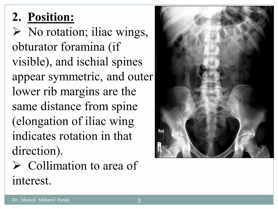
2. Position:No rotation; iliac wings,
obturator foramina (if visible), and ischial spines appear symmetric, and outer lower rib margins are the same distance from spine (elongation of iliac wing indicates rotation in that direction).
Collimation to area of interest.Dr. Ahmed Alsharef Farah 3

3. Exposure:No motion; ribs and all
gas bubble margins appear sharp.
Sufficient exposure visualize psoas muscle outlines, lumbar transverse processes, and ribs.
Margins of liver and kidneys should be visible on smaller to average-sized patients.
Dr. Ahmed Alsharef Farah 4
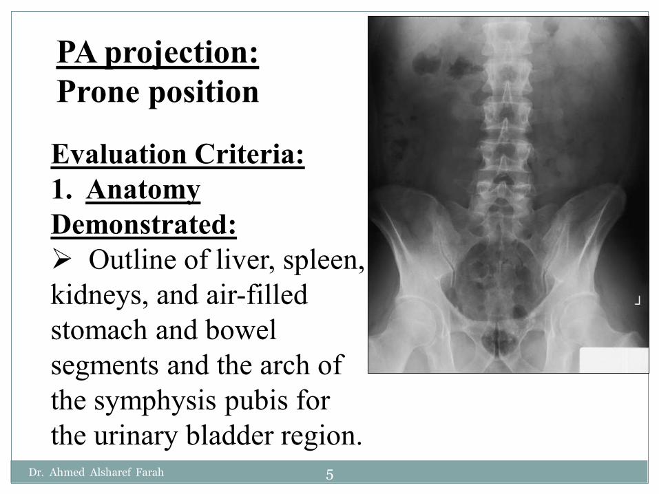
PA projection:Prone position
Evaluation Criteria:1. Anatomy Demonstrated:
Outline of liver, spleen, kidneys, and air-filled stomach and bowel segments and the arch of the symphysis pubis for the urinary bladder region.
Dr. Ahmed Alsharef Farah 5

2. Position:No rotation:
iliac wings appear symmetric, and sacroiliac joints and outer lower rib margins (if visible) should be the same distance from spine.
Collimation to area of interest.
Dr. Ahmed Alsharef Farah 6
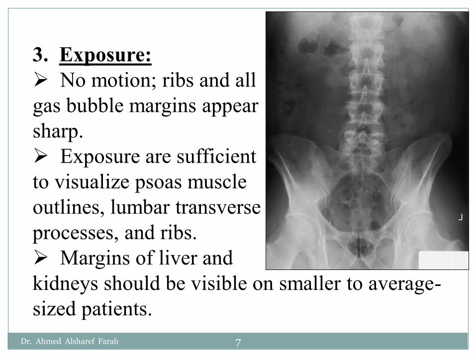
3. Exposure:No motion; ribs and all
gas bubble margins appear sharp.
Exposure are sufficient to visualize psoas muscle outlines, lumbar transverse processes, and ribs.
Margins of liver and kidneys should be visible on smaller to average-sized patients.
Dr. Ahmed Alsharef Farah 7
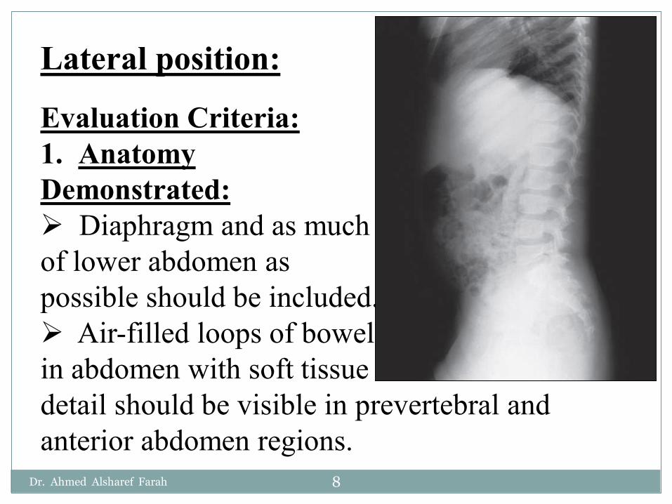
Lateral position:
Evaluation Criteria:1. Anatomy Demonstrated:
Diaphragm and as muchof lower abdomen as possible should be included.
Air-filled loops of bowel in abdomen with soft tissue detail should be visible in prevertebral and anterior abdomen regions.
Dr. Ahmed Alsharef Farah 8

2. Position:No rotation as evident
by superimposition of posterior ribs and posterior borders of iliac wings and bilateral ASIS.
Collimation to area of interest.
Dr. Ahmed Alsharef Farah 9

3. Exposure:No motion; rib and gas
bubble margins appear sharp.
Lumbar vertebrae may appear about 50% underexposed with soft tissue detail visible in anterior abdomen and in prevertebral region of lower lumbar vertebra.
Dr. Ahmed Alsharef Farah 10
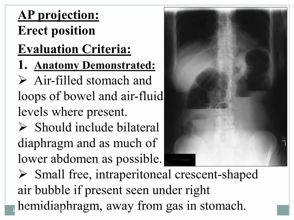
Evaluation Criteria:1. Anatomy Demonstrated:
Air-filled stomach and loops of bowel and air-fluid levels where present.
Should include bilateral diaphragm and as much of lower abdomen as possible.
Small free, intraperitoneal crescent-shaped air bubble if present seen under right hemidiaphragm, away from gas in stomach.
AP projection:Erect position
Dr. Ahmed Alsharef Farah 11

2. Position:No rotation; iliac wings
appear symmetric, and outer rib margins are the same distance from spine.
Spine should be straight (unless scoliosis is present), aligned with center of IR.
Collimation to area of interest.
Dr. Ahmed Alsharef Farah 12
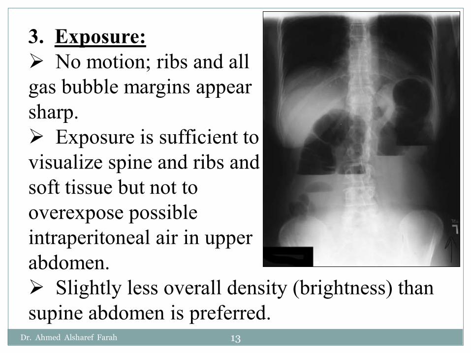
3. Exposure:No motion; ribs and all
gas bubble margins appear sharp.
Exposure is sufficient to visualize spine and ribs and soft tissue but not to overexpose possible intraperitoneal air in upper abdomen.
Slightly less overall density (brightness) than supine abdomen is preferred.
Dr. Ahmed Alsharef Farah 13

Skull x-ray
Dr. Ahmed Alsharef Farah 14
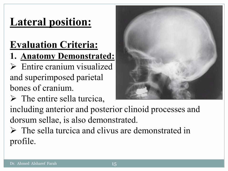
Lateral position:
Evaluation Criteria:1. Anatomy Demonstrated:
Entire cranium visualized and superimposed parietal bones of cranium.
The entire sella turcica, including anterior and posterior clinoid processes and dorsum sellae, is also demonstrated.
The sella turcica and clivus are demonstrated in profile.
Dr. Ahmed Alsharef Farah 15

Dr. Ahmed Alsharef Farah 16

2. Position:No rotation or tilt of the
cranium is evident.Rotation is evident by
anterior and posterior separation of symmetric vertical bilateral structures such as the EAM, mandibular rami, and greater wings of the sphenoid.
Tilt is evident by superior and inferior separation of symmetric horizontal structures such as the orbital roofs (plates) and greater wings of sphenoid.
Collimation to area of interest.Dr. Ahmed Alsharef Farah 17

3. Exposure:Density (brightness)
and contrast are sufficient to visualize bony detail of bony structures and surrounding skull.
Sharp bony margins indicate no motion.
Dr. Ahmed Alsharef Farah 18

PA projection: 0° CR
Evaluation Criteria:1. Anatomy Demonstrated:
Frontal bone, crista galli, internal auditory canals, frontal and anterior ethmoidsinuses, petrous ridges, greater and lesser wings of sphenoid, and dorsum sellaeare shown.
Dr. Ahmed Alsharef Farah 19

Dr. Ahmed Alsharef Farah 20

2. Position:No rotation is evident, as
indicated by equal distance bilaterally from lateral orbital margin to lateral cortex of skull.
Petrous ridges fill the orbits and level of the supraorbital margin.
Posterior and anterior clinoids are visualized just superior to ethmoid sinuses.
Collimation to area of interest.Dr. Ahmed Alsharef Farah 21

3. Exposure:Density (brightness)
and contrast are sufficient to visualize frontal bone and surrounding bony structures.
Sharp bony margins indicate no motion.
Dr. Ahmed Alsharef Farah 22
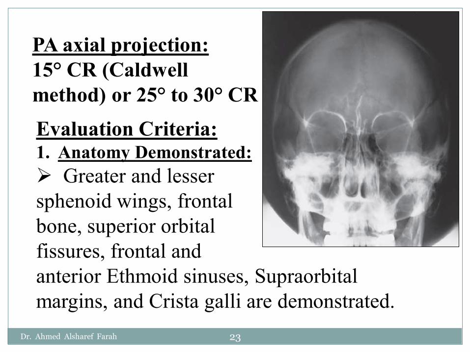
PA axial projection:15° CR (Caldwell method) or 25° to 30° CREvaluation Criteria:1. Anatomy Demonstrated:
Greater and lesser sphenoid wings, frontal bone, superior orbital fissures, frontal and anterior Ethmoid sinuses, Supraorbitalmargins, and Crista galli are demonstrated.
Dr. Ahmed Alsharef Farah 23
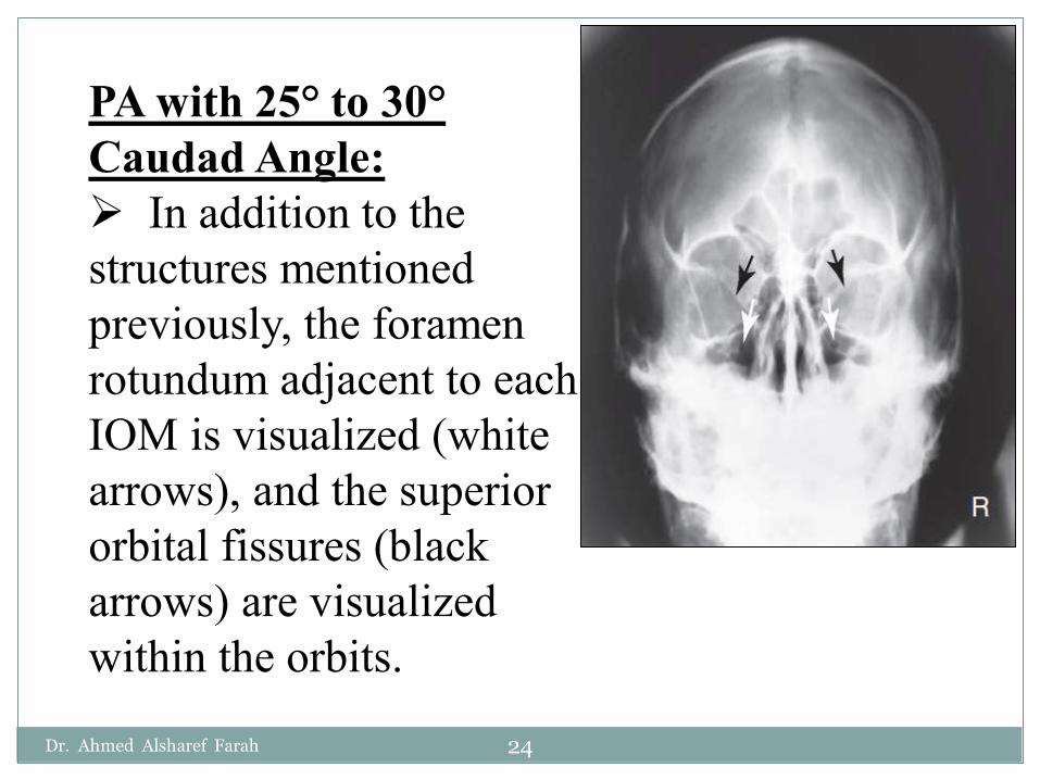
PA with 25° to 30°Caudad Angle:
In addition to the structures mentioned previously, the foramen rotundum adjacent to each IOM is visualized (white arrows), and the superior orbital fissures (black arrows) are visualized within the orbits.
Dr. Ahmed Alsharef Farah 24

2. Position:No rotation as assessed by equal distance
from the midlateral orbital margins to the lateral cortex of the cranium on each side, superior orbital fissures symmetric within the orbits, and correct extension of neck (OMLalignment).
If the distance between the right lateral orbit and lateral cranial cortex is greater than the left side, the face is rotated toward the left side.
No tilt with the MS P perpendicular to IR .Dr. Ahmed Alsharef Farah 25

PA with 15° Caudad Angle:
Petrous pyramids are projected into the lower one-third of the orbits.
Supraorbital margin is visualized without superimposition.
Dr. Ahmed Alsharef Farah 26

PA with 25° to 30°Caudad Angle:
Petrous pyramids are projected at or just below the IOM to allow visualization of the entire orbital margin.
Collimation to area of interest.
Dr. Ahmed Alsharef Farah 27
3. Exposure:Density (brightness) and contrast are
sufficient to visualize the frontal bone and sellar structures without overexposure to perimeter regions of skull.
Sharp bony margins indicate no motion.
Dr. Ahmed Alsharef Farah 28

Paranasal Sinuses
Dr. Ahmed Alsharef Farah 29

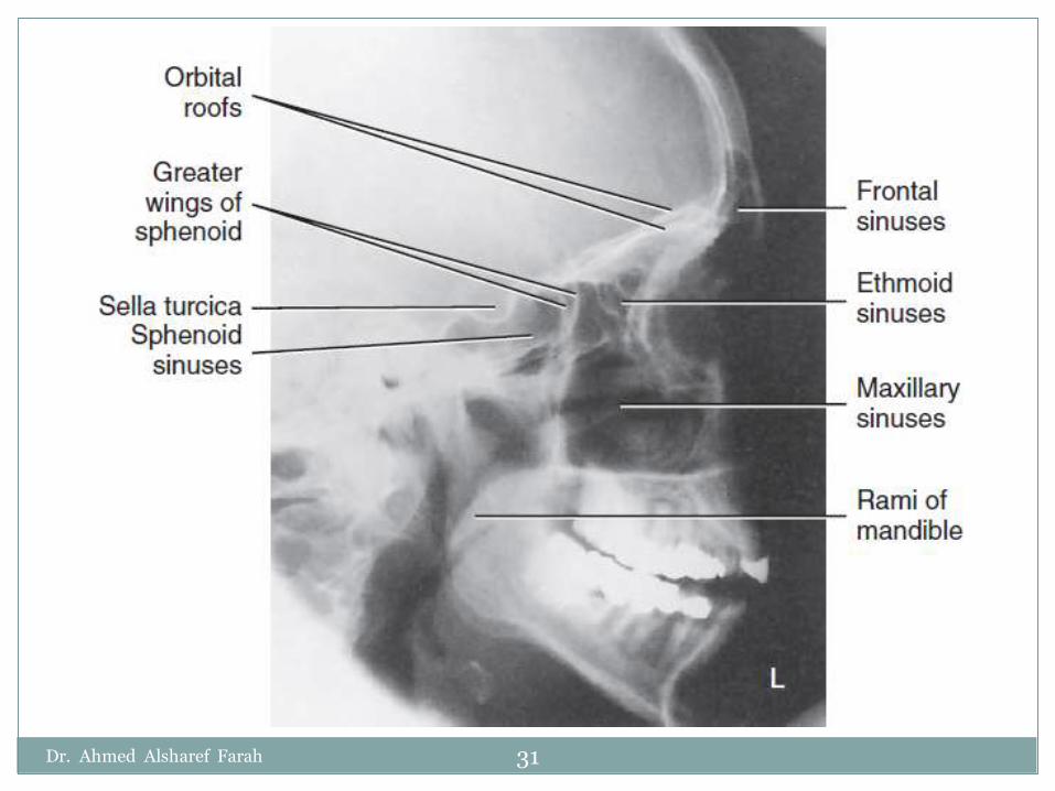
Lateral position:
Evaluation Criteria:1. Anatomy Demonstrated:
All four paranasalsinus groups are shown.
Dr. Ahmed Alsharef Farah 30
Dr. Ahmed Alsharef Farah 31
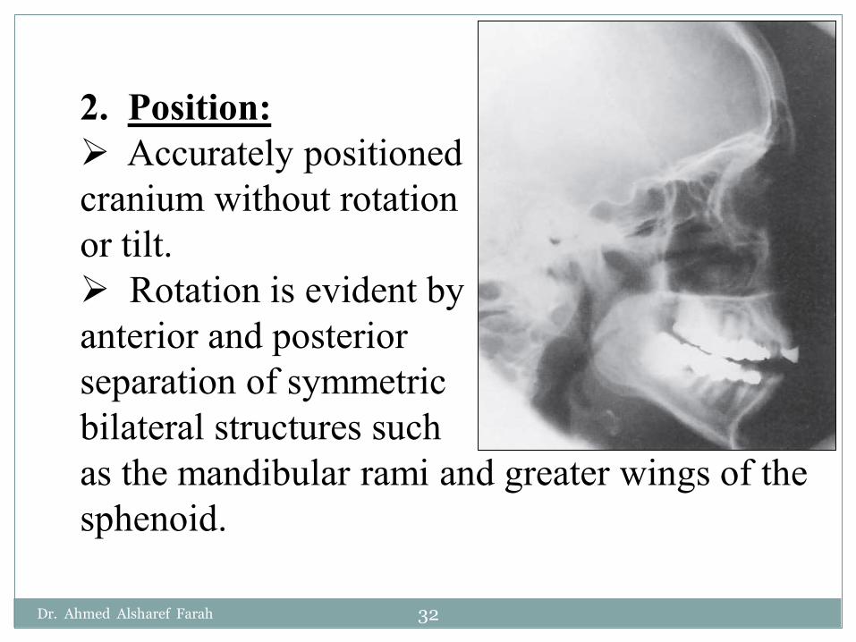
2. Position:Accurately positioned
cranium without rotation or tilt.
Rotation is evident by anterior and posterior separation of symmetric bilateral structures such as the mandibular rami and greater wings of the sphenoid.
Dr. Ahmed Alsharef Farah 32
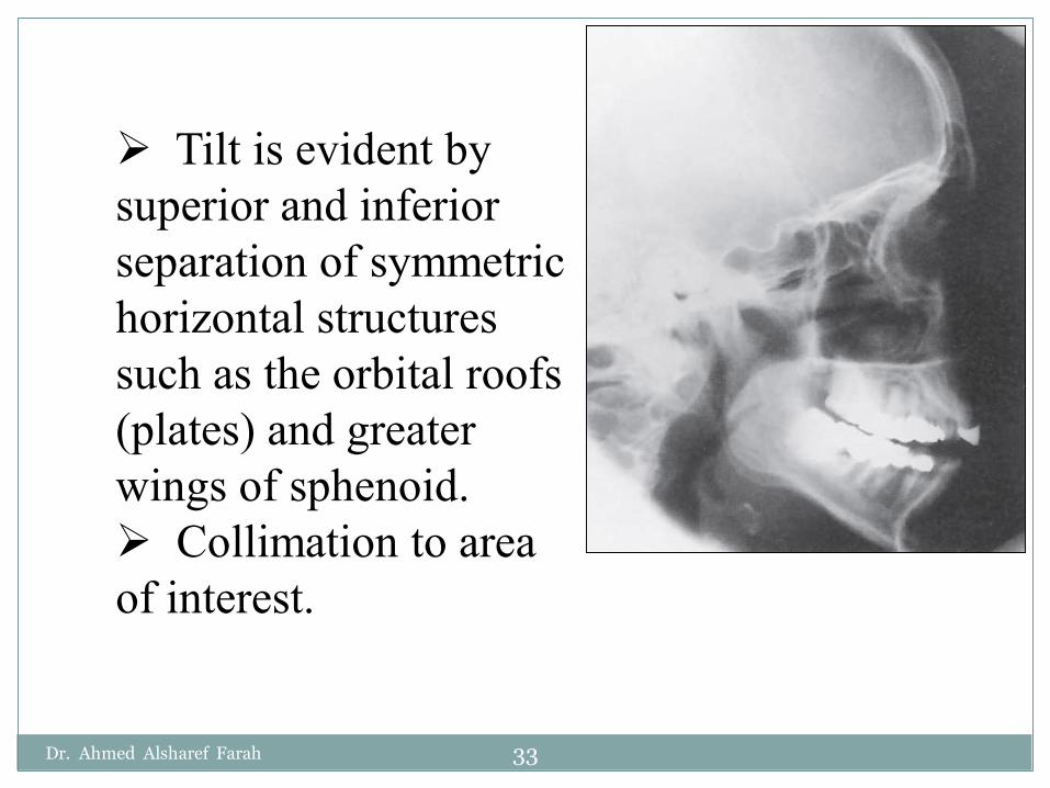
Tilt is evident by superior and inferior separation of symmetric horizontal structures such as the orbital roofs (plates) and greater wings of sphenoid.
Collimation to area of interest.
Dr. Ahmed Alsharef Farah 33

3. Exposure:Density (brightness)
and contrast are sufficient to visualize the sphenoid sinuses through the cranium without overexposing the maxillary and frontal sinuses.
Sharp bony margins indicate no motion.
Dr. Ahmed Alsharef Farah 34

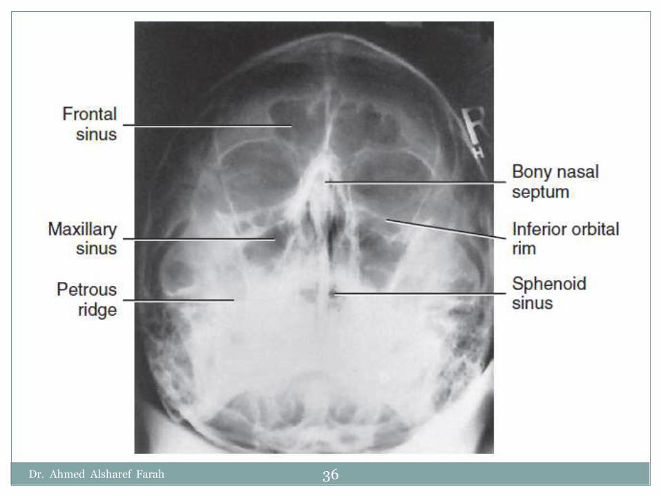
Parietoacanthialprojection:Watersmethod (OML 37° to IR).Evaluation Criteria:1. Anatomy Demonstrated:
Maxillary sinuses with the inferior aspect visualized free from superimposing alveolar processes and petrousridges, the inferior orbital rim, and an oblique view of the frontal sinuses.
Dr. Ahmed Alsharef Farah 35
Dr. Ahmed Alsharef Farah 36

2. Position:No rotation of the
cranium is indicated by the following:(1)Equal distance from MSP (identified by the bony nasal septum) to lateral orbital margin on both sides.(2) Equal distance from the lateral orbital margin to the lateral cortex of the cranium on both sides.
Dr. Ahmed Alsharef Farah 37

Adequate extension of neck demonstrates petrous ridges just inferior to the maxillary sinuses.
Collimation to area of interest.
Dr. Ahmed Alsharef Farah 38

3. Exposure:Density (brightness)
and contrast are sufficient to visualize maxillary sinuses.
Sharp bony margins indicate no motion.
Dr. Ahmed Alsharef Farah 39
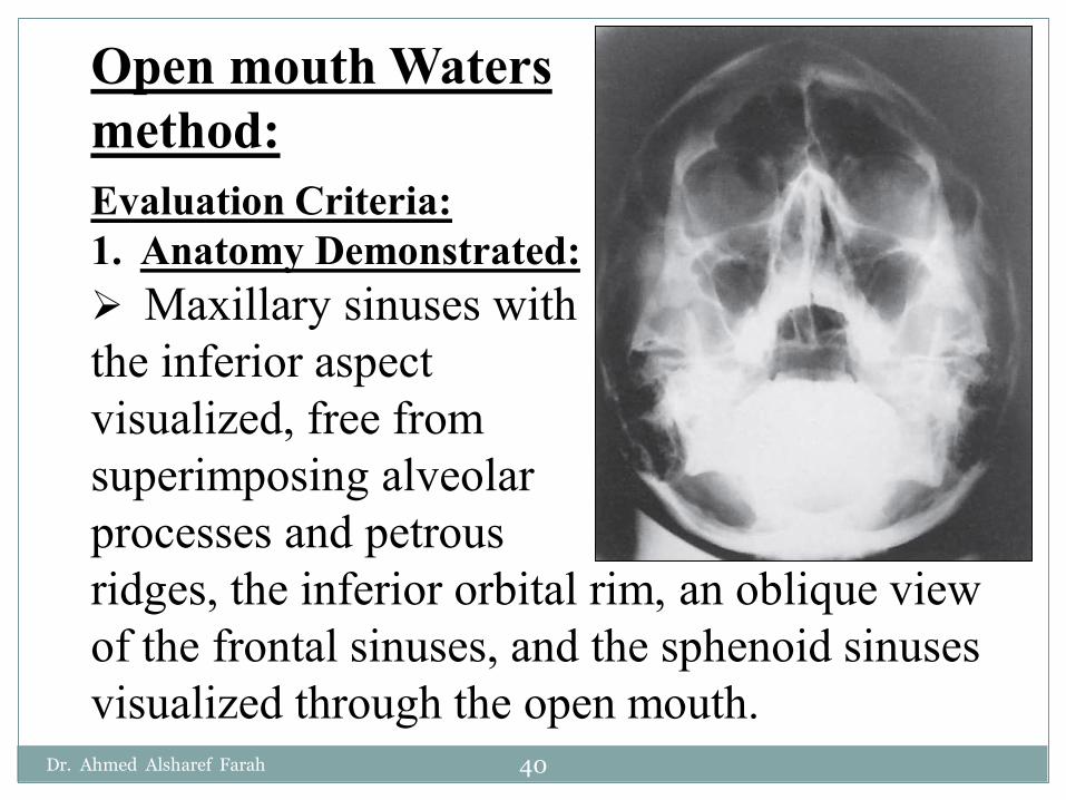
Open mouth Waters method:Evaluation Criteria:1. Anatomy Demonstrated:
Maxillary sinuses with the inferior aspect visualized, free from superimposing alveolar processes and petrousridges, the inferior orbital rim, an oblique view of the frontal sinuses, and the sphenoid sinuses visualized through the open mouth.
Dr. Ahmed Alsharef Farah 40

Dr. Ahmed Alsharef Farah 41
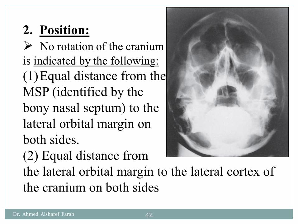
2. Position:No rotation of the cranium
is indicated by the following:(1)Equal distance from the MSP (identified by the bony nasal septum) to the lateral orbital margin on both sides.(2) Equal distance from the lateral orbital margin to the lateral cortex of the cranium on both sides
Dr. Ahmed Alsharef Farah 42

Accurate extension of the neck demonstrating petrousridges just inferior to the maxillary sinuses.
Collimation to area of interest.
Dr. Ahmed Alsharef Farah 43

3. Exposure:Density (brightness)
and contrast are sufficient to visualize the maxillary and sphenoid sinuses.
Sharp bony margins indicate no motion.
Dr. Ahmed Alsharef Farah 44

Thank You
Dr. Ahmed Alsharef Farah 45
Related Documents