A SYSTEMATIC APPROACH TO A SYSTEMATIC APPROACH TO X X - - RAY INTERPRETATION RAY INTERPRETATION Part 2 Part 2 Abdominal Plain Films, Anatomy Abdominal Plain Films, Anatomy & Common Pathologies & Common Pathologies Dr Meena Arunakirinathan West Middlesex Hospital

Welcome message from author
This document is posted to help you gain knowledge. Please leave a comment to let me know what you think about it! Share it to your friends and learn new things together.
Transcript

A SYSTEMATIC APPROACH TO A SYSTEMATIC APPROACH TO
XX--RAY INTERPRETATIONRAY INTERPRETATION
Part 2Part 2
Abdominal Plain Films, Anatomy Abdominal Plain Films, Anatomy
& Common Pathologies & Common Pathologies
Dr Meena Arunakirinathan
West Middlesex Hospital

Objectives
• To review the anatomy relevant to abdominal
x-rays.
• To learn a systematic approach to x-ray
interpretation.
• To apply this approach to interpreting
abdominal x-rays.
• To identify some common pathologies
detectable by abdominal x-ray.

Surface
anatomy of
the
abdomen

THE ABDOMINAL X-RAY (AXR)
• Of more limited value in diagnosis than CXR.
• Standard AXR is taken in supine position where x-rays are in AP projection with patient lying down on his/her back.
• May also be taken with patient in lateral decubitus or upright positions in order to visualise an air-fluid level.
• AXR is of most use in the patient with an acute abdomen.
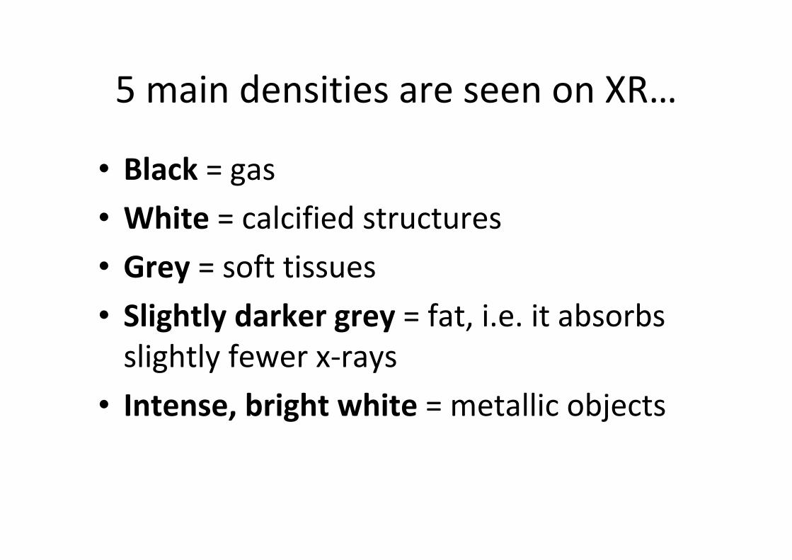
5 main densities are seen on XR…
• Black = gas
• White = calcified structures
• Grey = soft tissues
• Slightly darker grey = fat, i.e. it absorbs
slightly fewer x-rays
• Intense, bright white = metallic objects
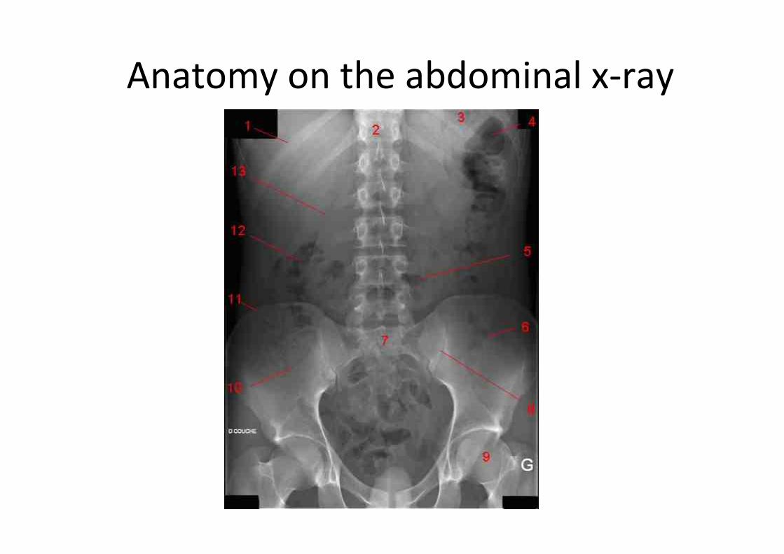
Anatomy on the abdominal x-ray

Take 10 seconds to examine this film…
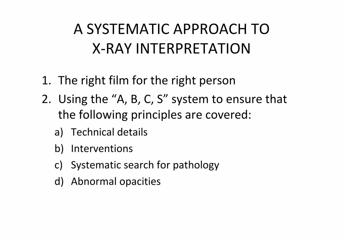
A SYSTEMATIC APPROACH TO
X-RAY INTERPRETATION
1. The right film for the right person
2. Using the “A, B, C, S” system to ensure that
the following principles are covered:
a) Technical details
b) Interventions
c) Systematic search for pathology
d) Abnormal opacities

The right film for the right person
• Is this the right patient?
– Name
– DOB
– Hospital number
• Is this the right film?
– Date of x-ray
– Time of x-ray

“A” is for adequacy, alignment and
apparatus
Adequate
penetration
No rotation
Surgical clips

“B” is for bones Fractured head and neck of
right femurClassic triad of
Paget’s disease
Anteroposterior compression injury to
pelvic ring

“C” is for cartilage & joints
Osteoarthritis in left
hip jointNormal hip joint

Work intraperitoneally to retroperitoneally to
evaluate outlines of the major abdominal
organs…
• Can you see gas in the stomach and/or bowel?
• Look at size and position of liver and spleen
• Bladder outline may be seen if bladder is full
• Look at size and position of kidneys lateral to T12
to L2 vertebrae
• Is there a clear outline of the psoas shadow?
“S” is for soft tissue

“S” is for soft tissue
Small bowel obstruction
Small
bowel
loops >
3cm
Valvulae conniventes
Loops of bowel are centrally located

“S” is for soft tissue
Large bowel obstruction
Loops of bowel are located peripherally
and follow characteristic pattern
Diameter of colon
> 5cmHaustra

“S” is for soft tissue
Left psoas shadow
Psoas shadow
is absent on
right side

“S” is for soft tissue
Pneumoperitoneum

“S” is for soft tissue
Check the following structures for calcification:
• Cartilage of ribs
• Blood vessels
• Pancreas
• Kidneys
• RUQ for gallbladder calculi

Systematically interpret this chest x-ray

CLINICAL
SCENARIOS

A 30 year-old man with a 6-month history of epigastric
pain occurring 2 to 3 hours after meals and anorexia
presented to A&E with sudden, severe epigastric pain
radiating to his back. Abdominal exam revealed
rebound tenderness, guarding and rigidity.
What are the differential diagnoses?
How could this condition be managed?

•An erect chest X-ray
showing free gas under
the diaphragm is
suggestive of a visceral
perforation, aka
pneumoperitoneum.
•Free gas under the
diaphragm is seen in
approximately 60% of
patients with a perforated
peptic ulcer.
•Absence of free gas does
not exclude a diagnosis of
visceral perforation.

A 70 year-old man presented with periumbilical
discomfort and abdominal bloating after meals and
fever. Upper GI endoscopy was found to be normal. A
barium meal and follow-through study was carried out.
What are the differential diagnoses?
How could this condition be treated?

•This image shows large
diverticulae of the proximal
small bowel with partial
intestinal obstruction.
•The incidence of diverticulitis
increases with age, with less
than 5% before age 40 to
greater than 65% by age 85.

A 16 year-old boy presented with a short history of
left iliac fossa pain and bloody diarrhoea streaked
with mucus. Stool cultures were found to be
negative. Flexible sigmoidoscopy showed an acute
colitis. Despite being given IV steroids he developed
abdominal distension and became systemically
unwell.
What are the differential diagnoses?
How could this condition be managed?

•This plain abdominal x-ray
was taken shows a dilated
colon with evidence of
mucosal oedema.
•The appearances are those
of toxic dilatation.
•TOXIC MEGACOLON =
radiological evidence of
colonic dilitation and any of
the 3 following conditions:
fever, tachycardia,
leukocyosis or anaemia.
Dilated colonMucosal oedema

An 89 year-old woman presented with a 4-day history of
absolute constipation and abdominal distension.
Examination revealed a grossly distended, non-tender
and tympanic abdomen. Sigmoidoscopy showed an
empty rectum, and at 25 cm a large amount of faecal
fluid and gas was encountered with relief of her
symptoms.
What is the differential diagnosis?
How could this condition be managed?

•This plain abdominal X-ray
shows the typical features of
a sigmoid volvulus, i.e.
coffee bean sign.
•Chronic constipation leads
to an overloaded sigmoid
colonic loop , and the weight
of this loaded loop makes it
susceptible to torsion along
the axis of the mesentery.
•A complete volvulus leads to
the development of a closed
loop obstruction of the
affected colonic segment..

This frail, 85 year-old woman presented with a 6-month
history of rectal bleeding, rapid weight loss and change
in bowel habit – in particular, increasing constipation.
Hepatomegaly and ascites were apparent on abdominal
examination. A barium enema revealed this finding.
What are the differential diagnoses?
How could this condition be managed?

•A barium enema showed the
presence of 'apple-core'
stricture in the proximal
sigmoid colon.
•This finding is typical of colonic
cancer and can be confirmed by
biopsies taken at flexible
sigmoidoscopy.
•Increasing age is a well-known
risk factor for colorectal cancer.

1. The right film for the right person
2. Using the “A, B, C, S” system to proceed:
• A = adequacy, alignment, apparatus
• B = bones
• C = cartilage and joints
• S = soft tissue – intraperitoneal →
retroperitoneal
Summary of systematic approach to
AXR interpretation


Bibliography
• http://anatomy.med.umich.edu
• http://www.esg.montana.edu/esg/kla/ta/digest.html
• Abdominal X Rays Made Easy, Student BMJ Series
• http://www.instantanatomy.net/
• http://www.docstoc.com/docs/451320/The-Abdominal-X-Ray
• http://www.surgical-tutor.org.uk/default-home.htm

Bibliography - continued
• http://library.med.utah.edu/WebPath/HISTHT
ML/ANATOMY/ANATOMY.html
• http://en.wikipedia.org/wiki/X-
ray_computed_tomography
• http://www.fmhs.auckland.ac.nz/sms/anatom
y/atlas/default.aspx
• http://www.med.wayne.edu/diagradiology/A
natomy_Modules/Mediastinum/Mediastinum
.html
Related Documents