A SYSTEMATIC APPROACH TO A SYSTEMATIC APPROACH TO X X - - RAY INTERPRETATION RAY INTERPRETATION Part 1 Part 1 Chest X Chest X - - rays, Anatomy & rays, Anatomy & Common Pathologies Common Pathologies Dr Meena Arunakirinathan West Middlesex Hospital

Welcome message from author
This document is posted to help you gain knowledge. Please leave a comment to let me know what you think about it! Share it to your friends and learn new things together.
Transcript

A SYSTEMATIC APPROACH TO A SYSTEMATIC APPROACH TO
XX--RAY INTERPRETATIONRAY INTERPRETATION
Part 1Part 1
Chest XChest X--rays, Anatomy & rays, Anatomy &
Common PathologiesCommon Pathologies
Dr Meena Arunakirinathan
West Middlesex Hospital
Objectives
• To review the anatomy relevant to chest x-
rays.
• To learn a systematic approach to x-ray
interpretation.
• To apply this approach to interpreting chest x-
rays.
• To identify some common pathologies
detectable by chest x-ray.
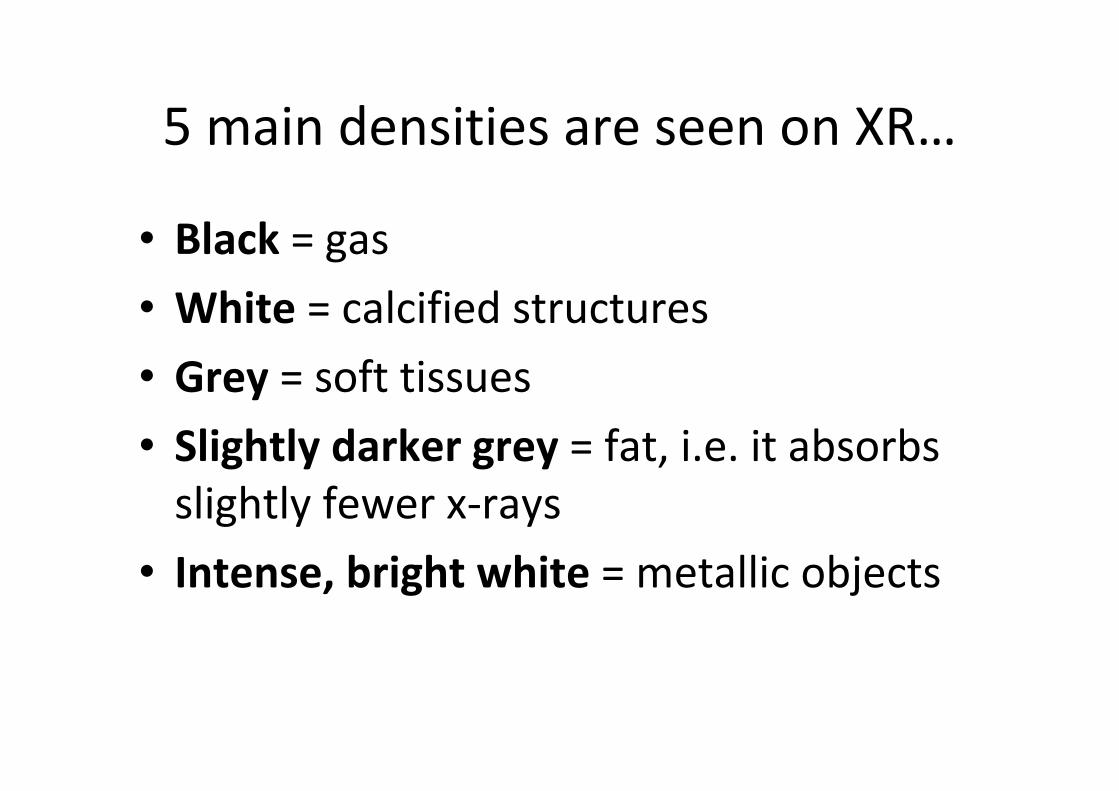
5 main densities are seen on XR…
• Black = gas
• White = calcified structures
• Grey = soft tissues
• Slightly darker grey = fat, i.e. it absorbs
slightly fewer x-rays
• Intense, bright white = metallic objects
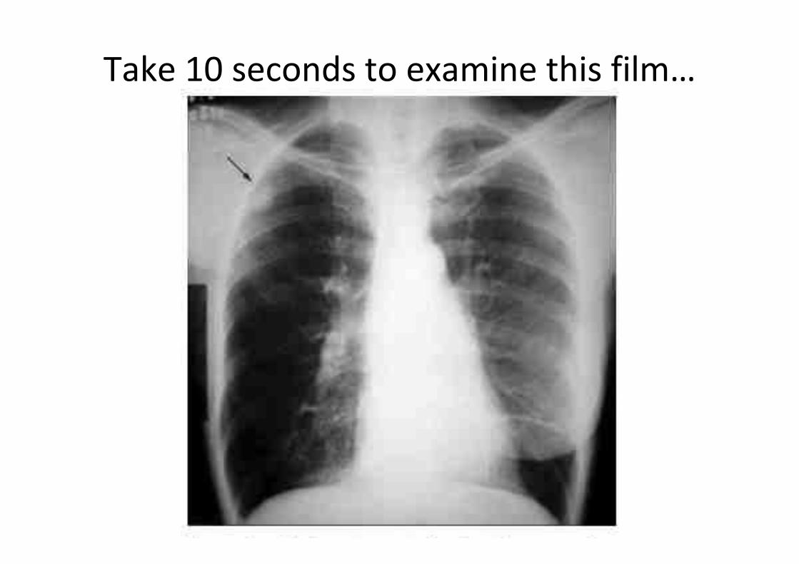
Take 10 seconds to examine this film…
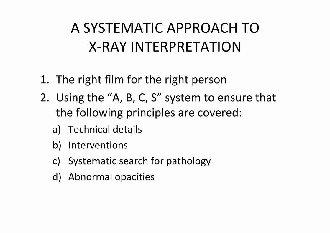
A SYSTEMATIC APPROACH TO
X-RAY INTERPRETATION
1. The right film for the right person
2. Using the “A, B, C, S” system to ensure that
the following principles are covered:
a) Technical details
b) Interventions
c) Systematic search for pathology
d) Abnormal opacities

The right film for the right person
• Is this the right patient:
– Name
– DOB
– Hospital number
• Is this the right film?
– Date of x-ray
– Time of x-ray
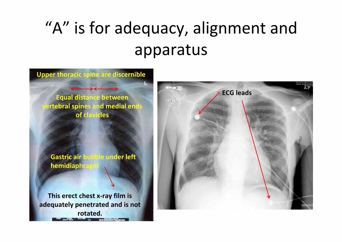
“A” is for adequacy, alignment and
apparatus
Upper thoracic spine are discernible
Equal distance between
vertebral spines and medial ends
of clavicles
This erect chest x-ray film is
adequately penetrated and is not
rotated.
Gastric air bubble under left
hemidiaphragm
ECG leads
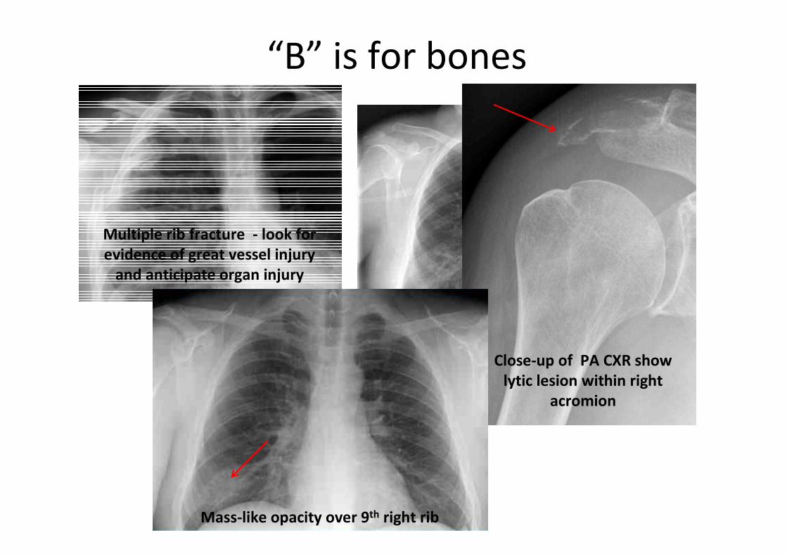
“B” is for bones
Multiple rib fracture - look for
evidence of great vessel injury
and anticipate organ injury
Close-up of PA CXR show
lytic lesion within right
acromion
Mass-like opacity over 9th right rib

“C” is for cartilage & joints• Do the anterior ribs appear to extend to the
sternum?
– Calcification of rib cartilages
• Examine all joints for degenerative joint
disease
– Joint space narrowing
– Osteophytes = bone spurs
– Osteopaenia = demineralisation of bone
– Marginal erosions where bone meet synovium
– Subluxation
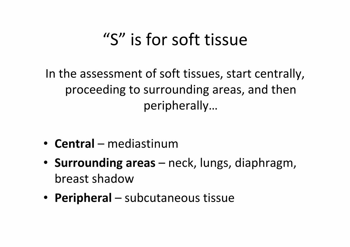
“S” is for soft tissue
In the assessment of soft tissues, start centrally,
proceeding to surrounding areas, and then
peripherally…
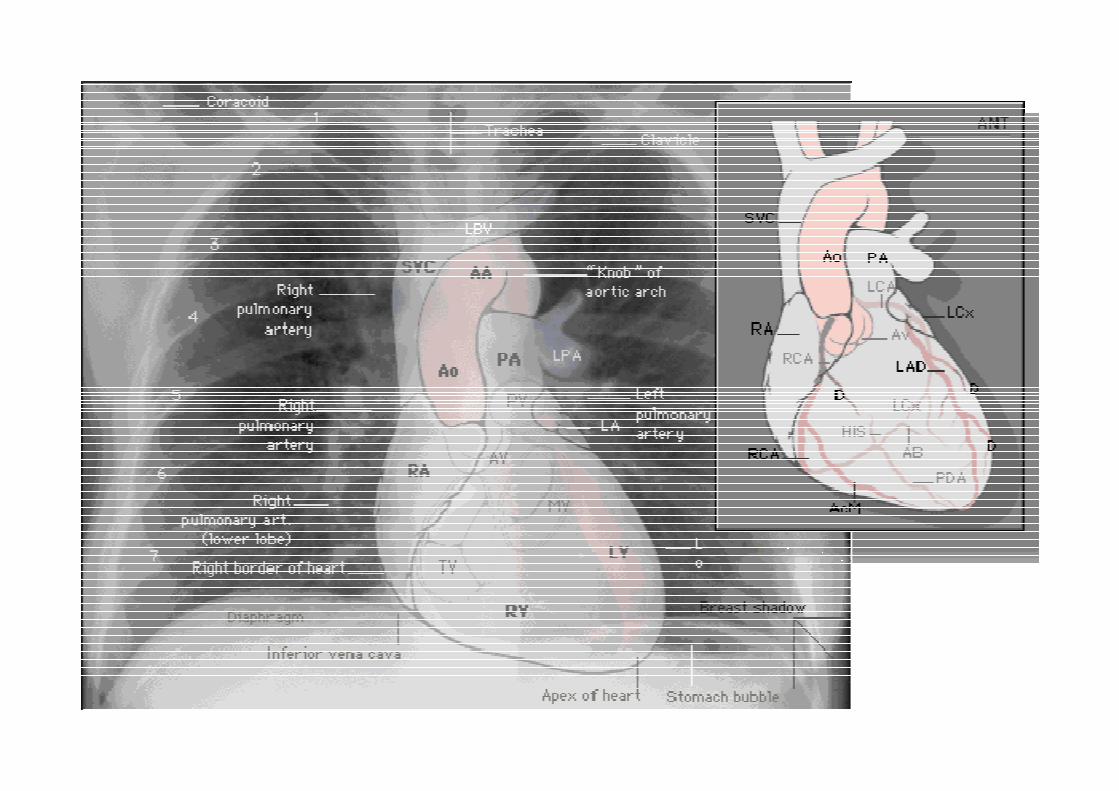
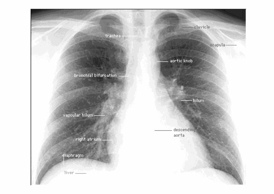
• Central – mediastinum
• Surrounding areas – neck, lungs, diaphragm,
breast shadow
• Peripheral – subcutaneous tissue
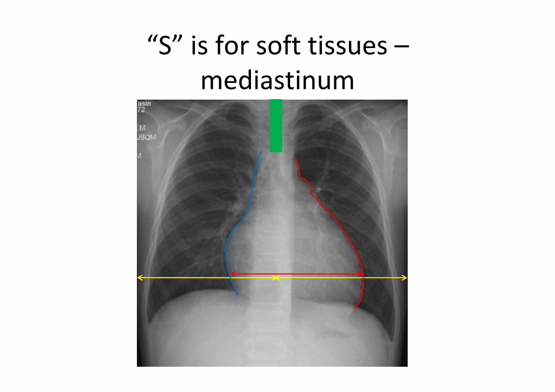
“S” is for soft tissues –
mediastinum
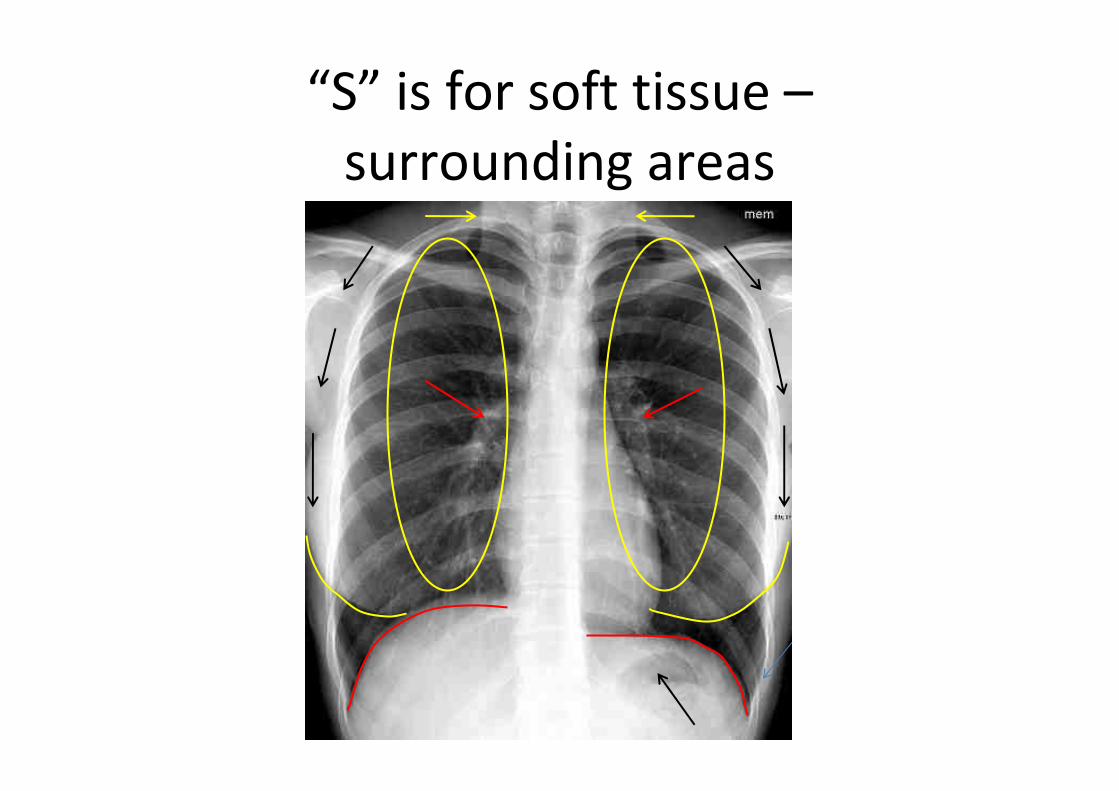
“S” is for soft tissue –
surrounding areas
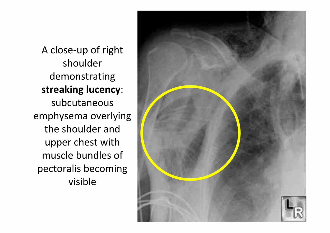
A close-up of right
shoulder
demonstrating
streaking lucency:
subcutaneous
emphysema overlying
the shoulder and
upper chest with
muscle bundles of
pectoralis becoming
visible

4 causes of “white out”
Consolidation Pleural effusion
Complete lung collapse Pneumectomy
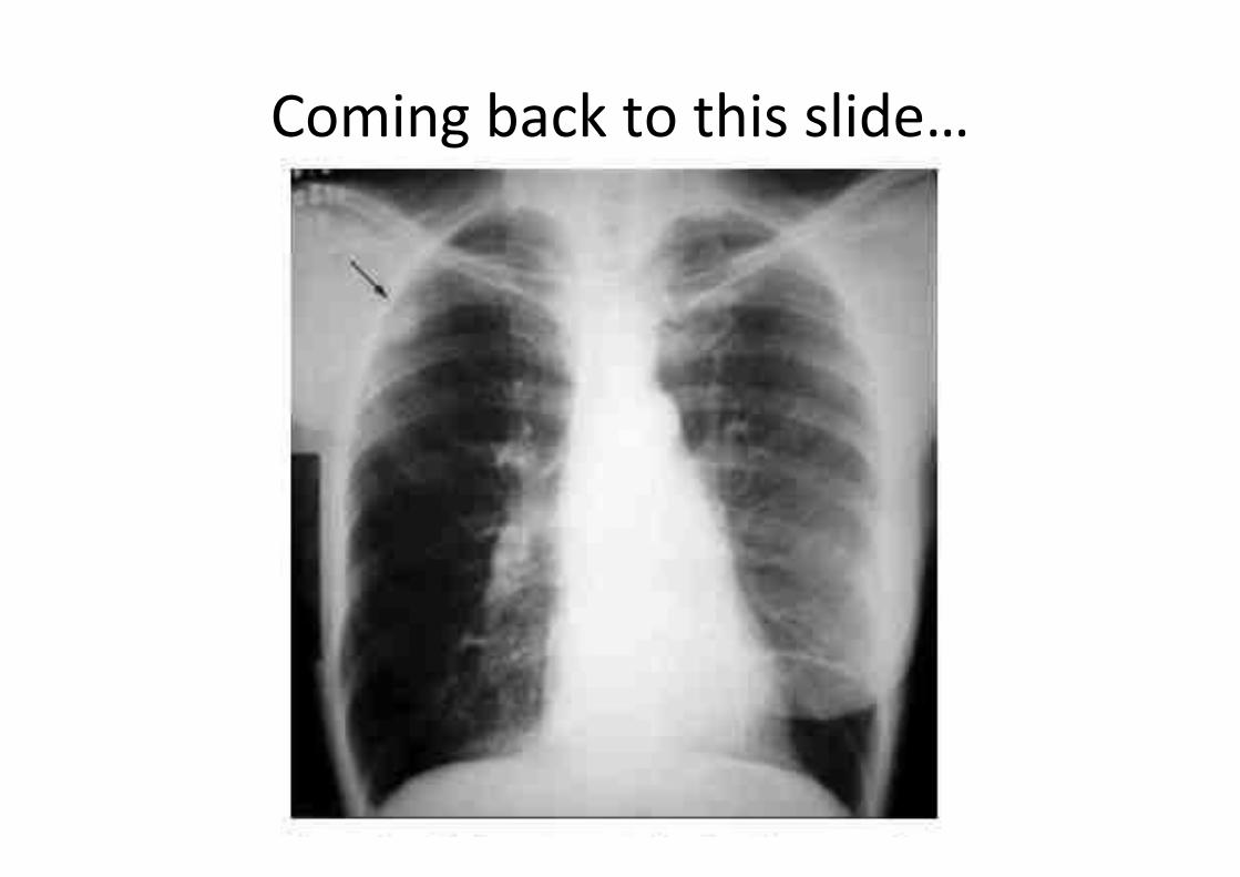
Coming back to this slide…

Systematically interpret this chest x-ray

CLINICAL
SCENARIOS
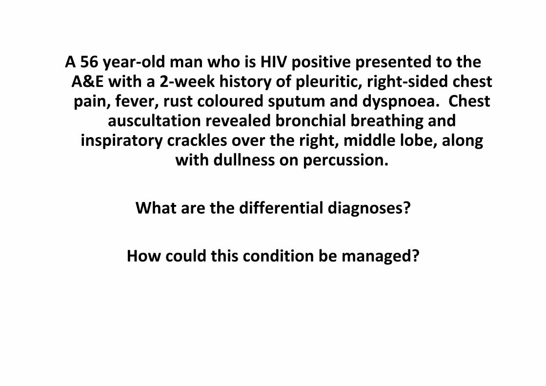
A 56 year-old man who is HIV positive presented to the A&E with a 2-week history of pleuritic, right-sided chest pain, fever, rust coloured sputum and dyspnoea. Chest
auscultation revealed bronchial breathing and inspiratory crackles over the right, middle lobe, along
with dullness on percussion.
What are the differential diagnoses?
How could this condition be managed?
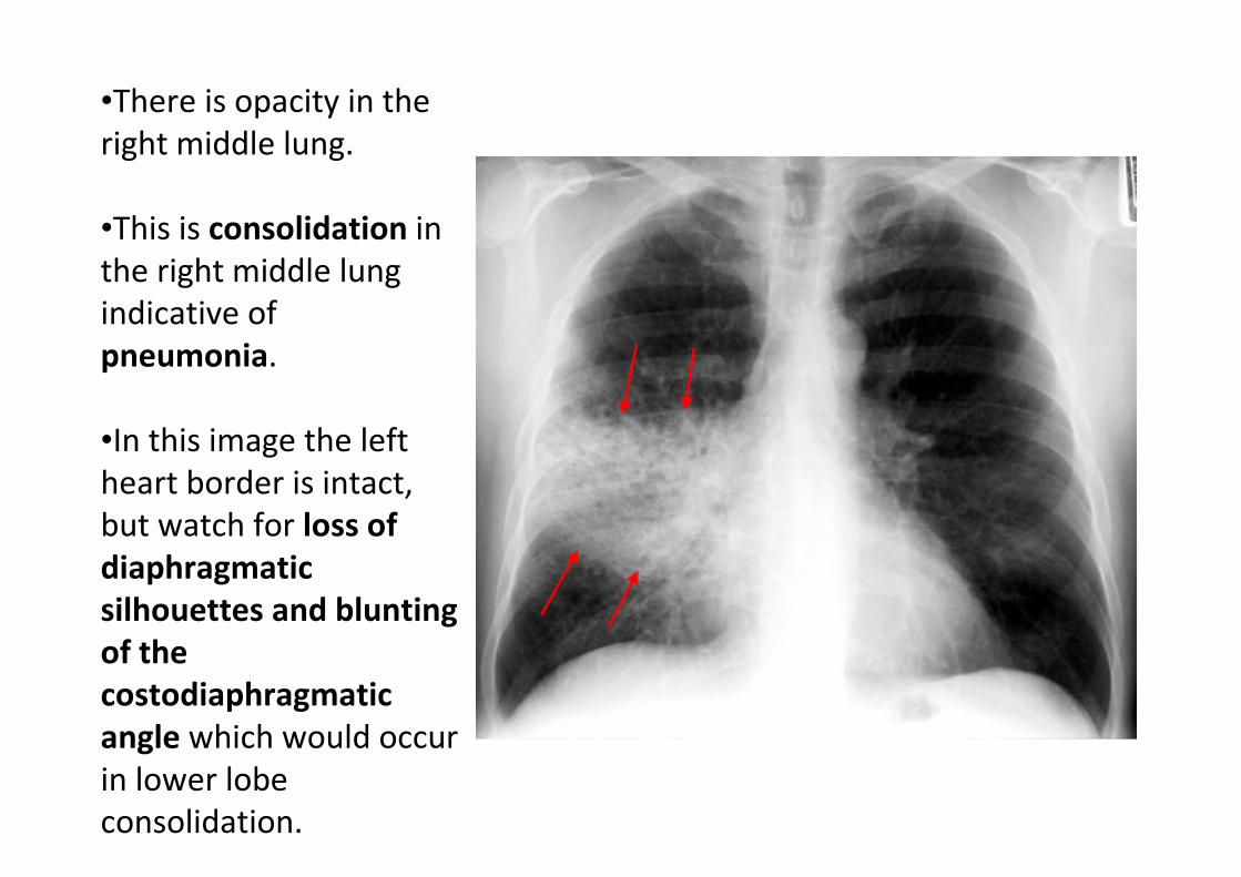
•There is opacity in the
right middle lung.
•This is consolidation in
the right middle lung
indicative of
pneumonia.
•In this image the left
heart border is intact,
but watch for loss of
diaphragmatic
silhouettes and blunting
of the
costodiaphragmatic
angle which would occur
in lower lobe
consolidation.

A tall and slim 21 year-old man presented to the A&E with sudden onset of chest pain, severe dyspnoea and
rapid heart rate. Physical exam findings revealed hyper-resonance of the left chest wall and diminished breath sounds on the left side. His blood pressure was
80/50 mmHg, and he was found to be cyanotic.
What are the differential diagnoses?
How might this condition be managed?
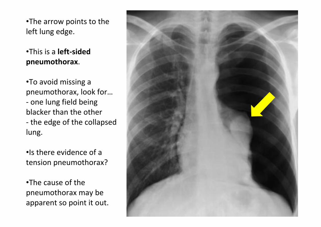
•The arrow points to the
left lung edge.
•This is a left-sided
pneumothorax.
•To avoid missing a
pneumothorax, look for…
- one lung field being
blacker than the other
- the edge of the collapsed
lung.
•Is there evidence of a
tension pneumothorax?
•The cause of the
pneumothorax may be
apparent so point it out.
A 78 year-old man presented to the A&E with dyspnoea,
stabbing chest pain exacerbated with deep inspiration
and haemoptysis. Physical examination revealed
dullness to percussion over the right chest wall,
inaudible breath sounds on the right side and
asymmetric chest wall expansion. He had been
diagnosed with lung cancer last year.
What are the differential diagnoses?
How could this condition be managed?
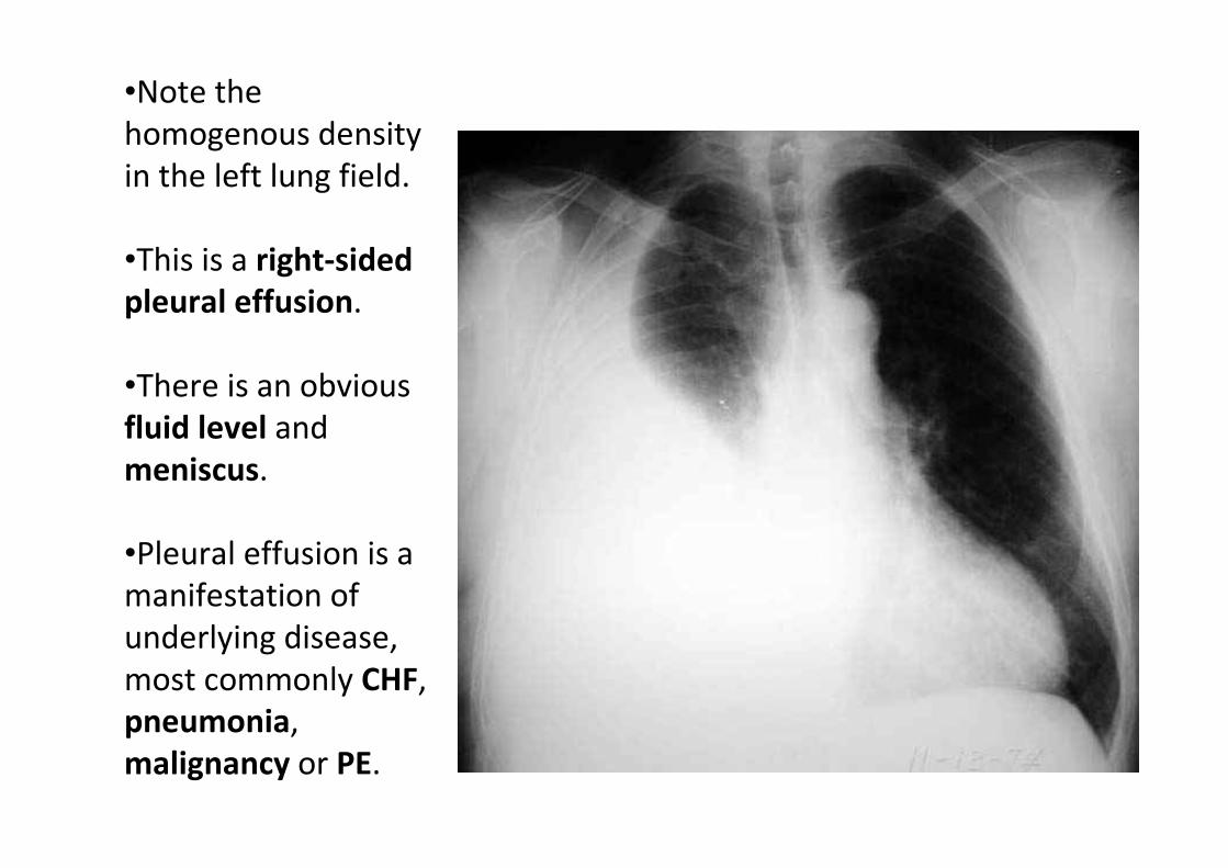
•Note the
homogenous density
in the left lung field.
•This is a right-sided
pleural effusion.
•There is an obvious
fluid level and
meniscus.
•Pleural effusion is a
manifestation of
underlying disease,
most commonly CHF,
pneumonia,
malignancy or PE.

Summary of systematic approach
to CXR interpretation
1. The right film for the right person
2. Using the “A, B, C, S” system to proceed:
– A = adequacy, alignment, apparatus
– B = bones
– C = cartilage and joints
– S = soft tissue – centrally →
surrounding areas → peripherally

X-RAY INTERPRETATION
EXERCISES


Bibliography
• http://info.med.yale.edu/intmed/cardio/imag
ing/contents.html
• Chest X Rays Made Easy, Student BMJ Series
• http://www.learningradiology.com/images/ch
estimages1/Pneumonectomy.JPG
• http://www.colorado.edu/intphys/Class/IPHY
3430-200/image/10-13.jpg
• http://www.heartandmetabolism.org/issues/
HM16/hm16refrcorner.asp

Bibliography (continued)
• http://chestjournal.chestpubs.org/content/11
3/1/256.1.full.pdf
• http://www.medscape.com/viewarticle/5601
63_4
Related Documents