Estrogen receptor-a regulates the degradation of insulin receptor substrates 1 and 2 in breast cancer cells Catia Morelli 1,2 , Cecilia Garofalo 1,2 , Monica Bartucci 1,2 and Eva Surmacz* ,1 1 Kimmel Cancer Center, Thomas Jefferson University, Philadelphia, PA 19107, USA; 2 Postgraduate School in Clinical Pathology, Faculty of Pharmacy, University of Calabria, Italy In breast cancer cells, 17-b-estradiol (E2) upregulates the expression of insulin receptor substrate 1 (IRS-1), a molecule transmitting insulin-like growth factor-I (IGF-I) signals through the PI-3K/Akt survival pathways. The stimulation of IRS-1 by E2 has been documented on the transcriptional level. Here we studied whether the expression of estrogen receptor (ER)-a affects IRS molecules post-transcriptionally. We used ER-a-negative MDA-MB-231 breast cancer cells and MDA-MB-231 cells with re-expressed ER-a. In MDA-MB-231 cells cultured under serum-free conditions, IRS-1 and IRS-2 were degraded through the 26S proteasome and calpain pathways. Re-expression of ER-a in MDA-MB-231 cells correlated with enhanced stability of IRS molecules. This effect coincided with significantly reduced ubiquitination of IRS-1 and IRS-2, but did not involve increased IRS-1 and IRS-2 transcription. The interference of ER-a with IRS-1 and IRS-2 turnover could rely on the competition for common degradation pathways, as in MDA-MB-231/ ER cells, ER-a processing was blocked by proteasome and calpain inhibitors. Notably, a fraction of the cytosolic ER- a colocalized and coprecipitated with IRS-1 and IRS-2, indicating a possible common destination for these proteins. The stabilization of IRS-1 in MDA-MB-231/ ER cells was paralleled by the upregulation of the IRS-1/ Akt/GSK-3 pathway and improved survival in the presence of IGF-I, whereas IRS-2 was not involved in IGF-I signaling. Oncogene (2003) 22, 4007–4016. doi:10.1038/sj.onc.1206436 Keywords: estrogen receptor; insulin receptor substrate; proteasome degradation; cell survival; breast cancer Introduction Insulin receptor substrates (IRS) 1 and 2, members of the IRS family of signaling molecules, are major signaling intermediates of the insulin and insulin- like growth factor I (IGF-I) receptors (IR and IGF- IR). In addition, IRS-1 and IRS-2 transmit signals of many other receptors (e.g., prolactin, growth hormone, several interleukins, and interferons, a6b4 integrins) (Yenush and White, 1997; Aguirre and White, 2000; Burks and White, 2001; Shaw, 2001). In response to ligand binding, IRS substrates are tyrosine phosphorylated, which results in sequestration of multiple effector molecules and stimulation of different signaling pathways (Yenush and White, 1997; Aguirre and White, 2000; Burks and White, 2001). The most notable is the PI-3K/Akt pathway that regulates cell growth and survival as well as different nonmitogenic functions in various cell systems (Shep- herd et al., 1998) Although the functions of IRS-1 and IRS-2 can partially overlap, the data obtained with knockout animals indicated that IRS-1 and IRS-2 may have unique roles (Burks and White, 2001; Fasshauer et al., 2001). Aberrant expression of IRS molecules has been associated with pathogenesis of diabetes as well as with the development of cancer of the breast, pancreas, and liver (Sasaki et al., 1993; Bergmann et al., 1996; Kornmann et al., 1998; Spector et al., 1999; Aguirre and White, 2000; Surmacz, 2000; Ducluzeau et al., 2001; Sachdev and Yee, 2001). However, the mechanisms controlling cellular abundance of these substrates are only partially known. Recent studies indicated that the expression of IRS-1 and IRS-2 is developmentally regulated and cell context-dependent, and may involve transcriptional and post-transcriptional events (Giovan- none et al., 2000). For instance, 17-b-estradiol (E2) has been shown to increase IRS-1 mRNA expression, while antiestrogens decrease IRS-1 mRNA levels (Guvakova and Surmacz, 1997; Lee et al., 1999; Salerno et al., 1999; Molloy et al., 2000; Surmacz, 2000; Mauro et al., 2001; Sachdev and Yee, 2001). Glucocorticoids inhibit IRS-1, but stimulate IRS-2 mRNA expression (Turnbow et al., 1994; Dupont et al., 1999; Sakoda et al., 2000). Furthermore, IRS-2 transcription can be induced by progesterone and PPAR gamma agonists and inhibited by insulin (Vassen et al., 1999; Smith et al., 2001; Zhang et al., 2001). In addition to the transcriptional control, cellular abundance of IRS proteins is regulated by post- transcriptional mechanisms, especially those controlling protein processing. For instance, in 3T3-L1 adipocytes, chronic exposure to insulin increased IRS-1 serine/ Received 4 October 2002; revised 24 January 2003; accepted 29 January 2003 *Correspondence: E Surmacz, Kimmel Cancer Center, Thomas Jefferson University, 233 S 10th Street, BLSB 631, Philadelphia, PA 19107, USA; E-mail: [email protected] Oncogene (2003) 22, 4007–4016 & 2003 Nature Publishing Group All rights reserved 0950-9232/03 $25.00 www.nature.com/onc

Welcome message from author
This document is posted to help you gain knowledge. Please leave a comment to let me know what you think about it! Share it to your friends and learn new things together.
Transcript

Estrogen receptor-a regulates the degradation of insulin receptor substrates1 and 2 in breast cancer cells
Catia Morelli1,2, Cecilia Garofalo1,2, Monica Bartucci1,2 and Eva Surmacz*,1
1Kimmel Cancer Center, Thomas Jefferson University, Philadelphia, PA 19107, USA; 2Postgraduate School in Clinical Pathology,Faculty of Pharmacy, University of Calabria, Italy
In breast cancer cells, 17-b-estradiol (E2) upregulates theexpression of insulin receptor substrate 1 (IRS-1), amolecule transmitting insulin-like growth factor-I (IGF-I)signals through the PI-3K/Akt survival pathways. Thestimulation of IRS-1 by E2 has been documented on thetranscriptional level. Here we studied whether theexpression of estrogen receptor (ER)-a affects IRSmolecules post-transcriptionally. We used ER-a-negativeMDA-MB-231 breast cancer cells and MDA-MB-231cells with re-expressed ER-a. In MDA-MB-231 cellscultured under serum-free conditions, IRS-1 and IRS-2were degraded through the 26S proteasome and calpainpathways. Re-expression of ER-a in MDA-MB-231 cellscorrelated with enhanced stability of IRS molecules. Thiseffect coincided with significantly reduced ubiquitinationof IRS-1 and IRS-2, but did not involve increased IRS-1and IRS-2 transcription. The interference of ER-a withIRS-1 and IRS-2 turnover could rely on the competitionfor common degradation pathways, as in MDA-MB-231/ER cells, ER-a processing was blocked by proteasome andcalpain inhibitors. Notably, a fraction of the cytosolic ER-a colocalized and coprecipitated with IRS-1 and IRS-2,indicating a possible common destination for theseproteins. The stabilization of IRS-1 in MDA-MB-231/ER cells was paralleled by the upregulation of the IRS-1/Akt/GSK-3 pathway and improved survival in thepresence of IGF-I, whereas IRS-2 was not involved inIGF-I signaling.Oncogene (2003) 22, 4007–4016. doi:10.1038/sj.onc.1206436
Keywords: estrogen receptor; insulin receptor substrate;proteasome degradation; cell survival; breast cancer
Introduction
Insulin receptor substrates (IRS) 1 and 2, membersof the IRS family of signaling molecules, are majorsignaling intermediates of the insulin and insulin-like growth factor I (IGF-I) receptors (IR and IGF-
IR). In addition, IRS-1 and IRS-2 transmit signalsof many other receptors (e.g., prolactin, growthhormone, several interleukins, and interferons, a6b4integrins) (Yenush and White, 1997; Aguirre andWhite, 2000; Burks and White, 2001; Shaw, 2001). Inresponse to ligand binding, IRS substrates are tyrosinephosphorylated, which results in sequestration ofmultiple effector molecules and stimulation ofdifferent signaling pathways (Yenush and White, 1997;Aguirre and White, 2000; Burks and White, 2001).The most notable is the PI-3K/Akt pathway thatregulates cell growth and survival as well as differentnonmitogenic functions in various cell systems (Shep-herd et al., 1998) Although the functions of IRS-1 andIRS-2 can partially overlap, the data obtained withknockout animals indicated that IRS-1 and IRS-2 mayhave unique roles (Burks and White, 2001; Fasshaueret al., 2001).
Aberrant expression of IRS molecules has beenassociated with pathogenesis of diabetes as well as withthe development of cancer of the breast, pancreas, andliver (Sasaki et al., 1993; Bergmann et al., 1996;Kornmann et al., 1998; Spector et al., 1999; Aguirreand White, 2000; Surmacz, 2000; Ducluzeau et al., 2001;Sachdev and Yee, 2001). However, the mechanismscontrolling cellular abundance of these substrates areonly partially known. Recent studies indicated that theexpression of IRS-1 and IRS-2 is developmentallyregulated and cell context-dependent, and may involvetranscriptional and post-transcriptional events (Giovan-none et al., 2000). For instance, 17-b-estradiol (E2) hasbeen shown to increase IRS-1 mRNA expression, whileantiestrogens decrease IRS-1 mRNA levels (Guvakovaand Surmacz, 1997; Lee et al., 1999; Salerno et al., 1999;Molloy et al., 2000; Surmacz, 2000; Mauro et al., 2001;Sachdev and Yee, 2001). Glucocorticoids inhibit IRS-1,but stimulate IRS-2 mRNA expression (Turnbow et al.,1994; Dupont et al., 1999; Sakoda et al., 2000).Furthermore, IRS-2 transcription can be induced byprogesterone and PPAR gamma agonists and inhibitedby insulin (Vassen et al., 1999; Smith et al., 2001; Zhanget al., 2001).
In addition to the transcriptional control, cellularabundance of IRS proteins is regulated by post-transcriptional mechanisms, especially those controllingprotein processing. For instance, in 3T3-L1 adipocytes,chronic exposure to insulin increased IRS-1 serine/
Received 4 October 2002; revised 24 January 2003; accepted 29 January2003
*Correspondence: E Surmacz, Kimmel Cancer Center, ThomasJefferson University, 233 S 10th Street, BLSB 631, Philadelphia, PA19107, USA;E-mail: [email protected]
Oncogene (2003) 22, 4007–4016& 2003 Nature Publishing Group All rights reserved 0950-9232/03 $25.00
www.nature.com/onc

threonine phosphorylation and resulted in IRS-1degradation through the 26S proteasome (Sun et al.,1999; Pederson et al., 2001). Similarly, prolongedexposure of MCF-7 breast cancer cells and prostateepithelial cells to IGF-I increased IRS-1 ubiquitinationand subsequent degradation in a 26S proteasome-dependent manner (Lee et al., 2000; Zhang et al.,2000). Other study with 3T3-L1 cells implicated calpain,a calcium-dependent neutral protease, in the degrada-tion of IRS-1 in response to chronic insulin treatment(Smith et al., 1993, 1996). The lysosomal pathway hasbeen reported as not involved in IRS-1 processing (Riceet al., 1993).
IRS-2 has been described to undergo degradationthrough the 26S proteasome in 3T3-L1 cells, Faohepatoma cells, mouse embryo fibroblasts (Rui et al.,2001) and mouse uterine cells (Richards et al., 2001).However, in CHO cells, prolonged insulin exposure didnot affect IRS-2 stability, while it resulted in proteaso-mal processing of IRS-1 (Sun et al., 1999).
The proliferation and survival of hormone-responsivebreast cancer cells is influenced by crosstalk betweenthe estrogen receptor-a (ER-a) and IGF-IR. Animportant feature of this interplay is upregulation ofIGF-IR signaling by activated ER-a (Surmacz, 2000;Sachdev and Yee, 2001). Especially, E2 has been shownto increase transcription and expression of IRS-1,potentiating the IRS-1/PI-3K/Akt pathway (Lee et al.,1999; Surmacz, 2000; Sachdev and Yee, 2001).The regulation of IRS molecules by ER-a on thepost-transcriptional level, especially on the level ofprotein degradation, has not been studied and is asubject of this work. Considering that the 26S protea-some and calpain pathways have been implicated inthe degradation of ER-a (Murayama et al., 1984;Shiba et al., 1996; Nawaz et al., 1999; Lonard et al.,2000), we explored the idea that ER-a and IRSsubstrates may compete for the same degradationprocesses. To better distinguish between the transcrip-tional and post-transcriptional regulation of IRS-1and IRS-2 by ER-a, we developed MDA-MB-231/ERbreast cancer cells, in which ER-a does not affect IRS-1and IRS-2 mRNA synthesis. The degradation ofIRS-1 and IRS-2 was studied in MDA-MB-231/ERcells and compared to that in ER-a-negative MDA-MB-231 cells.
Results
Re-expression of ER-a in MDA-MB-231 cells coincideswith decreased degradation of IRS-1 and IRS-2
MDA-MB-231 cells are ER-a-negative breast cancercells expressing IRS-1 and IRS-2. To investigate theexpression of IRS substrates in the presence or absenceof ER-a, we reintroduced ER-a into MDA-MB-231 cellsby stable transfection. Several clones positive for ER-aexpression were pooled to generate a mixed populationof MDA-MB-231/ER cells. In growing MDA-MB-231/ER cells (day 0), the levels of IRS-1 and IRS-2 were
greater than that in the parental ER-a-negative cells(Figure 1a) During cell culture in SFM, the abundanceof IRS-1 and IRS-2 in MDA-MB-231 cells furtherdeclined, reaching minimal levels at day 3, when the
Figure 1 Expression of ER-a in MDA-MB-231 cells preventsdegradation of IRS-1 and IRS-2. (a) Confluent cultures (70%) ofMDA-MB-231 (231) and MDA-MB-231/ER (231/ER) cells wereshifted to SFM for 0 (media change), 1, and 3 days. At indicatedtimes, the cells were lysed and 50 mg of protein lysates were probedby WB for the expression of IRS-1, IRS-2, ER-a (ER), and b-actin(actin), as described in Materials and methods. The statisticalvariation of IRS-1 and IRS-2 protein expression is shown in thegraph (bars, SE). (b) Total cellular RNA was isolated from MDA-MB-231 and MDA-MB-231/ER cells cultured in SFM for 0, 1, and3 days. The expression of IRS-1, IRS-2, and b-actin (control ofRNA input) mRNA was evaluated by RT–PCR as described inMaterials and methods. The PCR products corresponding to IRS-1, IRS-2, and b-actin cDNA fragments (762, 381, and 209bp,respectively) were obtained at 25 and 35 PCR cycles. To control thepurity of RNA, the RT step was omitted before the samples wereamplified by PCR. The experiments were repeated three times withthe same result. (c) The half-life of IRS-1 was determined in MDA-MB-231 and MDA-MB-231/ER cells by pulse-chase with 35S, asdescribed in Materials and methods. IRS-1 abundance wasanalysed at 0, 2, 4, 8, and 12 h in cells cultured in SFM. Theresults, expressed as percentage control (IRS-1 levels at time 0) arerepresented in the graph
ER-a regulates degradation of IRS-1 and IRS-2C Morelli et al
4008
Oncogene
expression of IRS-1 and IRS-2 relative to the basallevels decreased by B2.3- and 2.0-fold, respectively. Incontrast, in MDA-MB-231/ER cultured in SFM, thelevels of IRS-1 and IRS-2 remained nearly unchanged(Figure 1a).
To investigate whether the increased abundance ofIRS-1 and IRS-2 in the presence of ER-a reflectedincreased transcription, the expression of IRS-1 andIRS-2 mRNAs was assessed by RT–PCR at days 0, 1,and 3. At all time points, the abundance of the PCRproducts obtained at 25 and 35 cycles was similar inMDA-MB-231 and MDA-MB-231/ER cells, indicatingthat re-expression of ER-a did not affect IRS-1 andIRS-2 on the transcriptional level (Figure 1b).
Consequently, we explored the possibility that thepresence of ER-a might affect the stability of IRS-1 andIRS-2. We measured the half-life of the IRS moleculesby 35S pulse-chase labeling. Under SFM conditions, thehalf-life of IRS-1 in MDA-MB-231 cells was B3 h,while in MDA-MB-231/ER cells B10 h (Figure 1c) ER-a increased the half-life of IRS-2 by B6 h in our cellmodels (data not shown).
Degradation of IRS-1, IRS-2, and ER occurs through the26S proteasome and calpain pathways
To study the degradation pathways of IRS-1 and IRS-2in MDA-MB-231/ER cells, we used specific blockers of
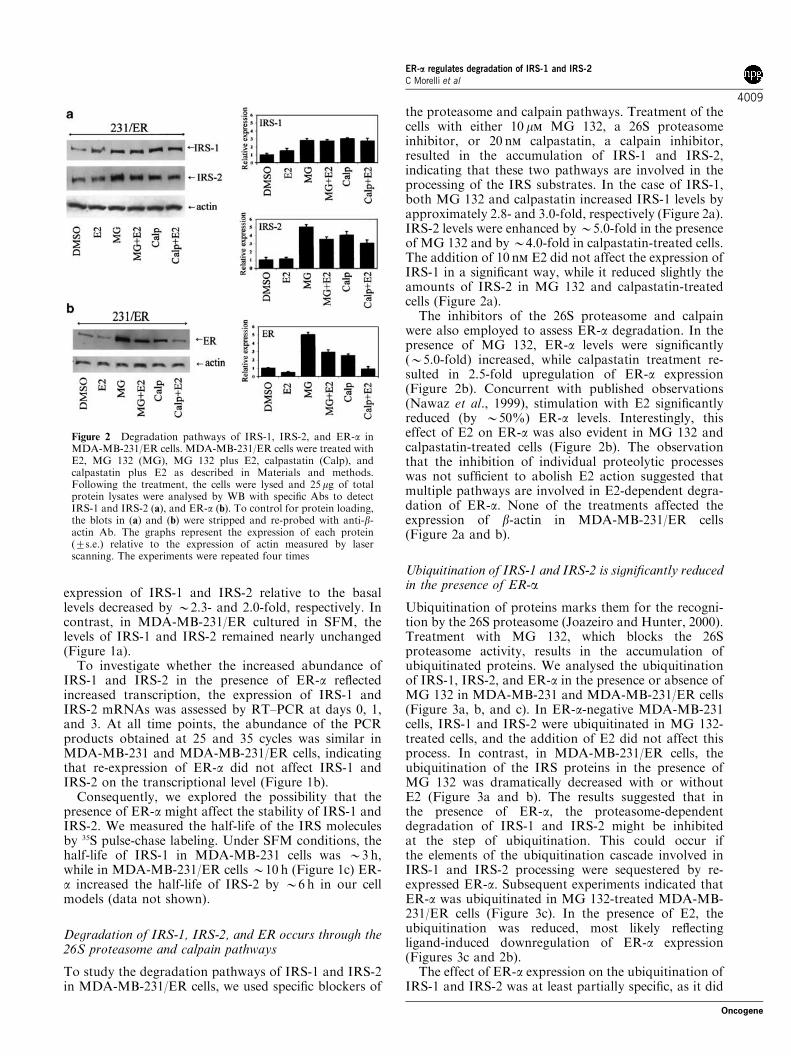
the proteasome and calpain pathways. Treatment of thecells with either 10 mm MG 132, a 26S proteasomeinhibitor, or 20 nm calpastatin, a calpain inhibitor,resulted in the accumulation of IRS-1 and IRS-2,indicating that these two pathways are involved in theprocessing of the IRS substrates. In the case of IRS-1,both MG 132 and calpastatin increased IRS-1 levels byapproximately 2.8- and 3.0-fold, respectively (Figure 2a).IRS-2 levels were enhanced by B5.0-fold in the presenceof MG 132 and byB4.0-fold in calpastatin-treated cells.The addition of 10 nm E2 did not affect the expression ofIRS-1 in a significant way, while it reduced slightly theamounts of IRS-2 in MG 132 and calpastatin-treatedcells (Figure 2a).
The inhibitors of the 26S proteasome and calpainwere also employed to assess ER-a degradation. In thepresence of MG 132, ER-a levels were significantly(B5.0-fold) increased, while calpastatin treatment re-sulted in 2.5-fold upregulation of ER-a expression(Figure 2b). Concurrent with published observations(Nawaz et al., 1999), stimulation with E2 significantlyreduced (by B50%) ER-a levels. Interestingly, thiseffect of E2 on ER-a was also evident in MG 132 andcalpastatin-treated cells (Figure 2b). The observationthat the inhibition of individual proteolytic processeswas not sufficient to abolish E2 action suggested thatmultiple pathways are involved in E2-dependent degra-dation of ER-a. None of the treatments affected theexpression of b-actin in MDA-MB-231/ER cells(Figure 2a and b).
Ubiquitination of IRS-1 and IRS-2 is significantly reducedin the presence of ER-a
Ubiquitination of proteins marks them for the recogni-tion by the 26S proteasome (Joazeiro and Hunter, 2000).Treatment with MG 132, which blocks the 26Sproteasome activity, results in the accumulation ofubiquitinated proteins. We analysed the ubiquitinationof IRS-1, IRS-2, and ER-a in the presence or absence ofMG 132 in MDA-MB-231 and MDA-MB-231/ER cells(Figure 3a, b, and c). In ER-a-negative MDA-MB-231cells, IRS-1 and IRS-2 were ubiquitinated in MG 132-treated cells, and the addition of E2 did not affect thisprocess. In contrast, in MDA-MB-231/ER cells, theubiquitination of the IRS proteins in the presence ofMG 132 was dramatically decreased with or withoutE2 (Figure 3a and b). The results suggested that inthe presence of ER-a, the proteasome-dependentdegradation of IRS-1 and IRS-2 might be inhibitedat the step of ubiquitination. This could occur ifthe elements of the ubiquitination cascade involved inIRS-1 and IRS-2 processing were sequestered by re-expressed ER-a. Subsequent experiments indicated thatER-a was ubiquitinated in MG 132-treated MDA-MB-231/ER cells (Figure 3c). In the presence of E2, theubiquitination was reduced, most likely reflectingligand-induced downregulation of ER-a expression(Figures 3c and 2b).
The effect of ER-a expression on the ubiquitination ofIRS-1 and IRS-2 was at least partially specific, as it did
Figure 2 Degradation pathways of IRS-1, IRS-2, and ER-a inMDA-MB-231/ER cells. MDA-MB-231/ER cells were treated withE2, MG 132 (MG), MG 132 plus E2, calpastatin (Calp), andcalpastatin plus E2 as described in Materials and methods.Following the treatment, the cells were lysed and 25mg of totalprotein lysates were analysed by WB with specific Abs to detectIRS-1 and IRS-2 (a), and ER-a (b). To control for protein loading,the blots in (a) and (b) were stripped and re-probed with anti-b-actin Ab. The graphs represent the expression of each protein(7s.e.) relative to the expression of actin measured by laserscanning. The experiments were repeated four times
ER-a regulates degradation of IRS-1 and IRS-2C Morelli et al
4009
Oncogene

not reduce total protein ubiquitination, or the ubiqui-tination of other proteins that are normally degradedthrough proteasome-dependent pathways (Figure 3d, e,and f). For instance, the ubiquitination of b-catenin andheat shock protein 90 (Hsp 90), both targets of the 26Sproteasome (Aberle et al., 1997; Ashok et al., 2001),were comparable in MDA-MB-231 and MDA-MB-231/ER cells ER (Figure 3e and f). Similarly, ER-a did notaffect ubiquitination of Hsp 70 and IRS-4 (data notshown).
IRS-1 and IRS-2 colocalize and coprecipitate with ER-a
Since ER-a appeared to affect the processing of IRS-1and IRS-2, we studied whether all these proteins residewithin the same cellular compartment. Using confocalmicroscopy and subcellular fractionation, we found thatin untreated MDA-MB-231/ER cells, ER-a is present inthe cytoplasm as well as in the nucleus, while IRS-1 andIRS-2 were mostly cytoplasmatic (Figure 4a and b, andunpublished data). Small amounts of IRS-1 and IRS-2
Figure 3 ER-a expression coincides with reduced ubiquitination of IRS-1 and IRS-2 in MDA-MB-231/ER cells. MDA-MB-231 andMDA-MB-231/ER cells were treated for 1 day with either E2 (10 nm), MG 132 (10mm), E2 plus MG 132, or DMSO (7mm) (controltreatment) and then lysed. IRS-1 (a), IRS-2 (b), or ER-a (c) were immunoprecipitated from 1mg of protein lysates, and their levels andubiquitination were evaluated by WB with specific Abs, as described in Materials and methods. The ubiquitination of total cytoplasmicproteins (d) was studied by WB in 50mg of protein lysates obtained from MDA-MB-231 and MDA-MB-231/ER cells treated with MG132 (10mm) or DMSO (7mm). The same lysates (500 mg) were used to immunoprecipitate b-catenin (beta-cat) (e) and Hsp 90 (f) andanalyse their ubiquitination by WB with appropriate Abs. The data are representative of at least three experiments.
ER-a regulates degradation of IRS-1 and IRS-2C Morelli et al
4010
Oncogene

were present in the nucleus of MDA-MB-231/ER cells,while nuclear IRS-1 and IRS-2 were undetectable inMDA-MB-231 cells (Figure 4b), confirming recentreports that IRS molecules can translocate to thenuclear compartment when coexpressed with nuclearproteins (Lassak et al., 2002; Prisco et al., 2002). InMDA-MB-231/ER cells, a significant fraction (B30%)of IRS-1 colocalized with ER-a in the cytoplasm(Figure 4a). Similar colocalization was found for IRS-2 and ER-a (data not shown).
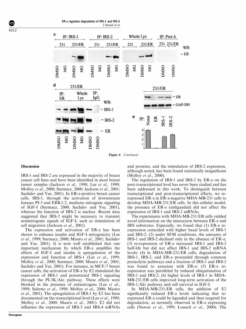
The subsequent experiments demonstrated that inMDA-MB-231/ER cells, ER-a can be found in IRS-1and IRS-2 immunoprecipitates, while it cannot bedetected in IRS-1 or IRS-2 precipitates fromER-a-negative MDA-MB-231 cells (Figure 4c). In acomplementary experiment, we located IRS-1 and IRS-2in ER-a precipitates in MDA-MB-231/ER cells(Figure 4d). Consistent with our previous findings(Figures 2 and 3), the abundance of ER-a : IRS-1 andER-a : IRS-2 complexes was always significantly greaterin MG 132-treated cells, and reflected increasedaccumulation of IRS-1, IRS-2, and ER-a in the presenceof the proteasome 26S inhibitor (Figure 4c and d). Toconfirm that a fraction of ER-a can associate with IRS-1and IRS-2, depletion experiments were performed,where the expression of ER-a was probed in cytosoliclysates before and after IRS-1 or IRS-2 precipitation.We found that depletion of IRS-1 and IRS-2 signifi-cantly (B35 and B25%, respectively) reduced cyto-plasmic abundance of ER-a (Figure 4e).
Re-expression of ER-a coincides with better cell survivalin the presence of IGF-I
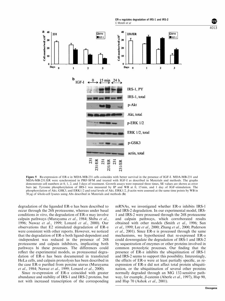
Previous results suggested that re-expression of ER-adecreases IRS-1 and IRS-2 turnover. Here, we addressedthe biologic consequences of this phenomenon. Growthprofiles confirmed our previous observations thatMDA-MB-231 cells die in SFM in the presence orabsence of IGF-I (Bartucci et al., 2001); however,compared with the parental cells, MDA-MB-231/ERcells exhibited significantly better survival in thepresence of IGF-I (Figure 5a). This effect coincidedwith increased IGF-I-dependent tyrosine phosphoryla-tion of IRS-1, increased activity of Akt, and enhancedserine phosphorylation of GSK3 a/b (at 15min and 1day of treatment). The upregulation of the Akt/GSK-3pathway in the presence of ER-a was noted in severalexperiments and was statistically significant. The in-creased activation of Akt was paralleled by down-regulation of ERK1/2 at 1 day (Figure 5b), which was inagreement with previously published observations ob-tained with this and other cellular models (Rommelet al., 1999; Zimmermann and Moelling, 1999; Bartucciet al., 2001). Interestingly, in MDA-MB-231/ER cells,IRS-2 tyrosine phosphorylation was not stimulatedupon IGF-I treatment (data not shown), confirmingfindings obtained in other breast cancer cell models(Jackson et al., 1998).
Figure 4 IRS-1 and IRS-2 colocalize and coprecipitate with ER-a inMDA-MB-231/ER cells. (a) The localization of IRS-1 and ER-a inunstimulatedMDA-MB-231/ER andMDA-MB-231 cells was studiedby confocal microscopy, as described in Materials and methods. Thecaptured images of IRS-1 (red fluorescence), ER-a (green fluores-cence), merged IRS-1 and ER-a (Merge) (yellow fluorescence), andbright field (BF) are shown. The representative images of severalexperiments are shown. (b) The expression of IRS-1, IRS-2, and ER-awas studied in 50mg of cytoplasmic (C) and nuclear (N) proteinlysates obtained from MDA-MB-231 (231) and MDA-MB-231/ER(231/ER) cells. The expression of a nuclear protein, c-Jun, and acytoplasmic enzyme, GAP-DH, was assessed (upon stripping andreprobing of the filters) to control the purity of fractions. The graphrepresents relative abundance of each protein (7SE). (c) MDA-MB-231 and MDA-MB-231/ER cells were treated for 1 day with 10mmMG 132 or 7mm DMSO and then lysed. IRS-1 and IRS-2 wereimmunoprecipitated from 1mg of protein lysates and their associationwith ER-a was probed by WB with anti-ER-a mAb. In parallel, theexpression of ER-a in the cell lines was studied by WB in 50mg ofwhole protein lysates. (d) ER-a was immunoprecipitated from 1mg ofprotein lysates and its association with IRS-1 and IRS-2 wasevaluated by WB with anti-IRS-1 and -IRS-2 Abs. The expressionof IRS-1 and IRS-2 in the cells was detected by WB in 25mg of wholecell lysates. In (c) and (d) the control samples were precipitated withcarrier beads only (protein A agarose and anti-mouse IgG agarose forIRS molecules and ER-a, respectively), with the omission of theprimary Abs, and then processed for WB. (e) The expression of ER-awas studied in 50mg of cytoplasmic lysates before and after IP of IRS-1, IRS-2, or ER-a. The expression of the cytoplasmic protein GAP-DH is shown as a control of loading. The graph represents theexpression of each protein relative to the expression of GAP-DH
ER-a regulates degradation of IRS-1 and IRS-2C Morelli et al
4011
Oncogene
Discussion
IRS-1 and IRS-2 are expressed in the majority of breastcancer cell lines and have been identified in most breasttumor samples (Jackson et al., 1998; Lee et al., 1999;Molloy et al., 2000; Surmacz, 2000; Jackson et al., 2001;Sachdev and Yee, 2001). In ER-a-positive breast cancercells, IRS-1, through the activation of downstreamkinases PI-3 and ERK1/2, mediates mitogenic signalingof IGF-I (Surmacz, 2000; Sachdev and Yee, 2001),whereas the function of IRS-2 is unclear. Recent datasuggested that IRS-2 might be necessary to transmitnonmitogenic signals of IGF-I, such as stimulation ofcell migration (Jackson et al., 2001).
The expression and activation of ER-a has beenshown to enhance insulin and IGF-I mitogenicity (Leeet al., 1999; Surmacz, 2000; Mauro et al., 2001; Sachdevand Yee, 2001). It is now well established that oneimportant mechanism by which ER-a amplifies theeffects of IGF-I and insulin is upregulation of theexpression and function of IRS-1 (Lee et al., 1999;Molloy et al., 2000; Surmacz, 2000; Mauro et al., 2001;Sachdev and Yee, 2001). For instance, in MCF-7 breastcancer cells, the activation of ER-a by E2 stimulated theexpression of IRS-1 and potentiated IRS-1 signalingthrough the PI-3K/Akt pathway. These effects wereblocked in the presence of antiestrogens (Lee et al.,1999; Salerno et al., 1999; Molloy et al., 2000; Mauroet al., 2001). The upregulation of IRS-1 by E2 has beendocumented on the transcriptional level (Lee et al., 1999;Molloy et al., 2000; Mauro et al., 2001). E2 did notinfluence the expression of IRS-3 and IRS-4 mRNAs
and proteins, and the stimulation of IRS-2 expression,although noted, has been found statistically insignificant(Molloy et al., 2000).
The regulation of IRS-1 and IRS-2 by ER-a on thepost-transcriptional level has never been studied and hasbeen addressed in this work. To distinguish betweentranscriptional and post-transcriptional effects, we re-expressed ER-a in ER-a-negative MDA-MB-231 cells todevelop MDA-MB-231/ER cells. In this cellular model,the presence of ER-a (unliganded) did not affect theexpression of IRS-1 and IRS-2 mRNAs.
The experiments with MDA-MB-231/ER cells yieldednovel information on the interaction between ER-a andIRS substrates. Especially, we found that (1) ER-a re-expression coincided with higher basal levels of IRS-1and IRS-2; (2) under SFM conditions, the amounts ofIRS-1 and IRS-2 declined only in the absence of ER-a;(3) re-expression of ER-a increased IRS-1 and IRS-2half-life but did not affect IRS-1 and IRS-2 mRNAlevels; (4) in MDA-MB-231/ER cells, degradation ofIRS-1, IRS-2, and ER-a proceeded through commonproteolytic pathways and a fraction of IRS-1 and IRS-2was found to associate with ER-a; (5) ER-a re-expression was paralleled by reduced ubiquitination ofIRS-1 and IRS-2; (6) higher levels of IRS-1 in MDA-MB-231/ER cells improved long-term activation of theIRS-1/Akt pathway and cell survival in IGF-I.
In MDA-MB-231/ER cells, the addition of E2significantly reduced ER-a levels indicating that re-expressed ER-a could be liganded and then targeted fordegradation, as normally observed in ER-a expressingcells (Nawaz et al., 1999; Lonard et al., 2000). The
Figure 4 Continued.
ER-a regulates degradation of IRS-1 and IRS-2C Morelli et al
4012
Oncogene
degradation of the liganded ER-a has been described tooccur through the 26S proteasome, whereas under basalconditions in vitro, the degradation of ER-a may involvecalpain pathways (Murayama et al., 1984; Shiba et al.,1996; Nawaz et al., 1999; Lonard et al., 2000). Ourobservations that E2 stimulated degradation of ER-awere consistent with other reports. However, we noticedthat the degradation of ER-a both ligand-dependent and-independent was reduced in the presence of 26Sproteasome and calpain inhibitors, implicating bothpathways hi these processes. The differences couldreflect the experimental models, as proteasomal degra-dation of ER-a has been documented in transfectedHeLa cells, and calpain proteolysis has been described inthe case ER-a purified from porcine uterus (Murayamaet al., 1984; Nawaz et al., 1999; Lonard et al., 2000).
Since re-expression of ER-a coincided with greaterabundance and stability of IRS-1 and IRS-2 proteins, butnot with increased transcription of the corresponding
mRNAs, we investigated whether ER-a inhibits IRS-1and IRS-2 degradation. In our experimental model, IRS-1 and IRS-2 were processed through the 26S proteasomeand calpain pathways, which corroborated resultsobtained with other models (Smith et al., 1996; Sunet al., 1999; Lee et al., 2000; Zhang et al., 2000; Pedersonet al., 2001). Since ER-a is processed through the samemechanisms, we hypothesized that re-expressed ER-acould downregulate the degradation of IRS-1 and IRS-2by sequestration of enzymes or other proteins involved incommon proteolytic processes. Our finding that thepresence of ER-a inhibits the ubiquitination of IRS-1and IRS-2 seems to support this possibility. Interestingly,the effects of ER-a were at least partially specific, as re-expression of ER-a did not affect total protein ubiquiti-nation, or the ubiquitination of several other proteinsnormally degraded through an MG 132-sensitive path-way, for example, b-catenin (Aberle et al., 1997), Hsp 90,and Hsp 70 (Ashok et al., 2001).
Figure 5 Re-expression of ER-a in MDA-MB-231 cells coincides with better survival in the presence of IGF-I. MDA-MB-231 andMDA-MB-231/ER were synchronized in PRF-SFM and treated with IGF-I as described in Materials and methods. The graphsdemonstrate cell numbers at 0, 1, 2, and 3 days of treatment. Growth assays were repeated three times, SE values are shown as errorbars (a). Tyrosine phosphorylation of IRS-1 was measured by IP and WB at 0, 15min, and 1 day of IGF-stimulation. Thephosphorylation of Akt, GSK3, and ERK1/2 and total levels of Akt, ERK1/2, b-actin were assessed at the same time points by WB in50mg of whole-cell lysates using Abs described in Materials and methods (b)
ER-a regulates degradation of IRS-1 and IRS-2C Morelli et al
4013
Oncogene

The stabilization of IRS molecules in the presence ofER-a appears to be biologically significant, especially forthe IGF-I phenotype. Our data suggest that MDA-MB-231/ER cells, when compared with MDA-MB-231 cells,are characterized by improved cell survival in IGF-I,enhanced tyrosine phosphorylation of IRS-1, and im-proved activation of the downstream Akt/GSK-3 path-way. We speculate that the restoration of this pathwaywas incomplete because some IGF-I effects that arecharacteristic for normal ER-a-positive cells (e.g., vigor-ous growth in IGF-I or IGF-IR/PI-3K-dependentdegradation of IRS-1) were not observed. The lack ofIRS-2 activation by IGF-I confirmed findings in otherbreast cancer cells (Jackson et al., 1998).
Since the IRS-1, IRS-2, and ER-a are degraded bycommon mechanisms, we analysed, using severaldifferent techniques, whether these proteins can befound in one cellular compartment. By confocalmicroscopy and subcellular protein fractionation, wedetermined that in unstimulated MDA-MB-231/ERcells, IRS-1, IRS-2, as well as a fraction of ER-a arefound in the cytoplasm. The cytoplasmic presence ofthese molecules has been additionally confirmed byimmunocytochemistry (data not shown). Our confocalmicroscopy and immunoprecipitation experiments sug-gested a physical interaction between ER-a and the IRSproteins. The association of ER-a with cytoplasmicsignaling molecules is not unusual; for instance, ER-ahas been shown to interact with the IGF-IR (Kahlertet al., 2000), PI-3K (Simoncini et al., 2000; Sun et al.,2001), and SHC (Song et al., 2002), in all cases,enhancing the signaling potential of these molecules incell stimulated with growth factors (Simoncini et al.,2000; Sun et al., 2001). Our experiments as well as datafrom other laboratories (Simoncini et al., 2000; Sunet al., 2001) suggest that only a fraction of ER-ainteracts with cytoplasmic signaling proteins. Theimmunoprecipitation and microscopic techniques sug-gested that approximately 20–30% of ER-a canassociate with IRS molecules. Our recent experimentsindicate that the ER-a : IRS-1 and ER-a : IRS-2 com-plexes are not unique to MDA-MB-231/ER cells as theyalso exist in MCF-7 cells and other ER-a-positive breastcancer cell lines and are regulated by E2 (Morelli et al.,unpublished data).
In summary, the expression of ER-a could decreaseproteolytic turnover of IRS molecules. We hypothesizethat ER-a : IRS-1/2 complex formation represents oneof the stages of the common degradation processes. Thepost-transcriptional interactions between ER-a andIRS-1 exemplify a new aspect of ER/IGF-IR crosstalkand a possible target in breast cancer therapy.
Materials and methods
Plasmids
The pcDNA3-ER expression plasmid (obtained from DrDiego Sisci, University of Calabria, Italy) encodes the wild-type ER-a under the CMV promoter and contains a neomycinresistance gene.
Cell lines
MDA-MB-231 cells were obtained from ATCC. MDA-MB-231/ER clones were developed by stable transfection of MDA-MB-231 cells with the plasmid pcDNA3-ER using Fugene 6transfection reagent (Roche) (DNA :Fugene 6 ratio was1mg : 3 ml). Transfectants resistant to 2mg/ml G418 (Gibco)were screened for ER-a expression by Western blotting (WB)using an anti-ER-a mouse monoclonal antibody (mAb) F-100.2mg/ml (Santa Cruz). To avoid clonal variation, we used amixed population of five ER-a-expressing clones (referred to asMDA-MB-231/ER cells).
Cell culture
MDA-MB-231 cells were grown in DMEM:F12 containing5% calf serum (CS). MDA-MB-231/ER cells were cultured inDMEM:F12 plus 5% CS plus 500mg/ml G418. In theexperiments requiring E2- and serum-free conditions, the cellswere cultured in phenol red-free serum-free medium (SFM)(Guvakova and Surmacz, 1997; Salerno et al., 1999).
Growth curves
The cells were plated in six-well plates at a concentration of1.5–2.0� 105 cells/plate in normal growth medium. Thefollowing day (day 0), the cells at approximately 50%confluence were shifted to SFM containing 50 ng/ml IGF-I.Cell number was determined at days 0, 1, 2, and 3. A fresh doseof IGF-I was added each day.
RT–PCR
Total cellular RNA was extracted from cells cultured for 0, 1,and 3 days using RNeasy Mini Kit (Qiagen). RNA (1 mg) wasreverse transcribed (RT) and then amplified by PCR to obtainproducts corresponding to cDNA fragments of IRS-1, IRS-2,or b-actin. RT–PCR was performed using the Superscript FirstStrand synthesis system (Gibco) and PCR Core kit (Roche).The following primers were used: IRS-1 upstream primer50-TCCACTGTGACACCAGAATAAT-30 (nt 4979–5000, hu-man IRS-1 cDNA; Araki et al., 1993), IRS-1 downstreamprimer 5-CGCCAACATTGTTCATTCCAA-30 (nt 5721–5741),IRS-2 upstream primer 50-GCTGCTGCTACAGCTCCT-30
(nt 2399–2414, human IRS-2 cDNA; Vassen et al., 1999),IRS-2 downstream primer 50-GGCTCGCCAAAGTCGA-TGT-30 (nt 2762–2780), b-actin upstream primer 50-TGGGAATGGGTCAGAAGGACT-30 (nt 224–244, humanactin cDNA; Powzaniuk et al., 2001), b-actin downstreamprimer 50-TTTCACGGTTGGCCTTAGGGTT-30 (nt 411–433). PCR was performed in a GeneAmp PCR System 9600thermocycler (Perkin-Elmer) using the following conditions:for IRS-1, 1min at 941C, 1min at 501C, 2min at 721C; forIRS-2, 30 s at 941C, 30 s at 481C, 40 s at 721C; for b-actin,1min at 941C, 1min at 501C, 2min at 721C. The amplificationproducts obtained in 15, 25, and 35 cycles were analysed in a1% agarose gel.
Treatment with inhibitors of protein degradation
Confluent cultures (70%) were shifted to SFM and treated for24 h with MG132 and calpastatin in the presence or absence ofE2. MG132 (carbobenzoxy-l-leucyl-l-leucyl-l-leucinal) (Cal-biochem) is an inhibitor of the 26S proteasome and was used atthe concentration of 10 mm. Calpastatin (a 27 residue peptideencoded by exon 1B of human calpastatin) (Calbiochem) is aninhibitor of calpains I and II, and was used at theconcentration 20 nm. E2 (Sigma) was used at the concentration
ER-a regulates degradation of IRS-1 and IRS-2C Morelli et al
4014
Oncogene

10 nm. Control cultures were treated with 7mm DMSO (ICNBiomedicals).
Immunoprecipitation and WB
Cell cultures (70%) were shifted to SFM for 0–72 h and thenlysed. Cytoplasmic protein lysates were obtained with a buffercontaining 50mm HEPES, pH 7.5, 150mm NaCl, 1% TritonX-100, 1.5mm MgCl2, EGTA 10mm pH 7.5, glycerol 10%,inhibitors (2 mm Na3VO4, 1% PMSF, 20mg/ml aprotinin).Following the collection of cytoplasmic proteins, the nucleiwere lysed with the buffer containing 20mm KOH–HEPES pH8, 0.1mm EDTA, 5mm MgCl2, 0.5m NaCl, 20% glycerol, 1%NP-40, and inhibitors (as above). The expression of targetproteins was analysed by WB using 25–50mg of cell lysate and/or by IP in 500 mg–1mg of lysate. The following antibodies(Abs) were used: anti-IRS-1 polyclonal Ab (pAb) (UBI) forWB and IP; anti-IRS-2 pAb (UBI) for WB and IP; anti-ER-amAb (Santa Cruz) for WB and IP; anti-b-catenin mAb(Transduction Laboratories) for WB and IP; anti-b-actinmAb (Sigma) for WB, anti-ubiquitin mAb (Santa Cruz) forWB; anti-Hsp 90mAb (Santa Cruz) for WB and IP; anti-activeAkt (Ser473) pAb (Cell Signaling) for WB; anti-total Akt pAb(Cell Signaling) for WB; anti-phospho-p44/42 MAP kinase(T202/Y204) mAb (Cell Signaling) for WB; anti-active GSK3a/b (Ser21/9) pAb (Cell Signaling) for WB, anti-GAP-DHmAb (Research Diagnostics Inc.) for WB, anti-c-Jun pAb(Santa Cruz) for WB. In all IPs, protein lysates were incubatedin HNTG buffer (20mm HEPES, pH 7.5, 150mm NaCl, 0.1%Triton X-100, 10% glycerol, and 0.2mm Na3VO4) at 41C for4 h with the primary antibodies, and then agarose beadsconjugated with Protein A (Calbiochem) (for IP of pAbs) oranti-mouse IgG (Sigma) (for IP of mouse mAbs) were addedfor another 1 h. In control samples, the primary Abs wereomitted. The immunoprecipitated proteins were washed threetimes with the HNTG buffer, separated by SDS–PAGE(polyacrilamide gel elecrophoresis), and analysed by WB andECL chemiluminescence (Amersham). The intensity of bandsrepresenting relevant proteins was measured by the ScionImage laser densitometry scanning program.
Pulse-chase labeling
To determine the half-life of IRS molecules, we followed themethodology described by Lee et al. (2000) and Zhang et al.(2000) with some modifications. Briefly, 70% cultures wereshifted to methionine- and cysteine-free DMEM (Gibco) for16 h and then metabolically labeled with 35S (100mCi/1ml,Express protein labeling mix, Perkin-Elmer) for 1 h. After that,the labeling medium was replaced with SFM. The cells werelysed at 2, 4, 8, and 12 h to obtain cytoplasmic proteins. Theprotein lysates were precipitated with anti-IRS-1 and IRS-2Abs for 4 h and IPs were separated by SDS–PAGE asdescribed above. Labeled IRS molecules were identified byautoradiography.
Confocal microscopy
Confluent cultures (50%) were fixed in 3% paraformaldehyde,permeabilized with 0.2% Triton X-100, washed 3� with PBS,and incubated for 1 h with primary antibodies (anti-IRS-1 CT(UBI) 2mg/ml and anti-ER-a F-10 (Santa Cruz) 2mg/ml), thenwashed with PBS 3� , and incubated with secondary Abs. Afluoresceine-conjugated donkey anti-mouse IgG (Calbiochem)was used as a secondary Ab for ER-a and a rhodamine-conjugated donkey anti-rabbit IgG (Calbiochem) was used forIRS-1. The cellular localization of IRS-1 and ER-a was studiedwith Bio-Rad MRC 1024 confocal microscope connected to aZeiss Axiovert 135M inverted microscope. The opticalsections were taken at the central plane. The fluorophoreswere imaged separately to ensure no excitation/emissionwavelength overlap.
Acknowledgements
This work was supported by the US Department of DefenseBreast Cancer Research Program Grants DAMD17-99-1-9407and DAMD 17-97-1-7211, and by the American–ItalianCancer Foundation.
References
Aberle H, Bauer A, Stappert J, Kispert A and Kemler R.(1997). EMBO J., 16, 3797–3804.
Aguirre V and White M. (2000). Curr. Opin. Endocrin. Diab.,7, 1–7.
Araki E, Sun XJ, Haag III BL, Chuang LM, Zhang Y, Yang-Feng TL, White MF and Kahn CR. (1993). Diabetes, 42,1041–1054.
Ashok BT, Kim E, Mittelman A and Tiwari RK. (2001). Int. J.Mol. Med., 8, 385–390.
Bartucci M, Morelli C, Mauro L, Ando’ S and Surmacz E.(2001). Cancer Res., 61, 6747–6754.
Bergmann U, Funatomi H, Kornmann M, Beger HG andKorc M. (1996). Biochem. Biophys. Res. Commun., 220,886–890.
Burks DJ and White MF. (2001). Diabetes, 50, S140–S145.Ducluzeau PH, Perretti N, Laville M, Andreelli F, Vega N,Riou JP and Vidal H. (2001). Diabetes, 50, 1134–1142.
Dupont J, Derouet M, Simon J and Taouis M. (1999). J.Endocrinol., 162, 67–76.
Fasshauer M, Klein J, Kriauciunas KM, Ueki K, Benito Mand Kahn CR. (2001). Mol. Cell. Biol., 21, 319–329.
Giovannone B, Scaldaferri ML, Federici M, Porzio O, LauroD, Fusco A, Sbraccia P, Borboni P, Lauro R, and Sesti G.(2000). Diabetes Metab. Res. Rev., 16, 434–441.
Guvakova MA and Surmacz E. (1997). Cancer Res., 57,2606–2610.
Jackson JG, White MF and Yee D. (1998). J. Biol. Chem., 273,9994–10003.
Jackson JG, Zhang X, Yoneda T and Yee D. (2001).Oncogene, 20, 7318–7325.
Joazeiro CAP and Hunter T. (2000). Science, 289,
2061–2062.Kahlert S, Nuedling S, Van Eickels M, Vetter H, Meyer R andGrohe’ C. (2000) J. Biol. Chem., 275, 18447–18453.
Kornmann M, Maruyama H, Bergmann U, Tangvoranunta-kul P, Beger HG, White MF and Korc M. (1998). CancerRes., 58, 4250–4254.
Lassak A, Del Valle L, Peruzzi F, Wang JY, Enam S, Croul S,Khalili K and Reiss K. (2002). J. Biol. Chem., 277,17231–17238.
Lee AV, Gooch JL, Oesterreich S, Guler RL and Yee D.(2000). Mol. Cell. Biol., 20, 1489–1496.
ER-a regulates degradation of IRS-1 and IRS-2C Morelli et al
4015
Oncogene

Lee AV, Jackson JG, Gooch JL, Hilsenbeck SG, Coronado-Heinsohn E, Osborne CK and Yee D. (1999). Mol.Endocrinol., 13, 787–796.
Lonard DM, Nawaz Z, Smith CL and O’Malley BW. (2000).Mol. Cell., 5, 939–948.
Mauro L, Salerno M, Panno ML, Bellizzi D, Sisci D, MigliettaA, Surmacz E and Ando’ S. (2001). Biochem. Biophys. Res.Commun., 288, 685–689.
Molloy CA, May FE and Westley BR (2000). J. Biol. Chem.,275, 12565–12571.
Murayama A, Fukai F and Murachi T. (1984). J. Biochem., 95,1697–1704.
Nawaz Z, Lonard DM, Dennis A, Smith CL and O’MalleyBW. (1999). Proc. Natl. Acad. Sci. USA, 96, 1858–1862.
Pederson TM, Kramer DL and Rondinone CM (2001).Diabetes, 50, 24–31.
Powzaniuk MA, Trotta R, Loza MJ, Harth A, Iozzo RV,Eisenlohr LC, Perussia B and Calabretta B. (2001).J. Immunol., 167, 242–249.
Prisco M, Santini F, Baffa R, Liu M, Drakas R, Wu A andBaserga R. (2002). J. Biol. Chem., 277, 32078–32085.
Rice KM, Turnbow MA and Gamer CW. (1993). Biochem.Biophys. Res. Commun., 190, 961–197.
Richards GR, Klotz DM, Bush MR, Walmer DK andDiAugustine RP. (2001). Endocrinology, 142, 3842–3849.
Rommel C, Clarke BA, Zimmermann S, Nunez L, RossmanR, Reid K, Moelling K, Yancopoulos GD and Glass DJ.(1999). Science, 286, 1738–1740.
Rui L, Fisher TL, Thomas J and White MF. (2001). J. Biol.Chem., 276, 40362–40367.
Sachdev D and Yee D. (2001). Endocr. Relat. Cancer, 8, 197–209.
Sakoda H, Ogihara T, Anai M, Funaki M, Inukai K, KatagiriH, Fukushima Y, Onishi Y, Ono H, Fujishiro M, KikuchiM, Oka Y and Asano T. (2000). Diabetes, 49, 1700–1708.
Salerno M, Sisci D, Mauro L, Guvakova MA, Ando S andSurmacz E. (1999). Int. J. Cancer, 81, 299–304.
Sasaki Y, Zhang XF, Nishiyama M, Avruch J and Wands JR.(1993). J. Biol. Chem., 268, 3805–3808.
Shaw LM. (2001). Mol. Cell. Biol., 21, 5082–5093.Shepherd PR, Withers D and Siddle K. (1998). Biochem. J.,333, 471–490.
Shiba E, Shoushin K, Fujitani M, Kambayashi J, KawamuraI, Tsujimoto S, Shimomura K, Tanji Y, Taguchi T, KimotoY, Izukura M and Takai S. (1996). Anticancer Res., 16,773–778.
Simoncini T, Hafezi-Moghadam A, Brazil DP, Ley K, ChinWW and Liao JK. (2000). Nature, 407, 538–541.
Smith LK, Bradshaw M, Croall DE and Garner CW. (1993).Biochem. Biophys. Res. Commun., 196, 767–772.
Smith LK, Rice KM and Garner CW. (1996). Mol. Cell.Endocrinol., 30, 81–92.
Smith U, Gogg S, Johansson A, Olausson T, Rotter V andSvalstedt B. (2001). FASEB J., 15, 215–220.
Song RX, McPherson RA, Adam L, Bao Y, Shupnik M,Kumar R and Santen RJ. (2002). Mol. Endocrinol., 16,116–127.
Spector SA, Olson ET, Gumbs AA, Friess H, Buchler MWand Seymour NE. (1999). J. Surg. Res., 83, 32–35.
Sun M, Paciga JE, Feldman RI, Yuan Z, Coppola D, Lu YY,Shelley SA, Nicosia SV and Cheng JQ. (2001). Cancer Res.,61, 5985–5991.
Sun XJ, Goldberg JL, Qiao L and Mitchell JJ. (1999).Diabetes, 48, 1359–1364.
Surmacz E. (2000). J. Mammary Gland Biol. Neoplasia, 5, 95–105.
Turnbow MA, Keller SR, Rice KM and Garner CW. (1994).J. Biol. Chem., 269, 2516–2520.
Vassen L, Wegrzyn W and Klein-Hitpass L. (1999). Mol.Endocrinol., 13, 485–494.
Yenush L and White MF. (1997). BioEssays, 19, 491–500.Zhang H, Hoff H and Sell C. (2000). J. Biol. Chem., 275,22558–22562.
Zhang J, Ou J, Bashmakov Y, Horton JD, Brown MSand Goldstein JL. (2001). Proc. Natl. Acad. Sci. USA, 27,3756–3761.
Zimmermann S and Moelling K. (1999). Science, 286, 1741–1744.
ER-a regulates degradation of IRS-1 and IRS-2C Morelli et al
4016
Oncogene
Related Documents