Leading Edge Perspective Establishment and Dysfunction of the Blood-Brain Barrier Zhen Zhao, 1 Amy R. Nelson, 1 Christer Betsholtz, 2 and Berislav V. Zlokovic 1, * 1 Department of Physiology and Biophysics and the Zilkha Neurogenetic Institute, Keck School of Medicine of the University of Southern California, Los Angeles, CA 90089, USA 2 Department of Immunology, Genetics, and Pathology, Rudbeck Laboratory, 75185 Uppsala, Sweden *Correspondence: [email protected] http://dx.doi.org/10.1016/j.cell.2015.10.067 Structural and functional brain connectivity, synaptic activity, and information processing require highly coordinated signal transduction between different cell types within the neurovascular unit and intact blood-brain barrier (BBB) functions. Here, we examine the mechanisms regulating the formation and maintenance of the BBB and functions of BBB-associated cell types. Furthermore, we discuss the growing evidence associating BBB breakdown with the pathogenesis of inherited monogenic neurological disorders and complex multifactorial diseases, including Alzheimer’s dis- ease. Introduction Blood vessels in the brain are organized with surprising preci- sion, supporting the major brain circuits tasked with sensation, memory, and motion (Andreone et al., 2015; Blinder et al., 2013; Zlokovic, 2011). Proper structural and functional brain connectivity, synaptic activity, and information processing all require precise regulation of cerebral blood flow (CBF), oxygen delivery, and energy metabolite supply (Attwell et al., 2010; Iade- cola, 2013). These key central nervous system (CNS) functions are maintained by the highly coordinated activity of multiple cell types within the neurovascular unit (NVU), including vascular cells (endothelial cells, pericytes, smooth muscle cells), glia (as- trocytes, oligodendroglia, microglia), and neurons (Figure 1)(Zlo- kovic, 2011). Within the NVU, the endothelial cells form the blood-brain bar- rier (BBB) that limits entry of potentially neurotoxic plasma com- ponents, blood cells, and pathogens into the brain (Winkler et al., 2011). Importantly, these endothelial cells express multiple sub- strate-specific transport systems that control transport of nutri- ents, energy metabolites, and other essential molecules from blood into the brain and the transport of metabolic waste prod- ucts from the brain’s interstitial fluid (ISF) into the blood. The meningeal lymphatic vessels contain cerebrospinal fluid (CSF) and immune cells and drain into the deep cervical lymph nodes (Aspelund et al., 2015; Louveau et al., 2015). Thus, the BBB serves as a key homeostatic site of the nervous system, con- necting CNS, systemic circulation, and major systems in the body such as respiratory, renal, hepatic, and immune systems. In this Perspective, first we examine the cellular and molecular mechanisms regulating the formation and maintenance of the BBB. We then look into how major NVU cell types contribute to BBB functions and how molecular alterations and aberrant signal transduction within the NVU leads to BBB breakdown that is associated with secondary neuronal injury and neurode- generation. In particular, we discuss the role of NVU and BBB breakdown in the etiology and pathogenesis of inherited mono- genic neurological disorders and complex neurodegenerative disorders such as Alzheimer’s disease (AD). Finally, we discuss key questions in the field for future investigation. BBB Development Different stages of BBB formation are illustrated in Figure 2A. The neural microenvironment provides initial cues for CNS angiogen- esis and induction of the BBB properties (Obermeier et al., 2013). At embryonic day E10 in mice, the angioblasts of the perineural vascular plexus penetrate the neuroectoderm guided by neuro- ectoderm-secreted vascular endothelial growth factor (VEGF), which results in formation of the nascent ‘‘leaky’’ blood vessels (Potente et al., 2011). Wnt ligands secreted by neural cells elicit canonical Wnt signaling in the endothelium by binding to the Frizzled receptors (Wang et al., 2012b) and the co-receptors low-density lipoprotein receptor-related protein (LRP) 5 and 6, which in turn activates b-catenin-dependent pathways (Dane- man et al., 2009; Liebner et al., 2008; Zhou et al., 2014) (Figure 2B). Activation of Wnt/b-catenin signaling leads to induc- tion of genes critical for the BBB formation, such as glucose transporter Glut1 (Stenman et al., 2008) and death receptors DR6 and TROY (Tam et al., 2012). In addition, an orphan G-pro- tein-coupled receptor, Gpr124, acts as a specific co-activator of Wnt/b-catenin signaling at the BBB (Kuhnert et al., 2010; Zhou and Nathans, 2014). The primitive BBB is formed at embryonic day E15 in mice (Ben-Zvi et al., 2014; Daneman et al., 2010), but the exact timing is species dependent and varies regionally. It is debat- able, whether humans and/or other mammals are born with a fully functional BBB (Saunders et al., 2013). Recruitment of peri- cytes to the developing endothelial capillary wall is critical for the formation and maintenance of the BBB (Armulik et al., 2010; Bell et al., 2010; Daneman et al., 2010). While some pathways have been implicated, it remains unclear exactly which signals are involved in the pericyte-mediated induction and regulation of the BBB (Figure 2B). Astrocytes recruited at a later stage further 1064 Cell 163, November 19, 2015 ª2015 Elsevier Inc.

Welcome message from author
This document is posted to help you gain knowledge. Please leave a comment to let me know what you think about it! Share it to your friends and learn new things together.
Transcript
Leading Edge
Perspective
Establishment and Dysfunctionof the Blood-Brain Barrier
Zhen Zhao,1 Amy R. Nelson,1 Christer Betsholtz,2 and Berislav V. Zlokovic1,*1Department of Physiology and Biophysics and the Zilkha Neurogenetic Institute, Keck School of Medicine of the University of Southern
California, Los Angeles, CA 90089, USA2Department of Immunology, Genetics, and Pathology, Rudbeck Laboratory, 75185 Uppsala, Sweden
*Correspondence: [email protected]://dx.doi.org/10.1016/j.cell.2015.10.067
Structural and functional brain connectivity, synaptic activity, and information processing requirehighly coordinated signal transduction between different cell types within the neurovascular unitand intact blood-brain barrier (BBB) functions. Here, we examine the mechanisms regulating theformation and maintenance of the BBB and functions of BBB-associated cell types. Furthermore,we discuss the growing evidence associating BBB breakdown with the pathogenesis of inheritedmonogenic neurological disorders and complex multifactorial diseases, including Alzheimer’s dis-ease.
IntroductionBlood vessels in the brain are organized with surprising preci-
sion, supporting the major brain circuits tasked with sensation,
memory, and motion (Andreone et al., 2015; Blinder et al.,
2013; Zlokovic, 2011). Proper structural and functional brain
connectivity, synaptic activity, and information processing all
require precise regulation of cerebral blood flow (CBF), oxygen
delivery, and energy metabolite supply (Attwell et al., 2010; Iade-
cola, 2013). These key central nervous system (CNS) functions
are maintained by the highly coordinated activity of multiple
cell types within the neurovascular unit (NVU), including vascular
cells (endothelial cells, pericytes, smooth muscle cells), glia (as-
trocytes, oligodendroglia, microglia), and neurons (Figure 1) (Zlo-
kovic, 2011).
Within the NVU, the endothelial cells form the blood-brain bar-
rier (BBB) that limits entry of potentially neurotoxic plasma com-
ponents, blood cells, and pathogens into the brain (Winkler et al.,
2011). Importantly, these endothelial cells express multiple sub-
strate-specific transport systems that control transport of nutri-
ents, energy metabolites, and other essential molecules from
blood into the brain and the transport of metabolic waste prod-
ucts from the brain’s interstitial fluid (ISF) into the blood. The
meningeal lymphatic vessels contain cerebrospinal fluid (CSF)
and immune cells and drain into the deep cervical lymph nodes
(Aspelund et al., 2015; Louveau et al., 2015). Thus, the BBB
serves as a key homeostatic site of the nervous system, con-
necting CNS, systemic circulation, and major systems in the
body such as respiratory, renal, hepatic, and immune systems.
In this Perspective, first we examine the cellular andmolecular
mechanisms regulating the formation and maintenance of the
BBB. We then look into how major NVU cell types contribute
to BBB functions and how molecular alterations and aberrant
signal transduction within the NVU leads to BBB breakdown
that is associated with secondary neuronal injury and neurode-
generation. In particular, we discuss the role of NVU and BBB
breakdown in the etiology and pathogenesis of inherited mono-
1064 Cell 163, November 19, 2015 ª2015 Elsevier Inc.
genic neurological disorders and complex neurodegenerative
disorders such as Alzheimer’s disease (AD). Finally, we discuss
key questions in the field for future investigation.
BBB DevelopmentDifferent stages of BBB formation are illustrated in Figure 2A. The
neural microenvironment provides initial cues for CNS angiogen-
esis and induction of the BBBproperties (Obermeier et al., 2013).
At embryonic day E10 in mice, the angioblasts of the perineural
vascular plexus penetrate the neuroectoderm guided by neuro-
ectoderm-secreted vascular endothelial growth factor (VEGF),
which results in formation of the nascent ‘‘leaky’’ blood vessels
(Potente et al., 2011). Wnt ligands secreted by neural cells elicit
canonical Wnt signaling in the endothelium by binding to the
Frizzled receptors (Wang et al., 2012b) and the co-receptors
low-density lipoprotein receptor-related protein (LRP) 5 and 6,
which in turn activates b-catenin-dependent pathways (Dane-
man et al., 2009; Liebner et al., 2008; Zhou et al., 2014)
(Figure 2B). Activation of Wnt/b-catenin signaling leads to induc-
tion of genes critical for the BBB formation, such as glucose
transporter Glut1 (Stenman et al., 2008) and death receptors
DR6 and TROY (Tam et al., 2012). In addition, an orphan G-pro-
tein-coupled receptor, Gpr124, acts as a specific co-activator of
Wnt/b-catenin signaling at the BBB (Kuhnert et al., 2010; Zhou
and Nathans, 2014).
The primitive BBB is formed at embryonic day E15 in mice
(Ben-Zvi et al., 2014; Daneman et al., 2010), but the exact
timing is species dependent and varies regionally. It is debat-
able, whether humans and/or other mammals are born with a
fully functional BBB (Saunders et al., 2013). Recruitment of peri-
cytes to the developing endothelial capillary wall is critical for the
formation and maintenance of the BBB (Armulik et al., 2010; Bell
et al., 2010; Daneman et al., 2010). While some pathways have
been implicated, it remains unclear exactly which signals are
involved in the pericyte-mediated induction and regulation of
the BBB (Figure 2B). Astrocytes recruited at a later stage further
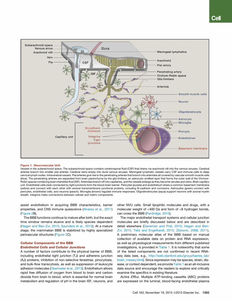
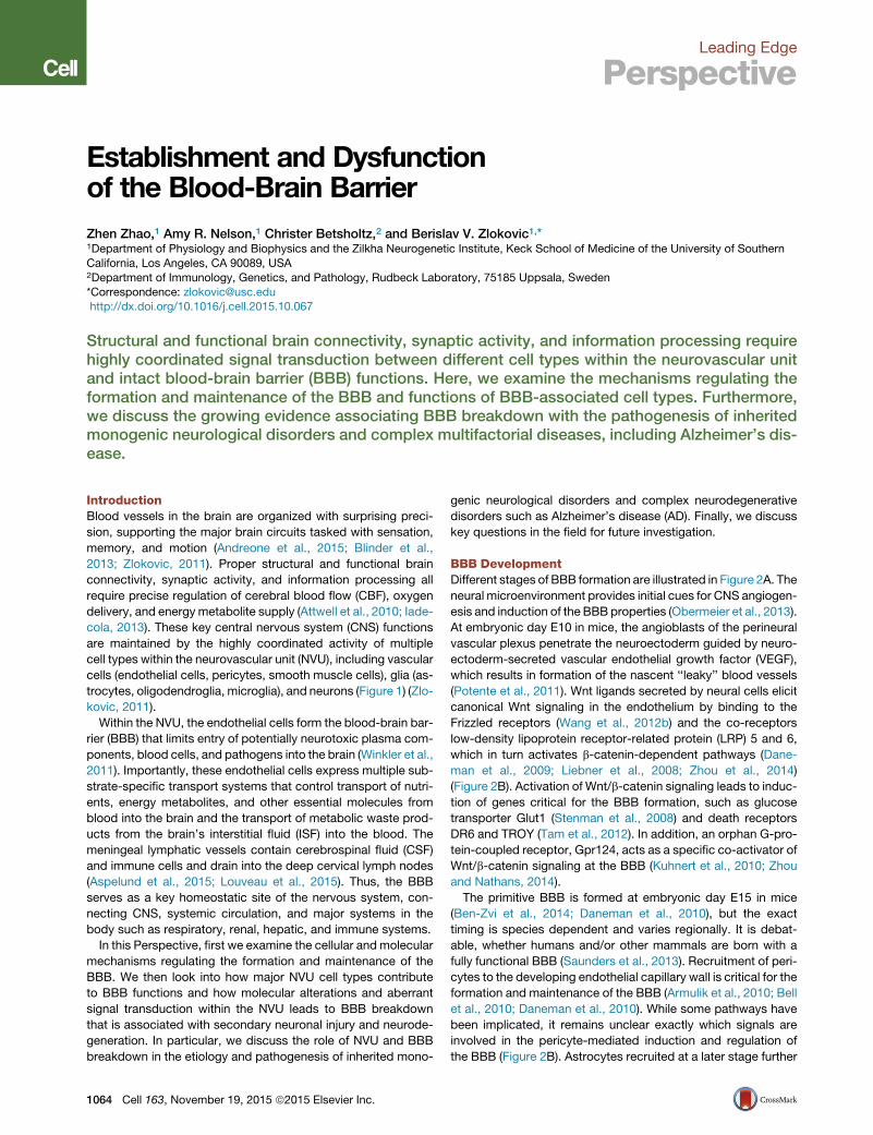
Figure 1. Neurovascular UnitVessels in the subarachnoid space. The subarachnoid space contains cerebrospinal fluid (CSF) that drains via arachnoid villi into the venous sinuses. Cerebralarteries branch into smaller pial arteries. Cerebral veins empty into dural venous sinuses. Meningeal lymphatic vessels carry CSF and immune cells to deepcervical lymph nodes. Intracerebral vessels. Pial arteries give rise to the penetrating arteries that branch into arterioles all covered by vascular smoothmuscle cells(blue). The penetrating arteries are separated from brain parenchyma by the glia limitans, an astrocytic endfeet layer that forms the outer wall of the Virchow-Robin spaces containing brain interstitial fluid (ISF). Arterioles branch off into capillaries, and the vessels enlarge as they become venules and veins.Brain capillaryunit. Endothelial cells (red) connected by tight junctions form the blood-brain barrier. Pericytes (purple) and endothelium share a common basement membrane(yellow) and connect with each other with several transmembrane junctional proteins, including N-cadherin and connexins. Astrocytes (green) connect withpericytes, endothelial cells, and neurons (peach). Microglia (brown) regulate immune responses. Oligodendrocytes (aqua) support neurons with axonal myelinsheath. Integrins make connections between cellular and matrix components.
assist endothelium in acquiring BBB characteristics, barrier
properties, and CNS immune quiescence (Alvarez et al., 2011)
(Figure 2B).
The BBB functions continue tomature after birth, but the exact
time window remains elusive and is likely species dependent
(Hagan and Ben-Zvi, 2015; Saunders et al., 2013). At a mature
stage, the mammalian BBB is stabilized by highly specialized
perivascular structures (Figure 2C).
Cellular Components of the BBBEndothelial Cells and Cellular Junctions
A number of factors contribute to the physical barrier of BBB,
including endothelial tight junction (TJ) and adherens junction
(AJ) proteins, inhibition of non-selective fenestrae, pinocytosis,
and bulk-flow transcytosis, as well as suppression of leukocyte
adhesionmolecules (Obermeier et al., 2013). Endothelium allows
rapid free diffusion of oxygen from blood to brain and carbon
dioxide from brain to blood, which is essential for normal brain
metabolism and regulation of pH in the brain ISF, neurons, and
other NVU cells. Small lipophilic molecules and drugs, with a
molecular weight of <400 Da and form of <8 hydrogen bonds,
can cross the BBB (Pardridge, 2015).
The major endothelial transport systems and cellular junction
molecules are briefly discussed below and are described in
detail elsewhere (Daneman and Prat, 2015; Hagan and Ben-
Zvi, 2015; Tietz and Engelhardt, 2015; Zlokovic, 2008, 2011).
A preliminary molecular atlas of the BBB based on manual
collection of available data on protein and RNA expression,
as well as physiological measurements from different published
investigations, is provided in Table 1. It is noteworthy that some
of the listed components are not confirmed in recent RNA-
seq data (see, e.g., http://web.stanford.edu/group/barres_lab/
brain_rnaseq.html). Since expression may be species, strain, dis-
ease, or context dependent, we provide Table 1 as an all-inclusive
data source and encourage the readers to explore and critically
examine the specifics in existing literature.
Active Efflux. Multiple ATP-binding cassette (ABC) proteins
are expressed on the luminal, blood-facing endothelial plasma
Cell 163, November 19, 2015 ª2015 Elsevier Inc. 1065
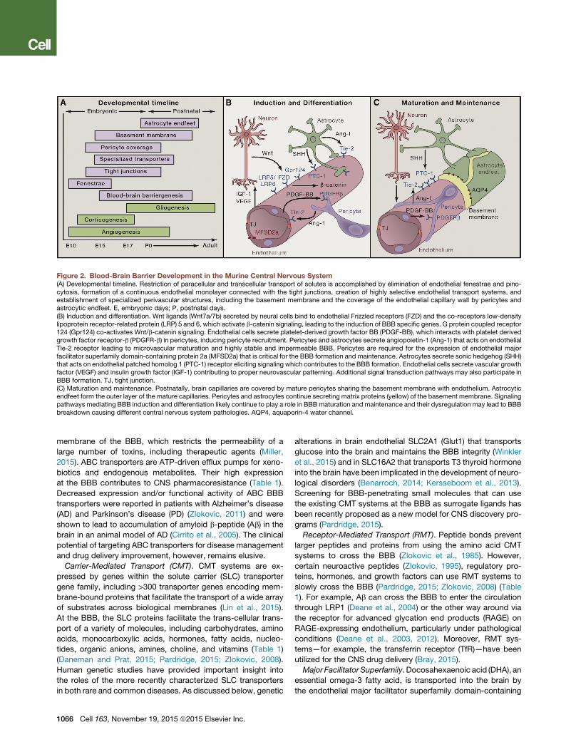
Figure 2. Blood-Brain Barrier Development in the Murine Central Nervous System(A) Developmental timeline. Restriction of paracellular and transcellular transport of solutes is accomplished by elimination of endothelial fenestrae and pino-cytosis, formation of a continuous endothelial monolayer connected with the tight junctions, creation of highly selective endothelial transport systems, andestablishment of specialized perivascular structures, including the basement membrane and the coverage of the endothelial capillary wall by pericytes andastrocytic endfeet. E, embryonic days; P, postnatal days.(B) Induction and differentiation. Wnt ligands (Wnt7a/7b) secreted by neural cells bind to endothelial Frizzled receptors (FZD) and the co-receptors low-densitylipoprotein receptor-related protein (LRP) 5 and 6, which activate b-catenin signaling, leading to the induction of BBB specific genes. G protein coupled receptor124 (Gpr124) co-activates Wnt/b-catenin signaling. Endothelial cells secrete platelet-derived growth factor BB (PDGF-BB), which interacts with platelet derivedgrowth factor receptor-b (PDGFR-b) in pericytes, inducing pericyte recruitment. Pericytes and astrocytes secrete angiopoietin-1 (Ang-1) that acts on endothelialTie-2 receptor leading to microvascular maturation and highly stable and impermeable BBB. Pericytes are required for the expression of endothelial majorfacilitator superfamily domain-containing protein 2a (MFSD2a) that is critical for the BBB formation and maintenance. Astrocytes secrete sonic hedgehog (SHH)that acts on endothelial patched homolog 1 (PTC-1) receptor eliciting signaling which contributes to the BBB formation. Endothelial cells secrete vascular growthfactor (VEGF) and insulin growth factor (IGF-1) contributing to proper neurovascular patterning. Additional signal transduction pathways may also participate inBBB formation. TJ, tight junction.(C) Maturation and maintenance. Postnatally, brain capillaries are covered by mature pericytes sharing the basement membrane with endothelium. Astrocyticendfeet form the outer layer of the mature capillaries. Pericytes and astrocytes continue secreting matrix proteins (yellow) of the basement membrane. Signalingpathways mediating BBB induction and differentiation likely continue to play a role in BBBmaturation and maintenance and their dysregulation may lead to BBBbreakdown causing different central nervous system pathologies. AQP4, aquaporin-4 water channel.
membrane of the BBB, which restricts the permeability of a
large number of toxins, including therapeutic agents (Miller,
2015). ABC transporters are ATP-driven efflux pumps for xeno-
biotics and endogenous metabolites. Their high expression
at the BBB contributes to CNS pharmacoresistance (Table 1).
Decreased expression and/or functional activity of ABC BBB
transporters were reported in patients with Alzheimer’s disease
(AD) and Parkinson’s disease (PD) (Zlokovic, 2011) and were
shown to lead to accumulation of amyloid b-peptide (Ab) in the
brain in an animal model of AD (Cirrito et al., 2005). The clinical
potential of targeting ABC transporters for disease management
and drug delivery improvement, however, remains elusive.
Carrier-Mediated Transport (CMT). CMT systems are ex-
pressed by genes within the solute carrier (SLC) transporter
gene family, including >300 transporter genes encoding mem-
brane-bound proteins that facilitate the transport of a wide array
of substrates across biological membranes (Lin et al., 2015).
At the BBB, the SLC proteins facilitate the trans-cellular trans-
port of a variety of molecules, including carbohydrates, amino
acids, monocarboxylic acids, hormones, fatty acids, nucleo-
tides, organic anions, amines, choline, and vitamins (Table 1)
(Daneman and Prat, 2015; Pardridge, 2015; Zlokovic, 2008).
Human genetic studies have provided important insight into
the roles of the more recently characterized SLC transporters
in both rare and common diseases. As discussed below, genetic
1066 Cell 163, November 19, 2015 ª2015 Elsevier Inc.
alterations in brain endothelial SLC2A1 (Glut1) that transports
glucose into the brain and maintains the BBB integrity (Winkler
et al., 2015) and in SLC16A2 that transports T3 thyroid hormone
into the brain have been implicated in the development of neuro-
logical disorders (Benarroch, 2014; Kersseboom et al., 2013).
Screening for BBB-penetrating small molecules that can use
the existing CMT systems at the BBB as surrogate ligands has
been recently proposed as a new model for CNS discovery pro-
grams (Pardridge, 2015).
Receptor-Mediated Transport (RMT). Peptide bonds prevent
larger peptides and proteins from using the amino acid CMT
systems to cross the BBB (Zlokovic et al., 1985). However,
certain neuroactive peptides (Zlokovic, 1995), regulatory pro-
teins, hormones, and growth factors can use RMT systems to
slowly cross the BBB (Pardridge, 2015; Zlokovic, 2008) (Table
1). For example, Ab can cross the BBB to enter the circulation
through LRP1 (Deane et al., 2004) or the other way around via
the receptor for advanced glycation end products (RAGE) on
RAGE-expressing endothelium, particularly under pathological
conditions (Deane et al., 2003, 2012). Moreover, RMT sys-
tems—for example, the transferrin receptor (TfR)—have been
utilized for the CNS drug delivery (Bray, 2015).
Major Facilitator Superfamily. Docosahexaenoic acid (DHA), an
essential omega-3 fatty acid, is transported into the brain by
the endothelial major facilitator superfamily domain-containing
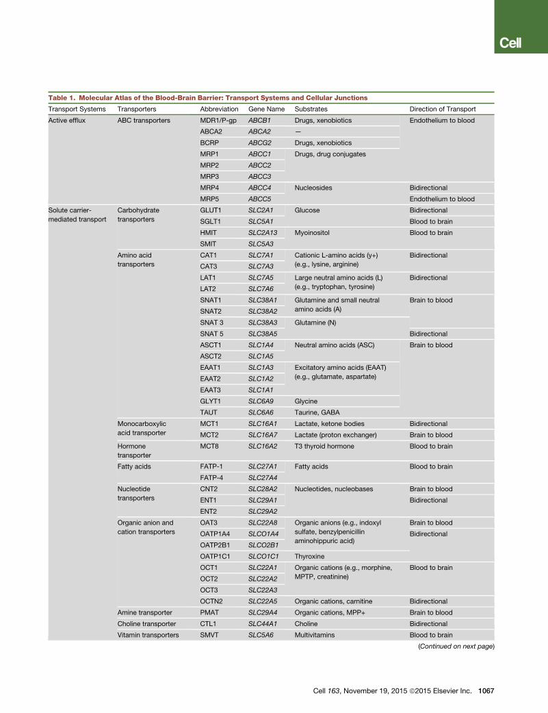
Table 1. Molecular Atlas of the Blood-Brain Barrier: Transport Systems and Cellular Junctions
Transport Systems Transporters Abbreviation Gene Name Substrates Direction of Transport
Active efflux ABC transporters MDR1/P-gp ABCB1 Drugs, xenobiotics Endothelium to blood
ABCA2 ABCA2 —
BCRP ABCG2 Drugs, xenobiotics
MRP1 ABCC1 Drugs, drug conjugates
MRP2 ABCC2
MRP3 ABCC3
MRP4 ABCC4 Nucleosides Bidirectional
MRP5 ABCC5 Endothelium to blood
Solute carrier-
mediated transport
Carbohydrate
transporters
GLUT1 SLC2A1 Glucose Bidirectional
SGLT1 SLC5A1 Blood to brain
HMIT SLC2A13 Myoinositol Blood to brain
SMIT SLC5A3
Amino acid
transporters
CAT1 SLC7A1 Cationic L-amino acids (y+)
(e.g., lysine, arginine)
Bidirectional
CAT3 SLC7A3
LAT1 SLC7A5 Large neutral amino acids (L)
(e.g., tryptophan, tyrosine)
Bidirectional
LAT2 SLC7A6
SNAT1 SLC38A1 Glutamine and small neutral
amino acids (A)
Brain to blood
SNAT2 SLC38A2
SNAT 3 SLC38A3 Glutamine (N)
SNAT 5 SLC38A5 Bidirectional
ASCT1 SLC1A4 Neutral amino acids (ASC) Brain to blood
ASCT2 SLC1A5
EAAT1 SLC1A3 Excitatory amino acids (EAAT)
(e.g., glutamate, aspartate)EAAT2 SLC1A2
EAAT3 SLC1A1
GLYT1 SLC6A9 Glycine
TAUT SLC6A6 Taurine, GABA
Monocarboxylic
acid transporter
MCT1 SLC16A1 Lactate, ketone bodies Bidirectional
MCT2 SLC16A7 Lactate (proton exchanger) Brain to blood
Hormone
transporter
MCT8 SLC16A2 T3 thyroid hormone Blood to brain
Fatty acids FATP-1 SLC27A1 Fatty acids Blood to brain
FATP-4 SLC27A4
Nucleotide
transporters
CNT2 SLC28A2 Nucleotides, nucleobases Brain to blood
ENT1 SLC29A1 Bidirectional
ENT2 SLC29A2
Organic anion and
cation transporters
OAT3 SLC22A8 Organic anions (e.g., indoxyl
sulfate, benzylpenicillin
aminohippuric acid)
Brain to blood
OATP1A4 SLCO1A4 Bidirectional
OATP2B1 SLCO2B1
OATP1C1 SLCO1C1 Thyroxine
OCT1 SLC22A1 Organic cations (e.g., morphine,
MPTP, creatinine)
Blood to brain
OCT2 SLC22A2
OCT3 SLC22A3
OCTN2 SLC22A5 Organic cations, carnitine Bidirectional
Amine transporter PMAT SLC29A4 Organic cations, MPP+ Brain to blood
Choline transporter CTL1 SLC44A1 Choline Bidirectional
Vitamin transporters SMVT SLC5A6 Multivitamins Blood to brain
(Continued on next page)
Cell 163, November 19, 2015 ª2015 Elsevier Inc. 1067
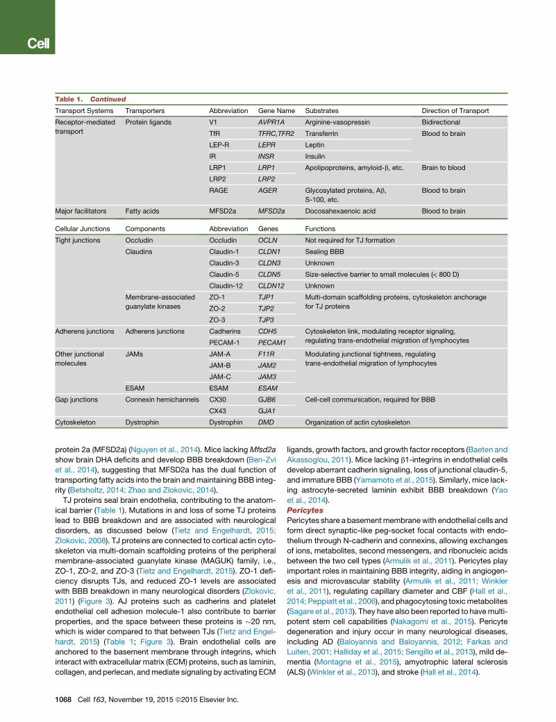
Table 1. Continued
Transport Systems Transporters Abbreviation Gene Name Substrates Direction of Transport
Receptor-mediated
transport
Protein ligands V1 AVPR1A Arginine-vasopressin Bidirectional
TfR TFRC,TFR2 Transferrin Blood to brain
LEP-R LEPR Leptin
IR INSR Insulin
LRP1 LRP1 Apolipoproteins, amyloid-b, etc. Brain to blood
LRP2 LRP2
RAGE AGER Glycosylated proteins, Ab,
S-100, etc.
Blood to brain
Major facilitators Fatty acids MFSD2a MFSD2a Docosahexaenoic acid Blood to brain
Cellular Junctions Components Abbreviation Genes Functions
Tight junctions Occludin Occludin OCLN Not required for TJ formation
Claudins Claudin-1 CLDN1 Sealing BBB
Claudin-3 CLDN3 Unknown
Claudin-5 CLDN5 Size-selective barrier to small molecules (< 800 D)
Claudin-12 CLDN12 Unknown
Membrane-associated
guanylate kinases
ZO-1 TJP1 Multi-domain scaffolding proteins, cytoskeleton anchorage
for TJ proteinsZO-2 TJP2
ZO-3 TJP3
Adherens junctions Adherens junctions Cadherins CDH5 Cytoskeleton link, modulating receptor signaling,
regulating trans-endothelial migration of lymphocytesPECAM-1 PECAM1
Other junctional
molecules
JAMs JAM-A F11R Modulating junctional tightness, regulating
trans-endothelial migration of lymphocytesJAM-B JAM2
JAM-C JAM3
ESAM ESAM ESAM
Gap junctions Connexin hemichannels CX30 GJB6 Cell-cell communication, required for BBB
CX43 GJA1
Cytoskeleton Dystrophin Dystrophin DMD Organization of actin cytoskeleton
protein 2a (MFSD2a) (Nguyen et al., 2014). Mice lacking Mfsd2a
show brain DHA deficits and develop BBB breakdown (Ben-Zvi
et al., 2014), suggesting that MFSD2a has the dual function of
transporting fatty acids into the brain andmaintaining BBB integ-
rity (Betsholtz, 2014; Zhao and Zlokovic, 2014).
TJ proteins seal brain endothelia, contributing to the anatom-
ical barrier (Table 1). Mutations in and loss of some TJ proteins
lead to BBB breakdown and are associated with neurological
disorders, as discussed below (Tietz and Engelhardt, 2015;
Zlokovic, 2008). TJ proteins are connected to cortical actin cyto-
skeleton via multi-domain scaffolding proteins of the peripheral
membrane-associated guanylate kinase (MAGUK) family, i.e.,
ZO-1, ZO-2, and ZO-3 (Tietz and Engelhardt, 2015). ZO-1 defi-
ciency disrupts TJs, and reduced ZO-1 levels are associated
with BBB breakdown in many neurological disorders (Zlokovic,
2011) (Figure 3). AJ proteins such as cadherins and platelet
endothelial cell adhesion molecule-1 also contribute to barrier
properties, and the space between these proteins is �20 nm,
which is wider compared to that between TJs (Tietz and Engel-
hardt, 2015) (Table 1; Figure 3). Brain endothelial cells are
anchored to the basement membrane through integrins, which
interact with extracellular matrix (ECM) proteins, such as laminin,
collagen, and perlecan, andmediate signaling by activating ECM
1068 Cell 163, November 19, 2015 ª2015 Elsevier Inc.
ligands, growth factors, and growth factor receptors (Baeten and
Akassoglou, 2011). Mice lacking b1-integrins in endothelial cells
develop aberrant cadherin signaling, loss of junctional claudin-5,
and immature BBB (Yamamoto et al., 2015). Similarly, mice lack-
ing astrocyte-secreted laminin exhibit BBB breakdown (Yao
et al., 2014).
Pericytes
Pericytes share a basementmembranewith endothelial cells and
form direct synaptic-like peg-socket focal contacts with endo-
thelium through N-cadherin and connexins, allowing exchanges
of ions, metabolites, second messengers, and ribonucleic acids
between the two cell types (Armulik et al., 2011). Pericytes play
important roles in maintaining BBB integrity, aiding in angiogen-
esis and microvascular stability (Armulik et al., 2011; Winkler
et al., 2011), regulating capillary diameter and CBF (Hall et al.,
2014; Peppiatt et al., 2006), and phagocytosing toxicmetabolites
(Sagare et al., 2013). They have also been reported to havemulti-
potent stem cell capabilities (Nakagomi et al., 2015). Pericyte
degeneration and injury occur in many neurological diseases,
including AD (Baloyannis and Baloyannis, 2012; Farkas and
Luiten, 2001; Halliday et al., 2015; Sengillo et al., 2013), mild de-
mentia (Montagne et al., 2015), amyotrophic lateral sclerosis
(ALS) (Winkler et al., 2013), and stroke (Hall et al., 2014).
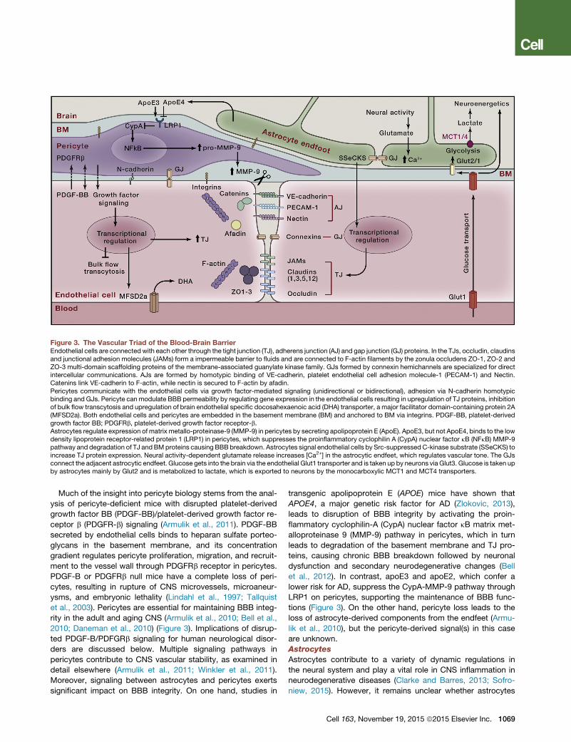
Figure 3. The Vascular Triad of the Blood-Brain BarrierEndothelial cells are connected with each other through the tight junction (TJ), adherens junction (AJ) and gap junction (GJ) proteins. In the TJs, occludin, claudinsand junctional adhesion molecules (JAMs) form a impermeable barrier to fluids and are connected to F-actin filaments by the zonula occludens ZO-1, ZO-2 andZO-3 multi-domain scaffolding proteins of the membrane-associated guanylate kinase family. GJs formed by connexin hemichannels are specialized for directintercellular communications. AJs are formed by homotypic binding of VE-cadherin, platelet endothelial cell adhesion molecule-1 (PECAM-1) and Nectin.Catenins link VE-cadherin to F-actin, while nectin is secured to F-actin by afadin.Pericytes communicate with the endothelial cells via growth factor-mediated signaling (unidirectional or bidirectional), adhesion via N-cadherin homotypicbinding and GJs. Pericyte can modulate BBB permeability by regulating gene expression in the endothelial cells resulting in upregulation of TJ proteins, inhibitionof bulk flow transcytosis and upregulation of brain endothelial specific docosahexaenoic acid (DHA) transporter, a major facilitator domain-containing protein 2A(MFSD2a). Both endothelial cells and pericytes are embedded in the basement membrane (BM) and anchored to BM via integrins. PDGF-BB, platelet-derivedgrowth factor BB; PDGFRb, platelet-derived growth factor receptor-b.Astrocytes regulate expression of matrix metallo-proteinase-9 (MMP-9) in pericytes by secreting apolipoprotein E (ApoE). ApoE3, but not ApoE4, binds to the lowdensity lipoprotein receptor-related protein 1 (LRP1) in pericytes, which suppresses the proinflammatory cyclophilin A (CypA) nuclear factor kB (NFkB) MMP-9pathway and degradation of TJ andBMproteins causing BBB breakdown. Astrocytes signal endothelial cells by Src-suppressed C-kinase substrate (SSeCKS) toincrease TJ protein expression. Neural activity-dependent glutamate release increases [Ca2+] in the astrocytic endfeet, which regulates vascular tone. The GJsconnect the adjacent astrocytic endfeet. Glucose gets into the brain via the endothelial Glut1 transporter and is taken up by neurons via Glut3. Glucose is taken upby astrocytes mainly by Glut2 and is metabolized to lactate, which is exported to neurons by the monocarboxylic MCT1 and MCT4 transporters.
Much of the insight into pericyte biology stems from the anal-
ysis of pericyte-deficient mice with disrupted platelet-derived
growth factor BB (PDGF-BB)/platelet-derived growth factor re-
ceptor b (PDGFR-b) signaling (Armulik et al., 2011). PDGF-BB
secreted by endothelial cells binds to heparan sulfate porteo-
glycans in the basement membrane, and its concentration
gradient regulates pericyte proliferation, migration, and recruit-
ment to the vessel wall through PDGFRb receptor in pericytes.
PDGF-B or PDGFRb null mice have a complete loss of peri-
cytes, resulting in rupture of CNS microvessels, microaneur-
ysms, and embryonic lethality (Lindahl et al., 1997; Tallquist
et al., 2003). Pericytes are essential for maintaining BBB integ-
rity in the adult and aging CNS (Armulik et al., 2010; Bell et al.,
2010; Daneman et al., 2010) (Figure 3). Implications of disrup-
ted PDGF-B/PDFGRb signaling for human neurological disor-
ders are discussed below. Multiple signaling pathways in
pericytes contribute to CNS vascular stability, as examined in
detail elsewhere (Armulik et al., 2011; Winkler et al., 2011).
Moreover, signaling between astrocytes and pericytes exerts
significant impact on BBB integrity. On one hand, studies in
transgenic apolipoprotein E (APOE) mice have shown that
APOE4, a major genetic risk factor for AD (Zlokovic, 2013),
leads to disruption of BBB integrity by activating the proin-
flammatory cyclophilin-A (CypA) nuclear factor kB matrix met-
alloproteinase 9 (MMP-9) pathway in pericytes, which in turn
leads to degradation of the basement membrane and TJ pro-
teins, causing chronic BBB breakdown followed by neuronal
dysfunction and secondary neurodegenerative changes (Bell
et al., 2012). In contrast, apoE3 and apoE2, which confer a
lower risk for AD, suppress the CypA-MMP-9 pathway through
LRP1 on pericytes, supporting the maintenance of BBB func-
tions (Figure 3). On the other hand, pericyte loss leads to the
loss of astrocyte-derived components from the endfeet (Armu-
lik et al., 2010), but the pericyte-derived signal(s) in this case
are unknown.
Astrocytes
Astrocytes contribute to a variety of dynamic regulations in
the neural system and play a vital role in CNS inflammation in
neurodegenerative diseases (Clarke and Barres, 2013; Sofro-
niew, 2015). However, it remains unclear whether astrocytes
Cell 163, November 19, 2015 ª2015 Elsevier Inc. 1069
are essential for BBB maintenance. The glial limitans ensheath-
ing the penetrating arterial blood vessels and the outer layer of
mature capillaries are formed by astrocytic endfeet (Figure 3).
Yet, genetic lineage tracing and ablation studies showed
that regional ablation had no effect on BBB permeability (Tsai
et al., 2012). In contrast, others have shown that Src-sup-
pressed C-kinase substrate (SSeCKS) in astrocyte progenitors
regulates angiogenesis and formation of TJs at the BBB by
modulating VEGF and Ang-1 expression (Lee et al., 2003).
Future studies should provide more definitive answers as to
whether astrocytes play a role in BBB maintenance in the adult
and aging brain.
BBB and Monogenic Neurological DisordersSeveral genetic diseases appear to originate in individual cell
types of the NVU and link to specific roles in BBB develop-
ment, function, and regulation. Such diseases, while rare, offer
insights into causal pathogenic links and chains of events.
Below, we briefly discuss some of these diseases, the relevant
cell types (Table 2), and the potential pathogenic role of BBB
dysfunction.
Endothelial Cells
Several inherited CNS diseases are caused by mutations in
genes that play pivotal roles in endothelial cells or in the endothe-
lium-derived ECM. These genes encode proteins that are either
structural components or regulators of the endothelial cell-cell
junctions, the vascular basement membrane, or transporters
critically involved in BBB maintenance. For instance, mutations
in the genes encoding the TJ proteins occludin (O’Driscoll
et al., 2010) and junctional adhesionmolecule C (JAM-C) (Wood-
fin et al., 2011) lead to severe problems in brain growth, hemor-
rhage, and calcification (Table 2). These pathologies may result
from uncontrolled leakage of solutes and plasma proteins across
the endothelial junctions and/or neuroinflammatory changes
due to increased trans-endothelial migration of leukocytes, a
process inhibited by JAM-C. Another example is familial cerebral
cavernous malformations (CCM), occurring in hereditary or spo-
radic forms and together affecting �0.5% of the population.
CCM is caused by mutations in three genes, CCM1–3, leading
to similar thin-walled, leaky vascular lesions of venous origin
(Fischer et al., 2013). The CCM proteins likely act together in
a complex that maintains endothelial junctional organization
and polarization and inhibits endothelial-to-mesenchymal transi-
tion (Maddaluno et al., 2013). Collagen COL4A1 and COL4A2 are
abundant in all basement membranes and are expressed by
many cell types, including vascular endothelial cells. Mutations
in these genes are associated with a diverse range of problems
in several organs, including the brain, where they are associated
with cerebral hemorrhage and small vessel disease (Gould et al.,
2006). Work in animal models with Col4a1 deficiency suggests
that increased vessel fragility could make the animal susceptible
to hemorrhage either upon mild trauma or due to the anatomy of
the vessel that is particularly sensitive to increased hemody-
namic stress (Kuo et al., 2012).
Among transporters mutated in brain disorders, two strik-
ing examples of BBB endothelial proteins are GLUT1 and
MFSD2a. GLUT1, the major glucose transporter at the BBB,
is mutated and functionally inactivated in human GLUT1 defi-
1070 Cell 163, November 19, 2015 ª2015 Elsevier Inc.
ciency syndrome, a disease associated with early-onset sei-
zures and microcephaly (Wang et al., 2000). This is consistent
with the importance of sufficient GLUT1 levels and glucose
transport across the BBB for brain function and its role in
maintaining the BBB integrity (Winkler et al., 2015). Similar to
GLUT1, MFSD2a is highly expressed on brain endothelial
cells. It transports lipids in the form of lysophoshatidylcholine
coupled to certain long fatty acyl chains and is critical for the
maintenance of the BBB integrity (Ben-Zvi et al., 2014; Nguyen
et al., 2014). Microcephaly syndrome was recently shown to
be caused by inactivating mutations in MFSD2A, the severity
of the syndrome correlating with the degree of functional inac-
tivation of the MFSD2A protein (Alakbarzade et al., 2015; Gue-
mez-Gamboa et al., 2015). These studies provide insight into
the cause of a rare disease but, perhaps more importantly,
are an illustration to how basic physiological knowledge may
come from human and mouse genetics and offer a funda-
mental insight into the mechanisms by which brain transports
lipids across the BBB (Betsholtz, 2015) and maintains BBB
integrity (Betsholtz, 2014; Zhao and Zlokovic, 2014). A third
example of a brain disease associated with a BBB transporter
is Allan-Herndon-Dudley syndrome, a psychomotor retarda-
tion syndrome caused by inactivating mutations in the triiodo-
thyronin (T3) transporter SLC16A2 (MCT8) (Dumitrescu et al.,
2004; Friesema et al., 2004). It is thought that the severe intel-
lectual disability and movement problems observed in these
patients are due to deficient transport of T3 from the blood
to the brain, resulting in impairment of neuronal development
and function. Indeed, different degrees of SLC16A2 inactiva-
tion correlate with the phenotypic consequences in patients
(Capri et al., 2013).
Vascular Mural Cells
CADASIL (cerebral autosomal dominant arteriopathy with
subcortical infarcts and leukoencephalopathy) is a relatively
common (2–4/100,000 individuals) autosomal-dominant stroke
syndrome caused by mutations in NOTCH3, a gene known
to be specifically expressed in vascular mural cells (Chabriat
et al., 2009). Although, the precise pathogenic mechanisms of
CADASIL remain unresolved, recent studies of Notch 3 null
mice demonstrated focal disruption of the BBB with tracer
leakage and perivascular fibrin deposits in the CNS (Henshall
et al., 2015). Primary familial brain calcification (PFBC, a.k.a.,
idiopathic basal ganglia calcification [IBGC] or Fahr’s disease)
is characterized by early-onset microvascular calcification
occurring in certain deep brain regions, most notably the
basal ganglia. Disease symptoms include motoric and cognitive
problems suggestive of significant neuronal dysfunction. The
recent description of loss-of-function mutations in PDGFB and
PDGFRB genes in PFBC (Keller et al., 2013; Nicolas et al.,
2013) suggests a role for pericytes in this disease. In different
mouse models based on mutations in Pdgfb that led to variable
levels of defect in PDGF-B/PDGFRb signaling, a correlation
was noted among the extent of pericyte loss, BBB deficiency,
and brain calcification (Keller et al., 2013). This is suggestive of
a role for BBB dysfunction in PFBC, possibly involving changes
in phosphate transport, since mutations in the phosphate trans-
porters SLC20A2 and XPR1 also cause PFBC (Legati et al., 2015;
Wang et al., 2012a).
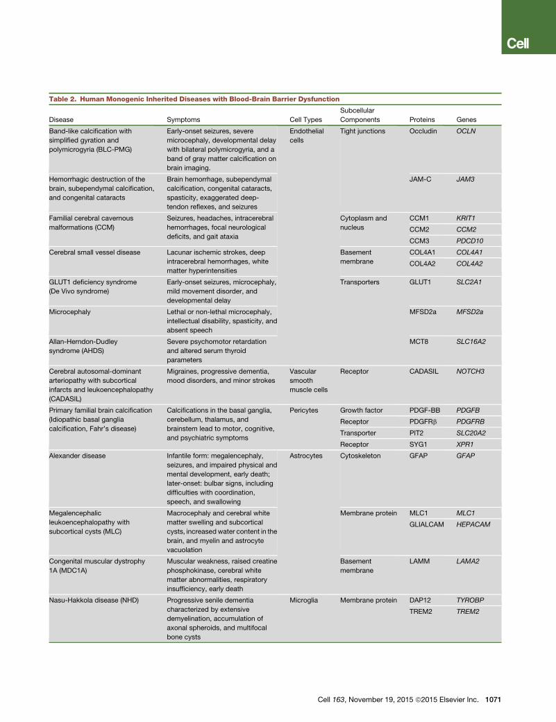
Table 2. Human Monogenic Inherited Diseases with Blood-Brain Barrier Dysfunction
Disease Symptoms Cell Types
Subcellular
Components Proteins Genes
Band-like calcification with
simplified gyration and
polymicrogyria (BLC-PMG)
Early-onset seizures, severe
microcephaly, developmental delay
with bilateral polymicrogyria, and a
band of gray matter calcification on
brain imaging.
Endothelial
cells
Tight junctions Occludin OCLN
Hemorrhagic destruction of the
brain, subependymal calcification,
and congenital cataracts
Brain hemorrhage, subependymal
calcification, congenital cataracts,
spasticity, exaggerated deep-
tendon reflexes, and seizures
JAM-C JAM3
Familial cerebral cavernous
malformations (CCM)
Seizures, headaches, intracerebral
hemorrhages, focal neurological
deficits, and gait ataxia
Cytoplasm and
nucleus
CCM1 KRIT1
CCM2 CCM2
CCM3 PDCD10
Cerebral small vessel disease Lacunar ischemic strokes, deep
intracerebral hemorrhages, white
matter hyperintensities
Basement
membrane
COL4A1 COL4A1
COL4A2 COL4A2
GLUT1 deficiency syndrome
(De Vivo syndrome)
Early-onset seizures, microcephaly,
mild movement disorder, and
developmental delay
Transporters GLUT1 SLC2A1
Microcephaly Lethal or non-lethal microcephaly,
intellectual disability, spasticity, and
absent speech
MFSD2a MFSD2a
Allan-Herndon-Dudley
syndrome (AHDS)
Severe psychomotor retardation
and altered serum thyroid
parameters
MCT8 SLC16A2
Cerebral autosomal-dominant
arteriopathy with subcortical
infarcts and leukoencephalopathy
(CADASIL)
Migraines, progressive dementia,
mood disorders, and minor strokes
Vascular
smooth
muscle cells
Receptor CADASIL NOTCH3
Primary familial brain calcification
(Idiopathic basal ganglia
calcification, Fahr’s disease)
Calcifications in the basal ganglia,
cerebellum, thalamus, and
brainstem lead to motor, cognitive,
and psychiatric symptoms
Pericytes Growth factor PDGF-BB PDGFB
Receptor PDGFRb PDGFRB
Transporter PIT2 SLC20A2
Receptor SYG1 XPR1
Alexander disease Infantile form: megalencephaly,
seizures, and impaired physical and
mental development, early death;
later-onset: bulbar signs, including
difficulties with coordination,
speech, and swallowing
Astrocytes Cytoskeleton GFAP GFAP
Megalencephalic
leukoencephalopathy with
subcortical cysts (MLC)
Macrocephaly and cerebral white
matter swelling and subcortical
cysts, increasedwater content in the
brain, and myelin and astrocyte
vacuolation
Membrane protein MLC1 MLC1
GLIALCAM HEPACAM
Congenital muscular dystrophy
1A (MDC1A)
Muscular weakness, raised creatine
phosphokinase, cerebral white
matter abnormalities, respiratory
insufficiency, early death
Basement
membrane
LAMM LAMA2
Nasu-Hakkola disease (NHD) Progressive senile dementia
characterized by extensive
demyelination, accumulation of
axonal spheroids, and multifocal
bone cysts
Microglia Membrane protein DAP12 TYROBP
TREM2 TREM2
Cell 163, November 19, 2015 ª2015 Elsevier Inc. 1071
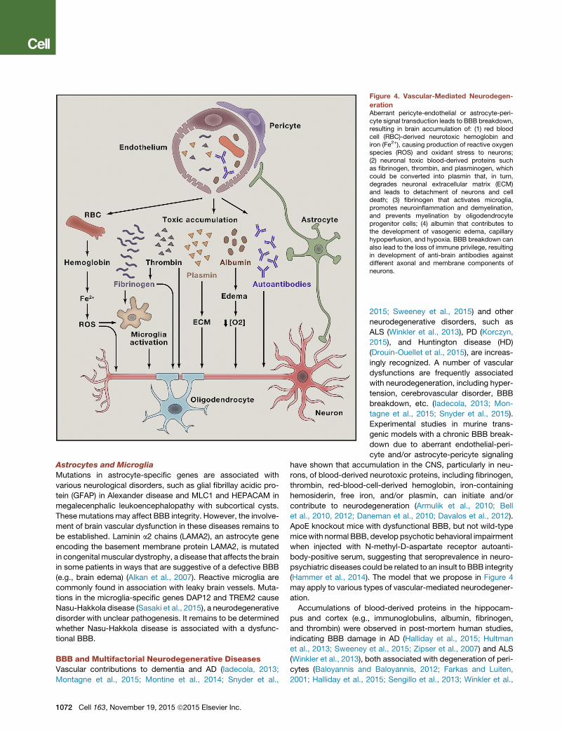
Figure 4. Vascular-Mediated Neurodegen-
erationAberrant pericyte-endothelial or astrocyte-peri-cyte signal transduction leads to BBB breakdown,resulting in brain accumulation of: (1) red bloodcell (RBC)-derived neurotoxic hemoglobin andiron (Fe2+), causing production of reactive oxygenspecies (ROS) and oxidant stress to neurons;(2) neuronal toxic blood-derived proteins suchas fibrinogen, thrombin, and plasminogen, whichcould be converted into plasmin that, in turn,degrades neuronal extracellular matrix (ECM)and leads to detachment of neurons and celldeath; (3) fibrinogen that activates microglia,promotes neuroinflammation and demyelination,and prevents myelination by oligodendrocyteprogenitor cells; (4) albumin that contributes tothe development of vasogenic edema, capillaryhypoperfusion, and hypoxia. BBB breakdown canalso lead to the loss of immune privilege, resultingin development of anti-brain antibodies againstdifferent axonal and membrane components ofneurons.
Astrocytes and Microglia
Mutations in astrocyte-specific genes are associated with
various neurological disorders, such as glial fibrillay acidic pro-
tein (GFAP) in Alexander disease and MLC1 and HEPACAM in
megalecenphalic leukoencephalopathy with subcortical cysts.
These mutations may affect BBB integrity. However, the involve-
ment of brain vascular dysfunction in these diseases remains to
be established. Laminin a2 chains (LAMA2), an astrocyte gene
encoding the basement membrane protein LAMA2, is mutated
in congenital muscular dystrophy, a disease that affects the brain
in some patients in ways that are suggestive of a defective BBB
(e.g., brain edema) (Alkan et al., 2007). Reactive microglia are
commonly found in association with leaky brain vessels. Muta-
tions in the microglia-specific genes DAP12 and TREM2 cause
Nasu-Hakkola disease (Sasaki et al., 2015), a neurodegenerative
disorder with unclear pathogenesis. It remains to be determined
whether Nasu-Hakkola disease is associated with a dysfunc-
tional BBB.
BBB and Multifactorial Neurodegenerative DiseasesVascular contributions to dementia and AD (Iadecola, 2013;
Montagne et al., 2015; Montine et al., 2014; Snyder et al.,
1072 Cell 163, November 19, 2015 ª2015 Elsevier Inc.
2015; Sweeney et al., 2015) and other
neurodegenerative disorders, such as
ALS (Winkler et al., 2013), PD (Korczyn,
2015), and Huntington disease (HD)
(Drouin-Ouellet et al., 2015), are increas-
ingly recognized. A number of vascular
dysfunctions are frequently associated
with neurodegeneration, including hyper-
tension, cerebrovascular disorder, BBB
breakdown, etc. (Iadecola, 2013; Mon-
tagne et al., 2015; Snyder et al., 2015).
Experimental studies in murine trans-
genic models with a chronic BBB break-
down due to aberrant endothelial-peri-
cyte and/or astrocyte-pericyte signaling
have shown that accumulation in the CNS, particularly in neu-
rons, of blood-derived neurotoxic proteins, including fibrinogen,
thrombin, red-blood-cell-derived hemoglobin, iron-containing
hemosiderin, free iron, and/or plasmin, can initiate and/or
contribute to neurodegeneration (Armulik et al., 2010; Bell
et al., 2010, 2012; Daneman et al., 2010; Davalos et al., 2012).
ApoE knockout mice with dysfunctional BBB, but not wild-type
micewith normal BBB, develop psychotic behavioral impairment
when injected with N-methyl-D-aspartate receptor autoanti-
body-positive serum, suggesting that seroprevalence in neuro-
psychiatric diseases could be related to an insult to BBB integrity
(Hammer et al., 2014). The model that we propose in Figure 4
may apply to various types of vascular-mediated neurodegener-
ation.
Accumulations of blood-derived proteins in the hippocam-
pus and cortex (e.g., immunoglobulins, albumin, fibrinogen,
and thrombin) were observed in post-mortem human studies,
indicating BBB damage in AD (Halliday et al., 2015; Hultman
et al., 2013; Sweeney et al., 2015; Zipser et al., 2007) and ALS
(Winkler et al., 2013), both associated with degeneration of peri-
cytes (Baloyannis and Baloyannis, 2012; Farkas and Luiten,
2001; Halliday et al., 2015; Sengillo et al., 2013; Winkler et al.,
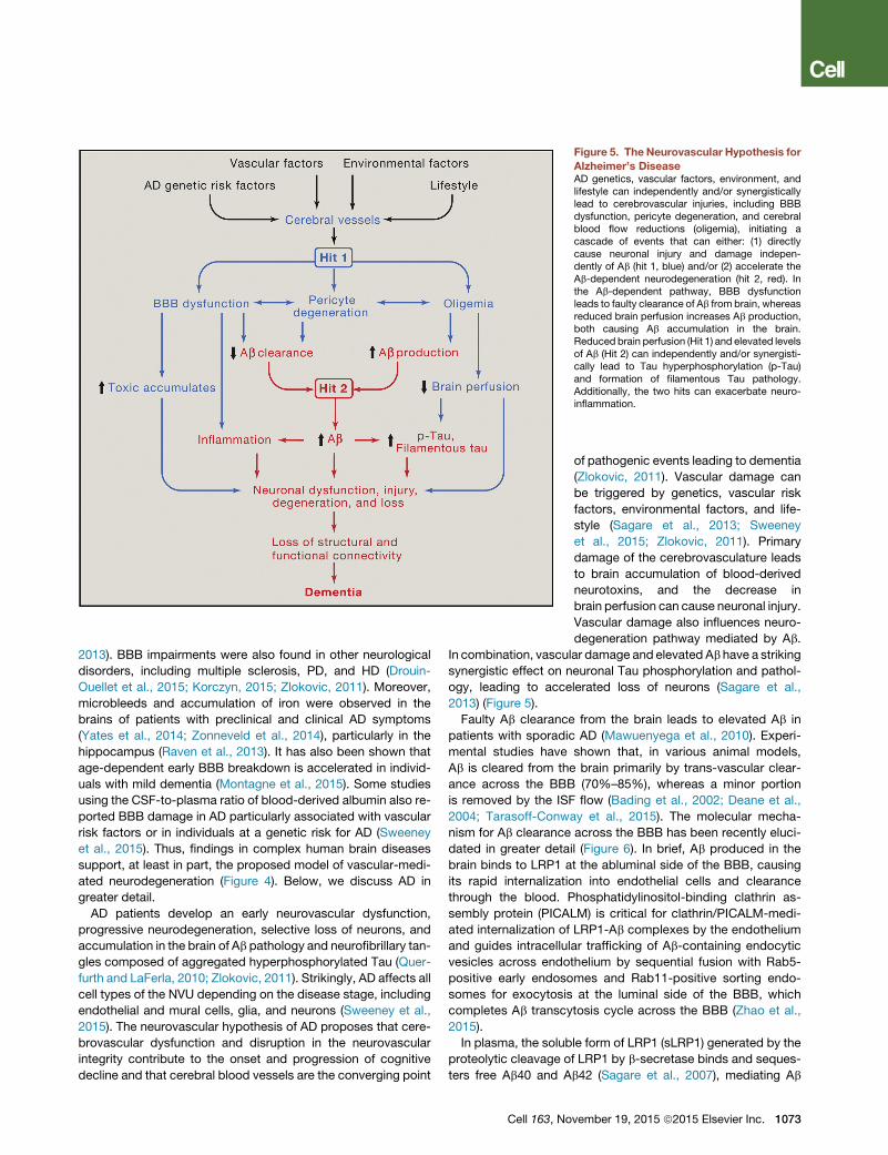
Figure 5. The Neurovascular Hypothesis for
Alzheimer’s DiseaseAD genetics, vascular factors, environment, andlifestyle can independently and/or synergisticallylead to cerebrovascular injuries, including BBBdysfunction, pericyte degeneration, and cerebralblood flow reductions (oligemia), initiating acascade of events that can either: (1) directlycause neuronal injury and damage indepen-dently of Ab (hit 1, blue) and/or (2) accelerate theAb-dependent neurodegeneration (hit 2, red). Inthe Ab-dependent pathway, BBB dysfunctionleads to faulty clearance of Ab from brain, whereasreduced brain perfusion increases Ab production,both causing Ab accumulation in the brain.Reduced brain perfusion (Hit 1) and elevated levelsof Ab (Hit 2) can independently and/or synergisti-cally lead to Tau hyperphosphorylation (p-Tau)and formation of filamentous Tau pathology.Additionally, the two hits can exacerbate neuro-inflammation.
2013). BBB impairments were also found in other neurological
disorders, including multiple sclerosis, PD, and HD (Drouin-
Ouellet et al., 2015; Korczyn, 2015; Zlokovic, 2011). Moreover,
microbleeds and accumulation of iron were observed in the
brains of patients with preclinical and clinical AD symptoms
(Yates et al., 2014; Zonneveld et al., 2014), particularly in the
hippocampus (Raven et al., 2013). It has also been shown that
age-dependent early BBB breakdown is accelerated in individ-
uals with mild dementia (Montagne et al., 2015). Some studies
using the CSF-to-plasma ratio of blood-derived albumin also re-
ported BBB damage in AD particularly associated with vascular
risk factors or in individuals at a genetic risk for AD (Sweeney
et al., 2015). Thus, findings in complex human brain diseases
support, at least in part, the proposed model of vascular-medi-
ated neurodegeneration (Figure 4). Below, we discuss AD in
greater detail.
AD patients develop an early neurovascular dysfunction,
progressive neurodegeneration, selective loss of neurons, and
accumulation in the brain of Ab pathology and neurofibrillary tan-
gles composed of aggregated hyperphosphorylated Tau (Quer-
furth and LaFerla, 2010; Zlokovic, 2011). Strikingly, AD affects all
cell types of the NVU depending on the disease stage, including
endothelial and mural cells, glia, and neurons (Sweeney et al.,
2015). The neurovascular hypothesis of AD proposes that cere-
brovascular dysfunction and disruption in the neurovascular
integrity contribute to the onset and progression of cognitive
decline and that cerebral blood vessels are the converging point
Cell 163, No
of pathogenic events leading to dementia
(Zlokovic, 2011). Vascular damage can
be triggered by genetics, vascular risk
factors, environmental factors, and life-
style (Sagare et al., 2013; Sweeney
et al., 2015; Zlokovic, 2011). Primary
damage of the cerebrovasculature leads
to brain accumulation of blood-derived
neurotoxins, and the decrease in
brain perfusion can cause neuronal injury.
Vascular damage also influences neuro-
degeneration pathway mediated by Ab.
In combination, vascular damage and elevated Ab have a striking
synergistic effect on neuronal Tau phosphorylation and pathol-
ogy, leading to accelerated loss of neurons (Sagare et al.,
2013) (Figure 5).
Faulty Ab clearance from the brain leads to elevated Ab in
patients with sporadic AD (Mawuenyega et al., 2010). Experi-
mental studies have shown that, in various animal models,
Ab is cleared from the brain primarily by trans-vascular clear-
ance across the BBB (70%–85%), whereas a minor portion
is removed by the ISF flow (Bading et al., 2002; Deane et al.,
2004; Tarasoff-Conway et al., 2015). The molecular mecha-
nism for Ab clearance across the BBB has been recently eluci-
dated in greater detail (Figure 6). In brief, Ab produced in the
brain binds to LRP1 at the abluminal side of the BBB, causing
its rapid internalization into endothelial cells and clearance
through the blood. Phosphatidylinositol-binding clathrin as-
sembly protein (PICALM) is critical for clathrin/PICALM-medi-
ated internalization of LRP1-Ab complexes by the endothelium
and guides intracellular trafficking of Ab-containing endocytic
vesicles across endothelium by sequential fusion with Rab5-
positive early endosomes and Rab11-positive sorting endo-
somes for exocytosis at the luminal side of the BBB, which
completes Ab transcytosis cycle across the BBB (Zhao et al.,
2015).
In plasma, the soluble form of LRP1 (sLRP1) generated by the
proteolytic cleavage of LRP1 by b-secretase binds and seques-
ters free Ab40 and Ab42 (Sagare et al., 2007), mediating Ab
vember 19, 2015 ª2015 Elsevier Inc. 1073
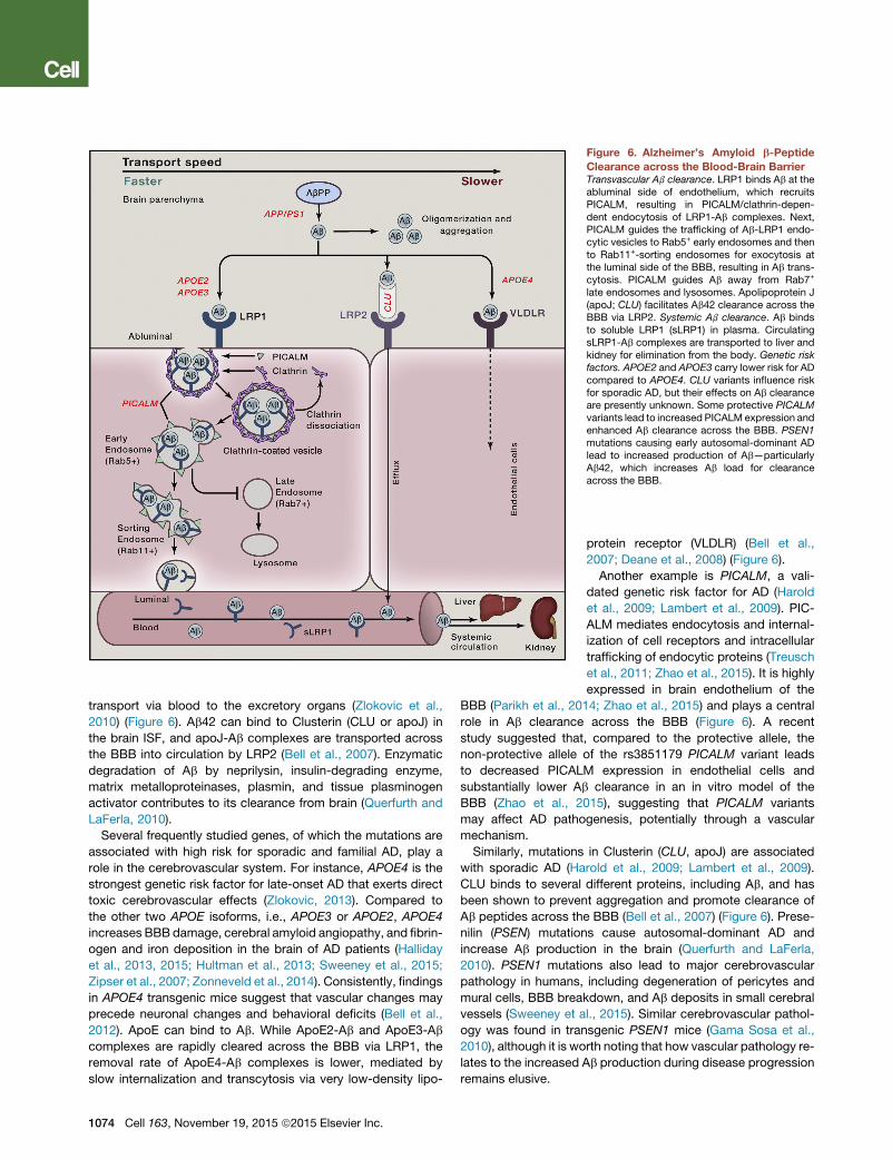
Figure 6. Alzheimer’s Amyloid b-Peptide
Clearance across the Blood-Brain BarrierTransvascular Ab clearance. LRP1 binds Ab at theabluminal side of endothelium, which recruitsPICALM, resulting in PICALM/clathrin-depen-dent endocytosis of LRP1-Ab complexes. Next,PICALM guides the trafficking of Ab-LRP1 endo-cytic vesicles to Rab5+ early endosomes and thento Rab11+-sorting endosomes for exocytosis atthe luminal side of the BBB, resulting in Ab trans-cytosis. PICALM guides Ab away from Rab7+
late endosomes and lysosomes. Apolipoprotein J(apoJ; CLU) facilitates Ab42 clearance across theBBB via LRP2. Systemic Ab clearance. Ab bindsto soluble LRP1 (sLRP1) in plasma. CirculatingsLRP1-Ab complexes are transported to liver andkidney for elimination from the body. Genetic riskfactors. APOE2 and APOE3 carry lower risk for ADcompared to APOE4. CLU variants influence riskfor sporadic AD, but their effects on Ab clearanceare presently unknown. Some protective PICALMvariants lead to increased PICALM expression andenhanced Ab clearance across the BBB. PSEN1mutations causing early autosomal-dominant ADlead to increased production of Ab—particularlyAb42, which increases Ab load for clearanceacross the BBB.
transport via blood to the excretory organs (Zlokovic et al.,
2010) (Figure 6). Ab42 can bind to Clusterin (CLU or apoJ) in
the brain ISF, and apoJ-Ab complexes are transported across
the BBB into circulation by LRP2 (Bell et al., 2007). Enzymatic
degradation of Ab by neprilysin, insulin-degrading enzyme,
matrix metalloproteinases, plasmin, and tissue plasminogen
activator contributes to its clearance from brain (Querfurth and
LaFerla, 2010).
Several frequently studied genes, of which the mutations are
associated with high risk for sporadic and familial AD, play a
role in the cerebrovascular system. For instance, APOE4 is the
strongest genetic risk factor for late-onset AD that exerts direct
toxic cerebrovascular effects (Zlokovic, 2013). Compared to
the other two APOE isoforms, i.e., APOE3 or APOE2, APOE4
increases BBBdamage, cerebral amyloid angiopathy, and fibrin-
ogen and iron deposition in the brain of AD patients (Halliday
et al., 2013, 2015; Hultman et al., 2013; Sweeney et al., 2015;
Zipser et al., 2007; Zonneveld et al., 2014). Consistently, findings
in APOE4 transgenic mice suggest that vascular changes may
precede neuronal changes and behavioral deficits (Bell et al.,
2012). ApoE can bind to Ab. While ApoE2-Ab and ApoE3-Ab
complexes are rapidly cleared across the BBB via LRP1, the
removal rate of ApoE4-Ab complexes is lower, mediated by
slow internalization and transcytosis via very low-density lipo-
1074 Cell 163, November 19, 2015 ª2015 Elsevier Inc.
protein receptor (VLDLR) (Bell et al.,
2007; Deane et al., 2008) (Figure 6).
Another example is PICALM, a vali-
dated genetic risk factor for AD (Harold
et al., 2009; Lambert et al., 2009). PIC-
ALM mediates endocytosis and internal-
ization of cell receptors and intracellular
trafficking of endocytic proteins (Treusch
et al., 2011; Zhao et al., 2015). It is highly
expressed in brain endothelium of the
BBB (Parikh et al., 2014; Zhao et al., 2015) and plays a central
role in Ab clearance across the BBB (Figure 6). A recent
study suggested that, compared to the protective allele, the
non-protective allele of the rs3851179 PICALM variant leads
to decreased PICALM expression in endothelial cells and
substantially lower Ab clearance in an in vitro model of the
BBB (Zhao et al., 2015), suggesting that PICALM variants
may affect AD pathogenesis, potentially through a vascular
mechanism.
Similarly, mutations in Clusterin (CLU, apoJ) are associated
with sporadic AD (Harold et al., 2009; Lambert et al., 2009).
CLU binds to several different proteins, including Ab, and has
been shown to prevent aggregation and promote clearance of
Ab peptides across the BBB (Bell et al., 2007) (Figure 6). Prese-
nilin (PSEN) mutations cause autosomal-dominant AD and
increase Ab production in the brain (Querfurth and LaFerla,
2010). PSEN1 mutations also lead to major cerebrovascular
pathology in humans, including degeneration of pericytes and
mural cells, BBB breakdown, and Ab deposits in small cerebral
vessels (Sweeney et al., 2015). Similar cerebrovascular pathol-
ogy was found in transgenic PSEN1 mice (Gama Sosa et al.,
2010), although it is worth noting that how vascular pathology re-
lates to the increased Ab production during disease progression
remains elusive.
Conclusions and Future DirectionsRecent advances in human genetics and the corresponding
transgenic models indicate that almost every non-neuronal
cell type of the NVU could be affected by some monogenic in-
herited disorders. This association can provide insights into the
potential pathogenic links among BBB dysfunction, neuronal
injury, neurodegeneration, and neurological disorders caused
by NVU disruption and BBB breakdown. On the other hand,
the relationship among neurovascular integrity, brain structural
and functional connectivity, cognitive function, and neurological
symptomatology in complex disorders such as AD still awaits
to be directly explored in the most relevant in vivo context,
which has only recently became possible with the development
of novel state-of-the art neuroimaging and molecular biomarker
approaches. Experimental studies combining genetic, environ-
mental, and lifestyle factors hold promise to further advance
our knowledge of multifactorial CNS disorders and to establish
the concept that the loss of healthy cerebral blood vessels and
BBB integrity influences the course and clinical phenotype of
neurological disorders in a region-specific manner.
Some important questions remain to be addressed. First, it
is still unclear whether in the living human brain, cerebrovas-
cular changes and BBB breakdown can drive the initial path-
ogenic events that lead to neuronal injury, disrupted structural
and functional brain connectivity, and early neurological
symptoms, such as cognitive decline in AD and motor
changes in ALS, PD, or HD. Second, further studies are war-
ranted to test whether the underlying molecular mechanisms
of NVU disruption and BBB breakdown might point to new
targets for therapeutic development to prevent and/or treat
neurodegenerative disorders. Third, technological advance is
required to determine whether neurovascular dysfunction
and BBB breakdown are detectable in the living human brain
prior to the development of the full spectrum of neurological
symptoms. Last but not least, future investigations need to
address whether molecular and imaging biomarkers of neuro-
vascular dysfunction can serve as reliable prognostic and/or
diagnostic tools to predict the development of neurodegener-
ative disorders.
From the basic science side, pushing the envelope further to
generate a comprehensive proteomics and RNA-seq molecular
atlas of the BBB in animals and humanswould provide a valuable
resource for discovering and studying new targets and signaling
pathways mediating the crosstalk among different cell types
within the NVU. This may lead to the development of new trans-
genic animal models—pluripotent stem cell models of the BBB,
NVU, and different neurological disorders—serving as valuable
platforms for drug discovery and for testing novel drug delivery
approaches.
AUTHOR CONTRIBUTIONS
Z.Z. and A.R.N. contributed equally in preparing some sections of this
Perspective.
ACKNOWLEDGMENTS
The work of B.V.Z. is supported by the National Institutes of Health grants
R01AG023084, R01NS090904, R01NS034467, and R01AG039452 and the
Cure Alzheimer’s Fund. The work of C.B. is supported by the European
Research Council (ERC advanced grant number 294556 BBBARRIER), the
Knut and Alice Wallenberg Foundation, Leducq Foundation (Sphingonet),
Swedish Cancer Foundation, the Swedish Science Council, and Uppsala
University. We apologize to authors whose work we could not cite because
of the limit on the number of references; in some instances, we mostly cited
the overview articles.
REFERENCES
Alakbarzade, V., Hameed, A., Quek, D.Q.Y., Chioza, B.A., Baple, E.L., Caze-
nave-Gassiot, A., Nguyen, L.N., Wenk, M.R., Ahmad, A.Q., Sreekantan-Nair,
A., et al. (2015). A partially inactivating mutation in the sodium-dependent lyso-
phosphatidylcholine transporter MFSD2A causes a non-lethal microcephaly
syndrome. Nat. Genet. 47, 814–817.
Alkan, A., Sigirci, A., Kutlu, R., Aslan, M., Doganay, S., and Yakinci, C. (2007).
Merosin-negative congenital muscular dystrophy: diffusion-weighted imaging
findings of brain. J. Child Neurol. 22, 655–659.
Alvarez, J.I., Dodelet-Devillers, A., Kebir, H., Ifergan, I., Fabre, P.J., Terouz, S.,
Sabbagh, M., Wosik, K., Bourbonniere, L., Bernard, M., et al. (2011). The
Hedgehog pathway promotes blood-brain barrier integrity and CNS immune
quiescence. Science 334, 1727–1731.
Andreone, B.J., Lacoste, B., and Gu, C. (2015). Neuronal and vascular interac-
tions. Annu. Rev. Neurosci. 38, 25–46.
Armulik, A., Genove, G., Mae, M., Nisancioglu, M.H., Wallgard, E., Niaudet, C.,
He, L., Norlin, J., Lindblom, P., Strittmatter, K., et al. (2010). Pericytes regulate
the blood-brain barrier. Nature 468, 557–561.
Armulik, A., Genove, G., and Betsholtz, C. (2011). Pericytes: developmental,
physiological, and pathological perspectives, problems, and promises. Dev.
Cell 21, 193–215.
Aspelund, A., Antila, S., Proulx, S.T., Karlsen, T.V., Karaman, S., Detmar, M.,
Wiig, H., and Alitalo, K. (2015). A dural lymphatic vascular system that drains
brain interstitial fluid and macromolecules. J. Exp. Med. 212, 991–999.
Attwell, D., Buchan, A.M., Charpak, S., Lauritzen, M., Macvicar, B.A., and
Newman, E.A. (2010). Glial and neuronal control of brain blood flow. Nature
468, 232–243.
Bading, J.R., Yamada, S., Mackic, J.B., Kirkman, L., Miller, C., Calero, M.,
Ghiso, J., Frangione, B., and Zlokovic, B.V. (2002). Brain clearance of
Alzheimer’s amyloid-beta40 in the squirrel monkey: a SPECT study in a pri-
mate model of cerebral amyloid angiopathy. J. Drug Target. 10, 359–368.
Baeten, K.M., and Akassoglou, K. (2011). Extracellular matrix and matrix re-
ceptors in blood-brain barrier formation and stroke. Dev. Neurobiol. 71,
1018–1039.
Baloyannis, S.J., and Baloyannis, I.S. (2012). The vascular factor in Alz-
heimer’s disease: a study in Golgi technique and electron microscopy.
J. Neurol. Sci. 322, 117–121.
Bell, R.D., Sagare, A.P., Friedman, A.E., Bedi, G.S., Holtzman, D.M., Deane,
R., and Zlokovic, B.V. (2007). Transport pathways for clearance of human
Alzheimer’s amyloid beta-peptide and apolipoproteins E and J in the mouse
central nervous system. J. Cereb. Blood Flow Metab. 27, 909–918.
Bell, R.D., Winkler, E.A., Sagare, A.P., Singh, I., LaRue, B., Deane, R., and
Zlokovic, B.V. (2010). Pericytes control key neurovascular functions and
neuronal phenotype in the adult brain and during brain aging. Neuron 68,
409–427.
Bell, R.D., Winkler, E.A., Singh, I., Sagare, A.P., Deane, R., Wu, Z., Holtzman,
D.M., Betsholtz, C., Armulik, A., Sallstrom, J., et al. (2012). Apolipoprotein E
controls cerebrovascular integrity via cyclophilin A. Nature 485, 512–516.
Ben-Zvi, A., Lacoste, B., Kur, E., Andreone, B.J., Mayshar, Y., Yan, H., and Gu,
C. (2014). Mfsd2a is critical for the formation and function of the blood-brain
barrier. Nature 509, 507–511.
Benarroch, E.E. (2014). Brain glucose transporters: implications for neurologic
disease. Neurology 82, 1374–1379.
Cell 163, November 19, 2015 ª2015 Elsevier Inc. 1075
Betsholtz, C. (2014). Physiology: Double function at the blood-brain barrier.
Nature 509, 432–433.
Betsholtz, C. (2015). Lipid transport and human brain development. Nat.
Genet. 47, 699–701.
Blinder, P., Tsai, P.S., Kaufhold, J.P., Knutsen, P.M., Suhl, H., and Kleinfeld, D.
(2013). The cortical angiome: an interconnected vascular network with nonco-
lumnar patterns of blood flow. Nat. Neurosci. 16, 889–897.
Bray, N. (2015). Biologics: Transferrin’ bispecific antibodies across the blood-
brain barrier. Nat. Rev. Drug Discov. 14, 14–15.
Capri, Y., Friesema, E.C.H., Kersseboom, S., Touraine, R., Monnier, A., Ey-
mard-Pierre, E., Des Portes, V., De Michele, G., Brady, A.F., Boespflug-Tan-
guy, O., et al. (2013). Relevance of different cellular models in determining
the effects of mutations on SLC16A2/MCT8 thyroid hormone transporter func-
tion and genotype-phenotype correlation. Hum. Mutat. 34, 1018–1025.
Chabriat, H., Joutel, A., Dichgans, M., Tournier-Lasserve, E., and Bousser,
M.-G. (2009). Cadasil. Lancet Neurol. 8, 643–653.
Cirrito, J.R., Deane, R., Fagan, A.M., Spinner, M.L., Parsadanian, M., Finn,
M.B., Jiang, H., Prior, J.L., Sagare, A., Bales, K.R., et al. (2005). P-glycoprotein
deficiency at the blood-brain barrier increases amyloid-beta deposition in an
Alzheimer disease mouse model. J. Clin. Invest. 115, 3285–3290.
Clarke, L.E., and Barres, B.A. (2013). Emerging roles of astrocytes in neural cir-
cuit development. Nat. Rev. Neurosci. 14, 311–321.
Daneman, R., and Prat, A. (2015). The blood-brain barrier. Cold Spring Harb.
Perspect. Biol. 7, a020412.
Daneman, R., Agalliu, D., Zhou, L., Kuhnert, F., Kuo, C.J., and Barres, B.A.
(2009). Wnt/beta-catenin signaling is required for CNS, but not non-CNS,
angiogenesis. Proc. Natl. Acad. Sci. USA 106, 641–646.
Daneman, R., Zhou, L., Kebede, A.A., and Barres, B.A. (2010). Pericytes are
required for blood-brain barrier integrity during embryogenesis. Nature 468,
562–566.
Davalos, D., Ryu, J.K., Merlini, M., Baeten, K.M., Le Moan, N., Petersen, M.A.,
Deerinck, T.J., Smirnoff, D.S., Bedard, C., Hakozaki, H., et al. (2012). Fibrin-
ogen-induced perivascular microglial clustering is required for the develop-
ment of axonal damage in neuroinflammation. Nat. Commun. 3, 1227.
Deane, R., Du Yan, S., Submamaryan, R.K., LaRue, B., Jovanovic, S., Hogg,
E., Welch, D., Manness, L., Lin, C., Yu, J., et al. (2003). RAGE mediates amy-
loid-beta peptide transport across the blood-brain barrier and accumulation in
brain. Nat. Med. 9, 907–913.
Deane, R., Wu, Z., Sagare, A., Davis, J., Du Yan, S., Hamm, K., Xu, F., Parisi,
M., LaRue, B., Hu, H.W., et al. (2004). LRP/amyloid beta-peptide interaction
mediates differential brain efflux of Abeta isoforms. Neuron 43, 333–344.
Deane, R., Sagare, A., Hamm, K., Parisi, M., Lane, S., Finn, M.B., Holtzman,
D.M., and Zlokovic, B.V. (2008). apoE isoform-specific disruption of amyloid
beta peptide clearance from mouse brain. J. Clin. Invest. 118, 4002–4013.
Deane, R., Singh, I., Sagare, A.P., Bell, R.D., Ross, N.T., LaRue, B., Love, R.,
Perry, S., Paquette, N., Deane, R.J., et al. (2012). A multimodal RAGE-specific
inhibitor reduces amyloid b-mediated brain disorder in a mouse model of Alz-
heimer disease. J. Clin. Invest. 122, 1377–1392.
Drouin-Ouellet, J., Sawiak, S.J., Cisbani, G., Lagace, M., Kuan, W.-L., Saint-
Pierre, M., Dury, R.J., Alata, W., St-Amour, I., Mason, S.L., et al. (2015). Cere-
brovascular and blood-brain barrier impairments in Huntington’s disease:
Potential implications for its pathophysiology. Ann. Neurol. 78, 160–177.
Dumitrescu, A.M., Liao, X.-H., Best, T.B., Brockmann, K., and Refetoff, S.
(2004). A novel syndrome combining thyroid and neurological abnormalities
is associated with mutations in a monocarboxylate transporter gene. Am. J.
Hum. Genet. 74, 168–175.
Farkas, E., and Luiten, P.G. (2001). Cerebral microvascular pathology in aging
and Alzheimer’s disease. Prog. Neurobiol. 64, 575–611.
Fischer, A., Zalvide, J., Faurobert, E., Albiges-Rizo, C., and Tournier-Lasserve,
E. (2013). Cerebral cavernous malformations: from CCM genes to endothelial
cell homeostasis. Trends Mol. Med. 19, 302–308.
1076 Cell 163, November 19, 2015 ª2015 Elsevier Inc.
Friesema, E.C.H., Grueters, A., Biebermann, H., Krude, H., von Moers, A., Re-
eser, M., Barrett, T.G., Mancilla, E.E., Svensson, J., Kester, M.H.A., et al.
(2004). Association between mutations in a thyroid hormone transporter and
severe X-linked psychomotor retardation. Lancet 364, 1435–1437.
Gama Sosa, M.A., Gasperi, R.D., Rocher, A.B., Wang, A.C.-J., Janssen,
W.G.M., Flores, T., Perez, G.M., Schmeidler, J., Dickstein, D.L., Hof, P.R.,
and Elder, G.A. (2010). Age-related vascular pathology in transgenic mice
expressing presenilin 1-associated familial Alzheimer’s disease mutations.
Am. J. Pathol. 176, 353–368.
Gould, D.B., Phalan, F.C., vanMil, S.E., Sundberg, J.P., Vahedi, K., Massin, P.,
Bousser, M.G., Heutink, P., Miner, J.H., Tournier-Lasserve, E., and John, S.W.
(2006). Role of COL4A1 in small-vessel disease and hemorrhagic stroke.
N. Engl. J. Med. 354, 1489–1496.
Guemez-Gamboa, A., Nguyen, L.N., Yang, H., Zaki, M.S., Kara, M., Ben-Om-
ran, T., Akizu, N., Rosti, R.O., Rosti, B., Scott, E., et al. (2015). Inactivating
mutations in MFSD2A, required for omega-3 fatty acid transport in brain,
cause a lethal microcephaly syndrome. Nat. Genet. 47, 809–813.
Hagan, N., and Ben-Zvi, A. (2015). The molecular, cellular, and morphological
components of blood-brain barrier development during embryogenesis.
Semin. Cell Dev. Biol. 38, 7–15.
Hall, C.N., Reynell, C., Gesslein, B., Hamilton, N.B., Mishra, A., Sutherland, B.A.,
O’Farrell, F.M.,Buchan,A.M., Lauritzen,M., andAttwell,D. (2014).Capillaryperi-
cytes regulate cerebral blood flow in health and disease. Nature 508, 55–60.
Halliday, M.R., Pomara, N., Sagare, A.P., Mack, W.J., Frangione, B., and
Zlokovic, B.V. (2013). Relationship between cyclophilin a levels and matrix
metalloproteinase 9 activity in cerebrospinal fluid of cognitively normal apoli-
poprotein e4 carriers and blood-brain barrier breakdown. JAMA Neurol. 70,
1198–1200.
Halliday, M.R., Rege, S.V., Ma, Q., Zhao, Z., Miller, C.A., Winkler, E.A., and Zlo-
kovic, B.V. (2015). Accelerated pericyte degeneration and blood-brain barrier
breakdown in apolipoprotein E4 carriers with Alzheimer’s disease. J. Cereb.
Blood Flow Metab. Off. J. Int. Soc. Cereb. Blood Flow Metab. http://dx.doi.
org/10.1038/jcbfm.2015.44.
Hammer, C., Stepniak, B., Schneider, A., Papiol, S., Tantra, M., Begemann,
M., Siren, A.-L., Pardo, L.A., Sperling, S., Mohd Jofrry, S., et al. (2014).
Neuropsychiatric disease relevance of circulating anti-NMDA receptor auto-
antibodies depends on blood-brain barrier integrity. Mol. Psychiatry 19,
1143–1149.
Harold, D., Abraham, R., Hollingworth, P., Sims, R., Gerrish, A., Hamshere,
M.L., Pahwa, J.S., Moskvina, V., Dowzell, K., Williams, A., et al. (2009).
Genome-wide association study identifies variants at CLU and PICALM asso-
ciated with Alzheimer’s disease. Nat. Genet. 41, 1088–1093.
Henshall, T.L., Keller, A., He, L., Johansson, B.R., Wallgard, E., Raschperger,
E., Mae, M.A., Jin, S., Betsholtz, C., and Lendahl, U. (2015). Notch3 is neces-
sary for blood vessel integrity in the central nervous system. Arterioscler.
Thromb. Vasc. Biol. 35, 409–420.
Hultman, K., Strickland, S., and Norris, E.H. (2013). The APOE 34/ 34 genotype
potentiates vascular fibrin(ogen) deposition in amyloid-laden vessels in
the brains of Alzheimer’s disease patients. J. Cereb. Blood Flow Metab. 33,
1251–1258.
Iadecola, C. (2013). The pathobiology of vascular dementia. Neuron 80,
844–866.
Keller, A., Westenberger, A., Sobrido, M.J., Garcıa-Murias, M., Domingo, A.,
Sears, R.L., Lemos, R.R., Ordonez-Ugalde, A., Nicolas, G., da Cunha,
J.E.G., et al. (2013). Mutations in the gene encoding PDGF-B cause brain
calcifications in humans and mice. Nat. Genet. 45, 1077–1082.
Kersseboom, S., Kremers, G.-J., Friesema, E.C.H., Visser, W.E., Klootwijk, W.,
Peeters, R.P., and Visser, T.J. (2013). Mutations inMCT8 in patients with Allan-
Herndon-Dudley-syndrome affecting its cellular distribution. Mol. Endocrinol.
27, 801–813.
Korczyn, A.D. (2015). Vascular parkinsonism–characteristics, pathogenesis
and treatment. Nat. Rev. Neurol. 11, 319–326.
Kuhnert, F., Mancuso, M.R., Shamloo, A., Wang, H.-T., Choksi, V., Florek, M.,
Su, H., Fruttiger, M., Young, W.L., Heilshorn, S.C., and Kuo, C.J. (2010).
Essential regulation of CNS angiogenesis by the orphan G protein-coupled
receptor GPR124. Science 330, 985–989.
Kuo, D.S., Labelle-Dumais, C., and Gould, D.B. (2012). COL4A1 and COL4A2
mutations and disease: insights into pathogenic mechanisms and potential
therapeutic targets. Hum. Mol. Genet. 21 (R1), R97–R110.
Lambert, J.-C., Heath, S., Even, G., Campion, D., Sleegers, K., Hiltunen, M.,
Combarros, O., Zelenika, D., Bullido, M.J., Tavernier, B., et al.; European
Alzheimer’s Disease Initiative Investigators (2009). Genome-wide association
study identifies variants at CLU and CR1 associated with Alzheimer’s disease.
Nat. Genet. 41, 1094–1099.
Lee, S.-W., Kim, W.J., Choi, Y.K., Song, H.S., Son, M.J., Gelman, I.H., Kim,
Y.-J., and Kim, K.-W. (2003). SSeCKS regulates angiogenesis and tight junc-
tion formation in blood-brain barrier. Nat. Med. 9, 900–906.
Legati, A., Giovannini, D., Nicolas, G., Lopez-Sanchez, U., Quintans, B.,
Oliveira, J.R.M., Sears, R.L., Ramos, E.M., Spiteri, E., Sobrido, M.-J., et al.
(2015). Mutations in XPR1 cause primary familial brain calcification associated
with altered phosphate export. Nat. Genet. 47, 579–581.
Liebner, S., Corada, M., Bangsow, T., Babbage, J., Taddei, A., Czupalla, C.J.,
Reis, M., Felici, A., Wolburg, H., Fruttiger, M., et al. (2008). Wnt/beta-catenin
signaling controls development of the blood-brain barrier. J. Cell Biol. 183,
409–417.
Lin, L., Yee, S.W., Kim, R.B., and Giacomini, K.M. (2015). SLC transporters
as therapeutic targets: emerging opportunities. Nat. Rev. Drug Discov. 14,
543–560.
Lindahl, P., Johansson, B.R., Leveen, P., and Betsholtz, C. (1997). Pericyte
loss and microaneurysm formation in PDGF-B-deficient mice. Science 277,
242–245.
Louveau, A., Smirnov, I., Keyes, T.J., Eccles, J.D., Rouhani, S.J., Peske, J.D.,
Derecki, N.C., Castle, D., Mandell, J.W., Lee, K.S., et al. (2015). Structural and
functional features of central nervous system lymphatic vessels. Nature 523,
337–341.
Maddaluno, L., Rudini, N., Cuttano, R., Bravi, L., Giampietro, C., Corada, M.,
Ferrarini, L., Orsenigo, F., Papa, E., Boulday, G., et al. (2013). EndMT contrib-
utes to the onset and progression of cerebral cavernous malformations.
Nature 498, 492–496.
Mawuenyega, K.G., Sigurdson, W., Ovod, V., Munsell, L., Kasten, T., Morris,
J.C., Yarasheski, K.E., and Bateman, R.J. (2010). Decreased clearance of
CNS beta-amyloid in Alzheimer’s disease. Science 330, 1774.
Miller, D.S. (2015). Regulation of ABC transporters blood-brain barrier: the
good, the bad, and the ugly. Adv. Cancer Res. 125, 43–70.
Montagne, A., Barnes, S.R., Sweeney, M.D., Halliday, M.R., Sagare, A.P.,
Zhao, Z., Toga, A.W., Jacobs, R.E., Liu, C.Y., Amezcua, L., et al. (2015).
Blood-brain barrier breakdown in the aging human hippocampus. Neuron
85, 296–302.
Montine, T.J., Koroshetz, W.J., Babcock, D., Dickson, D.W., Galpern, W.R.,
Glymour, M.M., Greenberg, S.M., Hutton, M.L., Knopman, D.S., Kuzmichev,
A.N., et al.; ADRD 2013 Conference Organizing Committee (2014). Recom-
mendations of the Alzheimer’s disease-related dementias conference.
Neurology 83, 851–860.
Nakagomi, T., Kubo, S., Nakano-Doi, A., Sakuma, R., Lu, S., Narita, A., Kawa-
hara, M., Taguchi, A., and Matsuyama, T. (2015). Brain vascular pericytes
following ischemia have multipotential stem cell activity to differentiate into
neural and vascular lineage cells. Stem Cells 33, 1962–1974.
Nguyen, L.N., Ma, D., Shui, G., Wong, P., Cazenave-Gassiot, A., Zhang, X.,
Wenk, M.R., Goh, E.L.K., and Silver, D.L. (2014). Mfsd2a is a transporter for
the essential omega-3 fatty acid docosahexaenoic acid. Nature 509, 503–506.
Nicolas, G., Pottier, C., Maltete, D., Coutant, S., Rovelet-Lecrux, A., Legallic,
S., Rousseau, S., Vaschalde, Y., Guyant-Marechal, L., Augustin, J., et al.
(2013). Mutation of the PDGFRB gene as a cause of idiopathic basal ganglia
calcification. Neurology 80, 181–187.
O’Driscoll, M.C., Daly, S.B., Urquhart, J.E., Black, G.C.M., Pilz, D.T., Brock-
mann, K., McEntagart, M., Abdel-Salam, G., Zaki, M., Wolf, N.I., et al.
(2010). Recessive mutations in the gene encoding the tight junction protein
occludin cause band-like calcification with simplified gyration and polymicro-
gyria. Am. J. Hum. Genet. 87, 354–364.
Obermeier, B., Daneman, R., and Ransohoff, R.M. (2013). Development, main-
tenance and disruption of the blood-brain barrier. Nat. Med. 19, 1584–1596.
Pardridge, W.M. (2015). Blood-brain barrier endogenous transporters as ther-
apeutic targets: a new model for small molecule CNS drug discovery. Expert
Opin. Ther. Targets 19, 1059–1072.
Parikh, I., Fardo, D.W., and Estus, S. (2014). Genetics of PICALM expression
and Alzheimer’s disease. PLoS ONE 9, e91242.
Peppiatt, C.M., Howarth, C., Mobbs, P., and Attwell, D. (2006). Bidirectional
control of CNS capillary diameter by pericytes. Nature 443, 700–704.
Potente, M., Gerhardt, H., and Carmeliet, P. (2011). Basic and therapeutic
aspects of angiogenesis. Cell 146, 873–887.
Querfurth, H.W., and LaFerla, F.M. (2010). Alzheimer’s disease. N. Engl. J.
Med. 362, 329–344.
Raven, E.P., Lu, P.H., Tishler, T.A., Heydari, P., and Bartzokis, G. (2013).
Increased iron levels and decreased tissue integrity in hippocampus of
Alzheimer’s disease detected in vivo with magnetic resonance imaging.
J. Alzheimers Dis. 37, 127–136.
Sagare, A., Deane, R., Bell, R.D., Johnson, B., Hamm, K., Pendu, R., Marky, A.,
Lenting, P.J., Wu, Z., Zarcone, T., et al. (2007). Clearance of amyloid-beta by
circulating lipoprotein receptors. Nat. Med. 13, 1029–1031.
Sagare, A.P., Bell, R.D., Zhao, Z., Ma, Q., Winkler, E.A., Ramanathan, A., and
Zlokovic, B.V. (2013). Pericyte loss influences Alzheimer-like neurodegenera-
tion in mice. Nat. Commun. 4, 2932.
Sasaki, A., Kakita, A., Yoshida, K., Konno, T., Ikeuchi, T., Hayashi, S., Matsuo,
H., and Shioda, K. (2015). Variable expression of microglial DAP12 and TREM2
genes in Nasu-Hakola disease. Neurogenetics 16, 265–276.
Saunders, N.R., Daneman, R., Dziegielewska, K.M., and Liddelow, S.A. (2013).
Transporters of the blood-brain and blood-CSF interfaces in development and
in the adult. Mol. Aspects Med. 34, 742–752.
Sengillo, J.D., Winkler, E.A., Walker, C.T., Sullivan, J.S., Johnson, M., and Zlo-
kovic, B.V. (2013). Deficiency in mural vascular cells coincides with blood-
brain barrier disruption in Alzheimer’s disease. Brain Pathol. 23, 303–310.
Snyder, H.M., Corriveau, R.A., Craft, S., Faber, J.E., Greenberg, S.M., Knop-
man, D., Lamb, B.T., Montine, T.J., Nedergaard, M., Schaffer, C.B., et al.
(2015). Vascular contributions to cognitive impairment and dementia including
Alzheimer’s disease. Alzheimers Dement. 11, 710–717.
Sofroniew, M.V. (2015). Astrocyte barriers to neurotoxic inflammation. Nat.
Rev. Neurosci. 16, 249–263.
Stenman, J.M., Rajagopal, J., Carroll, T.J., Ishibashi, M., McMahon, J., and
McMahon, A.P. (2008). Canonical Wnt signaling regulates organ-specific as-
sembly and differentiation of CNS vasculature. Science 322, 1247–1250.
Sweeney, M.D., Sagare, A.P., and Zlokovic, B.V. (2015). Cerebrospinal fluid
biomarkers of neurovascular dysfunction in mild dementia and Alzheimer’s
disease. J. Cereb. Blood Flow Metab. 35, 1055–1068.
Tallquist, M.D., French, W.J., and Soriano, P. (2003). Additive effects of PDGF
receptor beta signaling pathways in vascular smooth muscle cell develop-
ment. PLoS Biol. 1, E52.
Tam, S.J., Richmond, D.L., Kaminker, J.S., Modrusan, Z., Martin-McNulty, B.,
Cao, T.C., Weimer, R.M., Carano, R.A.D., van Bruggen, N., and Watts, R.J.
(2012). Death receptors DR6 and TROY regulate brain vascular development.
Dev. Cell 22, 403–417.
Tarasoff-Conway, J.M., Carare, R.O., Osorio, R.S., Glodzik, L., Butler, T.,
Fieremans, E., Axel, L., Rusinek, H., Nicholson, C., Zlokovic, B.V., et al.
(2015). Clearance systems in the brain-implications for Alzheimer disease.
Nat. Rev. Neurol. 11, 457–470.
Tietz, S., and Engelhardt, B. (2015). Brain barriers: Crosstalk between complex
tight junctions and adherens junctions. J. Cell Biol. 209, 493–506.
Cell 163, November 19, 2015 ª2015 Elsevier Inc. 1077
Treusch, S., Hamamichi, S., Goodman, J.L., Matlack, K.E.S., Chung, C.Y.,
Baru, V., Shulman, J.M., Parrado, A., Bevis, B.J., Valastyan, J.S., et al.
(2011). Functional links between Ab toxicity, endocytic trafficking, and Alz-
heimer’s disease risk factors in yeast. Science 334, 1241–1245.
Tsai, H.-H., Li, H., Fuentealba, L.C., Molofsky, A.V., Taveira-Marques, R.,
Zhuang, H., Tenney, A., Murnen, A.T., Fancy, S.P.J., Merkle, F., et al. (2012).
Regional astrocyte allocation regulates CNS synaptogenesis and repair.
Science 337, 358–362.
Wang, D., Kranz-Eble, P., and De Vivo, D.C. (2000). Mutational analysis of
GLUT1 (SLC2A1) in Glut-1 deficiency syndrome. Hum. Mutat. 16, 224–231.
Wang, C., Li, Y., Shi, L., Ren, J., Patti, M., Wang, T., de Oliveira, J.R.M.,
Sobrido, M.-J., Quintans, B., Baquero, M., et al. (2012a). Mutations in
SLC20A2 link familial idiopathic basal ganglia calcification with phosphate ho-
meostasis. Nat. Genet. 44, 254–256.
Wang, Y., Rattner, A., Zhou, Y., Williams, J., Smallwood, P.M., and Nathans, J.
(2012b). Norrin/Frizzled4 signaling in retinal vascular development and blood
brain barrier plasticity. Cell 151, 1332–1344.
Winkler, E.A., Bell, R.D., and Zlokovic, B.V. (2011). Central nervous system
pericytes in health and disease. Nat. Neurosci. 14, 1398–1405.
Winkler, E.A., Sengillo, J.D., Sullivan, J.S., Henkel, J.S., Appel, S.H., and
Zlokovic, B.V. (2013). Blood-spinal cord barrier breakdown and pericyte re-
ductions in amyotrophic lateral sclerosis. Acta Neuropathol. 125, 111–120.
Winkler, E.A., Nishida, Y., Sagare, A.P., Rege, S.V., Bell, R.D., Perlmutter, D.,
Sengillo, J.D., Hillman, S., Kong, P., Nelson, A.R., et al. (2015). GLUT1 reduc-
tions exacerbate Alzheimer’s disease vasculo-neuronal dysfunction and
degeneration. Nat. Neurosci. 18, 521–530.
Woodfin, A., Voisin, M.-B., Beyrau, M., Colom, B., Caille, D., Diapouli, F.-M.,
Nash, G.B., Chavakis, T., Albelda, S.M., Rainger, G.E., et al. (2011). The junc-
tional adhesion molecule JAM-C regulates polarized transendothelial migra-
tion of neutrophils in vivo. Nat. Immunol. 12, 761–769.
Yamamoto, H., Ehling, M., Kato, K., Kanai, K., van Lessen, M., Frye, M.,
Zeuschner, D., Nakayama, M., Vestweber, D., and Adams, R.H. (2015). Integ-
rin b1 controls VE-cadherin localization and blood vessel stability. Nat. Com-
mun. 6, 6429.
Yao, Y., Chen, Z.-L., Norris, E.H., and Strickland, S. (2014). Astrocytic laminin
regulates pericyte differentiation and maintains blood brain barrier integrity.
Nat. Commun. 5, 3413.
Yates, P.A., Desmond, P.M., Phal, P.M., Steward, C., Szoeke, C., Salvado, O.,
Ellis, K.A., Martins, R.N., Masters, C.L., Ames, D., et al.; AIBL Research Group
1078 Cell 163, November 19, 2015 ª2015 Elsevier Inc.
(2014). Incidence of cerebral microbleeds in preclinical Alzheimer disease.
Neurology 82, 1266–1273.
Zhao, Z., and Zlokovic, B.V. (2014). Blood-brain barrier: a dual life of MFSD2A?
Neuron 82, 728–730.
Zhao, Z., Sagare, A.P., Ma, Q., Halliday, M.R., Kong, P., Kisler, K., Winkler,
E.A., Ramanathan, A., Kanekiyo, T., Bu, G., et al. (2015). Central role for
PICALM in amyloid-b blood-brain barrier transcytosis and clearance. Nat.
Neurosci. 18, 978–987.
Zhou, Y., and Nathans, J. (2014). Gpr124 controls CNS angiogenesis and
blood-brain barrier integrity by promoting ligand-specific canonical wnt
signaling. Dev. Cell 31, 248–256.
Zhou, Y., Wang, Y., Tischfield, M., Williams, J., Smallwood, P.M., Rattner, A.,
Taketo,M.M., andNathans, J. (2014). CanonicalWNT signaling components in
vascular development and barrier formation. J. Clin. Invest. 124, 3825–3846.
Zipser, B.D., Johanson, C.E., Gonzalez, L., Berzin, T.M., Tavares, R., Hulette,
C.M., Vitek, M.P., Hovanesian, V., and Stopa, E.G. (2007). Microvascular injury
and blood-brain barrier leakage in Alzheimer’s disease. Neurobiol. Aging 28,
977–986.
Zlokovic, B.V. (1995). Cerebrovascular permeability to peptides: manipula-
tions of transport systems at the blood-brain barrier. Pharm. Res. 12, 1395–
1406.
Zlokovic, B.V. (2008). The blood-brain barrier in health and chronic neurode-
generative disorders. Neuron 57, 178–201.
Zlokovic, B.V. (2011). Neurovascular pathways to neurodegeneration in Alz-
heimer’s disease and other disorders. Nat. Rev. Neurosci. 12, 723–738.
Zlokovic, B.V. (2013). Cerebrovascular effects of apolipoprotein E: implica-
tions for Alzheimer disease. JAMA Neurol. 70, 440–444.
Zlokovic, B.V., Begley, D.J., and Chain-Eliash, D.G. (1985). Blood-brain barrier
permeability to leucine-enkephalin, D-alanine2-D-leucine5-enkephalin and
their N-terminal amino acid (tyrosine). Brain Res. 336, 125–132.
Zlokovic, B.V., Deane, R., Sagare, A.P., Bell, R.D., and Winkler, E.A. (2010).
Low-density lipoprotein receptor-related protein-1: a serial clearance homeo-
static mechanism controlling Alzheimer’s amyloid b-peptide elimination from
the brain. J. Neurochem. 115, 1077–1089.
Zonneveld, H.I., Goos, J.D.C., Wattjes, M.P., Prins, N.D., Scheltens, P., van
der Flier, W.M., Kuijer, J.P.A., Muller, M., and Barkhof, F. (2014). Prevalence
of cortical superficial siderosis in a memory clinic population. Neurology 82,
698–704.
Related Documents


![MRI-visible perivascular spaces are associated with cerebrospinal … · 2020. 12. 31. · blood-brain barrier (BBB) dysfunction [7]. These findings ... EPVS are associated with abnormal](https://static.cupdf.com/doc/110x72/60ec73afa4595a0ef648f565/mri-visible-perivascular-spaces-are-associated-with-cerebrospinal-2020-12-31.jpg)


