ErbB2 Receptor Over-Expression Improves Post- Traumatic Peripheral Nerve Regeneration in Adult Mice Giulia Ronchi 1,2 , Giovanna Gambarotta 1 , Federica Di Scipio 1 , Paolina Salamone 1 , Andrea E. Sprio 1 , Federica Cavallo 1,3 , Isabelle Perroteau 1 , Giovanni N. Berta 1 , Stefano Geuna 1,2 * 1 Department of Clinical and Biological Sciences, University of Turin, Orbassano (TO), Italy, 2 Neuroscience Institute of the ‘‘Cavalieri Ottolenghi’’ Foundation (NICO), University of Turin, Orbassano (TO), Italy, 3 Molecular Biotechnology Center, University of Turin, Turin, Italy Abstract In a transgenic mice (BALB-neuT) over-expressing ErbB2 receptor, we investigated the adult mouse median nerve in physiological and pathological conditions. Results showed that, in physiological conditions, the grip function controlled by the median nerve in BALB-neuT mice was similar to wild-type (BALB/c). Stereological assessment of ErbB2-overexpressing intact nerves revealed no difference in number and size of myelinated fibers compared to wild-type mice. By contrast, after a nerve crush injury, the motor recovery was significantly faster in BALB-neuT compared to BALB/c mice. Moreover, stereological assessment revealed a significant higher number of regenerated myelinated fibers with a thinner axon and fiber diameter and myelin thickness in BALB-neuT mice. At day-2 post-injury, the level of the mRNAs coding for all the ErbB receptors and for the transmembrane (type III) Neuregulin 1 (NRG1) isoforms significantly decreased in both BALB/c and BALB-neuT mice, as shown by quantitative real time PCR. On the other hand, the level of the mRNAs coding for soluble NRG1 isoforms (type I/II, alpha and beta) increased at the same post-traumatic time point though, intriguingly, this response was significantly higher in BALB-neuT mice with respect to BALB/c mice. Altogether, these results suggest that constitutive ErbB2 receptor over-expression does not influence the physiological development of peripheral nerves, while it improves nerve regeneration following traumatic injury, possibly through the up-regulation of soluble NRG1 isoforms. Citation: Ronchi G, Gambarotta G, Di Scipio F, Salamone P, Sprio AE, et al. (2013) ErbB2 Receptor Over-Expression Improves Post-Traumatic Peripheral Nerve Regeneration in Adult Mice. PLoS ONE 8(2): e56282. doi:10.1371/journal.pone.0056282 Editor: Guy Tear, King’s College London, United Kingdom Received June 27, 2012; Accepted January 12, 2013; Published February 21, 2013 Copyright: ß 2013 Ronchi et al. This is an open-access article distributed under the terms of the Creative Commons Attribution License, which permits unrestricted use, distribution, and reproduction in any medium, provided the original author and source are credited. Funding: This work was supported by grants from MIUR, Compagnia di San Paolo (MOVAG), Regione Piemonte (Progetto Ricerca Sanitaria Finalizzata) and AIRC (IG 5377). The funders had no role in study design, data collection and analysis, decision to publish, or preparation of the manuscript. Competing Interests: The authors have declared that no competing interests exist. * E-mail: [email protected] Introduction Peripheral nerve regeneration after a traumatic injury is regulated by the combined action of many factors [1]. In- vestigating the mechanisms underlying posttraumatic nerve re- generation is of great relevance because it can lead to effective new regenerative strategies to improve patient outcome after nerve reconstruction [2]. Over the past years, studies on nerve regeneration have increasingly employed the use of transgenic mice, leading to a better comprehension of nerve regenerative processes after trauma [3,4]. Schwann cells are the key element for the promotion of axonal regeneration [1,5,6] and thus identifying the signals that control Schwann cell response to nerve injury is of great biological and clinical interest. One interesting candidate for the regulation of Schwann cell proliferation is the tyrosine kinase receptor ErbB2 (also called HER-2 or Neu), which belongs to the epidermal growth factor (EGF) receptor family [7,8,9,10]. This receptor is involved in the signal transduction pathways leading to physiologic processes, such as embryogenesis, cell proliferation, and apoptosis [11] and in regenerative processes concerning nerve [12], heart [13], pancreas [14]. However, its deregulation can drive cancer development and progression in some histotypes; from this point of view ErbB2 is an oncogene having a role as a negative prognostic marker [15,16,17] and as a target for antineoplastic therapy [18,19,20,21]. No ErbB2 ligands have been identified yet. However, ligand binding to other ErbB family members induces heterodimerization and activation of ErbB2. Genetic evidence shows that ErbB2 participates in the transduction of signals downstream a family of ligands of the EGF family known as neuregulins [22]. The ligand of the ErbB2/ErbB3 heterodimer is the neuregulin- 1 (NRG1), which has a key role in the development of the peripheral nervous system. In mammals, there are several isoforms of NRG1, which can be soluble (type I/II) or transmembrane (type III), with different spatiotemporal patterns of expression [23,24,25,26]. It has been shown that juxtacrine and paracrine signaling mediated by transmembrane and soluble NRG1 isoforms play different roles during Schwann cell myelination and repair [27,28,29]; axons lacking NRG1 show a slower rate of re- generation with impaired remyelination [30]. In the present study, we investigated in the median nerve the effects of ErbB2 over-expression; median nerves from mice of the same offspring carrying (BALB-neuT mice) or not (BALB/c mice) the rat neu/ErbB2 gene under the control of the mouse mammary tumor virus (MMTV) promoter, were analyzed in both physio- logical conditions and after crush injury. PLOS ONE | www.plosone.org 1 February 2013 | Volume 8 | Issue 2 | e56282

Welcome message from author
This document is posted to help you gain knowledge. Please leave a comment to let me know what you think about it! Share it to your friends and learn new things together.
Transcript

ErbB2 Receptor Over-Expression Improves Post-Traumatic Peripheral Nerve Regeneration in Adult MiceGiulia Ronchi1,2, Giovanna Gambarotta1, Federica Di Scipio1, Paolina Salamone1, Andrea E. Sprio1,
Federica Cavallo1,3, Isabelle Perroteau1, Giovanni N. Berta1, Stefano Geuna1,2*
1Department of Clinical and Biological Sciences, University of Turin, Orbassano (TO), Italy, 2Neuroscience Institute of the ‘‘Cavalieri Ottolenghi’’ Foundation (NICO),
University of Turin, Orbassano (TO), Italy, 3Molecular Biotechnology Center, University of Turin, Turin, Italy
Abstract
In a transgenic mice (BALB-neuT) over-expressing ErbB2 receptor, we investigated the adult mouse median nerve inphysiological and pathological conditions. Results showed that, in physiological conditions, the grip function controlled bythe median nerve in BALB-neuT mice was similar to wild-type (BALB/c). Stereological assessment of ErbB2-overexpressingintact nerves revealed no difference in number and size of myelinated fibers compared to wild-type mice. By contrast, aftera nerve crush injury, the motor recovery was significantly faster in BALB-neuT compared to BALB/c mice. Moreover,stereological assessment revealed a significant higher number of regenerated myelinated fibers with a thinner axon andfiber diameter and myelin thickness in BALB-neuT mice. At day-2 post-injury, the level of the mRNAs coding for all the ErbBreceptors and for the transmembrane (type III) Neuregulin 1 (NRG1) isoforms significantly decreased in both BALB/c andBALB-neuT mice, as shown by quantitative real time PCR. On the other hand, the level of the mRNAs coding for solubleNRG1 isoforms (type I/II, alpha and beta) increased at the same post-traumatic time point though, intriguingly, this responsewas significantly higher in BALB-neuT mice with respect to BALB/c mice. Altogether, these results suggest that constitutiveErbB2 receptor over-expression does not influence the physiological development of peripheral nerves, while it improvesnerve regeneration following traumatic injury, possibly through the up-regulation of soluble NRG1 isoforms.
Citation: Ronchi G, Gambarotta G, Di Scipio F, Salamone P, Sprio AE, et al. (2013) ErbB2 Receptor Over-Expression Improves Post-Traumatic Peripheral NerveRegeneration in Adult Mice. PLoS ONE 8(2): e56282. doi:10.1371/journal.pone.0056282
Editor: Guy Tear, King’s College London, United Kingdom
Received June 27, 2012; Accepted January 12, 2013; Published February 21, 2013
Copyright: � 2013 Ronchi et al. This is an open-access article distributed under the terms of the Creative Commons Attribution License, which permitsunrestricted use, distribution, and reproduction in any medium, provided the original author and source are credited.
Funding: This work was supported by grants from MIUR, Compagnia di San Paolo (MOVAG), Regione Piemonte (Progetto Ricerca Sanitaria Finalizzata) and AIRC(IG 5377). The funders had no role in study design, data collection and analysis, decision to publish, or preparation of the manuscript.
Competing Interests: The authors have declared that no competing interests exist.
* E-mail: [email protected]
Introduction
Peripheral nerve regeneration after a traumatic injury is
regulated by the combined action of many factors [1]. In-
vestigating the mechanisms underlying posttraumatic nerve re-
generation is of great relevance because it can lead to effective new
regenerative strategies to improve patient outcome after nerve
reconstruction [2]. Over the past years, studies on nerve
regeneration have increasingly employed the use of transgenic
mice, leading to a better comprehension of nerve regenerative
processes after trauma [3,4].
Schwann cells are the key element for the promotion of axonal
regeneration [1,5,6] and thus identifying the signals that control
Schwann cell response to nerve injury is of great biological and
clinical interest. One interesting candidate for the regulation of
Schwann cell proliferation is the tyrosine kinase receptor ErbB2
(also called HER-2 or Neu), which belongs to the epidermal
growth factor (EGF) receptor family [7,8,9,10]. This receptor is
involved in the signal transduction pathways leading to physiologic
processes, such as embryogenesis, cell proliferation, and apoptosis
[11] and in regenerative processes concerning nerve [12], heart
[13], pancreas [14]. However, its deregulation can drive cancer
development and progression in some histotypes; from this point of
view ErbB2 is an oncogene having a role as a negative prognostic
marker [15,16,17] and as a target for antineoplastic therapy
[18,19,20,21]. No ErbB2 ligands have been identified yet.
However, ligand binding to other ErbB family members induces
heterodimerization and activation of ErbB2. Genetic evidence
shows that ErbB2 participates in the transduction of signals
downstream a family of ligands of the EGF family known as
neuregulins [22].
The ligand of the ErbB2/ErbB3 heterodimer is the neuregulin-
1 (NRG1), which has a key role in the development of the
peripheral nervous system. In mammals, there are several isoforms
of NRG1, which can be soluble (type I/II) or transmembrane (type
III), with different spatiotemporal patterns of expression
[23,24,25,26]. It has been shown that juxtacrine and paracrine
signaling mediated by transmembrane and soluble NRG1 isoforms
play different roles during Schwann cell myelination and repair
[27,28,29]; axons lacking NRG1 show a slower rate of re-
generation with impaired remyelination [30].
In the present study, we investigated in the median nerve the
effects of ErbB2 over-expression; median nerves from mice of the
same offspring carrying (BALB-neuT mice) or not (BALB/c mice)
the rat neu/ErbB2 gene under the control of the mouse mammary
tumor virus (MMTV) promoter, were analyzed in both physio-
logical conditions and after crush injury.
PLOS ONE | www.plosone.org 1 February 2013 | Volume 8 | Issue 2 | e56282
Materials and Methods
Mice and Surgery6-week old male BALB/c and BALB-neuT mice from the same
offspring were kindly provided by Prof. G. Forni (Molecular
Biotechnology Center, University of Turin). All these mice were
born from the mating of BALB-neuT male mice carrying the rat
ErbB2 transgene under the control of the mouse mammary tumor
virus (MMTV) promoter, in heterozygosis with wild-type BALB/c
females [31]. Following genotyping for rat ErbB2 mice, the
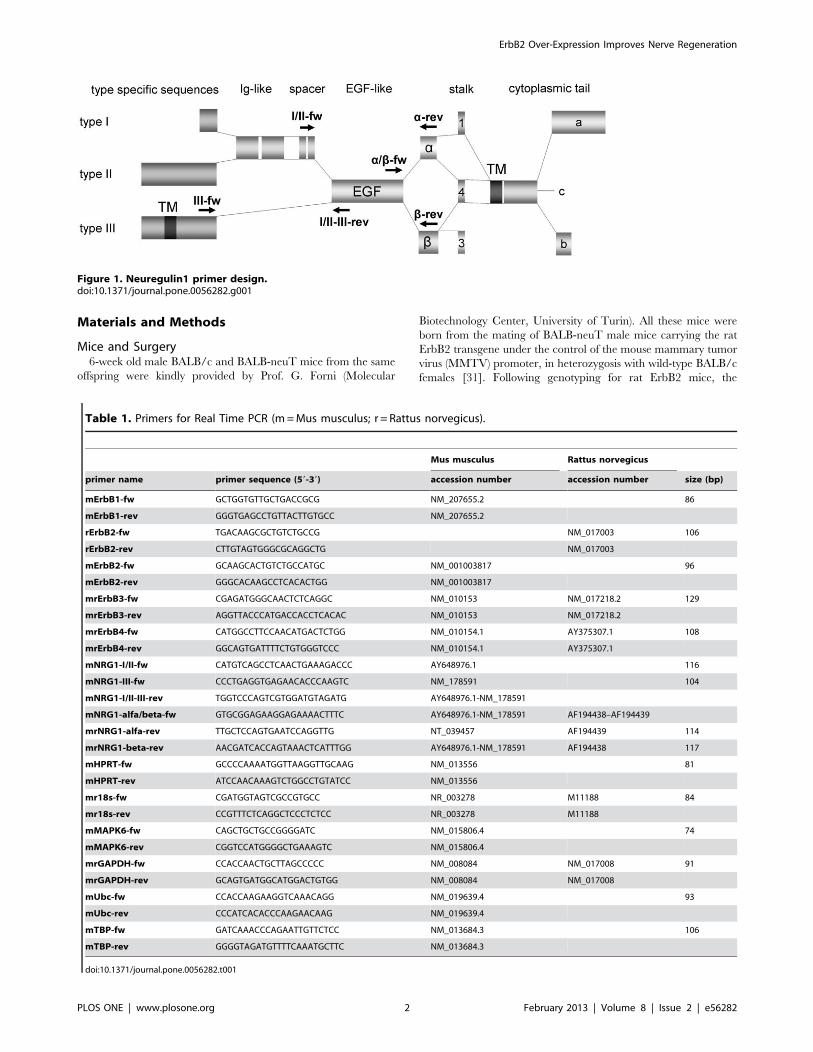
Figure 1. Neuregulin1 primer design.doi:10.1371/journal.pone.0056282.g001
Table 1. Primers for Real Time PCR (m=Mus musculus; r = Rattus norvegicus).
Mus musculus Rattus norvegicus
primer name primer sequence (59-39) accession number accession number size (bp)
mErbB1-fw GCTGGTGTTGCTGACCGCG NM_207655.2 86
mErbB1-rev GGGTGAGCCTGTTACTTGTGCC NM_207655.2
rErbB2-fw TGACAAGCGCTGTCTGCCG NM_017003 106
rErbB2-rev CTTGTAGTGGGCGCAGGCTG NM_017003
mErbB2-fw GCAAGCACTGTCTGCCATGC NM_001003817 96
mErbB2-rev GGGCACAAGCCTCACACTGG NM_001003817
mrErbB3-fw CGAGATGGGCAACTCTCAGGC NM_010153 NM_017218.2 129
mrErbB3-rev AGGTTACCCATGACCACCTCACAC NM_010153 NM_017218.2
mrErbB4-fw CATGGCCTTCCAACATGACTCTGG NM_010154.1 AY375307.1 108
mrErbB4-rev GGCAGTGATTTTCTGTGGGTCCC NM_010154.1 AY375307.1
mNRG1-I/II-fw CATGTCAGCCTCAACTGAAAGACCC AY648976.1 116
mNRG1-III-fw CCCTGAGGTGAGAACACCCAAGTC NM_178591 104
mNRG1-I/II-III-rev TGGTCCCAGTCGTGGATGTAGATG AY648976.1-NM_178591
mNRG1-alfa/beta-fw GTGCGGAGAAGGAGAAAACTTTC AY648976.1-NM_178591 AF194438–AF194439
mrNRG1-alfa-rev TTGCTCCAGTGAATCCAGGTTG NT_039457 AF194439 114
mrNRG1-beta-rev AACGATCACCAGTAAACTCATTTGG AY648976.1-NM_178591 AF194438 117
mHPRT-fw GCCCCAAAATGGTTAAGGTTGCAAG NM_013556 81
mHPRT-rev ATCCAACAAAGTCTGGCCTGTATCC NM_013556
mr18s-fw CGATGGTAGTCGCCGTGCC NR_003278 M11188 84
mr18s-rev CCGTTTCTCAGGCTCCCTCTCC NR_003278 M11188
mMAPK6-fw CAGCTGCTGCCGGGGATC NM_015806.4 74
mMAPK6-rev CGGTCCATGGGGCTGAAAGTC NM_015806.4
mrGAPDH-fw CCACCAACTGCTTAGCCCCC NM_008084 NM_017008 91
mrGAPDH-rev GCAGTGATGGCATGGACTGTGG NM_008084 NM_017008
mUbc-fw CCACCAAGAAGGTCAAACAGG NM_019639.4 93
mUbc-rev CCCATCACACCCAAGAACAAG NM_019639.4
mTBP-fw GATCAAACCCAGAATTGTTCTCC NM_013684.3 106
mTBP-rev GGGGTAGATGTTTTCAAATGCTTC NM_013684.3
doi:10.1371/journal.pone.0056282.t001
ErbB2 Over-Expression Improves Nerve Regeneration
PLOS ONE | www.plosone.org 2 February 2013 | Volume 8 | Issue 2 | e56282
offspring was divided in wild type (BALB/c) mice and transgenic
heterozygous (BALB-neuT) mice, over-expressing ErbB2. Only
male mice were studied since BALB-neuT female mice develop
fast growing mammary carcinomas [31,32]. A total of 32 mice
were used for this study: 4 mice were used for the western blot
analysis, 10 mice were used for the grasping test, morphological
and stereological analysis (5 BALB/c and 5 BALB-neuT) and 18
mice were used for the quantitative real time PCR analysis (9
BALB/c and 9 BALB-neuT).
All mouse experiments were performed with the approval of the
local Institution’s Animal Care and Ethics Committee of the
University of Turin and in accordance with the European
Communities Council Directive of 24 November 1986 (86/609/
EEC). Mice were housed in a temperature and humidity
controlled room with 12–12 h light/dark cycles and fed with
standard chow and water ad libitum. Measures were taken to
minimize pain and discomfort taking into account human
endpoints for animal suffering and distress.
For the surgery, mice were deeply anaesthetized via intraper-
itoneal injection with ketamine (9 mg/100 g-body weight),
xylazine (1.25 mg/100 g-body weight) and atropine (0.025 mg/
100 g-body weight). The median nerve of the left forelimb was
approached from the axillary region to the elbow. Under operative
microscope, the median nerve was carefully exposed and the crush
lesion was performed by compressing the nerve for 30 seconds
with a non-serrated clamp (manufactured by the Institute of
Industrial Electronic and Material Sciences, University of Tech-
nology, Vienna, Austria) [33,34]. The force applied to the nerve
was 61.3 N giving a final pressure of 20.43 Mpa [35]. In order to
prevent interferences with the grasping test, the contra-lateral
median nerve (right arm) was transected at the middle third of the
brachium and its proximal stump was sutured to the pectoralis
major muscle to avoid spontaneous reinnervation. The contro-
lateral uninjured median nerves were collected and used as
control. We therefore obtained four experimental groups: 1)
uninjured BALB/c nerves; 2) uninjured BALB-neuT nerves; 3)
injured BALB/c nerves; 4) injured BALB-neuT nerves.
For quantitative real time PCR analysis three independent
experiments were performed. For each experiment, six mice were
used: three BALB/c and three BALB-neuT mice. To obtain an
adequate amount of RNA, all animals underwent a unilateral
crush lesion on the median, ulnar and radial nerves following the
same protocol described above.
Postoperative Assessment of Functional RecoveryGrasping test was carried out weekly starting from the day
before the surgery until day-28. Grasping test was performed using
the BS-GRIP Grip Meter (2 Biological Instruments, Varese, Italy),
which is represented by a precision dynamometer connected to
a grid. Briefly, the mice were elevated by the tail and allowed to
grasp the grid [36]. The dynamometer records the maximum
weight that the mouse manages to hold up before losing the grip.
The maximal strength monitored by the dynamometer is an
indicator of muscle function. For each time point, each mouse was
tested three times and the average value was recorded.
ImmunocytochemistryMedian nerves from uninjured BALB/c and BALB-neuT mice
were fixed in 4% paraformaldehyde for 2 h, washed in a solution
of 0.01 M PBS (pH 7.2) for 30 min, dehydrated, and embedded in
paraffin. Sections were cut 8–10 mm thick, permeabilized, blocked
[0.1% triton X-100, 10% normal goat serum (NGS)/0.02%
NaN3, 1 h] and processed for an immunohistochemical protocol.
Mouse monoclonal primary antibody against ErbB2 (dilution 1:40,
Oncogene), rabbit polyclonal primary antibody against GFAP
(dilution 1:600, Sigma) and mouse monoclonal primary antibody
against tubulin bIII (dilution 1:1000, Sigma) were applied
overnight at room temperature for single or double staining.
Subsequently, the sections were incubated for 1 hour at room
temperature with goat a-rabbit IgG Cy3 (dilution 1:200 Jackson
ImmunoResearch) and/or goat a-mouse Alexa488 (dilution 1:200,
Molecular Probes). The samples were finally mounted with a Dako
fluorescent mounting medium and analyzed by a LSM 510
confocal laser microscopy system (Zeiss, Jena, Germany).
Resin Embedding, High-resolution Light Microscopy andElectron MicroscopyUnder deep anesthesia, mice were sacrificed at day-28 post-
injury and 5-mm long segment of the median nerve distal to the
site of the crush lesion from both BALB/c and BALB-neuT mice
was removed. A 4/0 stitch was used to mark the proximal stump
of the nerve segment. Nerve samples were fixed and embedded as
previously described [37].
Transversal 2.5 mm cross sections were obtained ,2 to 2.5 mm
distal to the lesion site of the withdrawn median nerve sample
using an Ultracut UCT ultramicrotome (Leica Microsystems,
Wetzlar, Germany) and stained by toluidine blue for high-
resolution light microscopy examination and design-based quan-
titative morphology. Photomicrographs were taken using
a DM4000B microscope equipped with a DFC320 digital camera
(Leica Microsystems, Wetzlar, Germany) and slightly adjusted for
brightness and contrast to obtain uniform plates.
Electron microscopy was performed on the same specimens
used for high-resolution light microscopy. Ultra-thin sections (70-
nm thick) were cut immediately after the series of semithin section
with the same ultramicrotome and double stained with saturated
aqueous solution of uranyl acetate and lead citrate. Ultra-thin
sections were analyzed using a JEM-1010 transmission electron
microscope (JEOL, Tokyo, Japan) equipped with a Mega-View-III
digital camera and a Soft-Imaging-System (SIS, Munster,
Germany).
Design-based Quantitative Morphology of Nerve FiberRegenerationDesign-based quantitative morphology was carried out on one
randomly selected toluidine blue stained semi-thin section.
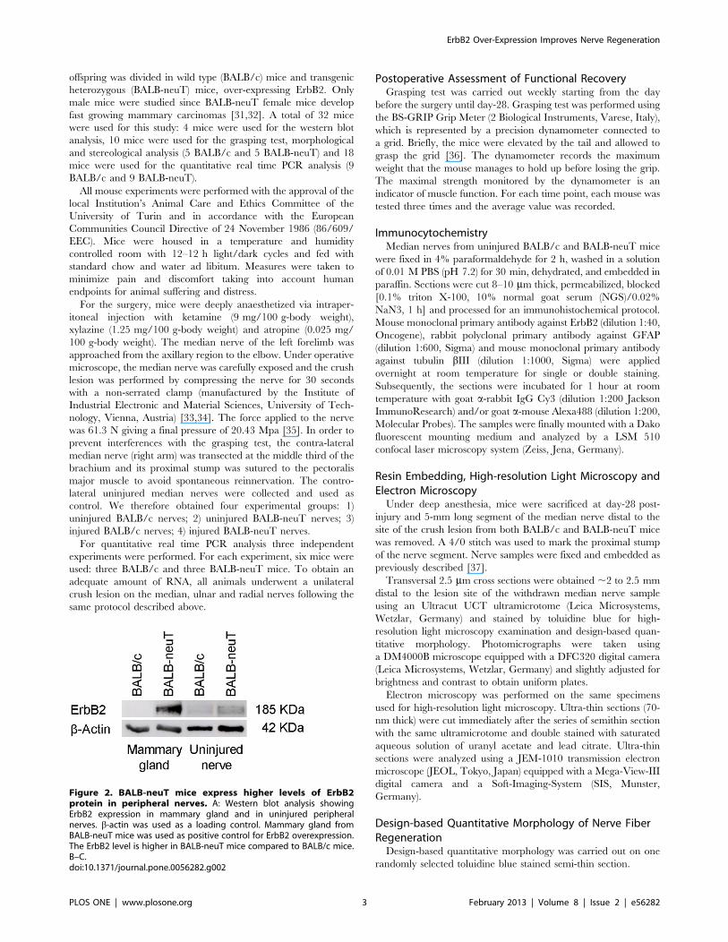
Figure 2. BALB-neuT mice express higher levels of ErbB2protein in peripheral nerves. A: Western blot analysis showingErbB2 expression in mammary gland and in uninjured peripheralnerves. b-actin was used as a loading control. Mammary gland fromBALB-neuT mice was used as positive control for ErbB2 overexpression.The ErbB2 level is higher in BALB-neuT mice compared to BALB/c mice.B–C.doi:10.1371/journal.pone.0056282.g002
ErbB2 Over-Expression Improves Nerve Regeneration
PLOS ONE | www.plosone.org 3 February 2013 | Volume 8 | Issue 2 | e56282

Stereological analysis of myelinated nerve fibers was carried out
using a protocol previously described [38,39,40]. On the randomly
selected section, the total cross-sectional area of the nerve was
measured and then an adequate number of fields (6–8) were
selected using a systematic random sampling protocol [38,41].
In each sampling field, the ‘‘edge effect’’ was avoided by
employing a two-dimensional dissector procedure which is based
on sampling the ‘‘tops’’ of fibers [42,43].
Mean fiber density was then calculated by dividing the total
number of nerve fibers within the sampling field (N) by its area (N/
mm2). The total number of fibers was estimated by multiplying the
mean fiber density by the total cross-sectional area of the whole
nerve cross section. In addition, in each fiber, both fiber and axon
area were measured and the fiber (D), axon (d) diameter, myelin
thicknessD{dð Þ2
h iand g-ratio d
D
� �� �were calculated.
Hence, we analyze the g-ratio/axon diameter correlation of
individual fibers by means of scatterplots, evaluating the differ-
ences in linear regression. Furthermore, we deepen that analysis
by means of predictive inference: considering uninjured group g-
ratio/axon diameter scatterplot as reference, we highlight
a graphical area bounded by 95% prediction interval of regression
line and 62 s of axon diameter. Statistically, about 90% of
functional myelinated fibers will fall inside this predictive area.
At the electron microscopic level, the number of Schwann cells
was quantified by counting the nuclear profiles numbers in
a defined area at 80006magnification (12,2616,2 mm2). The
‘‘edge effect’’ was avoided according to the stereological method
previously described [42,43].
Total Protein Extraction and Western Blot AnalysisTotal proteins were obtained from uninjured and crushed (2
days post-injury) BALB/c and BALB-neuT mice sacrificed under
deep anesthesia. A 6 mm segment of median, ulnar and radial
nerves just proximal and distal to the crushed site were harvested
for each animal.
Total proteins were extracted in boiling Laemmli buffer (2.5%
SDS, 0.125 M Tris-HCl, pH 6.8), followed by 3 min denaturation
at 100uC. Protein concentration was determined by the BCA
method, and equal amounts of proteins (50 mg) were denaturated
3 min at 100uC in 240 mM beta-mercaptoethanol, 18% glycerol,
Figure 3. ErbB2 transgene is expressed by Schwann cells. Immunohistochemistry analysis showing the localization of ErbB2 protein in BALB-neuT median merve. A, tubulinbIII staining; B, GFAP staining; C, ErbB2 staining; D: colocalization between GFAP and ErbB2 showing that the ErbB2protein is expressed by Schwann.Scale bar: 10 mm.doi:10.1371/journal.pone.0056282.g003
ErbB2 Over-Expression Improves Nerve Regeneration
PLOS ONE | www.plosone.org 4 February 2013 | Volume 8 | Issue 2 | e56282

loaded onto each lane, separated by SDS-PAGE, transferred to
a HybondTM C Extra membrane (Amersham Biosciences,
General Electric Healthcare Europe) and analyzed as previously
described [44]. The western blot was decorated with a polyclonal
primary antibody anti ErbB2 (Neu (C-18) # sc-284, 1:500 w.d.,
Santa Cruz Biotechnology, Inc., Santa Cruz, CA, USA) and the
horseradish peroxidase-linked donkey anti-rabbit secondary anti-
body (#NA934, 1:10.000 w.d., Amersham Biosciences).
RNA Extraction and Quantitative Real-time PCRTo obtain suitable amount of material, total RNA was extracted
from pools of median, ulnar and radial nerves of three mice for
each of three independent experiments. Under deep anesthesia,
mice were sacrificed at day-2 post-injury. A 6 mm segment of the
nerves just proximal and distal to the crushed site were harvested
for each animal. Total RNA was isolated using the RNeasy Mini
Kit (Qiagen S.r.l., Milano, Italy) according to the manufacturer’s
protocol.
0.5 mg total RNA were subjected to a reverse transcriptase (RT)
reaction in 25 ml reaction volume containing: 16 RT-Buffer
(Fermentas, Burlington, Canada); 0.1 mg/ml bovine serum albu-
min (BSA, Sigma); 0.05% Triton X-100; 1 mM dNTPs; 7.5 mMrandom exanucleotide primers (Fermentas); 1 U/ml RIBOlock
(Fermentas) and 200 U RevertAidTM M-MuLV reverse transcrip-
tase (Fermentas). The reaction was performed for 10 min at 25uC,90 min at 42uC, 10 min at 90uC.Quantitative real-time PCR for ErbB1, ErbB2, ErbB3, ErbB4,
NRG1-alpha, NRG1-beta, NRG1-I/II and NRG1-III was
performed using an ABI Prism 7300 (Applied Biosystems, Life
Technologies Europe BV, Monza, Italy) detection system using
SYBR Green chemistry. cDNA was diluted 10 times before
analysis and 5 ml were analyzed in a total volume of 25 mlcontaining 16 PowerGREEN Master Mix (Applied Biosystems),
and 300 nM of each primer. The reactions were carried out in 40
cycles (primer annealing temperature: 60uC). For each cDNA
sample, three technical replicates were averaged and dissociation
curves were routinely performed to check for the presence of
a single peak corresponding to the required amplicon. Normalized
reporter fluorescence (Rn) for each cycle was obtained by
normalizing SYBRGreen to ROX signal.
The data from the real-time PCR experiments were analyzed
using the DDCt method for the relative quantification. The
threshold cycle number (Ct) values of both the calibrator and the
samples of interest were normalized to the geometric average of six
endogenous housekeeping genes: Ubiquitin C (UbC), TATA box
binding protein (TBP), 18S ribosomal RNA (18S rRNA),
glyceraldehyde-3-phosphate dehydrogenase (GAPDH), hypoxan-
thine phosphoribosyltransferase 1 (HPRT1) and the signaling
molecule mitogen-activated protein kinase 6 (MAPK6). As
calibrator we used the RNA obtained from a pool of uninjured
nerves.
Primers were designed using Annhyb software (http://www.
bioinformatics.org/annhyb/) and synthesized by Invitrogen (Life
Technologies Europe BV, Monza, Italy). Primers sequences are
reported in Table 1. Neuregulin1 primers design was made as
summarized in Figure 1.
Statistical AnalysisStatistical analysis was performed using PASW statistics 18
(SPSS Inc., Chicago, IL, USA). For the values taken from the
different time-point assessments of the grasping test, one-way
repeated measures analysis of variance (RM-ANOVA) test
followed by post hoc multiple pair-wise comparisons using the
Student–Neuman–Keuls (SNK) test was used. For both stereolog-
ical and grasping test data, the N size used in the statistical
calculations was the number of animals (n = 5 for each experi-
mental group). Differences in predictive area fitting are evaluated
by Chi-Square test considering the number of fibers falling inside
the area versus those falling outside.
For quantitative real time PCR data, statistical analysis was
performed using the one-way ANOVA test followed by Bonferroni
post-hoc multiple comparison test. The interaction between the
effect of injury and ErbB2 over-expression was investigated by
two-way ANOVA test.
Results
BALB-neuT Mice Express Higher Levels of ErbB2 Proteinin the Peripheral Nervous SystemTo study the role of ErbB2 overexpression in physiological
conditions and after crush injury of peripheral nerves, we
performed a preliminary analysis to evaluate protein expression
in BALB-neuT mice carrying the rat neu/ErbB2 gene under the
control of the mouse mammary tumor virus (MMTV) promoter.
We detected a higher level of ErbB2 protein in the median, ulnar
and radial nerves of BALB-neuT compared with BALB/c mice
(Fig. 2A).
Figure 3 shows immune-labeling for tubulinbIII (A), GFAP (B)
and ErbB2 (C). Double labeling with GFAP and ErbB2 protein (D)
shows the co-localization between the two markers in Schwann
cells (Fig. 3D).
ErbB2 Over-expression has No Effects on Mice MedianNerve in Physiological ConditionsAlthough BALB-neuT mice weighted significantly (p,0.05) less
(23.22 g 61.38 g at the day of the sacrifice) than the BALB/c
(26.92 g 60.38 g at the day of the sacrifice), they showed no
evident behavioral abnormalities. In particular, no signs of motor
and sensory impairment were detectable in BALB-neuT compared
to BALB/c mice. Motor function was tested by means of the
grasping test the day before the injury (Fig. 4, day-1) and no
deficits were recorded in transgenic mice.
Figure 4. 14 days after the injury, grasping strength is greaterin BALB-neuT than in BALB/c mice. Graph showing the perfor-mance of the mice in the grasping test after normalizing the recordedvalues with the animal body weights. Data are reported as means 6SEM (*BALB/c vs BALB-neuT; # regenerating vs pre-injury wild-type,p,0.05).doi:10.1371/journal.pone.0056282.g004
ErbB2 Over-Expression Improves Nerve Regeneration
PLOS ONE | www.plosone.org 5 February 2013 | Volume 8 | Issue 2 | e56282

Figure 5. Morphological and stereological analysis of uninjured median nerves do not show detectable differences. A, B:representative light micrographs of transverse sections of median nerves stained with toluidine blue of BALB/c and BALB-neuT mice respectively. Inboth groups myelinated axons have a normal morphological appearance. Bar = 10 mm. C, D, E, F: histograms showing the results of
ErbB2 Over-Expression Improves Nerve Regeneration
PLOS ONE | www.plosone.org 6 February 2013 | Volume 8 | Issue 2 | e56282
To determine if any peripheral structural parameter correlates
with the over-expression of ErbB2, we compared semi-thin
sections stained with toluidine blue from the median nerve of
BALB/c (Fig. 5A) and BALB-neuT (Fig. 5B) mice. No significant
morphological differences were detected between the two groups.
Stereological analysis showed that the cross sectional area (Fig. 5C),
the number of total myelinated fibers (Fig. 5D) and the myelinated
fiber density (Fig. 5E) were not significantly different between
BALB/c and BALB-neuT mice. Also the parameters related to the
fiber size (axon diameter, fiber diameter and myelin thickness)
were not significantly different between BALB/c and BALB-neuT
mice (Fig. 5F). This result was further confirmed by g-ratio
measurement (expressed as scatterplots) of median nerve fibers
from BALB/c (0.6960.01) and BALB-neuT mice (0.7160.02)
(Fig. 5G).
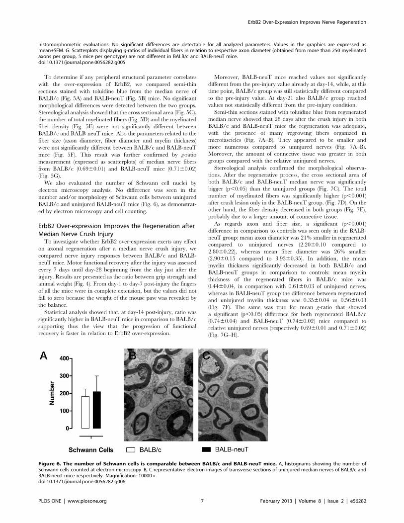
We also evaluated the number of Schwann cell nuclei by
electron microscopy analysis. No difference was seen in the
number and/or morphology of Schwann cells between uninjured
BALB/c and uninjured BALB-neuT mice (Fig. 6), as demonstrat-
ed by electron microscopy and cell counting.
ErbB2 Over-expression Improves the Regeneration afterMedian Nerve Crush InjuryTo investigate whether ErbB2 over-expression exerts any effect
on axonal regeneration after a median nerve crush injury, we
compared nerve injury responses between BALB/c and BALB-
neuT mice. Motor functional recovery after the injury was assessed
every 7 days until day-28 beginning from the day just after the
injury. Results are presented as the ratio between grip strength and
animal weight (Fig. 4). From day-1 to day-7 post-injury the fingers
of all the mice were in complete extension, but the values did not
fall to zero because the weight of the mouse paw was revealed by
the balance.
Statistical analysis showed that, at day-14 post-injury, ratio was
significantly higher in BALB-neuT mice in comparison to BALB/c
supporting thus the view that the progression of functional
recovery is faster in relation to ErbB2 over-expression.
Moreover, BALB-neuT mice reached values not significantly
different from the pre-injury value already at day-14, while, at this
time point, BALB/c group was still statistically different compared
to the pre-injury value. At day-21 also BALB/c group reached
values not statistically different from the pre-injury condition.
Semi-thin sections stained with toluidine blue from regenerated
median nerve showed that 28 days after the crush injury in both
BALB/c and BALB-neuT mice the regeneration was adequate,
with the presence of many regrowing fibers organized in
microfascicles (Fig. 7A–B). They appeared to be smaller and
more numerous compared to uninjured nerves (Fig. 7A–B).
Moreover, the amount of connective tissue was greater in both
groups compared with the relative uninjured nerves.
Stereological analysis confirmed the morphological observa-
tions. After the regenerative process, the cross sectional area of
both BALB/c and BALB-neuT median nerve was significantly
bigger (p,0.05) than the uninjured groups (Fig. 7C). The total
number of myelinated fibers was significantly higher (p,0.001)
after crush lesion only in the BALB-neuT group. (Fig. 7D). On the
other hand, the fiber density decreased in both groups (Fig. 7E),
probably due to a larger amount of connective tissue.
As regards axon and fiber size, a significant (p,0.001)
difference in comparison to controls was seen only in the BALB-
neuT group: mean axon diameter was 21% smaller in regenerated
compared to uninjured nerves (2.2060.10 compared to
2.8060.22), whereas mean fiber diameter was 26% smaller
(2.9060.15 compared to 3.9360.35). In addition, the mean
myelin thickness significantly decreased in both BALB/c and
BALB-neuT groups in comparison to controls: mean myelin
thickness of the regenerated fibers in BALB/c mice was
0.4460.04, in comparison with 0.6160.03 of uninjured nerves,
whereas in BALB-neuT group the difference between regenerated
and uninjured myelin thickness was 0.3560.04 vs 0.5660.08
(Fig. 7F). The same was true for mean g-ratio that showed
a significant (p,0.05) difference for both regenerated BALB/c
(0.7460.04) and BALB-neuT (0.7460.02) mice compared to
relative uninjured nerves (respectively 0.6960.01 and 0.7160.02)
(Fig. 7G–H).
histomorphometric evaluations. No significant differences are detectable for all analyzed parameters. Values in the graphics are expressed asmean+SEM. G: Scatterplots displaying g-ratios of individual fibers in relation to respective axon diameter (obtained from more than 250 myelinatedaxons per group, 5 mice per genotype) are not different in BALB/c and BALB-neuT mice.doi:10.1371/journal.pone.0056282.g005
Figure 6. The number of Schwann cells is comparable between BALB/c and BALB-neuT mice. A, histognams showing the number ofSchwann cells counted at electron microscopy. B, C representative electron images of transverse sections of uninjured median nerves of BALB/c andBALB-neuT mice respectively. Magnification: 100006.doi:10.1371/journal.pone.0056282.g006
ErbB2 Over-Expression Improves Nerve Regeneration
PLOS ONE | www.plosone.org 7 February 2013 | Volume 8 | Issue 2 | e56282

In order to explain the enhanced function recovery seen in
BALB-neuT group, we have evaluated the percentage of
myelinated fibers falling inside a g-ratio/axon diameter predictive
area in which fibers are considered as functional. By the
superposition of uninjured BALB-neuT and uninjured BALB/c
scatterplots, we have found that the 92.95% of BALB-neuT fibers
Figure 7. Morphological and stereological analysis of the regenerated median nerves show significant differences. A, B:representative light micrographs of transverse sections of median nerves stained with toluidine blue of BALB/c and BALB-neuT mice, respectively.Bar = 10 mm. C, D, E, F: histograms showing the results of histomorphometric evaluations of nerve regeneration. After the regenerative process, BALB-neuT nerves show more and smaller myelinated fibers compared to uninjured ones. Histograms are represented as percentage variation ofregenerated nerves with respect to relative uninjured nerves; values in the graphics are expressed as mean+SEM). G, H: scatterplots displaying g-ratios of individual fibers in relation to respective axon diameter (obtained from more than 250 myelinated axons per group, 5 mice per genotype)show that in both BALB/c (G) and BALB-neuT (H) mice g-ratio is higher after injury. Statistical significance: *p,0.05, **p,0.01, ***p,0.001.doi:10.1371/journal.pone.0056282.g007
ErbB2 Over-Expression Improves Nerve Regeneration
PLOS ONE | www.plosone.org 8 February 2013 | Volume 8 | Issue 2 | e56282
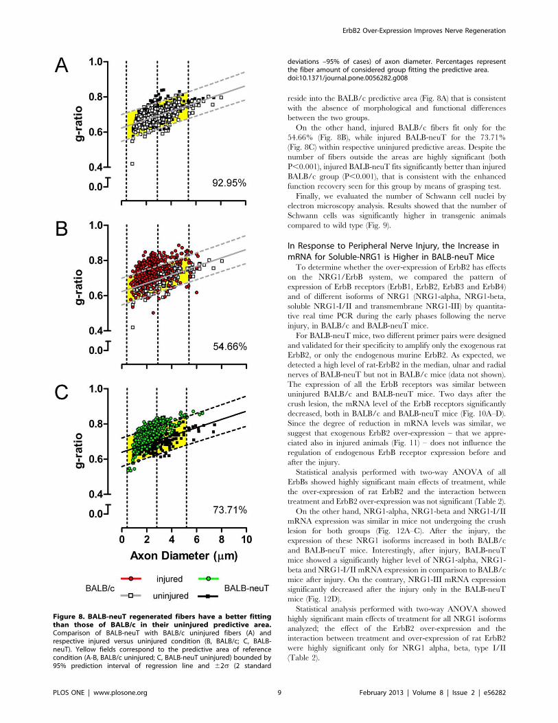
reside into the BALB/c predictive area (Fig. 8A) that is consistent
with the absence of morphological and functional differences
between the two groups.
On the other hand, injured BALB/c fibers fit only for the
54.66% (Fig. 8B), while injured BALB-neuT for the 73.71%
(Fig. 8C) within respective uninjured predictive areas. Despite the
number of fibers outside the areas are highly significant (both
P,0.001), injured BALB-neuT fits significantly better than injured
BALB/c group (P,0.001), that is consistent with the enhanced
function recovery seen for this group by means of grasping test.
Finally, we evaluated the number of Schwann cell nuclei by
electron microscopy analysis. Results showed that the number of
Schwann cells was significantly higher in transgenic animals
compared to wild type (Fig. 9).
In Response to Peripheral Nerve Injury, the Increase inmRNA for Soluble-NRG1 is Higher in BALB-neuT MiceTo determine whether the over-expression of ErbB2 has effects
on the NRG1/ErbB system, we compared the pattern of
expression of ErbB receptors (ErbB1, ErbB2, ErbB3 and ErbB4)
and of different isoforms of NRG1 (NRG1-alpha, NRG1-beta,
soluble NRG1-I/II and transmembrane NRG1-III) by quantita-
tive real time PCR during the early phases following the nerve
injury, in BALB/c and BALB-neuT mice.
For BALB-neuT mice, two different primer pairs were designed
and validated for their specificity to amplify only the exogenous rat
ErbB2, or only the endogenous murine ErbB2. As expected, we
detected a high level of rat-ErbB2 in the median, ulnar and radial
nerves of BALB-neuT but not in BALB/c mice (data not shown).
The expression of all the ErbB receptors was similar between
uninjured BALB/c and BALB-neuT mice. Two days after the
crush lesion, the mRNA level of the ErbB receptors significantly
decreased, both in BALB/c and BALB-neuT mice (Fig. 10A–D).
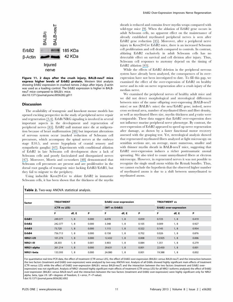
Since the degree of reduction in mRNA levels was similar, we
suggest that exogenous ErbB2 over-expression – that we appre-
ciated also in injured animals (Fig. 11) – does not influence the
regulation of endogenous ErbB receptor expression before and
after the injury.
Statistical analysis performed with two-way ANOVA of all
ErbBs showed highly significant main effects of treatment, while
the over-expression of rat ErbB2 and the interaction between
treatment and ErbB2 over-expression was not significant (Table 2).
On the other hand, NRG1-alpha, NRG1-beta and NRG1-I/II
mRNA expression was similar in mice not undergoing the crush
lesion for both groups (Fig. 12A–C). After the injury, the
expression of these NRG1 isoforms increased in both BALB/c
and BALB-neuT mice. Interestingly, after injury, BALB-neuT
mice showed a significantly higher level of NRG1-alpha, NRG1-
beta and NRG1-I/II mRNA expression in comparison to BALB/c
mice after injury. On the contrary, NRG1-III mRNA expression
significantly decreased after the injury only in the BALB-neuT
mice (Fig. 12D).
Statistical analysis performed with two-way ANOVA showed
highly significant main effects of treatment for all NRG1 isoforms
analyzed; the effect of the ErbB2 over-expression and the
interaction between treatment and over-expression of rat ErbB2
were highly significant only for NRG1 alpha, beta, type I/II
(Table 2).
Figure 8. BALB-neuT regenerated fibers have a better fittingthan those of BALB/c in their uninjured predictive area.Comparison of BALB-neuT with BALB/c uninjured fibers (A) andrespective injured versus uninjured condition (B, BALB/c; C, BALB-neuT). Yellow fields correspond to the predictive area of referencecondition (A-B, BALB/c uninjured; C, BALB-neuT uninjured) bounded by95% prediction interval of regression line and 62s (2 standard
deviations –95% of cases) of axon diameter. Percentages representthe fiber amount of considered group fitting the predictive area.doi:10.1371/journal.pone.0056282.g008
ErbB2 Over-Expression Improves Nerve Regeneration
PLOS ONE | www.plosone.org 9 February 2013 | Volume 8 | Issue 2 | e56282

Figure 9. The number of Schwann cells is higher in BALB-neuT mice after regeneration. A, histognams showing the number of Schwanncells counted at electron microscopy. B, C representative electron images of transverse sections of regenerated median nerves of BALB/c and BALB-neuT mice respectively. Magnification: 100006. Statistical significance: *p,0.05.doi:10.1371/journal.pone.0056282.g009
Figure 10. ErbB expression decreases following lesion, and is not influenced by rat ErbB2 over-expression. The relative quantificationof different ErbB receptors was obtained by quantitative real-time RT-PCR: data were normalized to the geometric mean of six endogenoushousekeeping genes (TBP, UbC, 18S, GAPDH, HPRT1 and MAPK6) and expressed as percentage. Values in the graphics are expressed as mean+SEM.Statistical analysis demonstrates that ErbB1, ErbB2, ErbB3 and ErbB4 expression level significantly decreased two days after the crush lesion(**p,0.01, ***p,0.001). No significant differences were observed between BALB/c and BALB-neuT mice both before and after the injury.doi:10.1371/journal.pone.0056282.g010
ErbB2 Over-Expression Improves Nerve Regeneration
PLOS ONE | www.plosone.org 10 February 2013 | Volume 8 | Issue 2 | e56282
Discussion
The availability of transgenic and knockout mouse models has
opened exciting perspective in the study of peripheral nerve repair
and regeneration [3,4]. ErbB/NRG signaling is involved in several
important aspects in the development and regeneration of
peripheral nerves [45]. ErbB2 null mutant mice die at midgesta-
tion because of heart malformation [46] but important alterations
of nervous system occur (marked reduction of Schwann cell
precursors, which accompany the spinal nerves at the embryo
stage E10.5, and severe hypoplasia of cranial sensory and
sympathetic ganglia) [47]. Experiments with conditional ablation
of ErbB2 in late Schwann cell development show a lack of
Schwann cells and poorly fasciculated and disorganized nerves
[47]. Moreover, Morris and co-workers [48] demonstrated that
Schwann cell precursors are present and are proliferative in the
dorsal root ganglia of transgenic mice lacking ErbB2/ErbB3, but
they fail to migrate to the periphery.
Using inducible Krox20-Cre to ablate ErbB2 in immature
Schwann cells, it has been shown that the thickness of the myelin
sheath is reduced and contains fewer myelin wraps compared with
wild-type mice [9]. When the ablation of ErbB2 gene occurs in
adult Schwann cells, no apparent effect on the maintenance of
already established myelinated peripheral nerves is seen after
ErbB2 gene reduction [45]. Moreover, after a peripheral nerve
injury in Krox20-Cre ErbB2 mice, there is an increased Schwann
cell proliferation and cell death compared to controls. In contrast,
ablating ErbB2 exclusively in adult Schwann cells has no
detectable effect on survival and cell division after injury. Thus,
Schwann cell responses to axotomy depend on the timing of
ErbB2 ablation [45].
While the effects of ErbB2 deletion in the peripheral nervous
system have already been analyzed, the consequences of its over-
expression have not been investigated to date. To fill this gap, we
examined the effect of the over-expression of ErbB2 on healthy
nerve and its role on nerve regeneration after a crush injury of the
median nerve.
We examined the peripheral nerves of healthy adult mice and
we did not detect morphological and stereological differences
between mice of the same offspring over-expressing (BALB-neuT
mice) or not (BALB/c mice) the neu/ErbB2 gene; indeed, nerve
cross sectional area, number of myelinated fibers and fiber density,
as well as myelinated fibers size, myelin thickness and g-ratio were
comparable. These data suggest that ErbB2 over-expression does
not influence murine peripheral nerve phenotype. By contrast, the
over-expression of ErbB2 appeared to speed up nerve regeneration
after damage, as shown by a faster functional motor recovery
assessed with the grasping test. Yet, stereological analysis showed
that regenerated myelinated fibers analyzed at light microscopy on
semithin sections are, on average, more numerous, smaller and
with thinner myelin sheath in BALB-neuT mice, suggesting that
ErbB2 over-expression induces a richer posttraumatic axonal
sprouting. We also tried to count unmyelinated fibers at electron
microscopy. However, in regenerated nerves it was not possible to
recognize the single small axons within the Remak bundles. Thus,
we cannot exclude the hypothesis that the observed higher number
of myelinated axons is due to a shift between unmyelinated to
myelinated axons.
Figure 11. 2 days after the crush injury, BALB-neuT miceexpress higher levels of ErbB2 protein. Western blot analysisshowing ErbB2 expression in crushed nerves 2-days after injury. b-actinwas used as a loading control. The ErbB2 expression is higher in BALB-neuT mice compared to BALB/c mice.doi:10.1371/journal.pone.0056282.g011
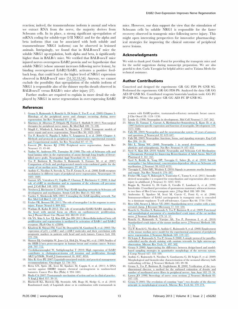
Table 2. Two-way ANOVA statistical analysis.
factor TREATMENT ErbB2 over-expression TREATMENT vs
(CTR vs LES) (WT vs ErbB2) ErbB2 over-expression
F df, E P F df, E P F df, E P
ErbB1 249.377 1, 8 0.000 6.078 1, 8 0.059 0.725 1, 8 0.419
ErbB2 242.377 1, 8 0.000 3.348 1, 8 0.105 0.009 1, 8 0.927
ErbB3 73.720 1, 8 0.000 1.115 1, 8 0.322 0.145 1, 8 0.954
ErbB4 756.713 1, 8 0.000 0.158 1, 8 0.702 0.026 1, 8 0.876
NRG1-I/II 101.274 1, 8 0.000 12.432 1, 8 0.008 13.925 1, 8 0.006
NRG1-III 28.303 1, 8 0.001 3.903 1, 8 0.084 1.351 1, 8 0.279
NRG1-alpha 261.214 1, 8 0.000 29.631 1, 8 0.001 22.450 1, 8 0.001
NRG1-beta 133.593 1, 8 0.000 24.080 1, 8 0.001 19.388 1, 8 0.002
For quantitative real time PCR data, the effect of treatment (CTR versus LES), the effect of ErbB2 over-expression (BALB/c versus BALB-neuT) and the interaction betweenthe two factors (treatment and ErbB2 over-expression) were analyzed by two-way ANOVA test. Analysis of all ErbBs showed highly significant main effects of treatment(CTR versus LES) while the effect of ErbB2 over-expression (BALB/c versus BALB-neuT) and the interaction between the two factors (treatment and ErbB2 over-expression) was not significant. Analysis of NRG1 showed highly significant main effects of treatment (CTR versus LES) for all NRG1 isoforms analyzed; the effect of ErbB2over-expression (BALB/c versus BALB-neuT) and the interaction between the two factors (treatment and ErbB2 over-expression) were highly significant only for NRG1alpha, beta, type I/II. (df = degrees of freedom, E = error, P = P-value).doi:10.1371/journal.pone.0056282.t002
ErbB2 Over-Expression Improves Nerve Regeneration
PLOS ONE | www.plosone.org 11 February 2013 | Volume 8 | Issue 2 | e56282

Since it has been shown that NRG1 stimulates the growth of
Schwann cells through the activation of ErbB2/ErbB3 hetero-
dimers [49,50], we hypothesize that the constitutive over-
expression of ErbB2 receptor makes Schwann cells more sensitive
to NRG1, which is significantly up-regulated already in wild type
mice. Up-regulated soluble NRG1 activates ErbB2/ErbB3 and,
through a positive feedback loop (helped by ErbB2 overexpres-
sion), augments its expression stimulating Schwann cell pro-
liferation [51,52,53,54]. Higher Schwann cell proliferation,
confirmed by electron microscopy quantification, could be at the
basis of the changes that we observed during posttraumatic nerve
regeneration. Nevertheless, we can’t exclude the possibility that
some of the effects observed in the transgenic mice are due to
enhanced ErbB2 signaling also in the axons.
To analyze the expression level of all ErbB family members and
of the different NRG1 isoforms, we performed relative quantifi-
cation using real time qPCR analysis. Peripheral nerve injury
interferes with the expression of many genes: the identification of
the optimal reference gene (whose expression level is constant in
both control and injured samples) is therefore an important issue
to deal with. Taking inspiration from the literature about accurate
normalization [55,56] we identified six good candidate house-
keeping genes whose expression is expected to be stable even after
nerve injury and we normalized the gene expression of ErbB and
NRG1 genes to the geometric average of these six housekeeping
genes.
We observed a strong significant decrease of mRNA expression
of all ErbB receptors in lesioned mice, both in BALB/c and
BALB-neuT mice.
The down-regulation of ErbB receptors is in contrast with data
of other authors [57]. However, this discrepancy can be explained
since our injury model and our analysis conditions are different:
they performed a surgical transection, while we carried out a crush
lesion; they observe a protein increase 5 days after axotomy, while
we are analyzing transcript expression 2 days after the crush
injury. The stability of protein and transcript can be different and
a decrease in mRNA at 2 days is not incompatible with an increase
of protein at 5 days.
We observed also down-regulation of transmembrane-type III
NRG1 in transgenic mice. Anyway, the expression level of this
isoform in our samples was already really low, as suggested by the
high threshold cycle of this isoform achieved in the qPCR
Figure 12. Soluble NRG1 expression increases following lesion, and its expression is positively influenced by ErbB2 over-expression. The relative quantification of different NRG1 isoforms was obtained by quantitative real-time RT-PCR. Results were normalized to thegeometric mean of six endogenous housekeeping genes (TBP, UbC, 18S, GAPDH, HPRT1 and MAPK6) and expressed as percentage. Values in thegraphics are expressed as mean+SEM. Statistical analysis demonstrates that NRG1-alpha, NRG1-beta and NRG1-I/II mRNA expression significantlyincreased two days after the crush lesion (*p,0.05, **p,0.01, ***p,0.001) both in BALB/c and BALB-neuT animals. Interestingly, in the BALB-neuTmice there was a significantly higher increase of mRNA expression compared to BALB/c animals. NRG1-III mRNA decreases after the crush injury, butonly in the BALB-neuT mice.doi:10.1371/journal.pone.0056282.g012
ErbB2 Over-Expression Improves Nerve Regeneration
PLOS ONE | www.plosone.org 12 February 2013 | Volume 8 | Issue 2 | e56282
reaction; indeed, the transmembrane isoform is axonal and when
we extract RNA from the nerve, the majority derives from
Schwann cells. In its place, a strong significant up-regulation of
mRNA coding for soluble-type I/II NRG1 and for the alpha and
beta isoforms (that can be associated with both soluble and
transmembrane NRG1 isoforms) can be observed in lesioned
animals. Intriguingly, we found that in BALB-neuT mice the
soluble NRG1 up-regulation, both alpha and beta, is significantly
higher than in BALB/c mice. We verified that BALB-neuT mice
injured nerves overexpress ErbB2 protein and we hypothesize that
soluble NRG1 (whose amount increases following nerve lesion), by
binding overexpressed ErbB2/ErbB3, activates a positive feed-
back loop, that could lead to the higher level of NRG1 expression
observed in BALB-neuT mice [51,52,53,54]. Anyway, we cannot
exclude the possibility that upregulation of the soluble isoform of
NRG1 is responsible also of the thinner myelin sheath observed in
BALB-neuT versus BALB/c mice after injury (27).
Further studies are required to explain in more detail the role
played by NRG1 in nerve regeneration in over-expressing ErbB2
mice. However, our data support the view that the stimulation of
Schwann cells by soluble NRG1 is responsible for the faster
recovery observed in transgenic mice following nerve injury. This
might open interesting perspectives for innovative pharmacolog-
ical strategies for improving the clinical outcome of peripheral
nerve lesions.
Acknowledgments
We wish to thank prof. Guido Forni for providing the transgenic mice and
for the useful suggestions during manuscript preparation. We are also
grateful to dott. Carla Taveggia for helpful advice and to Tiziana Merlo for
technical assistance.
Author Contributions
Conceived and designed the experiments: GR GG FDS PS GNB SG.
Performed the experiments: GR GG FDS PS. Analyzed the data: GR GG
AES IP GNB SG. Contributed reagents/materials/analysis tools: GG FC
IP GNB SG. Wrote the paper: GR GG AES FC IP GNB SG.
References
1. Geuna S, Raimondo S, Ronchi G, Di Scipio F, Tos P, et al. (2009) Chapter 3:
Histology of the peripheral nerve and changes occurring during nerve
regeneration. Int Rev Neurobiol 87: 27–46.
2. Martinez de Albornoz P, Delgado PJ, Forriol F, Maffulli N (2011) Non-surgical
therapies for peripheral nerve injury. Br Med Bull 100: 73–100.
3. Magill C, Whitlock E, Solowski N, Myckatyn T (2008) Transgenic models of
nerve repair and nerve regeneration. Neurol Res 30: 1023–1029.
4. Tos P, Ronchi G, Papalia I, Sallen V, Legagneux J, et al. (2009) Chapter 4:
Methods and protocols in peripheral nerve regeneration experimental research:
part I-experimental models. Int Rev Neurobiol 87: 47–79.
5. Fawcett JW, Keynes RJ (1990) Peripheral nerve regeneration. Annu Rev
Neurosci 13: 43–60.
6. Nadim W, Anderson PN, Turmaine M (1990) The role of Schwann cells and
basal lamina tubes in the regeneration of axons through long lengths of freeze-
killed nerve grafts. Neuropathol Appl Neurobiol 16: 411–421.
7. Tos P, Battiston B, Nicolino S, Raimondo S, Fornaro M, et al. (2007)
Comparison of fresh and predegenerated muscle-vein-combined guides for the
repair of rat median nerve. Microsurgery 27: 48–55.
8. Audisio C, Nicolino S, Scevola A, Tos P, Geuna S, et al. (2008) ErbB receptors
modulation in different types of peripheral nerve regeneration. Neuroreport 19:
1605–1609.
9. Garratt AN, Voiculescu O, Topilko P, Charnay P, Birchmeier C (2000) A dual
role of erbB2 in myelination and in expansion of the schwann cell precursor
pool. J Cell Biol 148: 1035–1046.
10. Newbern J, Birchmeier C (2010) Nrg1/ErbB signaling networks in Schwann cell
development and myelination. Semin Cell Dev Biol 21: 922–928.
11. Yarden Y, Sliwkowski MX (2001) Untangling the ErbB signalling network. Nat
Rev Mol Cell Biol 2: 127–137.
12. Fricker FR, Bennett DL (2011) The role of neuregulin-1 in the response to nerve
injury. Future Neurol 6: 809–822.
13. Wadugu B, Kuhn B (2012) The role of neuregulin/ErbB2/ErbB4 signaling in
the heart with special focus on effects on cardiomyocyte proliferation.
Am J Physiol Heart Circ Physiol 302: H2139–2147.
14. Oh YS, Shin S, Lee YJ, Kim EH, Jun HS (2011) Betacellulin-induced beta cell
proliferation and regeneration is mediated by activation of ErbB-1 and ErbB-2
receptors. PLoS One 6: e23894.
15. Khademi B, Shirazi FM, Vasei M, Doroudchi M, Gandomi B, et al. (2002) The
expression of p53, c-erbB-1 and c-erbB-2 molecules and their correlation with
prognostic markers in patients with head and neck tumors. Cancer Lett 184:
223–230.
16. Slamon DJ, Godolphin W, Jones LA, Holt JA, Wong SG, et al. (1989) Studies of
the HER-2/neu proto-oncogene in human breast and ovarian cancer. Science
244: 707–712.
17. Treekitkarnmongkol W, Suthiphongchai T (2010) High expression of ErbB2
contributes to cholangiocarcinoma cell invasion and proliferation through
AKT/p70S6K. World J Gastroenterol 16: 4047–4054.
18. Moy B, Goss PE (2007) Lapatinib-associated toxicity and practical management
recommendations. Oncologist 12: 756–765.
19. Berta GN, Sprio AE, Iezzi M, Spadaro M, Cappia S, et al. (2011) A DNA
vaccine against ERBB2 impairs chemical carcinogenesis in random-bred
hamsters. Cancer Prev Res (Phila) 4: 994–1001.
20. Hudis CA (2007) Trastuzumab–mechanism of action and use in clinical practice.
N Engl J Med 357: 39–51.
21. Blackwell KL, Burstein HJ, Storniolo AM, Rugo H, Sledge G, et al. (2010)
Randomized study of Lapatinib alone or in combination with trastuzumab in
women with ErbB2-positive, trastuzumab-refractory metastatic breast cancer.
J Clin Oncol 28: 1124–1130.
22. Lemke G (1996) Neuregulins in development. Mol Cell Neurosci 7: 247–262.
23. Meyer D, Yamaai T, Garratt A, Riethmacher-Sonnenberg E, Kane D, et al.(1997) Isoform-specific expression and function of neuregulin. Development 124:
3575–3586.
24. Falls DL (2003) Neuregulins and the neuromuscular system: 10 years of answers
and questions. J Neurocytol 32: 619–647.
25. Falls DL (2003) Neuregulins: functions, forms, and signaling strategies. Exp Cell
Res 284: 14–30.
26. Mei L, Xiong WC (2008) Neuregulin 1 in neural development, synaptic
plasticity and schizophrenia. Nat Rev Neurosci 9: 437–452.
27. Syed N, Kim HA (2010) Soluble Neuregulin and Schwann Cell Myelination:
a Therapeutic Potential for Improving Remyelination of Adult Axons. Mol CellPharmacol 2: 161–167.
28. Syed N, Reddy K, Yang DP, Taveggia C, Salzer JL, et al. (2010) Solubleneuregulin-1 has bifunctional, concentration-dependent effects on Schwann cell
myelination. J Neurosci 30: 6122–6131.
29. Taveggia C, Feltri ML, Wrabetz L (2010) Signals to promote myelin formation
and repair. Nat Rev Neurol 6: 276–287.
30. Fricker FR, Lago N, Balarajah S, Tsantoulas C, Tanna S, et al. (2011) Axonally
derived neuregulin-1 is required for remyelination and regeneration after nerveinjury in adulthood. J Neurosci 31: 3225–3233.
31. Boggio K, Nicoletti G, Di Carlo E, Cavallo F, Landuzzi L, et al. (1998)Interleukin 12-mediated prevention of spontaneous mammary adenocarcinomas
in two lines of Her-2/neu transgenic mice. J Exp Med 188: 589–596.
32. Ambrosino E, Spadaro M, Iezzi M, Curcio C, Forni G, et al. (2006)
Immunosurveillance of Erbb2 carcinogenesis in transgenic mice is concealed
by a dominant regulatory T-cell self-tolerance. Cancer Res 66: 7734–7740.
33. Beer GM, Steurer J, Meyer VE (2001) Standardizing nerve crushes with a non-
serrated clamp. J Reconstr Microsurg 17: 531–534.
34. Ronchi G, Nicolino S, Raimondo S, Tos P, Battiston B, et al. (2009) Functional
and morphological assessment of a standardized crush injury of the rat mediannerve. J Neurosci Methods 179: 51–57.
35. Ronchi G, Raimondo S, Varejao AS, Tos P, Perroteau I, et al. (2010)
Standardized crush injury of the mouse median nerve. J Neurosci Methods 188:
71–75.
36. Tos P, Ronchi G, Nicolino S, Audisio C, Raimondo S, et al. (2008) Employment
of the mouse median nerve model for the experimental assessment of peripheralnerve regeneration. J Neurosci Methods 169: 119–127.
37. Di Scipio F, Raimondo S, Tos P, Geuna S (2008) A simple protocol for paraffin-embedded myelin sheath staining with osmium tetroxide for light microscope
observation. Microsc Res Tech 71: 497–502.
38. Geuna S (2000) Appreciating the difference between design-based and model-
based sampling strategies in quantitative morphology of the nervous system.J Comp Neurol 427: 333–339.
39. Audisio C, Raimondo S, Nicolino S, Gambarotta G, Di Scipio F, et al. (2009)Morphological and biomolecular characterization of the neonatal olfactory bulb
ensheathing cell line. J Neurosci Methods 185: 89–98.
40. Geuna S, Tos P, Battiston B, Guglielmone R (2000) Verification of the two-
dimensional disector, a method for the unbiased estimation of density andnumber of myelinated nerve fibers in peripheral nerves. Ann Anat 182: 23–34.
41. Larsen JO (1998) Stereology of nerve cross sections. J Neurosci Methods 85:107–118.
42. Geuna S (2005) The revolution of counting ‘‘tops’’: two decades of the disectorprinciple in morphological research. Microsc Res Tech 66: 270–274.
ErbB2 Over-Expression Improves Nerve Regeneration
PLOS ONE | www.plosone.org 13 February 2013 | Volume 8 | Issue 2 | e56282
43. Schmitz C (1998) Variation of fractionator estimates and its prediction. Anat
Embryol (Berl) 198: 371–397.44. Gambarotta G, Garzotto D, Destro E, Mautino B, Giampietro C, et al. (2004)
ErbB4 expression in neural progenitor cells (ST14A) is necessary to mediate
neuregulin-1beta1-induced migration. J Biol Chem 279: 48808–48816.45. Atanasoski S, Scherer SS, Sirkowski E, Leone D, Garratt AN, et al. (2006)
ErbB2 signaling in Schwann cells is mostly dispensable for maintenance ofmyelinated peripheral nerves and proliferation of adult Schwann cells after
injury. J Neurosci 26: 2124–2131.
46. Lee KF, Simon H, Chen H, Bates B, Hung MC, et al. (1995) Requirement forneuregulin receptor erbB2 in neural and cardiac development. Nature 378: 394–
398.47. Woldeyesus MT, Britsch S, Riethmacher D, Xu L, Sonnenberg-Riethmacher E,
et al. (1999) Peripheral nervous system defects in erbB2 mutants followinggenetic rescue of heart development. Genes Dev 13: 2538–2548.
48. Morris JK, Lin W, Hauser C, Marchuk Y, Getman D, et al. (1999) Rescue of the
cardiac defect in ErbB2 mutant mice reveals essential roles of ErbB2 inperipheral nervous system development. Neuron 23: 273–283.
49. Morrissey TK, Levi AD, Nuijens A, Sliwkowski MX, Bunge RP (1995) Axon-induced mitogenesis of human Schwann cells involves heregulin and p185erbB2.
Proc Natl Acad Sci U S A 92: 1431–1435.
50. Salzer JL, Bunge RP (1980) Studies of Schwann cell proliferation. I. An analysis
in tissue culture of proliferation during development, Wallerian degeneration,and direct injury. J Cell Biol 84: 739–752.
51. Citri A, Yarden Y (2006) EGF-ERBB signalling: towards the systems level. Nat
Rev Mol Cell Biol 7: 505–516.52. Schulze A, Lehmann K, Jefferies HB, McMahon M, Downward J (2001)
Analysis of the transcriptional program induced by Raf in epithelial cells. GenesDev 15: 981–994.
53. Freeman M (2000) Feedback control of intercellular signalling in development.
Nature 408: 313–319.54. Wasserman JD, Freeman M (1998) An autoregulatory cascade of EGF receptor
signaling patterns the Drosophila egg. Cell 95: 355–364.55. Vandesompele J, De Preter K, Pattyn F, Poppe B, Van Roy N, et al. (2002)
Accurate normalization of real-time quantitative RT-PCR data by geometricaveraging of multiple internal control genes. Genome Biol 3: RESEARCH0034.
56. Bangaru ML, Park F, Hudmon A, McCallum JB, Hogan QH (2011)
Quantification of Gene Expression after Painful Nerve Injury: Validation ofOptimal Reference Genes. J Mol Neurosci.
57. Carroll SL, Miller ML, Frohnert PW, Kim SS, Corbett JA (1997) Expression ofneuregulins and their putative receptors, ErbB2 and ErbB3, is induced during
Wallerian degeneration. J Neurosci 17: 1642–1659.
ErbB2 Over-Expression Improves Nerve Regeneration
PLOS ONE | www.plosone.org 14 February 2013 | Volume 8 | Issue 2 | e56282
Related Documents