Epidermal Growth Factor Targeting of Bacteriophage to the Choroid Plexus for Gene Delivery to the Central Nervous System via Cerebrospinal Fluid Ana Maria Gonzalez 1 , Sonia Podvin 2 , Wendy Leadbeater 1 , Alexandra Borboa 2 , Michael Burg 2 , Ritsuko Sawada 2 , James Rayner 1 , Karen Sims 1 , Tetsuya Terasaki 3 , Conrad Johanson 4 , Edward Stopa 4 , Brian Eliceiri 2 , and Andrew Baird 1,2,* 1 School of Clinical and Experimental Medicine, University of Birmingham, Edgbaston, UK 2 Division of Trauma, Surgical Critical Care and Burns, Department of Surgery, University of California San Diego School of Medicine, San Diego Calif. USA 3 School of Pharmacy and Pharmaceutical Sciences, Tohoku University, Sendai, Japan 4 Neuroropathology and Neusurgery Research Labortaories, Rhode Island Hospital, Brown University, Providence RI USA Abstract Because the choroid plexus normally controls the production and composition of cerebrospinal fluid and as such, its many functions of the central nervous system, we investigated whether ligand-mediated targeting could deliver genes to its secretory epithelium. We show here that when bacteriophage are targeted with epidermal growth factor, they acquire the ability to enter choroid epithelial cells grown in vitro as cell cultures, ex vivo as tissue explants or in vivo by intra-cerebro- ventricular injection. The binding and internalization of these particles activates EGF receptors on targeted cells and the dose-, and time- dependant internalization of particles is inhibited by the presence of excess ligand. When the phage genome is further re-engineered to contain like green fluorescent protein or firefly luciferase under control of the cytomegalovirus promoter, gene expression is detectable in the choroid plexus and ependymal epithelium by immunohistochemistry or by non-invasive imaging respectively. Taken together, these data support the hypothesis that re-engineered ligand-mediated gene delivery should be considered a viable strategy to increase the specificity of gene delivery to the central nervous system and bypass the blood brain barrier so as to exploit the biological effectiveness of the choroid plexus as a portal of entry into the brain. Keywords Cerebrospinal fluid; epithelial cell; epidermal growth factor; phage; endocytosis; gene delivery Corresponding Author: Andrew Baird, Ph.D., Division of Trauma, Surgical Critical Care and Burns, Department of Surgery, UCSD School of Medicine, Hillcrest Campus, 212 Dickinson Street, CTF B310, MC 8236, San Diego, CA 92103, [email protected] Phone: 619-543-2905, Fax: 619-543-2325. The authors declare no conflicts of interest. Publisher's Disclaimer: This is a PDF file of an unedited manuscript that has been accepted for publication. As a service to our customers we are providing this early version of the manuscript. The manuscript will undergo copyediting, typesetting, and review of the resulting proof before it is published in its final citable form. Please note that during the production process errors may be discovered which could affect the content, and all legal disclaimers that apply to the journal pertain. NIH Public Access Author Manuscript Brain Res. Author manuscript; available in PMC 2011 November 4. Published in final edited form as: Brain Res. 2010 November 4; 1359: 1–13. doi:10.1016/j.brainres.2010.08.044. NIH-PA Author Manuscript NIH-PA Author Manuscript NIH-PA Author Manuscript

Welcome message from author
This document is posted to help you gain knowledge. Please leave a comment to let me know what you think about it! Share it to your friends and learn new things together.
Transcript
Epidermal Growth Factor Targeting of Bacteriophage to theChoroid Plexus for Gene Delivery to the Central Nervous Systemvia Cerebrospinal Fluid
Ana Maria Gonzalez1, Sonia Podvin2, Wendy Leadbeater1, Alexandra Borboa2, MichaelBurg2, Ritsuko Sawada2, James Rayner1, Karen Sims1, Tetsuya Terasaki3, ConradJohanson4, Edward Stopa4, Brian Eliceiri2, and Andrew Baird1,2,*
1 School of Clinical and Experimental Medicine, University of Birmingham, Edgbaston, UK2 Division of Trauma, Surgical Critical Care and Burns, Department of Surgery, University ofCalifornia San Diego School of Medicine, San Diego Calif. USA3 School of Pharmacy and Pharmaceutical Sciences, Tohoku University, Sendai, Japan4 Neuroropathology and Neusurgery Research Labortaories, Rhode Island Hospital, BrownUniversity, Providence RI USA
AbstractBecause the choroid plexus normally controls the production and composition of cerebrospinalfluid and as such, its many functions of the central nervous system, we investigated whetherligand-mediated targeting could deliver genes to its secretory epithelium. We show here that whenbacteriophage are targeted with epidermal growth factor, they acquire the ability to enter choroidepithelial cells grown in vitro as cell cultures, ex vivo as tissue explants or in vivo by intra-cerebro-ventricular injection. The binding and internalization of these particles activates EGF receptors ontargeted cells and the dose-, and time- dependant internalization of particles is inhibited by thepresence of excess ligand. When the phage genome is further re-engineered to contain like greenfluorescent protein or firefly luciferase under control of the cytomegalovirus promoter, geneexpression is detectable in the choroid plexus and ependymal epithelium byimmunohistochemistry or by non-invasive imaging respectively. Taken together, these datasupport the hypothesis that re-engineered ligand-mediated gene delivery should be considered aviable strategy to increase the specificity of gene delivery to the central nervous system andbypass the blood brain barrier so as to exploit the biological effectiveness of the choroid plexus asa portal of entry into the brain.
KeywordsCerebrospinal fluid; epithelial cell; epidermal growth factor; phage; endocytosis; gene delivery
Corresponding Author: Andrew Baird, Ph.D., Division of Trauma, Surgical Critical Care and Burns, Department of Surgery, UCSDSchool of Medicine, Hillcrest Campus, 212 Dickinson Street, CTF B310, MC 8236, San Diego, CA 92103, [email protected] Phone:619-543-2905, Fax: 619-543-2325.The authors declare no conflicts of interest.Publisher's Disclaimer: This is a PDF file of an unedited manuscript that has been accepted for publication. As a service to ourcustomers we are providing this early version of the manuscript. The manuscript will undergo copyediting, typesetting, and review ofthe resulting proof before it is published in its final citable form. Please note that during the production process errors may bediscovered which could affect the content, and all legal disclaimers that apply to the journal pertain.
NIH Public AccessAuthor ManuscriptBrain Res. Author manuscript; available in PMC 2011 November 4.
Published in final edited form as:Brain Res. 2010 November 4; 1359: 1–13. doi:10.1016/j.brainres.2010.08.044.
NIH
-PA Author Manuscript
NIH
-PA Author Manuscript
NIH
-PA Author Manuscript

1. INTRODUCTIONOf the many strategies to defeat the blood brain barrier (Johanson et al., 2005; Misra et al.,2003; Neuwelt et al., 2008; Pardridge, 2007a; Pardridge, 2007b; Patel et al., 2009;Schlachetzki et al., 2004; Soderquist and Mahoney, 2010), few have focused on the uniquebiological and biophysical features of the choroid plexus (CP), its intrinsic ability to producecerebrospinal fluid (CSF) and its capacity to deliver bioactive proteins. Over the last severalyears however, there are a number of isolated reports that have begun to investigate theprediction that it could be an important window for drug delivery into the CNS (Johanson etal., 2005; Stopa et al., 2001). Herenu et al (Herenu et al., 2007), exploited an earlyobservation by Bajocchi et al (Bajocchi et al., 1993) describing how adenoviral icvinjections transduce ependymal and CP epithelial cells. Because an ependymal route forinsulin like growth factor-1 (IGF-1) gene delivery circumvents the need to transport IGF-1from blood and eliminates the need for repeated IGF-1 injections icv (Carro et al., 2000;Carro et al., 2005), an ependymal route of IGF-1 gene delivery could be an effectiveapproach for therapeutics delivery.
While the mechanisms through which the biologically active molecules in CSF arrive attheir targets in parenchyma is remains controversial (Bickel et al., 2001; Brown et al., 2004;Patel et al., 2009; Soderquist and Mahoney, 2010; Vigh and Vigh-Teichmann, 1998; Vigh etal., 2004), there are unequivocal data showing that CSF is rich in biologically activepeptides (Veening and Barendregt, 2010). The introduction of biotherapeutics into CSF canhave dramatic biological effects on CNS function (Guan et al., 1996; Guan et al., 2001) butthere remain significant hurdles to delivering drugs into CSF. In as much as neuroactivepeptides regulate tissue function through the CNS, they can be widely distributed if they canaccess CSF (Veening and Barendregt, 2010). Accordingly, CNS active drugs might use thechoroid plexus as a direct entry into CSF, biotherapeutic proteins might target the CP to alterits function or alternatively genes might be targeted to CP so that the CP now producesfactors into CSF to alter CNS function. In all three cases, CP epithelial cell targeting isrequired and in the latter, a capacity to transduce CP epithelial cells.
To this end, we and several other investigators have been using phage display to identify andcharacterize ligands capable of cell-targeting for the purpose of specific genedelivery(Harbottle et al., 1998; Larocca et al., 1998; Larocca et al., 2002; Li et al., 2001;Poul and Marks, 1999). To date, naturally occurring ligands like epidermal growth factor(EGF), fibroblast growth factor (FGF) and integrin-related peptides (RGD) have been showncapable of delivering genes to cells using phage-based vectors.
In as much as ependymal and CP epithelia express EGF receptors (Danilov et al., 2009; Hallet al., 1990; Kuhn and Miller, 1996), we explored whether EGF might be a suitable agent toincrease targeting specificity to choroid epithelium for drug delivery to CSF. Wedemonstrate that (1) EGF phosphorylated receptors can be localized to epithelial cells of thechoroid plexus in vitro, ex vivo and in vivo, that (2) EGF-targeted phage show specific andselective targeting of the choroid plexus epithelium in vitro, ex vivo and in vivo and that (3)when these phage are engineered to also contain reporter genes like GFP and fireflyluciferase, then the EGF-targeted phage can transduce the CNS epithelia in ependyma andchoroid plexus in vivo. The possibility that these targeted phage might themselves becandidates to re-engineer specific CNS-gene delivery vectors is discussed.
Gonzalez et al. Page 2
Brain Res. Author manuscript; available in PMC 2011 November 4.
NIH
-PA Author Manuscript
NIH
-PA Author Manuscript
NIH
-PA Author Manuscript
2. RESULTSEGF targeting of choroid plexus in vitro
Because of the epithelial nature of the blood-CSF barrier, it seemed reasonable to evaluatethe possibility that EGF could target particles to epithelial cells of the mouse and rat CP.Accordingly, epithelial cells of a rat CP cell line (Hosoya et al., 2004; Kitazawa et al., 2001)were grown in culture as described in the Materials and Methods, and an anti-phosphorylated EGF receptor (EGFR) antibody used to confirm that these cells, whenstimulated by EGF, express an activated EGFR. As shown in Figure 1A, the EGF receptor isactivated by the EGF-targeted phage and targeted cells appear immunopositive for phospho-EGF receptor thereby supporting our hypothesis that the EGFR is the portal of entry forEGF displaying phage in the CP. When the EGF-targeted phage were incubated with thesecells, the phage were internalized by the cells (Figure 1B) and importantly, untargeted phagedid not internalize when they were incubated with CP cells (Figure 1C). Finally, when cellswere pre-incubated with the EGF-ligand so that the EGFR were occupied and downregulated (Figure 1D), the targeted phage failed to enter cells and no internalization isobserved.
EGF-targeting of phage to the cultured CP cells is also dose (Figure 2) and time dependant(Figure 3). As compared to controls, as little as 109 phage particles are needed to visualizeinternalization. We also found that it was critical that contaminating endotoxins andlipopolysaccharide (LPS) be removed from the phage preparation as they causes cell rufflingof membranes in vitro and in vivo and as such an increase non-specific particle entry andincreases background signal. For this reason all experiments were performed comparinginternalization with the highest untargeted particle concentrations. The time course ofinternalization (Figure 3) reinforced the notion that binding was specific as cell surfacelabeling decreased as internalization signal increased. By 48 hours, phage immunoreactivitywas undetectable inside cells due to degradation of intact phage particles.
EGF targeting of choroid plexus ex vivoWith the knowledge that EGF could target the choroid epithelial cells in vitro, it thenseemed reasonable to evaluate the possibility that EGF could also target these epithelial cellsex vivo and ensure that EGFR expression was not an artifact of cell culture. CP wereharvested as described in the Materials and Methods, and immunostaining withcommercially available anti-phosphorylated EGFR antibodies used to generateimmunohistochemical evidence that the EGF receptor is normally expressed in the ratchoroid plexus. As shown in Figure 4A, many but not all cells in the CP explants appearedimmuno-positive for phosphorylated EGFR supporting our hypothesis that the EGF receptorcould be used as a portal of entry for particle targeting to the CP. As shown in Figure 4B,when the EGF-targeted phage were incubated with these explants over 2 hours, we obtainedimmunohistochemical evidence that they have the ability to specifically internalize the EGF-phage. Again, untargeted phage did not internalize when they were incubated with CPexplants (Figure 4C). Furthermore, when explants were pre-incubated with EGF so that theEGFR are occupied by the ligand (Figure 4D), the EGF-targeted phage failed to enter cellsand the immunoreactive signal was significantly decreased.
EGF targeting of particles to CP explants was also dose (Figure 5) dependant. As comparedto controls, as little as 109particles were needed to visualize internalization into epithelialcells. As with TRCSF-B cells in culture however (Figure 2), when higher concentrations ofphage were used, there was an increase in non specific entry of untargeted phage into cellsof the explant. Similarly, we found that it was critical that LPS be removed from the phagepreparation to avoid this non-specific background.
Gonzalez et al. Page 3
Brain Res. Author manuscript; available in PMC 2011 November 4.
NIH
-PA Author Manuscript
NIH
-PA Author Manuscript
NIH
-PA Author Manuscript

EGF targeting of choroid plexus in vivoWhen EGF-targeted or untargeted phage were injected ICV into rat brain, we had theopportunity to evaluate target selectivity of the phage to determine the scope of cell targetsin vivo. It was clear that an injection of EGF-targeted phage had the capacity to internalizeinto the CP (Figure 6A) but there was also significant signal detected in the ependymal celllining of the ventricles (Figure 6B). In contrast, there was little, if any, signal in the CP(Figure 6C) or ependymal cells (Figure 6D) of animals treated with untargeted phage.Occasional cells showed phage immunoreactivity that appeared to correlate with the cilia-associated, macrophage-like cells in the CP (McMenamin, 1999). There was also asignificant immunohistochemical signal at the site of injection and along the needle track,presumably due to injury-induced expression of EGFR. Little non-specific signal wasdetected at the site of injection that could not be attributed to injury induced inflammatorycells.
EGF-mediated transduction of choroid plexus and ependyma in vivoWe exploited the fact that the constructs of phage and EGF-targeted phage were engineeredto contain the green fluorescent protein (GFP) under control of the cytomegalovirus (CMV)promoter. Accordingly, we used anti-GFP antibodies to evaluate whether there was anyevidence for transduction of cells targeted by the EGF-phage. As shown in Figure 7, GFPpositive cells were detected in the CP epithelium (Fig 7A), the epithelial cells of the lateral(Figure 7B) and third (Figure 7C) ventricles. No GFP was detected in untargeted phagetreated animals (Figure 7D). Although few cells are transduced, particularly when comparedto the effects of adenovirus, we explored whether in vivo we might be able to detecttransduction non-invasively using a method to detect bioluminescence induced by luciferasegene expression. To this end, the phage particle was re-engineered so that the GFP gene wasremoved and substituted by the firefly luciferase gene but still under control of the CMVpromoter. When this phage is added to EGF target cells, there is was a significant lightsignal generated by luciferase transgene expression that is was not present in cells treatedwith untargeted phage. Pre-treating cells with camptothecin, an inhibitor of topisomeraseincreased transduction (Burg et al., 2002) also gave a dose dependant increase in lightdetection to cells in culture (Figure 7A). Accordingly, it was used to enhance signaling invivo while cyclosporine used to decrease drug transport out of the brain. Using non-invasiveimaging of luciferase activity, significant light signal was detected in the brains of EGF-targeted but not untargeted phage (Figure 7B/C). Although lower the signal generated by thepositive control of the fluc gene delivered in adenovirus (Panel D), transduction wasdetectable and significant above a negative control background obtained with untargetedphage containing the fluc gene under control of the CMV promoter.
3. DISCUSSIONSeveral years ago, we proposed that it might be possible to target the CNS by exploiting theunique features of the choroid plexus which exists at the interface between blood and CSF(Johanson et al., 2005). If possible, we reasoned that drugs could either be designed totranslocate across the CP into CSF or, alternatively, to modify CP function so that thecomposition of CSF was altered. After all, the regulation of different brain states can bemodulated by neuroactive substances that distribute through the CNS via CSF (Veening andBarendregt, 2010). To this end, we show here that when phage are re-engineered to targetEGF receptor on the CP epithelium, they internalize into epithelial cells and that thetargeting is ligand specific, time and dose dependant and that it can lead to transduction oftarget cells in vivo. The findings raise the possibility of using combinatorial techniques toevolve a vector from these primordial particles that would be selectively and specificallyengineered for drug delivery to the CNS via CSF.
Gonzalez et al. Page 4
Brain Res. Author manuscript; available in PMC 2011 November 4.
NIH
-PA Author Manuscript
NIH
-PA Author Manuscript
NIH
-PA Author Manuscript

In 1985, Smith (Smith and Petrenko, 1997) conceived of a method whereby short nucleicacid sequences of DNA could be inserted into the coding sequence of the M13 phage glllgene to generate particles that display a peptide-plll fusion protein. Reasoning that thedisplayed peptides would confer phage with new intrinsic activities, he proposed that itshould be possible to introduce random and known sequences of DNA into the glll gene tocreate particles with new activities. Over the last twenty years, we and other investigatorshave been adapting this original phage display technique to identify novel peptides withdifferent specificities and activities for example, peptides have that confer physical stabilityto particles in organic solvents like chloroform, decrease complement activation ofmacromolecules in blood, modify immunogenicity, alter viral tropism in vitro and in vivo,internalize particles into cells, transduce cells, promote transcytosis in vitro and in vivo andeven promote transmigration of particles across cell barriers in vitro and in vivo(Muruganandam et al., 2002; Pasqualini and Ruoslahti, 1996b; Pasqualini and Ruoslahti,1996c; Tanha et al., 2003; Vitiello et al., 2005; Yip et al., 1999). These biopanningapproaches have been used to characterize organ and cell homing peptides that can target thevasculature and, parenchyma targeting peptides that can mediate transcytosis acrossepithelial cells in vitro and cell surface antibodies that transmigrate phage across the bloodbrain barrier. Our own laboratories’ focus has been to identify and exploit ligands thatinternalize into target cells. To this end, we and others previously re-engineered phagevectors for binding to mammalian cells and monitored their entry into cells byimmunohistochemistry for internalization, transfection for drug delivery PCR for DNAdelivery and transduction for gene expression (Barry et al., 1996; Burg et al., 2004;Harbottle et al., 1998; Ivanenkov and Menon, 2000; Kassner et al., 1999; Koivunen et al.,1999; Kolonin et al., 2004; Larocca et al., 1998; Mount et al., 2004; Pasqualini andRuoslahti, 1996a; Pasqualini, 1999; Poul and Marks, 1999; Rajotte et al., 1998; Trepel et al.,2000).
Although we prove the concept of CP targeting with EGF, it is not clear that this ligand isideally suited for CP targeting. First, EGF has intrinsic activity and is a naturally occurringgrowth factor. Second, EGF may be specific for epithelial cells in the brain as describedhere, but there are several alternative cell types that express EGF receptors includingastrocytes, activated glia and endothelial cells. The specificity that we observe in vivo islikely due to the compartmentalization of phage in the ventricular space while the specificityin vitro due to the limited number of cell types available for targeting. Accordingly, it wouldbe useful to take a more traditional approach of phage display to mine libraries and identifybiologically inert targeting peptides capable of redirecting particles to the CP.
There are at least three processes that could exploit the central role of the choroid plexus inthe brain for drug delivery to the CNS (Figure 8). The first, more classical approach, is totarget the CP epithelium for direct drug translocation into CSF. This involves exploitingcellular processes in the CP epithelia in the same way that has proposed exploiting receptorsin the blood brain barrier for translocation across brain endothelial cells into brainparenchyma (Pardridge, 2007a;Pardridge, 2007b). Whereas the studies presented heresupport the concept of CP-epithelial cell targeting, they were not aimed to demonstrate orevaluate the translocation of EGF-particles across the barrier. The second approach to drugtargeting for CNS delivery through the CP epithelium is to treat the CP directly so as to alterits function. Treatment of one cell could alter the function of its target and surrounding cells,for example in terms of CSF production and composition. The fact that EGF can targetparticles to CP epithelial cells certainly supports the concept that an EGF therapeutic mightalter CP function, but this was beyond the scope of the work presented here. Instead, themode of drug delivery we investigated is illustrated by the third approach. A gene isdelivered to the CP epithelium, either via blood or CSF, to alter CP function. Here, we showthat if the CP epithelium can be targeted (as with EGF in the experiments here), then the
Gonzalez et al. Page 5
Brain Res. Author manuscript; available in PMC 2011 November 4.
NIH
-PA Author Manuscript
NIH
-PA Author Manuscript
NIH
-PA Author Manuscript
particles can be processed inside the cell to deliver, transcribe and transduce a gene(luciferase and green fluorescent protein in the experiments here) and potentially modify theepithelial cell’s function to make CSF and modify its composition. Because the studies heredemonstrate the feasibility of targeting and transduction epithelial cells from the luminal(CSF) compartment, they support an initiative to achieve the same result via blood-bornedelivery.
The possibility of using the CP as a doorway into the CNS has gained momentum with thedifficulties in overcoming the blood brain barrier (Pardridge, 2007a; Pardridge, 2007b;Schlachetzki et al., 2004). Indeed, most investigators propose using brain’s endothelium as atarget for CNS drug delivery because brain endothelial cells can be used to deliver bioactiveagents into the brain parenchyma. While the effects are certainly localized to the CNS, thewide distribution of endothelium in the brain ensures wide distribution of drug if allendothelia is targeted. But CNS barriers also provide different areas of the CNS differentialprivate milieus that open to the portal vasculature and CSF (Rodriguez et al., 2010). Herenuet al however (Camihort et al., 2010; Herenu et al., 2009), used a different approach andtransduced ependyma so that cells now produce the growth factor IGF-1. In this case theyexploited the capacity of adenovirus to infect epithelial cells to increase the levels of IGF1 inCSF and mimic the concentrations achieved by the injection of protein into CSF.Presumably, adenoviral retargeting strategies that modify its pharmacokinetics, biologicalactivity and even potency could be used to improve its use as a gene delivery agent to theCNS. Alternatively, it is interesting to speculate that with an introduction of targetingligands, the re-engineering of primordial particles might be possible so as to create de novo,CNS-specific vectors for gene delivery. The observation reported here that the simpletargeting of a primordial particle like M13 bacteriophage with a ligand like EGF confers itwith a capacity to penetrate and transduce CP epithelia supports this hypothesis.
4. EXPERIMENTAL PROCEDURESEGF display on phage
Phagemid were engineered with the sequence human EGF fused onto the gene encoding theplll protein as described, prepared to titers of greater then 1013/ml with helper phage andpurified by PEG precipitation (Larocca et al., 2002). LPS was removed from all preparationsof phage using differential fractionation in detergent (Aida and Pabst, 1990). The resultingpurified phage average 2–3 copies of EGF displayed per particle as estimated by differentialimmunoblotting with anti-plll and and anti-EGF antibodies. As indicated, the EGF displayedon phage was prepared by fusion to amino acid 198 of plll (puc198EGF), amino acid 250 ofplll (puc250EGF) or there was no displayed ligand (puc198). No biological differences wereobserved between EGF-targeted phage although yields of puc250EGF are somewhat higher.Because both puc198 and puc198EGF plasmids also contain green fluorescent protein genedriven by the CMV promoter (Kassner et al., 1999), the genome of each was used as thestarting material to substitute firefly luciferase (fluc) for GFP into the phage genome toprepare puc198-fluc and puc198EGF-fluc. Briefly, the fluc gene was removed from thepGL4.14-luc2 plasmid (Promega, Madison, WI) by Hindlll digestion, DNA polymerase Iand Large Klenow Fragment (KF) treatment followed by Xbal/Stul digestion. The fluc genewas ligated into either the puc198 and puc198EGF plasmids that had their GFP gene excisedby BssH II digestion, treatment with KF, Xba I digestion and CIP treatment. XL1Blue MRF’bacteria were transformed and grown overnight using standard molecular techniques and theresulting plasmids were purified using the Qiagen plasmid DNA miniprep kit. Identity wasconfirmed by restriction enzyme analyses with Attlll and Xbal digestions. Renilla luciferase(rluc) was similarly ligated into the BssH II/KF treatment and Xbal excised puc198 andpuc198EGF plasmids using Hind III/KF treatment and Xba I digestion of the rluc excised
Gonzalez et al. Page 6
Brain Res. Author manuscript; available in PMC 2011 November 4.
NIH
-PA Author Manuscript
NIH
-PA Author Manuscript
NIH
-PA Author Manuscript

from a pGL4.76-rluc plasmid (Promega, Madison, WI). Only the findings with puc198flucand puc198EGFfluc are reported here.
Culture of rat choroid plexus (CP) epithelial cellsWe evaluated the effects of EGF targeting in vitro using an immortalized choroid plexus cellline (TRCSFB cells) developed from transgenic Wistar rats (Kitazawa et al., 2001).TRCSFB cells were plated on flasks coated with collagen type I (BD Biosciences) andcultured in DMEM + GlutaMAX (Invitrogen) supplemented with 4.5g/L glucose, 5mlSodium Pyruvate (100mM, Invitrogen), 5ml NEAA (non-essential amino acids, x100,(Invitrogen), 5ml Penicillin/Streptomycin (10,000 units/ml Penicillin, 10,000 g/mlStreptomycin, Invitrogen), and 10% FBS (Sigma).
Preparation of explants of mouse and rat choroid plexusAs indicated in each experiment, adult C/57 black and Balb/C mice, or Wistar and SpragueDawley rats were killed with an intraperitoneral overdose of pentobarbital and brainsimmediately dissected and placed in wet ice. All studies, protocols and procedures usinganimals were approved by the Institutional Animal Care Use Committees. The CP washarvested from 4th ventricle by carefully separating the cerebellum from the brain stem anddissecting the choroid plexus from the roof of the cerebellum with tweezers. The lateralventricle CP were dissected after removal of the 4th ventricle’s CP by immersing the brain inPBS and making two parallel sagital incisions 10 mm from the midline along the length ofthe brain to a depth of 4mm so as to cut through the corpus callosum. The cortex was thenpulled away to the side exposing the lateral ventricles and choroid plexus. With a pair oftweezers, each end of the CP was gently pulled away and placed in RPMI media containing10% fetal calf serum (FCS) and 5% normal horse serum (NHS). Experiments with CPexplants were performed immediately after harvest.
Incubation with phage in vitro, ex vivo and in vivoIn vitro: TRCSF-B cells or primary cultures of PC3 cells were incubated at 37C in 95%CO2controlled atmosphere with EGF-targeted or untargeted phage particles encoding the GFP orluciferase gene under the control of the CMV promoter as indicated in each of theexperiments. Primary cultures of PC3 cells were treated with 1 or 10ug of camptothecin toincrease transduction and at 48hrs, cells were incubated for 5 min with luciferin in order tovisualize luciferase expression using a Lumina CCD Imaging system (Caliper Life Sciences,Hopkinton, MA). Ex vivo: CP explants were incubated under the same conditions asTRCSF-B cells but incubation time did not extended beyond 2 hours to ensure patency ofthe epithelial cells. At the end of the indicated incubations, the cells or tissue were rinsedfirst in 10 mM PBS containing Ca+2 and Mg+2, and then washed briefly in either 50mMGlycine buffer pH 2.8 (cells in culture) or 10 mM PBS pH 7.4 containing 0.3% Tween 20.The amount of phage bound and internalized was evaluated by immunohistochemistry.
To evaluate targeting in vivo, EGF-targeted and untargeted phage were injected ICV intomice and rat brains as specifically indicated. All procedures involving the use of ratsfollowed strict adherence to Home Office guidelines, and with approval of the local ethicscommittee while all mice ICV injections were approved by the institutional animal care anduse committee at the University of California San Diego. Thirty minutes prior to receivingsurgery, adult male Wistar (Charles River) rats (200–250g) received analgesia through asubcutaneous injection of 0.03mg/kg buprenorphine. The rats were then anaesthetised using5% isofluorane, delivered with O2 at 1.7L/min through a mask, and full anaesthesia wasmaintained throughout the surgery.
Gonzalez et al. Page 7
Brain Res. Author manuscript; available in PMC 2011 November 4.
NIH
-PA Author Manuscript
NIH
-PA Author Manuscript
NIH
-PA Author Manuscript

During surgery, the rat was immobilised in a stereotactic frame with the head elevated andparallel to the table surface. A sagital incision was made along the midline of the scalp andteased apart to expose the skull. The landmark at co-ordinates 0/0 that indicate Bregmacrossing the midline was marked using a fine marker pen. A small hole was drilled into theskull 1.5mm lateral to the midline, 1mm posterior to Bregma. The needle of the Hamiltonsyringe was inserted 4mm deep from the brain surface and 20μl of phage solution wasinjected over 30 seconds. The syringe was left in the brain for one minute to prevent fluidreflux. Once the ICV injection was complete the syringe was removed, the skin sutured andthe animal was taken off the anaesthetic and allowed to recover on a warm fleece bed. Atspecific times after injection (24–72 hours), the rats were humanely killed by deliveringrising concentrations of CO2 and immediately perfused with 4% paraformaldehyde in PBS,pH 7.4. The brains were dissected and post-fixed for 4h at 4 °C. The tissues were thencryoprotected by placing them in raising concentrations of sucrose (10–30%) in PBS at 4 °C.Tissues were embedded in OCT (RA Lamb Labs) and stored at −80C until required. Twelvemicron-thick coronal midbrain sections were mounted onto positive charged slides toperform immunohistochemistry.
Immunohistochemistry and detection of internalized phagePrior to fixation, CP cells in culture and CP tissue explants were rinsed in 50mM glycinebuffer pH 2.8 for 5 min or 10mM PBS pH 7.4 containing 0.3% Tween 20 (Sigma),respectively. Cells and tissue samples were then rinsed in PBS (3X) and fixed for 20 min atroom temperature in 2.2% formaldehyde in 10mM PBS pH 7.4 containing 2% glucose and0.02% sodium azide. Cells and explants were rinsed once with PBS, permeabilized inmethanol for 6 min, and washed twice again with PBS. Immunofluorescence andimmunoperoxidase staining techniques using antibodies to the coat protein M13 (fd) phagewere used to detect internalized phage in either cultured epithelial cells in vitro, CP tissueexplants or midbrain coronal brain sections after ICV injection of phage particles. To blocknon specific staining, cells and tissue samples were incubated with PBS containing 1% BSA(Jackson Immuno Research), 0.3% Tween 20 (Sigma), and 10% NGS (Jackson ImmunoResearch) for 20 minutes at room temperature. Excess blocker solution was drained andsamples incubated with rabbit anti-M13 phage (1/200, Sigma) for 1h at room temperature.For immunofluorescence staining, samples were then incubated for 45 min at roomtemperature with Alexa 594 labelled goat anti-rabbit antibodies (Invitrogen). After washingwith PBS, cell and tissue samples were mounted using Vectashield mounting mediumcontaining DAPI (Vector Labs) and visualized under an epifluorescent microscope andconfocal microscopy. In some instances peroxidase staining was used to evaluate thedistribution of phage particles in the brain after ICV injection. In this instance, afterincubation of the tissues with rabbit anti-M13 phage antibodies, tissue sections were rinsedand incubated with biotinylated goat anti-rabbit IgG (Vector) for 45 min at roomtemperature. The tissue sections were rinsed in PBS and endogenous peroxidase quenchedby incubating the tissue sections in 0.3% hydrogen peroxide (Sigma) for 30 min, washed inPBS, and incubated with ABC solution (ABC Kit, Vector Labs) for 30 min. Sections wereincubated in Diaminobenzidine (DAB, Vector Labs) for 5–7 min rinsed, dehydrated, clearedin Histoclear and mounted. Immunostaining was visualized under bright field microscopyand DIC optics (Zeiss Axioplan).
When specifically stated, we evaluated the co-localization of immunoreactive phage proteinwith phosphorylated EGF receptor to demonstrate activation by targeted phage uponinternalization. Cells, explants or tissue sections were incubated with goat anti-phosphorylated EGFR (Santa Cruz Biotechnology) at a dilution of 1:50 and processed asdescribed above for anti-M13 phage antibodies. Normal donkey serum was used for theblocking solution and Alexa 594 donkey anti-goat (Invitrogen, San Diego CA) was used as a
Gonzalez et al. Page 8
Brain Res. Author manuscript; available in PMC 2011 November 4.
NIH
-PA Author Manuscript
NIH
-PA Author Manuscript
NIH
-PA Author Manuscript

secondary labeled antibody to visualize staining by fluorescence and confocal microscopy asindicated in the text. To localize GFP gene expression after ICV injection, brain sectionswere incubated with mouse anti-GFP antibodies (1:200, Invitrogen, San Diego CA) and thesignal was detected using Alexa 488 goat anti mouse antibodies as described above.
Evaluation of targeting specificityTo determine whether the internalization of EGF-targeted phage was ligand-receptorspecific, we performed competition assays with synthetic EGF peptide. TRCSFB cells ortissue explants were incubated for 2hr at 37C under 5% CO2 with EGF-targeted phage (1012
particles/ml) ten minutes after having added 200 ng synthetic mouse EGF (Invitrogen).
Non-invasive in vivo imaging of transductionMice were anesthetized with isofluorane and given an intra-peritoneal injection with 1.5mgof the substrate D-luciferin (Caliper Life Sciences, Hopkinton, MA) in 150 μL in saline.After allowing 5 minutes to attain steady state distribution kinetics of the injected substrate,luciferase activity was measured in the still-anesthetized mice using a Lumina CCD ImagingSystem. Light emitting images were acquired and data analyzed with Living Image software(Version 3.0, Caliper Life Sciences, Hopkinton, MA). As necessary all animal images wereexposure matched by matching color bar upper and lower limits in each panel. Regions ofinterest (ROI) showing bioluminescence in the head and matched sizes in the flank wereused for image acquisition. Each image was quantified in units of photons/sec/cm2/steradianto obtain a fold change as defined by targeted over untargeted ROI signal.
RESEARCH HIGHLIGHTS
1. Epidermal growth factor can target particles through its receptor and into thechoroid plexus epithelium.
2. The choroid plexus can be exploited as a portal of entry into the brain.
3. When genes are re-engineered into targeted bacteriophage, they can transducethe brain epithelia and ependyma.
4. It may be possible to direct the evolution of biological nanoparticles forsynthetic biology.
AcknowledgmentsResearch supported by the BBSRC grant BB/C50466X/1 (AMG), National Institutes of Health grants EY018479(AB), GM078421(AB), and HL73396 (BE), the CDMRP BC073891(AB). The authors wish to thank Ms EmelieAmburn, Ms Tran Ngyuen and Ms Shuman Sun at UCSD for cell culture, phage preparations and phagequalification, respectively. The authors would also like to thank Drs David Larocca (Mandala Biosciences), MartinBerry (University of Birmingham, UK) and Ann Logan (University of Birmingham, UK) for their insight andhelpful suggestions throughout the course of this work and the manuscript.
ReferencesAida Y, Pabst MJ. Removal of endotoxin from protein solutions by phase separation using Triton
X-114. J Immunol Methods. 1990; 132:191–5. [PubMed: 2170533]Bajocchi G, Feldman SH, Crystal RG, Mastrangeli A. Direct in vivo gene transfer to ependymal cells
in the central nervous system using recombinant adenovirus vectors. Nat Genet. 1993; 3:229–34.[PubMed: 8485578]
Barry MA, Dower WJ, Johnston SA. Toward cell-targeting gene therapy vectors: selection of cell-binding peptides from random peptide-presenting phage libraries. Nat Med. 1996; 2:299–305.[PubMed: 8612228]
Gonzalez et al. Page 9
Brain Res. Author manuscript; available in PMC 2011 November 4.
NIH
-PA Author Manuscript
NIH
-PA Author Manuscript
NIH
-PA Author Manuscript

Bickel U, Yoshikawa T, Pardridge WM. Delivery of peptides and proteins through the blood-brainbarrier. Adv Drug Deliv Rev. 2001; 46:247–79. [PubMed: 11259843]
Brown PD, Davies SL, Speake T, Millar ID. Molecular mechanisms of cerebrospinal fluid production.Neuroscience. 2004; 129:957–70. [PubMed: 15561411]
Burg M, Ravey EP, Gonzales M, Amburn E, Faix PH, Baird A, Larocca D. Selection of internalizingligand-display phage using rolling circle amplification for phage recovery. DNA Cell Biol. 2004;23:457–62. [PubMed: 15294095]
Burg MA, Jensen-Pergakes K, Gonzalez AM, Ravey P, Baird A, Larocca D. Enhanced phagemidparticle gene transfer in camptothecin-treated carcinoma cells. Cancer Res. 2002; 62:977–81.[PubMed: 11861367]
Camihort GA, Herenu CB, Luna GC, Rodriguez SS, Bracamonte MI, Goya RG, Console GM.Morphological changes induced by insulin-like growth factor-I gene therapy in pituitary cellpopulations in experimental prolactinomas. Cells Tissues Organs. 2010; 191:316–25. [PubMed:19923782]
Carro E, Nunez A, Busiguina S, Torres-Aleman I. Circulating insulin-like growth factor I mediateseffects of exercise on the brain. J Neurosci. 2000; 20:2926–33. [PubMed: 10751445]
Carro E, Spuch C, Trejo JL, Antequera D, Torres-Aleman I. Choroid plexus megalin is involved inneuroprotection by serum insulin-like growth factor I. J Neurosci. 2005; 25:10884–93. [PubMed:16306401]
Danilov AI, Gomes-Leal W, Ahlenius H, Kokaia Z, Carlemalm E, Lindvall O. Ultrastructural andantigenic properties of neural stem cells and their progeny in adult rat subventricular zone. Glia.2009; 57:136–52. [PubMed: 18709646]
Guan J, Skinner SJ, Beilharz EJ, Hua KM, Hodgkinson S, Gluckman PD, Williams CE. Themovement of IGF-I into the brain parenchyma after hypoxic-ischaemic injury. Neuroreport. 1996;7:632–6. [PubMed: 8730846]
Guan J, Miller OT, Waugh KM, McCarthy DC, Gluckman PD. Insulin-like growth factor-1 improvessomatosensory function and reduces the extent of cortical infarction and ongoing neuronal lossafter hypoxia-ischemia in rats. Neuroscience. 2001; 105:299–306. [PubMed: 11672597]
Hall WA, Merrill MJ, Walbridge S, Youle RJ. Epidermal growth factor receptors on ependymomasand other brain tumors. J Neurosurg. 1990; 72:641–6. [PubMed: 2319323]
Harbottle RP, Cooper RG, Hart SL, Ladhoff A, McKay T, Knight AM, Wagner E, Miller AD, CoutelleC. An RGD-oligolysine peptide: a prototype construct for integrin-mediated gene delivery. HumGene Ther. 1998; 9:1037–47. [PubMed: 9607415]
Herenu CB, Cristina C, Rimoldi OJ, Becu-Villalobos D, Cambiaggi V, Portiansky EL, Goya RG.Restorative effect of insulin-like growth factor-I gene therapy in the hypothalamus of senile ratswith dopaminergic dysfunction. Gene Ther. 2007; 14:237–45. [PubMed: 16988717]
Herenu CB, Sonntag WE, Morel GR, Portiansky EL, Goya RG. The ependymal route for insulin-likegrowth factor-1 gene therapy in the brain. Neuroscience. 2009; 163:442–7. [PubMed: 19531373]
Hosoya K, Hori S, Ohtsuki S, Terasaki T. A new in vitro model for blood-cerebrospinal fluid barriertransport studies: an immortalized choroid plexus epithelial cell line derived from the tsA58 SV40large T-antigen gene transgenic rat. Adv Drug Deliv Rev. 2004; 56:1875–85. [PubMed:15381338]
Ivanenkov VV, Menon AG. Peptide-mediated transcytosis of phage display vectors in MDCK cells.Biochem Biophys Res Commun. 2000; 276:251–7. [PubMed: 11006114]
Johanson CE, Duncan JA, Stopa EG, Baird A. Enhanced prospects for drug delivery and braintargeting by the choroid plexus-CSF route. Pharm Res. 2005; 22:1011–37. [PubMed: 16028003]
Kassner PD, Burg MA, Baird A, Larocca D. Genetic selection of phage engineered for receptor-mediated gene transfer to mammalian cells. Biochem Biophys Res Commun. 1999; 264:921–8.[PubMed: 10544031]
Kitazawa T, Hosoya K, Watanabe M, Takashima T, Ohtsuki S, Takanaga H, Ueda M, Yanai N,Obinata M, Terasaki T. Characterization of the amino acid transport of new immortalized choroidplexus epithelial cell lines: a novel in vitro system for investigating transport functions at theblood-cerebrospinal fluid barrier. Pharm Res. 2001; 18:16–22. [PubMed: 11336348]
Gonzalez et al. Page 10
Brain Res. Author manuscript; available in PMC 2011 November 4.
NIH
-PA Author Manuscript
NIH
-PA Author Manuscript
NIH
-PA Author Manuscript

Koivunen E, Arap W, Rajotte D, Lahdenranta J, Pasqualini R. Identification of receptor ligands withphage display peptide libraries. J Nucl Med. 1999; 40:883–8. [PubMed: 10319765]
Kolonin MG, Saha PK, Chan L, Pasqualini R, Arap W. Reversal of obesity by targeted ablation ofadipose tissue. Nat Med. 2004; 10:625–32. [PubMed: 15133506]
Kuhn PE, Miller MW. c-neu oncoprotein in developing rostral cerebral cortex: relationship toepidermal growth factor receptor. J Comp Neurol. 1996; 372:189–203. [PubMed: 8863125]
Larocca D, Witte A, Johnson W, Pierce GF, Baird A. Targeting bacteriophage to mammalian cellsurface receptors for gene delivery. Hum Gene Ther. 1998; 9:2393–9. [PubMed: 9829538]
Larocca D, Jensen-Pergakes K, Burg MA, Baird A. Gene transfer using targeted filamentousbacteriophage. Methods Mol Biol. 2002; 185:393–401. [PubMed: 11769003]
Li X, Stuckert P, Bosch I, Marks JD, Marasco WA. Single-chain antibody-mediated gene delivery intoErbB2-positive human breast cancer cells. Cancer Gene Ther. 2001; 8:555–65. [PubMed:11571533]
McMenamin PG. Distribution and phenotype of dendritic cells and resident tissue macrophages in thedura mater, leptomeninges, and choroid plexus of the rat brain as demonstrated in wholemountpreparations. J Comp Neurol. 1999; 405:553–62. [PubMed: 10098945]
Misra A, Ganesh S, Shahiwala A, Shah SP. Drug delivery to the central nervous system: a review. JPharm Pharm Sci. 2003; 6:252–73. [PubMed: 12935438]
Mount JD, Samoylova TI, Morrison NE, Cox NR, Baker HJ, Petrenko VA. Cell targeted phagemidrescued by preselected landscape phage. Gene. 2004; 341:59–65. [PubMed: 15474288]
Muruganandam A, Tanha J, Narang S, Stanimirovic D. Selection of phage-displayed llama single-domain antibodies that transmigrate across human blood-brain barrier endothelium. Faseb J. 2002;16:240–2. [PubMed: 11772942]
Neuwelt E, Abbott NJ, Abrey L, Banks WA, Blakley B, Davis T, Engelhardt B, Grammas P,Nedergaard M, Nutt J, Pardridge W, Rosenberg GA, Smith Q, Drewes LR. Strategies to advancetranslational research into brain barriers. Lancet Neurol. 2008; 7:84–96. [PubMed: 18093565]
Pardridge WM. Drug targeting to the brain. Pharm Res. 2007a; 24:1733–44. [PubMed: 17554607]Pardridge WM. Blood-brain barrier delivery. Drug DiscovToday. 2007b; 12:54–61.Pasqualini R, Ruoslahti E. Tissue targeting with phage peptide libraries. Mol Psychiatry. 1996a; 1:423.
[PubMed: 9154238]Pasqualini R, Ruoslahti E. Searching for a molecular address in the brain. Mol Psychiatry. 1996b;
1:421–2. [PubMed: 9154237]Pasqualini R, Ruoslahti E. Organ targeting in vivo using phage display peptide libraries. Nature.
1996c; 380:364–6. [PubMed: 8598934]Pasqualini R. Vascular targeting with phage peptide libraries. Q J Nucl Med. 1999; 43:159–62.
[PubMed: 10429511]Patel MM, Goyal BR, Bhadada SV, Bhatt JS, Amin AF. Getting into the brain: approaches to enhance
brain drug delivery. CNS Drugs. 2009; 23:35–58. [PubMed: 19062774]Poul MA, Marks JD. Targeted gene delivery to mammalian cells by filamentous bacteriophage. J Mol
Biol. 1999; 288:203–11. [PubMed: 10329137]Rajotte D, Arap W, Hagedorn M, Koivunen E, Pasqualini R, Ruoslahti E. Molecular heterogeneity of
the vascular endothelium revealed by in vivo phage display. J Clin Invest. 1998; 102:430–7.[PubMed: 9664085]
Rodriguez EM, Blazquez JL, Guerra M. The design of barriers in the hypothalamus allows the medianeminence and the arcuate nucleus to enjoy private milieus: the former opens to the portal bloodand the latter to the cerebrospinal fluid. Peptides. 2010; 31:757–76. [PubMed: 20093161]
Schlachetzki F, Zhang Y, Boado RJ, Pardridge WM. Gene therapy of the brain: the trans-vascularapproach. Neurology. 2004; 62:1275–81. [PubMed: 15111662]
Smith GP, Petrenko VA. Phage Display. Chem Rev. 1997; 97:391–410. [PubMed: 11848876]Soderquist RG, Mahoney MJ. Central nervous system delivery of large molecules: challenges and new
frontiers for intrathecally administered therapeutics. Expert Opin Drug Deliv. 2010; 7:285–93.[PubMed: 20201735]
Gonzalez et al. Page 11
Brain Res. Author manuscript; available in PMC 2011 November 4.
NIH
-PA Author Manuscript
NIH
-PA Author Manuscript
NIH
-PA Author Manuscript

Stopa EG, Berzin TM, Kim S, Song P, Kuo-LeBlanc V, Rodriguez-Wolf M, Baird A, Johanson CE.Human choroid plexus growth factors: What are the implications for CSF dynamics inAlzheimer’s disease? Exp Neurol. 2001; 167:40–7. [PubMed: 11161591]
Tanha J, Muruganandam A, Stanimirovic D. Phage display technology for identifying specificantigens on brain endothelial cells. Methods Mol Med. 2003; 89:435–49. [PubMed: 12958438]
Trepel M, Arap W, Pasqualini R. Exploring vascular heterogeneity for gene therapy targeting. GeneTher. 2000; 7:2059–60. [PubMed: 11223985]
Veening JG, Barendregt HP. The regulation of brain states by neuroactive substances distributed viathe cerebrospinal fluid; a review. Cerebrospinal Fluid Res. 2010; 7:1. [PubMed: 20157443]
Vigh B, Vigh-Teichmann I. Actual problems of the cerebrospinal fluid-contacting neurons. MicroscRes Tech. 1998; 41:57–83. [PubMed: 9550137]
Vigh B, Manzano e Silva MJ, Frank CL, Vincze C, Czirok SJ, Szabo A, Lukats A, Szel A. The systemof cerebrospinal fluid-contacting neurons. Its supposed role in the nonsynaptic signal transmissionof the brain. Histol Histopathol. 2004; 19:607–28. [PubMed: 15024719]
Vitiello CL, Merril CR, Adhya S. An amino acid substitution in a capsid protein enhances phagesurvival in mouse circulatory system more than a 1000-fold. Virus Res. 2005; 114:101–3.[PubMed: 16055223]
Yip YL, Hawkins NJ, Smith G, Ward RL. Biodistribution of filamentous phage-Fab in nude mice. JImmunol Methods. 1999; 225:171–8. [PubMed: 10365793]
Gonzalez et al. Page 12
Brain Res. Author manuscript; available in PMC 2011 November 4.
NIH
-PA Author Manuscript
NIH
-PA Author Manuscript
NIH
-PA Author Manuscript

Figure 1. Internalization of EGF-targeted phage is specific in CP cells in vitroTR-CSFB cells were incubated with EGF-targeted (Panels A, B & D) or non targeted phage(Panel C) (1×10¹²cfu/ml) for 2h at 37C. The cells were acid washed to remove any particleson the cell surface, fixed and then immuno-stained with antibodies against phosphorylatedEGF-R (Panel A) or M13 phage (Panels B-D) as described in the text. Binding of primaryantibodies was detected using 594 Alexa-donkey anti goat (Panel A) or 488 Alexa-goat antirabbit antibodies (Panels B-D). Positive immunostaining for the phosphorylated form of theEGFR receptor is observed in TR-CSFB cells after incubation with specific antibodies(Panel A). Internalization of EGF-phage is observed in the cytoplasm of these cells (PanelB). In contrast, no internalization is observed when the cells are incubated with untargetedphage (Panel C). Internalization of EGF targeted phage is blocked in the presence of anexcess of EGF (Panel D). Cells were counterstained with DAPI to visualize the cell nuclei.pEGFR: Red; M13 phage: Red; Cell nuclei: Blue.
Gonzalez et al. Page 13
Brain Res. Author manuscript; available in PMC 2011 November 4.
NIH
-PA Author Manuscript
NIH
-PA Author Manuscript
NIH
-PA Author Manuscript
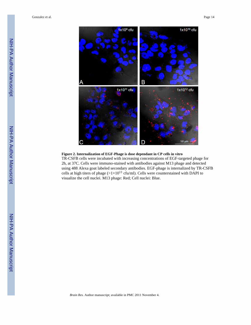
Figure 2. Internalization of EGF-Phage is dose dependant in CP cells in vitroTR-CSFB cells were incubated with increasing concentrations of EGF-targeted phage for2h, at 37C. Cells were immuno-stained with antibodies against M13 phage and detectedusing 488 Alexa goat labeled secondary antibodies. EGF-phage is internalized by TR-CSFBcells at high titers of phage (>1×10¹¹ cfu/ml). Cells were counterstained with DAPI tovisualize the cell nuclei. M13 phage: Red; Cell nuclei: Blue.
Gonzalez et al. Page 14
Brain Res. Author manuscript; available in PMC 2011 November 4.
NIH
-PA Author Manuscript
NIH
-PA Author Manuscript
NIH
-PA Author Manuscript
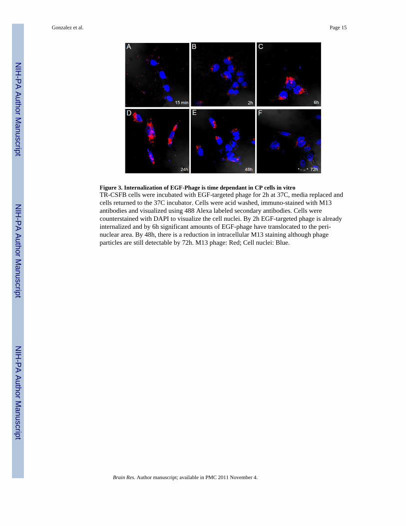
Figure 3. Internalization of EGF-Phage is time dependant in CP cells in vitroTR-CSFB cells were incubated with EGF-targeted phage for 2h at 37C, media replaced andcells returned to the 37C incubator. Cells were acid washed, immuno-stained with M13antibodies and visualized using 488 Alexa labeled secondary antibodies. Cells werecounterstained with DAPI to visualize the cell nuclei. By 2h EGF-targeted phage is alreadyinternalized and by 6h significant amounts of EGF-phage have translocated to the peri-nuclear area. By 48h, there is a reduction in intracellular M13 staining although phageparticles are still detectable by 72h. M13 phage: Red; Cell nuclei: Blue.
Gonzalez et al. Page 15
Brain Res. Author manuscript; available in PMC 2011 November 4.
NIH
-PA Author Manuscript
NIH
-PA Author Manuscript
NIH
-PA Author Manuscript
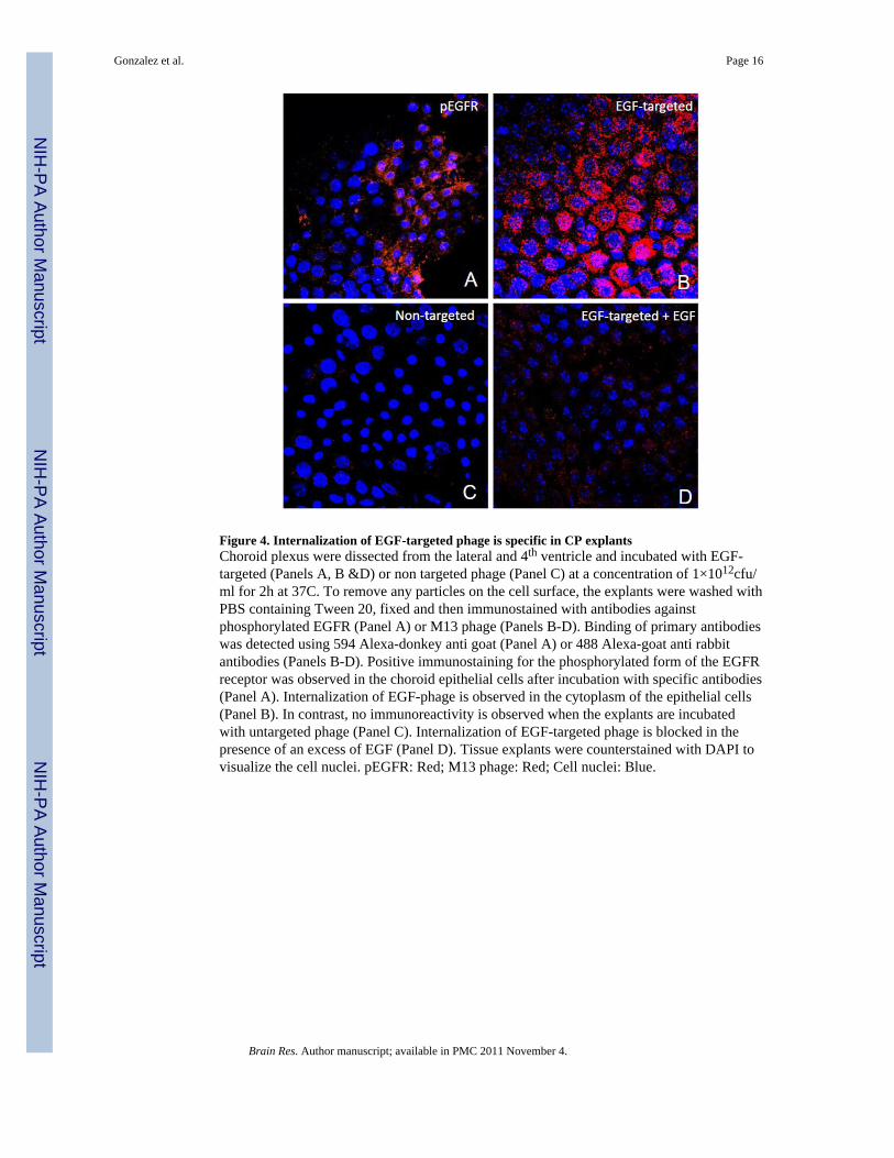
Figure 4. Internalization of EGF-targeted phage is specific in CP explantsChoroid plexus were dissected from the lateral and 4th ventricle and incubated with EGF-targeted (Panels A, B &D) or non targeted phage (Panel C) at a concentration of 1×10¹²cfu/ml for 2h at 37C. To remove any particles on the cell surface, the explants were washed withPBS containing Tween 20, fixed and then immunostained with antibodies againstphosphorylated EGFR (Panel A) or M13 phage (Panels B-D). Binding of primary antibodieswas detected using 594 Alexa-donkey anti goat (Panel A) or 488 Alexa-goat anti rabbitantibodies (Panels B-D). Positive immunostaining for the phosphorylated form of the EGFRreceptor was observed in the choroid epithelial cells after incubation with specific antibodies(Panel A). Internalization of EGF-phage is observed in the cytoplasm of the epithelial cells(Panel B). In contrast, no immunoreactivity is observed when the explants are incubatedwith untargeted phage (Panel C). Internalization of EGF-targeted phage is blocked in thepresence of an excess of EGF (Panel D). Tissue explants were counterstained with DAPI tovisualize the cell nuclei. pEGFR: Red; M13 phage: Red; Cell nuclei: Blue.
Gonzalez et al. Page 16
Brain Res. Author manuscript; available in PMC 2011 November 4.
NIH
-PA Author Manuscript
NIH
-PA Author Manuscript
NIH
-PA Author Manuscript

Figure 5. Internalization of EGF-targeted phage is dose dependant in CP explantsChoroid plexus explants were incubated with increasing concentrations of EGF-targetedphage for 2h, at 37C and processed as described in the text. Cells were immunostained withantibodies against M13 phage and the binding detected using 488 Alexa goat labeledsecondary antibodies. At low titers (1×10¹¹ cfu/ml), the internalization EGF-targeted phageis almost undetectable but significant increase of immunoreactive M13 internalization isobserved at 1×10¹² cfu/ml. At a concentration of 5×10¹² cfu/ml, almost all of choroidepithelial cells in the explants are immunopositive. Cells were counterstained with DAPI tovisualize the cell nuclei. M13 phage: Red; Cell nuclei: Blue.
Gonzalez et al. Page 17
Brain Res. Author manuscript; available in PMC 2011 November 4.
NIH
-PA Author Manuscript
NIH
-PA Author Manuscript
NIH
-PA Author Manuscript

Figure 6. EGF-targeted phage is internalised 24h after ICV injectionEGF-targeted phage (Panels A & B) or untargeted phage (Panels C & D) were injected intothe lateral ventricle and 24h later rats were killed and brain sections immunostained withanti-M13 antibodies. M13 immunoreactivity is observed in epithelial cells of the choroidplexus (Panel A) and the ependyma (Panel B). No immunoreactivity is observed in thesetissues when untargeted phage was injected ICV. (Red: M13; Blue: DAPI).
Gonzalez et al. Page 18
Brain Res. Author manuscript; available in PMC 2011 November 4.
NIH
-PA Author Manuscript
NIH
-PA Author Manuscript
NIH
-PA Author Manuscript

Figure 7. Gene expression after ICV injectionPanel (A) In vitro transduction: The EGF target PC3 cells were incubated with 1× 10¹¹
untargeted or EGF-targeted phage that were both re-engineered as described in the text tocontain the firefly luciferase (f-luc) gene under the control of the CMV promoter. Duplicatewells of cells were treated with 0, 1ug or 10 ug of camptothecin and either incubated with nophage (Ctl), EGF-targeted phage containing GFP gene (A), EGF-targeted phage containingf-luc gene (B) or untargeted phage containing the f-luc gene (C). After incubating cells for48 hrs, a 5 minute incubation with luciferin enabled a Lumina CCD Imaging system tovisualize gene expression in cells treated with EGF-targeted phage (Row B) and, asexpected the response was dose dependant on cells treated with camptothecin. No signal wasobserved in the absence of targeting (Row C) or when EGF-targeted particles do not containthe luciferase gene Row A.Panel B-F: Effects of ICV injection of EGF-targeted phage in vivo. Mice were injected icvwith 1×10¹² untargeted or EGF-targeted phage-luc and in Panels B and D, transgeneexpression of luciferase monitored on 48 hours later by Lumina CCD Imaging either noninvasively (Panel B) or after brain dissection (Panel C). To identify the cellular localizationof this gene expression similar experiments were performed with EGF-targeted phage-gfp(Panel D and E) or untargeted phage-gfp (Panel F) which demonstrated cell expression ofGFP in ependymal cells and in isolated cells in the choroid plexus. Whereas a 4-fold lowerbackground signal could be quantified after luciferase activity imaging, immunostaininglocalized this signal to macrophages and monocytes at the site of injection and not inependyma or choroid plexus (Panel F).
Gonzalez et al. Page 19
Brain Res. Author manuscript; available in PMC 2011 November 4.
NIH
-PA Author Manuscript
NIH
-PA Author Manuscript
NIH
-PA Author Manuscript

Figure 8. Drug Delivery to the Central Nervous System Via Choroid PlexusThere are three modes of drug delivery to the CNS using the choroid plexus as a portal ofentry. The first (1) is direct whereby molecules are targeted and directly translocated acrossthe CP epithelial barrier to find their targets from within the CNS. A second (2) is illustratedby entering and trateing the CP epithelium so that its capacity the produce and compositionof CSF is modified. These then find their targets in the CNS parenchyma. Finally the thirdstrategy, in the example here using gene delivery, results in longer term changes of thechoroid plexus, allows the epithelial cell to produce novel therapeutic factors itself and assuch can confer the epithelium with an ability to change the production and composition ofCSF. This latter process and the feasibility with luminal targeting (3) is demonstrated in theresults presented here.
Gonzalez et al. Page 20
Brain Res. Author manuscript; available in PMC 2011 November 4.
NIH
-PA Author Manuscript
NIH
-PA Author Manuscript
NIH
-PA Author Manuscript
Related Documents