~ 5 ~ International Journal of Fisheries and Aquatic Studies 2015; 2(3): 05-13 ISSN: 2347-5129 IJFAS 2015; 2(3): 05-13 © 2013 IJFAS www.fisheriesjournal.com Received: 05-11-2014 Accepted: 07-12-2014 Hany M. R. Abdel-Latif Department of Poultry and Fish diseases, Faculty of Veterinary Medicine, Alexandria University, Egypt. Riad H. Khalil Department of Poultry and Fish diseases, Faculty of Veterinary Medicine, Alexandria University, Egypt. Hanaa R. El-hofi Department of Fish diseases and hygiene, Animal Health Research Institute, Damanhur Branch, Egypt. Talaat T. Saad Department of Poultry and Fish diseases, Faculty of Veterinary Medicine, Alexandria University, Egypt. Shaimaa M. A. Zaied Department of Fish diseases and hygiene, Animal Health Research Institute, Damanhur Branch, Egypt. Correspondence Hany M. R. Abdel-Latif Department of Poultry and Fish diseases, Faculty of Veterinary Medicine, Alexandria University, Edfina, Behera, P.O. Box: 22758, Egypt. Tel: +201201147440; Fax: +2045 2963526, Email: [email protected] Epidemiological investigations of Mycotic infections of cultured Gilthead seabream, Sparus aurata at Marriott Lake, Egypt Hany M. R. Abdel-Latif, Riad H. Khalil, Hanaa R. El-hofi, Talaat T. Saad and Shaimaa M. A. Zaied Abstract Surveillance and descriptive studies of mycotic infections were investigated throughout a period of one year (2013 to 2014). A total number of one hundred (100) of cultured Gilthead seabream at Marriott Lake were surveyed for mycotic infections, whereas clinical and PM lesions were defined. Morphological and cultural characters of the isolated fungi and yeast were identified from fish tissues and organs. Moreover, their prevalence, incidence and relationship with physico-chemical properties and heavy metals content in water and tissues were evaluated. Infected fish have torned vertebral column, congested kidney with pale liver, fungal patches on the GIT and mottled appearance of the liver with severely congested heart. Results were confirmed with histopathological examination, which revealed the presence of fungal hyphae and spores in different organs. It was found that about eighty percentage (80%) of the examined fish were infected and total Aspergillus species were predominant in prevalence of mycotic isolates (32.12%) followed by Cladosporium (20.86%) and Fusarium species (14.45%). Moreover, the incidence of the mycotic isolates was higher in liver and kidney of the infected fish. The results of water quality parameters indicate that levels of nitrite, ammonia, organic matter as well as cadmium (Cd), lead (Pb) and copper (Cu) were higher than the permissible limits. We can conclude that the higher mycotic infections of cultured seabream were parallel together with unsuitable water quality and higher heavy metal levels. Keywords: Mycotic infections - Seabream - Marriott Lake – Epidemiology. 1. Introduction Seabream (Sparus aurata) is marine Fish with economic value and wide spread all over the world, especially in the Mediterranean Sea. Seabream culture was known recently in Egypt and need for progressive development especially in feeding and health care. Fungi are members of Thalophyta, lacking of chlorophyll completely therefore are bound to live as a saprophytic or parasites in existence. Fungi were reported to be responsible for many fish diseases. These fungi are belonging to wide range of genera which were found associated with several mycotic diseases of fish [1] . The importance of fungal diseases in freshwater fish not stopped only for incidence of mortalities but also as economic importance such as decrease growth rate, hatchability in chronaic infection or by mycotoxins production by contaminated fungus in case of bad storage feed [2] . In spite of the fungal infections importance our knowledge about them is still poor for two basic reasons: difficult identification of pathogenic fungi and the prolific growth of saprophytic fungi once the fish is dead [3] . Many of the fungi that affect fishes are considered opportunists, attacking the fishes when they are stressed or immunocompromised because of unfavorable environmental conditions, or secondary to bacterial or viral infections, or when they have lost their mucus protection because of trauma or excessive handling [4, 5] . Mycotic infections of fishes by Oomycetes are wide spread in freshwater and represent the most important fungal group affecting wild and cultured fishes. The Saprolegniaceae, in particular members of the genus Saprolegnia, are responsible for significant infections involving both living, dead fishes and eggs. Oomycetes are classical saprophytic opportunities, multiplying on fishes that are physically injured, stressed or infected [3] .

Welcome message from author
This document is posted to help you gain knowledge. Please leave a comment to let me know what you think about it! Share it to your friends and learn new things together.
Transcript

~ 5 ~
International Journal of Fisheries and Aquatic Studies 2015; 2(3): 05-13
ISSN: 2347-5129 IJFAS 2015; 2(3): 05-13 © 2013 IJFAS
www.fisheriesjournal.com Received: 05-11-2014 Accepted: 07-12-2014
Hany M. R. Abdel-Latif
Department of Poultry and Fish
diseases, Faculty of Veterinary
Medicine, Alexandria University,
Egypt.
Riad H. Khalil
Department of Poultry and Fish
diseases, Faculty of Veterinary
Medicine, Alexandria University,
Egypt.
Hanaa R. El-hofi
Department of Fish diseases and
hygiene, Animal Health Research
Institute, Damanhur Branch,
Egypt.
Talaat T. Saad
Department of Poultry and Fish
diseases, Faculty of Veterinary
Medicine, Alexandria University,
Egypt.
Shaimaa M. A. Zaied
Department of Fish diseases and
hygiene, Animal Health Research
Institute, Damanhur Branch,
Egypt.
Correspondence
Hany M. R. Abdel-Latif
Department of Poultry and Fish
diseases, Faculty of Veterinary
Medicine, Alexandria University,
Edfina, Behera, P.O. Box: 22758,
Egypt.
Tel: +201201147440;
Fax: +2045 2963526,
Email: [email protected]
Epidemiological investigations of Mycotic infections of
cultured Gilthead seabream, Sparus aurata at Marriott
Lake, Egypt
Hany M. R. Abdel-Latif, Riad H. Khalil, Hanaa R. El-hofi, Talaat T. Saad
and Shaimaa M. A. Zaied
Abstract Surveillance and descriptive studies of mycotic infections were investigated throughout a period of one
year (2013 to 2014). A total number of one hundred (100) of cultured Gilthead seabream at Marriott Lake
were surveyed for mycotic infections, whereas clinical and PM lesions were defined. Morphological and
cultural characters of the isolated fungi and yeast were identified from fish tissues and organs. Moreover,
their prevalence, incidence and relationship with physico-chemical properties and heavy metals content
in water and tissues were evaluated. Infected fish have torned vertebral column, congested kidney with
pale liver, fungal patches on the GIT and mottled appearance of the liver with severely congested heart.
Results were confirmed with histopathological examination, which revealed the presence of fungal
hyphae and spores in different organs. It was found that about eighty percentage (80%) of the examined
fish were infected and total Aspergillus species were predominant in prevalence of mycotic isolates
(32.12%) followed by Cladosporium (20.86%) and Fusarium species (14.45%). Moreover, the incidence
of the mycotic isolates was higher in liver and kidney of the infected fish. The results of water quality
parameters indicate that levels of nitrite, ammonia, organic matter as well as cadmium (Cd), lead (Pb)
and copper (Cu) were higher than the permissible limits. We can conclude that the higher mycotic
infections of cultured seabream were parallel together with unsuitable water quality and higher heavy
metal levels.
Keywords: Mycotic infections - Seabream - Marriott Lake – Epidemiology.
1. Introduction
Seabream (Sparus aurata) is marine Fish with economic value and wide spread all over the
world, especially in the Mediterranean Sea. Seabream culture was known recently in Egypt
and need for progressive development especially in feeding and health care.
Fungi are members of Thalophyta, lacking of chlorophyll completely therefore are bound to
live as a saprophytic or parasites in existence. Fungi were reported to be responsible for
many fish diseases. These fungi are belonging to wide range of genera which were found
associated with several mycotic diseases of fish [1]. The importance of fungal diseases in
freshwater fish not stopped only for incidence of mortalities but also as economic importance
such as decrease growth rate, hatchability in chronaic infection or by mycotoxins production
by contaminated fungus in case of bad storage feed [2]. In spite of the fungal infections
importance our knowledge about them is still poor for two basic reasons: difficult
identification of pathogenic fungi and the prolific growth of saprophytic fungi once the fish is
dead [3]. Many of the fungi that affect fishes are considered opportunists, attacking the fishes
when they are stressed or immunocompromised because of unfavorable environmental
conditions, or secondary to bacterial or viral infections, or when they have lost their mucus
protection because of trauma or excessive handling [4, 5].
Mycotic infections of fishes by Oomycetes are wide spread in freshwater and represent the
most important fungal group affecting wild and cultured fishes. The Saprolegniaceae, in
particular members of the genus Saprolegnia, are responsible for significant infections
involving both living, dead fishes and eggs. Oomycetes are classical saprophytic
opportunities, multiplying on fishes that are physically injured, stressed or infected [3].

~ 6 ~
International Journal of Fisheries and Aquatic Studies
Members of this group are generally considered agents of
secondary infection arising from conditions such as bacterial
infections, poor husbandry, and infestation by parasite and
social interaction. However, there are several reports of
Oomycetes as primary infectious agents of fishes [6] and their
eggs [7].
Moreover, there are other fungi that have been implicated in
fish diseases. Some of the genera involved include Aspergillus [8], Fusarium [9], Ichthyophonus [10], Branchiomycosis [11],
Phoma [12], Paecilomyces [2, 13], Exophiala [14], Phialophora [15],
Rhizomucor [16] and Candida [17]. Thus the present study was
aimed to spot light on isolation and identification of mycotic
infections of cultured seabream and their relationship with
physico-chemical and heavy metals of water.
2. Material and Methods
2.1. Fish samples
In our investigation, a total number of one hundred cage-
cultured marine fishes of Seabream (Sparus aurata L.), of
different body weight ranged (50 gm ± 30 gm), fishes were
from private fish farm at Wadi-Mariut region at west
Alexandria governorate, Egypt. The sources of water in Wadi-
Mariut are numerous; underground water, drainage of canal
originated from El-Banger area as well as from rainfall water
downstream from Borg-El-Arab city. Fishes were collected
showing clinical signs in plastic bags containing about 1/3
volume of water at site of collection and filled with oxygen.
They were transferred immediately to the laboratory in Animal
Health Research Institute Damanhur branch. The freshly dead
fish specimens were subjected to full clinical, postmortem
(PM) lesions, and mycological examinations.
2.2. Gross clinical examination
Clinical examination of naturally infected fishes was
performed to investigate any clinical abnormalities [18, 19].
2.3. Postmortem (PM) examination
Necropsy was performed on variable number of freshly dead
and moribund fishes for detection of PM lesions [20, 21].
2.4. Mycological examination
The fish surface was disinfected with a swab of cotton
moistened with 70% ethyl alcohol. Spleen, liver, heart and
kidney of killed fish were collected under complete aseptic
conditions and inoculated into Sabouraud's dextrose agar
medium (SDA) with 0.05 mg/L chloramphenicol, Potato-
dextrose agar and corn-meal agar plates [22]. The plates were
incubated at 25 – 28 °c for 3-5 days. Negative plates were not
discarded before 2 weeks [23]. All the positive moulds cultures
were purified by sub culturing on Sabouraud's dextrose agar
plates incubated at 25 - 28c for 3-5 days and examined for
gross and micro morphological characteristics [24, 25].
2.5. Identification of mycotic isolates
2.5.1. Identification of moulds
All the purified mould cultures were examined for macro and
micro-morphological characteristics. This was carried out
according to the methods of [25, 26, 27, 28, 29 & 30].
2.5.1. a. Microscopical examination
The gross morphological examination included the rate of
growth, texture, changes in color during growth, final color of
the surface and reverse sides of the colonies.
2.5.1. b. Microscopical examination (Solutip method)
For micro morphological studies, a small portion of the
periphery of a fresh colony was picked using the sticking
surface of a piece of solutip and placed with its sticking
surface down on a clean slide with a drop of lacto phenol
cotton blue stain [31] and examined microscopically.
Microscopically examination was carried out to detect
septation of hyphae, roughness or smoothness of
conidiophores, shape of vesicles, arrangement and number of
the rows of the strigmata.
2.5.2. Identification of yeasts
Suspected yeast like colonies were preliminary identified
according to the scheme [32]. Suspected Candida species were
scratched onto corn meal agar tween 80 for chlamydospore
production [33] in very short 3 or 4 parallel lines using a
mycological needle. The scratched lines were covered by
sterile cover slides to provide an aerobic condition. Incubation
was at 25 °C for 72 hours.
2.6. Collection of water samples 2.6.1. Assessment of physico-chemical water properties
The tools used for determination of Physico-chemical
properties of water quality were namely; Dissolved Oxygen
meter for measuring the level of Dissolved oxygen in the
water, Salinometer for measuring of % of water salinity, PH
meter for measuring the pH values and Kits for measuring the
levels of unionized ammonia and Sulphate in the water (USA,
Virginia Company, lot. No .201134).
2.6.2. Spectrophotometric method for detection the levels
of heavy metals in water and fish tissues
The method for analysis of the heavy metals in water [34] and
fish tissues [35] was carried out using Atomic Absorption
Spectrophotometry. Atomic Absorption Spectrophotometer
(Model Thermo Electron Corporation, S. Series AA
Spectrometer with Gravities furnace, UK,) instrument was
used to detect the heavy metals. The concentrations of heavy
metals were expressed as mg/l for water and μg/g dry wt. for
fish tissues.
Fish specimens were digested [36]. All frozen fish samples were
allowed to thaw at room temperature, washed with distilled
water and placed on filter paper to remove the excess liquid.
Their gills and musculature tissues were dissected separately
and minced using a domestic blender, then approximately 1.0
gm was placed in a 150 ml beaker and 10 ml concentrated
nitric acid was added. After a short soaking period, 5 ml of
60% perchloric acid was added and the mixture was slowly
heated on a hot plate until the conclusion of growth
(approximately 2 hrs). The mixture was then heated until the
appearance of dense white fumes that indicate the nitric acid
had evaporated and perchloric acid had reached its boiling
point. The mixture was cooled; 10 ml of 25% hydrochloric
acid was added then, the solution was transferred to a 100 ml
volumetric flask that was subsequently brought to volume with
de ionized water. Blank solution was prepared for the
background correction. Atomic absorption spectrophotometer
instrument was used to determine As, Zn, Cu, Pb, Hg, and Cd
concentrations which were expressed as μg / g dry weight in
the Toxicology Unit of Central Laboratory, Faculty of
Veterinary medicine, Alexandria University, Egypt.
~ 7 ~
International Journal of Fisheries and Aquatic Studies
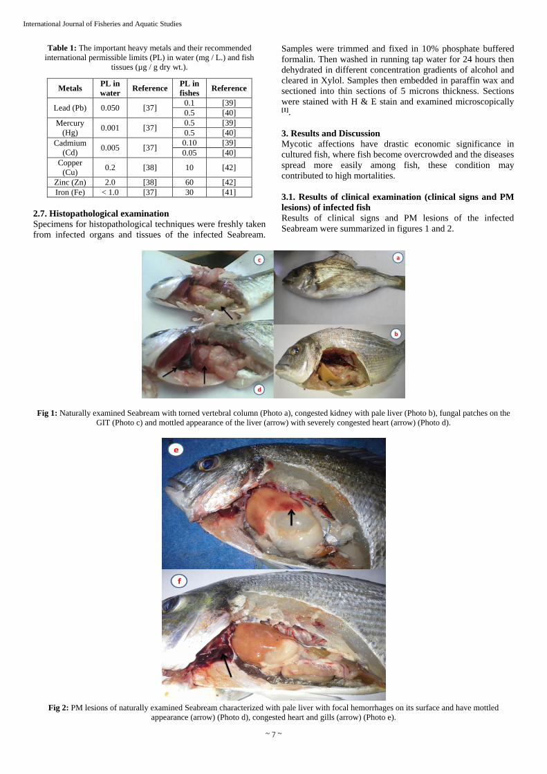
Table 1: The important heavy metals and their recommended
international permissible limits (PL) in water (mg / L.) and fish
tissues (µg / g dry wt.).
Metals PL in
water Reference
PL in
fishes Reference
Lead (Pb) 0.050 [37] 0.1 [39]
0.5 [40]
Mercury
(Hg) 0.001 [37]
0.5 [39]
0.5 [40]
Cadmium
(Cd) 0.005 [37]
0.10 [39]
0.05 [40]
Copper
(Cu) 0.2 [38] 10 [42]
Zinc (Zn) 2.0 [38] 60 [42]
Iron (Fe) < 1.0 [37] 30 [41]
2.7. Histopathological examination
Specimens for histopathological techniques were freshly taken
from infected organs and tissues of the infected Seabream.
Samples were trimmed and fixed in 10% phosphate buffered
formalin. Then washed in running tap water for 24 hours then
dehydrated in different concentration gradients of alcohol and
cleared in Xylol. Samples then embedded in paraffin wax and
sectioned into thin sections of 5 microns thickness. Sections
were stained with H & E stain and examined microscopically [1].
3. Results and Discussion
Mycotic affections have drastic economic significance in
cultured fish, where fish become overcrowded and the diseases
spread more easily among fish, these condition may
contributed to high mortalities.
3.1. Results of clinical examination (clinical signs and PM
lesions) of infected fish
Results of clinical signs and PM lesions of the infected
Seabream were summarized in figures 1 and 2.
Fig 1: Naturally examined Seabream with torned vertebral column (Photo a), congested kidney with pale liver (Photo b), fungal patches on the
GIT (Photo c) and mottled appearance of the liver (arrow) with severely congested heart (arrow) (Photo d).
Fig 2: PM lesions of naturally examined Seabream characterized with pale liver with focal hemorrhages on its surface and have mottled
appearance (arrow) (Photo d), congested heart and gills (arrow) (Photo e).

~ 8 ~
International Journal of Fisheries and Aquatic Studies
Results of clinical findings of Seabream were parallel to that
obtained by [43], and similarly the postmortem findings were in
agreement with those of [44].
3.2. Cultural and Morphological identification of mycotic
isolates
3.2.1. Moulds
3.2.1.1. Aspergillus species
3.2.1.1. a. Aspergillus flavus (A. flavus)
Macroscopically, the growth appeared velvety with numerous
aerial growths, at first the color was yellow and became
yellowish green by aging. While, microscopically, the
conidiophores were long and rough. The vesicles were large
and rounded. The strigmata were biseriate, loose, and radiate
and gave rise to ovoid rough conidia.
3.2.1.1. b. Aspergillus fumigatus (A. fumigatus)
Macroscopically, colonies have distinct margin with some
shades of green, blue-green, surface has a powedrey
appearance. A white apron was seen at the edge in the zone of
active growth. While, microscopically, characterized by
hyaline and distinctly septated hyphae, conidiophores were
long with club -shaped vesicle, spherical conidia were born
from single row of sterigmata.
3.2.1.1. c. Aspergillus niger (A. niger)
Macroscopically, the colonies were wooly in texture and
spread rapidly .They were black in color with radiated rouge.
While, microscopically, the conidiophores were very long,
smooth and yellowish color. The vesicles were very large and
globes while the strigmata were biseriate, compact and radiate.
The conidia were globes and smooth.
3.2.1.1. d. Aspergillus parasiticus (A. Parasiticus)
Its characters are those of A. flavus group but colony color is
predominantly greener, short stalks with usually a single series
of sterigmata. No sclerotia have been seen. The mycelium is
uncolored.
3.2.1.1. e. Aspergillus terreus (A. terreus)
Macroscopically, the colonies were buff to dark brown velvety
folded. While, microscopically, small hemispherical vesicle
with phialides born on prophialide.
3.2.1.2. Aphanomyces species
Macroscopically, characterized by flat, slight opaque colonies
with an uneven white velvets surface. The hyphal growth
increase with prolonged incubation to occupy the entire
surface of the plate and appear as linear growth within 14 days
of incubation. While, microscopically, characterized by
branched non septated hyphae with tapered end contain
cytoplasmic organelles.
3.2.1.3. Alternaria species
Macroscopically, colonies were dark greenish-black to grey-
brown with a light border. Reverse is black. Furthermore,
microscopically, the hyphae dark and septated. Conidia are
large, brown, muriform, club-shaped and occur singly or in
chains.
3.2.1.4. Cladosporium species
Macroscopically, colonies were dark, velvety and olive-green,
with dark reverse. Moreover, microscopically, the
conidiophores with varying lengths that produce long
branching chains of brown, smooth-walled, oval, pointed
conidia .The conidia are easily dispersed.
3.2.1.5. Fusarium species
Macroscopically, colonies were cottony or wooly in texture,
snow white, pink-violet or rosy-red in color, with specific
diffusion of colored pigments into the reverse surface of the
medium. While, microscopically, they were long, branched
and septated hyphae from which short conidiophores rose
singly or in groups, and sometimes branched. Two types of
conidia were observed, a large banana shaped, septated
macroconidia and a small, round, non septated microconidia.
3.2.1.6. Geotrichum species
Macroscopically, colonies were whitish, flat, and moist and
yeast like with a granular surface. Some strains produce short,
white, cottony aerial hyphae. Moreover, microscopically, the
septated mycelium fragments into arthrospores (arthroconidia),
which are formed consecutively and become round. No
blastoconidia, are produced.
3.2.1.7. Helminthosporium species
Macroscopically, colonies were cottony and dark grey to
black. Reverse is black. Furthermore, microscopically, the
unbranched conidiophores those are brown slightly curved,
with conidia forming along the sides. The later are large, dark,
multi-celled and club-shaped.
3.2.1.8. Ichthyophonus species
Macroscopically, characterized by growing culture appeared as
white hyphal growth with different levels both on the surface
and into the substrate of the S.D.A. media the hyphal growth
increased to full fill the plate within 10-14 days post
inoculation. Furthermore, microscopically, characterized by
branched non septated hyphae with spherical hyphal tips and
various forms of resting spores.
3.2.1.9. Nigrospora species
Macroscopically, colonies were compact and wooly, white at
first but black areas appear due to the production of black
globose conidia. Reverse is black. While, microscopically, the
short conidiophores that swell and then taper to the point of
conidia formation. Conidia are large, black, round but slightly
flattened.
3.2.1.10. Paecilomyces species
Macroscopically, colonies were flat surface, powdery or
velvety, yellowish-brown or light pastel shades of pink, violet
or gray green. Microscopically, resembles Penicillium (the
conidiophores formed brush -like branches resembling the
fingers, with long chains of small spherical conidia forming
the flask-shaped sterigmata (metula) but the phialides are more
elongated and taper into along slender tube. The conidia are
elliptical or oblong and occur in chains.
~ 9 ~
International Journal of Fisheries and Aquatic Studies
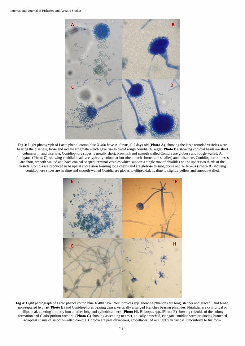
Fig 3: Light photograph of Lacto phenol cotton blue X 400 have A. flavus, 5-7 days old (Photo A), showing the large rounded vesicles were
bearing the biseriate, loose and radiate strigmata which gave rise to ovoid rough conidia, A. niger (Photo B), showing conidial heads are short
columnar in and biseriate. Conidiophore stipes is usually short, brownish and smooth walled Conidia are globose and rough-walled, A.
fumigatus (Photo C), showing conidial heads are typically columnar but often much shorter and smaller) and uniseriate. Conidiophore stipeses
are short, smooth-walled and have conical-shaped terminal vesicles which support a single row of phialides on the upper two thirds of the
vesicle. Conidia are produced in basipetal succession forming long chains and are globose to subglobose and A. terreus (Photo D) showing
conidiophore stipes are hyaline and smooth-walled Conidia are globes to ellipsoidal, hyaline to slightly yellow and smooth-walled.
Fig 4: Light photograph of Lacto phenol cotton blue X 400 have Paecilomyces spp. showing phialides are long, slender and graceful and broad,
non-septated hyphae (Photo E) and Conidiophores bearing dense, vertically arranged branches bearing phialides. Phialides are cylindrical or
ellipsoidal, tapering abruptly into a rather long and cylindrical neck (Photo H), Rhizopus spp. (Photo F) showing rhizoids of the colony
formation and Cladosporium carrionii (Photo G) showing ascending to erect, apically branched, elongate conidiophores producing branched
acropetal chains of smooth-walled conidia. Conidia are pale olivaceous, smooth-walled or slightly verrucose, limoniform to fusiform.

~ 10 ~
International Journal of Fisheries and Aquatic Studies
Fig 5: Light photograph of Lacto phenol cotton blue X 400 have Penicillium spp. (Photo I) showing conidiophores are hyaline, smooth walled
and bear terminal verticals of 3-5 metulae, each bearing 3-7 phialides. Conidia are globose to subglobose, smooth-walled and are produced in
basipetal succession from the phialides, Aphanomyces spp. (Photo J) Showing arrangement of zoospores in one row, Exophiala spp. (Photo K)
showing aggregations of cylindrical spores at the end of hyphae and Alternaria spp. (Photo L) showing macroconidia divided by alteration of
spores.
3.2.2. Yeasts
3.2.2.1. Torulopsis species
Characterized by white to cream colored, later become
grayish-white or brown on S.D.A. at 25oc to 37oc after 3-4
days. The colonies are moist, smooth and shiny initially. Older
cultures may become wrinkled. Microscopically, no pseudo
hyphae were formed on rice agar and growth at 37oc on
S.D.A.
3.2.2.2. Cryptococcus species
Could be identified by positive urease test, no pseudohyphae
on rice agar medium.
3.2.2.3. Rhodotorula species
Characterized by budding of round, oval cells, absence of
pseudohyphae on rice agar medium and colony on Sabouraud's
Dextrose Agar was characterized by formation of carotenoid
pigments; that vary from orange to red.
4. Histopathological findings of the infected Seabream
Fig 6: Histopathological section of liver of seabream (Photo a) showing hydropic degeneration and distributed fungal elements (arrows) in the
most disarrangement hepatic cells, while the musculature (Photo b) showing myelitis of the muscle fibers associated with embedded hyphal
elements along the course of muscle fibers (Arrows) (Periodic Acid Schiff stain 100X).

~ 11 ~
International Journal of Fisheries and Aquatic Studies
Fig 7: Histopathological section of the liver of Seabream (Photo c) showing severe congestion and hyaline cost masses as well as distributed
fungal elements (arrows) in the most disarrangement hepatic cells while the musculature of Seabream (Photo d) showing myelitis of the muscle
fibers associated with aggregation of budding spores of fungus in the center of muscle cells (Arrows) (Periodic Acid Schiff stain 100X).
5. Prevalence of mycotic infections in cultured Seabream
A total of 100 cultured Seabream were mycologically
examined. Specimens from different organs (600) of different
species were mycologically examined only 480 samples were
positive with affection percentage of 80%.
In this study 13 genera of mould and four genera of identified
yeast beside to the unidentified yeasts were isolated. This was
expected, as almost all these fungi were categorized [45] as
normal mycoflora. This does not mean that they cannot
produce disease. They can better be considered as
opportunistic fungi [30] as many of them possess virulence
factors, which enable them to cause diseases [46], particularly
under favorable predisposing condition.
Table 2: Prevalence of fungal isolates from cultured Seabream.
Fungal isolates Prevalence
No. %
A. niger 128 9.43
A. flavus 150 13.27
A. terreus 60 4.42
A. fumigatus 68 5.01
A. parasiticus 30 2.21
Total Aspergillus species 436 32.15
Paecilomyces species 104 7.66
Fusarium species 196 14.45
Ichthyophonus species 68 5.01
Aphanomyces invadans 12 0.88
Alternaria species 112 8.25
Cladosporium species 280 20.64
Helminthosporium species 8 0.58
Nigrospora species 32 2.35
Achlya species 12 0.88
Phomaherbarum 4 0.29
Legnadium 36 2.65
Exophiala 16 1.17
Geotrichum species 40 2.94
Total number of fungal isolates 1356
Table 3: Prevalence of yeast isolates from cultured Seabream.
Yeast isolates Prevalence
No. %
Unidentified yeast 280 66.03
Rhodotorula species 36 8.49
Candida species 94 22.16
Cryptococcus species 4 0.94
Torulopsis species 10 2.36
Total number of yeast isolates 424
6. Incidence of mycotic infections in different organs and
tissue of cultured Seabream
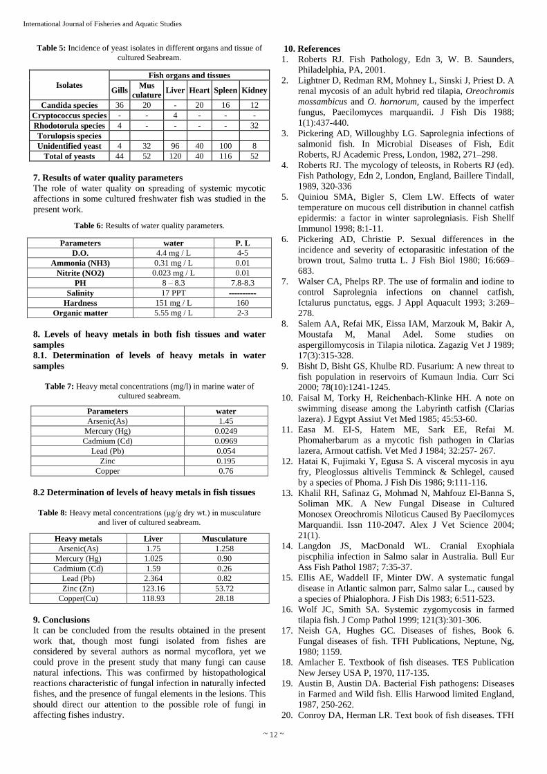
Incidence of fungal infections from different organs and
tissues of cultured Seabream were summarized in table 3.
Table 4: Incidence of fungal isolates in different organs and tissue of
cultured Seabream.
Isolates
Fish organs and tissues
Gills Musc
ulature Liver Heart Spleen Kidney
A. niger 8 4 36 8 32 40
A. flavus 56 16 56 8 16 28
A. terreus 4 36 8 - 4 8
A. fumigatus 12 4 12 12 12 16
A. parasiticus - - - - - -
Paecilomyces
species 40 4 4 4 12 40
Fusarium species 32 16 24 52 32 40
Ichthyophonus
species 20 24 12 - 4 8
Aphanomyces
invadans 8 - - - - 4
Alternaria species 20 24 4 20 16 28
Cladosporium
species 64 40 60 24 60 32
Helminthosporium
species - 4 4 - - -
Nigrospora species - - 4 8 8 12
Achlya species 8 - - - - 4
Phomaherbarum - - - - - 4
Legnadium species 4 - 4 20 4 4
Exophiala species 4 12 - - - -
Geotrichum
species 4 - - 32 4 -
Total of moulds 284 184 228 188 204 268
~ 12 ~
International Journal of Fisheries and Aquatic Studies
Table 5: Incidence of yeast isolates in different organs and tissue of
cultured Seabream.
Isolates
Fish organs and tissues
Gills Mus
culature Liver Heart Spleen Kidney
Candida species 36 20 - 20 16 12
Cryptococcus species - - 4 - - -
Rhodotorula species 4 - - - - 32
Torulopsis species
Unidentified yeast 4 32 96 40 100 8
Total of yeasts 44 52 120 40 116 52
7. Results of water quality parameters
The role of water quality on spreading of systemic mycotic
affections in some cultured freshwater fish was studied in the
present work.
Table 6: Results of water quality parameters.
P. L water Parameters
4-5 4.4 mg / L D.O.
0.01 0.31 mg / L Ammonia (NH3)
0.01 0.023 mg / L Nitrite (NO2)
7.8-8.3 8 – 8.3 PH
---------- 17 PPT Salinity
160 151 mg / L Hardness
2-3 5.55 mg / L Organic matter
8. Levels of heavy metals in both fish tissues and water
samples
8.1. Determination of levels of heavy metals in water
samples
Table 7: Heavy metal concentrations (mg/l) in marine water of
cultured seabream.
water Parameters 1.45 Arsenic(As)
0.0249 Mercury (Hg)
0.0969 Cadmium (Cd)
0.054 Lead (Pb)
0.195 Zinc
0.76 Copper
8.2 Determination of levels of heavy metals in fish tissues
Table 8: Heavy metal concentrations (μg/g dry wt.) in musculature
and liver of cultured seabream.
Heavy metals Liver Musculature
Arsenic(As) 1.75 1.258
Mercury (Hg) 1.025 0.90
Cadmium (Cd) 1.59 0.26
Lead (Pb) 2.364 0.82
Zinc (Zn) 123.16 53.72
Copper(Cu) 118.93 28.18
9. Conclusions
It can be concluded from the results obtained in the present
work that, though most fungi isolated from fishes are
considered by several authors as normal mycoflora, yet we
could prove in the present study that many fungi can cause
natural infections. This was confirmed by histopathological
reactions characteristic of fungal infection in naturally infected
fishes, and the presence of fungal elements in the lesions. This
should direct our attention to the possible role of fungi in
affecting fishes industry.
10. References
1. Roberts RJ. Fish Pathology, Edn 3, W. B. Saunders,
Philadelphia, PA, 2001.
2. Lightner D, Redman RM, Mohney L, Sinski J, Priest D. A
renal mycosis of an adult hybrid red tilapia, Oreochromis
mossambicus and O. hornorum, caused by the imperfect
fungus, Paecilomyces marquandii. J Fish Dis 1988;
1(1):437-440.
3. Pickering AD, Willoughby LG. Saprolegnia infections of
salmonid fish. In Microbial Diseases of Fish, Edit
Roberts, RJ Academic Press, London, 1982, 271–298.
4. Roberts RJ. The mycology of teleosts, in Roberts RJ (ed).
Fish Pathology, Edn 2, London, England, Baillere Tindall,
1989, 320-336
5. Quiniou SMA, Bigler S, Clem LW. Effects of water
temperature on mucous cell distribution in channel catfish
epidermis: a factor in winter saprolegniasis. Fish Shellf
Immunol 1998; 8:1-11.
6. Pickering AD, Christie P. Sexual differences in the
incidence and severity of ectoparasitic infestation of the
brown trout, Salmo trutta L. J Fish Biol 1980; 16:669–
683.
7. Walser CA, Phelps RP. The use of formalin and iodine to
control Saprolegnia infections on channel catfish,
Ictalurus punctatus, eggs. J Appl Aquacult 1993; 3:269–
278.
8. Salem AA, Refai MK, Eissa IAM, Marzouk M, Bakir A,
Moustafa M, Manal Adel. Some studies on
aspergillomycosis in Tilapia nilotica. Zagazig Vet J 1989;
17(3):315-328.
9. Bisht D, Bisht GS, Khulbe RD. Fusarium: A new threat to
fish population in reservoirs of Kumaun India. Curr Sci
2000; 78(10):1241-1245.
10. Faisal M, Torky H, Reichenbach-Klinke HH. A note on
swimming disease among the Labyrinth catfish (Clarias
lazera). J Egypt Assiut Vet Med 1985; 45:53-60.
11. Easa M. EI-S, Hatem ME, Sark EE, Refai M.
Phomaherbarum as a mycotic fish pathogen in Clarias
lazera, Armout catfish. Vet Med J 1984; 32:257- 267.
12. Hatai K, Fujimaki Y, Egusa S. A visceral mycosis in ayu
fry, Pleoglossus altivelis Temminck & Schlegel, caused
by a species of Phoma. J Fish Dis 1986; 9:111-116.
13. Khalil RH, Safinaz G, Mohmad N, Mahfouz El-Banna S,
Soliman MK. A New Fungal Disease in Cultured
Monosex Oreochromis Niloticus Caused By Paecilomyces
Marquandii. Issn 110-2047. Alex J Vet Science 2004;
21(1).
14. Langdon JS, MacDonald WL. Cranial Exophiala
piscphilia infection in Salmo salar in Australia. Bull Eur
Ass Fish Pathol 1987; 7:35-37.
15. Ellis AE, Waddell IF, Minter DW. A systematic fungal
disease in Atlantic salmon parr, Salmo salar L., caused by
a species of Phialophora. J Fish Dis 1983; 6:511-523.
16. Wolf JC, Smith SA. Systemic zygomycosis in farmed
tilapia fish. J Comp Pathol 1999; 121(3):301-306.
17. Neish GA, Hughes GC. Diseases of fishes, Book 6.
Fungal diseases of fish. TFH Publications, Neptune, Ng,
1980; 1159.
18. Amlacher E. Textbook of fish diseases. TES Publication
New Jersey USA P, 1970, 117-135.
19. Austin B, Austin DA. Bacterial Fish pathogens: Diseases
in Farmed and Wild fish. Ellis Harwood limited England,
1987, 250-262.
20. Conroy DA, Herman LR. Text book of fish diseases. TFH

~ 13 ~
International Journal of Fisheries and Aquatic Studies
publ West Sylvania, 1981.
21. Plumb JA, Bowser PR. A laboratory Manual of Microbial
fish Diseases. Auburn Univ. Auburn Alabama 1982, 77.
22. Whitman KA. Finfish and Shellfish Bacteriology Manual;
Techniques and Procedures. ISBN 0-8138-1952-0 – Iowa
State Press, 2004.
23. Feingold SM, Baron EJ. Bailey and Scoll's Diagnostic
Microbiology. The Cv. Mosby co., St. Louis, 1986.
24. Collins CH, Lyne PM. Microbiological Methods. Edn 5,
Butterworth's & Co. Publishers, Ltd, 1984.
25. Ellis MB. Dematiaceous Hyphomycetes. Commonwealth
Mycological Institute: Kew Surrey, UK, 1971.
26. Raper KB, Fennell DI. The Genus Aspergillus. Williams
and Wilkins, Baltimore, 1965.
27. Moss ES, McQuown AL. Atlas of medical mycology. Edn
3, Baltimore: The Williams and Wikins Company, 1969,
366.
28. Larone DH. Medically Important Fungi. A Guide to
Identification, 3rd ed. Harper and Row Publishers, New
York, 1976, 68, 73 and 102.
29. Frey D, Old-Field RJ, Bridjer RC. A colour atlas of
pathogenic fungi. Wolfe Medical Public Ltd Holland, 1979.
30. Refai M. Isolation and identification of fungi. Fac Vet
Mid Cairo University, 1987.
31. Cruickshank R, Duguid JP, Marimion BP, Swain RH.
Medical microbiology. Edn 12, Vol. 11: The practice of
medical microbiology. Churchill Livingstone, Edinburgh,
London, 1982.
32. Terrence CD. A practical approach to identification of
yeast like organisms. Amer. Jour Microbiol 1971;
35(5):580-590.
33. Larone, DH. Medically Important Fungi. A Guide to Identification, Edn 2, Washington, DC, 1987.
34. APHA (American Public Health Association). Standard
Methods for the Examination of Water and Wastewater.
19th Edn., Washington DC, 1995.
35. Clesceri LS. Standard methods for the examination of
water and waste water. In Collection and Preservation of
Samples and Metals (Eds Arnold E, Greenbergy and
Eaton AD), APHA, AWWA, WEF, Washington, DC,
1998.
36. AOAC (Association of Official Analytical Chemists).
“Official Methods for Minerals in Plants. Method 975.03.
B.b.” In: Official Methods of Analysis of AOAC
International, Edn 16, Vol 1, Arlington (Va.): AOAC
International Section 1996; 3:3-4.
37. WHO, World Health Organization. Guidelines for
drinking water quality. Geneva 1984, 111.
38. FAO. Water Quality for Agriculture. Irrigation and
Drainage Paper No. 29, Rev. 1. Food and Agriculture
Organization of the United Nations, Rome, 1985.
39. EOSQC. Egyptian organization for standardization and
Quality control. 2760- 2005. Physical and chemical
methods for testing fish and fishery products. Part 5:
Crustacea and Mollusca, Egyptian Organization for
Standardization and Quality Control. U.D.C.: 637/ 664-
2005 Arab Republic, 2005.
40. FAO/WHO. Codex alimentarius commission, standard
programme codex committee on food additives and
contaminates. 24th Session, Hague, 23- 28 March, 1992.
41. Food Stuff. Cosmetics and Disinfectants–Act No. 54-
Regulation No. R2064 Marin Food Government Gazett
Governorate Particular, Pretoria, 1972.
42. FAO/WHO. List of maximum levels recommended for
contaminants by the Joint FAO/ WHO Codex
Alimentarius Commission. Second Series. CAC/FAL,
Rome, 1984; 3:1–8.
43. Marzouk MS, Samira SR, El-Gamal MH. Mycological
investigations on cultured Tilapia in Kafer El- Sheikh
Governorate. Kafer El-Sheikh Vet Med J 2003; 1(2):97-
114.
44. Refai M, Abdel halim MM, Afify MMH, Youssef H,
Marzouk KM. Studies on aspergillomycosis in catfish
(Clarias Lazera). Allgemeine Pathologic and
pathologische Anatomic. Tagung der Deutachen Veterinar
- Medizinischen Gesellschaft. der Europeischen
Gesellschaft fur. Vet Pathol 1987; 63:1-12.
45. Shaheen AA. Mycoflora of some freshwater fish. MV Sc.
Thesis, Fac. Vet. Med., Zagazic Univ, 1986.
46. Refai MS Attia R, Salem M, El-Dahshan EM. Studies on
the pathogenicity of Aspergillus fumigatus, A. flavus and
A. niger isolated from chickens and their environment.
Egypt. J Comp Path Clinic Path 2004; 17(2):193-205.
Related Documents