Enhancement of Th1 Lung Immunity Induced by Recombinant Mycobacterium bovis Bacillus Calmette-Gue ´rin Attenuates Airway Allergic Disease Ana P. Christ 1 , Du ´ nia Rodriguez 2 , Juliana Bortolatto 2 ,E ´ rica Borducchi 2 , Alexandre Keller 2 , Daniel Mucida 2 , Joa ˜o S. Silva 3 , Luciana C. C. Leite 1 , and Momtchilo Russo 2 1 Centro de Biotecnologia, Instituto Butantan, Sa ˜o Paulo; and 2 Departamento de Imunologia, Instituto de Cie ˆncias Biome ´dicas, and 3 Faculdade de Medicina de Ribeira ˜o Preto, Universidade de Sa ˜o Paulo, Sa ˜o Paulo, Brazil Mycobacterium bovis Bacillus Calmette-Gue ´ rin (BCG) has been shown to down-regulate experimental allergic asthma, a finding that reinforced the hygiene hypothesis. We have previously found that recombinant BCG (rBCG) strain that express the genetically de- toxified S1 subunit of pertussis toxin (rBCG-S1PT) exerts an adjuvant effect that enhances Th1 responses against BCG proteins. Here we investigated the effect of this rBCG-S1PT on the classical ovalbumin- induced mouse model of allergic lung disease. We found that rBCG- S1PT was more effective than wild-type BCG in preventing Th2- mediated allergic immune responses. The inhibition of allergic lung disease was not associated with increased concentration of suppres- sive cytokines or with an increased number of pulmonary regulatory T cells but was positively correlated with the increase in IFN- g–producing T cells and T-bet expression in the lung. In addition, an IL-12–dependent mechanism appeared to be important to the inhibition of lung allergic disease. The inhibition of allergic inflam- mation was found to be restricted to the lung because when allergen challenge was given by the intraperitoneal route, rBCG-S1PT ad- ministration failed to inhibit peritoneal allergic inflammation and type 2 cytokine production. Our work offers a nonclassical interpre- tation for the hygiene hypothesis indicating that attenuation of lung allergy by rBCG could be due to the enhancement of local lung Th1 immunity induced by rBCG-S1PT. Moreover, it highlights the possi- ble use of rBCG strains as multipurpose immunomodulators by inducing specific immunity against microbial products while pro- tecting against allergic asthma. Keywords: airways allergic disease; eosinophils; rodent; Th1/Th2 cyto- kines; knockout mice Allergic airway inflammation and airway hyperreactivity (AHR) driven by T helper (Th) 2 cells are hallmarks of allergic asthma. There has been a rapid increase in allergic asthma and other atopic disorders in industrialized countries, the cause of which is still a matter of debate (1, 2). Epidemiologic studies suggest a direct relationship between high living standards and hygiene conditions and an increased risk to develop allergic diseases (3–6). The Hygiene Hypothesis proposes that a reduced contact with infectious diseases (e.g., tuberculosis and measles) may favor atopy (2, 7–9). More specifically, epidemiologic data obtained by Shirakawa and colleagues (10) indicated an asso- ciation between the exposures to Mycobacterium tuberculosis and a reduced risk of developing asthma. Experimentally, it has been shown that Mycobacterium bovis Bacille Calmette-Gue ´rin (BCG) and other mycobacterial infections can down-regulate the establishment of allergen-induced Th2-type cytokine re- sponses (11–15). However, it appears that the degree of sup- pression of allergic features is dependent on the mycobacterial strain and its preparation (live or dead microorganisms), dose, timing, and route of administration (16, 17). Two major immunologic mechanisms have been identified for the control of Th2 responses by mycobacterial infections: (1) induction of regulatory T cells (Treg) that secrete suppressive cytokines, which prevent the development or block the activi- ties of Th2 effector cells (18), or (2) development of allergen- specific Th1 cells or Th1-immune deviation, which antagonizes Th2 responses via IFN-g (1, 19). An experimental model with recombinant BCG (rBCG) expressing b-galactosidase was tested and found to be effective in preventing Th2 sensitization and consequent b-galactosidase– driven Th2 responses (20). However, in this study, lung-specific immune responses were not investigated. BCG has been explored for over a decade as a promising live vector for the presentation of heterologous antigens from a variety of pathogens. Specifically, we have constructed strains that express tetanus toxin fragment C (rBCG-FC) (21), a non- toxic fragment of the diphtheria toxin; CRM 197 (rBCG-DD) (22); and the genetically detoxified S1 subunit of pertussis toxin, S1PT (rBCG-S1PT) (23), aiming at the development of a tetra- valent vaccine against tuberculosis, tetanus, diphtheria, and pertussis (21). Bacterial toxins may have adjuvant properties. When we combined rBCG-FC with rBCG-DD, we observed a reciprocal adjuvant effect on antibody production against both bacterial components, whereas the rBCG-S1PT strain did not induce antibody production against PT but induced strong PT- specific IFN-g production (21–23). We also found that the expression of the toxin derivatives modified the immune re- sponse induced against BCG mycobacterial antigens when CLINICAL RELEVANCE The recombinant Bacillus Calmette-Guerin strain that expresses the genetically detoxified S1 subunit of pertussis toxin induces an enhanced lung Th1 immunity that in turn attenuates allergic lung disease. The major immunologic mechanism involved in the protection appears to be mediated by local lung immunity against Bacillus Calmette-Guerin. Our work highlights the possible use of recombinant Bacillus Calmette-Guerin strains as multi- purpose immunomodulators by inducing specific immunity against microbial products while protecting against allergic asthma. (Received in original form January 30, 2009 and in final form August 7, 2009) This work was supported by grants from FAPESP to M.R. (04/14297-6) and to L.C.C.L. (99/05202-1). Correspondence and requests for reprints should be addressed to Momtchilo Russo, M.D., Ph.D., Departamento de Imunologia Instituto de Cie ˆncias Biome ´d- icas, Universidade de Sa ˜o Paulo Av. Lineu Prestes, 1730 CEP 05508-000, Sa ˜o Paulo, SP, Brazil. E-mail: [email protected] This article contains an online supplement, which is accessible from this issue’s table of contents at www.atsjournals.org Am J Respir Cell Mol Biol Vol 43. pp 243–252, 2010 Originally Published in Press as DOI: 10.1165/rcmb.2009-0040OC on October 5, 2009 Internet address: www.atsjournals.org

Welcome message from author
This document is posted to help you gain knowledge. Please leave a comment to let me know what you think about it! Share it to your friends and learn new things together.
Transcript
Enhancement of Th1 Lung Immunity Induced byRecombinant Mycobacterium bovis BacillusCalmette-Guerin Attenuates Airway Allergic Disease
Ana P. Christ1, Dunia Rodriguez2, Juliana Bortolatto2, Erica Borducchi2, Alexandre Keller2, Daniel Mucida2,Joao S. Silva3, Luciana C. C. Leite1, and Momtchilo Russo2
1Centro de Biotecnologia, Instituto Butantan, Sao Paulo; and 2Departamento de Imunologia, Instituto de Ciencias Biomedicas, and 3Faculdade de
Medicina de Ribeirao Preto, Universidade de Sao Paulo, Sao Paulo, Brazil
Mycobacterium bovis Bacillus Calmette-Guerin (BCG)has beenshownto down-regulate experimental allergic asthma, a finding thatreinforced the hygiene hypothesis. We have previously found thatrecombinant BCG (rBCG) strain that express the genetically de-toxified S1 subunit of pertussis toxin (rBCG-S1PT) exerts an adjuvanteffect that enhances Th1 responses against BCG proteins. Here weinvestigated the effect of this rBCG-S1PT on the classical ovalbumin-induced mouse model of allergic lung disease. We found that rBCG-S1PT was more effective than wild-type BCG in preventing Th2-mediated allergic immune responses. The inhibition of allergic lungdisease was not associated with increased concentration of suppres-sive cytokines or with an increased number of pulmonary regulatoryT cells but was positively correlated with the increase in IFN-g–producing T cells and T-bet expression in the lung. In addition,an IL-12–dependent mechanism appeared to be important to theinhibition of lung allergic disease. The inhibition of allergic inflam-mation was found to be restricted to the lung because when allergenchallenge was given by the intraperitoneal route, rBCG-S1PT ad-ministration failed to inhibit peritoneal allergic inflammation andtype 2 cytokine production. Our work offers a nonclassical interpre-tation for the hygiene hypothesis indicating that attenuation of lungallergy by rBCG could be due to the enhancement of local lung Th1immunity induced by rBCG-S1PT. Moreover, it highlights the possi-ble use of rBCG strains as multipurpose immunomodulators byinducing specific immunity against microbial products while pro-tecting against allergic asthma.
Keywords: airways allergic disease; eosinophils; rodent; Th1/Th2 cyto-
kines; knockout mice
Allergic airway inflammation and airway hyperreactivity(AHR) driven by T helper (Th) 2 cells are hallmarks of allergicasthma. There has been a rapid increase in allergic asthma andother atopic disorders in industrialized countries, the cause ofwhich is still a matter of debate (1, 2). Epidemiologic studiessuggest a direct relationship between high living standards andhygiene conditions and an increased risk to develop allergicdiseases (3–6). The Hygiene Hypothesis proposes that a reducedcontact with infectious diseases (e.g., tuberculosis and measles)may favor atopy (2, 7–9). More specifically, epidemiologic dataobtained by Shirakawa and colleagues (10) indicated an asso-
ciation between the exposures to Mycobacterium tuberculosisand a reduced risk of developing asthma. Experimentally, it hasbeen shown that Mycobacterium bovis Bacille Calmette-Guerin(BCG) and other mycobacterial infections can down-regulatethe establishment of allergen-induced Th2-type cytokine re-sponses (11–15). However, it appears that the degree of sup-pression of allergic features is dependent on the mycobacterialstrain and its preparation (live or dead microorganisms), dose,timing, and route of administration (16, 17).
Two major immunologic mechanisms have been identifiedfor the control of Th2 responses by mycobacterial infections: (1)induction of regulatory T cells (Treg) that secrete suppressivecytokines, which prevent the development or block the activi-ties of Th2 effector cells (18), or (2) development of allergen-specific Th1 cells or Th1-immune deviation, which antagonizesTh2 responses via IFN-g (1, 19).
An experimental model with recombinant BCG (rBCG)expressing b-galactosidase was tested and found to be effectivein preventing Th2 sensitization and consequent b-galactosidase–driven Th2 responses (20). However, in this study, lung-specificimmune responses were not investigated.
BCG has been explored for over a decade as a promising livevector for the presentation of heterologous antigens froma variety of pathogens. Specifically, we have constructed strainsthat express tetanus toxin fragment C (rBCG-FC) (21), a non-toxic fragment of the diphtheria toxin; CRM197 (rBCG-DD)(22); and the genetically detoxified S1 subunit of pertussis toxin,S1PT (rBCG-S1PT) (23), aiming at the development of a tetra-valent vaccine against tuberculosis, tetanus, diphtheria, andpertussis (21). Bacterial toxins may have adjuvant properties.When we combined rBCG-FC with rBCG-DD, we observeda reciprocal adjuvant effect on antibody production against bothbacterial components, whereas the rBCG-S1PT strain did notinduce antibody production against PT but induced strong PT-specific IFN-g production (21–23). We also found that theexpression of the toxin derivatives modified the immune re-sponse induced against BCG mycobacterial antigens when
CLINICAL RELEVANCE
The recombinant Bacillus Calmette-Guerin strain thatexpresses the genetically detoxified S1 subunit of pertussistoxin induces an enhanced lung Th1 immunity that in turnattenuates allergic lung disease. The major immunologicmechanism involved in the protection appears to bemediated by local lung immunity against BacillusCalmette-Guerin. Our work highlights the possible useof recombinant Bacillus Calmette-Guerin strains as multi-purpose immunomodulators by inducing specific immunityagainst microbial products while protecting against allergicasthma.
(Received in original form January 30, 2009 and in final form August 7, 2009)
This work was supported by grants from FAPESP to M.R. (04/14297-6) and to
L.C.C.L. (99/05202-1).
Correspondence and requests for reprints should be addressed to Momtchilo
Russo, M.D., Ph.D., Departamento de Imunologia Instituto de Ciencias Biomed-
icas, Universidade de Sao Paulo Av. Lineu Prestes, 1730 CEP 05508-000, Sao
Paulo, SP, Brazil. E-mail: [email protected]
This article contains an online supplement, which is accessible from this issue’s
table of contents at www.atsjournals.org
Am J Respir Cell Mol Biol Vol 43. pp 243–252, 2010
Originally Published in Press as DOI: 10.1165/rcmb.2009-0040OC on October 5, 2009
Internet address: www.atsjournals.org
compared with wild-type (WT) BCG (unpublished results).Together these results indicate that, depending on the de-rivative expressed by the rBCG strains, an enhanced Th1skewing condition can be achieved.
In the present work we investigated whether these rBCGstrains would protect against allergic asthma-like lung disease.We found that rBCG-S1PT was the most effective strain inpreventing Th2-mediated lung immune responses. The inhibi-tion of allergic lung disease was not associated with an increasednumber of pulmonary regulatory T cells or with increasedsecretion of suppressive cytokines but was associated with theincrease in IFN-g–producing T cells and T-bet expression in thelung. An IL-12–dependent mechanism appeared to be crucial tothe inhibition of lung allergic disease. Moreover, the inhibitionof allergic inflammation was observed when allergen challengewas given by the intranasal route but not by the intraperitonealroute.
MATERIALS AND METHODS
Mice
Female BALB/c mice (6–8 wk old) were weight matched and usedthroughout this study. In some experiments we used IL-12 knockout(KO) and IFN-g KO mice on C57BL/6J background because theseKOs animals in BALB/c background were not available in ourbreeding unit. All mouse strains were kept under standard pathogen-free conditions and were bred for many generations in the breedingunit (Sao Paulo, Brazil). Mice were treated according to AnimalWelfare guidelines of Institute of Biomedical Sciences.
Bacterial Strains, Growth Conditions,
and Vaccine Preparation
M. bovis WT BCG Moreau and recombinant BCG strains expressingthe FC, CRM197, or S1PT toxin fragments were grown in Middlebrook7H9 medium supplemented with albumin-dextrose-catalase (Difco,Detroit, MI), with or without kanamycin (20 mg/ml), at 378C, usingstationary tissue culture flasks. BCG and rBCG vaccines were preparedfrom midlog-phase liquid cultures. The liquid cultures were centrifugedat 4,000 3 g, resuspended in 50% glycerol PBS, and maintained frozenat 2808C until used. Immediately before BCG or rBCG instillation,cells were thawed and diluted in saline to reach the appropriateconcentrations. The BCG and rBCG preparations were tested forendotoxin content, and we found that the endotoxin levels for allpreparations were below the limit of detection as measured by amoe-bocyte lysate assay (LAL QCL-1000 kit; Biowhittaker, Walkersville,MD).
Alum Gel Preparation
Alum (Al(OH)3) gel was prepared by precipitating 0.184 M ammo-nium aluminum sulfate dodecahydrate (AlH4NO8S2O�12H2O) with anexcess of 1 N NaOH, roughly 2.5V/1V. After precipitation, Al(OH)3
was suspended in water (Milli Q; Millipore, Etobicoke, ON, Canada)and washed five times at 3,000 rpm for 15 minutes. The final precipitatewas dissolved in water, and the concentration of Al(OH)3 was calculatedby determining the dry weight of 1 ml solution. For sensitization, alumgel stock solution (50 mg/ml) was dissolved in 10 ml PBS containingovalbumin (OVA) (10 mg/ml) in a multitube rotator for 30 minutes.
BCG and rBCG Immunization, OVA/Alum Sensitization,
and OVA Challenge
To test the effect of BCG and rBCG on pulmonary allergic reactioninduced by OVA, groups of six to eight mice were immunizedintranasally with 106 CFU of BCG or rBCG-S1PT or with a mixtureof 107 of rBCG-FC and rBCG-DD diluted in 50 ml of saline 30 daysbefore starting LPS-free OVA (Sigma-Aldrich, St. Louis, MO) sensi-tizations and challenges (Day 230). Mice were sensitized on Days0 and 7 by subcutaneous injection (0.4 ml total volume) in the nape ofthe neck with 4 mg OVA adsorbed to 2.0 mg of alum. On Days 14 and21, mice were challenged intranasally with 10 mg of OVA in 50 ml of
PBS as previously described (24). In some experiments, the OVAchallenge was given intraperitoneally, and the peritoneal allergicinflammation was determined by washing the peritoneal cavity with 3ml of cold PBS. Mice were anesthetized intramuscularly with 100 ml ofa solution containing ketamine (Ketamina Agener; Uniao QuımicaFamaceutica Nacional S/A, Sao Paulo, Brazil) and xylazine (Bayer,Pittsburgh, PA) before sensitization or challenge to ensure a completeinstillation to the lungs as previously described (25).
To check the growth and immune response against BCG or rBCGstrains, we killed five mice per group 1 or 4 weeks after nasalinstillation and plated the lungs in Middlebrook 7H10 mediumsupplemented with albumin-dextrose-catalase (Difco), with or withoutkanamycin (20 mg/ml), for 30 days at 378C. After 7 days, the number ofCFU/lung was similar to the initial inoculum (i.e., 106 CFU for BCGand rBCG-S1PT groups and 107 CFU for rBCG-FC/DD group),confirming that the mycobacteria was properly delivered (data notshown). At 30 days postinfection, the CFU/lung was still very high inanimals that received BCG or rBCG, and the infection was cleared atDay 120 (see Figure E1 in the online supplement).
Determination of Airway Responsiveness
Airway responsiveness to increasing doses of inhaled methacholine (3,6, 12, and 25 mg/ml) in conscious unrestrained mice was determinedusing a single-chamber, whole-body plethysmograph (Buxco Electron-ics Inc., Wilmington, NC) as previously described (26, 27). After eachnebulization with methacholine, recordings were taken for 5 minutes.The Penh values measured during each 5-minute sequence wereaveraged and expressed for each methacholine concentration. Al-though the noninvasive method is considered controversial, it wasestablished that when the C57BL/6 animals are challenged once ortwice with OVA via the airway route, the Penh measurementscorrelate with lung resistance and compliance (28). Preliminary resultsin our laboratory confirmed these findings in BALB/c animals (data notshown).
Bronchoalveolar Lavage and Peritoneal Exudate Cell Lavage
Immediately after assessment of AHR, mice were deeply anesthetizedby an intraperitoneal injection of urethane (Sigma-Aldrich) at 15 mgper 10 g body weight. The abdominal cavity was opened, and bloodsamples from the inferior cava vein were collected for serum antibodydeterminations. The trachea was cannulated, and lungs were lavagedtwice with 0.5 and 1.0 ml of cold PBS. After total cell counting,cytospin preparations of bronchoalveolar lavage (BAL) cells werestained with Diff-Quik (Baxter-Dade AG, Dudingen, Germany) anddifferential cell counts were performed on 200 cells on the basis ofmorphology and staining characteristics. Peritoneal lavage was per-formed with 5 ml of ice-cold PBS. Total and differential cell counts ofBAL fluid and peritoneal exudate cell (PEC) lavage were determinedby hemocytometer and cytospin preparation stained with Instant-Prov(Newprov, Brazil). Supernatants from BAL and PEC were collectedand frozen at 2208C for cytokines measurements.
Measurement of OVA-Specific IgG1, IgG2a,
and IgE Antibodies
OVA-specific antibodies were assayed by sandwich ELISA as pre-viously described (24). Briefly, serum samples were titrated for optimaldilutions for testing different isotypes. For OVA-specific IgE, IgG1,and IgG2a (Southern Biotech, Birmingham, AL), we used, respec-tively, 1/10, 1/10,000, and 1/100 dilutions. For OVA-specific IgEdeterminations, plates were coated with anti-IgE, followed by washingsand the addition of serum samples (1/10). Subsequently, biotin-labeledOVA was added to the wells, and bound OVA–biotin was revealed.OVA-specific IgE levels of samples were deduced from an internalstandard arbitrarily assigned as 1,000 units. For OVA-specific IgG1 andIgG2a antibodies, serum samples were plated on 96 wells previouslycoated with OVA (2 mg/well). The bound antibodies were revealedwith goat antimouse IgG1 or IgG2a, followed by peroxidase-labeledrabbit antigoat antibodies (all from Southern Biotech). The concen-trations of each OVA-specific isotype were estimated by comparisonwith IgG1 and IgG2a standards run in parallel as previously described(24).
244 AMERICAN JOURNAL OF RESPIRATORY CELL AND MOLECULAR BIOLOGY VOL 43 2010
Cytokine Measurements
Cytokine protein concentration in the BAL fluid or PEC lavage wasquantified by using ELISA kits specific for IL-4, IL-5, IFN-g, IL-10,and IL-12 (BD Biosciences PharMingen, San Diego, CA) for IL-13(R&D Systems, Minneapolis, MN) and for TGF b1 by ImmunoAssaySystem (Promega, Madison, WI). IL-17 was measured with purifiedand biotinylated antibodies and standard purified cytokines fromBiosciences PharMingen according to the manufacture’s recommenda-tions and as previously described (24). Values are expressed as pg/mldeduced from standards run in parallel with recombinant cytokines.
Histologic Analyses
After BAL collection, lungs were perfused via the right ventricle with10 ml of PBS to remove residual blood, immersed in 10% phosphate-buffered Formalin for 24 hours, and immersed in 70% ethanol untilembedded in paraffin. Lung sections (5 mm) were stained withhematoxylyn/eosin for evaluation of peribronchial and perivascularlung inflammation or with periodic acid-Schiff (PAS)/hematoxylin forevaluation of mucus production as previously described (29). Briefly,a quantitative digital morphometric analysis was performed using theapplication program Metamorph 6.0 (Universal Images Corp., CA).The circumference area of bronchi and the PAS-stained area wereelectronically measured, and the mucus index was determined by theformula (PAS stained area/bronchial circumference area) 3 100.
ELISPOT
Lung cells, purified from lung tissue after collagenase (2 mg/ml) andDNase (1 mg/ml) (Sigma-Aldrich) digestion or BAL cells isolated frommice submitted to the experimental protocols were incubated for 20hours with a-CD3 (1 mg/ml) or only media as control (nonstimulated)in ELISPOT plates (BD Biosciences) coated with antibodies againstIL-4 or IFN-g. The number of spot-forming cells (SFC) secreting IL-4or IFN-g was detected through ELISPOT technique according to themanufacture’s recommendations (BD Biosciences). The average num-ber of spots in triplicate wells was calculated by subtracting non-stimulated wells and considered as the number of SFC/105 culturedBAL/lung cells.
Foxp3 Staining
Foxp3 was accessed by intracellular staining. Briefly, cells isolated fromlung tissue after collagenase and DNase tratments were surface stainedwith PE-labeled anti-CD4 (clone L3T4) and Cy5-labeled anti-CD25(clone PC61) antibodies (BD Biosciences), fixed, permeabilized (BDCytofix/Cytoperm kit; BD-PharMingen), stained with FITC-labeledanti-Foxp3 antibody (clone FJK-16S; e-Biosciences, San Diego, CA),and analyzed in a FACSCalibur (BD Biosciences).
Real-Time PCR
After perfusion, the upper right lobule was placed in Trizol reagent(Invitrogen, Carlsbad, CA) and homogenized with a Fisher Powergen125 homogenizer. Samples were incubated at room temperature for 5minutes, and then 0.2 ml of chloroform per 1 ml of Trizol was added toperform the RNA extraction. Samples were centrifuged at 10,000 3 gat 48C for 20 minutes, and the aqueous phase was transferred to cleantube. After 0.5 ml of isopropanol was added, the samples wereincubated for 10 minutes at room temperature and centrifuged at4,000 3 g at 48C for 30 minutes. The pellet containing the total RNAwas washed twice with 75% ethanol and air dried. RNA samples wereressuspended in 0.1 ml of diethylpyrocarbonate-treated water andstored at 2808C. To obtain the RNA concentration and purity, sampleswere spectrophotometrically measured at 260/280 nm. ComplementaryDNA was synthesized using 3 mg of RNA through a reverse tran-scription reaction (Superscript II, Gibco). Real-time PCR quantitativeanalyses were performed in an ABI Prism 5700 Sequence DetectionSystem using the SYBR-green fluorescence quantification system(Applied Biosystems, Warrington, UK) for quantitation of amplicons.The standard PCR conditions were 958C (10 min), 568C (1 min), and728C (2 min), followed by the standard denaturation curve. Thesequences of the primers, the predicted amplicon sizes, and the meltingtemperatures (Mt) used were as follows: b-actin sense ATGTTTGAGACCTTCAACAC, antisense CACGTCACACTTCATGATGG,
which results in a 495–base pair (bp) amplification product (Mt,568C); T-bet sense AACCAGTATCCTGGTCCCA, antisense TGTCGCCACTGGAAGGATAG (436 bp; Mt, 578C); GATA-3 senseAAGGCATCCAGACCCGAAAC, antisense ACCCATGGCGGTGACCATGC (255 bp; Mt, 558C). PCR conditions for each target wereconscientiously optimized with regard to primer concentration, absenceof primer dimmer formation, and efficiency of amplification of targetgenes and housekeeping gene control. SYBR Green Master Mix(Applied Biosystems, Foster City, CA), 400 nM specific primers, and2.5 ng complementary DNA were used in each reaction. The thresholdfor positivity of real-time PCR was determined based on negativecontrols. The relative levels of gene expression were calculatedaccording to the instructions in the User’s Bulletin (P/N 4303859)from Applied Biosystems by reference to the b-actin in the sample,using the cycle threshold (Ct) method as previously described (30).Briefly, Ct is the point at which the exponential increase in signal(fluorescence) crosses a somewhat arbitrary signal level (usually 10times background). The mean Ct values from duplicate measurementswere used to calculate the expression of the target gene, withnormalization to b-actin, and then compared with the target-internalcontrol subjects to calculate the fold increase expression using the 2-DCt formula. Negative controls without RNA and without reversetranscriptase were also performed. The results show one of tworepresentative experiments.
Statistical Analysis
ANOVA was used to determine the levels of difference between allgroups. Comparisons of all pairs were performed by Tukey-Kramerhonest significant difference test. Values for all measurements areexpressed as means 6 SEM, and P values for significance were set to0.05.
RESULTS
Immunomodulatory Effects of Recombinant BCG Strains
on Airway Allergic Responses
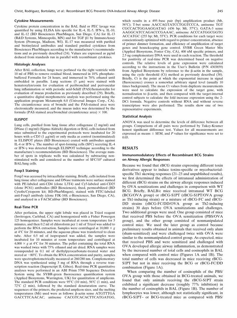
Because we found that rBCG strains expressing different toxinderivatives appear to induce toxins-specific or mycobacterial-specific Th1 skewing responses (21–23 and unpublished results),we first determined the effects of intranasal administration ofdifferent rBCG strains on the airway allergic responses inducedby OVA sensitizations and challenges in comparison with WTBCG. Briefly, BALB/c mice received intranasal WT BCG(BCG/OVA group) or rBCG-S1PT (rBCG-S1PT/OVA groupas Th1-inducing strain) or a mixture of rBCG-FC and rBCG-DD strains (rBCG-FC/DD/OVA group as Th2-inducingstrains) 30 days before OVA sensitizations and challenges.Two additional groups were used: One group consisted of micethat received PBS before the OVA sensitization (PBS/OVAgroup), and the other group consisted of nonmanipulatedcontrol mice. We used the later group as control becausepreliminary results obtained in animals that received only alum(sham-sensitized) and were challenged twice with OVA weresimilar to the nonmanipulated control group. As expected, micethat received PBS and were sensitized and challenged withOVA developed allergic airway inflammation, as demonstratedby the increased number of total cells and eosinophils in BALwhen compared with control mice (Figures 1A and 1B). Thetotal number of cells was decreased in mice receiving rBCG-S1PT but not in mice receiving the BCG or rBCG-FC/DDcombination (Figure 1A).
When comparing the number of eosinophils of the PBS/OVA group with those obtained in BCG-treated animals, wefound that only animals receiving the rBCG-S1PT strainexhibited a significant decrease (roughly 77% inhibition) inthe number of eosinophils in BAL (Figure 1B). The number oflymphocytes was lower, although not statistically significant, inrBCG-S1PT– or BCG-treated mice as compared with PBS/
Christ, Rodriguez, Bortolatto, et al.: Recombinant BCG Prevents OVA-Induced Airway Allergic Disease 245
OVA (Figure 1B), whereas the numbers of other cell types werecomparable among the studied groups (Figure 1B). Theseresults demonstrate that the intranasal administration of rBCGstrains can prevent OVA-induced airway inflammation, withrBCG-S1PT being the most effective in preventing the estab-lishment of the allergic symptoms.
Effect of Intranasal Administration of rBCG Strains on OVA-
Specific Antibody Production
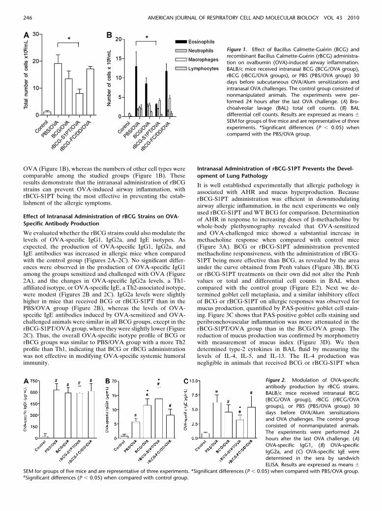
We evaluated whether the rBCG strains could also modulate thelevels of OVA-specific IgG1, IgG2a, and IgE isotypes. Asexpected, the production of OVA-specific IgG1, IgG2a, andIgE antibodies was increased in allergic mice when comparedwith the control group (Figures 2A–2C). No significant differ-ences were observed in the production of OVA-specific IgG1among the groups sensitized and challenged with OVA (Figure2A), and the changes in OVA-specific IgG2a levels, a Th1-affiliated isotype, or OVA-specific IgE, a Th2-associated isotype,were modest (Figures 2B and 2C). IgG2a levels were slightlyhigher in mice that received BCG or rBCG-S1PT than in thePBS/OVA group (Figure 2B), whereas the levels of OVA-specific IgE antibodies induced by OVA-sensitized and OVA-challenged animals were similar in all BCG groups, except in therBCG-S1PT/OVA group, where they were slightly lower (Figure2C). Thus, the overall OVA-specific isotype profile of BCG orrBCG groups was similar to PBS/OVA group with a more Th2profile than Th1, indicating that BCG or rBCG administrationwas not effective in modifying OVA-specific systemic humoralimmunity.
Intranasal Administration of rBCG-S1PT Prevents the Devel-
opment of Lung Pathology
It is well established experimentally that allergic pathology isassociated with AHR and mucus hyperproduction. BecauserBCG-S1PT administration was efficient in downmodulatingairway allergic inflammation, in the next experiments we onlyused rBCG-S1PT and WT BCG for comparison. Determinationof AHR in response to increasing doses of b-methacholine bywhole-body plethysmography revealed that OVA-sensitizedand OVA-challenged mice showed a substantial increase inmethacholine response when compared with control mice(Figure 3A). BCG or rBCG-S1PT administration preventedmethacholine responsiveness, with the administration of rBCG-S1PT being more effective than BCG, as revealed by the areaunder the curve obtained from Penh values (Figure 3B). BCGor rBCG-S1PT treatments on their own did not alter the Penhvalues or total and differential cell counts in BAL whencompared with the control group (Figure E2). Next we de-termined goblet cell metaplasia, and a similar inhibitory effectof BCG or rBCG-S1PT on allergic responses was observed formucus production, quantified by PAS-positive goblet cell stain-ing. Figure 3C shows that PAS-positive goblet cells staining andperibronchovascular inflammation was more attenuated in therBCG-S1PT/OVA group than in the BCG/OVA group. Thereduction of mucus production was confirmed by morphometrywith measurement of mucus index (Figure 3D). We thendetermined type-2 cytokines in BAL fluid by measuring thelevels of IL-4, IL-5, and IL-13. The IL-4 production wasnegligible in animals that received BCG or rBCG-S1PT when
Figure 1. Effect of Bacillus Calmette-Guerin (BCG) and
recombinant Bacillus Calmette-Guerin (rBCG) administra-
tion on ovalbumin (OVA)-induced airway inflammation.BALB/c mice received intranasal BCG (BCG/OVA group),
rBCG (rBCG/OVA groups), or PBS (PBS/OVA group) 30
days before subcutaneous OVA/Alum sensitizations and
intranasal OVA challenges. The control group consisted ofnonmanipulated animals. The experiments were per-
formed 24 hours after the last OVA challenge. (A) Bro-
choalveolar lavage (BAL) total cell counts. (B) BALdifferential cell counts. Results are expressed as means 6
SEM for groups of five mice and are representative of three
experiments. *Significant differences (P , 0.05) when
compared with the PBS/OVA group.
Figure 2. Modulation of OVA-specific
antibody production by rBCG strains.
BALB/c mice received intranasal BCG(BCG/OVA group), rBCG (rBCG/OVA
groups), or PBS (PBS/OVA group) 30
days before OVA/Alum sensitizations
and OVA challenges. The control groupconsisted of nonmanipulated animals.
The experiments were performed 24
hours after the last OVA challenge. (A)OVA-specific IgG1, (B) OVA-specific
IgG2a, and (C) OVA-specific IgE were
determined in the sera by sandwich
ELISA. Results are expressed as means 6
SEM for groups of five mice and are representative of three experiments. *Significant differences (P , 0.05) when compared with PBS/OVA group.#Significant differences (P , 0.05) when compared with control group.
246 AMERICAN JOURNAL OF RESPIRATORY CELL AND MOLECULAR BIOLOGY VOL 43 2010
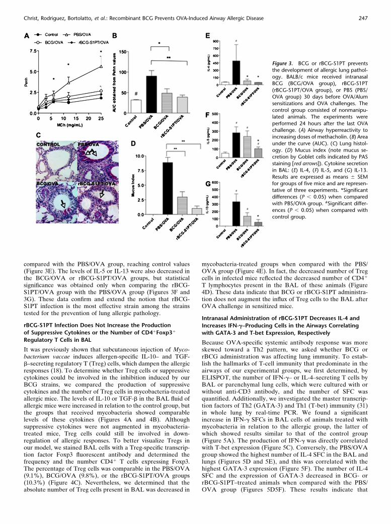
compared with the PBS/OVA group, reaching control values(Figure 3E). The levels of IL-5 or IL-13 were also decreased inthe BCG/OVA or rBCG-S1PT/OVA groups, but statisticalsignificance was obtained only when comparing the rBCG-S1PT/OVA group with the PBS/OVA group (Figures 3F and3G). These data confirm and extend the notion that rBCG-S1PT infection is the most effective strain among the strainstested for the prevention of lung allergic pathology.
rBCG-S1PT Infection Does Not Increase the Production
of Suppressive Cytokines or the Number of CD41Foxp31
Regulatory T Cells in BAL
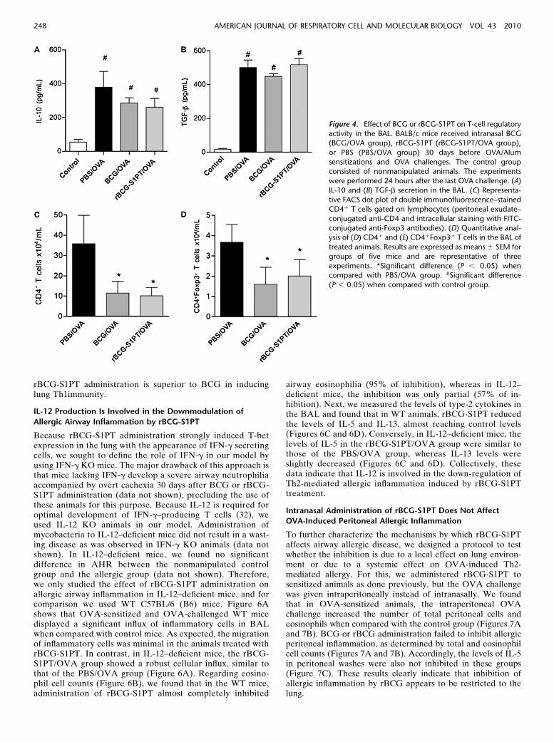
It was previously shown that subcutaneous injection of Myco-bacterium vaccae induces allergen-specific IL-10– and TGF-b–secreting regulatory T (Treg) cells, which dampen the allergicresponses (18). To determine whether Treg cells or suppressivecytokines could be involved in the inhibition induced by ourBCG strains, we compared the production of suppressivecytokines and the number of Treg cells in mycobacteria-treatedallergic mice. The levels of IL-10 or TGF-b in the BAL fluid ofallergic mice were increased in relation to the control group, butthe groups that received mycobacteria showed comparablelevels of these cytokines (Figures 4A and 4B). Althoughsuppressive cytokines were not augmented in mycobacteria-treated mice, Treg cells could still be involved in down-regulation of allergic responses. To better visualize Tregs inour model, we stained BAL cells with a Treg-specific transcrip-tion factor Foxp3 fluorescent antibody and determined thefrequency and the number CD41 T cells expressing Foxp3.The percentage of Treg cells was comparable in the PBS/OVA(9.1%), BCG/OVA (9.8%), or the rBCG-S1PT/OVA groups(10.3%) (Figure 4C). Nevertheless, we determined that theabsolute number of Treg cells present in BAL was decreased in
mycobacteria-treated groups when compared with the PBS/OVA group (Figure 4E). In fact, the decreased number of Tregcells in infected mice reflected the decreased number of CD41
T lymphocytes present in the BAL of these animals (Figure4D). These data indicate that BCG or rBCG-S1PT administra-tion does not augment the influx of Treg cells to the BAL afterOVA challenge in sensitized mice.
Intranasal Administration of rBCG-S1PT Decreases IL-4 and
Increases IFN-g–Producing Cells in the Airways Correlating
with GATA-3 and T-bet Expression, Respectively
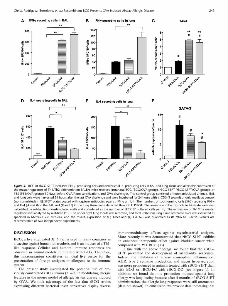
Because OVA-specific systemic antibody response was moreskewed toward a Th2 pattern, we asked whether BCG orrBCG administration was affecting lung immunity. To estab-lish the hallmarks of T-cell immunity that predominate in theairways of our experimental groups, we first determined, byELISPOT, the number of IFN-g– or IL-4–secreting T cells byBAL or parenchymal lung cells, which were cultured with orwithout anti-CD3 antibody, and the number of SFC wasquantified. Additionally, we investigated the master transcrip-tion factors of Th2 (GATA-3) and Th1 (T-bet) immunity (31)in whole lung by real-time PCR. We found a significantincrease in IFN-g SFCs in BAL cells of animals treated withmycobacteria in relation to the allergic group, the latter ofwhich showed results similar to that of the control group(Figure 5A). The production of IFN-g was directly correlatedwith T-bet expression (Figure 5C). Conversely, the PBS/OVAgroup showed the highest number of IL-4 SFC in the BAL andlungs (Figures 5D and 5E), and this was correlated with thehighest GATA-3 expression (Figure 5F). The number of IL-4SFC and the expression of GATA-3 decreased in BCG- orrBCG-S1PT–treated animals when compared with the PBS/OVA group (Figures 5D5F). These results indicate that
Figure 3. BCG or rBCG-S1PT prevents
the development of allergic lung pathol-
ogy. BALB/c mice received intranasalBCG (BCG/OVA group), rBCG-S1PT
(rBCG-S1PT/OVA group), or PBS (PBS/
OVA group) 30 days before OVA/Alumsensitizations and OVA challenges. The
control group consisted of nonmanipu-
lated animals. The experiments were
performed 24 hours after the last OVAchallenge. (A) Airway hyperreactivity to
increasing doses of methacholin. (B) Area
under the curve (AUC). (C) Lung histol-
ogy. (D) Mucus index (note mucus se-cretion by Goblet cells indicated by PAS
staining [red arrows]). Cytokine secretion
in BAL: (E) IL-4, (F) IL-5, and (G) IL-13.
Results are expressed as means 6 SEMfor groups of five mice and are represen-
tative of three experiments. *Significant
differences (P , 0.05) when comparedwith PBS/OVA group. #Significant differ-
ences (P , 0.05) when compared with
control group.
Christ, Rodriguez, Bortolatto, et al.: Recombinant BCG Prevents OVA-Induced Airway Allergic Disease 247
rBCG-S1PT administration is superior to BCG in inducinglung Th1immunity.
IL-12 Production Is Involved in the Downmodulation of
Allergic Airway Inflammation by rBCG-S1PT
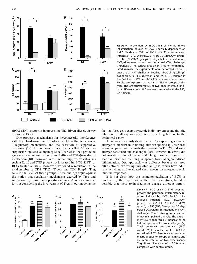
Because rBCG-S1PT administration strongly induced T-betexpression in the lung with the appearance of IFN-g secretingcells, we sought to define the role of IFN-g in our model byusing IFN-g KO mice. The major drawback of this approach isthat mice lacking IFN-g develop a severe airway neutrophiliaaccompanied by overt cachexia 30 days after BCG or rBCG-S1PT administration (data not shown), precluding the use ofthese animals for this purpose. Because IL-12 is required foroptimal development of IFN-g–producing T cells (32), weused IL-12 KO animals in our model. Administration ofmycobacteria to IL-12–deficient mice did not result in a wast-ing disease as was observed in IFN-g KO animals (data notshown). In IL-12–deficient mice, we found no significantdifference in AHR between the nonmanipulated controlgroup and the allergic group (data not shown). Therefore,we only studied the effect of rBCG-S1PT administration onallergic airway inflammation in IL-12–deficient mice, and forcomparison we used WT C57BL/6 (B6) mice. Figure 6Ashows that OVA-sensitized and OVA-challenged WT micedisplayed a significant influx of inflammatory cells in BALwhen compared with control mice. As expected, the migrationof inflammatory cells was minimal in the animals treated withrBCG-S1PT. In contrast, in IL-12–deficient mice, the rBCG-S1PT/OVA group showed a robust cellular influx, similar tothat of the PBS/OVA group (Figure 6A). Regarding eosino-phil cell counts (Figure 6B), we found that in the WT mice,administration of rBCG-S1PT almost completely inhibited
airway eosinophilia (95% of inhibition), whereas in IL-12–deficient mice, the inhibition was only partial (57% of in-hibition). Next, we measured the levels of type-2 cytokines inthe BAL and found that in WT animals, rBCG-S1PT reducedthe levels of IL-5 and IL-13, almost reaching control levels(Figures 6C and 6D). Conversely, in IL-12–deficient mice, thelevels of IL-5 in the rBCG-S1PT/OVA group were similar tothose of the PBS/OVA group, whereas IL-13 levels wereslightly decreased (Figures 6C and 6D). Collectively, thesedata indicate that IL-12 is involved in the down-regulation ofTh2-mediated allergic inflammation induced by rBCG-S1PTtreatment.
Intranasal Administration of rBCG-S1PT Does Not Affect
OVA-Induced Peritoneal Allergic Inflammation
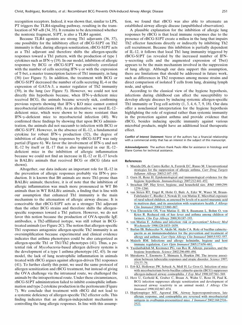
To further characterize the mechanisms by which rBCG-S1PTaffects airway allergic disease, we designed a protocol to testwhether the inhibition is due to a local effect on lung environ-ment or due to a systemic effect on OVA-induced Th2-mediated allergy. For this, we administered rBCG-S1PT tosensitized animals as done previously, but the OVA challengewas given intraperitoneally instead of intranasally. We foundthat in OVA-sensitized animals, the intraperitoneal OVAchallenge increased the number of total peritoneal cells andeosinophils when compared with the control group (Figures 7Aand 7B). BCG or rBCG administration failed to inhibit allergicperitoneal inflammation, as determined by total and eosinophilcell counts (Figures 7A and 7B). Accordingly, the levels of IL-5in peritoneal washes were also not inhibited in these groups(Figure 7C). These results clearly indicate that inhibition ofallergic inflammation by rBCG appears to be restricted to thelung.
Figure 4. Effect of BCG or rBCG-S1PT on T-cell regulatoryactivity in the BAL. BALB/c mice received intranasal BCG
(BCG/OVA group), rBCG-S1PT (rBCG-S1PT/OVA group),
or PBS (PBS/OVA group) 30 days before OVA/Alum
sensitizations and OVA challenges. The control groupconsisted of nonmanipulated animals. The experiments
were performed 24 hours after the last OVA challenge. (A)
IL-10 and (B) TGF-b secretion in the BAL. (C) Representa-tive FACS dot plot of double immunofluorescence–stained
CD41 T cells gated on lymphocytes (peritoneal exudate–
conjugated anti-CD4 and intracellular staining with FITC-
conjugated anti-Foxp3 antibodies). (D) Quantitative anal-ysis of (D) CD41 and (E) CD41Foxp31 T cells in the BAL of
treated animals. Results are expressed as means 6 SEM for
groups of five mice and are representative of three
experiments. *Significant difference (P , 0.05) whencompared with PBS/OVA group. #Significant difference
(P , 0.05) when compared with control group.
248 AMERICAN JOURNAL OF RESPIRATORY CELL AND MOLECULAR BIOLOGY VOL 43 2010
DISCUSSION
BCG, a live attenuated M. bovis, is used in many countries asa vaccine against human tuberculosis and is an inducer of a Th1-like response. Cellular and humoral immune responses areobserved in animal models immunized with BCG. Therefore,this microorganism constitutes an ideal live vector for thepresentation of foreign antigens or allergens to the immunesystem.
The present study investigated the potential use of pre-viously constructed rBCG strains (21–23) in modulating allergicfeatures in the mouse model of allergic airway disease inducedby OVA. We took advantage of the fact that rBCG strainsexpressing different bacterial toxin derivatives display diverse
immunomodulatory effects against mycobacterial antigens.More recently it was demonstrated that rBCG-S1PT exhibitsan enhanced therapeutic effect against bladder cancer whencompared with WT BCG (33).
In line with the above findings, we found that the rBCG-S1PT prevented the development of asthma-like responses.Indeed, the inhibition of airway eosinophilic inflammation,AHR, type 2 cytokine production, and mucus hypersecretionwas more pronounced in animals treated with rBCG-S1PT thanwith BCG or rBCG-FC with rBCG-DD (see Figure 1). Inaddition, we found that the protection induced against lungallergy was long lasting because after 4 months of rBCG-S1PTadministration, the allergic lung responses were still attenuated(data not shown). In conclusion, we provide data indicating that
Figure 5. BCG or rBCG-S1PT increases IFN-g–producing cells and decreases IL-4–producing cells in BAL and lung tissue and alters the expression ofthe master regulators of Th1/Th2 differentiation BALB/c mice received intranasal BCG (BCG/OVA group), rBCG-S1PT (rBCG-S1PT/OVA group), or
PBS (PBS/OVA group) 30 days before OVA/Alum sensitizations and OVA challenges. The control group consisted of nonmanipulated animals. BAL
and lung cells were harvested 24 hours after the last OVA challenge and were incubated for 24 hours with a-CD3 (1 mg/ml) or only media as control
(nonstimulated) in ELISPOT plates coated with capture antibodies against IFN-g an IL-4. The numbers of spot-forming cells (SFC) secreting IFN-gand IL-4 (A and B) in the BAL and (B and E) in the lung tissue were detected through ELISPOT. The average number of spots in triplicate wells was
calculated by subtracting nonstimulated wells and considered as the number of SFC/106 cultured cells per m/. The expression of Th1/Th2 master
regulators was analyzed by real-time PCR. The upper right lung lobule was removed, and total RNA from lung tissue of treated mice was extracted as
specified in MATERIALS AND METHODS, and the mRNA expression of (C) T-bet and (F) GATA-3 was quantified as its ratio to b-actin. Results arerepresentative of two independent experiments.
Christ, Rodriguez, Bortolatto, et al.: Recombinant BCG Prevents OVA-Induced Airway Allergic Disease 249
rBCG-S1PT is superior in preventing Th2-driven allergic airwaydisease to BCG.
One proposed mechanisms for mycobacterial interferencewith the Th2-driven lung pathology would be the induction ofT-regulatory mechanisms and the secretion of suppressivecytokines (18). It has been shown that a killed M. vaccae-suspension induced allergen-specific Treg cells that protectedagainst airway inflammation by an IL-10– and TGF-b–mediatedmechanism (18). However, in our model, suppressive cytokinessuch as IL-10 and TGF-b were not increased in rBCG-S1PT– orBCG-treated animals. Moreover, we found a reduction in thetotal number of CD41CD251 T cells and CD41Foxp31 Tregcells in the BAL of these groups. These findings argue againstthe notion that regulatory mechanisms exerted by Treg andsuppressive cytokines are operating in lung. Another argumentfor not considering the involvement of Treg in our model is the
fact that Treg cells exert a systemic inhibitory effect and that theinhibition of allergy was restricted to the lung but not to theperitoneal cavity.
It has been previously shown that rBCG expressing a specificallergen is efficient in inhibiting allergen-specific IgE responsewhen compared with animals that received WT BCG and wereallergen sensitized and challenged (20). However, this work didnot investigate the allergen-specific lung immune responses toascertain whether the lung is spared from allergen-inducedinflammation. Our approach was different because we usedrBCG strains expressing unrelated antigens, which have adju-vant activities, and evaluated their effects on allergen-specificimmune responses.
It is not clear how the immunomodulation of BCG ismodified by the expression of the toxin derivatives, but it ispossible that these toxin fragments engage different pattern
Figure 6. Prevention by rBCG-S1PT of allergic airwayinflammation induced by OVA is partially dependent on
IL-12. Wild-type (WT) or IL-12 KO B6 mice received
intranasal 106 CFU of rBCG-S1PT (rBCG-S1PT/OVA group)
or PBS (PBS/OVA group) 30 days before subcutaneousOVA/Alum sensitizations and intranasal OVA challenges
(intranasal). The control group consisted of nonmanipu-
lated animals. The experiments were performed 24 hoursafter the last OVA challenge. Total numbers of (A) cells, (B)
eosinophils, (C) IL-5 secretion, and (D) IL-13 secretion in
the BAL fluid of WT and IL-12 KO mice were determined.
Results are expressed as means 6 SEM for groups of fivemice and are representative of two experiments. Signifi-
cant differences (P , 0.05) when compared with the PBS/
OVA group.
Figure 7. BCG or rBCG-S1PT does not
prevent the peritoneal inflammatory re-action induced by OVA. BALB/c mice
received intranasal BCG (BCG/OVA
group), rBCG-S1PT (rBCG-S1PT/OVA
group), or PBS (PBS/OVA group) 30 daysbefore OVA/alum sensitizations and OVA
challenges. The control group consisted
of nonmanipulated animals. The experi-ments were performed 24 hours after the
last intraperitoneal OVA challenge. (A)
Total peritoneal exudate cell (PEC)
counts. (B) Eosinophils in PECs. (C) IL-5secretion in PECs. Results are expressed as
means 6 SEM for groups of six mice and
are representative of two experiments.#Significant differences (P , 0.05) whencompared with control group.
250 AMERICAN JOURNAL OF RESPIRATORY CELL AND MOLECULAR BIOLOGY VOL 43 2010
recognition receptors. Indeed, it was shown that, similar to LPS,PT triggers the TLR4-signaling pathway, resulting in the trans-location of NF-kB (34, 35). It remains to be determined whetherthe nontoxic fragment, S1PT, is also a TLR4 agonist.
Because TLR4 agonists are strong Th1 adjuvants (36, 37),one possibility for the inhibitory effect of rBCG-S1PT on Th2immunity is that, during allergen sensitization, rBCG-S1PT actsas a Th1 adjuvant and therefore shifts the allergen-specificresponses toward a Th1 pattern, with the production of type 1cytokines such as IFN-g (19). In our model, inhibition of allergicresponses by BCG or rBCG-S1PT was positively correlatedwith the number of cells secreting IFN-g or with the expressionof T-bet, a master transcription factors of Th1 immunity, in lung(38) (see Figure 5). In addition, the treatment with BCG orrBCG-S1PT decreased the number of cells secreting IL-4 or theexpression of GATA-3, a master regulator of Th2 immunity(39), in the lung (see Figure 5). However, we could not testdirectly this hypothesis because, when IFN-g–deficient micereceived BCG, the animals became severely sick, confirmingprevious reports showing that IFN-g KO mice cannot controlmycobacterial infections (40). As an alternative, we used IL-12–deficient mice, which were shown to be more resistant thanIFN-g–deficient mice to mycobacterial infection (40). Weconfirmed these findings by showing that upon BCG adminis-tration, the animals did not succumb to infection with BCG orrBCG-S1PT. However, in the absence of IL-12, a fundamentalcytokine for robust IFN-g production (32), the degree ofinhibition of allergic lung responses by rBCG-S1PT was onlypartial (Figure 6). We favor the involvement of IFN-g and notIL-12 by itself or IL-17 that is also impaired in our IL-12–deficient mice in the inhibition of allergic inflammationbecause we could not find an increase in IL-12 or IL-17 levelsin BALB/c animals that received BCG or rBCG (data notshown).
Altogether, our data indicate an important role for IL-12 inthe prevention of allergic responses probably via IFN-g pro-duction. It is known that B6 animals are more Th1-prone thanBALB/c animals; therefore, it is of note that the inhibition ofallergic inflammation was much more pronounced in WT B6animals than in WT BALB/c animals, a finding that is line withour assumption that enhanced Th1 immunity is the mainmechanism to the attenuation of allergic airway disease. It isconceivable that rBCG-S1PT acts as a stronger Th1 adjuvantthan the other BCG strains and therefore shifts the allergen-specific responses toward a Th1 pattern. However, we do notfavor this notion because the production of OVA-specific IgEantibodies, a Th2-affiliated isotype, was still robust in BCG-treated animals (see Figure 2C). The notion that allergen-specificTh1 responses antagonize allergen-specific Th2 immunity is anoversimplification because experimental and clinical evidenceindicates that asthma phenotypes could be also categorized asallergen-specific Th1 or Th1/Th2 phenotypes (41). Thus, a po-tential risk of Mycobacteria-based allergen delivery systems isthe development of a type 1 asthma phenotype (42, 43). In ourmodel, the lack of lung neutrophilic inflammation in animalstreated with rBCG argues against allergen-driven Th1 responses(41). To further clarify this point, we used the same protocol ofallergen sensitization and rBCG treatment, but instead of givingthe OVA challenge via the intranasal route, we challenged theanimals by the intraperitoneal route. Under these circumstances,rBCG-S1PT administration failed to inhibit eosinophilic inflam-mation and type 2 cytokine production in the peritoneum (Figure7). We conclude that treatment with rBCG did not lead toa systemic deficiency of allergen-specific Th2 effector cells. Thisfinding indicates that an allergen-independent mechanism iscontrolling the lung allergic responses. In line with this assump-
tion, we found that rBCG was also able to attenuate anestablished airway allergic disease (unpublished observation).
A plausible explanation for the inhibition of allergic lungresponses by rBCG is that local immune responses due to thepresence of rBCG-S1PT create a milieu in the lung that impairsTh2 effector functions directly or indirectly by inhibiting Th2cell recruitment. Because this inhibition is partially dependentof IL-12, it follows that local Th1 lung immunity triggered byrBCG-S1PT (as revealed by the increased number of IFN-g–secreting cells and the augmented expression of Tbet)appears to be the main mechanism involved in the suppressionof lung allergy. Although our data support this assumption,there are limitations that should be addressed in future work,such as differences in Th2 responses among mouse strains anddirect comparison of isolated T cells from lung, draining lymphnode, and spleen.
According to the classical view of the hygiene hypothesis,infections during childhood can affect the susceptibility todevelop allergic diseases by inducing systemic allergen-specificTh1 immunity or Treg cell activity (1, 3, 4, 6, 7, 9, 18). Our dataoffer a nonclassical interpretation for the hygiene hypothesishighlighting the role of regional rather than systemic immunityin the protection against asthma and provide evidence thatrBCG, besides inducing specific immunity against variousmicrobial products, might have an additional local therapeuticeffect.
Conflict of Interest Statement: None of the authors has a financial relationshipwith a commercial entity that has an interest in the subject of this manuscript.
Acknowledgments: The authors thank Paulo Albe for assistance in histology andEliane Gomes for technical assistance.
References
1. Mucida DS, de Castro Keller A, Fernvik EC, Russo M. Unconventionalstrategies for the suppression of allergic asthma. Curr Drug TargetsInflamm Allergy 2003;2:187–195.
2. Garn H, Renz H. Epidemiological and immunological evidence for thehygiene hypothesis. Immunobiology 2007;212:441–452.
3. Strachan DP. Hay fever, hygiene, and household size. BMJ 1989;299:1259–1260.
4. van Strien RT, Engel R, Holst O, Bufe A, Eder W, Waser M, Braun-Fahrlander C, Riedler J, Nowak D, von Mutius E. Microbial exposureof rural school children, as assessed by levels of n-acetyl-muramic acidin mattress dust, and its association with respiratory health. J AllergyClin Immunol 2004;113:860–867.
5. Von Ehrenstein OS, Von Mutius E, Illi S, Baumann L, Bohm O, vonKries R. Reduced risk of hay fever and asthma among children offarmers. Clin Exp Allergy 2000;30:187–193.
6. von Mutius E. Asthma and infection: risk or prevention? Schweiz MedWochenschr 1998;128:1833–1839.
7. Barlan IB, Bahceciler N, Akdis M, Akdis CA. Role of bacillus calmette-guerin as an immunomodulator for the prevention and treatment ofallergy and asthma. Curr Opin Allergy Clin Immunol 2005;5:552–557.
8. Maizels RM. Infections and allergy: helminths, hygiene and hostimmune regulation. Curr Opin Immunol 2005;17:656–661.
9. Yazdanbakhsh M, Kremsner PG, van Ree R. Allergy, parasites, and thehygiene hypothesis. Science 2002;296:490–494.
10. Shirakawa T, Enomoto T, Shimazu S, Hopkin JM. The inverse associ-ation between tuberculin responses and atopic disorder. Science 1997;275:77–79.
11. Erb KJ, Holloway JW, Sobeck A, Moll H, Le Gros G. Infection of micewith mycobacterium bovis-bacillus calmette-guerin (BCG) suppressesallergen-induced airway eosinophilia. J Exp Med 1998;187:561–569.
12. Herz U, Gerhold K, Gruber C, Braun A, Wahn U, Renz H, Paul K.BCG infection suppresses allergic sensitization and development ofincreased airway reactivity in an animal model. J Allergy ClinImmunol 1998;102:867–874.
13. Hopfenspirger MT, Agrawal DK. Airway hyperresponsiveness, lateallergic response, and eosinophilia are reversed with mycobacterialantigens in ovalbumin-presensitized mice. J Immunol 2002;168:2516–2522.
Christ, Rodriguez, Bortolatto, et al.: Recombinant BCG Prevents OVA-Induced Airway Allergic Disease 251
14. Hopfenspirger MT, Parr SK, Hopp RJ, Townley RG, Agrawal DK.Mycobacterial antigens attenuate late phase response, airwayhyperresponsiveness, and bronchoalveolar lavage eosinophilia ina mouse model of bronchial asthma. Int Immunopharmacol 2001;1:1743–1751.
15. Zuany-Amorim C, Manlius C, Trifilieff A, Brunet LR, Rook G, BowenG, Pay G, Walker C. Long-term protective and antigen-specific effectof heat-killed mycobacterium vaccae in a murine model of allergicpulmonary inflammation. J Immunol 2002;169:1492–1499.
16. Choi IS, Lin XH, Koh YA, Cui Y. Inoculation route-dependent andallergen-specific suppressive effects of bacille calmette-guerin vacci-nation on asthmatic reactions in balb/c mice. Lung 2007;185:179–186.
17. Choi IS, Lin XH, Koh YA, Koh YI, Lee HC. Strain-dependentsuppressive effects of BCG vaccination on asthmatic reactions inbalb/c mice. Ann Allergy Asthma Immunol 2005;95:571–578.
18. Zuany-Amorim C, Sawicka E, Manlius C, Le Moine A, Brunet LR,Kemeny DM, Bowen G, Rook G, Walker C. Suppression of airwayeosinophilia by killed mycobacterium vaccae-induced allergen-specificregulatory T-cells. Nat Med 2002;8:625–629.
19. Hopkin JM. Atopy, asthma, and the mycobacteria. Thorax 2000;55:443–445.
20. Kumar M, Behera AK, Matsuse H, Lockey RF, Mohapatra SS. Arecombinant BCG vaccine generates a Th1-like response and inhibitsIgE synthesis in Balb/c mice. Immunology 1999;97:515–521.
21. Mazzantini RP, Miyaji EN, Dias WO, Sakauchi D, Nascimento AL, RawI, Winter N, Gicquel B, Rappuoli R, Leite LC. Adjuvant activity ofmycobacterium bovis BCG expressing CRM197 on the immuneresponse induced by BCG expressing tetanus toxin fragment C.Vaccine 2004;22:740–746.
22. Miyaji EN, Mazzantini RP, Dias WO, Nascimento AL, Marcovistz R,Matos DS, Raw I, Winter N, Gicquel B, Rappuoli R, et al. Inductionof neutralizing antibodies against diphtheria toxin by priming withrecombinant mycobacterium bovis BCG expressing CRM197, a mutantdiphtheria toxin. Infect Immun 2001;69:869–874.
23. Nascimento IP, Dias WO, Mazzantini RP, Miyaji EN, Gamberini M,Quintilio W, Gebara VC, Cardoso DF, Ho PL, Raw I, et al.Recombinant mycobacterium bovis BCG expressing pertussis toxinsubunit S1 induces protection against an intracerebral challenge withlive bordetella pertussis in mice. Infect Immun 2000;68:4877–4883.
24. Russo M, Nahori MA, Lefort J, Gomes E, de Castro Keller A,Rodriguez D, Ribeiro OG, Adriouch S, Gallois V, de Faria AM,et al. Suppression of asthma-like responses in different mouse strainsby oral tolerance. Am J Respir Cell Mol Biol 2001;24:518–526.
25. Rodriguez D, Keller AC, Faquim-Mauro EL, de Macedo MS, CunhaFQ, Lefort J, Vargaftig BB, Russo M. Bacterial lipopolysaccharidesignaling through toll-like receptor 4 suppresses asthma-like re-sponses via nitric oxide synthase 2 activity. J Immunol 2003;171:1001–1008.
26. Keller AC, Mucida D, Gomes E, Faquim-Mauro EL, Caetano de FariaAM, Rodriguez D, Russo M. Hierarchial suppression of asthma-likeresponses by mucosal tolerance. JAllergy Clinical Immunol 2006;177:283–290.
27. Noulin N, Quesniaux VF, Schnyder-Candrian S, Schnyder B, Maillet I,Robert T, Vargaftig BB, Ryffel B, Couillin I. Both hemopoietic andresident cells are required for myd88-dependent pulmonary inflam-matory response to inhaled endotoxin. J Immunol 2005;175:6861–6869.
28. Albertine KH, Wang L, Watanabe S, Marathe GK, Zimmerman GA,McIntyre TM. Temporal correlation of measurements of airwayhyperresponsiveness in ovalbumin-sensitized mice. Am J PhysiolLung Cell Mol Physiol 2002;283:L219–L233.
29. Keller AC, Mucida D, Gomes E, Faquim-Mauro E, Faria AM,Rodriguez D, Russo M. Hierarchical suppression of asthma-likeresponses by mucosal tolerance. J Allergy Clin Immunol 2006;117:283–290.
30. Silva TA, Garlet GP, Lara VS, Martins W Jr, Silva JS, Cunha FQ.Differential expression of chemokines and chemokine receptors ininflammatory periapical diseases. Oral Microbiol Immunol 2005;20:310–316.
31. Chakir H, Wang H, Lefebvre DE, Webb J, Scott FW. T-bet/gata-3 ratioas a measure of the th1/th2 cytokine profile in mixed cell populations:predominant role of gata-3. J Immunol Methods 2003;278:157–169.
32. Macatonia SE, Hosken NA, Litton M, Vieira P, Hsieh CS, CulpepperJA, Wysocka M, Trinchieri G, Murphy KM, O’Garra A. Dendriticcells produce IL-12 and direct the development of th1 cells from naiveCD41 T cells. J Immunol 1995;154:5071–5079.
33. Chade DC, Borra RC, Nascimento IP, Villanova FE, Leite LC, AndradeEF, Ramos K, Srougi M, Andrade PM. Immunomodulatory effects ofrecombinant bcg expressing pertussis toxin on TNF-alpha and IL-10in a bladder cancer model. J Exp Clin Cancer Res 2008;27:78.
34. Fujimoto C, Yu CR, Shi G, Vistica BP, Wawrousek EF, Klinman DM,Chan CC, Egwuagu CE, Gery I. Pertussis toxin is superior to tlrligands in enhancing pathogenic autoimmunity, targeted at a neo-selfantigen, by triggering robust expansion of th1 cells and their cytokineproduction. J Immunol 2006;177:6896–6903.
35. Racke MK, Hu W, Lovett-Racke AE. Ptx cruiser: driving autoimmunityvia tlr4. Trends Immunol 2005;26:289–291.
36. Schroder NW. The role of innate immunity in the pathogenesis ofasthma. Curr Opin Allergy Clin Immunol 2009;9:38–43.
37. Schroder NW, Arditi M. The role of innate immunity in the pathogenesisof asthma: evidence for the involvement of toll-like receptor signal-ing. J Endotoxin Res 2007;13:305–312.
38. Finotto S, Neurath MF, Glickman JN, Qin S, Lehr HA, Green FH,Ackerman K, Haley K, Galle PR, Szabo SJ, et al. Development ofspontaneous airway changes consistent with human asthma in micelacking T-bet. Science 2002;295:336–338.
39. Kiwamoto T, Ishii Y, Morishima Y, Yoh K, Maeda A, Ishizaki K, IizukaT, Hegab AE, Matsuno Y, Homma S, et al. Transcription factors T-bet and GATA-3 regulate development of airway remodeling. Am JRespir Crit Care Med 2006;174:142–151.
40. Jouanguy E, Doffinger R, Dupuis S, Pallier A, Altare F, Casanova JL.IL-12 and IFN-gamma in host defense against mycobacteria andsalmonella in mice and men. Curr Opin Immunol 1999;11:346–351.
41. Kim YK, Oh SY, Jeon SG, Park HW, Lee SY, Chun EY, Bang B, LeeHS, Oh MH, Kim YS, et al. Airway exposure levels of lipopolysac-charide determine type 1 versus type 2 experimental asthma. JImmunol 2007;178:5375–5382.
42. Hansen G, Berry G, DeKruyff RH, Umetsu DT. Allergen-specific Th1cells fail to counterbalance Th2 cell-induced airway hyperreactivitybut cause severe airway inflammation. J Clin Invest 1999;103:175–183.
43. Takaoka A, Tanaka Y, Tsuji T, Jinushi T, Hoshino A, Asakura Y, Mita Y,Watanabe K, Nakaike S, Togashi Y, et al. A critical role for mouse cxcchemokine(s) in pulmonary neutrophilia during Th type 1-dependentairway inflammation. J Immunol 2001;167:2349–2353.
252 AMERICAN JOURNAL OF RESPIRATORY CELL AND MOLECULAR BIOLOGY VOL 43 2010
Related Documents