Immunology 1994 81 618-625 T-helper 1-like subset selection in Mycobacterium bovis bacillus Calmette-Guerin-infected resistant and susceptible mice I. KRAMNIK, D. RADZIOCH & E. SKAMENE McGill Centre for the Study of Host Resistance, The Montreal General Hospital Research Institute, Montreal, Quebec, Canada SUMMARY The Bcg gene has been shown to control natural resistance of mice to intravenous infection with low doses of Mycobacterium bovis (bacillus Calmette-Guerin; BCG). In the present study, we evaluated the impact of the Bcg gene on the development of T-cell reactivity during the early stages of infection. Congenic strains of mice, bearing 'r' and 's' alleles of the Bcg gene on BIO.A and BALB/c backgrounds, were studied at different time-points for 2 weeks after infection. The in vitro proliferative response of spleen cells, induced by mycobacteria or concanavalin A, was depressed in the Bcg; mice compared to the Bcg' congenic mice 14 days after infection with 105 colony-forming units (CFU) of BCG. Polymerase chain reaction (PCR)-based methodology was used to compare the level of lymphokine gene expression in the spleens of infected congenic mice both ex vivo and after in vitro stimulation. In both cases, preferential expression of interferon-y (IFN-y), lymphotoxin, interleukin-2 (IL-2) and IL-2 receptor genes was observed. The lymphokine gene expression profiles indicated that T lymphocytes activated in the course of the BCG infection preferentially expressed the T-helper 1-specific pattern, irrespective of the allele of the Bcg gene. We showed that this bias in T-cell differentiation could not be attributed to either down-regulation of IL-4 gene expression or modulation of the macrophage co-stimulatory activity by live M. bovis BCG. We conclude that the mechanism of phenotypic expression of the Bcg gene resides in the differential ability of macrophages to be activated by lymphokines produced by protective T cells, rather than in the lack of these lymphokines in susceptible animals. INTRODUCTION Host interactions with infectious agents are characterized by the involvement of different immune effector mechanisms, which could be implicated both in the elimination of pathogens and in disease progression. T lymphocytes play a key regulatory role in the development and expression of these mechanisms through lymphokine production. The role of T cells in host resistance to infection could be correlated with the pattern of lymphokines, secreted by pathogen-specific T lymphocytes. Since the T-helper 1 (Thl) and Th2 CD4+ clones were described initially, many approaches have been used to demon- strate that the restricted pattern of lymphokine secretion by parasite-specific T cells is responsible for disease susceptibility/ resistance.' In most cases, the development of the Thl-like subset, distinguished by the secretion of interleukin-2 (IL-2) and interferon-y (IFN-y), conferred resistance to intracellular patho- Received 24 September 1993; revised 3 November 1993; accepted 1 December 1993. Abbreviations: Con A, concanavalin A; PAGE, polyacrylamide gel electrophoresis; PCR, polymerase chain reaction; PPD, purified protein derivative; RT, reverse transcription. Correspondence: Dr D. Radzioch, McGill Centre for the Study of Host Resistance, Montreal General Hospital, Room 7113, 1650 Cedar Avenue, Montreal, Quebec, Canada H3G 1A4. gens (Listeria major, Mycobacterium leprae).2'3 It is conceivable that the protective potential of certain sets of lymphokines would depend on their ability to augment or suppress effector reactions crucial for the survival and multiplication of particular pathogens. The aim of the present study was to determine whether resistance/susceptibility to bacillus Calmette-Guerin (BCG) infection, conferred by the Bcg gene, correlates with the lymphokine spectrum expressed by the activated T cells and whether the quality of the macrophages, determined by the pleiotropic effects of the Bcg gene, contributes to the activation of the T cells, expressing a particular lymphokine profile. The Bcg gene is a single autosomal gene which controls the innate resistance of mice to several species of Mycobacteria.4 This gene is identical or very closely linked to two other host resistance genes which control resistance to infection with Salmonella typhymurium (Ity) and Leishmania donovani (Lsh).5 Recently, the most likely candidate for the Bcg gene has been cloned.6 Interestingly, the gene (Nrampl) was found to be expressed exclusively in macrophages. The Bcg gene acts very early in the host response to infection, at the time of the initiation of the immune response, and is phenotypically expressed as an intrinsic ability of macrophages to control the growth of intracellular pathogens.6 7 Recent reports have shown that Bcgr macrophages are better antigen-presenting cells (APC) than their Bcgf counterparts.8'9 Considering the tentative role of APC 618

Welcome message from author
This document is posted to help you gain knowledge. Please leave a comment to let me know what you think about it! Share it to your friends and learn new things together.
Transcript
Immunology 1994 81 618-625
T-helper 1-like subset selection in Mycobacterium bovis bacillusCalmette-Guerin-infected resistant and susceptible mice
I. KRAMNIK, D. RADZIOCH & E. SKAMENE McGill Centre for the Study of Host Resistance,The Montreal General Hospital Research Institute, Montreal, Quebec, Canada
SUMMARY
The Bcg gene has been shown to control natural resistance of mice to intravenous infection with lowdoses of Mycobacterium bovis (bacillus Calmette-Guerin; BCG). In the present study, we evaluatedthe impact of the Bcg gene on the development of T-cell reactivity during the early stages of infection.Congenic strains of mice, bearing 'r' and 's' alleles of the Bcg gene on BIO.A and BALB/cbackgrounds, were studied at different time-points for 2 weeks after infection. The in vitroproliferative response of spleen cells, induced by mycobacteria or concanavalin A, was depressed inthe Bcg; mice compared to the Bcg' congenic mice 14 days after infection with 105 colony-formingunits (CFU) of BCG. Polymerase chain reaction (PCR)-based methodology was used to compare thelevel of lymphokine gene expression in the spleens of infected congenic mice both ex vivo and after invitro stimulation. In both cases, preferential expression of interferon-y (IFN-y), lymphotoxin,interleukin-2 (IL-2) and IL-2 receptor genes was observed. The lymphokine gene expression profilesindicated that T lymphocytes activated in the course ofthe BCG infection preferentially expressed theT-helper 1-specific pattern, irrespective ofthe allele of the Bcg gene. We showed that this bias in T-celldifferentiation could not be attributed to either down-regulation of IL-4 gene expression or
modulation of the macrophage co-stimulatory activity by live M. bovis BCG. We conclude that themechanism ofphenotypic expression of the Bcg gene resides in the differential ability ofmacrophagesto be activated by lymphokines produced by protective T cells, rather than in the lack of theselymphokines in susceptible animals.
INTRODUCTION
Host interactions with infectious agents are characterized by theinvolvement of different immune effector mechanisms, whichcould be implicated both in the elimination of pathogens and indisease progression. T lymphocytes play a key regulatory role inthe development and expression of these mechanisms throughlymphokine production. The role ofT cells in host resistance toinfection could be correlated with the pattern of lymphokines,secreted by pathogen-specific T lymphocytes.
Since the T-helper 1 (Thl) and Th2 CD4+ clones weredescribed initially, many approaches have been used to demon-strate that the restricted pattern of lymphokine secretion byparasite-specific T cells is responsible for disease susceptibility/resistance.' In most cases, the development of the Thl-likesubset, distinguished by the secretion ofinterleukin-2 (IL-2) andinterferon-y (IFN-y), conferred resistance to intracellular patho-
Received 24 September 1993; revised 3 November 1993; accepted1 December 1993.
Abbreviations: Con A, concanavalin A; PAGE, polyacrylamide gelelectrophoresis; PCR, polymerase chain reaction; PPD, purified proteinderivative; RT, reverse transcription.
Correspondence: Dr D. Radzioch, McGill Centre for the Study ofHost Resistance, Montreal General Hospital, Room 7113, 1650 CedarAvenue, Montreal, Quebec, Canada H3G 1A4.
gens (Listeria major, Mycobacterium leprae).2'3 It is conceivablethat the protective potential of certain sets of lymphokineswould depend on their ability to augment or suppress effectorreactions crucial for the survival and multiplication ofparticularpathogens.
The aim of the present study was to determine whetherresistance/susceptibility to bacillus Calmette-Guerin (BCG)infection, conferred by the Bcg gene, correlates with thelymphokine spectrum expressed by the activated T cells andwhether the quality of the macrophages, determined by thepleiotropic effects of the Bcg gene, contributes to the activationof the T cells, expressing a particular lymphokine profile. TheBcg gene is a single autosomal gene which controls the innateresistance of mice to several species of Mycobacteria.4 This geneis identical or very closely linked to two other host resistancegenes which control resistance to infection with Salmonellatyphymurium (Ity) and Leishmania donovani (Lsh).5 Recently,the most likely candidate for the Bcg gene has been cloned.6Interestingly, the gene (Nrampl) was found to be expressedexclusively in macrophages. The Bcg gene acts very early in thehost response to infection, at the time of the initiation of theimmune response, and is phenotypically expressed as anintrinsic ability of macrophages to control the growth ofintracellular pathogens.67 Recent reports have shown that Bcgrmacrophages are better antigen-presenting cells (APC) thantheir Bcgf counterparts.8'9 Considering the tentative role ofAPC
618
T-lymphocyte gene expression in BCG infection
in T-lymphocyte subset activation, it seemed plausible that thecontribution ofinnate resistance, determined by the Bcg gene, tothe development ofimmune reactivity would be displayed at thelymphokine gene expression level, early in the course of theBCG infection.
MATERIALS AND METHODSMiceBl0.A (Bcgs) and A/J (Bcgt) mice were obtained from theNational Cancer Institute, Frederick, MD. Congenic B1O.A.Bcgr mice were constructed as described elsewhere'0 and main-tained in our animal facility under specific pathogen-free (SPF)conditions. CD.2 (BALB/c.Bcgr) mice were obtained as de-scribed elsewhere. " Both CD.2 and BALB/c mice were bred andmaintained under SPF conditions in the animal facility of theMontreal General Hospital Research Institute.
Infection ofmiceMycobacterium bovis BCG (strain Montreal) was grown inDubos liquid medium at 370 for 2 weeks before infection,filtered through a 5-pm filter (Millipore, Mississauga, Canada)to remove aggregates, and colony-forming units (CFU) ofviable bacteria were determined as described in detail pre-viously.'2 The BCG organisms were adjusted to the desiredconcentration in phosphate-buffered saline (PBS; Ca2+ andMg2+ free) and injected in a volume of 0-2 ml into the lateral tailvein.
Reagents and antibodiesAffinity-purified goat anti-mouse IgG (H + L) were purchasedfrom Cedarlane (Hornby, Canada). Protein G-purified anti-CD3 monoclonal antibodies (clone 145-2C1 1) were a kind giftof Dr Owens (McGill University, Montreal, Quebec, Canada).Purified protein derivative (PPD) was obtained from Statens(Seruminstitute, Copenhagen, Denmark). Live M. bovis (BCG)strain Montreal was obtained from the Institute Armand-Frappier, Laval, Quebec, Canada. Ten micrograms of frozenstock of M. bovis BCG used for the in vitro stimulationcontained 0-8 x 10⁵ CFU of the bacteria. Concanavalin A(Con A) was purchased from Pharmacia (Uppsala, Sweden)and used at a final concentration of 2 5 yg/ml.
CellsSingle-cell suspensions were prepared from the spleens ofinfected mice. Erythrocytes were lysed with a Tris-bufferedsolution of ammonium chloride (0- 15 M), washed twice withDulbecco's modified minimal essential medium (DMEM),containing 1% fetal calf serum (FCS), 20 mm HEPES and 100jg/ml gentamycin, and resuspended in complete RPMI-1640medium, containing 5% fetal bovine serum (FBS; HyClone,Logan, UT), 4 mm glutamine, 50 pg/ml gentamycin, 10 mMHEPES buffer, I mm sodium pyruvate and I x non-essentialamino acid concentrate (all components Gibco-BRL, GrandIsland, NY). Unseparated cell suspensions were stimulated at aconcentration of 2-5 x 10⁶ cells per ml in 96-well flat-bottomedplates (Nunc, Roskilde, Denmark).
Cell separationsSpleen cells were cultivated on plastic Petri dishes at concentra-tion of 5 x 10⁶ cells/ml for 2 hr, at 37° in a CO2 incubator. Plasticnon-adherent cells were enriched for T lymphocytes by passage
through a nylon-wool column (i.e. a 10-ml syringe containing0-7 g of nylon wool; PolyScience, Niles, IL). After incubation ofthe column for 1 hr at 370, nylon-wool non-adherent cells wereeluted with 10 ml of prewarmed medium. These cells weretreated with anti-Ia alloantiserum (Cedarlane), washed and theremaining Ia-positive and surface Ig-positive cells were removedby incubation on plastic Petri dishes coated with affinity-purified rabbit anti-mouse Ig antibodies (10 yg/ml). A panningprocedure was repeated twice for 45 min at room temperature.Purified T lymphocytes were cultured at 2-5 x 10⁵ cells per welland did not respond to stimulation with anti-CD3 antibodies ormycobacterial antigens in the absence of APC. Plastic adherentcells obtained after a 2-hr incubation of spleen cell suspensionswere washed extensively (six to seven times) with Ca2+-free PBSand incubated for 15 min at room temperature in PBScontaining EDTA (1 mg/ml). The cells were then detached bypipetting, washed twice and added to purified T lymphocytes at104-105 cells/well.
Proliferation assayAntigens or mitogens were added at the initiation of culture onthe day of the cell preparation. After 40-48 hr, cultures werepulsed with [3H]thymidine (specific activity 6-7 Ci/mM; ICN,Mississauga, Canada) at 0 5 pCi per well. Cells were harvestedonto filter mats (Skatron, Sterling, VA) after 16 hr and theradioactivity was counted in a liquid scintillation fl-counter(Beckman, Mississauga, Canada). Each culture was performedin triplicate. Spleen cells of two to three infected mice of eachstrain were processed separately and the statistical significanceof differences in the proliferative response between mousestrains was calculated according to the Student's test.
Macrophage cell linesC.D2MO and BALBMO macrophage cell lines were establishedfrom the bone marrow ofC.D2 (Bcgr) and BALB/c (Bcgs) mice,respectively, by transformation with J2 retrovirus, as previouslydescribed in detail.'2
Lymphokine-containing supernatantSpleen cells of intact BALB/c mice (5 x 10⁶ per ml) werestimulated with Con A (2 pg/ml) and the supernatant wascollected after 36 hr of incubation. After absorption of residualCon A with Sephadex G-25 (0-2 g/ml), the supernatant wassterilized by filtration and stored at -70° until analysis.
Analysis of lymphokine gene expressionA detailed description of the semi-quantitative analysis oflymphokine mRNA expression profiles based on reverse trans-cription-polymerase chain reaction (RT-PCR) is providedelsewhere.'3 In brief, the cells obtained from two wells in eachgroup were pooled together and solubilized in 100 ,l ofguanidine isothiocyanate solution containing 0-04 pg of stan-dard template (described below). Total cellular RNA wasprepared by a single-step acidic phenol extraction, as previouslydescribed.'3 The total amount of RNA was transcribed intosingle-stranded complementary DNA using random primingand 200 U per sample of Moloney murine leukaemia virus (M-MLV) reverse transcriptase (Gibco-BRL) in final volume of 20p1(390, 1 hr). Reverse transcriptase was then heat-inactivated(95°) and diluted with 30 p1 of water. Four microlitres of thefinal solution was used for PCR amplification, using 0 5-2 0 HM
619
L Kramnik, D. Radzioch & E. Skamene
Table 1. Proliferative responses to mycobacterial antigens of cells
ProliferationMouse strain Bcg allele Antigen (c.p.m.)
BlO.A-Bcgr r BCG 14,300 (2500)*PPD 7300 (1100)
1230 (210)
BIO.A s BCG 450 (85)PPD 320 (80)
960 (180)
CD.2 r BCG 18,000 (2800)PPD 24,000 (4500)
1780 (240)
BALB/c s BCG 6500 (2100)PPD 3200(1600)- 1460 (210)
Bcg-congenic mice were infected intravenously with 105 CFU of liveM. bovis BCG. Spleen cells were obtained 14-15 days after infection andstimulated with BCG (10 ig/ml, wet weight) or PPD (10 pg/ml) at aconcentration of 2-5 x 10⁶ cells/ml. After 48 hr, the cultures were pulsedwith [3H]thymidine and harvested 16 hr later.
* SD in parentheses.
concentration of primers and I U of Taq polymerase (Biocan,Mississauga, Canada) per sample. Primer pairs specific for IL-2,IL-4, IFN-y, tumour necrosis factor-# (TNF-fl; lymphotoxin)and IL-2 receptor (IL-2R) mRNA were purchased fromClontech (Palo Alto, CA); CD3-y chain-specific primers wereprepared at Sheldon Biotechnology Centre (Montreal, Quebec,Canada). For the quantification of relative mRNA levels, PCRproducts were labelled with 32P-dCTP (ICN; specific activity 10mCi/ml) present in the PCR reaction mixture (1-2 pCi persample). After 25 cycles of amplification (940, 45 seconds; 650,30 seconds; 720, 60 seconds), 32P-labelled PCR products were
separated by PAGE electrophoresis (4-2% of polyacrylamide).Fixed and dried gels were then exposed to X-ray films (Kodak,Rochester, NY). The amplification of cellular CD3 and IFN-ymRNA was performed simultaneously with standard templates,which represent respective PCR products subcloned in pGEM 3plasmid (Promega, Southampton, U.K.). The length of sub-cloned sequences was modified either by the insertion of a 123bp DNA piece (CD3-M) or the deletion of 51 bp (IFN-M).Standard templates were prepared by transcription of sub-cloned and modified CD3- and IFN-y-specific sequences usingT7 RNA polymerase. The variability of the standard banddensity did not exceed 15% within one experiment. Theintegrated optical density ofthe bands was measured on SciScan5000 (USB, Cleveland, OH) and the relative values of particularlymphokine gene expression were calculated as a percentage ofthe standard band. Each determination of the lymphokine geneexpression profile was repeated two to three times, and theresults were consistent and reproducible.
RESULTS
Proliferative responses of splenic T lymphocytes to mycobacterialantigens as a function of the Bcg gene
Spleen cells were obtained from mice of the Bcg-congenic pairsconstructed on both B1O.A and BALB/c genetic backgrounds,
(a) (b)(cIL-4
IL-2R ~~~NT
TNF-. -
IL-2
IFN--y
1 2 1 2 1 2 1 2Bcgr Bcg' Bcg' Bcgs Bcg' Bcgs
Figure 1. mRNA expression in bulk cultures of cells, obtained fromBlO.A and BlO.A-Bcgr mice infected with 105 CFU of BCG 2 weeksprior to in vitro stimulation. Cells (2-5 x 10⁶/ml) were stimulated withBCG (lanes 1) and PPD (lanes 2) for 24 hr (a) and 72 hr (b) in a 96-wellflat-bottomed plate. Cells from two wells per group were collected,pooled and lysed in guanidine isothiocyanate (GTC) solution, contain-ing 0 04 pg of IFN-y-specific control template (see the Materials andMethods) per sample. RNA was purified and amplified by RT-PCR, as
described. (c) Control, the same cells stimulated with Con A (2 5 Pg/ml)for 24 hr. NT, not tested.
(a) (b)IL-4
IL-2R
TNF-f3MOM
IL-2
IFN-y
1 2 | 2 1 2Bcgr BCgr Bcg'
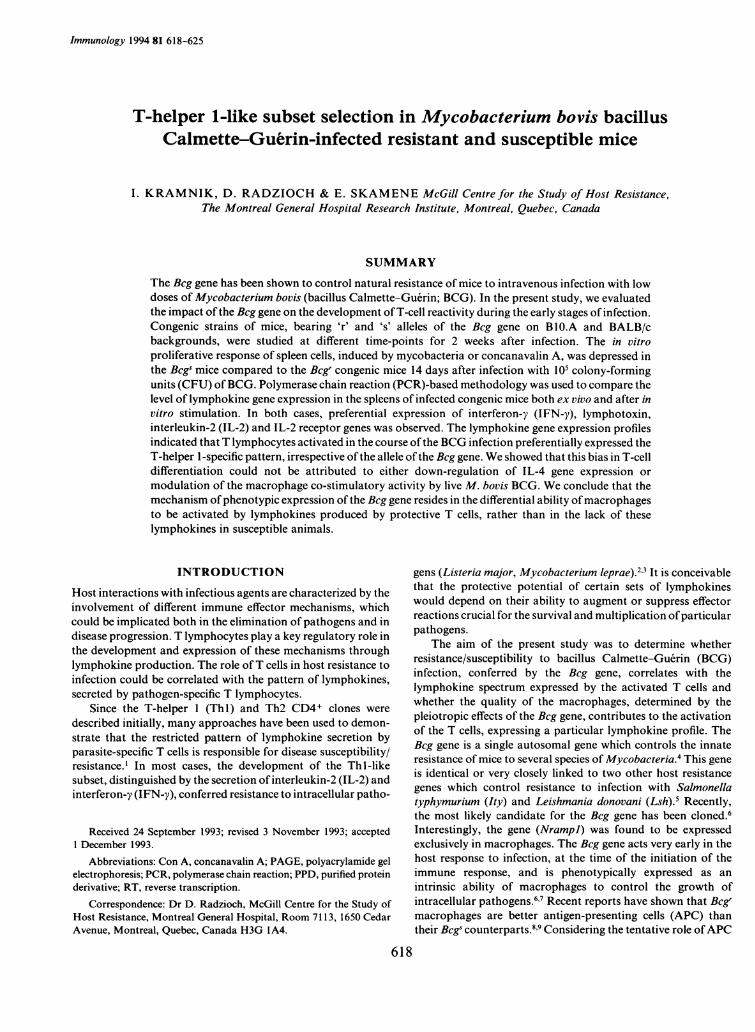
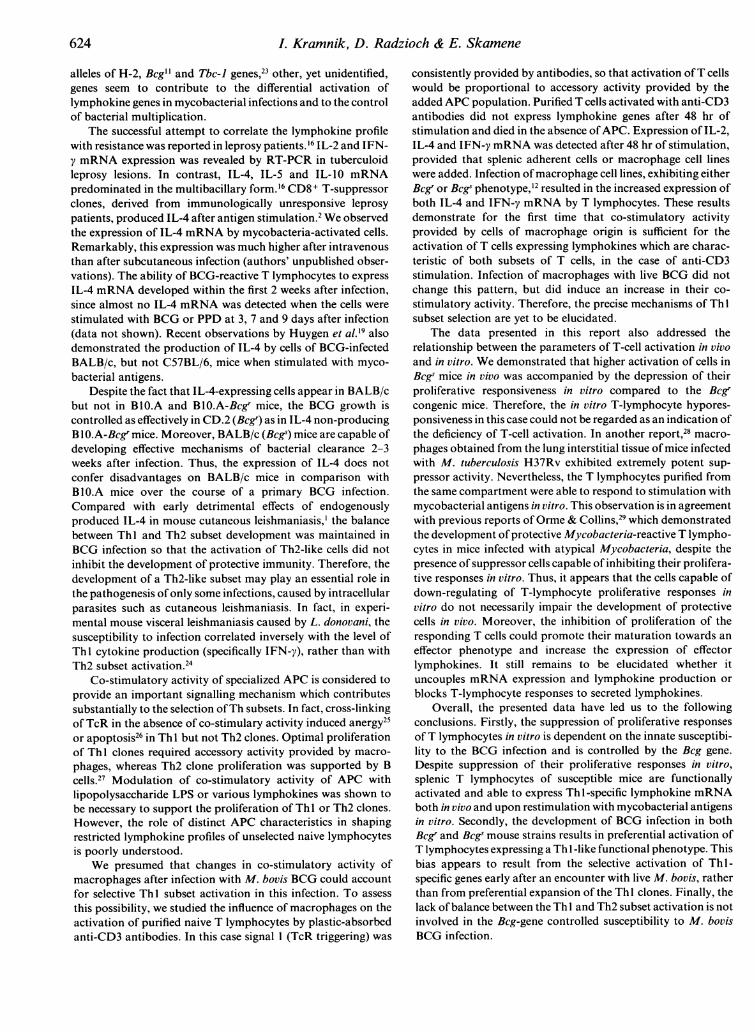
Figure 2. mRNA expression in cells, obtained from CD.2 and BALB/cmice infected with BCG 2 weeks before in vitro stimulation. (a) Cellswere obtained from CD.2 (Bcgr) mice infected with 106 CFU of BCG(Montreal). Lane 1, stimulation with PPD for 48 hr (10 pg/ml); lane 2,non-stimulated cells after 48 hr in vitro. (b) Cells were obtained fromCD.2 (Bcgr) and BALB/c (Bcg') infected with 105 CFU of BCG. Lane 1,stimulation with PPD for 48 hr; lanes 2, stimulation with M. bovis BCG(10 pg/ml) for 48 hr.
which were infected with 105 CFU of M. bovis BCG. The Bcg'strains of both genotypes displayed normal reactivity, whereasthe responses of Bcgs mice were depressed (Table 1). The degreeofT-cell hyporesponsiveness was consistently more pronouncedin Bcgs mice ofthe B 1 0.A background and, in some experiments,reached the level of complete proliferative unresponsiveness.Complete unresponsiveness, either to Con A or to BCG andPPD, was never observed in infected Bcgs mice of the BALB/cbackground.
620
T-lymphocyte gene expression in BCG infection
(a)
IFN-y
800 r (C)
600
IL-2
CD3-
CD3
Mgy- DLUy
400h
200
3 7 14v
Days after infection
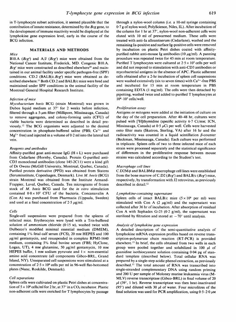
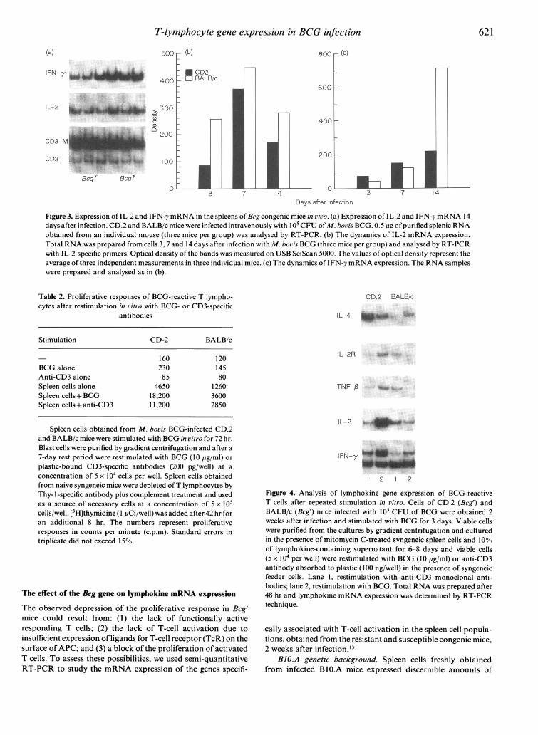
Figure 3. Expression of IL-2 and IFN-7 mRNA in the spleens of Bcg congenic mice in vivo. (a) Expression of IL-2 and IFN-y mRNA 14days after infection. CD.2 and BALB/c mice were infected intravenously with 105 CFU of M. bovis BCG. 0.5 pg ofpurified splenic RNAobtained from an individual mouse (three mice per group) was analysed by RT-PCR. (b) The dynamics of IL-2 mRNA expression.Total RNA was prepared from cells 3, 7 and 14 days after infection with M. bovis BCG (three mice per group) and analysed by RT-PCRwith IL-2-specific primers. Optical density of the bands was measured on USB SciScan 5000. The values of optical density represent theaverage of three independent measurements in three individual mice. (c) The dynamics of IFN-y mRNA expression. The RNA sampleswere prepared and analysed as in (b).
Table 2. Proliferative responses of BCG-reactive T lympho-cytes after restimulation in vitro with BCG- or CD3-specific
antibodies
Stimulation CD-2 BALB/c
160 120BCG alone 230 145Anti-CD3 alone 85 80Spleen cells alone 4650 1260Spleen cells+ BCG 18,200 3600Spleen cells+ anti-CD3 11,200 2850
Spleen cells obtained from M. bovis BCG-infected CD.2and BALB/c mice were stimulated with BCG in vitro for 72 hr.Blast cells were purified by gradient centrifugation and after a7-day rest period were restimulated with BCG (10 yg/ml) orplastic-bound CD3-specific antibodies (200 pg/well) at aconcentration of 5 x 10⁴ cells per well. Spleen cells obtainedfrom naive syngeneic mice were depleted ofT lymphocytes byThy- 1-specific antibody plus complement treatment and usedas a source of accessory cells at a concentration of 5 x 10⁵cells/well. [3H]thymidine (I pCi/well) was added after 42 hr foran additional 8 hr. The numbers represent proliferativeresponses in counts per minute (c.p.m). Standard errors intriplicate did not exceed 15%.
The effect of the Beg gene on lymphokine mRNA expression
The observed depression of the proliferative response in Bcegsmice could result from: (1) the lack of functionally activeresponding T cells; (2) the lack of T-cell activation due toinsufficient expression of ligands for T-cell receptor (TcR) on thesurface ofAPC; and (3) a block of the proliferation of activatedT cells. To assess these possibilities, we used semi-quantitativeRT-PCR to study the mRNA expression of the genes specifi-
CD.2 BALB/c
IL-4
IL-2R
TNF-p
{L-2_
IFN-y
12 1 2
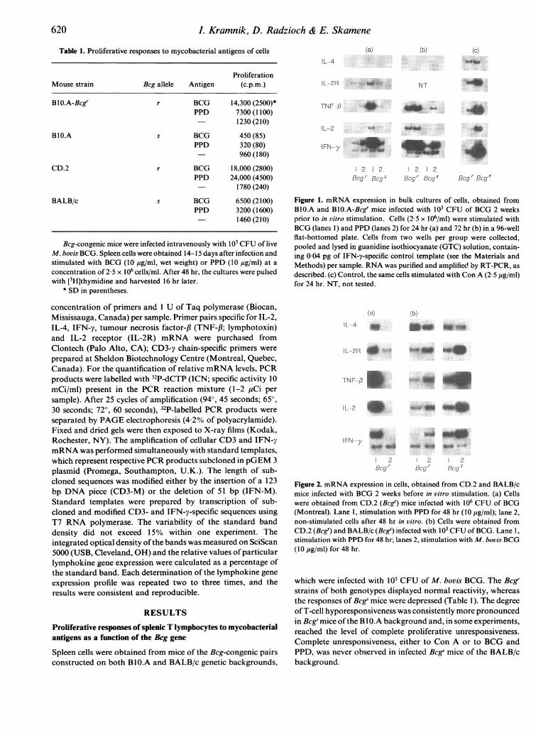
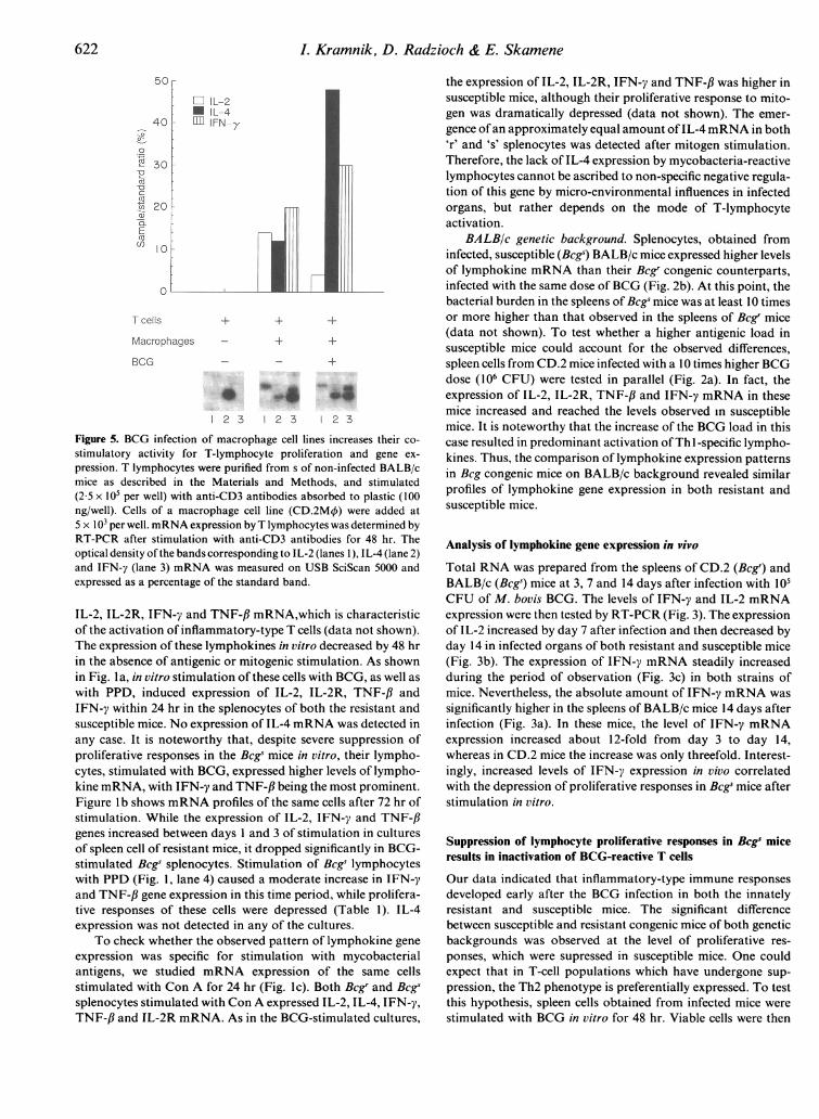
Figure 4. Analysis of lymphokine gene expression of BCG-reactiveT cells after repeated stimulation in vitro. Cells of CD.2 (Bcgs) andBALB/c (Bcgs) mice infected with 105 CFU of BCG were obtained 2weeks after infection and stimulated with BCG for 3 days. Viable cellswere purified from the cultures by gradient centrifugation and culturedin the presence of mitomycin C-treated syngeneic spleen cells and 100%of lymphokine-containing supernatant for 6-8 days and viable cells(5 x 10⁴ per well) were restimulated with BCG (10 yg/ml) or anti-CD3antibody absorbed to plastic (100 ng/well) in the presence of syngeneicfeeder cells. Lane 1, restimulation with anti-CD3 monoclonal anti-bodies; lane 2, restimulation with BCG. Total RNA was prepared after48 hr and lymphokine mRNA expression was determined by RT-PCRtechnique.
cally associated with T-cell activation in the spleen cell popula-tions, obtained from the resistant and susceptible congenic mice,2 weeks after infection.'3
BJO.A genetic background. Spleen cells freshly obtainedfrom infected BlO.A mice expressed discernible amounts of
621
6L Kramnik, D. Radzioch & E. Skamene
Cl IL-2* IL-4
IFN-y
'IR 30
C
.0
Macrophages + +
BCG _ - ~+
1 2 3 1 2 3 1 2 3
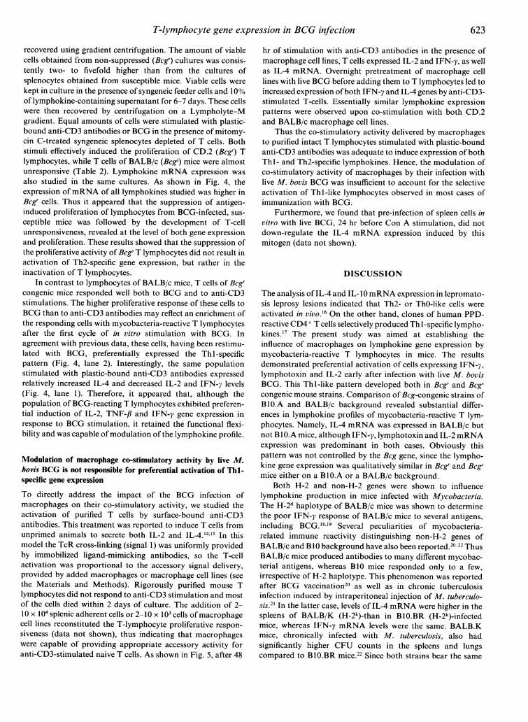
Figure 5. BCG infection of macrophage cell lines increases their co-
stimulatory activity for T-lymphocyte proliferation and gene ex-
pression. T lymphocytes were purified from s of non-infected BALB/cmice as described in the Materials and Methods, and stimulated(2 5 x10⁵ per well) with anti-CD3 antibodies absorbed to plastic (100ng/well). Cells of a macrophage cell line (CD.2M4) were added at5 x 103 per well. mRNA expression by T lymphocytes was determined byRT-PCR after stimulation with anti-CD3 antibodies for 48 hr. Theoptical density of the bands corresponding to IL-2 (lanes 1), IL-4 (lane 2)and IFN-y (lane 3) mRNA was measured on USB SciScan 5000 andexpressed as a percentage of the standard band.
IL-2, IL-2R, IFN-y and TNF-,B mRNA,which is characteristicof the activation of inflammatory-type T cells (data not shown).The expression of these lymphokines in vitro decreased by 48 hrin the absence of antigenic or mitogenic stimulation. As shownin Fig. I a, in vitro stimulation of these cells with BCG, as well as
with PPD, induced expression of IL-2, IL-2R, TNF-,B andIFN-y within 24 hr in the splenocytes of both the resistant andsusceptible mice. No expression of IL-4 mRNA was detected inany case. It is noteworthy that, despite severe suppression ofproliferative responses in the Bcge mice in vitro, their lympho-cytes, stimulated with BCG, expressed higher levels of lympho-kine mRNA, with IFN-y and TNF-fl being the most prominent.Figure b shows mRNA profiles of the same cells after 72 hr ofstimulation. While the expression of IL-2, IFN-y and TNF-,Bgenes increased between days I and 3 of stimulation in culturesof spleen cell of resistant mice, it dropped significantly in BCG-stimulated Bcgs splenocytes. Stimulation of Bcgs lymphocyteswith PPD (Fig. 1, lane 4) caused a moderate increase in IFN-yand TNF-,B gene expression in this time period, while prolifera-tive responses of these cells were depressed (Table 1). IL-4expression was not detected in any of the cultures.
To check whether the observed pattern of lymphokine gene
expression was specific for stimulation with mycobacterialantigens, we studied mRNA expression of the same cellsstimulated with Con A for 24 hr (Fig. lc). Both Bcgr and Bcgssplenocytes stimulated with Con A expressed IL-2, IL-4, IFN-y,TNF-,B and IL-2R mRNA. As in the BCG-stimulated cultures,
the expression of IL-2, IL-2R, IFN-y and TNF-f was higher insusceptible mice, although their proliferative response to mito-gen was dramatically depressed (data not shown). The emer-gence ofan approximately equal amount ofIL-4 mRNA in both'r' and 's' splenocytes was detected after mitogen stimulation.Therefore, the lack of IL-4 expression by mycobacteria-reactivelymphocytes cannot be ascribed to non-specific negative regula-tion of this gene by micro-environmental influences in infectedorgans, but rather depends on the mode of T-lymphocyteactivation.
BALBIc genetic background. Splenocytes, obtained frominfected, susceptible (Bcgs) BALB/c mice expressed higher levelsof lymphokine mRNA than their Bcgr congenic counterparts,infected with the same dose of BCG (Fig. 2b). At this point, thebacterial burden in the spleens of Bcgs mice was at least 10 timesor more higher than that observed in the spleens of Bcgr mice(data not shown). To test whether a higher antigenic load insusceptible mice could account for the observed differences,spleen cells from CD.2 mice infected with a 10 times higher BCGdose (106 CFU) were tested in parallel (Fig. 2a). In fact, theexpression of IL-2, IL-2R, TNF-/3 and IFN-y mRNA in thesemice increased and reached the levels observed in susceptiblemice. It is noteworthy that the increase of the BCG load in thiscase resulted in predominant activation ofTh 1-specific lympho-kines. Thus, the comparison of lymphokine expression patternsin Bcg congenic mice on BALB/c background revealed similarprofiles of lymphokine gene expression in both resistant andsusceptible mice.
Analysis of lymphokine gene expression in vivo
Total RNA was prepared from the spleens of CD.2 (Bcgr) andBALB/c (Bcgs) mice at 3, 7 and 14 days after infection with 105CFU of M. bovis BCG. The levels of IFN-y and IL-2 mRNAexpression were then tested by RT-PCR (Fig. 3). The expressionof IL-2 increased by day 7 after infection and then decreased byday 14 in infected organs of both resistant and susceptible mice(Fig. 3b). The expression of IFN-y mRNA steadily increasedduring the period of observation (Fig. 3c) in both strains ofmice. Nevertheless, the absolute amount of IFN-y mRNA wassignificantly higher in the spleens of BALB/c mice 14 days afterinfection (Fig. 3a). In these mice, the level of IFN-y mRNAexpression increased about 12-fold from day 3 to day 14,whereas in CD.2 mice the increase was only threefold. Interest-ingly, increased levels of IFN-y expression in vivo correlatedwith the depression of proliferative responses in Bcgs mice afterstimulation in vitro.
Suppression of lymphocyte proliferative responses in Bcgs miceresults in inactivation of BCG-reactive T cells
Our data indicated that inflammatory-type immune responsesdeveloped early after the BCG infection in both the innatelyresistant and susceptible mice. The significant differencebetween susceptible and resistant congenic mice of both geneticbackgrounds was observed at the level of proliferative res-ponses, which were supressed in susceptible mice. One couldexpect that in T-cell populations which have undergone sup-pression, the Th2 phenotype is preferentially expressed. To testthis hypothesis, spleen cells obtained from infected mice werestimulated with BCG in vitro for 48 hr. Viable cells were then
622
T-lymphocyte gene expression in BCG infection
recovered using gradient centrifugation. The amount of viablecells obtained from non-suppressed (Bcgr) cultures was consis-tently two- to fivefold higher than from the cultures ofsplenocytes obtained from susceptible mice. Viable cells werekept in culture in the presence of syngeneic feeder cells and 10%of lymphokine-containing supernatant for 6-7 days. These cellswere then recovered by centrifugation on a Lympholyte-Mgradient. Equal amounts of cells were stimulated with plastic-bound anti-CD3 antibodies or BCG in the presence of mitomy-cin C-treated syngeneic splenocytes depleted of T cells. Bothstimuli effectively induced the proliferation of CD.2 (Bcgr) Tlymphocytes, while T cells of BALB/c (Bcg) mice were almostunresponsive (Table 2). Lymphokine mRNA expression wasalso studied in the same cultures. As shown in Fig. 4, theexpression ofmRNA of all lymphokines studied was higher inBcgr cells. Thus it appeared that the suppression of antigen-induced proliferation of lymphocytes from BCG-infected, sus-ceptible mice was followed by the development of T-cellunresponsiveness, revealed at the level of both gene expressionand proliferation. These results showed that the suppression ofthe proliferative activity of Beg T lymphocytes did not result inactivation of Th2-specific gene expression, but rather in theinactivation of T lymphocytes.
In contrast to lymphocytes of BALB/c mice, T cells of Bcgrcongenic mice responded well both to BCG and to anti-CD3stimulations. The higher proliferative response of these cells toBCG than to anti-CD3 antibodies may reflect an enrichment ofthe responding cells with mycobacteria-reactive T lymphocytesafter the first cycle of in vitro stimulation with BCG. Inagreement with previous data, these cells, having been restimu-lated with BCG, preferentially expressed the Th 1-specificpattern (Fig. 4, lane 2). Interestingly, the same populationstimulated with plastic-bound anti-CD3 antibodies expressedrelatively increased IL-4 and decreased IL-2 and IFN-y levels(Fig. 4, lane 1). Therefore, it appeared that, although thepopulation of BCG-reacting T lymphocytes exhibited preferen-tial induction of IL-2, TNF-/3 and IFN-y gene expression inresponse to BCG stimulation, it retained the functional flexi-bility and was capable of modulation of the lymphokine profile.
Modulation of macrophage co-stimulatory activity by live M.bovis BCG is not responsible for preferential activation of Thl-specific gene expressionTo directly address the impact of the BCG infection ofmacrophages on their co-stimulatory activity, we studied theactivation of purified T cells by surface-bound anti-CD3antibodies. This treatment was reported to induce T cells fromunprimed animals to secrete both IL-2 and IL-4.14'5 In thismodel the TcR cross-linking (signal 1) was uniformly providedby immobilized ligand-mimicking antibodies, so the T-cellactivation was proportional to the accessory signal delivery,provided by added macrophages or macrophage cell lines (seethe Materials and Methods). Rigorously purified mouse Tlymphocytes did not respond to anti-CD3 stimulation and mostof the cells died within 2 days of culture. The addition of 2-10 X 104 splenic adherent cells or 2-10 x 103 cells of macrophagecell lines reconstituted the T-lymphocyte proliferative respon-siveness (data not shown), thus indicating that macrophageswere capable of providing appropriate accessory activity foranti-CD3-stimulated naive T cells. As shown in Fig. 5, after 48
hr of stimulation with anti-CD3 antibodies in the presence ofmacrophage cell lines, T cells expressed IL-2 and IFN-y, as wellas IL-4 mRNA. Overnight pretreatment of macrophage celllines with live BCG before adding them to T lymphocytes led toincreased expression ofboth IFN-y and IL-4 genes by anti-CD3-stimulated T-cells. Essentially similar lymphokine expressionpatterns were observed upon co-stimulation with both CD.2and BALB/c macrophage cell lines.
Thus the co-stimulatory activity delivered by macrophagesto purified intact T lymphocytes stimulated with plastic-boundanti-CD3 antibodies was adequate to induce expression of bothThl - and Th2-specific lymphokines. Hence, the modulation ofco-stimulatory activity of macrophages by their infection withlive M. bovis BCG was insufficient to account for the selectiveactivation of Thl-like lymphocytes observed in most cases ofimmunization with BCG.
Furthermore, we found that pre-infection of spleen cells invitro with live BCG, 24 hr before Con A stimulation, did notdown-regulate the IL-4 mRNA expression induced by thismitogen (data not shown).
DISCUSSION
The analysis of IL-4 and IL-I1mRNA expression in lepromato-sis leprosy lesions indicated that Th2- or ThO-like cells wereactivated in Vivo.16 On the other hand, clones of human PPD-reactive CD4 + T cells selectively produced Th I -specific lympho-kines.17 The present study was aimed at establishing theinfluence of macrophages on lymphokine gene expression bymycobacteria-reactive T lymphocytes in mice. The resultsdemonstrated preferential activation of cells expressing IFN-y,lymphotoxin and IL-2 early after infection with live M. bovisBCG. This Thl-like pattern developed both in Bcgr and Bcgscongenic mouse strains. Comparison of Bcg-congenic strains ofBl0.A and BALB/c background revealed substantial differ-ences in lymphokine profiles of mycobacteria-reactive T lym-phocytes. Namely, IL-4 mRNA was expressed in BALB/c butnot BIO.A mice, although IFN-y, lymphotoxin and IL-2 mRNAexpression was predominant in both cases. Obviously thispattern was not controlled by the Bcg gene, since the lympho-kine gene expression was qualitatively similar in Bcgr and Bcgsmice either on a Bl0.A or a BALB/c background.
Both H-2 and non-H-2 genes were shown to influencelymphokine production in mice infected with Mycobacteria.The H-2d haplotype of BALB/c mice was shown to determinethe poor IFN-y response of BALB/c mice to several antigens,including BCG.18"19 Several peculiarities of mycobacteria-related immune reactivity distinguishing non-H-2 genes ofBALB/c and B1O background have also been reported.20 22 ThusBALB/c mice produced antibodies to many different mycobac-terial antigens, whereas B10 mice responded only to a few,irrespective of H-2 haplotype. This phenomenon was reportedafter BCG vaccination20 as well as in chronic tuberculosisinfection induced by intraperitoneal injection of M. tuberculo-sis.21 In the latter case, levels of IL-4 mRNA were higher in thespleens of BALB/K (H-2k)-than in B10.BR (H-2k)_infectedmice, whereas IFN-y mRNA levels were the same. BALB.Kmice, chronically infected with M. tuberculosis, also hadsignificantly higher CFU counts in the spleens and lungscompared to B10.BR mice.22 Since both strains bear the same
623
624. I. Kramnik, D. Radzioch & E. Skamene
alleles of H-2, Beg" and Tbc-J genes,23 other, yet unidentified,genes seem to contribute to the differential activation oflymphokine genes in mycobacterial infections and to the controlof bacterial multiplication.
The successful attempt to correlate the lymphokine profilewith resistance was reported in leprosy patients.'6 IL-2 and IFN-y mRNA expression was revealed by RT-PCR in tuberculoidleprosy lesions. In contrast, IL-4, IL-5 and IL-l0 mRNApredominated in the multibacillary form.'6CD8+ T-suppressorclones, derived from immunologically unresponsive leprosypatients, produced IL-4 after antigen stimulation.2 We observedthe expression of IL-4 mRNA by mycobacteria-activated cells.Remarkably, this expression was much higher after intravenousthan after subcutaneous infection (authors' unpublished obser-vations). The ability of BCG-reactive T lymphocytes to expressIL-4 mRNA developed within the first 2 weeks after infection,since almost no IL-4 mRNA was detected when the cells werestimulated with BCG or PPD at 3, 7 and 9 days after infection(data not shown). Recent observations by Huygen et al.'9 alsodemonstrated the production of IL-4 by cells of BCG-infectedBALB/c, but not C57BL/6, mice when stimulated with myco-bacterial antigens.
Despite the fact that IL-4-expressing cells appear in BALB/cbut not in Bl0.A and BlO.A-Bcgr mice, the BCG growth iscontrolled as effectively in CD.2 (Bcgr) as in IL-4 non-producingB1O.A-BCgr mice. Moreover, BALB/c (Bcgs) mice are capable ofdeveloping effective mechanisms of bacterial clearance 2-3weeks after infection. Thus, the expression of IL-4 does notconfer disadvantages on BALB/c mice in comparison with
B0.A mice over the course of a primary BCG infection.
Compared with early detrimental effects of endogenouslyproduced IL-4 in mouse cutaneous leishmaniasis,' the balancebetween Thl and Th2 subset development was maintained inBCG infection so that the activation of Th2-like cells did notinhibit the development of protective immunity. Therefore, the
development of a Th2-like subset may play an essential role inthe pathogenesis ofonly some infections, caused by intracellularparasites such as cutaneous leishmaniasis. In fact, in experi-mental mouse visceral leishmaniasis caused by L. donovani, the
susceptibility to infection correlated inversely with the level ofThI cytokine production (specifically IFN-y), rather than withTh2 subset activation.24
Co-stimulatory activity of specialized APC is considered toprovide an important signalling mechanism which contributessubstantially to the selection ofTh subsets. In fact, cross-linkingof TcR in the absence of co-stimulary activity induced anergy25or apoptosis26 in Th I but not Th2 clones. Optimal proliferationof Th1 clones required accessory activity provided by macro-phages, whereas Th2 clone proliferation was supported by B
cells.27 Modulation of co-stimulatory activity of APC withlipopolysaccharide LPS or various lymphokines was shown tobe necessary to support the proliferation of Th 1 or Th2 clones.However, the role of distinct APC characteristics in shapingrestricted lymphokine profiles of unselected naive lymphocytesis poorly understood.
We presumed that changes in co-stimulatory activity ofmacrophages after infection with M. bovis BCG could accountfor selective Th 1 subset activation in this infection. To assessthis possibility, we studied the influence of macrophages on theactivation of purified naive T lymphocytes by plastic-absorbedanti-CD3 antibodies. In this case signal I (TcR triggering) was
consistently provided by antibodies, so that activation ofT cellswould be proportional to accessory activity provided by theadded APC population. Purified T cells activated with anti-CD3antibodies did not express lymphokine genes after 48 hr ofstimulation and died in the absence of APC. Expression of IL-2,IL-4 and IFN-y mRNA was detected after 48 hr of stimulation,provided that splenic adherent cells or macrophage cell lineswere added. Infection of macrophage cell lines, exhibiting eitherBegr or Begs phenotype,'2 resulted in the increased expression ofboth IL-4 and IFN-y mRNA by T lymphocytes. These resultsdemonstrate for the first time that co-stimulatory activityprovided by cells of macrophage origin is sufficient for theactivation of T cells expressing lymphokines which are charac-teristic of both subsets of T cells, in the case of anti-CD3stimulation. Infection of macrophages with live BCG did notchange this pattern, but did induce an increase in their co-
stimulatory activity. Therefore, the precise mechanisms of Th 1subset selection are yet to be elucidated.
The data presented in this report also addressed the
relationship between the parameters of T-cell activation in vivoand in vitro. We demonstrated that higher activation of cells in
Bcgs mice in vivo was accompanied by the depression of their
proliferative responsiveness in vitro compared to the Bcgrcongenic mice. Therefore, the in vitro T-lymphocyte hypores-
ponsiveness in this case could not be regarded as an indication ofthe deficiency of T-cell activation. In another report,28 macro-
phages obtained from the lung interstitial tissue of mice infectedwith M. tuberculosis H37Rv exhibited extremely potent sup-
pressor activity. Nevertheless, the T lymphocytes purified fromthe same compartment were able to respond to stimulation with
mycobacterial antigens in vitro. This observation is in agreementwith previous reports of Orme & Collins,29 which demonstratedthe development of protective Mycobacteria-reactive T lympho-cytes in mice infected with atypical Mycobacteria, despite the
presence of suppressor cells capable of inhibiting their prolifera-tive responses in vitro. Thus, it appears that the cells capable of
down-regulating of T-lymphocyte proliferative responses invitro do not necessarily impair the development of protectivecells in vivo. Moreover, the inhibition of proliferation of the
responding T cells could promote their maturation towards aneffector phenotype and increase the expression of effector
lymphokines. It still remains to be elucidated whether it
uncouples mRNA expression and lymphokine production orblocks T-lymphocyte responses to secreted lymphokines.
Overall, the presented data have led us to the followingconclusions. Firstly, the suppression of proliferative responsesof T lymphocytes in vitro is dependent on the innate susceptibi-lity to the BCG infection and is controlled by the Bcg gene.Despite suppression of their proliferative responses in vitro,
splenic T lymphocytes of susceptible mice are functionallyactivated and able to express Thl-specific lymphokine mRNAboth in vivo and upon restimulation with mycobacterial antigensin vitro. Secondly, the development of BCG infection in both
Bcg' and Bcgs mouse strains results in preferential activation ofT lymphocytes expressing a Th I-like functional phenotype. Thisbias appears to result from the selective activation of Thl-specific genes early after an encounter with live M. bovis, ratherthan from preferential expansion of the Th 1 clones. Finally, thelack of balance between the Th I and Th2 subset activation is notinvolved in the Bcg-gene controlled susceptibility to M. bovisBCG infection.
624
T-lymphocyte gene expression in BCG infection 625
REFERENCES
1. HEINZEL F.P., SADICK M.D., HOLADAY B.J., COFFMAN R.L. &LOCKSLEY R.M. (1989) Reciprocal expression of interferon-y orinterleukin-4 during the resolution or progression of murineleishmaniasis. Evidence for expansion of distinct helper T cellsubsets. J. exp. Med. 169, 59.
2. SALGAME P., ABRAMS J.S., CLAYBERGER C., GOLDSTEIN H., CONVITJ., MODLIN R.L. & BLOOM B.R. (1991) Differing lymphokineprofiles of functional subsets of human CD41 and CD8+ T cellclones. Science, 254, 279.
3. MOSSMAN T.R. & COFFMAN R.L. (1989) THI and TH2 cells:different patterns of lymphokine secretion lead to different func-tional properties. Ann. Rev. Immunol. 7, 145.
4. SKAMENE E. (1989) Genetic control of susceptibility to mycobac-terial infections. Rev. Infect. Dis. 11, 394.
5. SKAMENE E., GROS P., FORGET A., KONGSHAVN P.A.L., ST CHARLESC. & TAYLOR B.A. (1982) Genetic regulation of resistance tointracellular pathogens. Nature, 297, 506.
6. VIDAL, S.M., MALO D., VOGAN K., SKAMENE E. & GROS P. (1993)Natural resistance to infection with intracellular parasites; Isolationof a candidate for Bcg. Cell, 73, 469.
7. STACH J.-L., GROS P., FORGET A. & SKAMENE E. (1984) Phenotypicexpression of genetically-controlled natural resistance to Mycobac-terium bovis BCG. J. Immunol. 132, 888.
8. DENIS M., FORGET A., PELLETIER M. & SKAMENE E. (1988)Pleiotropic effects of the Bcg gene: I. Antigen presentation ingenetically susceptible and resistant congenic mouse strains.J. Immunol. 140, 2395.
9. KAYE P.M. & BLACKWELL J.M. (1989) Lsh, antigen presentation &the development of CMI. Res. Immunol. 140, 810.
10. NESBITT M.N. & SKAMENE E. (1984) Recombinant inbred mousestrains derived from A/J and C57B1/6J: a tool for the study ofgenetic mechanisms in host resistance to infection and malignancy.J. Leukoc. Biol. 36, 357.
11. POTTER M., O'BRIEN A., SKAMENE E., GROS P., FORGET A.,KONGSHAVN P.A.L. & WAX J.C. (1983) A BALB/c congenic strainof mice that carries a genetic locus (Ityr) controlling resistance forintracellular parasites. Infect. Immun. 40, 1234.
12. RADZIOCH D., HUDSON T., BOULE M., BARRERA L., URBANCE J.W.,VARESIO L. & SKAMENE E. (1991) Genetic resistance/susceptibility tomycobacteria: phenotypic expression in bone marrow derivedmacrophage cell lines. J. Leukoc. Biol. 50, 263.
13. KRAMNIK I., SKAMENE E. & RADZIOCH D. (1993) Assessment oflymphokine profiles by semiquantitative PCR in lymphocytesactivated in vitro and in vivo. J. immunol. Meth. 162, 143.
14. BEN-SASSON S.Z., LE GROS G., CONDRAD D.H., FINKELMAN F.D. &PAUL W.E. (1990) IL-4 production by T cells from naive donors.IL-2 is required for IL-4 production. J. Immunol. 145, 1127.
15. FLAMAND V., ABRAMovIcz D., GOLDMAN M., BIERNAUX C., HUEZG., URBAIN J., MOSER M. & LEO 0. (1990) Anti-CD3 antibodiesinduce T cells from unprimed animals to secrete IL-4 both in vitroand in vivo. J. Immunol. 144, 2875.
16. YAMAMURA M., UYEMURA K., DEANS R.J., WEINBERG K., REA T.H.,BLOOM B.R. & MODLIN R.L. (1991) Defining protective responsesto pathogens: cytokine profiles in leprosy lesions. Science, 254, 277.
17. HAANEN J.B.A.G., DE WAAL MALEFUJT R., RES P.C.M., KRAAKMANE.M., OTTENHOFF T.H.M., DE VRIES R.R.P. & SPITS H. (1991)Selection of a human T helper cell subset by mycobacteria. J. exp.Med. 174, 583.
18. ASHERSON G.L., DIELI F., GAUTAM Y., SiEw L.K. & ZEMBALA M.(1990) Major histocompatibility complex regulation of the class ofthe immune response: the H-2d haplotype determines poor inter-feron-y response to several antigens. Eur. J. Immunol. 20.
19. HUYGEN K., ABRAMOVICZ D., VANDENBUSSCHE P., JACOBS F., DEBRUYN J., KENTOs A., DROWART A., VAN VOOREN J.-P. & GOLDMANM. (1992) Cell cytokine secretion in Mycobacterium bovis BCG-infected mice. Infect. Immun. 60, 2880.
20. HUYGEN K., LJUNGQVIST L., TEN BERG L. & VAN VOOREN J.P. (1990)Repertoires of antibodies to culture filtrate antigens in differentmouse strains infected with Mycobacterium bovis BCG. Infect.Immun. 58, 2192.
21. BRETT S.J. & IVANYI J. (1990) Genetic influences on the immunerepertoire following tuberculous infection in mice. Immunology,71, 113.
22. BRETT S., ORRELL J.M., BECK J.S. & IVANYI J. (1992) Influence ofH-2 genes on growth of Mycobacterium tuberculosis in the lungs ofchronically infected mice. Immunology, 76, 129.
23. NIKONENKO B.V., APT A.S., MOROz A.M. & AVERBAKH M.M.(1985) Genetic analysis of susceptibility of mice to H37Rv tubercu-losis infection: sensitivity versus relative resistance. Prog. LeukocyteBiol. 3, 291.
24. KAYE P.M., CURRY A.J. & BLACKWELL J.M. (1991) Differentialproduction of Th 1- and Th2-derived cytokines does not determinethe genetically controlled or vaccine-induced rate of cure in murinevisceral leishmaniasis. J. Immunol. 146, 2763.
25. MUELLER D., JENKINS M.K. & SCHWARTZ R.H. (1989) Clonalexpansion versus functional clonal inactivation: a costimulatorysignalling pathway determines the outcome of T cell antigenreceptor occupancy. Ann. Rev. Immunol. 7,445.
26. Liu Y. & JANEWAY C.A. (1990) Interferon-y plays a critical role ininduced cell death of effector T cell: a possible third mechanism ofself-tolerance. J. exp. Med. 172, 1735.
27. GAJEWSKY T.F., PINNAS M., WONG T. & FITCH F.W. (1991) MurineTH 1 & TH2 clones proliferate optimally in response to distinctantigen-presenting cell populations. J. Immunol. 146, 1750.
28. APT A.S., KRAMNIK I. & MOROz A.M. (1991) Regulation of T cellproliferative responses by cells from solid lung tissue of M.tuberculosis-infected mice. Immunology, 73, 173.
29. ORME I.M. & COLLINS F.M. (1984) Immune response to atypicalmycobacteria: immunocompetence of heavily infected micemeasured in vivo fails to substantiate immunosuppression dataobtained in vitro. Infect. Immun. 43, 32.
Related Documents