Enhanced Potency of Nucleotide–Dendrimer Conjugates as Agonists of the P2Y 14 Receptor: Multivalent Effect in G Protein- Coupled Receptor Recognition Arijit Das † , Yixing Zhou ‡ , Andrei A. Ivanov +,† , Rhonda L. Carter ‡ , T. Kendall Harden ‡ , and Kenneth A. Jacobson *,† Laboratory of Bioorganic Chemistry, National Institute of Diabetes and Digestive and Kidney Diseases, National Institutes of Health, Bethesda, Maryland 20892, and Department of Pharmacology, University of North Carolina School of Medicine, Chapel Hill, North Carolina 27599 Abstract The P2Y 14 receptor is a G protein-coupled receptor activated by uridine-5′-diphosphoglucose and other nucleotide sugars that modulates immune function. Covalent conjugation of P2Y 14 receptor agonists to PAMAM (polyamidoamine) dendrimers enhanced pharmacological activity. Uridine-5′-diphosphoglucuronic acid (UDPGA) and its ethylenediamine adduct were suitable functionalized congeners for coupling to several generations (G2.5–6) of dendrimers (both terminal carboxy and amino). Prosthetic groups, including biotin for avidin complexation, a chelating group for metal complexation (and eventual magnetic resonance imaging), and a fluorescent moiety, also were attached with the eventual goals of molecular detection and characterization of the P2Y 14 receptor. The activities of conjugates were assayed in HEK293 cells stably expressing the human P2Y 14 receptor. A G3 PAMAM conjugate containing 20 bound nucleotide moieties (UDPGA) was 100-fold more potent (EC 50 2.4 nM) than the native agonist uridine-5′-diphosphoglucose. A molecular model of this conjugate docked in the human P2Y 14 receptor showed that the nucleotide-substituted branches could extend far beyond the dimensions of the receptor and be available for multivalent docking to receptor aggregates. Larger dendrimer carriers and greater loading favored higher potency. A similar conjugate of G6 with 147 out of 256 amino groups substituted with UDPGA displayed an EC 50 value of 0.8 nM. Thus, biological activity was either retained or dramatically enhanced in the multivalent dendrimer conjugates in comparison with monomeric P2Y 14 receptor agonists, depending on size, degree of substitution, terminal functionality, and attached prosthetic groups. Introduction G protein-coupled receptors (GPCRs)1 are cell membrane-spanning receptors that respond to extracellular signaling molecules to control cell function via specific signaling pathways. Direct or indirect modulation of GPCRs serves as the basis of many disease treatments (1). The P2 receptors for purine and pyrimidine nucleotides have diverse biological roles (2–4). © 2009 American Chemical Society * Corresponding author. Molecular Recognition Section, Laboratory of Bioorganic Chemistry, National Institute of Diabetes and Digestive and Kidney Diseases, National Institutes of Health, Bethesda, Maryland 20892, USA; [email protected]. † National Institutes of Health. ‡ University of North Carolina School of Medicine. + Current address: Emory University School of Medicine, Department of Biochemistry, Atlanta, GA 30322. Supporting Information Available: 1 H NMR and MALDI spectra. This material is available free of charge via the Internet at http://pubs.acs.org. NIH Public Access Author Manuscript Bioconjug Chem. Author manuscript; available in PMC 2011 June 2. Published in final edited form as: Bioconjug Chem. 2009 August 19; 20(8): 1650–1659. doi:10.1021/bc900206g. NIH-PA Author Manuscript NIH-PA Author Manuscript NIH-PA Author Manuscript

Welcome message from author
This document is posted to help you gain knowledge. Please leave a comment to let me know what you think about it! Share it to your friends and learn new things together.
Transcript
Enhanced Potency of Nucleotide–Dendrimer Conjugates asAgonists of the P2Y14 Receptor: Multivalent Effect in G Protein-Coupled Receptor Recognition
Arijit Das†, Yixing Zhou‡, Andrei A. Ivanov+,†, Rhonda L. Carter‡, T. Kendall Harden‡, andKenneth A. Jacobson*,†
Laboratory of Bioorganic Chemistry, National Institute of Diabetes and Digestive and KidneyDiseases, National Institutes of Health, Bethesda, Maryland 20892, and Department ofPharmacology, University of North Carolina School of Medicine, Chapel Hill, North Carolina27599
AbstractThe P2Y14 receptor is a G protein-coupled receptor activated by uridine-5′-diphosphoglucose andother nucleotide sugars that modulates immune function. Covalent conjugation of P2Y14 receptoragonists to PAMAM (polyamidoamine) dendrimers enhanced pharmacological activity.Uridine-5′-diphosphoglucuronic acid (UDPGA) and its ethylenediamine adduct were suitablefunctionalized congeners for coupling to several generations (G2.5–6) of dendrimers (bothterminal carboxy and amino). Prosthetic groups, including biotin for avidin complexation, achelating group for metal complexation (and eventual magnetic resonance imaging), and afluorescent moiety, also were attached with the eventual goals of molecular detection andcharacterization of the P2Y14 receptor. The activities of conjugates were assayed in HEK293 cellsstably expressing the human P2Y14 receptor. A G3 PAMAM conjugate containing 20 boundnucleotide moieties (UDPGA) was 100-fold more potent (EC50 2.4 nM) than the native agonisturidine-5′-diphosphoglucose. A molecular model of this conjugate docked in the human P2Y14receptor showed that the nucleotide-substituted branches could extend far beyond the dimensionsof the receptor and be available for multivalent docking to receptor aggregates. Larger dendrimercarriers and greater loading favored higher potency. A similar conjugate of G6 with 147 out of 256amino groups substituted with UDPGA displayed an EC50 value of 0.8 nM. Thus, biologicalactivity was either retained or dramatically enhanced in the multivalent dendrimer conjugates incomparison with monomeric P2Y14 receptor agonists, depending on size, degree of substitution,terminal functionality, and attached prosthetic groups.
IntroductionG protein-coupled receptors (GPCRs)1 are cell membrane-spanning receptors that respondto extracellular signaling molecules to control cell function via specific signaling pathways.Direct or indirect modulation of GPCRs serves as the basis of many disease treatments (1).The P2 receptors for purine and pyrimidine nucleotides have diverse biological roles (2–4).
© 2009 American Chemical Society* Corresponding author. Molecular Recognition Section, Laboratory of Bioorganic Chemistry, National Institute of Diabetes andDigestive and Kidney Diseases, National Institutes of Health, Bethesda, Maryland 20892, USA; [email protected].†National Institutes of Health.‡University of North Carolina School of Medicine.+Current address: Emory University School of Medicine, Department of Biochemistry, Atlanta, GA 30322.Supporting Information Available: 1H NMR and MALDI spectra. This material is available free of charge via the Internet athttp://pubs.acs.org.
NIH Public AccessAuthor ManuscriptBioconjug Chem. Author manuscript; available in PMC 2011 June 2.
Published in final edited form as:Bioconjug Chem. 2009 August 19; 20(8): 1650–1659. doi:10.1021/bc900206g.
NIH
-PA Author Manuscript
NIH
-PA Author Manuscript
NIH
-PA Author Manuscript
P2 receptors are divided into two structurally unrelated subfamilies–the P2X receptors,which are ligand-gated ion channels (5), and the P2Y receptors (2), which are GPCRs. P2Yreceptors are divided into two subgroups. The first subgroup consisting of the P2Y1, P2Y2,P2Y4, P2Y6, and P2Y11 receptors activate Gq and promote phospholipase C (PLC)-dependent inositol lipid signaling. The second consists of P2Y12, P2Y13, and P2Y14receptors, which preferentially activate Gi and inhibit adenylyl cyclase (2). P2Y receptorsare activated by adenine and/or uracil nucleotides, while P2X receptors are principallyactivated by adenine nucleotides. P2Y receptors are widely distributed, for example, in theimmune system (and widely distributed on hematopoietic cells), cardiovascular system,central and peripheral nervous system, endocrine system, and the renal and pulmonarysystems (3–10).
The P2Y14 receptor is activated by uridine-5′-diphosphoglucose (UDPG) 1 (Chart 1) andother UDP-sugars (11, 12). The P2Y14 receptor affects immune function, but thephysiological function of the receptor is not clearly established (13, 14). Extracellular 1 alsoacts as a weak full agonist at the P2Y2 receptor (EC50 10 μM) (15). We have studied theSAR of nucleotide derivatives at this receptor, which is among the most structurallyrestrictive of the P2Y family (15–18). Activity of synthetic analogues of 1 at the P2Y14receptor has been followed through the stimulation of phosphoinositide hydrolysis bycoexpression in COS-7 cells of a PLC-activating chimeric G protein that responds to Gi-coupled receptors (18).
Dendrimers are tree-like polymeric macromolecular nanostructures,which can serve asnanocarriers for drug delivery (19–21). Dendrimer chemistry offers versatility in the controlof the component functional groups and in feasibility for conjugation of multiple functionalunits at both the periphery and interior. Dendrimers recently have attracted considerableattention in biomedical research in the context of drug delivery (targeted/ controlled release,encapsulation, or covalent/electrostatic attachment) (22–24), protein–carbohydrateinteractions (multivalent effect) (25–30), medical diagnostics (signal amplification) (31, 32),and tissue engineering (33, 34).
Polyamidoamine (PAMAM) dendrimers have well-defined chemical structures in which acore is surrounded by successively added (and bifurcating) layers of methyl acrylate andethylenediamine, which form each dendrimeric shell (or “generation”) of the polymer.Different generations of PAMAM dendrimers have either primary amine (integralgenerations) or carboxylic acid (half-integral generations) groups on their surface, and thenumber of surface groups for conjugation to multiple functional units depends on thegeneration of the dendrimer. PAMAM dendrimers are relatively biocompatible, whichallows many applications in biomedicine (35–38).
We recently reported the first example of application of dendrimers to multivalent GPCR-promoted signal transduction in a study of adenosine receptor-targeted dendrimers (39–41).Various adenosine receptor agonists were attached covalently to PAMAM dendrimers, andthe resulting nucleoside conjugates displayed distinct biological properties, including potentinhibition of platelet aggregation.
Here, we report the first example of application of PAMAM dendrimers to covalentlyconjugate a P2Y receptor ligand resulting in the enhancement of its pharmacological
1Abbreviations: DTPA, diethylenetriaminepentaacetic acid; EDC, N-ethyl-N′-dimethylaminopropylcarbodiimide; GPCR, G protein-coupled receptor; HEK, human embryonic kidney; IBMX, 3-isobutyl-1-methylxanthine; MES, 2-(N-morpholino)ethanesulfonic acid;MRI, magnetic resonance imaging; NHS, N′-hydroxysuccinimide; PAMAM, polyamidoamine; SAR, structure activity relationship; p-SCN-Bn-DTPA, 2-(4-isothiocyanatobenzyl)-diethylenetriaminepentaacetic acid; UDPG, uridine-5′-diphosphoglucose; UDPGA,uridine-5′-diphosphoglucuronic acid.
Das et al. Page 2
Bioconjug Chem. Author manuscript; available in PMC 2011 June 2.
NIH
-PA Author Manuscript
NIH
-PA Author Manuscript
NIH
-PA Author Manuscript
activity. The P2Y14 receptor agonist uridine-5′-diphosphoglucuronic acid (UDPGA) 2 andits ethylenediamine adduct 3a were utilized as appropriately functionalized congeners tocouple with several generations of PAMAM dendrimers with the goal of modulatingpharmacological interaction with this receptor. We also attached prosthetic groups, such asbiotin, AlexaFluor488, and the metal chelating group diethylenetriaminepentaacetic acid(DTPA) on the dendrimer-nucleotide conjugates for targeting the P2Y14 receptor in vivo.Our long-term goal is to initiate the genesis of receptor probes that lead to improvedbiomedical diagnostics and possibly to novel conjugates for drug treatment.
Experimental ProceduresChemical Synthesis
Materials and Methods—All reactions were carried out under a nitrogen atmosphere.G2.5 PAMAM (10 wt % solution in methanol), G3 PAMAM (20 wt % solution inmethanol), G5.5 PAMAM (5 wt % solution in methanol), and G6 PAMAM (5 wt % solutionin methanol), dendrimer with an ethylenediamine core, EDC, UDPGA, Gd(OAc)3, andethylenediamine were purchased from Aldrich. Sulfo-NHS-LC-biotin was purchased fromPierce (Rockford, IL), and Alex-aFluor488-carboxylic acid 2,3,5,6-tetrafluorophenyl ester(AlexaFluor488–5-TFP) was purchased from Invitrogen Corp. (Carlsbad, CA) and 2-(4-isothiocyanatobenzyl)-diethylenetriaminepentaacetic acid (p-SCN-Bn-DTPA) purchasedfrom Macrocyclic (Dallas, TX). Dialysis membranes (Spectra/Pore Membrane, MWCO3500, flat width 18 mm) were purchased from Spectrum Laboratories, Inc. (RanchoDominguez, CA).
Nuclear magnetic resonance (NMR) spectra were recorded on a Bruker DRX-600spectrometer using D2O as a solvent. The chemical shifts are expressed as relative ppm fromHOD (4.80).
The electrospray ionization mass spectrometry (ESI MS) and matrix-assisted laserdesorption ionization time-of-flight (MALDI-TOF) MS experiments were performed on aWaters LCT Premier mass spectrometer at the Mass Spectrometry Facility, NIDDK, NIH.
Synthesis of G3 or G6 PAMAM-UDPGA Conjugates 8, 9, and 12—A commercialsolution of G3 PAMAM 5 (100 μL, 2 μmol) or G6 PAMAM 7 (167 μL, 0.11 μmol) wasadded to a round-bottom flask, and the methanol was evaporated using a rotary evaporator.The dendrimer was treated with EDC-HCl (2.1 mg, 5.5 equiv for compound 8; 13.8 mg, 36equiv for compound 9; and 5.4 mg, 256 equiv for compound 12) and then dissolved withminimum volume of 0.1 M 2-(N-morpholino)ethanesulfonic acid (MES), pH 5 (1 mL). Thereaction mixture was stirred and UDPGA 2 (7.7 mg, 6 equiv for compound 8; 82.7 mg, 64equiv for compound 9; and 27.2 mg, 384 equiv for compound 12) was added. The pH of thereaction was adjusted to the range 4.5–5.0 using 0.1 M HCl. The reaction mixture wasstirred at room temperature under nitrogen atmosphere for 2 days and then diluted withwater (1 mL). After that, the product was purified by extensive dialysis in water. Themixture was then lyophilized to give the pure product of the conjugates 8, 9, and 12.
G3 PAMAM-UDPGA Conjugate 8 (Low Loading)—Compound 8 (13.5 mg, 71%) wasobtained as a white solid following the general procedure. The product was analyzed byNMR and MALDI-TOFF MS, which indicated approximately 4.3 UDPGA moietiesattached per dendrimer. 1H NMR (D2O) δ 7.84 (d, J = 8.8 Hz, 4.3 H), 5.91 (d, J = 5.7 Hz,8.6 H), 5.53 (m, 4.3 H), 4.28 (m, 8.6 H), 4.17 (m, 8.6 H), 4.08 (m, 8.6 H), 3.85 (m, 4.3 H),3.63 (m, 8.6 H), 3.44 (m, 180 H), 3.3 (m, 120 H), 3.14 (m, 60 H), 2.72 (m, 120 H); m/z(M +ESI MS) found 9848.0; calc 9519.0.
Das et al. Page 3
Bioconjug Chem. Author manuscript; available in PMC 2011 June 2.
NIH
-PA Author Manuscript
NIH
-PA Author Manuscript
NIH
-PA Author Manuscript
G3 PAMAM-UDPGA Conjugate 9 (High Loading)—Compound 9 (26 mg, 68%) wasobtained as a white solid following the general procedure. The product was analyzed byNMR and MALDI-TOFF MS, which indicated approximately 20.1 UD-PGA moietiesattached per dendrimer.1H NMR (D2O) δ 7.88 (d, J = 7.4 Hz, 20.1 H), 5.81 (d, J = 5.4 Hz,40.2 H), 5.51 (dd, J = 3.3, 6.1 Hz, 20.1 H), 4.3 (m, 40.2 H), 4.2 (m, 40.2 H), 4.1 (m, 40.2 H),3.65 (m, 20.1 H), 3.52 (m, 40.2 H), 3.41 (m, 180 H), 3.3 (m, 120 H), 3.14 (m, 60 H), 2.72(m, 120 H); m/z (M +ESI MS) found 18721.4; calc 19113.7.
G6 PAMAM-UDPGA Conjugate 12—Compound 12 (9.86 mg, 61%) was obtained as awhite solid following the general procedure. The product was analyzed by NMR andMALDI-TOFF MS, which indicated approximately 147 UDPGA moieties attached perdendrimer. 1H NMR (D2O) δ 7.87 (d, J = 7.5 Hz, 147 H), 5.89 (d, J = 5.4 Hz, 294 H), 5.51(m, 147 H), 4.29 (m, 294 H), 4.17 (m, 294 H), 4.09 (m, 294 H), 3.81 (m, 147 H), 3.7 (m, 294H), 3.51 (m, 512 H), 3.41 (m, 512 H), 3.28 (m, 1020 H) 3.12 (m, 504 H), 3.07 (m, 504 H),2.78 (m, 1020 H). The molecular weight was unable to be determined using ESI or MALDI-TOF MS, possibly due to stacking of the PAMAM dendrimer or because of the highmolecular weight.
Synthesis of G2.5 or G5.5 PAMAM–UDPGA Conjugates 10 and 11—Acommercial solution of G2.5 PAMAM 4 (13.76 μL, 0.2 μmol) or G5.5 PAMAM 6 (115 μL,0.1 μmol) was added to a round-bottom flask, and the methanol was evaporated. EDC-HCl(1.4 mg, 36 equiv, for compound 8; and 1.9 mg, 100 equiv for compound 9) was added tothe residue, and then the mixture was dissolved in a minimum volume of 0.1 M MES, pH 5(0.5 mL), and stirred under a nitrogen atmosphere. UDPGA-ethylenediamine (3a) wasadded to the reaction mixture (6.5 mg, 50 equiv, for compound 10; and 13.1 mg, 200 equiv,for compound 11). The pH of the reaction mixture was maintained within the range 4.5–5.0using 0.1 M HCl. After 48 h, the small molecule impurities were removed by extensivedialysis in water. After dialysis, the mixture was lyophilized to give conjugate 10 or 11.
G2.5 PAMAM-UDPGA Conjugate 10—Compound 10 (2.4 mg, 66%) was obtained as awhite solid following the general procedure. The product was analyzed by NMR andMALDI-TOF MS, which indicated approximately 17.3 UDPGA moieties attached perdendrimer. 1H NMR (D2O) δ 7.82 (d, J = 7.7 Hz, 17.34 H), 5.84 (d, J = 5.5 Hz, 34.68 H),5.51 (dd, J = 3, 5.1 Hz, 17.34 H), 4.29 (m, 52.0 H), 4.15 (m, 52.0 H), 3.7 (m, 17.34 H), 3.51(m, 98.7 H), 3.41 (m, 120 H), 3.31 (m, 90.7 H), 3.14 (m, 60 H), 2.68 (m, 34.7 H), 2.51 (m,56 H); m/z (M +ESI MS) found 18151.4, calc 18477.9.
G5.5 PAMAM-UDPGA Conjugate 11—Compound 11 (4.5 mg, 63%) was obtained as awhite solid following the general procedure. The product was analyzed by NMR andMALDI-TOF MS, which indicated approximately 29.9 UDPGA moieties attached per G5.5dendrimer. 1H NMR (D2O) δ 7.85 (d, J = 8.3 Hz, 29.94 H), 5.88 (d, J = 6.5 Hz, 59.9 H),5.57 (dd, J = 3.1, 5.7 Hz, 29.9 H), 4.26 (m, 89.8 H), 4.14 (m, 89.8 H), 3.72 (m, 29.94 H),3.44 (m, 571 H), 3.23 (m, 1075.88 H), 2.84 (m, 504 H), 2.67 (m, 508 H), 2.48 (m, 564 H);m/z (M +ESI MS) found 72626, calc 71651.
Synthesis of G3 PAMAM-UDPGA-Biotin Conjugate 13—A mixture of G3 PAMAMcomplex 9 (20 mg, 1.04 μmol) and Sulfo-NHS-LC-Biotin 17 (361 μg, 0.65 μmol) in a flaskwas dissolved in bicarbonate buffer (1 mL, pH 8.5, 0.002 M Na2CO3, 0.048 M NaHCO3,0.15 M NaCl). After 48 h of stirring at room temperature, the reaction mixture was dilutedwith 1 mL water, and the product was purified by dialysis against water. The solution wasthen lyophilized, which provided G3 PAMAMbotin 13 (14.4 mg, 67%) as a white solid. Theproduct was analyzed by NMR and MALDI-TOF MS, which indicated that approximately
Das et al. Page 4
Bioconjug Chem. Author manuscript; available in PMC 2011 June 2.
NIH
-PA Author Manuscript
NIH
-PA Author Manuscript
NIH
-PA Author Manuscript
4.87 biotin moieties attached per dendrimer. 1H NMR (D2O) δ 7.87 (d, J = 7.4 Hz, 20.1 H),5.82 (d, J = 5.4 Hz, 40.2 H), 5.51 (m 20.1 H),4.55 (m, 9.74 H) 4.28 (m, 40.2 H), 4.18 (m,40.2 H), 4.09 (m, 40.2 H), 3.67 (m, 20.1 H), 3.53 (m, 40.2 H), 3.42 (m, 180 H), 3.28 (m, 135H), 3.11 (m, 60 H), 2.91(m, 9.74 H) 2.71 (m, 120 H), 2.12 (m, 19.5 H), 1.52 (m, 39.0 H),1.27 (m, 19.5 H); m/z (M +ESI MS) found 21114.0, calc 20767.0.
Synthesis of G3 PAMAM-UDPGA-AlexaFluor488 Conjugate 14—G3 PAMAMcomplex 9 (20 mg, 1.04 μmol) was reacted with AlexaFluor488-5-TFP 18 (575 μg, 0.65μmol) in bicarbonate buffer (1 mL, pH 8.5, 0.002 M Na2CO3, 0.048 M NaHCO3, 0.15 MNaCl). The reaction mixture was stirred for 2 days at room temperature and then dilutedwith 1 mL of water. The mixture was purified by dialysis with water. Lyophilization of thesolution gave the G3 PAMAM–AlexaFluor488 complex 14 (13.4 mg, 62%) as a red solid.The product was analyzed by NMR and MALDI-TOFF MS, which indicated approximately2.3 AlexaFluor488 moieties attached per dendrimer. 1H NMR (D2O) δ 8.28 (m, 2.3 H), 8.01(d, J = 7.4 Hz, 20.1 H), 7.85 (m, 4.6 H), 6.1 (m, 2.3 H), 6.9 (m, 2.3 H), 5.9 (d, J = 5.4 Hz,40.2 H), 5.75 (m, 2.3 H), 5.53 (dd, J = 3.1, 5.9 Hz, 20.1 H), 5.3 (m, 2.3 H), 4.32 (m, 40.2 H),4.21 (m, 40.2 H), 4.18 (m, 40.2 H), 3.94 (m, 40.2 H), 3.68 (m, 20.1 H), 3.43 (m, 180 H),3.28 (m, 120 H), 3.15 (m, 60 H), 2.63 (m, 120 H); m/z (M +ESI MS) found 22336.0, calc20766.3.
Synthesis of G3 PAMAM-UDPGA-DTPA Conjugate 15 and G3 PAMAM-UDPGA-DTPA-Gd Conjugate 16—G3 PAM-AM complex 9 (20 mg, 1.04 μmol) was stirred withp-SCN-Bn-DTPA 19 (351 μg, 0.65 μmol) in bicarbonate buffer (1 mL, pH 8.5, 0.002 MNa2CO3, 0.048 M NaHCO3, 0.15 M NaCl). The reaction mixture was stirred for 48 h, atroom temperature, and then the small molecule impurities were removed by extensivedialysis in water followed by lyophilization to yield the G3 PAMAM-DTPA complex 15(15.2 mg, 68%) as a white solid. The product was analyzed by NMR and MALDI-TOF MS,which indicated approximately 4.5 DTPA moieties attached per dendrimer. 1H NMR (D2O)δ 7.89 (d, J = 7.4 Hz, 20.1 H), 7.21 (m, 9 H), 7.31 (m, 9 H), 5.85 (d, J = 5.4 Hz, 40.2 H),5.53 (dd, J = 3.3, 6.1 Hz, 20.1 H), 4.31 (m, 40.2 H), 4.24 (m, 40.2 H), 4.15 (m, 80.4 H), 3.71(m, 20.1 H), 3.64 (m, 49.5 H), 3.44 (m, 180 H), 3.33 (m, 120 H), 3.16 (m, 69 H), 2.63 (m,120 H), 2.66 (m, 27 H); m/z (M +ESI MS) found 22744.0, calc 21546.1.
The chelating prosthetic group on conjugate 15 was used to complex Gd(III) by addition ofGd(OAc)3. G3 PAMAM-UDPGA-DTPA 15 (10 mg, 0.46 μmol), Gd (OAc)2 (94 μg, 0.28μmol) were dissolved in 0.3 M citrate buffer (1 mL, pH 4.5). The mixture was stirred atroom temperature for 48 h in dark. The mixture was dialyzed sequentially with 0.1 MNH4OAc (pH 7.0) to remove the uncomplexed Gd(III) and then extensively against water.Lyophilization of the solution gave the product 16 (7.5 mg, 74%). Since the compound 16contains Gd(III), we could not analyze the NMR spectra. We assume that all the DTPAmoieties would complex with Gd(III), because we reacted 15 with excess Gd(OAC)3 in thistheoretically quantitative reaction. So, we assumed that compound G3 PAMAM-UDPGA-DTPA-Gd(III) 16 contained approximately 4.5 Gd(III) moieties attached per dendrimerbased on the stoichiometry of the precursor, which was comparable to mass spectral data. m/z (M +ESI MS) found 24175.0, calc 22311.2.
Biological MethodsMaterials—IBMX was purchased from SigmaAldrich (St. Louis, MO). Compounds 1 and2 were manufactured by Fluka and were purchased from SigmaAldrich (St. Louis, MO).[3H]Adenine was purchased from American Radiolabeled Chemicals (St. Louis, MO). Allcell culture media and sera were from Gibco (Invitrogen, Carlsbad, CA).
Das et al. Page 5
Bioconjug Chem. Author manuscript; available in PMC 2011 June 2.
NIH
-PA Author Manuscript
NIH
-PA Author Manuscript
NIH
-PA Author Manuscript
Cell Culture—Human embryonic kidney-293 cells stably expressing the human P2Y14-R(P2Y14–HEK293 cells) were generated as previously described by Fricks et al. (46). P2Y14–HEK293 cells were grown in Dulbecco's Modified Eagle Medium (DMEM) supplementedwith 10% fetal bovine serum (FBS), 1% genticin (Gibco), and 1% antibiotic–antimocotic(Gibco) at 37 °C in a 5% CO2 environment.
Cyclic AMP Accumulation—P2Y14–HEK293 cells were grown in 24-well plates andincubated with 1 μCi [3H]adenine/well in serum-free DMEM for at least 2 h prior to assay.Assays were initiated by addition of HEPES-buffered, serum-free DMEM containing 200μM 3-isobutyl-1-methyl-xanthine (IBMX) and 30 μM forskolin, with or without drugs, andincubation continued for 15 min at 37 °C. Incubations were terminated by aspiration of themedium and addition of 450 μL ice-cold 5% trichloroacetic acid. [3H]Cyclic AMP wasisolated by sequential Dowex and alumina chromatography and quantified by liquidscintillation counting as previously described by Harden et al. (55).
Data Analysis—The concentrations of the dendrimer–ligand complexes were measured bythe concentration of the dendrimer, not the attached ligand. EC50 values were determinedusing Prism software (GraphPad, San Diego, CA) and are presented as mean ± SE. Allexperiments were repeated at least three times.
ResultsChemical Synthesis
We recently investigated the SAR (structure–activity relationship) of analogues of UDPG 1at the P2Y14 receptor (15, 16), but few of the ligands displayed comparable pharmacologicalproperties to those of the native ligand, and nearly all modifications of the uracil and ribosemoieties abolished activity. We also reported that functionalized congeners of UDPGA 2 inwhich an amide-linked chain was extended from the terminal carboxylic acid group via N-acetylethylenediamine (e.g., 3b) or N-(t-butyloxycarbonyl)ethylenediamine moieties did notsignificantly differ from 1 in their pharmacological activity (15). This suggests that it ispossible to incorporate structural modifications of the carboxylic acid chain in 2 that do notsubstantively affect interaction with the P2Y14 receptor.
In this study, we first coupled 2 with the peripheral amino groups of a third-generation (G3)PAMAM dendrimer to yield 8 and 9 and to a sixth-generation (G6) PAMAM dendrimer toyield 12. The coupling conditions used were the water-soluble carbodiimide reagent EDC inaqueous medium at pH 4.5 in 0.1 M MES buffer and under a nitrogen atmosphere (Scheme1). The degree of substitution of the G3 dendrimer was varied to determine how the fractionof drug loading on the PAMAM surface would affect the pharmacological activity.Compound 8 contained an average of 4.3 bound nucleotide moieties (2) per dendrimer, andcompound 9 an average of 20.1 bound nucleotide moieties per dendrimer, out of atheoretical 32. The degree of substitution was calculated using mass spectroscopy andintegration of the 600 MHz 1H NMR spectra. For compound 12, we added excess nucleotidemonomer to derivatize the maximum number of nucleotide moieties on the surface of a G6dendrimer, in which the average number of drug moieties attached was found to be anaverage of 147 out of a theoretical 256. Following the coupling reaction, we removed allresidual compound 2 that remained unreacted by extensive dialysis in water. Thus, wesuccessively varied the number of covalently attached ligands, determined to be an averageof 4.3, 20.1, and 147 per dendrimer in compounds 8, 9, and 12, respectively.
A G3 PAMAM-biotin conjugated UDPGA complex 13 also was prepared. Since biotin andits amide derivatives are known to bind strongly with the tetrameric protein avidin,compound 13 was designed as a multifunctional chemical probe of the P2Y14 receptor. For
Das et al. Page 6
Bioconjug Chem. Author manuscript; available in PMC 2011 June 2.
NIH
-PA Author Manuscript
NIH
-PA Author Manuscript
NIH
-PA Author Manuscript
the preparation of compound 13, PAMAM-UDPGA 9 was reacted with a water-soluble N-hydroxysuccinimide (NHS) ester of a chain-elongated biotin 17 in bicarbonate buffer at pH8.5 (Scheme 2) and the product 13 was subjected to dialysis in water. From the 1H NMR andmass spectra, we confirmed that the average number of biotin units attached in compound 13was 4.9 per dendrimer.
With the eventual goal of direct microscopic visualization of the P2Y14 receptor inbiological systems, we prepared a fluorescent dendrimer–UDPGA derivative 14. Compound14 was synthesized by reaction of 9 and an equimolar amount of 5-carboxytetrafluorophenyl ester of AlexaFluor488 18 in the presence of triethylamine in a minimumvolume of water. AlexaFluor488 was chosen, because although its fluorescent properties aresimilar to fluorescein, it exhibits higher photostability, pH insensitivity, and good watersolubility (42). We subjected compound 14 to dialysis in water to remove low MWimpurities. The conjugated product was analyzed by 1H NMR and mass spectroscopy, whichshowed that the average number of AlexaFluor488 units attached per dendrimer moleculewas 2.3.
PAMAM dendrimers recently were used for magnetic resonance imaging (MRI) (43). MRIis one of the fastest growing diagnostic methods in medical technology because of itseffectiveness in visualizing soft tissues with good resolution. Ionic gadolinium is a widelyused reagent for MRI, and a chelated Gd(III) complex recently was covalently attached toPAMAM dendrimers for molecular imaging (44). As such, we derivatized the PAMAM–UDPGA conjugate 9 with the same MRI-active reagent for chelation of Gd(III). Compound9 was reacted with an electrophilic reactive derivative (aryl isothiocyanate) ofdiethylenetriaminepentaacetic acid (DTPA) 19 in bicarbonate buffer at pH = 8.5 for twodays to obtain compound 15, which was purified by dialysis in water. The molecular weightof 15 was >10 000 D, and 1H NMR and mass spectroscopic analyses revealed that theaverage number of DTPA moieties attached in compound 15 was 4.5 per dendrimer. Wesubsequently reacted compound 15 with excess Gd(OAc)3 in the presence of 0.3 M citratebuffer (pH = 4.5) to obtain compound 16, which was subjected to dialysis using 0.1 Mammonium acetate to remove low MW impurities. It was not possible to obtain a 1H NMRspectrum for compound 16, so we have assumed that the number of Gd(III) ions incompound 16 was 4.5 based on quantitative complexation of the attached DTPA with excessionic Gd. We used Gd(OAc)3 as a biological control to ensure that Gd(III) does not exhibitactivity in the system examined and to anticipate eventual replacement of nonradioactiveGd(OAc)3 with radioactive Gd(OAc)3 for in vivo imaging.
The unreacted dendrimer surface in compounds 8, 9, and 12–16 contains amino groups,which are known to be associated with cell toxicity (45). Therefore, we also producedconjugates of carboxylic acid-containing PAMAM dendrimers by coupling an amine-functionalized congener of UDPGA 3a to G2.5 (4) and G5.5 (6) PAMAM dendrimers toform compounds 10 and 11. Compound 3a was further purified by a subsequent HPLC stepfor subsequent biological testing. Compound 3a was reacted with PAMAM dendrimer G2.54 and G5.5 6 by EDC coupling in 0.1 M MES buffer under a nitrogen atmosphere and at pH4.5 to 5.0 to form products 10 and 11, respectively. Each polymeric product was subjected todialysis in water to remove low MW impurities. Compounds 10 and 11 were analyzed by 1HNMR and mass spectroscopy, which showed that the average number of monomers 3a incompounds 10 and 11 was 17.3 and 29.9 per dendrimer, respectively.
Biological ActivityThe activities of dendrimer conjugates were tested in human embryonic kidney (HEK293)cells stably expressing the human P2Y14 receptor. Concentration effect curves weregenerated as previously described (46) comparing the relative capacities of UDPG 1 and the
Das et al. Page 7
Bioconjug Chem. Author manuscript; available in PMC 2011 June 2.
NIH
-PA Author Manuscript
NIH
-PA Author Manuscript
NIH
-PA Author Manuscript
denrimer conjugates to inhibit forskolin (30 μM)-stimulated cyclic AMP accumulation.Neither compound 1 (46) nor any of the dendrimers tested (data not shown) exhibitedactivity in wild-type HEK293 cells.
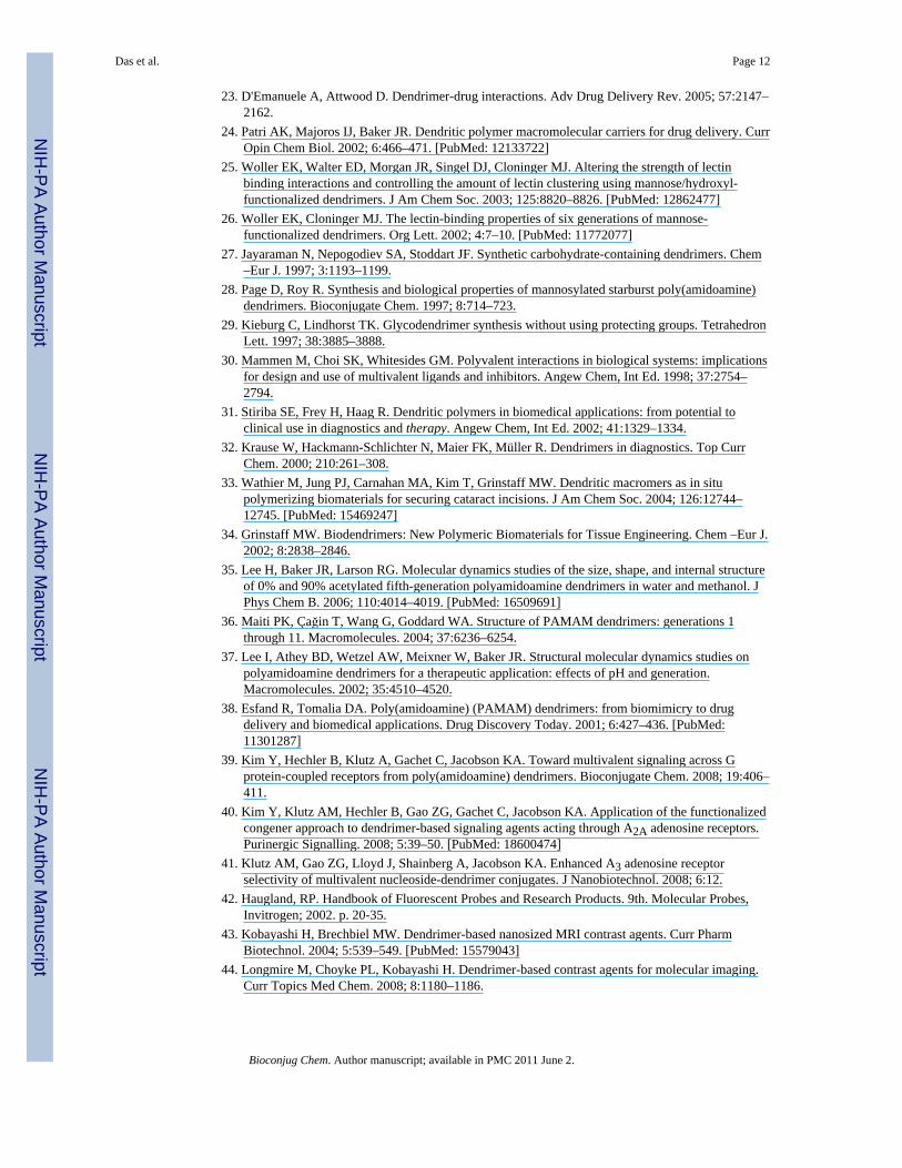
The control dendrimers 4 and 5 were inactive in this test system. In contrast, compound 9,derived from the G3 dendrimer and containing free residual amino groups, was a very potentagonist that exhibited an EC50 of 2.4 nM at the P2Y14 receptor (Figure 1). This potency wasapproximately 100-fold greater than that of the native agonist UDPG 1, which had an EC50of 261 nM. UDPGA 2 was approximately equipotent to 1 in this assay. The correspondingdendrimer 8 (Figure 1), which had a lower degree of loading of the nucleotide on a G3dendrimer, was considerably less potent than 9, but similar in potency to 1. Therefore, adistinct multivalent effect occurs in these compounds.
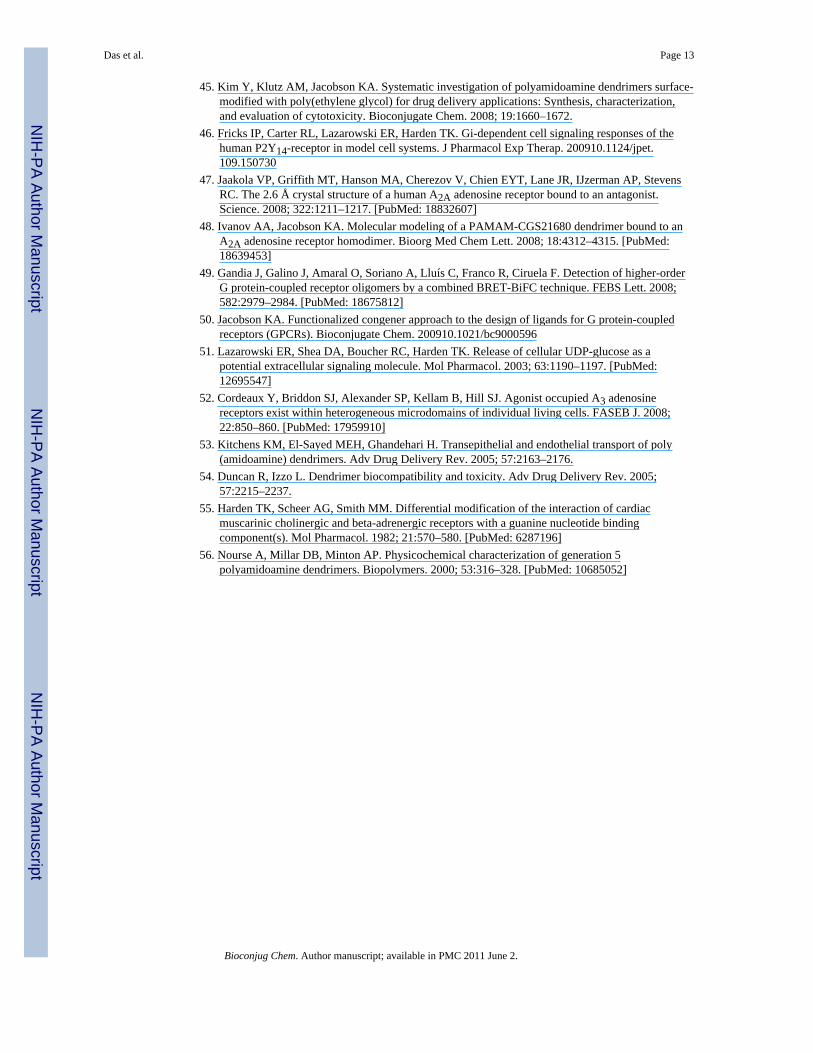
Compounds 10 and 11, both of which were derived from half-integral dendrimers andcontain free residual carboxylate groups, were very potent at the P2Y14 receptor with EC50values of 3.2 and 3.1 nM, respectively (Figure 2). The corresponding amine derivative 3a ofUDPGA, which also served as the synthetic precursor of conjugates 10 and 11, wasconsiderably less potent.
The higher molecular weight G6 dendrimer conjugate 12 was similar in relative compositionto the smaller conjugate 9, which had a high degree of loading of nucleotide. Compound 12(EC50 = 0.8 nM) was significantly more potent than 9 (Figure 3). Both 9 and 12 containsignificant fractions (but less than 50%) of residual amino groups.
The G3 dendrimer conjugates 13 and 15 were equivalent to the precursor dendrimerconjugate 12, except that they both contain covalently attached prosthetic groups in additionto the nucleotide. Each of these dendrimers was essentially as potent as 9. The G3 dendrimerconjugate 14, which had approximately two covalently attached AlexaFluor488 moieties,was 17-fold less potent than the corresponding 9 but 7-fold more potent than compound 1.However, when the DTPA conjugate 15 was complexed with gadolinium, the potency wasgreatly reduced, to the level of 1, the native agonist.
Molecular ModelingA molecular model of compound 3b docked to the human P2Y14 receptor recently wasconstructed on the basis of the crystal structure of the human A2A adenosine receptor (15,47). This model was utilized to build a complex of the P2Y14 receptor bound to a PAMAMG3 dendrimer using the same approach as reported for the docking of a polyvalentdendrimer–nucleoside conjugate to the human A2A adenosine receptor (48). In particular,our published model of the conjugate of PAMAM (G3) with an A2A receptor agonist (2-[p-(2-carboxyethyl)phenyl-ethylamino]-5′-N-ethylcarboxamidoadenosine, CGS21680) wasutilized to build a model of dendrimer–nucleotide conjugate 9, which was partiallysubstituted with nucleotide moieties. Initially, all fragments of the carboxylic acidCGS21680 were removed from the model of PAMAM-CGS21680, and hydrogen atomswere added to the remaining PAMAM structure. Twenty moieties of UDPGA 2 then werecoupled to randomly selected amino groups of the PAMAM chains. The remaining 12unacylated chains of the dendrimer were substituted with protonated amino groups.
The model of 9 was minimized in the OPLS2005 force field. The Polak–Ribier conjugategradient (PRCG) minimization method with a maximum of 5000 iterations and with aconvergence threshold of 0.05 kJ · mol−1 · Å−1 was applied. One randomly selectedUDPGA moiety attached to the dendrimer then was removed from the PAMAM model, andthe UDPGA docked to the P2Y14 receptor was attached to the dendrimer. Thus, an initialcomplex of 9 docked to the P2Y14 receptor was obtained. The orientation of a few UDPGA
Das et al. Page 8
Bioconjug Chem. Author manuscript; available in PMC 2011 June 2.
NIH
-PA Author Manuscript
NIH
-PA Author Manuscript
NIH
-PA Author Manuscript
moieties was manually adjusted to avoid overlays with the amino acid residues of the P2Y14receptor. The model was subjected to geometry optimization using the same protocol asmentioned above. After minimization, the binding mode of the UDPGA moiety locatedinside the P2Y14 receptor was found to be similar to the binding mode of 1 itself (15). Theethylenediamine chain of PAMAM connected to the receptor-bound UDPGA moiety waslocated between extracellular loops of P2Y14 receptor. In particular, the nucleotide moietywas surrounded by Ile173 (EL2), and Leu175 (EL2), Arg274 (EL3), and Lys277 (7.35). Inaddition, Lys277 formed an H-bond with the amide oxygen atom of the chain of PAMAM.The molecular model of compound 9 docked in the human P2Y14 receptor (Figure 4)showed that the nucleotide-substituted branches of the dendrimer extended far beyond thedimensions of the receptor protein and thus would be available for multivalent docking toreceptor dimers and higher-order aggregates (49).
DiscussionA functionalized ethylenediamine-containing chain was incorporated in agonists of thehuman P2Y14 receptor, and this chain provided a conjugation strategy for couplingnucleoside agonists to PAMAM dendrimer carriers. Uridine-5′-diphosphoglucuronic acidand its ethylenediamine adduct were suitable functionalized congeners (50) for coupling toseveral generations (G2.5–6) of dendrimers (both terminal carboxy and amino) to modulatepotency of the intact conjugates. The observed biological activity of the dendrimerconjugates was clearly P2Y14 receptor-dependent; the control dendrimers 4 and 5 lackingnucleotides were inactive. A G3 PAMAM conjugate containing 20 bound nucleotidemoieties was 100-fold more potent than the native agonist 1. Larger dendrimer carriers andthose with greater loading favored higher potency as P2Y14 receptor agonists. All of thedendrimer–nucleotide conjugates were full agonists at the receptor.
The molecular mechanism whereby these dendrimers exhibit increased activity relative tothe native cognate agonist UDPG is unclear. One possible explanation is that thesemultivalent compounds may bridge multiple P2Y14 receptor binding sites simultaneously.Molecular modeling has indicated this possibility for an analogous G3 PAMAM dendrimerconjugate of an A2A adenosine receptor agonist (48). The dramatic enhancement in potency,especially in the more heavily substituted dendrimer derivatives, is highly suggestive of amultivalent effect. The ability to span multiple receptor binding sites would be furtherpromoted by the possible aggregation of dendrimer derivatives (56). Nanomolar andsubnanomolar potencies have been achieved, which are unprecedented for any monomericnucleotides acting at the P2Y14 receptor. Dendrimer conjugates 10 and 11, which are bothmore than half substituted with nucleotide moieties, are more than 800-fold more potentthan a corresponding monomer, i.e., the ethylenediamine analogue 3a. Lower loading of thesame dendrimer with nucleotides resulted in a conjugate (compound 9) that exhibitedrelatively lower potency. Variability of the potency depending on degree of loading will bethe subject of future systematic studies. Such studies also might benefit from quantificationof the relative activities of these compounds across a wide range of receptor expressionlevels in the same cell system.
It is possible that conjugation to dendrimers might protect the nucleotides against enzymaticdegradation, but this is not the basis for the enhancement of potency in cAMP-inhibitionwith the multivalent dendrimers. Our earlier studies (51) indicated that little metabolism ofextracellular 1 occurs under the conditions of these assays. Moreover, the concentrationeffect curve of 1 for decreasing cyclic AMP levels is similar whether quantified at times of<5 min or times up to 30 min.
Das et al. Page 9
Bioconjug Chem. Author manuscript; available in PMC 2011 June 2.
NIH
-PA Author Manuscript
NIH
-PA Author Manuscript
NIH
-PA Author Manuscript
Dendrimers containing several different prosthetic groups also were synthesized. Dendrimerconjugate 14, which is tagged with AlexaFluor488, potentially will be useful for receptorvisualization. Although the potency of 14 was 17-fold lower than that of the correspondingdendrimer lacking the fluorescent group (i.e., 9), the EC50 of approximately 40 nM wascomparable to previously introduced fluorescent ligand probes for other GPCRs (52). Asimilar approach in a previous study, using a fluorescent AlexaFluor488-tagged G3PAMAM dendrimer, was used to detect the expression of the A3 adenosine receptor incultured cells (41). Gd(III) is a paramagnetic ion that is used for magnetic resonanceimaging (44). Dendrimer conjugate 15, which is tagged with the chelating group DTPA, issubstantially less potent as its Gd(III) complex, thus limiting the potential utility of thisderivative. The in vivo application of dendrimer conjugates is normally limited to i.p. or i.v.injection, but novel approaches for gaining greater bioavailability of dendrimers by otherroutes of administration have been discussed (53, 54).
In conclusion, we have identified a site on a nucleotide agonist of the P2Y14 receptor forchemical tethering to a macromolecular carrier without losing the ability to activate thereceptor. Moreover, we have shown that the potency of the native ligand is greatly enhancedin these multivalent ligands. These biologically active drug conjugates do not requirecleavage or cellular internalization for their action at the GPCR; in fact, these processeswould reduce activity. Covalent conjugation of P2Y14 receptor agonists to PAMAMdendrimers qualitatively altered their pharmacological activity. Thus, potency was eitherretained or dramatically enhanced in the multivalent dendrimer conjugates in comparison tothe monomeric P2Y14 receptor agonists, depending on size, degree of substitution, terminalfunctionality, and attached prosthetic groups. The ability to modulate the potency of a givenGPCR ligand by the mode and stoichiometry of attachment to dendrimer carriers promisesto have general applicability to this therapeutically important class of receptor proteins.
Supplementary MaterialRefer to Web version on PubMed Central for supplementary material.
AcknowledgmentsThis research was supported in part by the Intramural Research Program of the NIH, NIDDK, and by NIH grantGM38213 to T.K.H. We thank Dr. Herman Yeh and Dr. Athena M. Keene-Klutz of NIDDK for helpful advice onthe NMR experiments and chemical synthesis. We thank Dr. John Lloyd (NIDDK) for mass spectral measurements.
Literature cited1. Bridges TM, Lindsley CW. G-protein-coupled receptors: from classical modes of modulation to
allosteric mechanisms. ACS Chem Biol. 2008; 3:530–541. [PubMed: 18652471]2. Abbracchio MP, Burnstock G, Boeynaems JM, Barnard EA, Boyer JL, Kennedy C, Fumagalli M,
King BF, Gachet C, Jacobson KA, Weisman GA. International Union of Pharmacology. Update ofthe P2Y G protein-coupled nucleotide receptors: from molecular mechanisms and pathophysiologyto therapy. Pharmacol Rev. 2006; 58:281–341. [PubMed: 16968944]
3. Gachet C. Regulation of platelet functions by P2 receptors. Annu Rev Pharmacol Toxicol. 2006;46:277–300. [PubMed: 16402906]
4. Burnstock G. Purinergic signalling and disorders of the central nervous system. Nat Rev DrugDiscovery. 2008; 7:575–590.
5. Idzko M, Hammad H, van Nimwegen M, Kool M, Willart MA, Muskens F, Hoogsteden HC,Luttmann W, Ferrari D, Di Virgilio F, Virchow JC, Lambrecht BN. Extracellular ATP triggers andmaintains asthmatic airway inflammation by activating dendritic cells. Nat Med. 2007; 13:913–919.[PubMed: 17632526]
Das et al. Page 10
Bioconjug Chem. Author manuscript; available in PMC 2011 June 2.
NIH
-PA Author Manuscript
NIH
-PA Author Manuscript
NIH
-PA Author Manuscript
6. Shin A, Toy T, Rothenfusser S, Robson N, Vorac J, Dauer M, Stuplich M, Endres S, Cebon J,Maraskovsky E, Schnurr M. P2Y receptor signaling regulates phenotype and IFN-alpha secretion ofhuman plasmacytoid dendritic cells. Blood. 2008; 111:3062–3069. [PubMed: 17993619]
7. Malin SA, Davis BM, Richard KH, Reynolds IJ, Albers KM, Molliver DC. Thermal nociception andTRPV1 function are attenuated in mice lacking the nucleotide receptor P2Y2. Pain. 2008; 138:484–496. [PubMed: 18343036]
8. Lugo-Garcia L, Filhol R, Lajoix AD, Gross R, Petit P, Vignon J. Expression of purinergic P2Yreceptor subtypes by INS-1 insulinoma beta-cells: a molecular and binding characterization. Eur JPharmacol. 2007; 568:54–60. [PubMed: 17509560]
9. Lazarowski ER, Tarran R, Grubb BR, van Heusden CA, Okada S, Boucher RC. Nucleotide releaseprovides a mechanism for airway surface liquid homeostasis. J Biol Chem. 2004; 279:36855–36864. [PubMed: 15210701]
10. Müller T, Bayer H, Myrtek D, Ferrari D, Sorichter S, Ziegenhagen MW, Zissel G, Virchow JC,Luttmann W, Norgauer J, Di Virgilio F, Idzko M. The P2Y14 receptor of airway epithelial cellscoupling to intracellular Ca2+ and IL-8 secretion. Am J Respir Cell Mol Biol. 2005; 33:601–609.[PubMed: 16109883]
11. Chambers JK, Macdonald LE, Sarau HM, Ames RS, Freeman K, Foley JJ, Zhu Y, McLaughlinMM, Murdock P, McMillan L, Trill J, Swift A, Aiyar N, Taylor P, Vawter L, Naheed S, SzekeresP, Hervieu G, Scott C, Watson JM, Murphy AJ, Duzic E, Klein C, Bergsma DJ, Wilson S, LiviGP. A G protein-coupled receptor for UDP-glucose. J Biol Chem. 2000; 275:10767–10771.[PubMed: 10753868]
12. Abbracchio MP, Boeynaems JM, Barnard EA, Boyer JL, Kennedy C, Miras-Portugal MT, KingBF, Gachet C, Jacobson KA, Weisman GA, Burnstock G. Characterization of the UDP-glucosereceptor (re-named here the P2Y14 receptor) adds diversity to the P2Y receptor family. TrendsPharmacol Sci. 2003; 24:52–55. [PubMed: 12559763]
13. Scrivens M, Dickenson JM. Functional expression of P2Y14 receptor in human neutrophils. Eur JPharmacol. 2006; 543:166–173. [PubMed: 16820147]
14. Bassil AK, Bourdu S, Townson KA, Wheeldon A, Jarvie EM, Zebda N, Abuin A, Grau E, Livi GP,Punter L, Latcham J, Grimes AM, Hurp DP, Downham KM, Sanger GJ, Winchester WJ, MorrisonAD, Moore GBT. UDP-glucose modulates gastric function through P2Y14 receptor-dependent and-independent mechanisms. Am J Physiol Gastrointest Liver Physiol. 2009; 296:G923–G930.[PubMed: 19164486]
15. Ko H, Das A, Carter RL, Fricks IP, Zhou Y, Ivanov AA, Melman A, Joshi BV, Kováč P, HajduchJ, Kirk KL, Harden TK, Jacobson KA. Molecular recognition in the P2Y14 receptor: Probing thestructurally permissive terminal sugar moiety of UDP-glucose. Bioorg Med Chem. 10.1016/j.bmc.2009.05.024
16. Ko H, Fricks I, Ivanov AA, Harden TK, Jacobson KA. Structure activity relationship of uridine 5′-diphosphoglucose (UDP-Glucose) analogues as agonists of the human P2Y14 receptor. J MedChem. 2007; 50:2030–2039. [PubMed: 17407275]
17. Ivanov AA, Fricks I, Harden TK, Jacobson KA. Molecular dynamics simulation of the P2Y14receptor. Ligand docking and identification of a putative binding site of the distal hexose moiety.Bioorg Med Chem Lett. 2007; 17:761–766. [PubMed: 17088057]
18. Fricks I, Maddiletti S, Carter R, Lazarowski ER, Nicholas RA, Jacobson KA, Harden TK. UDP isa competitive antagonist at the human P2Y14 receptor and a full agonist at the rat P2Y14 receptor.J Pharm Exp Therap. 2008; 325:588–594.
19. Tomalia DA, Reyna LA, Svenson S. Dendrimers as multi-purpose nanodevices for oncology drugdelivery and diagnostic imaging. Cellular delivery of therapeutic macromolecules. Biochem SocTrans. 2007; 35:61–67. [PubMed: 17233602]
20. Lee CC, MacKay JA, Fréchet JMJ, Szoka FC. Designing dendrimers for biological applications.Nat Biotechnol. 2005; 23:1517–1526. [PubMed: 16333296]
21. Zeng F, Zimmerman SC. Dendrimers in supramolecular chemistry: from molecular recognition toself-assembly. Chem Rev. 1997; 97:1681–1712. [PubMed: 11851463]
22. Gillies ER, Fréchet JMJ. Dendrimers and dendritic polymers in drug delivery. Drug DiscoveryToday. 2005; 10:35–43. [PubMed: 15676297]
Das et al. Page 11
Bioconjug Chem. Author manuscript; available in PMC 2011 June 2.
NIH
-PA Author Manuscript
NIH
-PA Author Manuscript
NIH
-PA Author Manuscript
23. D'Emanuele A, Attwood D. Dendrimer-drug interactions. Adv Drug Delivery Rev. 2005; 57:2147–2162.
24. Patri AK, Majoros IJ, Baker JR. Dendritic polymer macromolecular carriers for drug delivery. CurrOpin Chem Biol. 2002; 6:466–471. [PubMed: 12133722]
25. Woller EK, Walter ED, Morgan JR, Singel DJ, Cloninger MJ. Altering the strength of lectinbinding interactions and controlling the amount of lectin clustering using mannose/hydroxyl-functionalized dendrimers. J Am Chem Soc. 2003; 125:8820–8826. [PubMed: 12862477]
26. Woller EK, Cloninger MJ. The lectin-binding properties of six generations of mannose-functionalized dendrimers. Org Lett. 2002; 4:7–10. [PubMed: 11772077]
27. Jayaraman N, Nepogodiev SA, Stoddart JF. Synthetic carbohydrate-containing dendrimers. Chem–Eur J. 1997; 3:1193–1199.
28. Page D, Roy R. Synthesis and biological properties of mannosylated starburst poly(amidoamine)dendrimers. Bioconjugate Chem. 1997; 8:714–723.
29. Kieburg C, Lindhorst TK. Glycodendrimer synthesis without using protecting groups. TetrahedronLett. 1997; 38:3885–3888.
30. Mammen M, Choi SK, Whitesides GM. Polyvalent interactions in biological systems: implicationsfor design and use of multivalent ligands and inhibitors. Angew Chem, Int Ed. 1998; 37:2754–2794.
31. Stiriba SE, Frey H, Haag R. Dendritic polymers in biomedical applications: from potential toclinical use in diagnostics and therapy. Angew Chem, Int Ed. 2002; 41:1329–1334.
32. Krause W, Hackmann-Schlichter N, Maier FK, Müller R. Dendrimers in diagnostics. Top CurrChem. 2000; 210:261–308.
33. Wathier M, Jung PJ, Carnahan MA, Kim T, Grinstaff MW. Dendritic macromers as in situpolymerizing biomaterials for securing cataract incisions. J Am Chem Soc. 2004; 126:12744–12745. [PubMed: 15469247]
34. Grinstaff MW. Biodendrimers: New Polymeric Biomaterials for Tissue Engineering. Chem –Eur J.2002; 8:2838–2846.
35. Lee H, Baker JR, Larson RG. Molecular dynamics studies of the size, shape, and internal structureof 0% and 90% acetylated fifth-generation polyamidoamine dendrimers in water and methanol. JPhys Chem B. 2006; 110:4014–4019. [PubMed: 16509691]
36. Maiti PK, Çağin T, Wang G, Goddard WA. Structure of PAMAM dendrimers: generations 1through 11. Macromolecules. 2004; 37:6236–6254.
37. Lee I, Athey BD, Wetzel AW, Meixner W, Baker JR. Structural molecular dynamics studies onpolyamidoamine dendrimers for a therapeutic application: effects of pH and generation.Macromolecules. 2002; 35:4510–4520.
38. Esfand R, Tomalia DA. Poly(amidoamine) (PAMAM) dendrimers: from biomimicry to drugdelivery and biomedical applications. Drug Discovery Today. 2001; 6:427–436. [PubMed:11301287]
39. Kim Y, Hechler B, Klutz A, Gachet C, Jacobson KA. Toward multivalent signaling across Gprotein-coupled receptors from poly(amidoamine) dendrimers. Bioconjugate Chem. 2008; 19:406–411.
40. Kim Y, Klutz AM, Hechler B, Gao ZG, Gachet C, Jacobson KA. Application of the functionalizedcongener approach to dendrimer-based signaling agents acting through A2A adenosine receptors.Purinergic Signalling. 2008; 5:39–50. [PubMed: 18600474]
41. Klutz AM, Gao ZG, Lloyd J, Shainberg A, Jacobson KA. Enhanced A3 adenosine receptorselectivity of multivalent nucleoside-dendrimer conjugates. J Nanobiotechnol. 2008; 6:12.
42. Haugland, RP. Handbook of Fluorescent Probes and Research Products. 9th. Molecular Probes,Invitrogen; 2002. p. 20-35.
43. Kobayashi H, Brechbiel MW. Dendrimer-based nanosized MRI contrast agents. Curr PharmBiotechnol. 2004; 5:539–549. [PubMed: 15579043]
44. Longmire M, Choyke PL, Kobayashi H. Dendrimer-based contrast agents for molecular imaging.Curr Topics Med Chem. 2008; 8:1180–1186.
Das et al. Page 12
Bioconjug Chem. Author manuscript; available in PMC 2011 June 2.
NIH
-PA Author Manuscript
NIH
-PA Author Manuscript
NIH
-PA Author Manuscript
45. Kim Y, Klutz AM, Jacobson KA. Systematic investigation of polyamidoamine dendrimers surface-modified with poly(ethylene glycol) for drug delivery applications: Synthesis, characterization,and evaluation of cytotoxicity. Bioconjugate Chem. 2008; 19:1660–1672.
46. Fricks IP, Carter RL, Lazarowski ER, Harden TK. Gi-dependent cell signaling responses of thehuman P2Y14-receptor in model cell systems. J Pharmacol Exp Therap. 200910.1124/jpet.109.150730
47. Jaakola VP, Griffith MT, Hanson MA, Cherezov V, Chien EYT, Lane JR, IJzerman AP, StevensRC. The 2.6 Å crystal structure of a human A2A adenosine receptor bound to an antagonist.Science. 2008; 322:1211–1217. [PubMed: 18832607]
48. Ivanov AA, Jacobson KA. Molecular modeling of a PAMAM-CGS21680 dendrimer bound to anA2A adenosine receptor homodimer. Bioorg Med Chem Lett. 2008; 18:4312–4315. [PubMed:18639453]
49. Gandia J, Galino J, Amaral O, Soriano A, Lluís C, Franco R, Ciruela F. Detection of higher-orderG protein-coupled receptor oligomers by a combined BRET-BiFC technique. FEBS Lett. 2008;582:2979–2984. [PubMed: 18675812]
50. Jacobson KA. Functionalized congener approach to the design of ligands for G protein-coupledreceptors (GPCRs). Bioconjugate Chem. 200910.1021/bc9000596
51. Lazarowski ER, Shea DA, Boucher RC, Harden TK. Release of cellular UDP-glucose as apotential extracellular signaling molecule. Mol Pharmacol. 2003; 63:1190–1197. [PubMed:12695547]
52. Cordeaux Y, Briddon SJ, Alexander SP, Kellam B, Hill SJ. Agonist occupied A3 adenosinereceptors exist within heterogeneous microdomains of individual living cells. FASEB J. 2008;22:850–860. [PubMed: 17959910]
53. Kitchens KM, El-Sayed MEH, Ghandehari H. Transepithelial and endothelial transport of poly(amidoamine) dendrimers. Adv Drug Delivery Rev. 2005; 57:2163–2176.
54. Duncan R, Izzo L. Dendrimer biocompatibility and toxicity. Adv Drug Delivery Rev. 2005;57:2215–2237.
55. Harden TK, Scheer AG, Smith MM. Differential modification of the interaction of cardiacmuscarinic cholinergic and beta-adrenergic receptors with a guanine nucleotide bindingcomponent(s). Mol Pharmacol. 1982; 21:570–580. [PubMed: 6287196]
56. Nourse A, Millar DB, Minton AP. Physicochemical characterization of generation 5polyamidoamine dendrimers. Biopolymers. 2000; 53:316–328. [PubMed: 10685052]
Das et al. Page 13
Bioconjug Chem. Author manuscript; available in PMC 2011 June 2.
NIH
-PA Author Manuscript
NIH
-PA Author Manuscript
NIH
-PA Author Manuscript
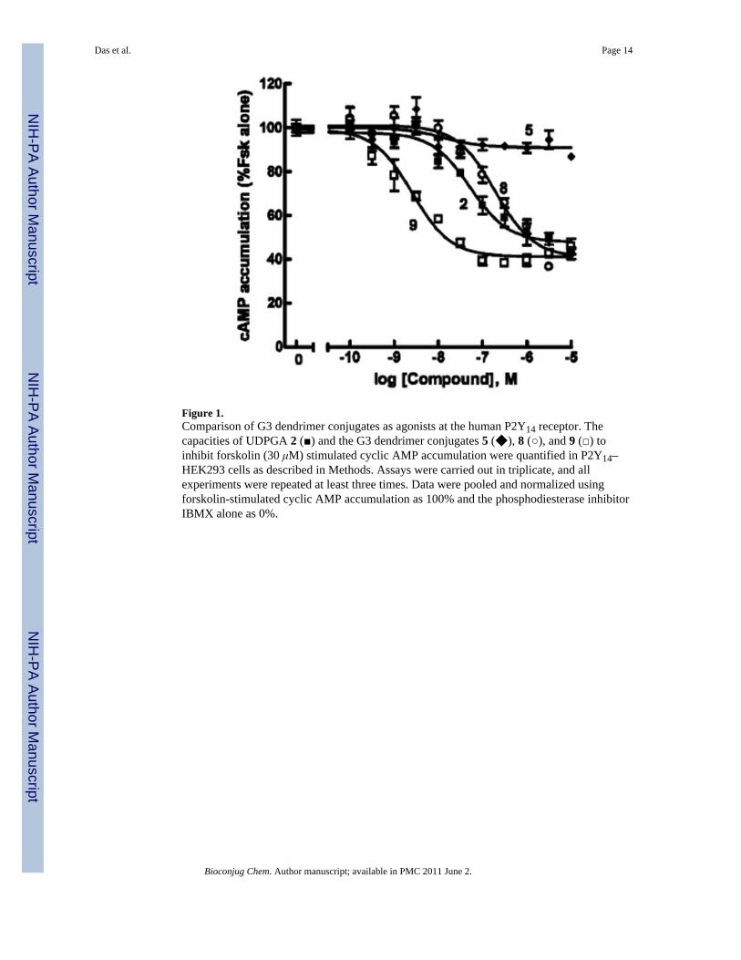
Figure 1.Comparison of G3 dendrimer conjugates as agonists at the human P2Y14 receptor. Thecapacities of UDPGA 2 (■) and the G3 dendrimer conjugates 5 (◆), 8 (○), and 9 (□) toinhibit forskolin (30 μM) stimulated cyclic AMP accumulation were quantified in P2Y14–HEK293 cells as described in Methods. Assays were carried out in triplicate, and allexperiments were repeated at least three times. Data were pooled and normalized usingforskolin-stimulated cyclic AMP accumulation as 100% and the phosphodiesterase inhibitorIBMX alone as 0%.
Das et al. Page 14
Bioconjug Chem. Author manuscript; available in PMC 2011 June 2.
NIH
-PA Author Manuscript
NIH
-PA Author Manuscript
NIH
-PA Author Manuscript
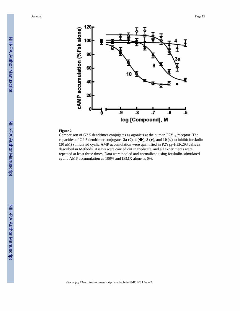
Figure 2.Comparison of G2.5 dendrimer conjugates as agonists at the human P2Y14 receptor. Thecapacities of G2.5 dendrimer conjugates 3a (◊), 4 (◆), 8 (●), and 10 (○) to inhibit forskolin(30 μM) stimulated cyclic AMP accumulation were quantified in P2Y14–HEK293 cells asdescribed in Methods. Assays were carried out in triplicate, and all experiments wererepeated at least three times. Data were pooled and normalized using forskolin-stimulatedcyclic AMP accumulation as 100% and IBMX alone as 0%.
Das et al. Page 15
Bioconjug Chem. Author manuscript; available in PMC 2011 June 2.
NIH
-PA Author Manuscript
NIH
-PA Author Manuscript
NIH
-PA Author Manuscript

Figure 3.Comparison of larger dendrimer conjugates as agonists at the human P2Y14 receptor. Thecapacities of UDPGA 2 (■), G3 dendrimer conjugate 9 (□), and G6 dendrimer conjugate 12(○) to inhibit forskolin (30 μM)-stimulated cyclic AMP accumulation were quantified inP2Y14–HEK293 cells as described in Methods. Assays were carried out in triplicate, and allexperiments were repeated at least three times. Data were pooled and normalized usingforskolin-stimulated cyclic AMP accumulation as 100% and IBMX alone as 0%.
Das et al. Page 16
Bioconjug Chem. Author manuscript; available in PMC 2011 June 2.
NIH
-PA Author Manuscript
NIH
-PA Author Manuscript
NIH
-PA Author Manuscript

Figure 4.Molecular model of compound 9 docked in the human P2Y14 receptor, obtained afterdocking to the receptor homology model built based on the crystal structure of the A2Aadenosine receptor (47). The receptor helices are colored by residue position: N-terminus inred, TM 1 in orange, TM 2 in ochre, TM 3 in yellow, TM 4 in green, TM 5 in cyan, TM 6 inblue, and TM 7 and C-terminus in purple.
Das et al. Page 17
Bioconjug Chem. Author manuscript; available in PMC 2011 June 2.
NIH
-PA Author Manuscript
NIH
-PA Author Manuscript
NIH
-PA Author Manuscript
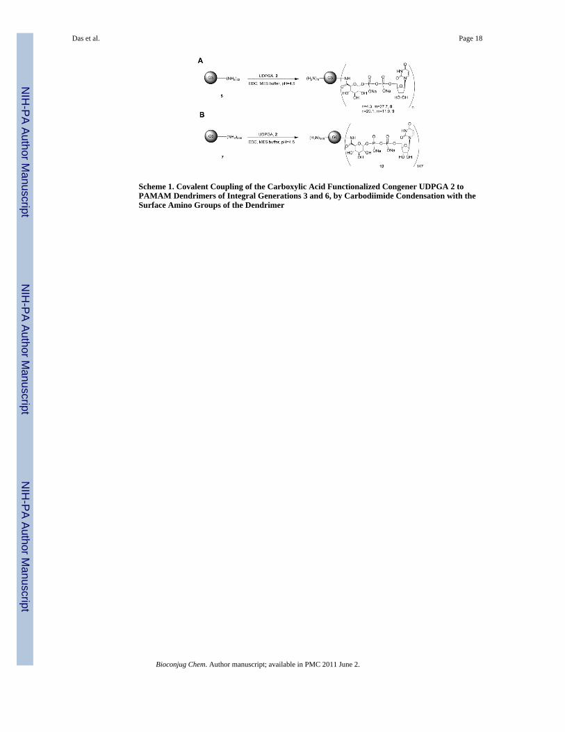
Scheme 1. Covalent Coupling of the Carboxylic Acid Functionalized Congener UDPGA 2 toPAMAM Dendrimers of Integral Generations 3 and 6, by Carbodiimide Condensation with theSurface Amino Groups of the Dendrimer
Das et al. Page 18
Bioconjug Chem. Author manuscript; available in PMC 2011 June 2.
NIH
-PA Author Manuscript
NIH
-PA Author Manuscript
NIH
-PA Author Manuscript
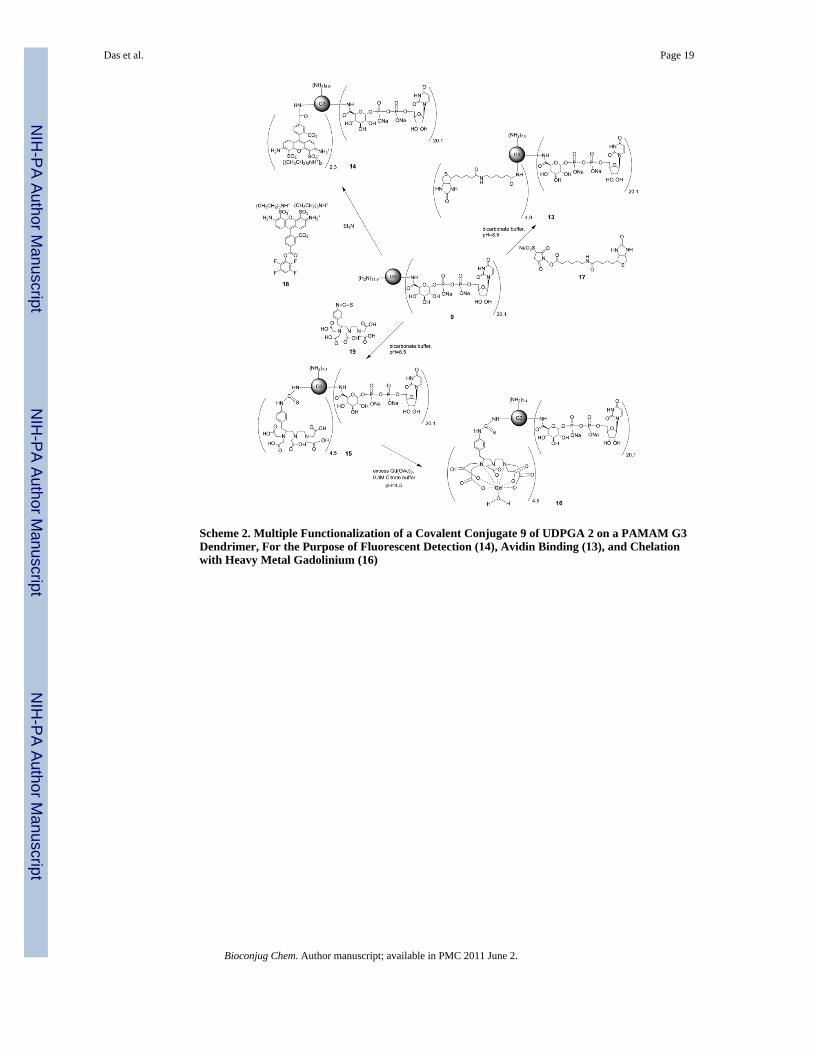
Scheme 2. Multiple Functionalization of a Covalent Conjugate 9 of UDPGA 2 on a PAMAM G3Dendrimer, For the Purpose of Fluorescent Detection (14), Avidin Binding (13), and Chelationwith Heavy Metal Gadolinium (16)
Das et al. Page 19
Bioconjug Chem. Author manuscript; available in PMC 2011 June 2.
NIH
-PA Author Manuscript
NIH
-PA Author Manuscript
NIH
-PA Author Manuscript
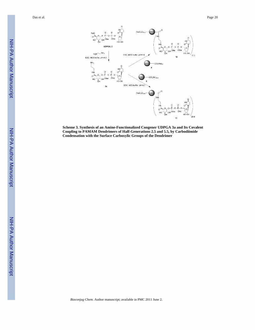
Scheme 3. Synthesis of an Amine-Functionalized Congener UDPGA 3a and Its CovalentCoupling to PAMAM Dendrimers of Half-Generations 2.5 and 5.5, by CarbodiimideCondensation with the Surface Carboxylic Groups of the Dendrimer
Das et al. Page 20
Bioconjug Chem. Author manuscript; available in PMC 2011 June 2.
NIH
-PA Author Manuscript
NIH
-PA Author Manuscript
NIH
-PA Author Manuscript
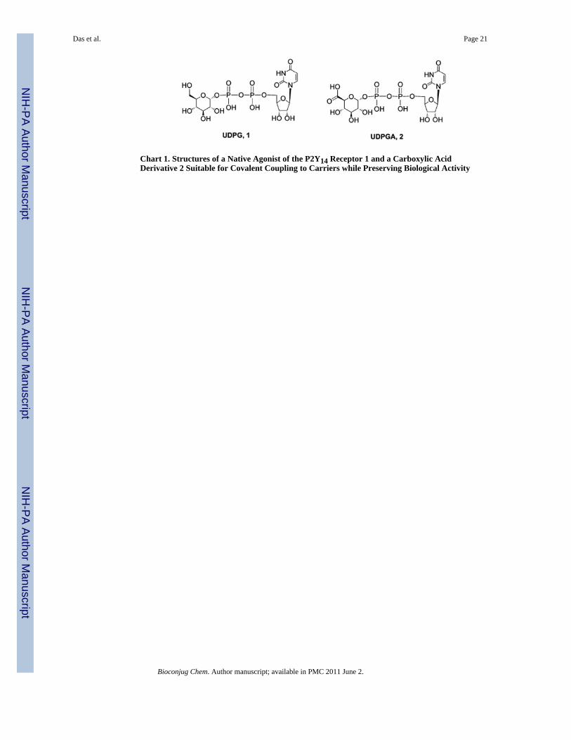
Chart 1. Structures of a Native Agonist of the P2Y14 Receptor 1 and a Carboxylic AcidDerivative 2 Suitable for Covalent Coupling to Carriers while Preserving Biological Activity
Das et al. Page 21
Bioconjug Chem. Author manuscript; available in PMC 2011 June 2.
NIH
-PA Author Manuscript
NIH
-PA Author Manuscript
NIH
-PA Author Manuscript
NIH
-PA Author Manuscript
NIH
-PA Author Manuscript
NIH
-PA Author Manuscript
Das et al. Page 22
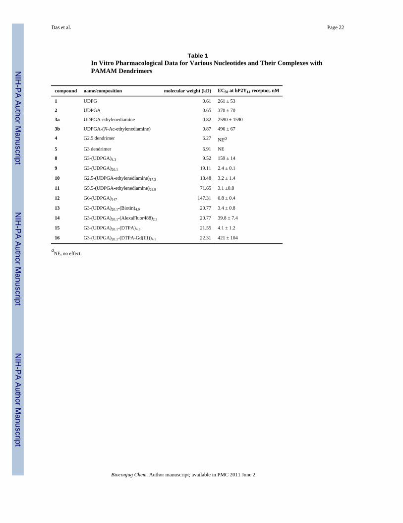
Table 1In Vitro Pharmacological Data for Various Nucleotides and Their Complexes withPAMAM Dendrimers
compound name/composition molecular weight (kD) EC50 at hP2Y14 receptor, nM
1 UDPG 0.61 261 ± 53
2 UDPGA 0.65 370 ± 70
3a UDPGA-ethylenediamine 0.82 2590 ± 1590
3b UDPGA-(N-Ac-ethylenediamine) 0.87 496 ± 67
4 G2.5 dendrimer 6.27 NEa
5 G3 dendrimer 6.91 NE
8 G3-(UDPGA)4.3 9.52 159 ± 14
9 G3-(UDPGA)20.1 19.11 2.4 ± 0.1
10 G2.5-(UDPGA-ethylenediamine)17.3 18.48 3.2 ± 1.4
11 G5.5-(UDPGA-ethylenediamine)29.9 71.65 3.1 ±0.8
12 G6-(UDPGA)147 147.31 0.8 ± 0.4
13 G3-(UDPGA)20.1-(Biotin)4.9 20.77 3.4 ± 0.8
14 G3-(UDPGA)20.1-(AlexaFluor488)2.3 20.77 39.8 ± 7.4
15 G3-(UDPGA)20.1-(DTPA)4.5 21.55 4.1 ± 1.2
16 G3-(UDPGA)20.1-(DTPA-Gd(III))4.5 22.31 421 ± 104
aNE, no effect.
Bioconjug Chem. Author manuscript; available in PMC 2011 June 2.
Related Documents