The Regulated Expression, Intracellular Trafficking, and Membrane Recycling of the P2Y-like Receptor GPR17 in Oli-neu Oligodendroglial Cells * Received for publication, July 31, 2012, and in revised form, January 2, 2013 Published, JBC Papers in Press, January 3, 2013, DOI 10.1074/jbc.M112.404996 Alessandra Fratangeli ‡1,2 , Elena Parmigiani ‡§1 , Marta Fumagalli ¶ , Davide Lecca ¶ , Roberta Benfante ‡ , Maria Passafaro ‡3 , Annalisa Buffo § , Maria P. Abbracchio ¶ , and Patrizia Rosa ‡4 From the ‡ Consiglio Nazionale delle Ricerche-Institute of Neuroscience, Department of Medical Biotechnologies and Translational Medicine (BIOMETRA), University of Milan, Milan, 20129, Italy, the ¶ Laboratory of Molecular and Cellular Pharmacology of Purinergic Transmission, Department of Pharmacological and Biomolecular Sciences, University of Milan, Milan, 20133, Italy, and the § Department of Neuroscience, Section of Physiology, University of Turin, Neuroscience Institute of Turin (NIT), Neuroscience Institute Cavalieri Ottolenghi, Regione Gonzole, Orbassano, 10043, Turin, Italy Background: GPR17 is a key player in oligodendrocyte differentiation. By regulating the availability of receptors at the cell surface, agonist-induced GPR17 trafficking may influence terminal cell fate. Results: UDP-glucose and LTD 4 induce GPR17 endocytosis and distribution in lysosomes or recycling compartments. Conclusion: Agonist-activated GPR17 undergoes partial degradation and fast membrane recycling. Significance: Understanding GPR17 trafficking may increase our knowledge of oligodendrocyte differentiation and myelination. GPR17 is a G-protein-coupled receptor that is activated by two classes of molecules: uracil-nucleotides and cysteinyl-leu- kotrienes. GPR17 is required for initiating the differentiation of oligodendrocyte precursors but has to be down-regulated to allow cells to undergo terminal maturation. Although a great deal has been learned about GPR17 expression and signaling, no information is currently available about the trafficking of native receptors after the exposure of differentiating oligodendrocytes to endogenous agonists. Here, we demonstrate that neuron- conditioned medium induces the transcriptionally mediated, time-regulated expression of GPR17 in Oli-neu, an oligoden- drocyte precursor cell line, making these cells suitable for study- ing the endocytic traffic of the native receptor. Agonist-induced internalization, intracellular trafficking, and membrane recy- cling of GPR17 were analyzed by biochemical and immunofluo- rescence assays using an ad hoc-developed antibody against the extracellular N-terminal of GPR17. Both UDP-glucose and LTD 4 increased GPR17 internalization, although with different efficiency. At early time points, internalized GPR17 co-localized with transferrin receptor, whereas at later times it partially co- localized with the lysosomal marker Lamp1, suggesting that a portion of GPR17 is targeted to lysosomes upon ligand binding. An analysis of receptor recycling and degradation demonstrated that a significant aliquot of GPR17 is recycled to the cell surface. Furthermore, internalized GPR17 displayed a co-localization with the marker of the “short loop” recycling endosomes, Rab4, while showing very minor co-localization with the “long loop” recycling marker, Rab11. Our results provide the first data on the agonist-induced trafficking of native GPR17 in oligodendro- glial cells and may have implications for both physiological and pathological myelination. Recent work has led to the “deorphanization” of the G pro- tein-coupled receptor (GPCR) 5 referred to as GPR17, which is located at an intermediate phylogenetic position between the purinergic P2Y receptors and the CysLT 1 and CysLT 2 receptors for cysteinyl-leukotrienes (cysLTs; 1– 6). Both recombinant and native GPR17 receptors respond to uracil nucleotides (e.g. UDP and UDP-glucose) and arachidonic acid-derived cysLTs (e.g. LTD 4 and LTE 4 ). The physiological role of GPR17 has been deeply investigated in both in vivo and in vitro systems, and a number of studies have revealed its crucial role in oligodendrocyte precursor cell (OPC) differentiation (2, 7–11). Receptor expression, almost absent in early OPCs, gradually increases in more mature pre- cursors, reaches a plateau in immature/pre-oligodendrocytes, and then gradually decreases during terminal differentiation. In line with these findings, GPR17 is co-expressed with the early oligodendrocyte marker NG2 and markers of pre/immature oligodendrocyte phenotype (such as O4 and DM-20) but is down-regulated in cells expressing myelin proteins such as * This work was supported by Progetti di Ricerca di Interesse Nazionale COFIN-PRIN 2008, Ministero dell’Istruzione dell’ Università e Della Ricerca “Purinoceptors and neuroprotection: focus on the new purinergic receptor GPR17,” Regione Lombardia Project NUTEC ID 30263049 and Cariplo Fuon- dation rif.no. 2012-0546 (to P. R.). This work was supported in part by FISM Fondazione Italiana Sclerosi Multipla Grant 2010/R2 (to M. P. A.) and CNR- Progetto Invecchiamento, CUP B51J11000820005. 1 Both authors contributed equally to this work. 2 Recipient of a Italo Monzino Foundation fellowship. 3 Recipient of a Fondazione Italiana Sclerosi Multipla research fellowship. 4 To whom correspondence should be addressed: CNR Institute of Neurosci- ence, Dept. of Medical Biotechnologies and Translational Medicine, Uni- versity of Milan, via Vanvitelli 32, 20129 Milan, Italy. Tel.: 0039-0250316974; E-mail: [email protected]. 5 The abbreviations used are: GPCR, G protein-coupled receptor; LT, leukotri- ene; cysLT, cysteinyl-leukotriene; CM, conditioned medium; OPCs, oligo- dendrocyte precursor cells; Lamp1, lysosomal-associated membrane pro- tein 1; MAG, myelin-associated glycoprotein; TfR, transferrin receptor; FoxO, Forkhead box protein O; MesNA, sodium 2-mercaptoethane sulfon- ate; Nt, N-terminal. THE JOURNAL OF BIOLOGICAL CHEMISTRY VOL. 288, NO. 7, pp. 5241–5256, February 15, 2013 © 2013 by The American Society for Biochemistry and Molecular Biology, Inc. Published in the U.S.A. FEBRUARY 15, 2013 • VOLUME 288 • NUMBER 7 JOURNAL OF BIOLOGICAL CHEMISTRY 5241 by guest on May 28, 2016 http://www.jbc.org/ Downloaded from

Welcome message from author
This document is posted to help you gain knowledge. Please leave a comment to let me know what you think about it! Share it to your friends and learn new things together.
Transcript

The Regulated Expression, Intracellular Trafficking, andMembrane Recycling of the P2Y-like Receptor GPR17 inOli-neu Oligodendroglial Cells*
Received for publication, July 31, 2012, and in revised form, January 2, 2013 Published, JBC Papers in Press, January 3, 2013, DOI 10.1074/jbc.M112.404996
Alessandra Fratangeli‡1,2, Elena Parmigiani‡§1, Marta Fumagalli¶, Davide Lecca¶, Roberta Benfante‡,Maria Passafaro‡3, Annalisa Buffo§, Maria P. Abbracchio¶, and Patrizia Rosa‡4
From the ‡Consiglio Nazionale delle Ricerche-Institute of Neuroscience, Department of Medical Biotechnologies and TranslationalMedicine (BIOMETRA), University of Milan, Milan, 20129, Italy, the ¶Laboratory of Molecular and Cellular Pharmacology ofPurinergic Transmission, Department of Pharmacological and Biomolecular Sciences, University of Milan, Milan, 20133, Italy, andthe §Department of Neuroscience, Section of Physiology, University of Turin, Neuroscience Institute of Turin (NIT), NeuroscienceInstitute Cavalieri Ottolenghi, Regione Gonzole, Orbassano, 10043, Turin, Italy
Background:GPR17 is a key player in oligodendrocyte differentiation. By regulating the availability of receptors at the cellsurface, agonist-induced GPR17 trafficking may influence terminal cell fate.Results: UDP-glucose and LTD4 induce GPR17 endocytosis and distribution in lysosomes or recycling compartments.Conclusion: Agonist-activated GPR17 undergoes partial degradation and fast membrane recycling.Significance: Understanding GPR17 trafficking may increase our knowledge of oligodendrocyte differentiation andmyelination.
GPR17 is a G-protein-coupled receptor that is activated bytwo classes of molecules: uracil-nucleotides and cysteinyl-leu-kotrienes. GPR17 is required for initiating the differentiation ofoligodendrocyte precursors but has to be down-regulated toallow cells to undergo terminal maturation. Although a greatdeal has been learned aboutGPR17 expression and signaling, noinformation is currently available about the trafficking of nativereceptors after the exposure of differentiating oligodendrocytesto endogenous agonists. Here, we demonstrate that neuron-conditioned medium induces the transcriptionally mediated,time-regulated expression of GPR17 in Oli-neu, an oligoden-drocyte precursor cell line,making these cells suitable for study-ing the endocytic traffic of the native receptor. Agonist-inducedinternalization, intracellular trafficking, and membrane recy-cling of GPR17 were analyzed by biochemical and immunofluo-rescence assays using an ad hoc-developed antibody against theextracellular N-terminal of GPR17. Both UDP-glucose andLTD4 increased GPR17 internalization, although with differentefficiency.At early timepoints, internalizedGPR17 co-localizedwith transferrin receptor, whereas at later times it partially co-localized with the lysosomal marker Lamp1, suggesting that aportion of GPR17 is targeted to lysosomes upon ligand binding.An analysis of receptor recycling anddegradationdemonstrated
that a significant aliquot of GPR17 is recycled to the cell surface.Furthermore, internalized GPR17 displayed a co-localizationwith the marker of the “short loop” recycling endosomes, Rab4,while showing very minor co-localization with the “long loop”recycling marker, Rab11. Our results provide the first data onthe agonist-induced trafficking of nativeGPR17 in oligodendro-glial cells and may have implications for both physiological andpathological myelination.
Recent work has led to the “deorphanization” of the G pro-tein-coupled receptor (GPCR)5 referred to as GPR17, which islocated at an intermediate phylogenetic position between thepurinergic P2Y receptors and theCysLT1 andCysLT2 receptorsfor cysteinyl-leukotrienes (cysLTs; 1–6). Both recombinantand native GPR17 receptors respond to uracil nucleotides (e.g.UDP and UDP-glucose) and arachidonic acid-derived cysLTs(e.g. LTD4 and LTE4).The physiological role ofGPR17has been deeply investigated
in both in vivo and in vitro systems, and a number of studieshave revealed its crucial role in oligodendrocyte precursor cell(OPC) differentiation (2, 7–11). Receptor expression, almostabsent in early OPCs, gradually increases in more mature pre-cursors, reaches a plateau in immature/pre-oligodendrocytes,and then gradually decreases during terminal differentiation. Inline with these findings, GPR17 is co-expressed with the earlyoligodendrocyte marker NG2 and markers of pre/immatureoligodendrocyte phenotype (such as O4 and DM-20) but isdown-regulated in cells expressing myelin proteins such as
* This work was supported by Progetti di Ricerca di Interesse NazionaleCOFIN-PRIN 2008, Ministero dell’Istruzione dell’ Università e Della Ricerca“Purinoceptors and neuroprotection: focus on the new purinergic receptorGPR17,” Regione Lombardia Project NUTEC ID 30263049 and Cariplo Fuon-dation rif.no. 2012-0546 (to P. R.). This work was supported in part by FISMFondazione Italiana Sclerosi Multipla Grant 2010/R2 (to M. P. A.) and CNR-Progetto Invecchiamento, CUP B51J11000820005.
1 Both authors contributed equally to this work.2 Recipient of a Italo Monzino Foundation fellowship.3 Recipient of a Fondazione Italiana Sclerosi Multipla research fellowship.4 To whom correspondence should be addressed: CNR Institute of Neurosci-
ence, Dept. of Medical Biotechnologies and Translational Medicine, Uni-versity of Milan, via Vanvitelli 32, 20129 Milan, Italy. Tel.: 0039-0250316974;E-mail: [email protected].
5 The abbreviations used are: GPCR, G protein-coupled receptor; LT, leukotri-ene; cysLT, cysteinyl-leukotriene; CM, conditioned medium; OPCs, oligo-dendrocyte precursor cells; Lamp1, lysosomal-associated membrane pro-tein 1; MAG, myelin-associated glycoprotein; TfR, transferrin receptor;FoxO, Forkhead box protein O; MesNA, sodium 2-mercaptoethane sulfon-ate; Nt, N-terminal.
THE JOURNAL OF BIOLOGICAL CHEMISTRY VOL. 288, NO. 7, pp. 5241–5256, February 15, 2013© 2013 by The American Society for Biochemistry and Molecular Biology, Inc. Published in the U.S.A.
FEBRUARY 15, 2013 • VOLUME 288 • NUMBER 7 JOURNAL OF BIOLOGICAL CHEMISTRY 5241
by guest on May 28, 2016
http://ww
w.jbc.org/
Dow
nloaded from

myelin basic protein, which is highly synthesized in fullymature cells (7, 10, 11). Consistent with the role of GPR17 inoligodendrocyte ontogenesis, its activation by natural agonistspromotes OPC differentiation under physiological conditions(2, 10). In contrast, the inhibition of GPR17 expression causesan impairment in oligodendrocyte differentiation andmyelina-tion in both in vivo (7) and in vitro systems (10). Altogether,these studies indicate that GPR17 is an integral signaling com-ponent controlling oligodendrocyte ontogenesis and suggestthat the appropriate activation and deactivation of GPR17 arecrucial steps in OPC maturation.As it has been reported for many GPCRs, after ligand bind-
ing, GPR17 may undergo endocytosis and subsequent sortinginto lysosomes for degradation and/or into recycling endo-somes for re-incorporation into the plasma membrane. Thebalance of this dynamic intracellular trafficking is physiologi-cally relevant because it modulates receptor levels at the cellsurface. This process has important implications for the activa-tion or silencing of GPR17-signaling pathway(s), and in turn,for OPC differentiation (12–16). It may even be hypothesizedthat GPR17 endocytosis may represent a key event necessary toallow OPCs to proceed to myelination. A similar process hasbeen associated with the specification of other cell lineages,where the down-regulation of membrane receptors has beenproposed to be necessary to allow cells to proceed toward ter-minal differentiation (17). Interestingly, the abnormal up-reg-ulation of GPR17 has been associated with defective myelina-tion during development and with multiple sclerosis (7). Thus,the characterization of the mechanisms involved in the expres-sion of GPR17 in the plasma membrane may help us to betterunderstand the molecular mechanisms of the contribution ofGPR17 to oligodendrogenesis and may set the background forinterpreting the consequences of GPR17 dysfunction indisease.At present, however, there are very few studies available on
the trafficking of GPR17 both under basal conditions and uponactivation. In 1321N1 cells heterologously expressing hGPR17,it has been demonstrated that the GPR17 agonists UDP-glu-cose and LTD4 determine receptor desensitization/re-sensiti-zation (6). On the other hand, a previous study has failed todemonstrate the direct activation of GPR17 by agonists, pro-posing that the receptor may function exclusively as a negativeregulator for the cysLT1 receptor response to LTD4 treatment(18). Furthermore, Benned-Jensen and Rosenkilde (19) re-ported that mouse or human GPR17 is activated by uracilnucleotides but apparently not by LTD4 or LTC4 and showedthat LTD4 did not significantly increase the internalization ofFLAG-tagged hGPR17 in transiently transfected HEK293 cells.Moreover, despite the crucial role of GPR17 in OPCs (seeabove) and the evidence that the receptor is clearly down-reg-ulated in cells achieving terminal maturation, no studies are yetavailable on the agonist-induced regulation of native GPR17 incells of the oligodendroglial lineage.In this study, therefore, we decided to analyze the endocytic
trafficking of native GPR17 after activation with uracil nucleo-tides or cysLTs using a physiological expression system.Although OPC primary cultures would represent an ideal sys-tem, the necessity to isolate them from tissue for each experi-
ment and the relatively low number of cells obtained from eachpreparationmarkedly limited their use in the detailed biochem-ical analysis planned for the present work. To avoid this prob-lem, we selected Oli-neu cells, an OPC cell line immortalizedfrom E16 mouse brains because these cells can be induced torecapitulate several features of differentiatingOPCs in vitro (20,21). Therefore, we first established the conditions for stimulat-ing the endogenous expression of GPR17 in Oli-neu and thenused these cells for the analysis of the endocytic trafficking ofnative GPR17. We found that Oli-neu cells express significantamounts of GPR17 after incubation with medium conditionedfromneuronal primary cultures. The expression of the receptorwas transient and depended on the maturational stages of Oli-neu toward a more differentiated phenotype; GPR17 synthesiswas up-regulated during the early stages of differentiation andwas down-regulated when the cells reached later stages of mat-uration, as shown in primaryOPCs (2, 10, 11). Furthermore, theGPR17 gene promoter appeared to be highly activated by fac-tors released in the medium by neurons and/or astrocytes.Finally, we demonstrate that UDP-glucose and LTD4, althoughwith low efficiency, stimulated clathrin-mediated endocytosisof GPR17. After internalization, the receptor is delivered intoearly endosomes and then sorted either to lysosomes for deg-radation or recycled to the cell surface via the small G-proteinRab4-dependent pathway.
EXPERIMENTAL PROCEDURES
Materials, Antibodies, and Plasmids—The UDP-glucose,protease inhibitor cocktails, dithiothreitol (DTT), sodium2-mercaptoethane sulfonate (MesNA), monoclonal antibodiesagainst FLAG, anti-rabbit, andmouse IgG conjugated to horse-radish peroxidase, and anti-rabbit IgG-light chain conjugatedto horseradish peroxidase came from Sigma; LTD4 was fromCayman Europe. Montelukast was a gift fromMerck, and can-grelor was a gift from TheMedicines Co. (Parsippany, NJ). Thesulfosuccinimidyl-2-(biotinamido)ethyl-1,3�dithiopropionate(EZ-LinkTM, Sulfo-NHS-SS-Biotin) was from Thermo Scien-tific (Milan, Italy), and streptavidin Plus UltraLink Resin wasfrom Pierce. The antibodies againstMAG came fromMillipore(Billerica, MA), the anti-Lamp1 (lysosome-associated mem-brane protein-1) was from BD Biosciences, and the anti-trans-ferrin receptor (TfR) was from Zymed Laboratories Inc. (SanFrancisco, CA). The polyclonal antibody against FoxO1(C29H4, Cell Signaling Technology) was from Millipore. Anantibody against a C-terminal region of GPR17 (anti-Ct-GPR17) was raised in rabbits and affinity-purified as previouslydescribed (1). The fluorescein-, rhodamine- or Cy3-conjugatedanti-mouse, rabbit, or rat IgGs were purchased from JacksonImmunoResearch Laboratories (West Grove, PA). The siRNAagainst GPR17 were designed and synthesized by Qiagen(Milan, Italy), and the transfection reagent INTERFERinTM
came from Polyplus-transfection (Illkirch, France).Antibodies against the N-terminal Region of GPR17—An
antibody was raised in rabbit using a synthetic peptide corre-sponding to the N-terminal sequence of mouse GPR17 (MNG-LEAALPSLTDNSSLAYSEQC) coupled to keyhole-limpethemocyanin (22). The antibodies (from here on referred to as
GPR17 Endocytic Trafficking in Oligodendroglial Cells
5242 JOURNAL OF BIOLOGICAL CHEMISTRY VOLUME 288 • NUMBER 7 • FEBRUARY 15, 2013
by guest on May 28, 2016
http://ww
w.jbc.org/
Dow
nloaded from

anti-Nt-GPR17)were affinity-purified and tested for their spec-ificity for in vivo immunolabeling.Cloning of the GPR17 Promoter Region and the Generation of
Luciferase Constructs—The human GPR17 promoter was ana-lyzedwith theGenomatix suite. A small fragment of 909 bpwashypothesized to be a regulatory region, and it was cloned into apGL4.17 vector (Promega), upstream to the luc2 reporter gene,using the primers 5�-CGCTCGAGTTCCCATGTTGCTGGA-TGTA-3� (forward) and 5�-CAAGCTTCGCTGAGTGTTTC-CTCTGCT-3� (reverse) containing a cassette for the restrictionenzymes XhoI and HindIII, respectively. The generated vectorwas called pGL4-h909.Cell Culture and Differentiation—The Oli-neu murine OPC
cell line was kindly provided by Prof. J. Trotter (University ofMainz) and cultured in Sato medium containing 1% horseserum as previously described (20). The neuron-conditionedmedium (CM) was obtained from primary cultured neuronsprepared from the cerebral cortex of 18-day-old rat embryosandmaintained in neurobasal medium supplemented with B27as previously described (23). The CM was collected after 7–10days and passed through a 22-�m filter. The Oli-neu cells werecultured in normalmediumor normalmediumplusCM in a 1:1ratio. COS-7 cells were grown in Dulbecco’s modified Eagle’smedium supplemented with 10% fetal bovine serum.Pulse-chase Experiments—Differentiated Oli-neu cells were
preincubated for 1 h in methionine- and cysteine-free Oli-neumedium and labeled with 200 �Ci/ml Express 35S-proteinlabeling mix ([35S]methionine/[35S]cysteine; PerkinElmer LifeSciences) for 90 min. They were then washed twice with com-plete medium containing a 2-fold excess of methionine/cys-teine and chased in the absence or presence of the drugs for thetimes indicated under “Results.” At the end of the chase, thecells were lysed in ice-cold buffer A and centrifuged at 20,000�g for 20 min. The extracts were immediately analyzed by gelelectrophoresis or after immunoprecipitation (see above). Thegels were exposed to phosphor screens and analyzed with aStorm PhosphorImager (Molecular Dynamics). Band intensitywas quantified with ImageQuant software.RNA Interference and Plasmid Transfections—For RNA
interference, the Oli-neu cells were plated the day before andincubated in CM for 6 h before transfection. The cells weretransfected using the transfection reagent INTERFERinTMwithout siRNA (mock transfection control), 2.5 nM scrambledsiRNA (negative control), or a siRNAdesigned to silencemouseGPR17 (Qiagen; target sequences), in accordance with themanufacturer’s instructions. Then, 24 or 48 h after transfection,the cells were fixed for immunofluorescence analysis or thecellular proteins extracted for immunoblotting. For forkheadbox protein (Fox)O1 knock-down, Oli-neu cells were trans-fected with 50 nM siRNA designed to silence mouse FoxO1(Cell Signaling Technology, Millipore). Thirty-six hours aftertransfection cells were incubated for 24 h with CM and thenfixed as described above.For cDNA transfection, the day before transfection, Oli-neu
cells were seeded onto 24 � 24-mm glass coverslips (75,000cells/coverslip) and incubated in CM. After 24 h, the cells weretransfected with plasmids (0.5 �g/coverslip) encoding theN-terminally enhanced GFP-tagged Rab5a (24), Rab4a, or
Rab11a (25), a generous gift of Dr. C. Bucci (Dept. of Science,University of Salento) and B. Chini (Institute of Neuroscience,Milan, Italy). cDNAsweremixedwith jetPEI reagent (Polyplus-Transfection) in a 1:2 ratio, incubated for 20 min at room tem-perature, and gently added to the cells. After 24 h of transfec-tion, the medium was removed, and fresh CM was added.Analysis of GPR17 internalization was performed �48 h aftertransfection. For FoxO1 overexpression, Oli-neu were trans-fected with cDNAs encoding GFP-FoxO1 or FLAG-FoxO1-al-anine-aspartic acid-alanine (ADA) obtained from Dr. D. Accili(Columbia University, New York) and provided by Addgene.After 48 h of incubation in Sato medium, cells were fixed andprocessed for immunofluorescence. For the analysis of pro-moter induction, Oli-neu cells were transfected with the nor-malizing vector pGL4-TK-Renilla (Promega) along with eitherthe reporter construct pGL4-h909 or the corresponding emptyvector (as a negative control) in a 1:50 ratio. A total of 250 ng ofDNA was transfected with the JetPEI reagent. The day aftertransfection, the cells were incubated for 24 or 48 h with theselected treatments. The Dual Reporter Luminometer Assay(Promega) was performed according to the manufacturer’sinstructions.Pharmacological Treatments and Cell Solubilization—Ago-
nists were added to differentiatingOli-neu cells at the followingconcentrations: 100 �M UDP- glucose and 50 nM LTD4. Cellswere then incubated at 37 °C for the indicated times. Whenrequired, cells were preincubated for 10min at 4 °C with antag-onists (10 �M cangrelor; 1 �Mmontelukast). Cells were lysed inbuffer A (150 mM NaCl, 2 mM EGTA, 50 mM Tris-HCl, pH 7.5,and a Sigmaprotease inhibitormixture diluted 1:1000) contain-ing 1%TritonX-100 and then centrifuged 20,000� g for 20minat 4 °C.Immunoprecipitation, Endoglycosidase Digestions, andWest-
ern Blotting—For the immunoprecipitation of GPR17, aliquotsof the cell extracts (100–200 �g of protein) were incubated for1 h with protein A- or G-Sepharose beads. The beads wereremoved by centrifugation, and the “precleared” supernatantswere added to protein A orG beads that had been preincubatedwith the primary antibodies or non-immune IgG for 2 h at 4 °C.After overnight incubation, the beads were extensively washedwith 50 mM Tris-HCl, pH 7.5, 150 mM NaCl, and 0.3% (w/v)TritonX-100. For endoglycosidase digestions, proteins isolatedby immunoprecipitation were eluted from protein A beads byheating at 65 °C in a solution containing 0.5% SDS and 1%�-mercaptoethanol. Aliquots of each sample were then dilutedwith Nonidet P-40 (1% final concentration) and 1⁄10 concen-trated endoglycosidase F or endoglycosidase H reaction buffers(G7 or G5, New England Biolabs) in accordance with the man-ufacturer’s instructions and digested with 250 units of endogly-cosidase F orH for 1 h at 37 or 0 °C. The reactions were stoppedby the addition of Laemmli sample buffer and analyzed byWestern blotting as previously described (22) using anti-rabbitIgG light chains or anti-mouse IgG conjugated to peroxidase(diluted 1:50,000) as secondary antibodies. The peroxidase wasrevealed using a chemiluminescent substrate (Pierce). Forquantitative analysis, unsaturated autoradiograms wereacquired using an ARCUS II scanner (Agfa-Gevaert, Mortsel,Germany), and the density of each band was quantified using
GPR17 Endocytic Trafficking in Oligodendroglial Cells
FEBRUARY 15, 2013 • VOLUME 288 • NUMBER 7 JOURNAL OF BIOLOGICAL CHEMISTRY 5243
by guest on May 28, 2016
http://ww
w.jbc.org/
Dow
nloaded from

NIH Image J software (National Technical Information Service,Springfield, VA).RNA Extraction, Real-time and Reverse Transcription-PCR—
Total RNA was purified from tissue, platelets, or cell culturesusing an RNeasy Plus Mini kit (Qiagen) in accordance with themanufacturer’s instructions. The target sequences for thequantitative, real-time PCR were amplified from 1 �g cDNA;for analysis of gene expression, we used the ABI Prism� 7000Sequence Detection System, SDS software Version 1.2.3(Applied Biosystems, CA). The target sequences wereamplified using a pre-programmed thermal profile ofenzyme activation at 50 °C followed by 40 identical cycles ofdenaturation at 95 °C for 15 s and annealing and amplifica-tion at 60 °C for 1 min; these conditions were previouslydetermined to be optimal by Applied Biosystems. TheTaqMan� primer and probe assays used were GPR17(Mm02619401_s1) and the endogenous control glyceralde-hyde-3-phosphate dehydrogenase (Mm99999915_g1). Con-trol reactions that were performed in the absence of Quan-tiscript reverse transcriptase (Qiagen) excluded the possibilityof genomic traces. The results were calculated using the 2���Ct
method, allowing for the normalization to glyceraldehyde-3-phosphate dehydrogenase (GAPDH)with the calibrator set to avalue of 1 (26). For reverse transcription-PCR, 1�g of RNAwastranscribed to cDNA using the QuantiTect reverse transcrip-tion kit (Qiagen), and aliquots (1 �l) of the total cDNA wereamplified in 40 cycles of PCR prepared using TopTaq MasterMix kit (Qiagen). The primers are indicated in Table 1. TheDNA fragments were then analyzed by means of agarose gelelectrophoresis.Biotinylation Assay—Cells were incubated in neuron-condi-
tioned medium for 48 or 72 h and then biotinylated using 0.3mg/ml of sulfo-NHS-SS-Biotin (Thermo Scientific) dissolvedin PBS with 0.1 mM CaCl2 and 1 mM MgCl2 for 30 min at 4 °C.The labeled cells were washed 3 times for 10 min with 50 mM
glycine in TBS (25 mM Tris, 85 mM NaCl, 5 mM KCl, 1 mM
CaCl2, 1 mMMgCl2) to quench free biotin. When required, thecells were subsequently incubated in Oli-neu medium in the
absence or presence of the drugs for the times indicated in theresults. After a 4 °C wash with ice-cold PBS containing 0.1 mM
CaCl2 and 1 mM MgCl2, the remaining surface biotin labelingwas removed by incubating the cells twice with 50 mM DTT orMesNA, a membrane-impermeable reducing agent, at 4 °C.Both reducing agents were then neutralized with iodoacet-amide (10mM) in PBS with 0.1mMCaCl2 and 1mMMgCl2, andthe cells were lysed in buffer A containing 1%Triton X-100 anda protease inhibitor mixture. After centrifugation (20,000 � gfor 20 min at 4 °C), supernatants containing equal amounts ofprotein were incubated with streptavidin beads to isolate thebiotinylated proteins. After extensive washes in extractionbuffer, the proteinswere eluted from the streptavidin beads andanalyzed by SDS-PAGE followed byWestern blotting. Unsatu-rated autoradiograms were acquired using an ARCUS II scan-ner (Agfa-Gevaert, Mortsel, Germany), and the density of eachband was quantified using NIH Image J software (NationalTechnical Information Service, Springfield, VA).Datawere col-lected from at least three experiments.Antibody Labeling of Cell Surface-exposed GPR17 in Living
Cells—Cells that were cultured on coverslips as describedabove were chilled at 4 °C and washed once in ice-cold PBScontaining 0.1 mM CaCl2, 1 mM MgCl2, and 1% horse serum.Cells were then incubated at 4 °C for 45 min with rabbit anti-Nt-GPR17 antibody (10 �g IgG/24 � 24-mm coverslips)diluted in ice-cold PBSwith 0.1mMCaCl2, 1mMMgCl2, and 1%horse serum.After the incubation period, the cells werewashed5 timeswith PBS containing 0.1mMCaCl2, 1mMMgCl2, and 1%horse serumand either fixedwith formaldehyde or incubated inSato medium (controls) supplemented with agonists in theabsence or presence of antagonists, 0.45 M sucrose, or 80 �M
dynasore as reported under “Results.“ After an incubation at37 °C for different times, the cells were chilled at 4 °C to blockendocytosis and washed with PBS containing 0.1 mM CaCl2, 1mM MgCl2, and when required, 50 mM glycine in Hanks’ bal-anced salt solution (glycine buffer). Finally, the cells were fixedand processed for immunofluorescence.Immunofluorescence—The cells were fixed for 8–10 min
with 4% paraformaldehyde in phosphate buffer, pH 7.3, con-taining 4% sucrose at 37 °C andpermeabilized for 5min at roomtemperature in PBS containing 0.3% Triton X-100. Afterimmunostaining as previously described (27), images wererecorded using either a Zeiss LSM510 Meta or an MRC-1024laser-scanning microscope (Bio-Rad) equipped with a 60�objective or an Axiovert 200M (Zeiss) confocal systemequipped with a spinning disc (PerkinElmer Life Sciences) witha 63� or 40� objectives. To compare the double-stained pat-terns, images from the fluorescein or rhodamine channels wereacquired separately and superimposed. The images were pro-cessed using Photoshop (Adobe Systems,Mountain View, CA).Image Analyses and Quantitation—We quantified the inter-
nalization of GPR17 in Oli-neu cells by measuring the immu-nofluorescence intensity of anti-Nt-GPR17 in cells before (time0) or after incubations with or without agonists at 37 °C fol-lowed bywasheswith glycine buffer to strip the antibody boundto the remaining cell surface-exposed receptors. At least threeindependent experiments were performed for each condition,and �180 cells were examined in each experiment. All images
TABLE 1Primers used in reverse transcription-PCR experimentsFw, forward; Rv, reverse.
Targetgene Primers
Productlength
bpP2Y1 Fw 5�-CCTGCGAAGTTATTTCATCTA-3� 318
Rv 5�-GTTGAGACTTGCTAGACCTCT-3�P2Y2 Fw 5�-GCAGCATCCTCTTCCTCACCT-3� 499
Rv 5�-CATGTTGATGGCGTTGAGGGT-3�P2Y4 Fw 5�-GGCATTGTCAGACACCTTGTA-3� 550
Rv 5�-AAGGCACGAAGCAGACAGCAA-3�P2Y6 Fw 5�-CGCTTCCTCTTCTATGCCAA-3� 481
Rv 5�-GTAGGCTGTCTTGGTGATGTG-3�P2Y12 Fw 5�-CAGGTTCTCTTCCCATTGCT -3� 638
Rv 5�-CAGCAATGATGATGAAAACC-3�P2Y14 Fw 5�-TGTCTGCCGTGATCTTCT-3� 589
Rv 5�-GGGTCCAGACACACATTG-3�GPR17 Fw 5�-CTGCTTCTACCTTCTGGACTTCAT-3� 344
Rv 5�-AGGTGAGCATAGAGAGGTCTTCGA-3�cysLT1 Fw 5�-CTTGAACGTACTCTGACACTACAA-3� 241
Rv 5�-GAGATGTCGTCAGATTTT-3�cysLT2 Fw 5�-AGATAAGATGTCCATCATGT-3� 264
Rv 5�-ACAGTTCCTGTTACTATAAC-3�GAPDH Fw 5�-GCCATCAACGACCCCTTCATTG-3� 597
Rv 5�-TGCCAGTGAGCTTCCCGTTC-3�
GPR17 Endocytic Trafficking in Oligodendroglial Cells
5244 JOURNAL OF BIOLOGICAL CHEMISTRY VOLUME 288 • NUMBER 7 • FEBRUARY 15, 2013
by guest on May 28, 2016
http://ww
w.jbc.org/
Dow
nloaded from

were recorded with an MRC-1024 laser-scanning microscope(Bio-Rad) using identical parameters. Serial sections wereacquired for each field, and the images were exported as TIFFfiles for analysis with Image J software. Values are expressed asthe -fold increase from the intensity detected in the cells at time0 (background).Statistical Analysis—We analyzed the data using GraphPad
Prism software and expressed the results as mean values �S.E.The significance of the differences was assessed using either atwo-tailed, non-paired Student’s t test or a two-way analysis ofvariance, and the customary threshold of p � 0.05 was used todeclare statistical significance (confidence intervals 95%).
RESULTS
Endogenous Expression and Biochemical Characterization ofGPR17 in Oli-neu—To investigate whether Oli-neu cellsexpress GPR17, cell cultures maintained in normal conditions(Satomedium)were analyzed by confocal immunofluorescencemicroscopy after immunolabeling with antibodies directedagainst the C terminus of GPR17 (anti-Ct-GPR17; 1) or proteinmarkers typical of different stages of oligodendrocyte differen-tiation. In linewith previous data,Oli-neu cells showed very fewcell processes and expressed classical markers of an immatureoligodendrocyte phenotype (Olig2 and NG2, data not shown)when grown in Sato medium. At this stage very few (�4%)GPR17-positive cells were detected, whereas the majority ofcells did not show immunoreactivity for GPR17 or MAG (Fig.1A). As a number of studies have reported thatOli-neu cells canbe induced to acquire a more mature phenotype if they areplaced under differentiating conditions (20, 28), we decided to
incubate Oli-neu cells in selected conditions to promote theirdifferentiation and possibly modulate their expression ofGPR17. However, many of the tested treatments (e.g.ATP, leu-kemia inhibitory factor, and insulin-like growth factor admin-istration, data not shown)were ineffective or induced low levelsof GPR17 expression after 72 h (e.g. dibutyryl-cAMP, data notshown). Conversely, when the cells were cultured in the pres-ence of medium collected from primary cultures of corticalneurons (from here on referred to as CM), a sustained increasein the levels of GPR17 expressionwas observed. Notably, after a24-h incubation in CM, immunoreactivity for GPR17 wasdetected in a large number of Oli-neu cells. Twenty-four hourslater (�CM48 h),�80% of cells appeared intensively immuno-labeled for GPR17. These GPR17-positive cells showed anincreased number of processes, and some started to co-expressthemyelin protein,MAG (Fig. 1A). To confirm the specificity ofthe immunostaining for GPR17, 48-h differentiated cells weretransfected with a small interfering (si) RNA specific to silencemouse GPR17. As shown in Fig. 1B, GPR17 labeling was largelyreduced upon treatment with the specific siRNA but remainedunchanged when the cells were transfected with a nonspecific(scrambled) siRNA.The expression of GPR17 in CM-stimulated Oli-neu cells
was also analyzed in cell extracts byWestern blotting. As shownin Fig. 1C, a broad band of �46–48 kDa and a sharper band of38 kDa were both detected with the anti-Ct-GPR17 antibody indifferentiated but not in undifferentiated Oli-neu cells. Theexpression of these two polypeptides was largely reduced uponreceptor knockdown with the specific siRNA (Fig. 1D). In con-
FIGURE 1. GPR17 expression in Oli-neu cells. Oli-neu cells were cultured in Sato medium ([minus]CM) or conditioned medium (�CM, A) or transfected withscrambled siRNA (�siRNAsc) or a specific siRNA targeted against GPR17 (�siRNAGPR17) and then incubated in CM (�CM, B). At selected time points, the cellswere fixed and double-immunolabeled with the anti-Ct-GPR17 antibody (�1 �g/100 �l; green) and a monoclonal antibody against MAG (1:400, red) followedby fluorochrome-conjugated secondary antibodies. Images were recorded using a confocal microscope; merged images selected from four independentexperiments are shown. Note the morphological changes and the increase in GPR17-labeling in Oli-neu cells maintained in medium supplemented with CM.Immunoreactivity for GPR17 was largely reduced upon transfection with a specific siRNA. Scale bar 20 �m. In C, total membrane extracts (30 �g proteins) ofOli-neu cells cultured in Sato medium or CM (indicated as CM � or �) were analyzed by Western blotting using the anti-Ct-GPR17 antibody (1 �g/ml); the arrowand arrowhead indicate bands of 46 – 48 and 38 kDa, respectively, specifically immunodetected in differentiated cells. In D, total membrane extracts (30 �g) ofOli-neu cells cultured for 48 h in CM with scrambled siRNA (siRNAsc) or siRNAGPR17 were immunoblotted using antibodies against TfR (1:1000 dilution) orGPR17. Note the specific reduction of the 38 and 46 – 48 kDa bands after GPR17 knockdown, whereas Tfr and the 57-kDa band are unchanged. E, quantitativeanalysis of the intensity of the 46 – 48-kDa bands detected in Western blots of control and silenced cells; the data are expressed as arbitrary units and representthe mean values �S.E. of three independent experiments (*, p � 0.05).
GPR17 Endocytic Trafficking in Oligodendroglial Cells
FEBRUARY 15, 2013 • VOLUME 288 • NUMBER 7 JOURNAL OF BIOLOGICAL CHEMISTRY 5245
by guest on May 28, 2016
http://ww
w.jbc.org/
Dow
nloaded from

trast, a polypeptide of �57 kDa, which was also recognized bythe antibody in undifferentiated and differentiated Oli-neucells, was unaffected byGPR17 knockdown, suggesting that thisband is not related to GPR17 but, rather, corresponds to a non-specific signal. Altogether, these data further demonstrate theexpression of GPR17 in differentiating Oli-neu cells and indi-cate that the fully mature, post-translationally modified recep-tor displays a molecular mass of 46–48 kDa, whereas thedetected band of 38 kDa likely represents a precursor form.These conclusions are also supported by two additional obser-vations (Fig. 2). First, pulse-chase experiments with [35S]Met/Cys followed by immunoprecipitation of 35S-labeled GPR17from cell extracts indicated the presence of two major 35S-la-beled polypeptides of 46–48 and 38 kDa, respectively, afterlabeling (pulse), whereas the 38-kDa polypeptide disappearedafter the chase, suggesting that it indeed corresponds to a pre-cursor form of the receptor that is converted to the maturereceptor during the chase experiment (Fig. 2A). Second,endoglycosidase F digestion (to remove all N-linked carbohy-drates) converted both polypeptides of 46–48 and 38 kDa intoa single band of 33 kDa (Fig. 2B). In contrast, the 46–48-kDaband remained unchanged after endoglycosidase H digestion,which only removes high mannose N-linked carbohydrates,whereas the 38-kDa band was largely converted to the 33-kDaband. This result indicates that the 38-kDa polypeptide is aprecursor form of GPR17 carrying high mannose oligosaccha-ride chains (Fig. 2B).Next, we evaluated the expression of GPR17 in Oli-neu cells
at different time points after the administration of CM. Previ-ous data have demonstrated that in OPCs, GPR17 expressionincreases during differentiation from the precursor stage topre-oligodendrocytes and subsequently decreases during thetransition from pre-oligodendrocytes to mature oligodendro-cytes (2, 7, 10, 11). Importantly, in Oli-neu cells incubated inCM, the protein levels of GPR17 showed a similarly transientexpression. As shown in Fig. 3, after 48–72 h incubation in CM,a strong immunoreactivity for GPR17 was detected in �80–90% of cells. This signal declined at later times, and few GPR17highly positive cells were detected after 96–120 h. In contrast,the number of cells expressing detectable levels of myelin pro-teins increased strongly after 72 h in CM, and almost 80% of the
cells were highly positive for myelin proteins after 96 h (Fig. 3).These differences in GPR17 and MAG expression were alsoconfirmed byWestern blotting followed by densitometric anal-ysis. As shown in Fig. 3, B and C, the peak of GPR17 proteinexpression preceded that of MAG and then rapidly declined,whereas the levels of the myelin protein continued to increasefor up to 96 h. Changes in GPR17 protein expression after incu-bation with CMwere consistent with the changes in the level ofGPR17 mRNA during Oli-neu cell differentiation. As revealedby real-time PCR (Fig. 3D), the mRNA coding for GPR17 wasup-regulated during differentiation and increased 10-fold aftera 48-h incubation in CM. In accordance with the proteinexpression levels, the levels of GPR17 mRNA declined rapidlyand returned to a lower level after 96 h. In contrast, the expres-sion of most of the other members of the purinergic or cysLTreceptor families detected in Oli-neu cells were almostunchanged, with the only exception being P2Y2, whose mRNAlevels were increased upon incubation with CM (Fig. 4). Inter-estingly, P2Y12 and cysLT1 receptor mRNAs were undetectedat any time.To evaluate whether the increase in GPR17 expression in
Oli-neu cells during differentiation occurred via a mechanisminvolving gene activation, one of the putative promoter regionsidentified on the humanGPR17 gene (h909) and correspondingto highly conserved orthologous sequences on the mouse andrat genes (Fig. 5A) was cloned into a luciferase reporter vectorand transfected in Oli-neu cells to measure promoter activityupon the exposure of cells to various stimuli. As reported in Fig.5B, a statistically significant stimulation of theGPR17promoterand luciferase activity was observed after a 48-h exposure ofOli-neu cells to CM. A slight, but not statistically significantstimulation of GPR17 promoter activity was detected after theexposure of Oli-neu cells to neurobasal medium supplementedwith B27 (which is normally used to culture cortical neurons,indicated as �NB in Fig. 5B). In accordance with these results,Western blot analysis of cell extracts from parallel culturesrevealed a slight increase in the GPR17 protein in Oli-neu cellsafter incubation in neurobasalmedium supplementedwith B27(�NB in Fig. 5C) at later times (72–96 h), whereas receptorprotein levels were strongly increased after a 48-h incubation inCM, suggesting for the first time that the GPR17 promoter ishighly activated by factors released by neurons and/or astro-cytes. To shed light on the mechanisms underlying the induc-tion of GPR17 in Oli-neu, we turned our attention to recentdata demonstrating that the transcription factor FoxO1 bindsto the GPR17 promoter and increases receptor expression in asubset of hypothalamic neurons critical for initiating foodintake (29). To investigate whether FoxO1 is also implicated inthe expression of GPR17 in differentiating Oli-neu, we firstexamined the levels of FoxO1 in Oli-neu incubated in Satomedium or after administration of CM.Western blotting anal-ysis demonstrated the presence of a faint band of �78 kDa inextracts of cells incubated for 24 h, suggesting that very lowlevels of FoxO1 are normally expressed in Oli-neu (Fig. 5D).We then investigated the possible role of FoxO1 on GPR17
expression bymRNA silencing aswell as by protein overexpres-sion. After transfection with either a siRNA targeting mouseFoxO1 or scrambled siRNA, Oli-neu were incubated for 36 h in
FIGURE 2. Synthesis and biochemical characterization of GPR17. A, immu-noprecipitates with anti-Ct-GPR17 (�GPR) or preimmune rabbit IgG (pIg) fromaliquots (100 �g of protein) of cell extracts from labeled (Lb) or 5 h “chased”(Ch) samples were analyzed by Western blotting followed by phosphorimag-ing analysis. Square brackets 46 – 48-kDa bands; arrow 38-kDa band.B, GPR17 glycosylation is shown. Protein (100 �g) from Triton X-100 extractsof undifferentiated (lanes 1 and 3) or 72 h-differentiated (lanes 2 and 4 – 8)Oli-neu cells were immunoprecipitated (IP) with anti-GPR17 or preimmunerabbit IgG (pIg). Before Western blot analysis, the samples in lanes 5– 8were incubated with endoglycosidase F (EdF) or H (EdH) at 37 or 0 °C. Squarebracket 46 – 48-kDa band, mature GPR17; arrow 38-kDa band, immatureGPR17; arrowhead 33 kDa band un-glycosylated GPR17.
GPR17 Endocytic Trafficking in Oligodendroglial Cells
5246 JOURNAL OF BIOLOGICAL CHEMISTRY VOLUME 288 • NUMBER 7 • FEBRUARY 15, 2013
by guest on May 28, 2016
http://ww
w.jbc.org/
Dow
nloaded from

CM to induce GPR17 expression. Confocal images revealed nodifferences between specific or negative siRNA-transfectedcells (data not shown). Next, we transfectedOli-neuwith eitherwild type GFP-FoxO1 or a constitutively active FoxO1 (FLAG-FoxO1ADA) inwhich phosphorylation sites have beenmutated
to prevent FoxO1 inactivation (29). After a 48-h incubation ofcells in Sato medium, high levels of FoxO1 were detected in thecytoplasm or in the nucleus of �10% of the cells, but noincreases in GPR17 expression were observed in transfectedcompared with untransfected cells, thus suggesting that FoxO1does not regulate GPR17, at least in Oli-neu oligodendroglialcells and under the tested experimental conditions (Fig. 5E).Altogether, these data demonstrate that GPR17 expression inOli-neu cells can be modulated during differentiation, asobserved in OPC primary cultures or in vivo (2, 7, 10, 11), andsupport the conclusion that these cells are an appropriatemodel for investigating GPR17 trafficking.Agonist-induced Internalization ofNativeGPR17—In light of
these results, we investigated the internalization of GPR17 indifferentiated Oli-neu cells after the administration of eitheragonists or antagonists. Cells were incubated with sulfo-NHS-SS-Biotin under conditions that prevent plasma membraneinternalization (4 °C). Preliminary experiments revealed thatGPR17 and TfRs exposed to the cell surface were efficientlybiotinylated, whereas, as expected, cytosolic proteins such asactin were not. Furthermore, biotin labeling of GPR17 and TfRwas abolished by stripping with disulfide reducing reagents(such asDTTorMesNA), further indicating that nomembraneinternalization occurred at 4 °C. Conversely, when biotinylatedOli-neu cells were incubated for 15 min at 37 °C, a portion ofbiotinylated receptors (10.82% � 6.63% of total labeling, n 3independent experiments; Fig. 6C) was protected from strip-ping with reducing agents, suggesting that GPR17, similar tomany other GPCRs, undergoes constitutive internalization. To
FIGURE 3. Transient expression of GPR17 in Oli-neu cells incubated in neuron-conditioned medium. In A, Oli-neu cells were cultured in CM for 48 h, 72 h,or 96 h (�CM) and then fixed and double-immunolabeled with antibodies against GPR17 or MAG (A�–C�). Images were recorded using a confocal microscope,and superimposed images (merges) are shown. Selected images from four independent experiments are shown. Scale bar 20 �m. In B, cell extracts fromOli-neu cells cultured either in Sato medium (�) or in medium supplemented with CM (�) for 24, 48, 72, or 96 h were analyzed by Western blotting usingantibodies against TfR, GPR17, or MAG (1:2,000 dilution). C, quantitative analysis of the Western blots from three independent experiments is shown; theintensity of the bands as measured by the ImageJ program is given as arbitrary units (*, p � 0.05; **, p � 0.01; ***, p � 0.0001). D, shown is real time PCR analysisof GPR17 gene expression in Oli-neu cells cultured in Sato medium (�) or exposed to CM (�) for the selected time points. GPR17 mRNA levels are relative to thehousekeeping gene GAPDH and are expressed as -fold increase with respect to the mRNA levels (1) detected in Oli-neu cells maintained in Sato medium. Dataare from three independent experiments and represent the mean � S.E. Statistical analysis was performed using a two-tailed, non-paired Student’s t test (*, p �0.05; **, p � 0.01; ***, p � 0.0001).
FIGURE 4. Analysis of purinergic and CysLT receptors in Oli-neu cells.Reverse transcription-PCR amplification was performed using specific prim-ers (see “Experimental Procedures”) and 1 �g of RNA extracted from Oli-neucells cultured in Sato medium alone (�) or in medium supplemented with CD(�) for 24 or 48 h. RNA from mouse brain (Br) and mouse platelets (Pl) wasused as positive controls as indicated under “Experimental Procedures.”GAPDH was used as an internal standard. The final products were separatedon an agarose gel (5 �l of the reaction mixtures were loaded on each lane).The bands revealed by ethidium bromide staining corresponded to the pre-dicted sizes of the amplified DNA fragments.
GPR17 Endocytic Trafficking in Oligodendroglial Cells
FEBRUARY 15, 2013 • VOLUME 288 • NUMBER 7 JOURNAL OF BIOLOGICAL CHEMISTRY 5247
by guest on May 28, 2016
http://ww
w.jbc.org/
Dow
nloaded from

investigate agonist-induced endocytosis, biotinylated Oli-neucells were incubated with micromolar concentrations of uracilnucleotides (100 �M UDP-glucose) and nanomolar concentra-tions of cysLTs (50 nM LTD4); these agonist concentrationswere shown previously to activate the receptors in OPC pri-mary cultures (10). Cells were exposed to the agonists for 5, 15,and 30min at 37 °C and at the end of each time point incubatedwith a reducing agent as described above. The results of West-ern blotting indicated that the agonists activated the endocyto-sis of native GPR17 in a time-dependent manner (Fig. 6D) ashas been previously reported for the receptor in a heterologousexpression system (6). Quantitative analysis of the internaliza-tion of biotinylated receptors after a 15-min incubation withthe agonists demonstrated that UDP-glucose was more effi-cient at stimulating the endocytosis of GPR17 compared withLTD4. The amount of internalized receptor was indeedincreased to 392.9 � 97.2% (n 3; p 0.0241) or to 157.9 �12.21% (p� 0.0091) compared with the constitutively internal-
ized receptor after an incubation with UDP-glucose or LTD4,respectively (Fig. 6, F and G). Moreover, the effects of the twoagonists were counteracted by the addition of cangrelor ormontelukast, two antagonists previously shown to inhibitreceptor activationmediated by UDP-glucose or LTD4, respec-tively (1, 2, 10).The Distribution of GPR17 in the Endocytic Compartments
after Agonist Administration—To analyze the trafficking ofendocytosed GPR17 after the administration of UDP-glucoseor LTD4, we employed protocols for tracking cell surface recep-tors by using antibodies directed against extracellular epitopes(30). To this endwe raised antibodies directed against the extra-cellularN-terminal (Nt) domain ofGPR17 and tested their abil-ity to recognize the receptor on the plasmamembranes of livingcells. An anti-Nt-GPR17 antibody was found to primarily labelthe plasmamembrane of differentiatedOli-neu cells after incu-bation at 4 °C (Fig. 7A). The specificity of the antibody wasdemonstrated by (i) the absence of labeling on the plasmamem-
FIGURE 5. The GPR17 promoter is selectively activated by CM. A, shown is a schematic representation of the human (h), mouse (m), and rat (r) GPR17 gene.The region upstream to the coding sequence has been analyzed with the Genomatix suite. Small blocks (from 600 bp to 1 kb) have been identified as putativepromoter regions. Five regions in human, two in mouse, and two in rat were found (in gray). Only the region 1 is highly conserved among the three species. Thehuman sequence 1 (909 bp) has been cloned in a pGL4 vector for the reporter assay. B, results of dual reporter luciferase assays show the GPR17 promoteractivity in Oli-neu cells after transfection with the reporter construct pGL4-h909 or the corresponding empty vector. This was followed by incubation in Satomedium (control), neurobasal medium plus B27 (�NB), or CM for 24 – 48 h. Data are from three independent experiments and represent the mean � S.E.Statistical analysis was performed using a two-way analysis of variance (***, p � 0.0001). C, cell extracts (30 �g protein) from Oli-neu cells incubated with eithermedium alone (�) or CM or neurobasal medium plus B27 (�NB) for the selected time points were analyzed by Western blotting using antibodies againstGPR17. D, cell extracts from COS-7 (30 �g) or Oli-neu cells incubated with either Sato medium alone (�) or supplemented with CM for the selected time pointswere analyzed by Western blotting using antibodies against FoxO1 or GPR17. E, Oli-neu cells transiently transfected with cDNAs encoding for GFP-FoxO1(FoxO1) or FLAG-FoxO1ADA were cultured in Sato medium for 48, fixed, and then labeled with anti-Ct-GPR17 antibodies and anti-FLAG antibody followed bysecondary antibodies. Cells were examined using an Axiovert 200M confocal system equipped with a spinning disc. Arrows indicate FoxO1-transfected cells.Note that wild type protein is accumulated mainly in cytoplasm, whereas the constitutively active form (FoxO1ADA) is accumulated in the nucleus. The imagesare representatives from three independent experiments. Bar 20 �m.
GPR17 Endocytic Trafficking in Oligodendroglial Cells
5248 JOURNAL OF BIOLOGICAL CHEMISTRY VOLUME 288 • NUMBER 7 • FEBRUARY 15, 2013
by guest on May 28, 2016
http://ww
w.jbc.org/
Dow
nloaded from

brane of undifferentiated cells (data not shown) and (ii) theabolishment of immunostaining after antibody preadsorptionwith the antigen (Fig. 7B).This antibody was then used to follow the endocytic path-
ways taken by GPR17. Receptors exposed to the cell surfacewere labeled with the anti-Nt-GPR17 antibody at 4 °C and thenincubated at 37 °C with or without agonists for different times.To allow for a better localization of the internalized receptor,after an incubation at 37 °C, the cells were washed with glycinebuffer, pH 2.8, to strip the antibodies bound to the receptorsremaining at the cell surface. Cells were then fixed, permeabi-lized, and labeled with anti-rabbit IgG conjugated to Cy3 ordouble-labeled using monoclonal antibodies directed againstTfR or Lamp1 followed by species-specific secondary antibod-ies. As a first step, we examined whether labeling of cells withanti-Nt-GPR17 antibody could per se increase the basal inter-nalization of the receptor. Quantitative analysis of the intensityof immunofluorescence detected on Oli-neu after labeling at4 °C or after labeling followed by a 15-min incubation at 37 °C
without agonists indicated that the antibody increased theinternalization of GPR17 only slightly compared with biotin(18.30% � 1.29% of total labeling, n 158 cells from two inde-pendent experiments), thus suggesting that it has little effect onGPR17.After 2 min of incubation at 37 °C with agonists, immuno-
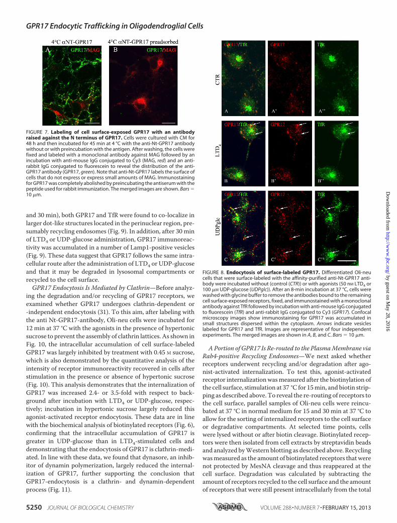
staining for the anti-Nt-GPR17 was still largely detected at thecell surface, although the labeling appeared more clustered,suggesting that receptor stimulation results in a more efficientrecruitment of the receptor to endocytic patches and vesicles.At later times (8 and 12 min at 37 °C), an increasing number ofdot-like structures that were immunolabeled for GPR17appeared concentrated in the cytoplasm of cells following theadministration of agonists. We traced the endocytic route ofGPR17 by double-labeling with antibodies against proteinmarkers of early/recycling endosomes (TfR) or lysosomes(Lamp1). As shown in Fig. 8, after 8 min of endocytosis, GPR17partially co-localized with TfR in small vesicles that were dis-persed throughout the cytoplasm, whereas at later times (15
FIGURE 6. Agonist-induced internalization of GPR17. Internalization of GPR17 was measured in differentiated Oli-neu cells after cell surface labeling at 4 °Cwith the thiol-cleavable sulfo-NHS-SS-biotin. A, after labeling, cells were washed without (�) or with (�) DTT to remove the biotin from surface, and cell extracts(250 �g) were incubated with streptavidin. The proteins bound to streptavidin beads (B) and 5% of the cell extracts used for streptavidin-bead incubation (C.Ex)and also 5% of the supernatants from the streptavidin incubation (unbound proteins (Ub)) were analyzed by Western blotting with antibodies against TfR,GPR17, and actin. B, after labeling, Oli-neu cells were incubated for 15 min at 37 °C to allow for receptor internalization and then incubated without (�) or with(�) DTT. The cell extracts were analyzed as described above. In C, quantitative analysis of Western blots revealed that 10.82 � 6.63% of the total biotinylatedGPR17 underwent internalization under basal conditions. Data are from three independent experiments (the mean � S.E.). ***, p � 0.0001. In D and E, afterlabeling cells were extracted or incubated with either UDP-glucose (100 �M, UDPg) or LTD4 (50 nM). At selected time points, the cells were cooled at 4 °C,incubated with the membrane-impermeable reducing agent MesNA to remove biotin from the remaining cell surface receptors, and solubilized. Aliquots (250�g) of cell extracts were incubated with streptavidin beads and proteins bound to streptavidin (E) and also 10 �g of total cell extracts (D) were analyzed byWestern blotting with antibodies against GPR17. F, after biotin labeling, Oli-neu cells were incubated at 37 °C in the absence (�) or presence of UDP-glucose,UDP-glucose (UDPg) and cangrelor (10 �M, Cng), LTD4, or LTD4 and montelukast (1 �M, MTL). After 15 min, cells were cooled at 4 °C, treated with reducingagents, and solubilized. Cell extracts (200 �g) were incubated with streptavidin beads, and bound proteins were analyzed by Western blotting with anti-GPR17antibodies. The image shows representative blots from three independent experiments. G, graphs represent the quantitative analysis of blots, and the data areexpressed as the percentage of biotinylated GPR17 internalized in the absence of agonists (�) and represent the mean � S.E. (*, p � 0.05; **, p � 0.01,two-tailed, non-paired Student’s t test).
GPR17 Endocytic Trafficking in Oligodendroglial Cells
FEBRUARY 15, 2013 • VOLUME 288 • NUMBER 7 JOURNAL OF BIOLOGICAL CHEMISTRY 5249
by guest on May 28, 2016
http://ww
w.jbc.org/
Dow
nloaded from
and 30 min), both GPR17 and TfR were found to co-localize inlarger dot-like structures located in the perinuclear region, pre-sumably recycling endosomes (Fig. 9). In addition, after 30 minof LTD4 or UDP-glucose administration, GPR17 immunoreac-tivity was accumulated in a number of Lamp1-positive vesicles(Fig. 9). These data suggest that GPR17 follows the same intra-cellular route after the administration of LTD4 or UDP-glucoseand that it may be degraded in lysosomal compartments orrecycled to the cell surface.GPR17 Endocytosis Is Mediated by Clathrin—Before analyz-
ing the degradation and/or recycling of GPR17 receptors, weexamined whether GPR17 undergoes clathrin-dependent or-independent endocytosis (31). To this aim, after labeling withthe anti Nt-GPR17-antibody, Oli-neu cells were incubated for12 min at 37 °C with the agonists in the presence of hypertonicsucrose to prevent the assembly of clathrin lattices. As shown inFig. 10, the intracellular accumulation of cell surface-labeledGPR17 was largely inhibited by treatment with 0.45 M sucrose,which is also demonstrated by the quantitative analysis of theintensity of receptor immunoreactivity recovered in cells afterstimulation in the presence or absence of hypertonic sucrose(Fig. 10). This analysis demonstrates that the internalization ofGPR17 was increased 2.4- or 3.5-fold with respect to back-ground after incubation with LTD4 or UDP-glucose, respec-tively; incubation in hypertonic sucrose largely reduced thisagonist-activated receptor endocytosis. These data are in linewith the biochemical analysis of biotinylated receptors (Fig. 6),confirming that the intracellular accumulation of GPR17 isgreater in UDP-glucose than in LTD4-stimulated cells anddemonstrating that the endocytosis of GPR17 is clathrin-medi-ated. In line with these data, we found that dynasore, an inhib-itor of dynamin polymerization, largely reduced the internal-ization of GPR17, further supporting the conclusion thatGPR17-endocytosis is a clathrin- and dynamin-dependentprocess (Fig. 11).
APortion of GPR17 Is Re-routed to the PlasmaMembrane viaRab4-positive Recycling Endosomes—We next asked whetherreceptors underwent recycling and/or degradation after ago-nist-activated internalization. To test this, agonist-activatedreceptor internalizationwasmeasured after the biotinylation ofthe cell surface, stimulation at 37 °C for 15min, and biotin strip-ping as described above. To reveal the re-routing of receptors tothe cell surface, parallel samples of Oli-neu cells were reincu-bated at 37 °C in normal medium for 15 and 30 min at 37 °C toallow for the sorting of internalized receptors to the cell surfaceor degradative compartments. At selected time points, cellswere lysed without or after biotin cleavage. Biotinylated recep-tors were then isolated from cell extracts by streptavidin beadsand analyzed byWestern blotting as described above. Recyclingwasmeasured as the amount of biotinylated receptors thatwerenot protected by MesNA cleavage and thus reappeared at thecell surface. Degradation was calculated by subtracting theamount of receptors recycled to the cell surface and the amountof receptors that were still present intracellularly from the total
FIGURE 7. Labeling of cell surface-exposed GPR17 with an antibodyraised against the N terminus of GPR17. Cells were cultured with CM for48 h and then incubated for 45 min at 4 °C with the anti-Nt-GPR17 antibodywithout or with preincubation with the antigen. After washing, the cells werefixed and labeled with a monoclonal antibody against MAG followed by anincubation with anti-mouse IgG conjugated to Cy3 (MAG, red) and an anti-rabbit IgG conjugated to fluorescein to reveal the distribution of the anti-GPR17 antibody (GPR17, green). Note that anti-Nt-GPR17 labels the surface ofcells that do not express or express small amounts of MAG. Immunostainingfor GPR17 was completely abolished by preincubating the antiserum with thepeptide used for rabbit immunization. The merged images are shown. Bars 10 �m.
FIGURE 8. Endocytosis of surface-labeled GPR17. Differentiated Oli-neucells that were surface-labeled with the affinity-purified anti-Nt-GPR17 anti-body were incubated without (control (CTR)) or with agonists (50 nM LTD4 or100 �M UDP-glucose (UDPglc)). After an 8-min incubation at 37 °C, cells werewashed with glycine buffer to remove the antibodies bound to the remainingcell surface-exposed receptors, fixed, and immunostained with a monoclonalantibody against TfR followed by incubation with anti-mouse IgG conjugatedto fluorescein (TfR) and anti-rabbit IgG conjugated to Cy3 (GPR17). Confocalmicroscopy images show immunostaining for GPR17 was accumulated insmall structures dispersed within the cytoplasm. Arrows indicate vesicleslabeled for GPR17 and TfR. Images are representative of four independentexperiments. The merged images are shown in A, B, and C. Bars 10 �m.
GPR17 Endocytic Trafficking in Oligodendroglial Cells
5250 JOURNAL OF BIOLOGICAL CHEMISTRY VOLUME 288 • NUMBER 7 • FEBRUARY 15, 2013
by guest on May 28, 2016
http://ww
w.jbc.org/
Dow
nloaded from

amount of internalized receptors after agonist administration(time 0). The results clearly indicate that a significant amount ofGPR17 is recycled to the plasma membrane (Fig. 12). Densito-metric analysis ofWestern blots from three independent exper-iments demonstrated that although �50–45% of endocytosedGPR17 underwent degradation after agonist administration, aconsistent portion of the receptor (�25 and �30% after LTD4or UDP-glucose, respectively) was also recycled to the cell sur-face (Fig. 12). The amounts of recycled or degraded receptorswere not significantly different after stimulation with eitheragonist.To further characterize the recycling pathways taken by
GPR17,we compared its distribution to that of the smallG-pro-
teins Rab4, Rab5, and Rab11 at different times after the admin-istration of agonists. A number of important studies have dem-onstrated the relevance of these Rab proteins as regulators ofthe recycling/endosomal compartments. It has also been dem-onstrated that early/sorting endosomes in the cell peripherycontain primarily Rab5 and Rab4, whereas recycling endo-somes in the perinuclear area contain primarily Rab4 or Rab11(32–35). To investigate the distribution of GPR17 and Rab pro-teins, we transfected Oli-neu cells with cDNAs (0.5–0.2 �g)encoding for Rab4a, Rab5a, or Rab11a bearing a green fluores-cent protein EGFP tag at theN terminus. After transfection, the
FIGURE 9. GPR17 is accumulated in TfR- or Lamp1-positive compartmentsafter 15 and 30 min of exposure to agonists. Differentiated Oli-neu cellswere labeled with anti-Nt-GPR17 antibodies and incubated for 15 and 30 minat 37 °C with agonists (LTD4 or UDP-glucose (UDPglc)). After glycine washing,cells were fixed and labeled with monoclonal antibodies against TfR or Lamp1followed by incubation with anti-rabbit IgG conjugated to Cy3 (GPR, red) andanti-mouse or anti-rat IgG conjugated to fluorescein (TfR or LAMP, green).Merged images represent individual confocal sections (0.5 �M) and are rep-resentative of four independent experiments. Note a subset of cytoplasmicstructures co-labeled (yellow) for GPR17 and TfR or Lamp1. Bar 10 �m.
FIGURE 10. GPR17 is internalized via clathrin-dependent endocytosis. InA, differentiated Oli-neu cells were surface-labeled with anti-Nt-GPR17 anti-bodies and then incubated for 12 min at 37 °C without (control (CTR)) or withagonists (LTD4 or UDP-glucose (UDPglc)) in Sato medium minus (�) or sup-plemented (�) with 450 mM sucrose. After glycine washing, cells were fixedand labeled with monoclonal antibodies against Lamp1 followed by incuba-tion with anti-rat IgG conjugated to fluorescein (LAMP) and anti-rabbit IgGconjugated to Cy3 (GPR17). Confocal microscopy images show no accumula-tion of GPR17 immunostaining in the cells incubated in hypertonic sucrose.Images are representative of three independent experiments. The mergedimages are shown. Bar 10 �m. B, to quantify the effects of clathrin latticeinhibition on the endocytosis of GPR17, confocal images were collected fromOli-neu cells incubated with or without hypertonic sucrose (�), and the pixelintensity was determined by ImageJ software. The graph represents theincrease of GPR17 immunoreactivity in the cells over background (back-ground the immunofluorescence detected in the cells after labeling at 4 °Cfollowed by acid stripping). The values are the mean (� S.E.) of three inde-pendent experiments. *, p 0.05; **, p � 0.01; ***, p � 0.0001 (two-tailed,non-paired Student’s t test).
GPR17 Endocytic Trafficking in Oligodendroglial Cells
FEBRUARY 15, 2013 • VOLUME 288 • NUMBER 7 JOURNAL OF BIOLOGICAL CHEMISTRY 5251
by guest on May 28, 2016
http://ww
w.jbc.org/
Dow
nloaded from

cells were differentiatedwithCM for 48 h and then processed asdescribed above to investigate the internalization and intracel-lular distribution of GPR17. Cells expressingmoderate levels ofGFP-Rabs (�5% of total cells) were selected for analysis. Underthis condition, Rab5, Rab4, and Rab11 show an intracellular
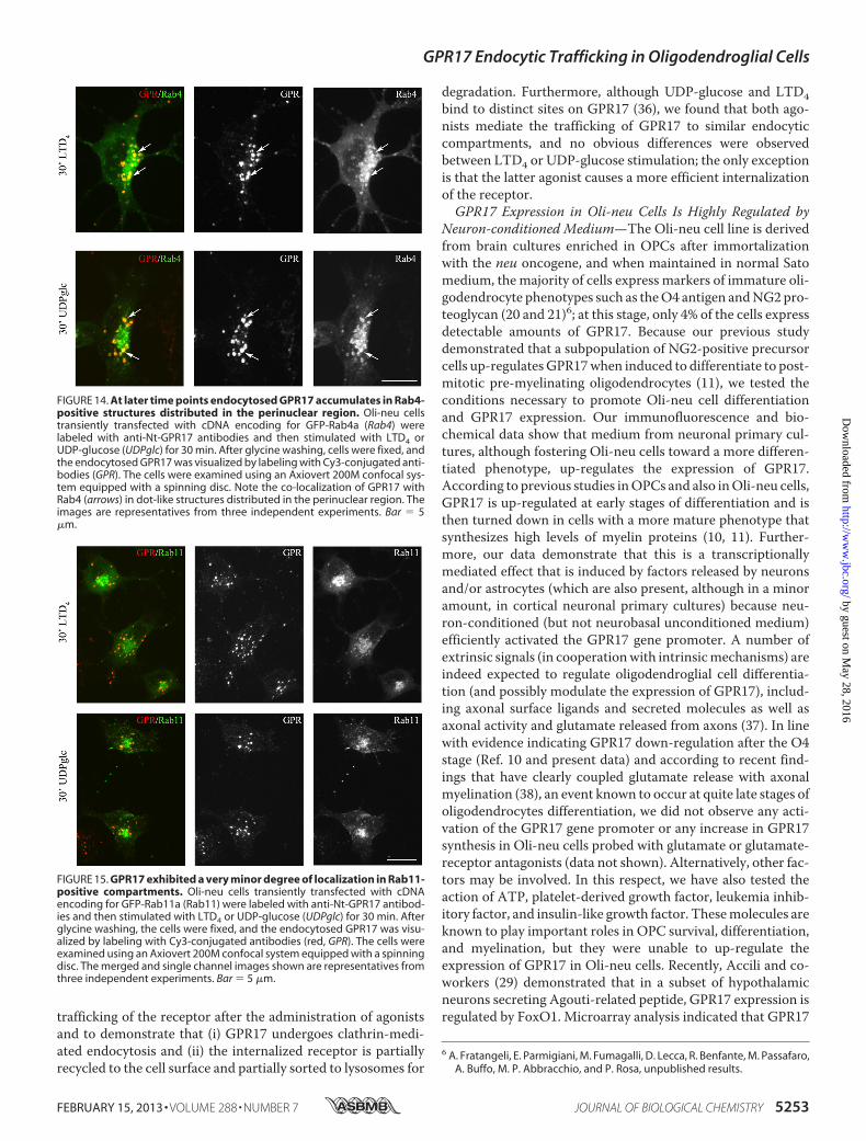
distribution similar to that previously reported (32). Based onthe confocal images (Fig. 13), GPR17 largely co-localized withGFP-Rab5 and Rab4 in small vesicles that were primarily scat-tered in the cytoplasm or located at the periphery of the cells atearly times of internalization. At later times, GPR17 partiallyco-localized with Rab4 primarily in large, dot-like structuresthat were distributed in the perinuclear region (Fig. 14). A veryminor co-localization of GPR17 with Rab11a could also bedetected, as reported in Fig. 15. Altogether, these data supportthe conclusion that a portion of GPR17 is also recycled to theplasma membrane after internalization upon agonist stimula-tion through a fast recycling loop as revealed by co-localizationwith Rab4.
DISCUSSION
To investigate the intracellular trafficking of GPR17 in anative expressionmodel, we have first characterized its expres-sion in Oli-neu cells, an immortalized OPC cell line, andshowed thatmediumderived fromcortical neuron primary cul-tures stimulates the transient expression of GPR17, as previ-ously observed in maturing cultured OPCs (2, 7, 10, 11). UsingOli-neu cells, we were then able to investigate the endocytic
FIGURE 11. Dynamin inhibition affects GPR17 endocytosis. Oli-neu cellswere labeled with anti-Nt-GPR17 antibodies, pretreated at 4 °C with or with-out the dynamin inhibitor dynasore, and then incubated at 37 °C for 12 min inSato medium (CTR) or medium supplemented with dynasore and UDP-glu-cose or LTD4. After fixation, cells were double-immunolabeled with monoclo-nal antibodies against Lamp1 followed by incubation with anti-rat IgG conju-gated to fluorescein (LAMP, green) and anti-rabbit IgG conjugated to Cy3(GPR17, red). Note the large reduction of GPR17 internalization in Oli-neuincubated with the inhibitor of dynamin (Bar 20 �m).
FIGURE 12. Recycling and degradation of GPR17 after agonist-inducedendocytosis. Oli-neu cells after cell surface labeling with sulfo-NHS-SS-biotinat 4 °C were incubated at 37 °C with UDP-glucose (100 �M, UDPglc) or LTD4 (50nM). After 15 min, cells were cooled at 4 °C, incubated with MesNA to removebiotin from the remaining cell surface receptors, and solubilized (time 0) orfurther incubated in normal medium. At selected times, cells were either sol-ubilized (�) or re-incubated with MesNA to remove biotinylated receptorsre-exposed to the cell surface. In A aliquots (200 �g) of cell extracts wereincubated with streptavidin beads, and the proteins bound to streptavidinwere analyzed by Western blotting with antibodies against GPR17. In B,graphs represent the quantitative analysis of GPR17 recycling and degrada-tion, which is reported as a percentage of the amount of receptor internalizedafter 15 min at 37 °C with LTD4 or UDP-glucose; the values are the mean offour independent experiments �S.E.
FIGURE 13. At early time points endocytosed GPR17 accumulates in Rab5-or Rab4-positive vesicles. Oli-neu cells transiently transfected with cDNAencoding for GFP-Rab5a (Rab5) or GFP-Rab4a (Rab4) were labeled with anti-Nt-GPR17 antibodies and then stimulated with LTD4 or UDP-glucose (UDPglc)for 15 min. After glycine washing, the cells were fixed, and the endocytosedGPR17 was visualized by labeling with Cy3-conjugated antibodies (red). Thecells were then examined using an Axiovert 200M confocal system equippedwith a spinning disc. Note the co-localization of Rab proteins with GPR17(arrows) in small vesicles scattered throughout the cytoplasm. The images arerepresentatives from three independent experiments. Bar 5 �m.
GPR17 Endocytic Trafficking in Oligodendroglial Cells
5252 JOURNAL OF BIOLOGICAL CHEMISTRY VOLUME 288 • NUMBER 7 • FEBRUARY 15, 2013
by guest on May 28, 2016
http://ww
w.jbc.org/
Dow
nloaded from
trafficking of the receptor after the administration of agonistsand to demonstrate that (i) GPR17 undergoes clathrin-medi-ated endocytosis and (ii) the internalized receptor is partiallyrecycled to the cell surface and partially sorted to lysosomes for
degradation. Furthermore, although UDP-glucose and LTD4bind to distinct sites on GPR17 (36), we found that both ago-nists mediate the trafficking of GPR17 to similar endocyticcompartments, and no obvious differences were observedbetween LTD4 or UDP-glucose stimulation; the only exceptionis that the latter agonist causes a more efficient internalizationof the receptor.GPR17 Expression in Oli-neu Cells Is Highly Regulated by
Neuron-conditioned Medium—The Oli-neu cell line is derivedfrom brain cultures enriched in OPCs after immortalizationwith the neu oncogene, and when maintained in normal Satomedium, the majority of cells express markers of immature oli-godendrocyte phenotypes such as theO4 antigen andNG2pro-teoglycan (20 and 21)6; at this stage, only 4% of the cells expressdetectable amounts of GPR17. Because our previous studydemonstrated that a subpopulation of NG2-positive precursorcells up-regulatesGPR17when induced to differentiate to post-mitotic pre-myelinating oligodendrocytes (11), we tested theconditions necessary to promote Oli-neu cell differentiationand GPR17 expression. Our immunofluorescence and bio-chemical data show that medium from neuronal primary cul-tures, although fostering Oli-neu cells toward a more differen-tiated phenotype, up-regulates the expression of GPR17.According to previous studies inOPCs and also inOli-neu cells,GPR17 is up-regulated at early stages of differentiation and isthen turned down in cells with a more mature phenotype thatsynthesizes high levels of myelin proteins (10, 11). Further-more, our data demonstrate that this is a transcriptionallymediated effect that is induced by factors released by neuronsand/or astrocytes (which are also present, although in a minoramount, in cortical neuronal primary cultures) because neu-ron-conditioned (but not neurobasal unconditioned medium)efficiently activated the GPR17 gene promoter. A number ofextrinsic signals (in cooperationwith intrinsicmechanisms) areindeed expected to regulate oligodendroglial cell differentia-tion (and possibly modulate the expression of GPR17), includ-ing axonal surface ligands and secreted molecules as well asaxonal activity and glutamate released from axons (37). In linewith evidence indicating GPR17 down-regulation after the O4stage (Ref. 10 and present data) and according to recent find-ings that have clearly coupled glutamate release with axonalmyelination (38), an event known to occur at quite late stages ofoligodendrocytes differentiation, we did not observe any acti-vation of the GPR17 gene promoter or any increase in GPR17synthesis in Oli-neu cells probed with glutamate or glutamate-receptor antagonists (data not shown). Alternatively, other fac-tors may be involved. In this respect, we have also tested theaction of ATP, platelet-derived growth factor, leukemia inhib-itory factor, and insulin-like growth factor. Thesemolecules areknown to play important roles in OPC survival, differentiation,and myelination, but they were unable to up-regulate theexpression of GPR17 in Oli-neu cells. Recently, Accili and co-workers (29) demonstrated that in a subset of hypothalamicneurons secreting Agouti-related peptide, GPR17 expression isregulated by FoxO1. Microarray analysis indicated that GPR17
6 A. Fratangeli, E. Parmigiani, M. Fumagalli, D. Lecca, R. Benfante, M. Passafaro,A. Buffo, M. P. Abbracchio, and P. Rosa, unpublished results.
FIGURE 14. At later time points endocytosed GPR17 accumulates in Rab4-positive structures distributed in the perinuclear region. Oli-neu cellstransiently transfected with cDNA encoding for GFP-Rab4a (Rab4) werelabeled with anti-Nt-GPR17 antibodies and then stimulated with LTD4 orUDP-glucose (UDPglc) for 30 min. After glycine washing, cells were fixed, andthe endocytosed GPR17 was visualized by labeling with Cy3-conjugated anti-bodies (GPR). The cells were examined using an Axiovert 200M confocal sys-tem equipped with a spinning disc. Note the co-localization of GPR17 withRab4 (arrows) in dot-like structures distributed in the perinuclear region. Theimages are representatives from three independent experiments. Bar 5�m.
FIGURE 15. GPR17 exhibited a very minor degree of localization in Rab11-positive compartments. Oli-neu cells transiently transfected with cDNAencoding for GFP-Rab11a (Rab11) were labeled with anti-Nt-GPR17 antibod-ies and then stimulated with LTD4 or UDP-glucose (UDPglc) for 30 min. Afterglycine washing, the cells were fixed, and the endocytosed GPR17 was visu-alized by labeling with Cy3-conjugated antibodies (red, GPR). The cells wereexamined using an Axiovert 200M confocal system equipped with a spinningdisc. The merged and single channel images shown are representatives fromthree independent experiments. Bar 5 �m.
GPR17 Endocytic Trafficking in Oligodendroglial Cells
FEBRUARY 15, 2013 • VOLUME 288 • NUMBER 7 JOURNAL OF BIOLOGICAL CHEMISTRY 5253
by guest on May 28, 2016
http://ww
w.jbc.org/
Dow
nloaded from

is highly decreased in FoxO1-deficient AgRP neurons, whereasFoxO1 overexpression increased the expression of GPR17mRNA in neuro2A neuroblastoma cells. Our present results,however, indicate that the mechanism(s) underlying GPR17expression in Oli-neu is different from that reported for AgRPneurons and FoxO1 does not appear to be involved. This con-clusion comes from three observations: (i) the amount ofFoxO1 inOli-neu is very low even after incubationwithCM, (ii)its knockdown does not reduce GPR17-expression, and (iii) itsoverexpression (even of the constitutively active forms) doesnot induce expression of GPR17. Although FoxO1 does notregulate GPR17 in Oli-neu, FoxO proteins may have an impor-tant function in OPC proliferation and oligodendrogenesis.Recent studies have indeed demonstrated that after hypoxiathese transcription factors, mainly FoxO1, can affect theexpression of p27Kip1, a key regulator of oligodendrogenesis(39).Concerning Oli-neu, we cannot exclude the possibility that
factors present in the CM could counteract the signaling of theneu-oncogene, which has been used to immortalize the precur-sor cells isolated from E16mouse brains and foster their prolif-eration (20). Exit from themitotic cycle may trigger the expres-sion of GPR17 and promote differentiation as has beenreported for OPCs (10, 11). This hypothesis is supported by theobservation that an erbB inhibitor (PD174,265) induced theexpression of GPR17 and the rapid (24 h) differentiation ofOli-neu cells (data not shown). Further work is required toidentify additional molecules that may promote GPR17 geneactivation and receptor protein synthesis.Agonist-dependent GPR17 Trafficking—Our previous work
has shown that GPR17 is a “dualistic” receptor that is activatedby uracil nucleotides and cysLTs (1, 2, 10).Moreover, studies inheterologously transfected cells demonstrated that, similar toother GPCRs, recombinant GPR17 can undergo a loss of func-tion upon prolonged exposure to its endogenous agonists asshown by desensitization, i.e. the inability to further respond toagonist stimulation, followed by re-sensitization upon agonistremoval (6). However, the relevance of these findings to theregulation of native GPR17 in physiological systems has notbeen investigated previously. Moreover, in our previous study,the role of endocytic trafficking in agonist-induced GPR17desensitization/re-sensitization was only marginally analyzed.The dynamics of intracellular trafficking of the receptor is, nev-ertheless, physiologically relevant, as it modulates receptor lev-els at the cell surface (12, 14–16, 40). The internalization andsubsequent sorting of the receptor into degradative or recyclingcompartments may have important implications for the activa-tion/silencing of signaling pathway(s). In the case of GPR17, itmay represent a key event necessary to achieve GPR17 down-regulation and in turn to allow immature oligodendroglial cellsto proceed to a more mature phenotype. A similar process hasbeen associated with the differentiation of erythrocytes, wherethe down-regulation of erythropoietin receptors via lysosomaldegradation has been proposed to be necessary to allow cells toproceed toward terminal maturation (17). Therefore, it wasimportant to investigate in detail the trafficking of GPR17 in amore physiological model such as differentiating Oli-neu cells.Furthermore, given the dualistic nature of GPR17 and the likely
presence of two distinct binding sites, one for uracil nucleotidesand one for cysLTs, in its structure (36), it was important toverify whether the two distinct classes of natural agonists mod-ulate the endocytic pathways taken by the receptor in differentways.In this study we demonstrate that native GPR17 in Oli-neu
cells is internalized in a time- and dose-dependentmanner afterthe administration of agonists, although the efficacy of LTD4appeared to be less than that of UDP-glucose. This conclusionis supported by quantitative analysis performed by using bio-chemical (biotinylation of cell surface receptors) and immuno-cytochemical (immunolabeling of cell surface receptors with aspecific antibody) approaches aimed at revealing the internal-ization of GPR17. Moreover, we demonstrate that, at least inour cell system, LTD4 acts by directly activatingGPR17 and notvia the CysLT1 receptor; this conclusion is based on the findingthat this receptor is not expressed in Oli-neu cells. These dataare in line with our previous data obtained in a heterologousexpression system in which CysLTR1 is also not expressed (6),but we cannot exclude the possibility that GPR17 may be dif-ferentially regulated and, therefore, act as a negative regulatorof CysLTR1 when both receptors are co-expressed in the samesystem as previously reported (18). However, this possibilitywill never occur in OPCs and pre-oligodendrocytes whereCyLTR1 has not been detected (10), thus further confirmingthat in oligodendroglial cells, cysLTs act by directly promotingthe activation of GPR17. Finally, the mechanisms and func-tional consequences of this difference in the internalization ofGPR17 after exposure to either uracil nucleotides or cysLTs arenot yet known. Differences in the efficiency of receptor inter-nalization may be due to differences in binding to arrestinsand/or the activation of different intracellular signals.Althoughwedid observe somequantitative differences in the
efficiency of internalization, we did not observe any significantdifferences in the pathway taken by internalized receptors afterUDP or LTD4 stimulation. Using a protocol that has allowed usto visualize the fate of internalized receptors in endocytic com-partments by means of confocal microscopy analysis, we dem-onstrated that GPR17 is internalized via clathrin-mediatedendocytosis after both LTD4 and UDP-glucose administration.At early times after internalization, the receptor reaches earlyendosomes independent of the agonist used for stimulation. Atlater times, it accumulated in dot-like structures and partiallyco-localized with a marker of lysosomes, thus suggesting thatboth agonists stimulated receptor degradation. This conclusionis also supported by the quantification of the amounts of bioti-nylated receptor recovered in cells after stimulation and agonistretrieval. In addition, these experiments indicate that althoughan aliquot of the receptor is degraded, a consistent portion of itis still recycled to the cell surface. Furthermore, using an immu-nocytochemical approach, we investigated whether internal-ized GPR17 is recycled via a rapid or slow recycling loop bycomparing the distribution of GPR17 with that of the Rab pro-teins involved in endocytic traffic. After transfection inOli-neucells, Rab proteins exhibited a localization similar to thatreported previously (32). Rab5 was mainly concentrated in ves-icles that were dispersed in the cytoplasm, and Rab4 waslocated in small vesicles and in dotted structures at the perinu-
GPR17 Endocytic Trafficking in Oligodendroglial Cells
5254 JOURNAL OF BIOLOGICAL CHEMISTRY VOLUME 288 • NUMBER 7 • FEBRUARY 15, 2013
by guest on May 28, 2016
http://ww
w.jbc.org/
Dow
nloaded from

clear region, whereas given its distribution and cycling in thetrans-Golgi-network, Rab11wasmainly detected in the perinu-clear region. Our results demonstrate that GPR17 is trans-ported into the early/sorting endosomes and can be recycled tothe membrane via the fast recycling loop. This conclusion issupported by the observation that soon after internalization,GPR17 co-localized with Rab5 and Rab4 in a compartment atthe periphery of the cell. At later times, GPR17 also accumu-lated in a Rab4-positive compartment known to be involved inthe rapid recycling loop. Only a very small amount of co-local-izationwas observedwith Rab11, amarker of the slow recyclingloop.In conclusion, we demonstrate that after agonist activation,
GPR17 undergoes degradation and recycling. The balance ofreceptor sorting into recycling and/or degradative compart-mentsmay have important implications in the down-regulationof GPR17 signaling pathway(s) in differentiating OPCs.
Acknowledgments—We aremost grateful to Jacqueline Trotter for theOli-neu cells and Cecilia Bucci and Bice Chini for the Rab constructs.Special thanks to theMonzino Foundation,Milan, Italy, for the gift ofthe Zeiss LSM510 Meta confocal microscope and the Axiovert 200Mconfocal system equipped with a spinning disc (PerkinElmer LifeSciences).
REFERENCES1. Ciana, P., Fumagalli, M., Trincavelli, M. L., Verderio, C., Rosa, P., Lecca,
D., Ferrario, S., Parravicini, C., Capra, V., Gelosa, P., Guerrini, U., Bel-credito, S., Cimino, M., Sironi, L., Tremoli, E., Rovati, G. E., Martini, C.,and Abbracchio, M. P. (2006) The orphan receptor GPR17 identified as anew dual uracil nucleotides/cysteinyl-leukotrienes receptor. EMBO J. 25,4615–4627
2. Lecca, D., Trincavelli, M. L., Gelosa, P., Sironi, L., Ciana, P., Fumagalli, M.,Villa, G., Verderio, C., Grumelli, C., Guerrini, U., Tremoli, E., Rosa, P.,Cuboni, S., Martini, C., Buffo, A., Cimino, M., and Abbracchio, M. P.(2008)The recently identified P2Y-like receptorGPR17 is a sensor of braindamage and a new target for brain repair. PLoS One 3, e3579
3. Parravicini, C., Ranghino, G., Abbracchio, M. P., and Fantucci, P. (2008)GPR17. Molecular modeling and dynamics studies of the 3-D structureand purinergic ligand binding features in comparison with P2Y receptors.BMC Bioinformatics 9, 263–282
4. Pugliese, A. M., Trincavelli, M. L., Lecca, D., Coppi, E., Fumagalli, M.,Ferrario, S., Failli, P., Daniele, S., Martini, C., Pedata, F., and Abbracchio,M. P. (2009) Functional characterization of two isoforms of the P2Y-likereceptor GPR17. [35S]GTP�S binding and electrophysiological studies in1321N1 cells. Am. J. Physiol. Cell. Physiol. 297, C1028–C1040
5. Daniele, S., Lecca, D., Trincavelli, M. L., Ciampi, O., Abbracchio, M. P.,andMartini, C. (2010) Regulation of PC12 cell survival and differentiationby the new P2Y-like receptor GPR17. Cell. Signal. 22, 697–706
6. Daniele, S., Trincavelli, M. L., Gabelloni, P., Lecca, D., Rosa, P., Abbrac-chio,M. P., andMartini, C. (2011)Agonist-induced desensitization/resen-sitization of human G protein-coupled receptor 17. A functional cross-talk between purinergic and cysteinyl-leukotriene ligands. J. Pharmacol.Exp. Ther. 338, 559–567
7. Chen, Y.,Wu, H.,Wang, S., Koito, H., Li, J., Ye, F., Hoang, J., Escobar, S. S.,Gow, A., Arnett, H. A., Trapp, B. D., Karandikar, N. J., Hsieh, J., and Lu,Q. R. (2009) The oligodendrocyte-specific G protein-coupled receptorGPR17 is a cell-intrinsic timer of myelination. Nat. Neurosci. 12,1398–1406
8. Ceruti, S., Villa, G., Genovese, T., Mazzon, E., Longhi, R., Rosa, P., Bra-manti, P., Cuzzocrea, S., and Abbracchio, M. P. (2009) The P2Y-like re-ceptor GPR17 as a sensor of damage and a new potential target in spinalcord injury. Brain 132, 2206–2218
9. Ceruti, S., Viganò, F., Boda, E., Ferrario, S., Magni, G., Boccazzi, M., Rosa,P., Buffo, A., and Abbracchio, M. P. (2011) Expression of the new P2Y-likereceptor GPR17 during oligodendrocyte precursor cell maturation regu-lates sensitivity to ATP-induced death. Glia 59, 363–378
10. Fumagalli, M., Daniele, S., Lecca, D., Lee, P. R., Parravicini, C., Fields,R. D., Rosa, P., Antonucci, F., Verderio, C., Trincavelli, M. L., Bramanti,P., Martini, C., and Abbracchio, M. P. (2011) Phenotypic changes, sig-naling pathway, and functional correlates of GPR17-expressing neuralprecursor cells during oligodendrocyte differentiation. J. Biol. Chem.286, 10593–10604
11. Boda, E., Viganò, F., Rosa, P., Fumagalli, M., Labat-Gest, V., Tempia, F.,Abbracchio,M. P., Dimou, L., and Buffo, A. (2011) TheGPR17 receptor inNG2 expressing cells. Focus on in vivo cellmaturation and participation inacute trauma and chronic damage. Glia 59, 1958–1973
12. Ferguson, S. S. (2001) Evolving concepts in G protein-coupled receptorendocytosis. The role in receptor desensitization and signaling. Pharma-col. Rev. 53, 1–24
13. Marchese, A., Paing, M. M., Temple, B. R., and Trejo, J. (2008) G protein-coupled receptor sorting to endosomes and lysosomes. Annu. Rev. Phar-macol. Toxicol. 48, 601–629
14. Dunham, J. H., and Hall, R. A. (2009) Enhancement of the surface expres-sion of G protein-coupled receptors. Trends Biotechnol. 27, 541–545
15. Ritter, S. L., and Hall, R. A. (2009) Fine-tuning of GPCR activity by recep-tor-interacting proteins. Nat. Rev. Mol. Cell Biol. 10, 819–830
16. Sorkin, A., and von Zastrow, M. (2009) Endocytosis and signaling. Inter-twining molecular networks. Nat. Rev. Mol. Cell Biol. 10, 609–622
17. Walrafen, P., Verdier, F., Kadri, Z., Chrétien, S., Lacombe, C., andMayeux,P. (2005) Both proteasomes and lysosomes degrade the activated erythro-poietin receptor. Blood 105, 600–608
18. Maekawa, A., Balestrieri, B., Austen, K. F., and Kanaoka, Y. (2009) GPR17is a negative regulator of the cysteinyl leukotriene 1 receptor response toleukotriene D4. Proc. Natl. Acad. Sci. U.S.A. 106, 11685–11690
19. Benned-Jensen, T., and Rosenkilde, M. M. (2010) Distinct expression andligand binding profiles of two constitutively active GPR17 splice variants.Br. J. Pharmacol. 159, 1092–1105
20. Jung, M., Krämer, E., Grzenkowski, M., Tang, K., Blakemore, W., Aguzzi,A., Khazaie, K., Chlichlia, K., von Blankenfeld, G., and Kettenmann, H.(1995) Lines of murine oligodendroglial precursor cells immortalized byan activated neu tyrosine kinase show distinct degrees of interaction withaxons in vitro and in vivo. Eur. J. Neurosci. 7, 1245–1265
21. Winterstein, C., Trotter, J., and Krämer-Albers, E. M. (2008) Distinct en-docytic recycling of myelin proteins promotes oligodendroglial mem-brane remodeling. J. Cell Sci. 121, 834–842
22. Taverna, E., Saba, E., Linetti, A., Longhi, R., Jeromin, A., Righi, M., Clem-enti, F., and Rosa, P. (2007) Localization of synaptic proteins involved inneurosecretion in different membranemicrodomains. J. Neurochem. 100,664–677
23. Passafaro, M., Piëch, V., and Sheng, M. (2001) Subunit-specific temporaland spatial patterns of AMPA receptor exocytosis in hippocampal neu-rons. Nat. Neurosci. 4, 917–926
24. Bucci, C., Lütcke, A., Steele-Mortimer, O., Olkkonen, V. M., Dupree, P.,Chiariello,M., Bruni, C. B., Simons, K., and Zerial,M. (1995) Co-operativeregulation of endocytosis by three Rab5 isoforms. FEBS Lett. 366, 65–71
25. Conti, F., Sertic, S., Reversi, A., and Chini, B. (2009) Intracellular traffick-ing of the human oxytocin receptor. Evidence of receptor recycling via aRab4/Rab5 “short cycle.” Am. J. Physiol. Endocrinol. Metab. 296,E532–E542
26. Livak, K. J., and Schmittgen, T. D. (2001) Analysis of relative gene expres-sion data using real-time quantitative PCR and the 2(���CT) method.Methods 25, 402–408
27. Linetti, A., Fratangeli, A., Taverna, E., Valnegri, P., Francolini, M.,Cappello, V., Matteoli, M., Passafaro, M., and Rosa, P. (2010) Choles-terol reduction impairs exocytosis of synaptic vesicles. J. Cell Sci. 123,595–605
28. Kippert, A., Trajkovic, K., Fitzner, D., Opitz, L., and Simons, M. (2008)Identification of Tmem10/Opalin as a novel marker for oligodendrocytesusing gene expression profiling. BMC Neurosci. 9, 40–52
29. Ren, H., Orozco, I. J., Su, Y., Suyama, S., Gutiérrez-Juárez, R., Horvath,
GPR17 Endocytic Trafficking in Oligodendroglial Cells
FEBRUARY 15, 2013 • VOLUME 288 • NUMBER 7 JOURNAL OF BIOLOGICAL CHEMISTRY 5255
by guest on May 28, 2016
http://ww
w.jbc.org/
Dow
nloaded from

T. L., Wardlaw, S. L., Plum, L., Arancio, O., and Accili, D. (2012) FoxO1target Gpr17 activates AgRP neurons to regulate food intake. Cell 149,1314–1326
30. Klein, U., and von Zastrow, M. (2000) in Regulation of G-protein-coupledReceptor Function and Expression (Benovic, J. L., ed.) pp. 4–13, NewYork,Wiley-Liss, Inc.
31. Doherty, G. J., and McMahon, H. T. (2009) Mechanisms of endocytosis.Annu. Rev. Biochem. 78, 857–902
32. Sönnichsen, B., De Renzis, S., Nielsen, E., Rietdorf, J., and Zerial,M. (2000)Distinct membrane domains on endosomes in the recycling pathway vi-sualized by multicolor imaging of Rab4, Rab5, and Rab11. J. Cell Biol. 149,901–914
33. Seachrist, J. L., and Ferguson, S. S. (2003) Regulation of G protein-coupledreceptor endocytosis and trafficking byRabGTPases.Life Sci.74, 225–235
34. Platta, H. W., and Stenmark, H. (2011) Endocytosis and signaling. Curr.Opin. Cell Biol. 23, 393–403
35. Zerial, M., andMcBride, H. (2001) Rab proteins as membrane organizers.
Nat. Rev. Mol. Cell Biol. 2, 107–11736. Parravicini, C., Abbracchio, M. P., Fantucci, P., and Ranghino, G. (2010)
Forced unbinding of GPR17 ligands from wild type and R255I mutantreceptormodels through a computational approach.BMCStruct. Biol. 10,8–26
37. Emery, B. (2010) Regulation of oligodendrocyte differentiation andmyeli-nation. Science 330, 779–782
38. Wake, H., Lee, P. R., and Fields, R. D. (2011) Control of local proteinsynthesis and initial events in myelination by action potentials. Science333, 1647–1651
39. Jablonska, B., Scafidi, J., Aguirre, A., Vaccarino, F., Nguyen, V., Borok, E.,Horvath, T. L., Rowitch, D. H., and Gallo V. (2012) Oligodendrocyte re-generation after neonatal hypoxia requires FoxO1-mediated p27Kip1 ex-pression. J. Neurosci. 32, 14775–14793
40. Hanyaloglu, A. C., and von Zastrow, M. (2008) Regulation of GPCRs byendocytic membrane trafficking and its potential implications.Annu. Rev.Pharmacol. Toxicol. 48, 537–568
GPR17 Endocytic Trafficking in Oligodendroglial Cells
5256 JOURNAL OF BIOLOGICAL CHEMISTRY VOLUME 288 • NUMBER 7 • FEBRUARY 15, 2013
by guest on May 28, 2016
http://ww
w.jbc.org/
Dow
nloaded from

Benfante, Maria Passafaro, Annalisa Buffo, Maria P. Abbracchio and Patrizia RosaAlessandra Fratangeli, Elena Parmigiani, Marta Fumagalli, Davide Lecca, Roberta
the P2Y-like Receptor GPR17 in Oli-neu Oligodendroglial CellsThe Regulated Expression, Intracellular Trafficking, and Membrane Recycling of
doi: 10.1074/jbc.M112.404996 originally published online January 3, 20132013, 288:5241-5256.J. Biol. Chem.
10.1074/jbc.M112.404996Access the most updated version of this article at doi:
Alerts:
When a correction for this article is posted•
When this article is cited•
to choose from all of JBC's e-mail alertsClick here
http://www.jbc.org/content/288/7/5241.full.html#ref-list-1
This article cites 39 references, 14 of which can be accessed free at
by guest on May 28, 2016
http://ww
w.jbc.org/
Dow
nloaded from
Related Documents


