Introduction A fine equilibrium of cardiac output and peripheral resistance regulates arterial blood pressure. A number of neurohormonal and mechanical factors participate in the fine tuning of blood pressure, involving the sympathetic nervous system (SNS), which appears to play a pivotal role 1 for its ability to regulate at the same time cardiac output and peripheral vascular tone. e release of norepinephrine, the major SNS neurotransmitter, in neuromuscular synapses at the vascular level produces the activation of two classes of adrenergic receptors (ARs): α 1 ARs that drive to vasoconstriction and βARs, which cause vasodilatation. Under normal physiological conditions vascular tone is a result from a balance between α 1 AR-mediated vasoconstriction and βAR vasodilatation. In hypertension, this balance is shiſted toward increased vasoconstriction presumably due to defective vasodilatation in response to βAR stimulation. Indeed, βAR agonist administration in the human brachial artery causes forearm vasodilatation, and this response is attenuated in hypertension. 2–4 An impaired signal transduction due to a decreased production of second messengers has been observed in several tissues of hypertensive patients and has been advocated as the possible mechanism underlying the blunted βAR vasodilatation. 5 is, by increasing peripheral vascular resistance can play a causal role in hypertension. βAR signaling is highly regulated to allow for a quick and versatile response, which is needed for rapid adjustments of the vascular tone. Desensitization of βARs, that is, the functional uncoupling of the receptor from inducing second messenger is part of this regulation. It allows the rapid shut-off of the signaling and starts a series of events that produce receptor internalization and resensitization. Desensitization of these receptors occurs aſter agonist-induced βAR phosphorylation by a family of Ser/ r kinases, known as G protein-coupled receptor kinases (GRKs). 6–9 e prototype GRK for βARs is GRK2, also known as βAR kinase (βARK). is enzyme is ubiquitous in human tissues, and is abundantly expressed in the heart and in the vessels. 6,10 In transgenic mice, overexpression of GRK2 in cardiovascular tissues causes an impairment of βAR-mediated physiological responses. 11,12 Interestingly, a significant increase of GRK2 expression has been shown in the heart and the vessels in humans during chronic cardiovascular diseases 13,14 and in animal models of hypertension. 15 Because an increase in GRK expression and activity leads to βAR desensitization, it has been hypothesized that the increased GRK2 levels could play a mechanistic role in hypertension. However, there is still a debate on whether vascular βAR desensitization really occurs in physiological conditions in humans. On one hand, it has been demonstrated that in the dorsal vein of the hand, the exposure to the βAR agonist isoproterenol (ISO) for 4 hours leads to attenuation of the vasodilatation. 16 Opposite results were gathered aſter 4 hours of infusion of βAR agonist in the brachial artery, a maneuver that causes a steady vasodilatation, without any evidence of βAR desensitization. 3 Even less is known regarding excessive desensitization as a possible mechanism in hypertension, and the only evidence available has been obtained in experimental models such as genetically manipulated animals. 11 Major limitation for the investigation of this issue in humans is the difficulty to retrieve tissue samples for biochemical assessments. Surrogate specimens, such as peripheral blood lymphocytes represent a valid alternative for biochemical assessments of βAR signaling, given the similarities between lymphocyte and vascular βAR. 6,13 us, to clarify in vivo the occurrence of βAR desensitization and its role, if any, in the control of vascular responses to βAR stimulation, we evaluated βAR signaling in lymphocytes and forearm vasodilatation induced by ISO administration in normotensive subjects and hypertensive patients. Because it is well known that heparin can be used as inhibitor of GRK activity in vitro to lower receptor phosphorylation induced by ISO, 17 we designed an experimental protocol, using this inhibitor, to evaluate the process of in vivo desensitization of ISO-induced vasodilatation. Enhanced GRK2 Expression and Desensitization of βAR Vasodilatation in Hypertensive Patients Raffaele Izzo 1 , Ersilia Cipolletta 1 , Michele Ciccarelli 1 , Alfonso Campanile 1 , Gaetano Santulli 1 , Gianluigi Palumbo 1 , Antonio Vasta 1 , Salvatore Formisano 2 , Bruno Trimarco 1 , and Guido Iaccarino 1 Abstract Increased levels of G protein coupled receptor kinase GRK2 appear to participate in hypertension presumably through the desensitization of β adrenergic receptors (βARs) that mediate vasodilatation. There are contrasting data on the occurrence of βAR desensitization in the vasculature, we therefore investigated βAR vasodilatation and desensitization in normotensives and in hypertensive humans. In blood lymphocytes, we assessed βAR signaling and GRK2 expression and found βAR signaling alterations and, consistent with desensitization, increased GRK2 levels in hypertensives. We studied in vivo vasodilatation to the βAR agonist isoproterenol (ISO) injected in the brachial artery in control conditions and during the concomitant infusion of heparin, a known in vitro nonspecific GRK inhibitor. ISO induced a dose-dependent vasorelaxation that was attenuated in hypertensives indicating a loss of βAR signaling. Intra-arterial infusion of heparin inhibited lymphocyte GRK2 activity and prevented desensitization of βAR vasodilatation in normotensives. In hypertensives, heparin restored vasodilatation to ISO, to levels observed in normotensives. Our results suggest that βAR desensitization does indeed occur at the vascular levels in vivo, and that heparin by acting as a GRK inhibitor prevents this in normotensives and restores impaired βAR vasodilation in hypertensives. We conclude that desensitization participates to impaired βAR vasodilation in hypertension. Keywords: desensitization, vasodilatation, hypertension, peripheral blood lymphocytes, βAR signaling 1 Dipartimento di Medicina Clinica Scienze Cardiovascolari ed Immunologiche; 2 Dipartimento di Biologia e Patologia Cellulare e Molecolare, Federico II University of Naples, Italy. Correspondence: G Iaccarino ([email protected]) DOI: 10.1111/j.1752-8062.2008.00050.x WWW.CTSJOURNAL.COM 215 Volume 1 • Issue 3

Welcome message from author
This document is posted to help you gain knowledge. Please leave a comment to let me know what you think about it! Share it to your friends and learn new things together.
Transcript
IntroductionA fine equilibrium of cardiac output and peripheral resistance regulates arterial blood pressure. A number of neurohormonal and mechanical factors participate in the fine tuning of blood pressure, involving the sympathetic nervous system (SNS), which appears to play a pivotal role1 for its ability to regulate at the same time cardiac output and peripheral vascular tone. The release of norepinephrine, the major SNS neurotransmitter, in neuromuscular synapses at the vascular level produces the activation of two classes of adrenergic receptors (ARs): α1ARs that drive to vasoconstriction and βARs, which cause vasodilatation. Under normal physiological conditions vascular tone is a result from a balance between α1AR-mediated vasoconstriction and βAR vasodilatation. In hypertension, this balance is shifted toward increased vasoconstriction presumably due to defective vasodilatation in response to βAR stimulation. Indeed, βAR agonist administration in the human brachial artery causes forearm vasodilatation, and this response is attenuated in hypertension.2–4 An impaired signal transduction due to a decreased production of second messengers has been observed in several tissues of hypertensive patients and has been advocated as the possible mechanism underlying the blunted βAR vasodilatation.5 This, by increasing peripheral vascular resistance can play a causal role in hypertension.
βAR signaling is highly regulated to allow for a quick and versatile response, which is needed for rapid adjustments of the vascular tone. Desensitization of βARs, that is, the functional uncoupling of the receptor from inducing second messenger is part of this regulation. It allows the rapid shut-off of the signaling and starts a series of events that produce receptor internalization and resensitization. Desensitization of these receptors occurs after agonist-induced βAR phosphorylation by a family of Ser/Thr kinases, known as G protein-coupled receptor kinases (GRKs).6–9 The prototype GRK for βARs is GRK2, also known as βAR kinase (βARK). This enzyme is ubiquitous in human tissues, and is abundantly expressed in the heart and in the vessels.6,10
In transgenic mice, overexpression of GRK2 in cardiovascular tissues causes an impairment of βAR-mediated physiological responses.11,12 Interestingly, a significant increase of GRK2 expression has been shown in the heart and the vessels in humans during chronic cardiovascular diseases13,14 and in animal models of hypertension.15 Because an increase in GRK expression and activity leads to βAR desensitization, it has been hypothesized that the increased GRK2 levels could play a mechanistic role in hypertension. However, there is still a debate on whether vascular βAR desensitization really occurs in physiological conditions in humans. On one hand, it has been demonstrated that in the dorsal vein of the hand, the exposure to the βAR agonist isoproterenol (ISO) for 4 hours leads to attenuation of the vasodilatation.16 Opposite results were gathered after 4 hours of infusion of βAR agonist in the brachial artery, a maneuver that causes a steady vasodilatation, without any evidence of βAR desensitization.3 Even less is known regarding excessive desensitization as a possible mechanism in hypertension, and the only evidence available has been obtained in experimental models such as genetically manipulated animals.11 Major limitation for the investigation of this issue in humans is the difficulty to retrieve tissue samples for biochemical assessments. Surrogate specimens, such as peripheral blood lymphocytes represent a valid alternative for biochemical assessments of βAR signaling, given the similarities between lymphocyte and vascular βAR. 6,13
Thus, to clarify in vivo the occurrence of βAR desensitization and its role, if any, in the control of vascular responses to βAR stimulation, we evaluated βAR signaling in lymphocytes and forearm vasodilatation induced by ISO administration in normotensive subjects and hypertensive patients. Because it is well known that heparin can be used as inhibitor of GRK activity in vitro to lower receptor phosphorylation induced by ISO,17 we designed an experimental protocol, using this inhibitor, to evaluate the process of in vivo desensitization of ISO-induced vasodilatation.
Enhanced GRK2 Expression and Desensitization of βAR Vasodilatation in Hypertensive PatientsRaffaele Izzo1, Ersilia Cipolletta1, Michele Ciccarelli1, Alfonso Campanile1, Gaetano Santulli1, Gianluigi Palumbo1, Antonio Vasta1, Salvatore Formisano2, Bruno Trimarco1, and Guido Iaccarino1
AbstractIncreased levels of G protein coupled receptor kinase GRK2 appear to participate in hypertension presumably through the desensitization of β adrenergic receptors (βARs) that mediate vasodilatation. There are contrasting data on the occurrence of βAR desensitization in the vasculature, we therefore investigated βAR vasodilatation and desensitization in normotensives and in hypertensive humans. In blood lymphocytes, we assessed βAR signaling and GRK2 expression and found βAR signaling alterations and, consistent with desensitization, increased GRK2 levels in hypertensives. We studied in vivo vasodilatation to the βAR agonist isoproterenol (ISO) injected in the brachial artery in control conditions and during the concomitant infusion of heparin, a known in vitro nonspecific GRK inhibitor. ISO induced a dose-dependent vasorelaxation that was attenuated in hypertensives indicating a loss of βAR signaling. Intra-arterial infusion of heparin inhibited lymphocyte GRK2 activity and prevented desensitization of βAR vasodilatation in normotensives. In hypertensives, heparin restored vasodilatation to ISO, to levels observed in normotensives. Our results suggest that βAR desensitization does indeed occur at the vascular levels in vivo, and that heparin by acting as a GRK inhibitor prevents this in normotensives and restores impaired βAR vasodilation in hypertensives. We conclude that desensitization participates to impaired βAR vasodilation in hypertension.
Keywords: desensitization, vasodilatation, hypertension, peripheral blood lymphocytes, βAR signaling
1Dipartimento di Medicina Clinica Scienze Cardiovascolari ed Immunologiche; 2Dipartimento di Biologia e Patologia Cellulare e Molecolare, Federico II University of Naples, Italy.Correspondence: G Iaccarino ([email protected])
DOI: 10.1111/j.1752-8062.2008.00050.x
www.CTSjouRnAl.CoM 215Volume 1 • Issue 3
Methods
Study populationThe study was performed on 22 volunteers (13 males, 9 females; mean age 40 ± 4) who gave their written informed consent to participate in the study.
All subjects were Caucasian, born within the Campania region in Southern Italy. We studied 12 normotensive subjects and 10 patients with mild–moderate hypertension (grade 1–2).18 All hypertensive volunteers were either never treated or placed on treatment suspension for up to a month to rule out any cause of secondary hypertension or the concurrence of other relevant disease. The study design adheres to the principle of the Declaration of Helsinki and the protocol was performed in compliance with human study committee regulations of Federico II University.
Experimental protocolStudies were performed in a calm and quiet room, starting at 8:00 AM, with a constant temperature of 20–25°C. We used the described human forearm perfusion technique.19 Briefly, after an overnight fast, the volunteer rested in the supine position, with the right arm at the same resting level of the heart. Cardiac activity was monitored by EKG. Blood pressure was measured by connecting a catheter placed into the brachial artery to a small displacement volume transducer and amplifier (Powerlab, AD Instruments, Colorado Springs, CO, USA). Blood flow of the right forearm (FBF, mL/min/100 mL of forearm volume), was measured by strain gauge plethysmography (Hokanson) as previously described.19
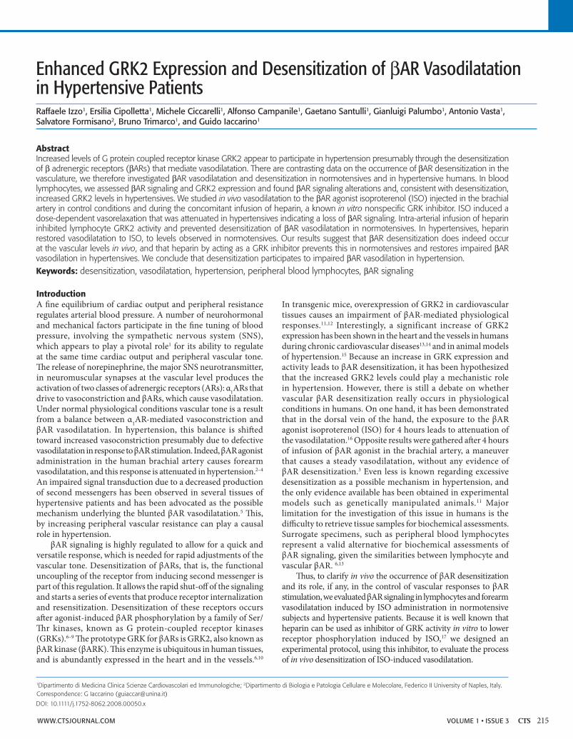
Forearm βAR-induced vasodilatation was assessed by intra-arterial administration of the βAR agonist ISO (1, 3, 6 ng/100 mL of forearm volume/min, for 5 minutes, 7 subjects), the muscarinic receptor agonist Acetylcholine (Ach, 0.5, 1.0, 2.0 μg/kg/min, 5 subjects) and the receptor-independent vasodilator Nitroprusside (NP, 0.8, 1.6, 3.2 μg/kg/min, same 5 subjects of Ach).20 Desensitization of the βAR response was evaluated comparing two vasodilatation curves to ISO generated 15 minutes apart from each other, according to the protocol depicted in Figure 1A.
Doses were identified to have an effect restricted only to the forearm, without any systemic involvement, as indicated by continuous monitoring of blood pressure and heart rate.
To assess the role of phosphorylation in the process of desensitization of βAR we repeated, in the same session, the identical protocol after a phase of recovery of at least 30 min, in the presence of intrarterial infusion heparin (administered as a bolus of heparin [250 IU] followed by continous infusion of 1,000 IU/hr), and evaluated the two curves of vascular responses to ISO, as described in Figure 1B.
We also performed two additional studies in two independent healthy volunteers, which were subjected to intra-arterial infusion of heparin according to the above described protocol, but that did not receive any vasodilating agent.
Vasodilatation is reported as fold over resting FBF.
Peripheral lymphocyte samplesBlood (20 mL) was collected from the antecubital vein and anticoagulated with EDTA. Lymphocytes were isolated by ficoll gradient using HISTOPAQUE-1077 (Sigma), frozen, and stored at –80°C until the day of the assay.13
-AR density and membrane adenylyl cyclase activity assaysCrude cell membranes were prepared from lymphocytes as previously described.13 β-AR density was determined by radioligand binding with the nonselective β-AR ligand [125I]-CYP and membrane adenylyl cyclase activity under basal conditions or in the presence of either 10 µmol/L isoproterenol or 10 mmol/L NaF and cAMP was quantified using standard methods.13 All β-AR signaling results were normalized to the amount of protein added during the experiments.
Protein immunoblottingImmunodetection of lymphocyte levels of GRK2 and actin were performed using detergent-solubilized cell extracts or cytosol fractions obtained by centrifugations, using polyclonal anti-GRK2 and anti-actin antibodies (Santa Cruz Biotechnology) after SDS-PAGE and Western Blot.13 The 80 kDa GRK2 protein was visualized using standard enhanced chemiluminescence (ECL Kit, Amersham Biosciences, Piscataway, NJ, USA). Quantization of immunoreactive GRK2 was done by scanning the autoradiography film and using ImageQuant software (Molecular Dynamics, Piscataway, NJ, USA), and corrected for actin expression.
GRK activitySoluble cytosolic fractions were separated and GRK activity was assessed on 100 μg protein by light dependent phosphorylation of rhodopsin-enriched rod outer segment membranes in lysis buffer with 10 mM MgCl2 and 0.1 mM ATP (containing γ[32P] ATP).13 After incubating on light for 15 minutes at room temperature, reactions were quenched with ice-cold lysis buffer, and centrifuged for 15 minutes at 13,000 g. The pelleted material was resuspended in 35 μL protein gel loading dye and electrophorosed through 4%–20% polyacrylamide Tris/glycine gels. Phosphorylated rhodopsin was visualized by autoradiography of dried gels.
Izzo et al. n GRK2 and Forearm Vasodilatation
Figure 1. The forearm perfusion technique was applied to induce local vasodilatation assessed by strain gauge plethysmography. (A) To induce vasodilatation, increasing amounts of drug were infused in the brachial artery through means of a plastic cannula. Each level of infusion was maintained for 5 minutes. At the fifth minute of infusion, blood flow was assessed for 1 minute twice consecutively (indicated by the arrows) and then the drug infusion rate was switched to the next step. After the last measurement on the last dose, the infusion was stopped and the FBF allowed to recover for 15 minutes. Then, blood flow was measured twice, and the drug infusion was started again, according to the same procedure described above. (B) In normotensives, after adequate recovery time, heparin was infused into the brachial artery, by a bolus injection of 250 units, followed by a constant infusion of 1,000 units per hour. After 15 minutes of infusion, the ISO administration was started again according to the same protocol described in A, while heparin was still being infused.
SecondFirst
15 min
Heparin: Bolus 250 UI, 1000 UI/hr
Second ISOFirst ISO
15 min
A
B
216 Volume 1 • Issue 3 www.CTSjouRnAl.CoM
basal level (12.9 ± 1.6 mL/100 mL/min, NS vs. basal) and the second administration of ISO induced again a vasodilatation, which was significantly attenuated as compared to the previous hemodynamic response (Figure 4A, p < 0.05, ANOVA).
Statistical analysisThe t-test was used to compare βAR signaling in lymphocytes. One-way repeated-measurements analysis of variance (ANOVA) was used to evaluate the responses to ISO, Ach, and sodium nitroprusside. To estimate the effect of heparin on the responses to ISO, repeated-measurements ANOVA with a grouping factor was performed. Post hoc simultaneous multiple comparisons were done by Bonferroni’s analysis.21 Results are presented as mean ± SE.
Results
PopulationCharacteristics of our population are reported in Table 1. There are not significant differences in age and body mass index between normotensives and hypertensive patients. As expected, values of systolic and diastolic blood pressure were significantly higher in hypertensives.
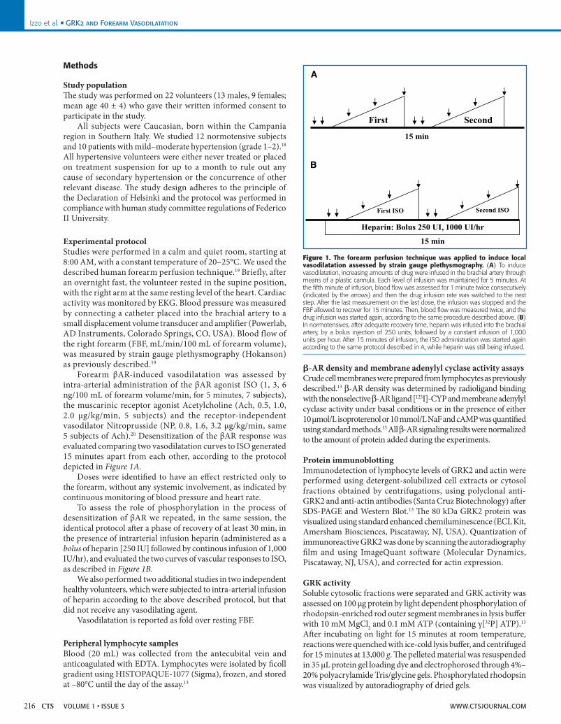
Lymphocyte βAR signaling in normotensive and hypertensive patientsThe signaling capability of the βAR system was assessed on peripheral blood lymphocyte membranes, given the similarities between the βAR signaling of the vasculature and peripheral lymphocytes.22 A reduced basal production of cAMP was observed in nonstimulated membranes in hypertensive patients as compared to normotensives (38.5 ± 10 vs. 125 ± 31 pmol/mg/min, respectively, p < 0.02). In normotensives, challenges with the βAR agonist ISO (Figure 2A) or the adenylyl cyclase activator NaF (5208 ± 278 pmol/mg/min) caused an increase in the production of the second messenger cAMP. In hypertensive patients the βAR response to ISO was blunted (Figure 2A), but not that to NaF (5374 ± 296 pmol/mg/min, NS vs. normotensives). The impairment of the βAR response could not be imputed to downregulation of βARs, since hypertensive and normotensive showed similar βAR densities (Figure 2B). Thus, it could be ascribed to the higher GRK2 expression observed in hypertensive patients compared to normotensives (Figure 2C).
Forearm vasodilatationNormotensive subjects.The intra-brachial artery administration of ISO did not cause significant alterations of blood pressure or heart rate (Figure 3). ISO induced a dose-dependent increase of forearm blood flow (FBF: from 9.14 ± 0.78 to 27.5 ± 1.4 mL/100 mL/min, p < 0.05 ANOVA; Figure 4A). After 15 minutes of rest, FBF returned to
Izzo et al. n GRK2 and Forearm VasodilatationIzzo et al. n GRK2 and Forearm Vasodilatation
Normotensives Hypertensives p
(n = 12) (n = 10)
Age (years) 39 ± 4 41 ± 4 nS
Sex (M/F) 7/5 6/4 nS
Weight (kg) 66 ± 4 60 ± 4 nS
Height (cm) 168 ± 3 166 ± 4 nS
BMI 23.3 ± 0.9 21.6 ± 1.3 nS
SBP (mm Hg) 126 ± 6.5 167 ± 5.0 <0.05
DBP (mm Hg) 72 ± 4 86 ± 3.5 <0.05
MBP (mm Hg) 90 ± 4.5 113 ± 3.0 <0.05
NS = not significant; BMI = body mass index; SBP = systolic blood pressure; BP = diastolic blood pressure; MBP = mean blood pressure.
Table 1. Characteristics of the population.
Figure 2. AR signaling in peripheral blood lymphocytes from normotensives and hypertensive patients. (A) cAMP production was assessed on lymphocyte membranes. Stimulation with ISO induced an increase, which was attenuated in hypertensive patients compared to normotensives. *p < 0.05, n = 5 in duplicate. (B) Receptor density was similar between hypertensive patients and normotensives, although there is a slight reduction in the number of receptors in hypertensive patients that was not statistically significant. n = 5 in triplicate. (C) GRK2 and actin expression were evaluated by western blot and quantified by digital densitometry (Inset). GRK2 levels were higher in hypertensive patients than in normotensives, and this difference held significant also after correction for actin expression levels. *p < 0.05, n = 5.
www.CTSjouRnAl.CoM 217Volume 1 • Issue 3

The administration of Ach or NP both induced dose-dependent vasodilatations that did not desensitize over time (Figure 4B and 4C).
Effects of heparin on βAR desensitization in normotensives.Intra-brachial artery infusion of heparin did not modify the basal blood flow in the forearm (from 11.37 ± 0.8 to 11.23 ± 1.6 mL/min/100 mL, NS), nor changed the ISO vasodilatation (Figure 4A). Interestingly, heparin prevented the desensitization of the ISO response (Figure 4A).
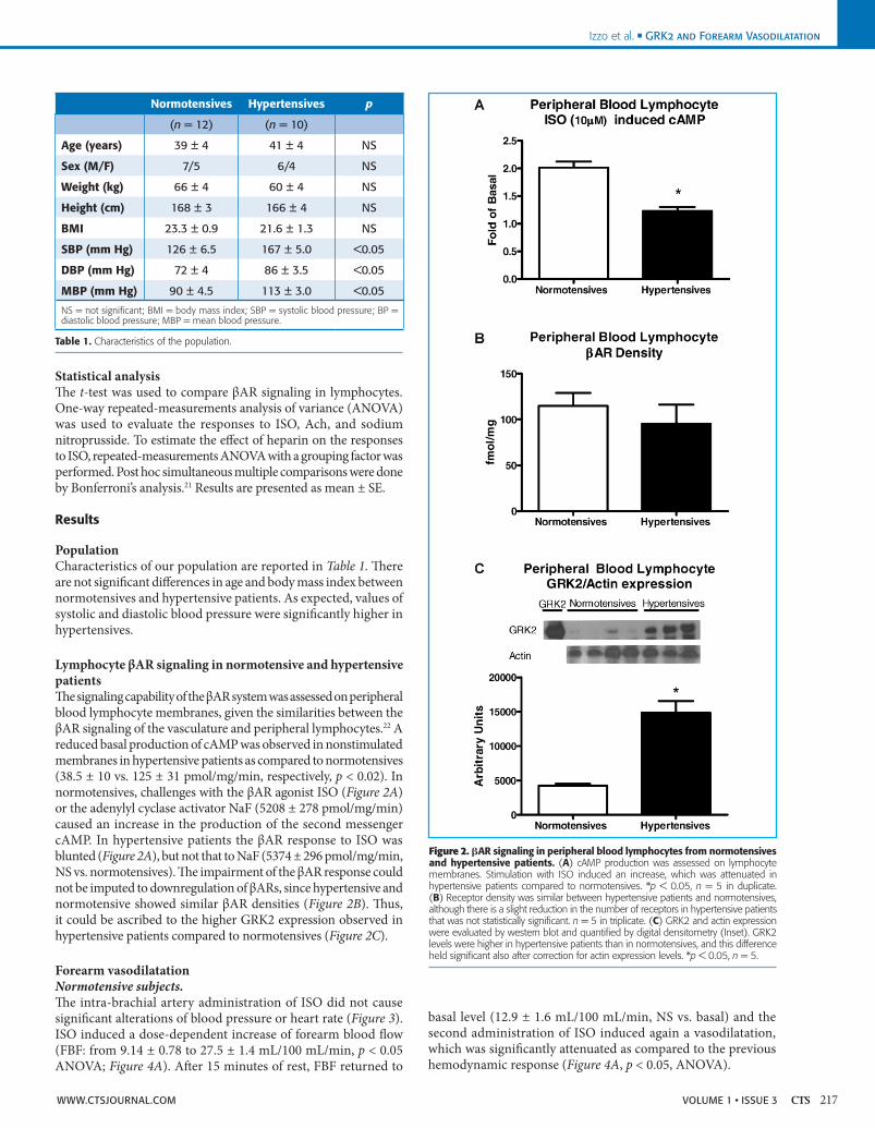
Effects of heparin on lymphocyte cytosol GRK activity and GRK2 expression.In two healthy volunteers, we assessed GRK activity of venous lymphocytes collected before and after intrabrachial artery infusion of heparin. This maneuver inhibited cytosol kinase activity as indicated in Figure 5, without changing GRK2 expression.
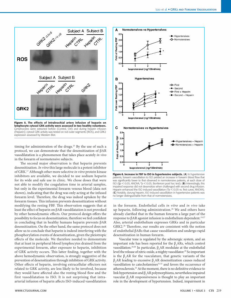
Hypertensive patients.Resting FBF in hypertensive patients did not differ from normotensives (FBF: 12.2 ± 9.4 mL/min/100 mL, NS). As observed in normotensive subjects, also in hypertensive patients the administration of ISO in the brachial artery did not cause significant alterations of blood pressure or heart rate (Figure 3). ISO caused a forearm vasodilatation response, which was smaller than that of normotensives (Figure 6A; ANOVA, p < 0.0.5). This response did not deteriorate further when the forearm was challenged for the second dose–response curve (Figure 6B). Heparin induced a slight albeit not significant increase in basal FBF (from 11.9 ± 1.03 to 12.45 ± 3.0 mL/100 mL/min), but enhanced in a significant manner the responses to ISO (Figure 6B). Thus, the increases in FBF were no longer distinguishable from those of the normotensive subjects (Figure 6C).
DiscussionThe first major finding of the present article is the evidence that in humans in vivo, βAR vasodilation undergoes desensitization. This is a controversial issue because of several conflicting studies in the literature: Stein et al. described that chronic exposure to the βAR agonist terbutalin leads to a decreased vasodilatation to ISO in dorsal veins of the hand.23 However, the same group of authors showed that continuous administration of ISO for 4 hours in the brachial artery produces a constant increase in forearm blood flow, without attenuation of the response.4 This observation seems to deny the existence of an acute desensitization of vascular βARs, but using a similar protocol, Vincent et al. showed that chronic exposure to ISO (4 hours) causes a desensitization of venodilatation.16 We believe that our results help to clarify this issue, in virtue of the use of an experimental protocol which is more appropriate for the evaluation of the desensitization. Indeed, the study of desensitization phenomena can be carried out according to different experimental procedures.24 We assessed the amplitude of the reduction in vasodilatation in a second curve performed after a short interval from the first one. This protocol is borrowed from in vitro studies and requires a tight respect of
Izzo et al. n GRK2 and Forearm Vasodilatation
Figure 3. Blood pressure and heart rate were measured throughout the study in normotensives (n = 7) and hypertensive patients (n = 10), continuously, by means of an EKG lead and a blood pressure amplifier connected to the arterial cannula. BP and HR did not change throughout the ISO infusion. A similar pattern was observed in subjects infused with Ach and NP (data not shown).
Figure 4. Increase in FBF in normotensive subjects. (A) ISO induced a first dose dependent response, followed by a second dose dependent response, which was attenuated. When the protocol was repeated during concomitant heparin infusion, no desensitization was observed in the second vasodilatation. *p < 0.05, second versus first, ANOVA. (B) Acetylcholine, on the contrary, did not desensitize, and the second response was similar to the first (n = 5). (C) Similarly, no changes were observed in the responses to NP (n = 5).
218 Volume 1 • Issue 3 www.CTSjouRnAl.CoM
timing for administration of the drugs.25 By the use of such a protocol, we can demonstrate that the desensitization of βAR vasodilatation is a phenomenon that takes place acutely in vivo in the forearm of normotensive subjects.
The second major observation is that heparin prevents desensitization. In vitro this large molecule is a potent inhibitor of GRK.17 Although other more selective in vitro protein kinase inhibitors are available, we decided to use sodium heparin for its wide and safe use in clinic. We chose doses that were not able to modify the coagulation time in arterial samples, but only in the experimental forearm venous blood (data not shown), indicating that the drug was only acting at the infused forearm level. Therefore, the drug was indeed uptaken by the forearm tissues. This infusion prevents desensitization without modifying the resting FBF. This observation suggests that at least the effect of heparin on βAR vasodilatation is not provoked by other hemodynamic effects. Our protocol design offers the possibility to focus on desensitization, therefore we feel confident in concluding that in healthy humans heparin prevents βAR desensitization. On the other hand, the same protocol does not allow us to conclude that heparin is indeed interfering with the phosphorylation events of desensitization, given the pleiotropic effects of the molecule. We therefore needed to demonstrate that at least in peripheral blood lymphocytes drained from the experimental forearm, after exposure to heparin, inhibition of GRK activity occurs. This latter finding, together with the above hemodynamic observation, is strongly suggestive of the prevention of desensitization through inhibition of GRK activity. Other effects of heparin, involving extracellular effectors not related to GRK activity, are less likely to be involved, because they would have affected also the resting blood flow and the first vasodilatation to ISO. It is not surprising that intra-arterial infusion of heparin affects ISO-induced vasodilatation
in the forearm. Endothelial cells in vitro and in vivo take up heparin, following administration.26 We and others have already clarified that in the human forearm a large part of the response to βAR agonist infusion is endothelium dependent.19,27 Also, arterial endothelium expresses GRKs and in particular GRK2.28 Therefore, our results are consistent with the notion of endothelial βARs that cause vasodilation and undergo rapid desensitization in human forearm.
Vascular tone is regulated by the adrenergic system, and an important role has been reported for the β2ARs, which control vasodilation.29,30 In particular, β2AR modulate at the endothelial level the release of nitric oxide, a mighty vasodilator.29 So important is the β2AR for the vasculature, that genetic variants of the β2AR leading to excessive β2AR desensitization causes reduced vasodilation to catecholamines31,32 and favors the occurrence of atherosclerosis.33 At the moment, there is no definitive evidence to link hypertension and β2AR polymorphisms, nevertheless impaired vascular β2AR responsiveness is thought to play an important role in the development of hypertension. Indeed, impairment in
Izzo et al. n GRK2 and Forearm VasodilatationIzzo et al. n GRK2 and Forearm Vasodilatation
Figure 5. The effects of intrabrachial artery infusion of heparin on lymphocyte cytosol GRK activity were assessed in two healthy volunteers. Lymphocytes were extracted before (Control, Ctrl) and during heparin infusion (Heparin); cytosol GRK activity was tested on rod outer segments (ROS), and GRK2 expression assessed by Western Blot.
Figure 6. Increase in FBF to ISO in hypertensive subjects. (A) In hypertensive patients, forearm vasodilation to ISO yielded an increase in forearm blood flow that was significantly lower to that observed in normotensive patients, at each dose of ISO (§p < 0.05, ANOVA; *p < 0.05, Bonferroni post hoc test). (B) Interestingly, this impaired response did not desensitize when challenged with second drug infusion. Heparin enhanced the ISO induced vasodilation (*p < 0.05 vs. first curve, ANOVA). (C) Notably, durung heparin, ISO induced vasodilation in hypertensive patients was no longer distinguishable from that of normotensives.
www.CTSjouRnAl.CoM 219Volume 1 • Issue 3
Izzo et al. n GRK2 and Forearm Vasodilatation
β2AR-mediated vasodilatation has been described in both human and animal models of hypertension.2,34 This impairment may be due to an alteration in GRK2 expression; in particular Gros et al. have demonstrated that GRK2 activity is increased in lymphocytes from hypertensive subjects.35 It is possible to speculate that this increase is causative for the impaired βAR response in hypertensive state. To test this hypothesis, we repeated our experimental desensitization protocol in untreated hypertensive patients. The present data confirm our previous report that hypertensive patients show an impaired βAR vasodilatation.21 It is interesting to note that in hypertensive patients, the vasodilatation cannot be further desensitized and the first and second ISO-vasodilatation overlap. Heparin action results to be effective on the impaired βAR response, and enhance the vasodilatation to ISO to a level similar to those of normotensives. This change occurs without any modification in blood pressure and heart rate, suggesting that there is no systemic hemodynamic involvement in this heparin effect. Our data in vivo are also sustained by an in vitro counterpart, showing that at least in peripheral blood lymphocytes, the main determinant of impaired βAR response in hypertensives is the increased level of GRK2. Therefore, the possible explanation for the improved hemodynamic to ISO is that heparin exerts its favorable effect by ameliorating βAR response, probably by inhibiting GRK activity at the endothelial level.
PerspectivesIncreased GRK expression is probably causal to impaired vasodilatation and increased peripheral resistance in hypertension; as a consequence, GRK inhibition by systemic administration should result in reduced blood pressure. In perspective, the monitoring of peripheral GRK levels could be used as a biomarker for monitoring prognosis and effectiveness of treatment in hypertensive patients.13 Further studies ongoing in the lab are actively pursuing this issue, which requires an appropriate follow-up and a larger patient population.
References1. Head GA. The sympathetic nervous system in hypertension: assessment by blood pressure variability and ganglionic blockade. J Hypertens. 2003; 21(9): 1619–1621.
2. Feldman RD. Defective venous beta-adrenergic response in borderline hypertensive subjects is corrected by a low sodium diet. J Clin Invest. 1990; 85(3): 647–652.
3. Stein CM, Nelson R, Deegan R, He H, Wood M, Wood AJ. Forearm beta adrenergic receptor-mediated vasodilation is impaired, without alteration of forearm norepinephrine spillover, in borderline hypertension. J Clin Invest. 1995; 96(1): 579–585.
4. Stein CM, Nelson R, Deegan R, He H, Inagami T, Frazer M, Badr KF, Wood M, Wood AJ. Tachyphylaxis of human forearm vascular responses does not occur rapidly after exposure to isoproterenol. Hypertension. 1995; 25(6): 1294–1300.
5. Feldman RD, Limbird LE, Nadeau J, Robertson D, Wood AJ. Leukocyte beta-receptor alterations in hypertensive subjects. J Clin Invest. 1984; 73(3): 648–653.
6. Iaccarino G, Campanile A, Santulli G, Zuppieri F, Koch W. G protein-coupled receptor kinases and hypertension: a review of disease mechanisms. High Blood Press Cardiovasc Prev. 2006; 13(4): 151–158.
7. Hausdorff WP, Caron MG, Lefkowitz RJ. Turning off the signal: desensitization of beta-adrenergic receptor function. Faseb J. 1990; 4(11): 2881–2889.
8. Whalen EJ, Foster MW, Matsumoto A, Ozawa K, Violin JD, Que LG, Nelson CD, Benhar M, Keys JR, Rockman HA, Koch WJ, Daaka Y, Lefkowitz RJ, Stamler JS. Regulation of beta-adrenergic receptor signaling by S-nitrosylation of G-protein-coupled receptor kinase 2. Cell. 2007; 129(3): 511–522.
9. Zeiders JL, Seidler FJ, Iaccarino G, Koch WJ, Slotkin TA. Ontogeny of cardiac beta-adrenoceptor desensitization mechanisms: agonist treatment enhances receptor/G-protein transduction rather than eliciting uncoupling. J Mol Cell Cardiol. 1999; 31(2): 413–423.
10. Penela P, Ribas C, Mayor F, Jr. Mechanisms of regulation of the expression and function of G protein-coupled receptor kinases. Cell Signal. 2003; 15(11): 973–981.
11. Eckhart AD, Ozaki T, Tevaearai H, Rockman HA, Koch WJ. Vascular-targeted overexpression of G protein-coupled receptor kinase-2 in transgenic mice attenuates beta-adrenergic receptor signaling and increases resting blood pressure. Mol Pharmacol. 2002; 61(4): 749–758.
12. Koch WJ, Rockman HA, Samama P, Hamilton RA, Bond RA, Milano CA, Lefkowitz RJ. Cardiac function in mice overexpressing the beta-adrenergic receptor kinase or a beta ARK inhibitor. Science N.Y. 1995; 268(5215): 1350–1353.
13. Iaccarino G, Barbato E, Cipolletta E, De Amicis V, Margulies KB, Leosco D, Trimarco B, Koch WJ. Elevated myocardial and lymphocyte GRK2 expression and activity in human heart failure. Eur Heart J. 2005; 26(17): 1752–1758.
14. Iaccarino G, Keys JR, Rapacciuolo A, Shotwell KF, Lefkowitz RJ, Rockman HA, Koch WJ. Regulation of myocardial betaARK1 expression in catecholamine-induced cardiac hypertrophy in transgenic mice overexpressing alpha1B-adrenergic receptors. J Am Coll Cardiol. 2001; 38(2): 534–540.
15. Choi DJ, Koch WJ, Hunter JJ, Rockman HA. Mechanism of beta-adrenergic receptor desensitization in cardiac hypertrophy is increased beta-adrenergic receptor kinase. J Biol Chem. 1997; 272(27): 17223–17229.
16. Vincent J, Blaschke TF, Hoffman BB. Desensitization of beta-adrenoceptor- and prostaglandin E1 receptor-mediated human vascular smooth muscle relaxation. J Cardiovasc Pharmacol. 1992; 19(3): 447–452.
17. Lohse MJ, Lefkowitz RJ, Caron MG, Benovic JL. Inhibition of beta-adrenergic receptor kinase prevents rapid homologous desensitization of beta 2-adrenergic receptors. Proc Natl Acad Sci USA. 1989; 86(9): 3011–3015.
18. Mancia G, De Backer G, Dominiczak A, Cifkova R, Fagard R, Germano G, Grassi G, Heagerty AM, Kjeldsen SE, Laurent S, Narkiewicz K, Ruilope L, Rynkiewicz A, Schmieder RE, Boudier HA, Zanchetti A. ESH-ESC Practice Guidelines for the Management of Arterial Hypertension: ESH-ESC Task Force on the Management of Arterial Hypertension. J Hypertens. 2007; 25(9): 1751–1762.
19. Lembo G, Iaccarino G, Vecchione C, Barbato E, Izzo R, Fontana D, Trimarco B. Insulin modulation of an endothelial nitric oxide component present in the alpha2- and beta-adrenergic responses in human forearm. J Clin Invest. 1997; 100(8): 2007–2014.
20. Miller MR, Megson IL. Recent developments in nitric oxide donor drugs. Br J Pharmacol. 2007; 151(3): 305–321.
21. Lembo G, Iaccarino G, Vecchione C, Rendina V, Parrella L, Trimarco B. Insulin modulation of beta-adrenergic vasodilator pathway in human forearm. Circulation. 1996; 93(7): 1403–1410.
22. Gros R, Chorazyczewski J, Meek MD, Benovic JL, Ferguson SS, Feldman RD. G-Protein-coupled receptor kinase activity in hypertension: increased vascular and lymphocyte G-protein receptor kinase-2 protein expression. Hypertension. 2000; 35(1 Pt 1): 38–42.
23. Stein M, Deegan R, Wood AJ. Long-term exposure to beta 2-receptor agonist specifically desensitizes beta-receptor-mediated venodilation. Clin Pharmacol Ther. 1993; 54(2): 187–193.
24. Model MA, Omann GM. Experimental approaches for observing homologous desensitisation and their pitfalls. Pharmacol Res. 1998; 38(1): 41–44.
25. Kazanietz MG, Enero MA. Desensitization of the beta-2 adrenoceptor-mediated vasodilation in rat aorta after prolonged treatment with the beta-2 adrenoceptor agonist clenbuterol. J Pharmacol Exp Ther. 1990; 252(2): 758–764.
26. Hiebert LM, McDuffie NM. The internalization and release of heparins by cultured endothelial cells: the process is cell source, heparin source, time and concentration dependent. Artery. 1990; 17(2): 107–118.
27. Panza JA, Garcia CE, Kilcoyne CM, Quyyumi AA, Cannon RO, 3rd. Impaired endothelium-dependent vasodilation in patients with essential hypertension. Evidence that nitric oxide abnormality is not localized to a single signal transduction pathway. Circulation. 1995; 91(6): 1732–1738.
28. Leosco D, Iaccarino G, Cipolletta E, De Santis D, Pisani E, Trimarco V, Ferrara N, Abete P, Sorriento D, Rengo F, Trimarco B. Exercise restores beta-adrenergic vasorelaxation in aged rat carotid arteries. Am J Physiol. 2003; 285(1): H369–H374.
29. Iaccarino G, Cipolletta E, Fiorillo A, Annecchiarico M, Ciccarelli M, Cimini V, Koch WJ, Trimarco B. Beta(2)-adrenergic receptor gene delivery to the endothelium corrects impaired adrenergic vasorelaxation in hypertension. Circulation. 2002; 106(3): 349–355.
30. Ciccarelli M, Cipolletta E, Santulli G, Campanile A, Pumiglia K, Cervero P, Pastore L, Astone D, Trimarco B, Iaccarino G. Endothelial beta(2) adrenergic signaling to AKT: role of G(i) and SRC. Cell Signal. 2007; 19(9): 1949–1955.
31. Bruck H, Leineweber K, Park J, Weber M, Heusch G, Philipp T, Brodde OE. Human beta2-adrenergic receptor gene haplotypes and venodilation in vivo. Clin Pharmacol Ther. 2005; 78(3): 232–238.
32. Khalaila JM, Elami A, Caraco Y. Interaction between beta2 adrenergic receptor polymorphisms determines the extent of isoproterenol-induced vasodilatation ex vivo. Pharmacogen Genomics. 2007; 17(10): 803–811.
33. Barbato E, Berger A, Delrue L, Van Durme F, Manoharan G, Boussy T, Heyndrickx GR, De Bruyne B, Ciampi Q, Vanderheyden M, Wijns W, Bartunek J. GLU-27 variant of beta2-adrenergic receptor polymorphisms is an independent risk factor for coronary atherosclerotic disease. Atherosclerosis. 2007; 194(2): e80–e86.
34. Feldman RD, Gros R. Impaired vasodilator function in hypertension: the role of alterations in receptor-G protein coupling. Trends Cardiovasc Med. 1998; 8(7): 297–305.
35. Gros R, Benovic JL, Tan CM, Feldman RD. G-protein-coupled receptor kinase activity is increased in hypertension. J Clin Invest. 1997; 99(9): 2087–2093.
220 Volume 1 • Issue 3 www.CTSjouRnAl.CoM
Related Documents