eScholarship provides open access, scholarly publishing services to the University of California and delivers a dynamic research platform to scholars worldwide. Lawrence Berkeley National Laboratory Title: Energy metabolism in Desulfovibrio vulgaris Hildenborough: insights from transcriptome analysis Author: Pereira, Patricia M. Publication Date: 06-18-2009 Publication Info: Lawrence Berkeley National Laboratory Permalink: http://escholarship.org/uc/item/234821wf Keywords: Bioenergetic metabolism, Hydrogen metabolism, Microarrays, Respiratory complexes, Sulphate- reducing bacteria Abstract: Sulphate-reducing bacteria are important players in the global sulphur and carbon cycles, with considerable economical and ecological impact. However, the process of sulphate respiration is still incompletely understood. Several mechanisms of energy conservation have been proposed, but it is unclear how the different strategies contribute to the overall process. In order to obtain a deeper insight into the energy metabolism of sulphate-reducers whole-genome microarrays were used to compare the transcriptional response of Desulfovibrio vulgaris Hildenborough grown with hydrogen/sulphate, pyruvate/sulphate, pyruvate with limiting sulphate, and lactate/ thiosulphate, relative to growth in lactate/sulphate. Growth with hydrogen/sulphate showed the largest number of differentially expressed genes and the largest changes in transcript levels. In this condition the most up-regulated energy metabolism genes were those coding for the periplasmic [NiFeSe] hydrogenase, followed by the Ech hydrogenase. The results also provide evidence for the involvement of formate cycling and the recently proposed ethanol pathway during growth in hydrogen. The pathway involving CO cycling is relevant during growth on lactate and pyruvate, but not during growth in hydrogen as the most down-regulated genes were those coding for the CO- induced hydrogenase. Growth on lactate/thiosulphate reveals a down-regulation of several energy metabolism genes similar to what was observed in the presence of nitrite. This study identifies the role of several proteins involved in the energy metabolism of D. vulgaris and highlights several novel genes related to this process, revealing a more complex bioenergetic metabolism than previously considered.

Welcome message from author
This document is posted to help you gain knowledge. Please leave a comment to let me know what you think about it! Share it to your friends and learn new things together.
Transcript
eScholarship provides open access, scholarly publishingservices to the University of California and delivers a dynamicresearch platform to scholars worldwide.
Lawrence Berkeley National Laboratory
Title:Energy metabolism in Desulfovibrio vulgaris Hildenborough: insights from transcriptome analysis
Author:Pereira, Patricia M.
Publication Date:06-18-2009
Publication Info:Lawrence Berkeley National Laboratory
Permalink:http://escholarship.org/uc/item/234821wf
Keywords:Bioenergetic metabolism, Hydrogen metabolism, Microarrays, Respiratory complexes, Sulphate-reducing bacteria
Abstract:Sulphate-reducing bacteria are important players in the global sulphur and carbon cycles, withconsiderable economical and ecological impact. However, the process of sulphate respiration isstill incompletely understood. Several mechanisms of energy conservation have been proposed,but it is unclear how the different strategies contribute to the overall process. In order to obtaina deeper insight into the energy metabolism of sulphate-reducers whole-genome microarrayswere used to compare the transcriptional response of Desulfovibrio vulgaris Hildenboroughgrown with hydrogen/sulphate, pyruvate/sulphate, pyruvate with limiting sulphate, and lactate/thiosulphate, relative to growth in lactate/sulphate. Growth with hydrogen/sulphate showed thelargest number of differentially expressed genes and the largest changes in transcript levels. In thiscondition the most up-regulated energy metabolism genes were those coding for the periplasmic[NiFeSe] hydrogenase, followed by the Ech hydrogenase. The results also provide evidence forthe involvement of formate cycling and the recently proposed ethanol pathway during growth inhydrogen. The pathway involving CO cycling is relevant during growth on lactate and pyruvate, butnot during growth in hydrogen as the most down-regulated genes were those coding for the CO-induced hydrogenase. Growth on lactate/thiosulphate reveals a down-regulation of several energymetabolism genes similar to what was observed in the presence of nitrite. This study identifies therole of several proteins involved in the energy metabolism of D. vulgaris and highlights severalnovel genes related to this process, revealing a more complex bioenergetic metabolism thanpreviously considered.
1
Energy metabolism in Desulfovibrio vulgaris Hildenborough: insights from
transcriptome analysis
Patrícia M. Pereira1, Qiang He2,3,4, Filipa M. A. Valente1, António V. Xavier1, Jizhong
Zhou2,5,6, Inês A. C. Pereira1 & Ricardo O. Louro1*
1 Instituto de Tecnologia Química e Biológica, Universidade Nova de Lisboa, Portugal;
2 Environmental Sciences Division, Oak Ridge National Laboratory, Oak Ridge, Tennessee 37831;
3 Department of Civil and Environmental Engineering, the University of Tennessee, Knoxville, Tennessee
37996;
4 The Center for Environmental Biotechnology, the University of Tennessee, Knoxville, Tennessee 37996;
5 Virtual Institute for Microbial Stress and Survival, Berkeley, California 94720;
6 Institute for Environmental Genomics, Department of Botany and Microbiology, University of Oklahoma,
Norman, Oklahoma 73019.
To whom correspondence should be addressed at: ITQB, Rua da Quinta Grande, 6
Apt. 126, 2780-156 Oeiras, Portugal. Tel.351-214469309; Fax 351-214411277
http://www.itqb.unl.pt, [email protected]

2
Keywords: Bioenergetic metabolism, Hydrogen metabolism, Microarrays,
Respiratory complexes, Sulphate-reducing bacteria.
ABSTRACT
Sulphate-reducing bacteria are important players in the global sulphur and carbon
cycles, with considerable economical and ecological impact. However, the process of
sulphate respiration is still incompletely understood. Several mechanisms of energy
conservation have been proposed, but it is unclear how the different strategies
contribute to the overall process. In order to obtain a deeper insight into the energy
metabolism of sulphate-reducers whole-genome microarrays were used to compare
the transcriptional response of Desulfovibrio vulgaris Hildenborough grown with
hydrogen/sulphate, pyruvate/sulphate, pyruvate with limiting sulphate, and
lactate/thiosulphate, relative to growth in lactate/sulphate. Growth with
hydrogen/sulphate showed the largest number of differentially expressed genes and
the largest changes in transcript levels. In this condition the most up-regulated energy
metabolism genes were those coding for the periplasmic [NiFeSe] hydrogenase,
followed by the Ech hydrogenase. The results also provide evidence for the
involvement of formate cycling and the recently proposed ethanol pathway during
growth in hydrogen. The pathway involving CO cycling is relevant during growth on
lactate and pyruvate, but not during growth in hydrogen as the most down-regulated
genes were those coding for the CO-induced hydrogenase. Growth on
lactate/thiosulphate reveals a down-regulation of several energy metabolism genes
similar to what was observed in the presence of nitrite. This study identifies the role of
several proteins involved in the energy metabolism of D. vulgaris and highlights several
novel genes related to this process, revealing a more complex bioenergetic metabolism
than previously considered.

3
Abbreviations:
HS- hydrogen/sulphate medium, LS- lactate/sulphate medium, LT-
lactate/thiosulphate medium, PS- pyruvate/sulphate medium, P-pyruvate medium,
SRB- Sulphate-reducing bacteria, Type I cytochrome c3 - TpIc3
4
INTRODUCTION
Sulphate-reducing bacteria (SRB) are anaerobic prokaryotes widespread in natural
habitats like marine or freshwater sediments, soil, and also the gastrointestinal tract of
many animals, including man [1]. They are particularly abundant in habitats with high
concentration of sulphate such as sea water, effectively linking the global sulphur and
carbon cycles [2, 3]. SRB use sulphate or other sulphur oxo-anions as electron
acceptors for the anaerobic oxidation of inorganic or organic substrates such as
hydrogen, lactate, acetate, ethanol and propionate. As a consequence of this
metabolism, large amounts of sulphide are produced and accumulated in their natural
habitats. This aggressive metabolic end-product poses an important economic problem
for the oil industry due to its involvement in biocorrosion of metal structures [4] in
addition to souring of oil and gas deposits [3]. However, SRB can also reduce a wide
variety of other substrates, including heavy metals and radionuclides. Examples
include U(VI), Tc(VII), As (V) and Cr (VI), making these bacteria interesting targets to
use for bioremediation of contaminated anaerobic sediments and groundwater [5, 6]. In
situ stimulation of microbial populations using specific electron donors or acceptors is
an attractive strategy for environmental bioremediation processes. A better
understanding of energy metabolism of SRB growing with different energy sources or
electron acceptors is thus crucial for exploring their potential in bioremediation, as well
as to control their activity wherever it leads to undesirable consequences.
Despite numerous studies, the bioenergetic mechanism of sulphate respiration is
still far from being understood. Odom and Peck proposed a bioenergetic pathway to
explain energy conservation in Desulfovibrio, the most studied genus of SRB, which
involved cycling of hydrogen during growth in lactate/sulphate [7]. The proposal
considers that protons and electrons resulting from the oxidation of lactate to acetate
are converted to hydrogen in the cytoplasm by a cytoplasmic hydrogenase. The
hydrogen then diffuses across the membrane to the periplasm where it is converted to
5
protons and electrons by a periplasmic hydrogenase. Transfer of the resulting electrons
back to the cytoplasm for sulphate reduction results in a transmembrane proton
gradient that can drive ATP synthesis by the ATP synthase complex. This mechanism
has been disputed because hydrogen formation from lactate oxidation to pyruvate is
energetically unfavourable, even in conditions where there is a large excess of lactate
in the presence of small hydrogen concentrations [8, 9]. Furthermore, this mechanism
requires the presence of a cytoplasmic hydrogenase, which is already known to be
absent in the genome of some SRB such as Desulfovibrio desulfuricans G20
(www.jgi.doe.gov) and Desulfotalea psycrophila [10]. Alternative chemiosmotic models
have been proposed in which electrons generated from substrate oxidation are
transported through membrane-bound electron carriers to sulphate reduction (vectorial
electron transport), and in the process translocate protons to the periplasm (vectorial
proton transport) [11, 12]. This electron transport chain most likely involves the
participation of menaquinone [13], plus two membrane-bound redox complexes that
are conserved in sulphate reducers, the Qmo and Dsr complexes [14-16]. There is also
evidence that cycling of other reduced intermediates like CO or formate may also be
involved in the energy conservation mechanisms of Desulfovibrio vulgaris
Hildenborough (referred hereafter as D. vulgaris) [17-19]. Biochemical, genetic and
genomic studies of D. vulgaris have allowed the identification and initial
characterization of several of the participants in the electron transfer network involved
in sulphate respiration [20]. However, the genome reveals a large number of genes
involved in energy metabolism with an unknown function. Furthermore, different
electron transport pathways seem to be involved in the oxidation of pyruvate, lactate or
hydrogen [17, 21], and different metabolic strategies may be used by different
organisms [20]. In this work, the global gene expression profile of D. vulgaris grown in
different electron donors and acceptors was investigated in order to obtain a broad
view of the transcriptional response of the energy metabolism genes. It has been

6
shown that there is a good correlation between mRNA levels and protein abundance of
Central Intermediary Metabolism and Energy Metabolism genes [22], thus enabling a
physiological interpretation of the transcriptional response. This study complements a
previous one comparing transcriptional changes for growth in formate/sulphate relative
to lactate/sulphate [23]. The results provide several insights into the bioenergetic
pathways that are operative under the different growth conditions.
MATERIALS & METHODS
Growth conditions
D. vulgaris (DSM 644) was grown in modified medium C [24] at 37ºC, under an
atmosphere of N2. The modified medium C contained 25 µM FeSO4.7H2O and 1 µM
NiCl2.6H2O. It was previously shown that in medium C without supplements the amount
of adventitious selenium and nickel is below the detection limit using atomic absorption
spectrometry (<0.04mg/l and <0.01mg/l, respectively) [25]. Medium C-LS contained 40
mM lactate, 40 mM sulphate, and was used as a control. Pyruvate (40 mM) and
thiosulphate (40 mM) were used as an alternative electron donor and acceptor,
respectively. Both pyruvate and thiosulphate were added from stock solutions in sterile
conditions with a 0.2 µm filter. Growth with limiting sulphate was carried out with
pyruvate (40 mM), using a 10% pyruvate/sulphate grown innoculum. Media (1000-ml)
were dispensed in 2000-ml DURAN® SHOTT bottles, flushed with N2 and sealed with
butyl rubber stoppers. Cultures with hydrogen as electron donor were grown in a 3L
fermentor containing 2L of growth medium with 40 mM acetate, gassed with a mixture
of 80 % (vol/vol) H2, 20 % (vol/vol) CO2 at 900 ml/min, stirred at 250 rpm and with a
constant pH of 6.8. As a control, D. vulgaris was grown in the same conditions but in
Medium C-LS, and gassed with N2. In both procedures a 10% (vol/vol) inoculum of a
freshly grown culture of D. vulgaris in the same conditions of the experiment was used,

7
with exception to pyruvate culture where the inoculum was grown in pyruvate/sulphate
medium. The optical density of the cultures at 600 nm (OD600) was determined with a
Shimadzu UV-1603 spectrophotometer. Cultures were harvested by centrifugation
(10,000 x g for 15 min) in the mid-exponential phase with an OD600 of 0.47 for
lactate/sulphate (LS), 0.42 for H2+CO2+acetate/sulphate (HS), 0.45 for
lactate/thiosulphate (LT), 0.6 for pyruvate/sulphate (PS) and 0.47 for growth on
pyruvate (P). The cells were stored at -70ºC until used.
Global expression analysis
Microarray expression profiling was carried out using D. vulgaris whole genome
microarrays which contain 3,482 of the 3,531 protein-coding sequences of the D.
vulgaris genome [26]. All microarray procedures including total RNA extraction, cDNA
labelling, microarray hybridization and washing were performed using previously
published protocols [26]. RNA extraction, purification, and labelling were performed
independently on each cell sample. Four samples of each total RNA preparation were
labelled, two with Cy3-dUTP and another two with Cy5-dUTP, for microarray
hybridization. To hybridize microarray glass slides, the Cy5-dUTP-labelled cDNA
targets from one lactate/sulphate culture (control culture) were mixed with the Cy3-
dUTP-labelled cDNA targets from one culture grown with a different electron donor or
acceptor and vice versa (dye swap). As a result, each biological sample was hybridized
to four microarray slides. The arrays were visualized by ScanArray Express confocal
laser scanner (Perkin Elmer), and hybridization signal intensities were quantified using
ImaGene software version 6.0 (Biodiscovery, Marina Del Rey, CA).
Two biological replicates obtained from independently grown D. vulgaris cultures
were used in the gene expression analysis for all growth conditions. In addition, each
microarray slide contained duplicate sets of gene fragments and the RNA isolated from
each replicate sample was hybridized with four microarray slides. This provided a total
8
of 16 data points per experimental condition enabling the use of rigorous statistical
tests to determine significant changes in gene expression. The resulting data files were
subjected to Lowess intensity-based normalization and further analyzed using
GeneSpring version 5.1 (Silicon Genetics, Redwood City, Calif.). Lowess normalization
was performed on each microarray slide, and results of the two replicate cultures of
each experimental condition were used for statistical analysis. To assess the statistical
significance of individual data points, the Student t-test was used to calculate a p-value
to test the null hypothesis that the expression level was unchanged. Gene expression
changes observed in these arrays were extensively validated by RT-PCR in previous
studies [26-29]. The annotation of the ORFs was obtained from the comprehensive
microbial resource at The Institute for Genomic Research (TIGR). The data were
deposited in NCBIs Gene Expression Omnibus (GEO,
http://www.ncbi.nlm.nih.gov/geo/) and are accessible through GEO Series accession
number GSE8069.
RESULTS AND DISCUSSION
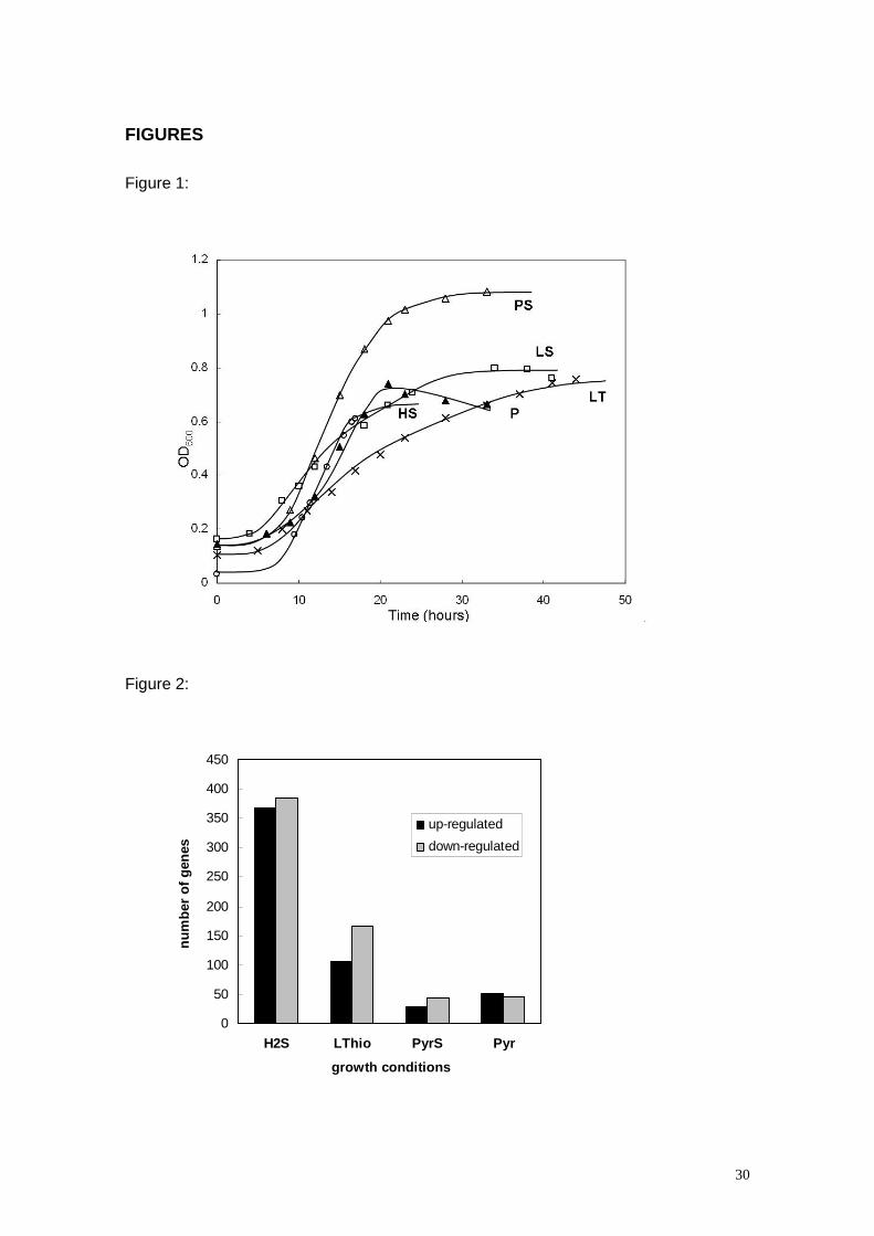
Gene expression profiling was performed using cells from D. vulgaris collected at
mid-exponential phase (Figure 1). The transcriptomic profiles of D. vulgaris cultures
grown in these conditions were determined using DNA microarrays representing
approximately 99% of the total protein-coding capacity of the D. vulgaris genome [18,
26]. Relative expression ratios were derived by comparing mRNA abundance levels in
cells grown in HS, PS, LT and P relative to mRNA levels in LS grown cells. The
number of genes displaying more than two fold change in transcript abundance, either
up or down and corresponding to a p value of 0.05 or smaller was 761 in HS, 272 in
LT, 73 in PS, and 96 in P (Figure 2). Of the 3,379 chromosomal genes in D. vulgaris,
2,315 genes have so far been classified into one of the 20 clusters of orthologous
groups of functional categories (COG) [30, 31]. Growth on HS shows considerable
9
changes in the transcription profile versus the LS reference across all COGs, whereas
the most significant changes found during growth on LT, PS and P as compared to LS,
involve primarily genes with functions in energy and central intermediary metabolism
(Table 1). A detailed list of energy metabolism genes that display changes in their
expression levels is presented in Table 2.
Changes in gene expression using hydrogen as electron donor
The use of hydrogen as electron donor lead to very significant changes in gene
expression relative to growth with lactate, with approximately 30% of the genes
involved in energy metabolism being affected (Table 2).
Periplasmic hydrogenases. As might be expected, some of the most affected genes
include those that code for hydrogenases. The genome of D. vulgaris includes four
periplasmic-facing hydrogenases, the [FeFe] hydrogenase (DVU1769-70; hydAB), two
[NiFe] hydrogenase isoenzymes (DVU1921-22; hynAB-1 and DVU2525-26; hynAB-2)
and a [NiFeSe] hydrogenase (DVU1917-18; hysAB). The genome also contains genes
for two cytoplasmically oriented hydrogenases, the Ech hydrogenase (DVU0429-34;
echABCDEF) and the Coo hydrogenase (DVU2286-93; cooMKLXUHF). It has not been
clearly established if these hydrogenases may have specific roles, or if this redundancy
allows functional compensation under different conditions [32, 33]. Recently, it was
shown that expression of the three main hydrogenases detected in D. vulgaris ([FeFe],
[NiFe]1 and [NiFeSe]) is affected by the metals available in the growth medium, and
that the [NiFeSe] hydrogenase is dominant when nickel and selenium are available
[25]. In the absence of selenium the [NiFeSe] hydrogenase is not detected and the
major hydrogenases present are the [NiFe]1 and the [FeFe] hydrogenases. In the
present case the hysAB genes displayed a strong increase in transcript level with
growth on HS, whereas the hydA gene transcript was slightly down-regulated and the
hynAB-1 genes showed no change. These data show that transcription of the [NiFeSe]

10
hydrogenase gene is strongly up-regulated when hydrogen is supplied as electron
donor, even when selenium is not available in the medium and thus the protein is not
synthesised. This indicates that the [NiFeSe] is the most responsive hydrogenase to
the presence of hydrogen. Western blot in similar growth conditions reveals a modest
increase of both the [NiFe]1 and [FeFe] hydrogenases when comparing HS to LS
conditions [25]. However, when nickel is present and selenium is not, the [NiFe]1
hydrogenase is also strongly increased with growth in hydrogen relative to lactate. Very
recently, it was also described that the transcription levels of the different
hydrogenases of D. vulgaris are affected by the hydrogen concentration in the gas
phase [29]. The hysAB transcript level is higher with hydrogen at 5% (v/v) than with
50% and lowest with LS, whereas the opposite is observed for hydAB. These results,
corroborated with studies of knock-out mutants, suggest that the high-activity, low-
affinity [FeFe] hydrogenase is preferred when H2 is plentiful, whereas the lower-activity,
higher-affinity [NiFeSe] hydrogenase is preferred when there is a low concentration of
H2. In our studies the hydrogen concentration used was 80% and the hysAB genes
were strongly up-regulated, but the other differences in growth conditions may preclude
a direct comparison of the results. Overall, these studies reveal that the regulation of
the periplasmic-facing hydrogenases of D. vulgaris is quite intricate and fine-tuned to
changes in the environmental conditions. The presence of several hydrogenases with
different properties and cofactors undoubtedly provides this organism with a great
flexibility in terms of hydrogen metabolism, which is a crucial metabolite in anaerobic
environments and also plays an essential role in its energetic metabolism.
Cytoplasmic facing hydrogenases. The two membrane-bound cytoplasmic-facing
hydrogenases showed opposite response when hydrogen was used as electron donor,
with the echABCDEF prominent among the gene clusters up-regulated and the
cooMKLXUHF prominent among those down-regulated. A similar situation was
reported for cells grown in formate/sulphate [23]. In Methanosarcina barkeri the Ech

11
hydrogenase complex has been shown to be multifunctional, with one of its roles being
to drive ferredoxin reduction with H2, which is used for CO2 fixation [34, 35]. This is a
crucial activity for D. vulgaris cells grown in hydrogen, since acetate and CO2 are the
only carbon sources available. Thus, the increased expression of the Ech hydrogenase
in these conditions points to its role in reducing ferredoxin for carbon fixation.
Unfortunately, no replicates were obtained for the ferredoxin I gene and therefore its
transcriptional response cannot be evaluated. The down-regulation of genes coding for
the Coo hydrogenase suggests that the CO pathway is operative during growth with
lactate, but does not function when H2 is used as electron donor.
Formate dehydrogenases. All three formate dehydrogenases encoded in the D.
vulgaris genome (DVU0587-88; DVU2481-85; DVU2809-12) and the pyruvate:formate
lyase activating enzyme (DVU2271; pflA) showed increased expression during growth
on hydrogen, as reported also for growth on formate [23]. This suggests that formate
cycling is occurring during growth with HS, providing an alternative pathway for energy
generation: CO2 and acetate are converted to formate, which is transported across the
membrane and is oxidised in the periplasm by the formate dehydrogenases, thus
contributing to the proton motive force as electrons are transferred back to the
cytoplasm to reduce sulphate [18, 19, 36].
Transmembrane complexes. Contrary to most organisms, the terminal reductases of
SRB are located in the cytoplasm, and therefore, are not directly involved in charge
translocation across the membrane. The electrons generated in the periplasm from
hydrogen oxidation have to be transported across the membrane to be used in the
reduction of sulphate. Desulfovibrio spp. contain several membrane-bound redox
complexes that can accept electrons from an abundant pool of periplasmic
cytochromes c that act as electron acceptors for the hydrogenases and formate
dehydrogenases [20]. The first such complex to be identified was the transmembrane
Hmc complex of D. vulgaris, which was proposed to accept electrons from periplasmic
12
hydrogenases via the type I cytochrome c3 (TpIc3), and to transfer them to the
cytoplasmic reduction of sulphate [37]. In support of this proposal increased expression
of the Hmc complex was observed when using hydrogen as electron donor [38], and a
∆hmc mutant where the hmc operon was knocked-out grew slower than the wild type in
these conditions [39]. Contrary to these results in the present study the hmc operon
was one of those more down-regulated with growth on H2. It is possible that the
conditions used here for HS growth (constant pH and with H2/CO2 bubbled through the
medium, removing H2S), which were very different from the referred studies, led to
down-regulation of the hmc genes. Our observation is corroborated by recent results
from the same group obtained with D. vulgaris grown with 5% and 50% hydrogen
showing also a reduced transcript level of the hmc operon relative to lactate growth
conditions [29]. An alternative transmembrane electron transfer pathway may involve
the Tmc complex, whose protein subunits are homologous to those of Hmc [40], given
that the gene encoding the cytochrome c subunit of this complex (tmcA, DVU0263)
was up-regulated in HS relative to LS. The TmcA cytochrome is actually a much better
electron acceptor for the periplasmic hydrogenases via TpIc3 than HmcA [41, 42].
The two membrane complexes QmoABC and DsrMKJOP, conserved in all SRB
sequenced to date, are thought to be involved in the sulphate reduction pathway as
electron donors to the enzymes APS reductase and sulphite reductase, respectively
[14-16]. Genes from both these complexes were down-regulated in H2-grown cells,
indicating that electron transport through Qmo and Dsr complexes is less important
during growth on hydrogen than on lactate. This may be related to the up-regulation of
the genes of the Tmc complex that may provide an alternative route for the flow of
electrons to the cytoplasmic terminal reductases. The three complexes, Tmc, Hmc and
Dsr have a homologous cytoplasmic subunit, which displays spectroscopic features
indicative of the presence of a special [4Fe4S]3+ center [16, 40], and this suggests this
13
subunit may play similar roles in the cytoplasm. The transmembrane electron flow
through the Tmc complex may allow a reduced electron flow through the Qmo and Dsr-
associated pathways. This proposal is in agreement with the fact that the genes
encoding the APS reductase (DVU0846/7) and sulphite reductase (DVU0402/4) did not
display significant differences in transcript levels.
Another membrane redox complex of unknown function, RnfCDGEAB (DVU2792-
97), showed up-regulation for several genes. This complex may interact with a
decaheme cytochrome c encoded in an adjacent gene (DVU2791; DhcA). The rnf
genes may be associated with different functions in different organisms. They were first
identified in Rhodobacter capsulatus as being involved in nitrogen fixation [43]. The
three integral membrane subunits RnfADE, and the cytoplasmic RnfG subunit, show
similarity to subunits of the Nqr complex of Vibrio spp., a Na+ - translocating
NADH:quinone oxidoreductase [44]. In E. coli the Rnf complex is named Rsx and is
involved in keeping the redox-sensitive transcriptional factor SoxR in its inactive
reduced state during aerobic growth [45]. There is so far no information as to the
possible role of the Rnf complex in Desulfovibrio spp.
Ethanol pathway. Some studies have suggested the involvement of an additional
bioenergetic pathway in D. vulgaris involving ethanol, which can be oxidised by an
alcohol dehydrogenase and the reducing equivalents transferred to sulphate reduction,
through a still uncertain mechanism involving hdrABC and other gene products [46]. A
relationship between ethanol and H2 metabolism was first revealed by the fact that a D.
vulgaris mutant lacking the [FeFe] hydrogenase had very low levels of the DVU2405-
encoded alcohol dehydrogenase, which is one of the most highly expressed proteins in
several growth conditions [46, 47]. This pathway is more active during exponential than
stationary growth phases [23]. In HS-grown cells there is an up-regulation of this
alcohol dehydrogenase gene (DVU2405; adh), as previously reported [46], and the
gene for a subunit of a putative heterodisulphide reductase (DVU2404; hdrC),

14
suggesting that this pathway is more important than in LS growth conditions. This
pathway provides an alternative route for electron transfer to sulphate reduction, and
may also be associated with the down-regulation of genes encoding the Qmo and Dsr
complexes, which contain subunits homologous to Hdr proteins.
Changes in gene expression using pyruvate as energy source
The number of energy metabolism genes with modified expression in PS relative to
LS was quite small and all changes were below four-fold either up or down (Table 2).
This agrees with the fact that oxidation of lactate proceeds via pyruvate. Three of the
ech genes were up-regulated. In these growth conditions carbon fixation is not required
and it is more likely that the Ech hydrogenase is acting to generate H2 from ferredoxin,
which is reduced by the pyruvate:ferredoxin oxidoreductase. This process is associated
with energy conservation as proposed in the hydrogen-cycling hypothesis, and
suggests a greater relevance for this pathway with pyruvate than with lactate. The
observed up-regulation of the ech operon in HS and PS conditions suggests that in D.
vulgaris, as in M. barkeri [34], the Ech hydrogenase is able to function bi-directionally
depending on the growth conditions. Among the few energy metabolism genes down-
regulated were the hmc genes (DVU0533; hmcD), the fhdA gene (DVU0587) coding for
the catalytic subunit of the periplasmic FdhAB formate dehydrogenase, and the gene
coding for the redox protein flavodoxin (DVU2680; fla).
In cells grown with pyruvate and a restricted amount of sulphate more energy
metabolism genes were up-regulated than in PS-grown cells, but all expression ratios
were below three-fold (Table 2). D. vulgaris does not grow on pyruvate alone unless a
small amount of sulphate is present at the beginning of growth, probably due to the
inhibitory effect of accumulation of reduced compounds during the fermentation burst
[17, 48]. In this study a small amount of sulphate was present in the medium because
the inoculum was made using cells grown in PS conditions. Interestingly, and contrary
15
to our expectation, in these sulphate-limited conditions the genes for several proteins
thought to be involved in sulphate respiration showed increased expression. These
included APS reductase, some subunits of the Qmo and Dsr complexes, the TpIc3
(DVU3171), the [NiFe]2 hydrogenase (DVU2524-25), and several genes of the Hmc
complex. The genes for two other membrane redox complexes of unknown function
were also up-regulated (DVU0692/3 and DVU3143/4). The increased expression of the
Hmc complex agrees with published experiments, which showed that in similar growth
conditions a ∆hmc mutant accumulates large amounts of hydrogen, in contrast to the
wild type [17]. These results suggest that during growth in pyruvate with limiting
sulphate, cycling of hydrogen is also occurring. Only two energy metabolism genes
were down-regulated, fla and fhdA genes as observed also in PS. The reduced level of
transcription of the gene coding for flavodoxin in both pyruvate growth conditions
suggests a role for this protein in the lactate oxidation pathway upstream of pyruvate.
Changes in gene expression using thiosulphate as electron acceptor
Although previous studies have addressed the effect of different electron donors in
the transcriptional and translational response of D. vulgaris [23, 46] this is the first time
that the effect of a different electron acceptor is explored. In contrast to sulphate,
thiosulphate does not require prior activation to be used as electron acceptor and
therefore a higher cell yield would be expected. However, the yield of cells grown in LT
is lower than in LS as previously reported [48]. The down-regulation of genes coding
for ATP synthase (DVU0774-79), APS reductase (DVU0846-47; apsAB), the Qmo and
Dsr complexes, and the [NiFe]2 hydrogenase (Table 2) suggests a low energy state of
the cells, which agrees with the slower growth rate (Figure 1). Reduction of
thiosulphate yields sulphite, which at high concentrations is toxic [49]. It is possible that
accumulation of this compound led to down-regulation of genes involved in the
16
sulphate reduction pathway as observed for nitrite, which inhibits the sulphite reductase
and thus leads to an accumulation of sulphite [14, 26]. In agreement with this proposal
several of the genes mentioned above were also down-regulated in D. vulgaris cells
grown in the presence of nitrite [14, 26]. Some genes of the membrane-bound
(DVU2482; fdnG) and the three-subunit (DVU2811; fdhB) formate dehydrogenases
were also down-regulated, whereas the genes coding for the periplasmic two-subunit
formate dehydrogenase (DVU0587/8; fdhAB) were up-regulated indicating different
regulation for these proteins in LT, in contrast with what was observed in HS
conditions. The up-regulation of the genes coding for the [FeFe] hydrogenase, which
displays the highest rates of hydrogen uptake when compared with the other
periplasmic hydrogenases [50], may serve to improve energy recovery through H2
cycling. The gene coding for a periplasmic octaheme cytochrome c (DVU3107) of
unknown function is the most down-regulated energy metabolism gene in these
conditions. The physiological role of this cytochrome is not known but the considerable
down regulation in these conditions and lack of transcriptional response of this gene in
the other conditions tested suggests a role in sulphate reduction. This is supported by
the significant down-regulation of this gene in the transition from exponential to
stationary phase in D. vulgaris cells grown with lactate/sulphate and with
formate/sulphate [23]. Unexpectedly, the gene for one of the subunits of the putative
thiosulphate reductase (DVU0172; phsB) was down-regulated, as well as a putative
lactate dehydrogenase (DVU2784; lldD), which suggests that these genes may code
for proteins with different functions from those indicated in the genome annotation.
Finally, the gene for an iron-sulphur flavoprotein (DVU0819; isf) is noteworthy
because it was up-regulated in all conditions tested. This protein is homologous to the
Isf protein of Methanosarcina thermophilus, where it plays a role in electron transport
during fermentation of acetate to methane by accepting electrons from ferredoxin [51].
The role of Isf in Desulfovibrio metabolism has not been investigated.

17
Overall view of the bioenergetic metabolism of D. vulgaris from the transcriptional
studies
In this work, the global gene expression response of D. vulgaris to growth on
different electron donors and acceptors provided several important insights into the
bioenergetic pathways of this organism, and its response to different growth conditions.
A simplified scheme of these pathways deduced from the transcriptional results in this
work, and taking into account previously published results [17, 23] is presented in
Figure 3. The change from an organic carbon and energy source (lactate) to H2 as
energy source and CO2/acetate as carbon source results in a strong shift in the
transcriptional pattern of D. vulgaris. Since hydrogen is a ubiquitous metabolite in
anaerobic environments, and it is likely to be an important energy source for
Desulfovibrio spp. in their natural habitats, these results are of great physiological
significance. The evidence indicates a shift in metabolic trafficking involving different
bioenergetic pathways when changing from lactate to H2. The formate cycling pathway
is more relevant for growth with H2 than lactate, whereas the contrary is observed for
the CO cycling pathway. The results provide also evidence for the existence of a
pathway involving ethanol that is up-regulated during growth in H2, and provides a
soluble route for electron transfer to sulphate reduction. This pathway involves several
novel proteins of unknown function that should be further investigated. Regarding the
complexes involved in the transmembrane electron transfer the evidence indicates that
the Tmc is preferred during growth with H2, whereas the Qmo and Dsr are more
relevant during growth with carbon sources.
An interesting observation from the present results is that when several, seemingly
redundant, isoenzymes are present, as in the case of hydrogenases or formate
dehydrogenases, there are different transcriptional responses to the changes in growth
conditions, indicating that these isoenzymes have specialized roles and are not
18
completely interchangeable. All the studies published so far indicate that there is a fine-
tuning of the activity of the periplasmic hydrogenases, which should permit a rapid
adaptation to changing environmental conditions, including different intracellular and
extracellular concentrations of H2 or metals available. The present study also gives
strong support for a bifunctional role of the cytoplasmic Ech hydrogenase that may
reduce ferredoxin for carbon fixation during growth in H2, or oxidize ferredoxin forming
H2 for hydrogen cycling during growth in PS.
Finally, the results reported in this work identified several novel gene products that
are involved in energy metabolism and that merit further study to clarify their function.
Examples are the Rnf complex (and other membrane redox complexes), the proteins
involved in the ethanol pathway, the octaheme cytochrome c, the Isf protein, flavodoxin
and several others. Our study highlights the high complexity and plasticity of the D.
vulgaris energetic metabolism and show that further studies are warranted before
sulphate respiration can be fully understood.
ACKNOWLEDGEMENTS
This work was supported by Fundação para a Ciência e Tecnologia grants
POCI/2004/QUI/55690 to ROL and PTDC/QUI/68368/2006 to IACP, co-funded by
FEDER program, and was part of the Virtual Institute for Microbial Stress and Survival
(http://VIMSS.lbl.gov) supported by the U. S. Department of Energy, Office of Science,
Office of Biological and Environmental Research, Genomics Program:GTL through
contractDE-AC02-05CH11231 between Lawrence Berkeley National Laboratory and
the U. S. Department of Energy. PMP was a recipient of the FCT PhD grant
SFRH/BD/5231/2001. The authors are grateful to Prof. Gerrit Voordouw for helpful
discussions and sharing data prior to publication.

19
REFERENCES
1 Rabus, R., Hansen, T. and Widdel, F. (2001) Dissimilatory sulfate- and sulfur-
reducing prokaryotes. In The prokaryotes: an evolving electronic resource for
the microbiologic community [online] (Dworkin, M., Falkow, S., Rosenberg, E.,
Schleifer, K.-H. and Stackebrandt, E., eds.), Springer Science Online,
Heidelberg, Germany
2 Truper, H. G. (1984) In Sulfur: Its Significance for Chemistry, for the Geo-, Bio-
and Cosmophere, and Technology (Müller, A. K., B., ed.), pp. 351-365,
Elsevier, Amsterdam
3 Hamilton, W. A. (1998) Bioenergetics of sulphate-reducing bacteria in relation to
their environmental impact. Biodegradation 9: 201-212
4 Dinh, H. T., Kuever, J., Mussmann, M., Hassel, A. W., Stratmann, M. and
Widdel, F. (2004) Iron corrosion by novel anaerobic microorganisms. Nature
427: 829-832
5 Lloyd, J. R. (2003) Microbial reduction of metals and radionuclides. FEMS
Microbiol Rev 27: 411-425
6 Wall, J. D. and Krumholz, L. R. (2006) Uranium Reduction. Ann Rev Microbiol
60: 149-166
7 Odom, J. M. and Peck, H. D. (1981) Hydrogen cycling as a general mechanism
for energy coupling in the sulphate-reducing bacterium Desulfovibrio sp. FEMS
Microbiol Lett 12: 47-50
8 Pankhania, I. P., Spormann, A. M., Hamilton, W. A. and Thauer, R. K. (1988)
Lactate conversion to acetate, CO2 and H2 in cell-suspensions of Desulfovibrio
vulgaris (Marburg) - Indications for the involvement of an energy driven
reaction. Arch Microbiol 150: 26-31
9 Traoré, A. S., Hatchikian, C. E., Belaich, J. P. and Le Gall, J. (1981)
Microcalorimetric studies of the growth of sulfate-reducing bacteria: energetics
of Desulfovibrio vulgaris growth. J Bacteriol 145: 191-199
10 Rabus, R., Ruepp, A., Frickey, T., Rattei, T., Fartmann, B., Stark, M., Bauer, M.,
Zibat, A., Lombardot, T., Becker, I., Amann, J., Gellner, K., Teeling, H.,
Leuschner, W. D., Glockner, F. O., Lupas, A. N., Amann, R. and Klenk, H. P.

20
(2004) The genome of Desulfotalea psychrophila, a sulfate-reducing bacterium
from permanently cold Arctic sediments. Environ Microbiol 6: 887-902
11 Lupton, F. S., Conrad, R. and Zeikus, J. G. (1984) Physiological function of
hydrogen metabolism during growth of sulfidogenic bacteria on organic
substrates. J Bacteriol 159: 843-849
12 Wood, P. M. (1978) A chemiosmotic model for sulphate respiration. FEBS Letts
95: 12-18
13 Maroc, J., Azerad, R., Kamen, M. D. and Le Gall, J. (1970) Menaquinone (MK-
6) in the sulfate-reducing obligate anaerobe, Desulfovibrio. Biochim Biophys
Acta 197: 87-89
14 Haveman, S. A., Greene, E. A., Stilwell, C. P., Voordouw, J. K. and Voordouw,
G. (2004) Physiological and gene expression analysis of inhibition of
Desulfovibrio vulgaris Hildenborough by nitrite. J Bacteriol 186: 7944-7950
15 Pires, R. H., Lourenco, A. I., Morais, F., Teixeira, M., Xavier, A. V., Saraiva, L.
M. and Pereira, I. A. (2003) A novel membrane-bound respiratory complex from
Desulfovibrio desulfuricans ATCC 27774. Biochim Biophys Acta 1605: 67-82
16 Pires, R. H., Venceslau, S. S., Morais, F., Teixeira, M., Xavier, A. V. and
Pereira, I. A. C. (2006) Characterization of the Desulfovibrio desulfuricans
ATCC 27774 DsrMKJOP complex - a membrane-bound redox complex
involved in sulfate respiration. Biochemistry 45: 249-262
17 Voordouw, G. (2002) Carbon monoxide cycling by Desulfovibrio vulgaris
Hildenborough. J Bacteriol 184: 5903-5911
18 Heidelberg, J. F., Seshadri, R., Haveman, S. A., Hemme, C. L., Paulsen, I. T.,
Kolonay, J. F., Eisen, J. A., Ward, N., Methe, B., Brinkac, L. M., Daugherty, S.
C., Deboy, R. T., Dodson, R. J., Durkin, A. S., Madupu, R., Nelson, W. C.,
Sullivan, S. A., Fouts, D., Haft, D. H., Selengut, J., Peterson, J. D., Davidsen, T.
M., Zafar, N., Zhou, L. W., Radune, D., Dimitrov, G., Hance, M., Tran, K.,
Khouri, H., Gill, J., Utterback, T. R., Feldblyum, T. V., Wall, J. D., Voordouw, G.
and Fraser, C. M. (2004) The genome sequence of the anaerobic, sulfate-
reducing bacterium Desulfovibrio vulgaris Hildenborough. Nat Biotechnol 22:
554-559
19 Tang, Y., Pingitore, F., Mukhopadhyay, A., Phan, R., Hazen, T. C. and
Keasling, J. D. (2007) Pathway confirmation and flux analysis of central
metabolic pathways in Desulfovibrio vulgaris Hildenborough using gas
21
chromatography-mass spectrometry and Fourier transform-ion cyclotron
resonance mass spectrometry. J Bacteriol 189: 940-949
20 Pereira, I. A. C., Haveman, S. A. and Voordouw, G. (2007) Biochemical, genetic
and genomic characterization of anaerobic electron transport pathways in
sulphate-reducing delta-proteobacteria. In Sulphate-Reducing Bacteria:
Environmental and Engineered Systems (L.L., B. and W.A., H., eds.),
Cambridge University Press, Cambridge, UK
21 Rapp-Giles, B. J., Casalot, L., English, R. S., Ringbauer, J. A., Jr., Dolla, A. and
Wall, J. D. (2000) Cytochrome c3 mutants of Desulfovibrio desulfuricans. Appl
Environ Microbiol 66: 671-677
22 Nie, L., Wu, G. and W., Z. (2006) Correlation between mRNA and protein
abundance in Desulfovibrio vulgaris: a multiple regression to identify sources of
variations. Biochem Bioplys Res Commun 339: 603-610
23 Zhang, W., Culley, D. E., Scholten, J. C., Hogan, M., Vitiritti, L. and Brockman,
F. J. (2006) Global transcriptomic analysis of Desulfovibrio vulgaris on different
electron donors. Antonie Van Leeuwenhoek 89: 221-237
24 Postgate, J. R. (1984) The sulphate-reducing bacteria. Cambridge University
Press, Cambridge, United Kingdom
25 Valente, F. A. A., Almeida, C. C., Pacheco, I., Carita, J., Saraiva, L. M. and
Pereira, I. A. C. (2006) Selenium is involved in regulation of periplasmic
hydrogenase gene expression in Desulfovibrio vulgaris Hildenborough. J
Bacteriol 188: 3228-3235
26 He, Q., Huang, K. H., He, Z., Alm, E. J., Fields, M. W., Hazen, T. C., Arkin, A.
P., Wall, J. D. and Zhou, J. (2006) Energetic Consequences of Nitrite Stress in
Desulfovibrio vulgaris Hildenborough, Inferred from Global Transcriptional
Analysis. Appl Environ Microbiol 72: 4370-4381
27 Clark, M. E., He, Q., He, Z., Huang, K. H., Alm, E. J., Wan, X. F., Hazen, T. C.,
Arkin, A. P., Wall, J. D., Zhou, J. Z. and Fields, M. W. (2006) Temporal
transcriptomic analysis as Desulfovibrio vulgaris hildenborough transitions into
stationary phase during electron donor depletion. Appl Environ Microbiol 72:
5578-5588
28 Mukhopadhyay, A., Redding, A., Joachimiak, M. P., Arkin, A., Borglin, S. C.,
Dehal, P. S., Chakraborty, R., Geller, J. T., Hazen, T. C., He, Q., Joyner, D. C.,
Martin, V. J. J., Wall, J., Yang, Z. K. and Keasling, J. D. (2007) Cell wide
22
responses to low oxygen exposure in Desulfovibrio vulgaris Hildenborough. J
Bacteriol 189: 5996-6010
29 Caffrey, S. M., Park, H.-S., Voordouw, J. K., He, Z., Zhou, J. and Voordouw, G.
(2007) Function of periplasmic hydrogenases in the sulfate reducing bacterium
Desulfovibrio vulgaris Hildenborough. J Bacteriol 189: 6159-6167
30 Tatusov, R. L., Koonin, E. V. and Lipman, D. J. (1997) A genomic perspective
on protein families. Science 278: 631-637
31 Tatusov, R. L., Fedorova, N. D., Jackson, J. D., Jacobs, A. R., Kiryutin, B.,
Koonin, E. V., Krylov, D. M., Mazumder, R., Mekhedov, S. L., Nikolskaya, A. N.,
Rao, B. S., Smirnov, S., Sverdlov, A. V., Vasudevan, S., Wolf, Y. I., Yin, J. J.
and Natale, D. A. (2003) The COG database: an updated version includes
eukaryotes. BMC Bioinformatics 4: 41
32 Goenka, A., Voordouw, J. K., Lubitz, W., Gartner, W. and Voordouw, G. (2005)
Construction of a [NiFe]-hydrogenase deletion mutant of Desulfovibrio vulgaris
Hildenborough. Biochem Soc Trans 33: 59-60
33 Pohorelic, B. K., Voordouw, J. K., Lojou, E., Dolla, A., Harder, J. and Voordouw,
G. (2002) Effects of deletion of genes encoding Fe-only hydrogenase of
Desulfovibrio vulgaris Hildenborough on hydrogen and lactate metabolism. J
Bacteriol 184: 679-686
34 Meuer, J., Kuettner, H. C., Zhang, J. K., Hedderich, R. and Metcalf, W. W.
(2002) Genetic analysis of the archaeon Methanosarcina barkeri Fusaro reveals
a central role for Ech hydrogenase and ferredoxin in methanogenesis and
carbon fixation. Proc Natl Acad Sci 99: 5632-5637
35 Hedderich, R. and Forzi, L. (2005) Energy converting [NiFe] hydrogenases:
more than just H2 activation. J Mol Microbiol Biotechnol 10: 92-104
36 Badziong, W., Bernhard, D. and Thauer, R. K. (1979) Acetate and carbon
dioxide assimilation by Desulfovibrio vulgaris (Marburg), growing on hydrogen
and sulfate as sole energy source. Arch Microbiol 123: 301-305
37 Rossi, M., Pollock, W. B., Reij, M. W., Keon, R. G., Fu, R. and Voordouw, G.
(1993) The hmc operon of Desulfovibrio vulgaris subsp. vulgaris Hildenborough
encodes a potential transmembrane redox protein complex. J Bacteriol 175:
4699-4711
38 Keon, R. G., Fu, R. and Voordouw, G. (1997) Deletion of two downstream
genes alters expression of the hmc operon of Desulfovibrio vulgaris subsp.
vulgaris Hildenborough. Arch Microbiol 167: 376-383
23
39 Dolla, A., Pohorelic, B. K., Voordouw, J. K. and Voordouw, G. (2000) Deletion
of the hmc operon of Desulfovibrio vulgaris subsp. vulgaris Hildenborough
hampers hydrogen metabolism and low-redox-potential niche establishment.
Arch Microbiol 174: 143-151
40 Pereira, P. M., Teixeira, M., Xavier, A. V., Louro, R. O. and Pereira, I. A. C.
(2006) The Tmc complex from Desulfovibrio vulgaris Hildenborough is involved
in transmembrane electron transfer from periplasmic hydrogen oxidation.
Biochemistry 45: 10359-10367
41 Pereira, I. A., Romão, C. V., Xavier, A. V., Le Gall, J. and Teixeira, M. (1998)
Electron transfer between hydrogenases and mono and multiheme
cytochromes in Desulfovibrio spp. J. Biol. Inorg. Chem. 3: 494-498
42 Valente, F. M., Saraiva, L. M., LeGall, J., Xavier, A. V., Teixeira, M. and Pereira,
I. A. (2001) A membrane-bound cytochrome c3: a type II cytochrome c3 from
Desulfovibrio vulgaris Hildenborough. Chembiochem 2: 895-905
43 Schmehl, M., Jahn, A., Vilsendorf, A. M. Z., Hennecke, S., Masepohl, B.,
Schuppler, M., Marxer, M., Oelze, J. and Klipp, W. (1993) Identification of a new
class of nitrogen-fixation genes in Rhodobacter capsulatus - a putative
membrane complex involved in electron-transport to nitrogenase. Mol Gen
Genet 241: 602-615
44 Steuber, J. (2001) Na(+) translocation by bacterial NADH:quinone
oxidoreductases: an extension to the complex-I family of primary redox pumps.
Biochim Biophys Acta 1505: 45-56
45 Koo, M. S., Lee, J. H., Rah, S. Y., Yeo, W. S., Lee, J. W., Lee, K. L., Koh, Y. S.,
Kang, S. O. and Roe, J. H. (2003) A reducing system of the superoxide sensor
SoxR in Escherichia coli. Embo J 22: 2614-2622
46 Haveman, S. A., Brunelle, V., Voordouw, J. K., Voordouw, G., Heidelberg, J. F.
and Rabus, R. (2003) Gene expression analysis of energy metabolism mutants
of Desulfovibrio vulgaris Hildenborough indicates an important role for alcohol
dehydrogenase. J Bacteriol 185: 4345-4353
47 Zhang, W., Gritsenko, M. A., Moore, R. J., Culley, D. E., Nie, L., Petritis, K.,
Strittmatter, E. F., Camp, D. G., 2nd, Smith, R. D. and Brockman, F. J. (2006) A
proteomic view of Desulfovibrio vulgaris metabolism as determined by liquid
chromatography coupled with tandem mass spectrometry. Proteomics 6: 4286-
4299

24
48 Magee, E. L., Ensley, B. D., Jr. and Barton, L. L. (1978) An assessment of
growth yields and energy coupling in Desulfovibrio. Arch Microbiol 117: 21-26
49 Badziong, W. and Thauer, R. K. (1978) Growth yields and growth rates of
Desulfovibrio vulgaris (Marburg) growing on hydrogen plus sulfate and
hydrogen plus thiosulfate as the sole energy sources. Arch Microbiol 117: 209-
214
50 Valente, F. M., Oliveira, A. S., Gnadt, N., Pacheco, I., Coelho, A. V., Xavier, A.
V., Teixeira, M., Soares, C. M. and Pereira, I. A. (2005) Hydrogenases in
Desulfovibrio vulgaris Hildenborough: structural and physiologic
characterisation of the membrane-bound [NiFeSe] hydrogenase. J Biol Inorg
Chem 10: 667-682
51 Zhao, T., Cruz, F. and Ferry, J. G. (2001) Iron-sulfur flavoprotein (Isf) from
Methanosarcina thermophila is the prototype of a widely distributed family. J
Bacteriol 183: 6225-6233

25
TABLES
Table 1. Distribution of up- and down-regulated genes in D. vulgaris grown with different electron donors and acceptors for
the COG functional categories presenting greater changes. The total number refers to the number of the all genes changed in each
growth condition. Only those genes with p-value ≤ 0.05 and a ratio value ≤ 0.5 and ≥ 2.0 are included.
COG functional category
HS LT PS P
Total 761 Total 272 Total 73 Total 96
No. of genes No. of genes No. of genes No. of genes
up down up down up down up down
Cell envelope 22 29 7 14 1 3 9 3
Energy and central intermediary metabolism 45 35 14 27 7 4 20 3
Protein synthesis 2 42 1 8 0 0 1 0
Regulatory functions 22 17 8 7 3 3 3 2
Transport and binding proteins 34 31 4 25 0 6 1 8
Hypothetical proteins 129 120 36 39 11 18 12 21
Other or unknown function 115 118 37 45 7 10 5 8
26
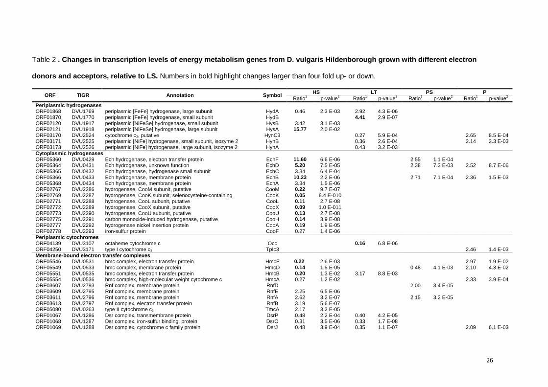
Table 2 . Changes in transcription levels of energy metabolism genes from D. vulgaris Hildenborough grown with different electron
donors and acceptors, relative to LS. Numbers in bold highlight changes larger than four fold up- or down.
ORF TIGR Annotation Symbol HS LT PS P Ratio1 p-value2 Ratio1 p-value2 Ratio1 p-value2 Ratio1 p-value2
Periplasmic hydrogenases ORF01868 DVU1769 periplasmic [FeFe] hydrogenase, large subunit HydA 0.46 2.3 E-03 2.92 4.3 E-06 ORF01870 DVU1770 periplasmic [FeFe] hydrogenase, small subunit HydB 4.41 2.9 E-07 ORF02120 DVU1917 periplasmic [NiFeSe] hydrogenase, small subunit HysB 3.42 3.1 E-03 ORF02121 DVU1918 periplasmic [NiFeSe] hydrogenase, large subunit HysA 15.77 2.0 E-02 ORF03170 DVU2524 cytochrome c3, putative HynC3 0.27 5.9 E-04 2.65 8.5 E-04 ORF03171 DVU2525 periplasmic [NiFe] hydrogenase, small subunit, isozyme 2 HynB 0.36 2.6 E-04 2.14 2.3 E-03 ORF03173 DVU2526 periplasmic [NiFe] hydrogenase, large subunit, isozyme 2 HynA 0.43 3.2 E-03 Cytoplasmic hydrogenases ORF05360 DVU0429 Ech hydrogenase, electron transfer protein EchF 11.60 6.6 E-06 2.55 1.1 E-04 ORF05364 DVU0431 Ech hydrogenase, unknown function EchD 5.20 7.5 E-05 2.38 7.3 E-03 2.52 8.7 E-06 ORF05365 DVU0432 Ech hydrogenase, hydrogenase small subunit EchC 3.34 6.4 E-04 ORF05366 DVU0433 Ech hydrogenase, membrane protein EchB 10.23 2.2 E-06 2.71 7.1 E-04 2.36 1.5 E-03 ORF05368 DVU0434 Ech hydrogenase, membrane protein EchA 3.34 1.5 E-06 ORF02767 DVU2286 hydrogenase, CooM subunit, putative CooM 0.22 9.7 E-07 ORF02769 DVU2287 hydrogenase, CooK subunit, selenocysteine-containing CooK 0.05 8.4 E-010 ORF02771 DVU2288 hydrogenase, CooL subunit, putative CooL 0.11 2.7 E-08 ORF02772 DVU2289 hydrogenase, CooX subunit, putative CooX 0.09 1.0 E-011 ORF02773 DVU2290 hydrogenase, CooU subunit, putative CooU 0.13 2.7 E-08 ORF02775 DVU2291 carbon monoxide-induced hydrogenase, putative CooH 0.14 3.9 E-08 ORF02777 DVU2292 hydrogenase nickel insertion protein CooA 0.19 1.9 E-05 ORF02778 DVU2293 iron-sulfur protein CooF 0.27 1.4 E-06 Periplasmic cytochromes ORF04139 DVU3107 octaheme cytochrome c Occ 0.16 6.8 E-06 ORF04250 DVU3171 type I cytochrome c3 TpIc3 2.46 1.4 E-03 Membrane-bound electron transfer complexes ORF05546 DVU0531 hmc complex, electron transfer protein HmcF 0.22 2.6 E-03 2.97 1.9 E-02 ORF05549 DVU0533 hmc complex, membrane protein HmcD 0.14 1.5 E-05 0.48 4.1 E-03 2.10 4.3 E-02 ORF05551 DVU0535 hmc complex, electron transfer protein HmcB 0.20 1.3 E-02 3.17 8.8 E-03 ORF05554 DVU0536 hmc complex, high-molecular weight cytochrome c HmcA 0.27 1.2 E-02 2.33 3.9 E-04 ORF03607 DVU2793 Rnf complex, membrane protein RnfD 2.00 3.4 E-05 ORF03609 DVU2795 Rnf complex, membrane protein RnfE 2.25 6.5 E-06 ORF03611 DVU2796 Rnf complex, membrane protein RnfA 2.62 3.2 E-07 2.15 3.2 E-05 ORF03613 DVU2797 Rnf complex, electron transfer protein RnfB 3.19 5.6 E-07 ORF05080 DVU0263 type II cytochrome c3 TmcA 2.17 3.2 E-05 ORF01067 DVU1286 Dsr complex, transmembrane protein DsrP 0.48 2.2 E-04 0.40 4.2 E-05 ORF01068 DVU1287 Dsr complex, iron-sulfur binding protein DsrO 0.31 3.5 E-06 0.33 1.7 E-08 ORF01069 DVU1288 Dsr complex, cytochrome c family protein DsrJ 0.48 3.9 E-04 0.35 1.1 E-07 2.09 6.1 E-03

27
ORF01070 DVU1289 Dsr complex, iron-sulfur binding subunit DsrK 0.37 2.9 E-06 0.36 3.2 E-06 2.16 1.8 E-04 ORF01072 DVU1290 Dsr complex, membrane cytochrome b protein DsrM 0.29 6.1 E-07 0.38 7.3 E-05 ORF00341 DVU0848 Qmo complex, flavin protein QmoA 0.37 1.0 E-06 0.44 1.4 E-02 2.27 2.3 E-02 ORF00343 DVU0849 Qmo complex, flavin protein QmoB 0.46 8.7 E-03 2.36 1.6 E-02 ORF00345 DVU0850 Qmo complex, membrane iron-sulfur protein QmoC 0.47 1.2 E-02 ORF04199 DVU3143 iron-sulfur cluster binding protein OhcB 2.63 1.6 E-04 ORF04200 DVU3144 octaheme cytochrome c OhcA 2.13 2.5 E-04 ORF00078 DVU0692 molybdopterin oxidoreductase, transmembrane subunit 2.05 5.0 E-02 ORF00079 DVU0693 molybdopterin oxidoreductase, iron-sulfur cluster-binding subunit 2.45 8.9 E-03 Formate dehydrogenases ORF05640 DVU0587 formate dehydrogenase, α subunit FdhA 4.05 5.7E-03 3.89 3.2E-07 0.43 1.3E-04 0.44 1.4E-04 ORF05642 DVU0588 formate dehydrogenase, β subunit FdhB 3.00 1.5E-03 3.07 1.0E-06 ORF03099 DVU2481 formate dehydrogenase, β subunit CfdB 2.73 4.0E-06 ORF03102 DVU2482 formate dehydrogenase, α subunit CfdA 2.11 6.2 E-03 0.49 4.6 E-03 ORF03638 DVU2809 cytochrome c3 FdhC3 3.95 7.1 E-04 ORF03639 DVU2810 formate dehydrogenase, formation protein, putative FdhE 3.10 3.6 E-03 ORF03640 DVU2811 formate dehydrogenase, β subunit FdhB 5.24 8.9 E-05 ORF03641 DVU2812 formate dehydrogenase, α subunit FdhA 5.07 6.9 E-05 Enzymes involved in the reduction of sulfur oxo-anions ORF00336 DVU0846 adenylyl sulfate reductase , β subunit ApsB 0.49 4.9 E-04 2.47 5.7 E-03 ORF00338 DVU0847 adenylyl sulfate reductase , α subunit ApsA 0.29 6.0 E-04 2.25 2.8 E-02 ORF04933 DVU0172 thiosulfate reductase, iron-sulfur binding protein phsB 2.14 1.2 E-04 0.42 1.1 E-05 Electron transfer proteins ORF00292 DVU0819 iron-sulfur flavoprotein Isf 3.12 1.3 E-04 4.15 1.5 E-05 3.66 1.7 E-07 2.58 3.5 E-05 ORF02976 DVU2404 heterodissulfide oxidoreductase, subunit C hdrC 2.01 1.2 E-03 ORF03423 DVU2680 flavodoxin Fla 0.32 1.3 E-06 0.37 9.8 E-05 Oxidative stress responsive proteins ORF04274 DVU3185 rubredoxin-oxygen oxidoreductase roO 0.43 2.0 E-04 ORF04112 DVU3093 rubredoxin-like protein rdl 2.45 1.8 E-04 ORF01341 DVU1457 thioredoxin reductase trxB 4.81 1.5 E-07 ORF01989 DVU1839 Thioredoxin trxA 2.55 3.6 E-05 ATP synthase ORF00217 DVU0774 ATP synthase, F1 ε subunit AtpC 0.50 4.9 E-03 ORF00219 DVU0775 ATP synthase, F1 β subunit AtpD 0.43 1.9 E-03 ORF00220 DVU0776 ATP synthase, F1 γ subunit AtpG 0.37 1.4 E-04 ORF00223 DVU0777 ATP synthase, F1 α subunit AtpA 0.29 7.9 E-04 ORF00224 DVU0778 ATP synthase, F1 δ subunit AtpH 0.46 5.2 E-03 ORF00226 DVU0779 ATP synthase, F0, B subunit, putative AtpF2 0.46 7.1 E-04 ORF00462 DVU0918 ATP synthase, F0, A subunit AtpB 0.32 2.5 E-07 Carbon metabolizing enzymes ORF05664 DVU0600 L-lactate dehydrogenase ldh 2.85 1.2 E-05 ORF03593 DVU2784 Lactate dehydrogenase, FMN-dependent family lldD 4.29 1.7 E-10 0.36 3.5 E-02 ORF02745 DVU2271 Pyruvate:formate-lyase pflA 4.43 7.6 E-05 ORF02977 DVU2405 alcohol dehydrogenase adh 2.21 1.3 E-02
1 mRNA abundance levels in cells grown in HS, PS, LT and P relative to mRNA levels in cells grown in LS

28
2 probability that the mRNA abundance remained unchaged

29
FIGURE LEGENDS
Figure 1- Growth curves of D. vulgaris. �-LS; �-HS; �-PS; �-P; �-LT. Points
are averages of two independent growth experiments.
Figure 2- Distribution of up- and down-regulated genes in D. vulgaris
Hildenborough as a function of different growth conditions. Only those genes with
p -value ≤ 0.05 and a ratio value ≤ 0.5 and ≥ 2.0 are included in the plot.
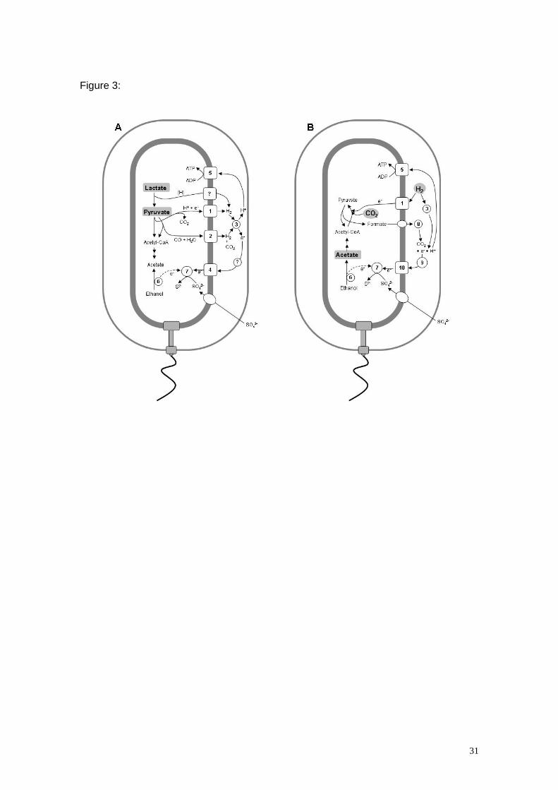
Figure 3- Comparative scheme of the bioenergetic pathways operative in D.
vulgaris grown in different conditions. Energy and/or carbon sources are in a grey
background. [H] represents hydrogen equivalents. The pathway that accepts electrons
from lactate oxidation or the pathway leading to the production of ethanol have not
been elucidated. For the sake of simplicity the role of the membrane menaquinone pool
is not considered.
Panel A- Growth with lactate or pyruvate and sulphate. Numbers correspond to the
following enzymes or proteins: 1- Ech hydrogenase, 2- CO-dehydrogenase and
associated hydrogenase, 3- Periplasmic hydrogenases, 4- Electron transfer complexes
including Dsr and Qmo, 5- ATP synthase, 6- Alcohol dehydrogenase and other proteins
of the pathway, 7- Sulphate reducing enzymes.
Panel B- Growth with hydrogen as electron donor and acetate/CO2 as carbon
sources. Numbers correspond to the following enzymes or proteins: 1- Ech
hydrogenase, 3- Periplasmic hydrogenases, 5- ATP synthase, 6- Alcohol
dehydrogenase and other proteins of the pathway, 7- Sulphate reducing enzymes, 8-
Formate dehydrogenases, 9- Pool of periplasmic cytochromes c, 10- Transmembrane
electron transfer complexes including Tmc.
30
FIGURES
Figure 1:
Figure 2:
0
50
100
150
200
250
300
350
400
450
H2S LThio PyrS Pyr
growth conditions
nu
mb
er o
f g
enes
up-regulated
down-regulated
31
Figure 3:
Related Documents