This is the pre-peer reviewed version of the following article: Endothelial nanoparticle binding kinetics are matrix and size dependent, Amber L. Doiron, Brendan Clark, Kristina D. Rinker; Biotechnology and Bioengineering, 108(12), pages 2988–2998, December 2011, which has been published in final form at http://onlinelibrary.wiley.com/doi/10.1002/bit.23253/abstract Accepted 20 June 2011 Endothelial nanoparticle binding kinetics are matrix and size dependent Amber L. Doiron 1,2,4,6,7 , Brendan Clark 1 , and Kristina D. Rinker 1,2,3,5 1 Cellular and Molecular Bioengineering Research Laboratory, University of Calgary, Calgary, Canada 2 Department of Chemical and Petroleum Engineering, University of Calgary, Calgary, Canada 3 Department of Physiology and Pharmacology, University of Calgary, Calgary, Canada 4 Department of Radiology, University of Calgary, Calgary, Canada 5 Centre for Bioengineering Research and Education, University of Calgary, Calgary, Canada, 6 Hotchkiss Brain Institute, University of Calgary, Calgary, Canada 7 Seaman Family MR Research Centre, Foothills Medical Centre, Calgary, Canada Corresponding Author: Kristina D. Rinker, University of Calgary, 2500 University Drive NW, Calgary, Department of Chemical and Petroleum Engineering, AB T2N 1N4, 403-210-9733, [email protected] Running Title: Particle Binding Kinetics Dependence on Size

Welcome message from author
This document is posted to help you gain knowledge. Please leave a comment to let me know what you think about it! Share it to your friends and learn new things together.
Transcript

This is the pre-peer reviewed version of the following article: Endothelial nanoparticle binding
kinetics are matrix and size dependent, Amber L. Doiron, Brendan Clark, Kristina D. Rinker;
Biotechnology and Bioengineering, 108(12), pages 2988–2998, December 2011, which has been
published in final form at http://onlinelibrary.wiley.com/doi/10.1002/bit.23253/abstract
Accepted 20 June 2011
Endothelial nanoparticle binding kinetics are matrix and size dependent
Amber L. Doiron1,2,4,6,7, Brendan Clark1, and Kristina D. Rinker1,2,3,5
1Cellular and Molecular Bioengineering Research Laboratory, University of Calgary, Calgary,
Canada
2Department of Chemical and Petroleum Engineering, University of Calgary, Calgary, Canada
3Department of Physiology and Pharmacology, University of Calgary, Calgary, Canada
4Department of Radiology, University of Calgary, Calgary, Canada
5Centre for Bioengineering Research and Education, University of Calgary, Calgary, Canada,
6Hotchkiss Brain Institute, University of Calgary, Calgary, Canada
7Seaman Family MR Research Centre, Foothills Medical Centre, Calgary, Canada
Corresponding Author: Kristina D. Rinker, University of Calgary, 2500 University Drive NW,
Calgary, Department of Chemical and Petroleum Engineering, AB T2N 1N4, 403-210-9733,
Running Title: Particle Binding Kinetics Dependence on Size
1
ABSTRACT
Nanoparticles are increasingly important in medical research for application to areas such as
drug delivery and imaging. Understanding the interactions of nanoparticles with cells in
physiologically relevant environments is vital for their acceptance, and cell/particle interactions
likely vary based on the design of the particle including its size, shape, and surface chemistry.
For this reason, the kinetic interactions of fluorescent nanoparticles of sizes 20, 100, 200, and
500 nm with human umbilical vein endothelial cells (HUVEC) were determined by 1) measuring
nanoparticles per cell at 37oC and 4oC (to inhibit endocytosis) and 2) modeling experimental
particle uptake data with equations describing particle attachment, detachment, and
internalization. Additionally, the influence of cell substrate compliance on nanoparticle
attachment and uptake was investigated. Results show that the number of binding sites per cell
decreased with increasing nanoparticle size, while the attachment coefficient increased. By
comparing HUVEC grown on either a thin coating of collagen or on top of three-dimensional
collagen hydrogel, nanoparticle attachment and internalization were shown to be influenced
significantly by the substrate on which the cells are cultured. This study concludes that both
particle size and cell culture substrate compliance appreciably influence the binding of
nanoparticles; important factors in translating in vitro studies of nanoparticle interactions to in
vivo studies focused on therapeutic or diagnostic applications.
Keywords: polystyrene nanoparticle uptake, nanotoxicity, 3D cell culture, compliance, HUVEC,
collagen, dextran

2
Introduction
Nanotechnology has rapidly progressed into an exciting field, and the push towards the nano-
scale has led to many medical advances. The use of nanoparticles to deliver an encapsulated or
attached agent to a specific site has promise to enhance efficacy, reduce toxicity, improve
imaging, and tune the biodistribution of a given therapeutic or imaging agent (Singh and Lillard,
2009). Nanoparticles of various shapes, sizes, and surface chemistries have been investigated for
therapy and imaging applications. While these properties are highly advantageous in tuning
nanoparticles for a particular application (Caldera-Moore et al., 2010), they also contribute to the
complexity of understanding how nanoparticles interact with their target cells.
Currently, it is customary for researchers to evaluate nanoparticles for toxicity, targetability,
and efficacy using in vitro cell cultures before proceeding to animal studies to evaluate the
nanoparticle in vivo. The two-dimensional (2D) nature of most adherent culture systems,
whereby cells are grown either on a bare or protein-coated tissue culture plate or glass substrate,
fails to translate into in vivo relevance, possibly because the 2D environment is a poor mimic of
the biological complexity of the three dimensional (3D) in vivo environment including the
extracellular matrix, matrix-cell interactions, transport limitations, and external mechanical cues
(Ng and Pun, 2008; Griffith and Swartz, 2006; Goldman et al., 2005). Two-dimensionally
cultured cells and in vivo cells differ in many aspects including gene expression (Griffith and
Swartz, 2006), biosynthesis of extracellular molecules (Albrecht et al., 2006) and cytoskeletal
components (Cukierman et al., 2001), and biological activity such as proliferation (Bhatia et al.,
1998; Heldin et al., 2004). For these reasons, the biological interactions of nanoparticles may be
more appropriately studied in vitro using a 3D culture environment. In both a 3D and 2D
environment in vitro, substrate compliance has been shown to greatly affect the compliance of

3
the endothelial cells themselves cultured atop the substrate resulting from a change in actin stress
fibers (Byfield et al., 2009). To the authors’ knowledge, no study to date explores the influence
of cell substrate compliance on the binding of nanoparticles, a potentially important factor in
translating in vitro studies of nanoparticle binding behavior to in vivo relevance. In this study, we
use cells cultured on a thin coating of collagen or on the surface of a collagen hydrogel of non-
negligible height to differentiate between the 2D and 3D extracellular environment of different
compliance in vitro.
Many nanotherapies rely on vascular targeting or travel through the vasculature to reach an
intended target. Nanoparticles of several sizes have been shown to deposit preferentially in the
endothelium of vessels and largely avoid penetration into the media and adventitia (Chesler and
Enyinna, 2003). While the main strategy for delivery of nanoparticles to tissues up to now has
been the enhanced permeability and retention (EPR) effect, recent research in the area of
vascular biodiversity (Sergeeva et al., 2006) suggests wide utility for nanoparticle targeting to
the endothelium. Human endothelial cells are studied here in vitro as a model of the vessel wall
that nanomaterials encounter upon intravenous injection.
This study explores the kinetics of binding of polystyrene nanoparticles with diameters from
20 nm to 500 nm to elucidate how particles of varying size bind to the cell surface and are
internalized. Sedimentation varies over the range of nanoparticle diameters studied and is
accounted for in a system of equations based on a single cell model of attachment (Goodman et
al., 2008; Wilhelm et al., 2002). These equations are used in conjunction with data collected
using human umbilical vein endothelial cells (HUVEC) cultured on top of 2D or 3D collagen
exposed to polystyrene nanoparticles to obtain the kinetic coefficients.
Materials and Methods

4
Cell Culture
HUVEC were cultured according to the supplier’s directions with modifications described here.
HUVEC (Lonza, Walkersville, MD, USA) were seeded from cryopreservation at passage 4,
cultured on 0.1% gelatin (BD Difco, Mississauga ON, Canada) coated tissue culture flasks in
endothelial growth medium (EGM, Lonza) containing 2% serum. Well plates were prepared
prior to introduction of cells by coating with collagen or preparation of 3D collagen gels.
HUVEC were grown to confluence on 48 well plates before addition of nanoparticles. Studies
involving dextran were conducted using EGM adjusted to a viscosity of 3.0 cP by including
dextran (Spectrum Chemical Mfg, Gardena, CA, USA) at a concentration of 3.02 g per 100 mL
media. Culture medium was replaced with dextran-containing EGM just prior to introduction of
particles.
Preparation of Collagen Substrates
Thin collagen coatings (2D) were prepared by mixing rat tail type I collagen (5 mg/mL stock,
Gibco, Invitrogen, Grand Island, NY, USA) with room temperature acetic acid (0.02M, Fisher
Scientific, Ottawa ON, Canada) at 172.5 µg/mL final collagen concentration, and 150 µL of this
solution was evenly pipetted into each well. After three hours incubation at room temperature,
the solution was aspirated, and wells were gently rinsed with DPBS (Sigma-Aldrich, Oakville
ON, Canada) and placed under ultraviolet (UV) light for 15 minutes prior to seeding with
HUVEC at 7,500 cells/cm2.
Alternatively, to form three-dimensional 3.5 mg/mL collagen hydrogels in the wells of a 48
well plate, the following cold reagents were mixed: 1.36 mL deionized water, 130 µL 1M NaOH
(Fisher), 0.75 mL 10x DPBS (Sigma), and 5.25 mL collagen; 150 µL of the solution was added
to each well. Plates were incubated overnight at 37˚C before gels were washed three times (15
5
minutes each) with DPBS. Well plates were placed under a UV light for 15 minutes prior to
seeding with HUVEC at 10,000 cells/ cm2, a higher cell density than 2D collagen cultures due to
lower initial cell adhesion to the substrate. HUVEC were seeded on top of the pre-formed gels
and did not readily migrate into the gel during the time periods relevant to this study. Gels were
visually inspected to verify intactness during washes and media changes.
Nanoparticle Uptake Studies
Fluosphere® particles (Molecular Probes, Invitrogen, Eugene, OR, USA) were supplied as 2%
w/v suspensions of polystyrene spheres of diameters 0.02, 0.100, 0.200, and 0.500 µm loaded
with red fluorophore and with surface carboxylic acid moieties. Particles were used after
thorough mixing via sonication and vortexing. Standard curves were prepared for each
nanoparticle size sample to equate signal to concentration. All samples were run in triplicate. In
both 2D and 3D culture systems, care was taken to ensure the confluence of cells prior to
exposure to nanoparticles in order to avoid nanoparticles sticking to the cell substrate as opposed
to interacting with cells.
To determine concentration dependence, nanoparticles were added to cells at concentrations
of 1 µL of stock solution per mL EGM to 320 µL/mL and incubated for one hour at 4˚C to
inhibit endocytosis. Time dependence was studied at a nanoparticle concentration of 10 µL/mL
for 15 minutes to 48 hours at 37˚C. After addition of particles, plates were agitated on a shaker
(Lab-Line Instruments Inc., Melrose Park, IL, USA) for 30 seconds. After incubation, the media
was aspirated and wells were gently washed three times with DPBS. DPBS (0.5 mL) was added
to each well prior to reading on a plate reader (SpectraMax M2, Molecular Devices, Sunnyvale,
CA, USA) with an excitation wavelength of 580 nm and emission of 605 nm.
Confocal Microscopy
6
Cell samples were seeded on glass slides coated with 2D or 3D collagen. For collagen coating, a
central area on the slide was masked out using 0.0508 cm thick silicon gasket (Specialty
Manufacturing, Saginaw, MI, USA) with a 1.5 cm by 5 cm cutout, and 400 µl collagen solution
(2D or 3D) was added to this cutout area. Slides were incubated, washed, and seeded with cells
as described in previous section. Confluent cells were incubated with nanoparticles at 10 µl/mL
at 37˚C for 1 hour. Slides were washed three times with DPBS and incubated for 15 minutes at
room temperature with 1 µl/mL Hoechst 33258 (Invitrogen) and CellMask Deep Red
(Invitrogen) in DPBS. Slides were washed with DPBS, fixed for 10 minutes at 37˚C in 4%
paraformaldehyde (Sigma), and washed three times with DPBS prior to mounting with
VectaShield (Vector Laboratories, Burlingame, CA, USA) and coverslip. Imaging of slides was
carried out on an Olympus FluoView™ FV1000 confocal laser scanning microscope and
accompanying software, version 2.1b (Markham, Ontario, Canada) using a 60x objective.
Theoretical Model of Nanoparticle Binding Kinetics and Internalization
Particle interactions with cells are characterized by particle movement to the cell surface, particle
attachment and detachment to cells, and the internalization of particles, assuming homogeneity
of the system, as modeled similarly to previous studies (Goodman et al. 2008).
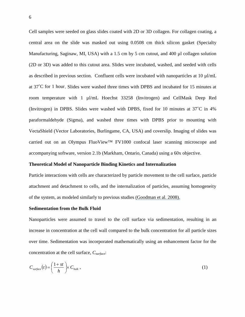
Sedimentation from the Bulk Fluid
Nanoparticles were assumed to travel to the cell surface via sedimentation, resulting in an
increase in concentration at the cell wall compared to the bulk concentration for all particle sizes
over time. Sedimentation was incorporated mathematically using an enhancement factor for the
concentration at the cell surface, Csurface:
bulksurface Ch
sttC
1
, (1)

7
where t is time, h is the height of media (1 cm) over which gravity is acting, Cbulk is the starting
bulk concentration at time zero, and s is the Einstein-Stokes sedimentation rate defined as:
9
2 2lpgr
s
, (2)
where r is the particle radius, g is the force of gravity, µ is the viscosity of the liquid with cell
media approximated here as water (µ = 0.79 cP at 37˚C and µ = 1.5 cP at 4̊ C) , ρp is the density
of the polystyrene particle (1.05 g/cm3), and ρl is the density of the liquid (water, ρl = 0.992
g/cm3at 37˚C and ρl 1.00 g/cm3= at 4˚C). The concentration of particles near the cell surface
changes over time as sedimentation occurs, as described by equations 1 and 2.
Single Cell Model of Reaction Kinetics
In order to determine the three kinetic rate coefficients, the single cell model of particle
interaction (Wilhelm et al. 2002) was utilized. Equations 3 – 7 describe particle interactions with
an individual cell in terms of the number of particles bound per cell, Nb, the particles internalized
per cell, Ni, the number of binding sites, Nbs, the number of binding sites available at time zero,
B, and the total number of particles bound and internalized, Nt, evolving over time t. The kinetic
rate coefficients for particle adsorption (binding), desorption, and internalization are ka (in M-1s-
1), kd (in s-1), and ki (in s-1), respectively.
The number of particles bound on a cell per unit time is proportional to the concentration of
nanoparticles in the reservoir at the cell surface (considered to change over time due to
sedimentation as given in equations 3 and 4) and the number of binding sites on the cell. Both
the number of particles detaching and particles internalized are proportional to the number of
bound particles, Nb(t). The number of bound nanoparticles changes over time following equation
3 and the number of internalized particles follows equation 4.

8
bibdbsab NkNkCNk
dt
dN (3)
bii Nk
dt
dN (4)
The binding capacity of the cell was assumed to remain the same over time (B=constant),
i.e., the cell surface was unobstructed by the surrounding cells and the number of binding sites
present (whether occupied by bound nanoparticles or not) was assumed constant due to
continuous turnover of the membrane. The number of binding sites available for binding (Nbs)
changes over time based on the number of particles already bound (Nb).
bbs NBtN (5)
Solutions to equations 3 - 5 give the number of particles bound to the cell surface (equation
6) and the number of bound and internalized particles (equation 7) evolving over time.
tb eN 1
CBka ida kkCk (6)
ti
iibt ek
tktNtNtN
1 (7)
The number of binding sites and the kinetic rate coefficients for particle attachment,
detachment, and internalization were determined using the above equations and experimental
data of the number of particles attached to the cell at various bulk concentrations over time.
Assumptions made in equations 3 – 7 included the following: exocytosis was represented as a
reduction in ki, endocytosis was assumed to account for all particle internalization based on
studies of latex nanoparticles of similar sizes to those used here (Rejman et al., 2004), and
binding sites were assumed to regenerate immediately upon endocytosis. To estimate the
parameter B, concentration-dependent experimental data at 4˚C were fitted to equation 5 using

9
non-linear least squares fitting in Matlab® version 7.8, where t was infinite due to saturated
binding sites and ki was zero due to the absence of endocytosis at 4˚C. For determination of
reaction parameters ka, kd, and ki, experimental time-dependence data at 37˚C were fitted to
equation 6 using the value obtained for B and non-linear least squares fitting in MatLab®.
Statistical Analysis
Error bars represent the standard error calculated from triplicate samples. Goodness of fit was
determined by finding the coefficient of determination (R2) between the fitted curve derived
from coefficients and equation 6 and the experimentally obtained data points in MatLab®.
Results
Confocal Imaging of Nanoparticles and HUVEC
HUVEC grown on top of either 2D or 3D collagen were incubated in the presence of
nanoparticles of various sizes for one hour prior to rinsing and subsequent staining of nuclei with
Hoechst 33258 and cell membrane with CellMask Deep Red. The absence of cell migration into
the gel as well as the absence of nanoparticles in the underlying collagen substrate was
confirmed with image stacks in the z-direction, as shown in Figure 1 with the compressed z-stack
as the central image and projections through the z-plane along the yellow lines in the x- and y-
directions given as the sidebars.
Additionally, z-stacks were taken and compressed into a single view as shown in Figures 2
and 3 of HUVEC grown on 2D and 3D collagen, respectively. For both 2D and 3D, images show
an increase in red fluorescence (particles) with decreasing size indicating an increase in the
binding and/or internalization of particles for smaller particles. Individual particles are below the
resolution of the microscope so that counting of particles is not possible, and smaller particles
cause a diffuse area of fluorescence rather than being particulate.

10
For the case of 20 nm particles and HUVEC grown on 2D collagen, red fluorescence appears
to overlay very closely with the white outline of the entire cell indicating almost no cell
membrane area present without being covered with nanoparticles. Conversely, with the other
sized particles and all 3D samples, the cell membrane is quite visible while particles are present
within the outline of the cell membrane. Qualitatively, it appears from the images that the 2D
samples have higher levels of red fluorescence arising from nanoparticles than 3D samples.
Further studies were then conducted to quantify differences in nanoparticle uptake between
endothelial cells grown on 2D or 3D surfaces for a range of nanoparticle sizes.
Endothelial Nanoparticle Adhesion/Uptake Differs with Particle Size
For endothelial cells cultured on thin coatings of collagen (2D), nanoparticle adhesion and
internalization were shown to depend on particle size and exposure time. Particle adhesion and
uptake increased with time until saturation was reached. While the 20 nm particles did not reach
saturation after 24 hours, the other particles saturated around 1-5 hours (100 nm and 200 nm) to
4-8 hours (500 nm). The highest levels of adhesion and uptake were seen for the smallest
particles (20 nm), which after 24 hours reached levels 300 fold higher than the next highest
adherent nanoparticle (100 nm; Figure 4).
Nanoparticle Adhesion/Uptake Is Affected by Extracellular Matrix Compliance
Overall, 20 nm nanoparticle uptake was less abundant on endothelial cells on 3D versus 2D
substrates as shown in microscopy images (Figures 2 and 3) and quantitative fluorescence data of
nanoparticles (Figures 4 and 5), while nanoparticle adhesion and uptake is greater for 3D than for
2D for all other sizes. In order to more accurately compare data from different nanoparticle sizes,
computational modeling was performed to correct for differences in sedimentation velocity and
endocytosis in determination of kinetic parameters.

11
Number of Binding Sites Decreases with Increasing Nanoparticle Size
Confluent cell cultures were exposed to particles at various concentrations at 4˚C to examine
attachment and detachment of particles in the absence of endocytosis. Nanoparticle adhesion to
and uptake by endothelial cells was found to be dose dependent at 4˚C (Figure 6). The number of
particles adherent to the cells increased significantly with decreasing particle size for HUVEC
grown on both collagen substrates. Data indicate a leveling off of nanoparticle adherence to cells
with increasing concentration of nanoparticles in the reservoir of media. The smaller particles
require larger concentrations of nanoparticles in order for this leveling off to occur, i.e., the cells
have a higher binding capacity for the smaller particles.
The fluorescence data from Figure 6 were fitted to equation 5 with ki equal to zero and the
number of HUVEC approximated as the expected cell counts from confluent 48 well plate wells.
The total number of binding sites per cell, B, was determined using non-linear least curve fitting
in Matlab (Table I and Figure 7). The number of available binding sites decreased four orders of
magnitude (from ~1 x 10⁸ to 1 x 10⁴) as the particle size increased; larger particles take up more
space on the cell surface, likely blocking binding of other particles. Also, HUVEC grown on 3D
collagen had more available binding sites compared to HUVEC grown on 2D collagen coatings,
particularly at low particle sizes.
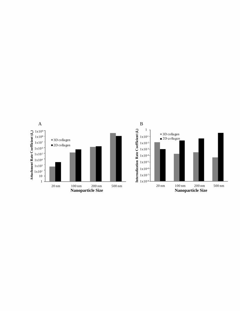
Determining Reaction Kinetic Coefficients
The rate coefficients (Figure 8) for attachment (ka), detachment (kd), and internalization (ki)
versus size for cells grown on top of 2D and 3D collagen are summarized in Table I. The
adsorption rate coefficient, ka, is higher for the 2D culture system for each size except 500 nm
diameter particles which have a 3.1 fold change for 3D over 2D. For the 20 nm, 100 nm, and
200 nm particles, the adsorption rate coefficient is 6.0, 3.6, and 1.4-fold higher for 2D versus 3D-

12
grown HUVEC. The internalization rate coefficient is larger (closer to zero) in the 3D culture
system for all sizes except 20 nm particles which have a 11-fold higher ki than the same particles
in the 2D culture system. For the 100 nm, 200 nm, and 500 nm particles, the internalization rate
coefficient is 120, 140, and 6700-fold higher for 3D versus 2D-grown HUVEC.
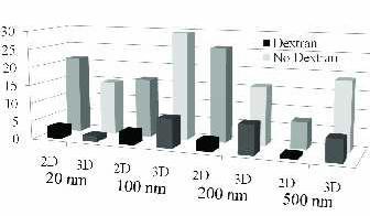
Dextran Effects on Particle Uptake
The inclusion of dextran in the cell growth media during exposure to nanoparticles
significantly reduced uptake and adhesion of nanoparticles. For 2D culture conditions after an
hour of exposure to 20 nm, 100 nm, 200 nm, or 500 nm particles, signal from dextran samples
was only 15%, 22%, 11%, and 10%, respectively, of that from the corresponding experiments
conducted in the absence of dextran. For the 3D samples, these percentages of dextran signal to
no-dextran signal for each ascending particle size were as follows: 11%, 27%, 50%, and 32%.
Data were normalized to the particle concentration at the cell surface to account for viscosity
differences. Figure 9 shows nanoparticle adsorption/internalization at each particle size for both
culture conditions compared between dextran and non-dextran samples.
DISCUSSION
In this work, the influence of cell culture substrate on the binding kinetics of nanoparticles over a
range of particle sizes was studied using HUVEC and fluorescent nanoparticles. The effect of
cell substrate was examined by culturing endothelial cells on a thin coating of collagen or on the
surface of a collagen hydrogel, and a noteworthy difference in kinetic parameters resulted from
this variation. The number of nanoparticles per cell was determined for particles with diameters
from 20 nm to 500 nm over a range of concentrations and time. This data was fit to an adaptation
of a single cell model of nanoparticle-cell interactions (Goodman et al., 2008; Wilhelm et al.,
2002) incorporating sedimentation. Non-linear least squares fitting of the experimental data was

13
used to determine interaction parameters including the number of binding sites available and
kinetic rate coefficients of attachment, detachment, and internalization. The parameters vary over
the size range of nanoparticles used here (20 nm to 500 nm) and may hold predictive value for
negatively charged particles interacting with endothelial cells.
Researchers typically evaluate nanoparticles using in vitro cell cultures, yet the two-
dimensional nature and surface rigidity of most adherent culture systems does not accurately
represent the biological complexity of the three dimensional in vivo environment (Ng and Pun,
2008; Griffith and Swartz, 2006). In this study, we used HUVEC cultured either on a thin
coating of collagen or on the surface of a collagen hydrogel to differentiate between the 2D and
3D extracellular environment of different compliance in vitro. The two cell substrates were
seemingly similar in chemical composition and varied primarily in the mechanical properties
experienced by the cells. Nanoparticle adhesion and uptake varied significantly depending on
whether cells were grown on top of 2D or 3D collagen. This is a striking finding and may carry
important implications for in vitro studies mimicking in vivo responses to nanoparticles. It is
postulated here that the 3D culture environment more accurately mimics the in vivo environment
experienced by a cell. Our findings support those of others that show surface compliance alters
cell responses.
At a given concentration, the binding of nanoparticles to the cell surface can be understood as
an interplay between the number of available binding sites and the adsorption coefficient ka. The
rate at which particles bind to the surface is balanced by the number of available sites to bind.
Across the range of nanoparticle sizes studied here, ka increases with increasing particle
diameter. Larger particles bind at a higher rate per unit concentration than do smaller particles.
Conversely, as particle size increases, the number of available binding sites on the cell decreases.

14
This is postulated to occur due to steric hindrance of the plasma membrane from larger particles,
restricting the space available for binding. These statements hold true for both the 2D and 3D
collagen substrate cell growth conditions examined here.
For all nanoparticle sizes, the number of binding sites per cell is larger for the 3D-collagen
cultured HUVEC than for cells cultivated on the 2D substrate. The number of binding sites may
differ due to either upregulation at the protein (receptor) level or increased surface area due to
compliance of the 3D collagen gel. Additionally, in all cases except the 500 nm particles, the ka
values for 2D collagen grown HUVEC were larger than those for 3D culture conditions. The fold
change in ka between 2D and 3D conditions decreases with increasing nanoparticle size up to
200 nm, while 500 nm particles have an approximately three-fold change from 3D to 2D. The
number of nanoparticles per cell after 24 hours was higher for cells grown on the 3D substrate
for all particle sizes except 20nm. Therefore, it appears that the increase in number of binding
sites dominates over the decrease in attachment kinetics in determining the number of
nanoparticles per cell on 3D substrates for 100-200nm particles.
For cells grown on 2D collagen, the rate of internalization decreases with increasing particle
size. This result is somewhat intuitive in that the larger particles are slower to enter the cell. This
result has been shown previously with latex particles (Rejman et al., 2004), viral particles
(Matlin et al., 1982), and polyplexes (Godbey et al., 1999). However, this trend is not seen in our
work with HUVEC cultures on the surface of 3D collagen hydrogels. In fact, in examining the
two particle size extremes, 20 nm and 500 nm, the larger particles are internalized at a higher
rate than the small-sized particles. The explanation for this result is not clear here and requires
further examination into the mechanism of uptake.

15
The desorption rate coefficient for 3D collagen-grown HUVEC increases with increasing
particle size. However, for 2D collagen grown cells, the trend does not hold, and in fact, three of
the four nanoparticle sizes have negative values for kd. While this result at first seems erroneous,
it is possible that the negative values are in fact a result of a more complex occurrence at the cell
membrane such as particle aggregation. If one particle binds to the cell surface and a second
particle subsequently aggregates to this particle without itself adsorbing to the cell surface, there
is no parameter in this single-cell attachment model to account for such an occurrence. The
particle would not accurately be described by the ka value or accounted for in the number of
binding sites. Therefore, these negative values for desorption may be explained by an
agglomeration process or more than one particle being attached per binding site.
Dextran largely blocks nanoparticle adsorption and uptake as shown in these results. Dextran
is a typical additive to media to increase viscosity for in vitro flow experiments (Blackman et al.,
2000; Rinker et al., 2001; Rouleau et al., 2010). Results suggest that the presence of dextran
blocks interactions between cells and particles, decreasing adherence and internalization.
Previous results support this finding as the dextran sugar molecules can be used to decrease
aggregation of particles by blocking charge-based interactions at the particle surface.
Nanoparticle design is a continually expanding area of research with the intended function
depending much on design factors such as size, surface chemistry, shape, surface charge, and
other factors. Size has been suggested by Morose as an important factor in determining the safety
of nanomaterials (2010). As shown in the results presented here, varying nanoparticle size has a
large influence on the attachment and internalization of particles. This information may be used
in the design of nanotherapies and other nanomaterials to result in a cell specific response. In
addition, these results may aide in the understanding of the possible toxic effects of

16
nanoparticles, i.e., nanotoxicity. While the uptake of nanoparticles has been shown to be
concentration, time, and energy-dependent (Davda and Labhasetwar, 2002; Panyam et al., 2003)
and uptake of polystyrene nanoparticles has been studied in several cell types (Clift et al., 2008;
Löhbach et al., 2006), the kinetics of uptake have not been determined over a range of sizes for
nanoparticles until the present study.
Conclusions
This study explored the adsorption and internalization kinetics of variously sized nanoparticles in
endothelial cells grown on two substrates of different compliance. Experimental data were fitted
to equations to extract information on the number of binding sites and kinetic coefficients for
adsorption, desorption, and internalization. Nanoparticle attachment and internalization were
influenced significantly by changing the substrate on which the cells were cultured. This study
elucidates the importance of particle size in nanoparticle-endothelial cell interactions, as the
number of binding sites per cell decreases with increasing nanoparticle size while the attachment
coefficient increases. Further, cell culture dimensionality and substrate compliance influenced
the binding of nanoparticles; this is a potentially important factor in translating in vitro studies of
nanoparticle binding to in vivo relevance.
Acknowledgements
The authors acknowledge the National Sciences and Engineering Research Council of Canada
(NSERC) Collaborative Health Research Projects (CHRP) grant for funding of the project, the T.
Chen Fong Postdoctoral Fellowship in Medical Imaging for funding of Amber Doiron, and the
Markin Undergraduate Student Research Program Scholarship for funding of Brendan Clark.
The authors additionally thank Robert Shepherd for helpful discussions and editorial comments.

17
References
Albrecht DR, Underhill GH, Wassermann TB, Sah RL, Bhatia SN. 2006. Probing the role of
multicellular organization in three-dimensional microenvironments. Nat Methods 3:369-
375.
Bhatia SN, Balis UJ, Yarmush ML, Toner M. 1998. Microfabrication of hepatocyte/fibroblast
co-cultures: role of homotypic cell interactions. Biotechnol Prog 14:378–387.
Blackman BR, Barbee KA, Thibault LE. 2000. In vitro cell shearing device to investigate the
dynamic response of cells in a controlled hydrodynamic environment. Ann Biomed Eng
28:363–372.
Byfield FJ, Reen RK, Shentu TP, Levitan I, Gooch KJ. 2009. Endothelial actin and cell stiffness
is modulated by substrate stiffness in 2D and 3D. J Biomech 42:1114-1119.
Caldorera-Moore M, Guimard N, Shi L, Roy K. 2010. Designer nanoparticles: incorporating
size, shape and triggered release into nanoscale drug carriers. Expert Opin Drug Delivery
7:479-495.
Chesler NC, Enyinna OC. 2003. Particle deposition in arteries ex vivo: effects of pressure, flow,
and waveform. J Biomech Eng 125:389.
Clift MJ, Rothen-Rutishauser B, Brown DM, Duffin R, Donaldson K, Proudfoot L, Guy K,
Stone V. 2008. The impact of different nanoparticle surface chemistry and size on uptake
and toxicity in a murine macrophage cell line. Toxicol Appl Pharmacol 232:418–427.
Cukierman E, Pankov R, Stevens DR, Yamada KM. 2001. Taking cell-matrix adhesions to the
third dimension. Science 294:1708-1712.
Davda J, Labhasetwar V. 2002. Characterization of nanoparticle uptake by endothelial cells. Int J
Pharm 233:51–59.
18
Godbey WT, Wu KK, Mikos AG. 1999. Tracking the intracellular path of poly
(ethylenimine)/DNA complexes for gene delivery. Proc Natl Acad Sci USA 96:5177-
5181.
Goldman J, Le TX, Skobe M, Swartz MA. 2005. Overexpression of VEGF-C causes transient
lymphatic hyperplasia but not increased lymphangiogenesis in regenerating skin. Circ
Res 96:1193-1199.
Goodman TT, Chen J, Matveev K, Pun SH. 2008. Spatio-temporal modeling of nanoparticle
delivery to multicellular tumor spheroids. Biotechnol Bioeng 101:388–399.
Griffith L, Swartz M. 2006. Capturing complex 3D tissue physiology in vitro. Nat Rev Mol Cell
Biol 7:211-224.
Heldin C, Rubin K, Pietras K, Ostman A. 2004. High interstitial fluid pressure - an obstacle in
cancer therapy. Nat Rev Cancer 4:806-813.
Löhbach C, Neumann D, Lehr C, Lamprecht A. 2006. Human vascular endothelial cells in
primary cell culture for the evaluation of nanoparticle bioadhesion. J Nanosci
Nanotechnol 6:3303-3309.
Matlin KS, Reggio H, Helenius A, Simons K. 1982. Pathway of vesicular stomatitis virus entry
leading to infection. J Mol Biol 156:609–631.
Morose G. 2010. The 5 principles of "design for safer nanotechnology". J Cleaner Prod 18:285–
289.
Ng CP, Pun SH. 2008. A perfusable 3D cell-matrix tissue culture chamber for in situ evaluation
of nanoparticle vehicle penetration and transport. Biotechnol Bioeng 99:1490-1501.

19
Panyam J, Sahoo S, Prabha S, Bargar T, Labhasetwar V. 2003. Fluorescence and electron
microscopy probes for cellular and tissue uptake of poly(D,L-lactide-co-glycolide)
nanoparticles. Int J Pharm 262:1-11.
Rejman J, Oberle V, Zuhorn IS, Hoekstra D. 2004. Size-dependent internalization of particles via
the pathways of clathrin-and caveolae-mediated endocytosis. Biochem J 377:159-169.
Rinker KD, Prabhakar V, Truskey GA. 2001. Effect of contact time and force on monocyte
adhesion to vascular endothelium. Biophys J 80:1722–1732.
Rouleau L, Rossi J, Leask RL. 2010. Concentration and time effects of dextran exposure on
endothelial cell viability, attachment, and inflammatory marker expression in vitro. Ann
Biomed Eng 38:1451-1462.
Sergeeva A, Kolonin MG, Molldrem JJ, Pasqualini R, Arap W. 2006. Display technologies:
application for the discovery of drug and gene delivery agents. Adv Drug Delivery Rev
58:1622–1654.
Singh R, Lillard Jr JW. 2009. Nanoparticle-based targeted drug delivery. Exp Mol Pathol
86:215–223.
Wilhelm C, Gazeau F, Roger J, Pons JN, Bacri J. 2002. Interaction of anionic superparamagnetic
nanoparticles with cells: kinetic analyses of membrane adsorption and subsequent
internalization. Langmuir 18:8148-8155.

20
Table Caption
Table I. Values for interaction parameters B (number of binding sites per cell) and kinetic rate
coefficients for attachment (ka), detachment (kd), and internalization (ki) for HUVEC grown on
2D and 3D collagen substrates with nanoparticles of various sizes. Obtained by fitting
experimental data to the single cell model of particle-cell interaction.
Figure Captions
signal shown in white is the cell membrane labeled with CellMask, blue is the cell nuclei, and
Figure 2. Confocal microscopy images of HUVEC grown on 2D collagen and exposed to no
nanoparticles (control), 20 nm, 100 nm, 200 nm, 500 nm nanoparticles for a period of one hour.
For each region of interest, z-stacks taken with a 60x objective were compressed into a single
view where fluorescence signal shown white is the cell membrane labeled with CellMask, blue is
the cell nuclei, and red is the nanoparticles. The final column is the overlay of all three. Scale
bar in the top left panel applies to all images and denotes 50 μm.
red is the nanoparticles. Compressed z-stack taken with 60x objective are shown as the central
image (A and D), and projections through the z-plane along the yellow lines in the y- (B and E)
and x-directions (C and F) are given as the sidebars. Green line along the sidebars denotes
location of glass slide and scale bar in A applies to all images and represents 50 μm.
(D-F) collagen exposed to 20 nm nanoparticles for a period of one hour where fluorescence
Figure 1. Confocal microscopy images of HUVEC grown on the surface of 2D (A-C) and 3D

21
Figure 3. Confocal microscopy images of HUVEC grown on 3D collagen and exposed to no
nanoparticles (control), 20 nm, 100 nm, 200 nm, 500 nm nanoparticles for a period of one hour.
For each region of interest, z-stacks taken with a 60x objective were compressed into a single
view where fluorescence signal shown white is the cell membrane labeled with CellMask, blue is
the cell nuclei, and red is the nanoparticles. The final column is the overlay of all three. Scale
bar in the top left panel applies to all images and denotes 50 μm.
Figure 4. Number of adherent and internalized particles over time per HUVEC (n=3). Cells
cultured on 2D collagen were incubated at 37˚C with particles of various sizes: 20 nm (A), 100
nm (B), 200 nm (C), and 500 nm (D), and fluorescence intensity was measured. The goodness
of fit (R2) for each fitted curve is 0.961 (A), 0.958 (B), 0.911 (C), and 0.951 (D).
Figure 5. Number of adherent and internalized particles over time per HUVEC (n=3). Cells
cultured on 3D collagen were incubated at 37˚C with particles of various sizes: 20 nm (A), 100
nm (B), 200 nm (C), and 500 nm (D), and fluorescence intensity was measured. The goodness
of fit (R2) for each fitted curve is 0.989 (A), 0.987 (B), 0.978 (C), and 0.982 (D).
Figure 6. Number of adherent particles per HUVEC. Cells cultured on either 2D (A-D) or 3D
(E-H) collagen were incubated at 4˚C with nanoparticles of various sizes: 20 nm (A and E), 100
nm (B and F), 200 nm (C and G), and 500 nm (D and H) over a range of concentrations for one
hour. The goodness of fit (R2) for each fitted curve is 0.992 (A), 0.975 (B), 0.991 (C), 0.964 (D),
0.976 (E), 0.925 (F), 0.956 (G), and 0.976 (H).

22
Figure 7. The number of nanoparticle binding sites per cell grown on either 2D or 3D collagen
substrate over the range of particle sizes. Data from Figure 6 were used to determine the number
of binding sites.
Figure 8. (A) Rate coefficient for attachment (ka) of nanoparticles to the cell surface for HUVEC
grown on either 2D collagen thin coatings or 3D collagen hydrogel. (B) Rate coefficient for
internalization (ki) of nanoparticles by HUVEC grown on either 2D collagen thin coatings or 3D
collagen hydrogel. Data from Figures 4 and 5 were used to determine kinetic coefficients.
Figure 9. Normalized number of nanoparticles bound per cell grown on either 2D or 3D
collagen, with sedimentation accounted for, after 1 hour of exposure in media with or without
dextran.

y
xz
z
A B
C
y
xz
z
D E
F

Control
20 nm
100 nm
200 nm
500 nm

Control
20 nm
100 nm
200 nm
500 nm

0 3 6 9 12 15 18 21 240
0 3 6 9 12 15 18 21 240
0 3 6 9 12 15 18 21 240
0 3 6 9 12 15 18 21 240
4 x 10⁴
4 x 10⁸
3 x 10⁸
2 x 10⁸
1 x 10⁸
1 x 10⁴
2 x 10⁴
3 x 10⁴
4 x 10⁴
2 x 10⁴
8 x 10⁵
6 x 10⁵
4 x 10⁵
2 x 10⁵
1 x 10⁶
1.2 x 10⁶
6 x 10⁴
8 x 10⁴
A
D
B
C
Time (hours)

0 3 6 9 12 15 18 21 240
0 3 6 9 12 15 18 21 240
0 3 6 9 12 15 18 21 240
0 3 6 9 12 15 18 21 240
1 x 10⁸
1.5 x 10⁸
5 x 10⁷
4 x 10⁴
3 x 10⁴
2 x 10⁴
1 x 10⁴
2 x 10⁶
3 x 10⁶
1 x 10⁶
4 x 10⁵
3 x 10⁵
2 x 10⁵
1 x 10⁵
Time (hours)
A
D
B
C5 x 104

0 0.5 1.0 1.5 2.0 2.50
Nanomolar
0 2 4 6 8 10 12 14 16 18 200
Nanomolar0 0.5 1.0 1.5 2.0 2.5
0
Micromolar
0 20 40 60 80 100 120 140 1600
Picomolar
2 x 10⁷
4 x 10⁷
6 x 10⁷
8 x 10⁷
2 x 10⁵
4 x 10⁵
6 x 10⁵
1 x 10⁵
8 x 10⁴
6 x 10⁴
4 x 10⁴
2 x 10⁴
1 x 10⁴
8 x 10³
6 x 10³
4 x 10³
2 x 10³
0 0.5 1.0 1.5 2.0 2.50
Micromolar0 2 4 6 8 10 12 14 16 18 20
0
Nanomolar
0 0.5 1.0 1.5 2.0 2.50
Nanomolar0 20 40 60 80 100 120 140 160
0
Picomolar
5 x 10⁵
1 x 10⁶
1.5 x 10⁶
2 x 10⁶
2.5 x 10⁶
1 x 10⁵
5 x 10⁴
1 x 10⁸
5 x 10⁷
2 x 10⁸
1.5 x 10⁸
2 x 10⁵
1.5 x 10⁵
2.5 x 10⁵ 1.5 x 10⁴
1.25 x 10 4
1 x 10⁴
7.5 x 10³
5 x 10³
2.5 x 10³
3 x 1062.5 x 108
A
F
D
B
HG
E
C
Concentration of Nanoparticles in Media

20 nm 100 nm 200 nm 500 nm
3D collagen
2D collagen
Nanoparticle Size
1
1x10²
1x10⁴
1x10³
1x10⁵
1x10⁶
1x10⁷
1x10⁸
1x10⁹
10
20 nm 100 nm 200 nm 500 nm
3D collagen
2D collagen
1
1x10²
1x10⁴
1x10³
1x10 5
1x10 6
1x10⁷
1x10⁸
1x10⁹
10
A B
20 nm 100 nm 200 nm 500 nm
3D collagen1
1x10⁻⁸
1x10⁻⁶
1x10⁻⁷
1x10⁻⁵
1x10⁻⁴
1x10⁻³
1x10⁻²
1x10⁻¹
Nanoparticle Size Nanoparticle Size
Related Documents