Patricia Garrido Vásquez: Emotion Processing in Parkinson's Disease: The Role of Motor Symptom Asymmetry. Leipzig: Max Planck Institute of Cognitive Neuroscience, 2012 (MPI Series in Cognitive Neuroscience; 137)

Welcome message from author
This document is posted to help you gain knowledge. Please leave a comment to let me know what you think about it! Share it to your friends and learn new things together.
Transcript

Patricia Garrido Vásquez: Emotion Processing in Parkinson's Disease: The Role of Motor Symptom Asymmetry. Leipzig: Max Planck Institute of Cognitive Neuroscience, 2012 (MPI Series in Cognitive Neuroscience; 137)

Emotion Processing in Parkinson’s Disease:The Role of Motor Symptom Asymmetry
Von der Fakultät für
Biowissenschaften, Pharmazie und Psychologie
der Universität Leipzig
genehmigte
DISSERTATIONzur Erlangung des akademischen Grades
doctor rerum naturalium
Dr. rer. nat.
vorgelegt
von Dipl.-Psych Patricia Garrido Vásquez
geboren am 17. Dezember 1982 in Dresden
Dekan: Prof. Dr. Erich SchrögerGutachter: Prof. Dr. Sonja A. Kotz
Prof. Dr. Marc D. Pell
Tag der Verteidigung: Leipzig, den 23.02.2012


Acknowledgements
This thesis would not have been possible without the support of many people.
Particularly, I would like to thank Sonja Kotz and Angela Friederici for giving me the
opportunity to work at the Max Planck Institute for Human Cognitive and Brain Sciences.
It was an inspiring experience to be part of such a prestigious institute and I enjoyed this
time very much.
I am especially grateful to Sonja Kotz for getting me into this highly interesting research
project, for her excellent supervision throughout the whole course of this dissertation, and
for teaching me so many valuable lessons in scientific writing. Finally, of course, thanks for
reviewing this thesis.
I would also like to thank Marc Pell for making this whole project possible and for very
helpful comments and suggestions at several stages of this work, despite of such a large
distance between us! Finally, thank you for agreeing to review this thesis.
Another person who needs to be mentioned here is Silke Paulmann; thank you for
teaching me event-related potentials, thanks for all the help throughout the last years, and
particularly for taking the time to read parts of the present thesis and for providing helpful
comments.
During the last years, I enjoyed the company of many wonderful colleagues at the MPI,
most notably (in alphabetical order, as I would not know how to put them into a ranking)
Julia Groh, Tam Ho, Christian Obermeier, Patricia Román, and Paula Roncaglia-Denissen.
Thank you for your friendship, support and all those fruitful discussions.
There are numerous people from the MPI’s technical and administrative staff who
were amazingly helpful in so many aspects. Thanks to Kerstin Flake, Andrea Gast-
Sandmann, and Heike Schmidt-Duderstedt for graphics support, Cornelia Schmidt, Heike
Böthel, Ina Koch, Anne-Kathrin Franz and Kristiane Klein for help in organizing and
conducting the experiments, the whole IT department for making everything concerning
computation possible, the library team for getting me so many papers, and the secretaries
of the neuropsychology department for help with many different things.

4
Furthermore and very importantly, I would like to thank the patients and the elderly
controls who participated in the experiments of this thesis. Sincere thanks to all of you for
devoting so much time and effort to this project which would not have worked without you.
Finally, I thank Antonio and Mauricio for building such a wonderful family with me
and for all their love and support during the last years.

Contents
Preface v
I Theoretical and empirical background 1
1 Event-related potentials 31.1 Electroencephalography . . . . . . . . . . . . . . . . . . . . . . . . . . . 3
1.2 Event-related potentials technique . . . . . . . . . . . . . . . . . . . . . . 5
1.3 Specific ERP components . . . . . . . . . . . . . . . . . . . . . . . . . . . 7
1.4 Summary . . . . . . . . . . . . . . . . . . . . . . . . . . . . . . . . . . . 8
2 Parkinson’s disease 92.1 Neuropathological changes in PD . . . . . . . . . . . . . . . . . . . . . . 9
2.1.1 The neurodegenerative process . . . . . . . . . . . . . . . . . . . . 9
2.1.2 Changes in basal ganglia loops . . . . . . . . . . . . . . . . . . . . 10
2.2 Symptomatology in PD . . . . . . . . . . . . . . . . . . . . . . . . . . . . 12
2.2.1 Motor symptoms . . . . . . . . . . . . . . . . . . . . . . . . . . . 12
2.3 Non-motor symptoms . . . . . . . . . . . . . . . . . . . . . . . . . . . . . 14
2.4 Summary . . . . . . . . . . . . . . . . . . . . . . . . . . . . . . . . . . . 15
3 Emotion 173.1 What is emotion? . . . . . . . . . . . . . . . . . . . . . . . . . . . . . . . 17
3.2 Neural basis of emotion perception . . . . . . . . . . . . . . . . . . . . . . 18
3.2.1 Emotion network in the brain . . . . . . . . . . . . . . . . . . . . 18
3.2.2 Emotion and the basal ganglia . . . . . . . . . . . . . . . . . . . . 20
3.2.3 Lateralization of emotion in the brain . . . . . . . . . . . . . . . . 20
3.3 Vocal emotion processing . . . . . . . . . . . . . . . . . . . . . . . . . . . 21
3.3.1 Models of vocal emotion processing . . . . . . . . . . . . . . . . . 22
i

ii CONTENTS
3.3.2 Neural correlates and hemispheric lateralization of vocal emotion
processing . . . . . . . . . . . . . . . . . . . . . . . . . . . . . . 23
3.3.3 The role of the basal ganglia in vocal emotion processing . . . . . . 25
3.4 Emotion perception from faces . . . . . . . . . . . . . . . . . . . . . . . . 26
3.4.1 Models of facial emotion processing . . . . . . . . . . . . . . . . . 26
3.4.2 Neural correlates and hemispheric lateralization of facial emotion
processing . . . . . . . . . . . . . . . . . . . . . . . . . . . . . . 29
3.4.3 The role of the basal ganglia in facial emotion processing . . . . . . 30
3.5 (Cross-modal) emotional priming . . . . . . . . . . . . . . . . . . . . . . . 30
3.5.1 Priming: the basics . . . . . . . . . . . . . . . . . . . . . . . . . . 30
3.5.2 Emotional priming . . . . . . . . . . . . . . . . . . . . . . . . . . 31
3.6 Summary . . . . . . . . . . . . . . . . . . . . . . . . . . . . . . . . . . . 32
4 Emotion perception in PD 334.1 Perception of vocal emotion in PD . . . . . . . . . . . . . . . . . . . . . . 33
4.2 Perception of facial emotion in PD . . . . . . . . . . . . . . . . . . . . . . 36
4.3 Emotional priming in PD . . . . . . . . . . . . . . . . . . . . . . . . . . . 39
4.4 Summary . . . . . . . . . . . . . . . . . . . . . . . . . . . . . . . . . . . 40
II Experiments 41
5 Experiment 1: Vocal emotion 43
6 Experiment 1A – Pilot study 456.1 Methods . . . . . . . . . . . . . . . . . . . . . . . . . . . . . . . . . . . . 45
6.1.1 Participants . . . . . . . . . . . . . . . . . . . . . . . . . . . . . . 45
6.1.2 Stimulus material . . . . . . . . . . . . . . . . . . . . . . . . . . . 46
6.1.3 Procedure . . . . . . . . . . . . . . . . . . . . . . . . . . . . . . . 46
6.1.4 Data acquisition and analysis . . . . . . . . . . . . . . . . . . . . . 48
6.2 Results . . . . . . . . . . . . . . . . . . . . . . . . . . . . . . . . . . . . . 49
6.2.1 Behavioral data . . . . . . . . . . . . . . . . . . . . . . . . . . . . 49
6.2.2 ERP data . . . . . . . . . . . . . . . . . . . . . . . . . . . . . . . 50
6.3 Discussion . . . . . . . . . . . . . . . . . . . . . . . . . . . . . . . . . . . 55
7 Experiment 1B – Patient study 617.1 Methods . . . . . . . . . . . . . . . . . . . . . . . . . . . . . . . . . . . . 62
7.1.1 Participants . . . . . . . . . . . . . . . . . . . . . . . . . . . . . . 62
7.1.2 Stimulus material . . . . . . . . . . . . . . . . . . . . . . . . . . . 63

CONTENTS iii
7.1.3 Procedure . . . . . . . . . . . . . . . . . . . . . . . . . . . . . . . 65
7.1.4 Data acquisition and analysis . . . . . . . . . . . . . . . . . . . . . 66
7.2 Results . . . . . . . . . . . . . . . . . . . . . . . . . . . . . . . . . . . . . 68
7.2.1 Test scores . . . . . . . . . . . . . . . . . . . . . . . . . . . . . . 68
7.2.2 P300 data . . . . . . . . . . . . . . . . . . . . . . . . . . . . . . . 69
7.2.3 Behavioral data . . . . . . . . . . . . . . . . . . . . . . . . . . . . 69
7.2.4 ERP data . . . . . . . . . . . . . . . . . . . . . . . . . . . . . . . 71
7.2.5 Correlations . . . . . . . . . . . . . . . . . . . . . . . . . . . . . . 76
7.2.6 Prosody categorization study . . . . . . . . . . . . . . . . . . . . . 77
7.3 Discussion . . . . . . . . . . . . . . . . . . . . . . . . . . . . . . . . . . . 78
8 Experiment 2: Emotional priming 83
9 Experiment 2A – Pilot study 879.1 Rating study . . . . . . . . . . . . . . . . . . . . . . . . . . . . . . . . . . 88
9.2 ERP study . . . . . . . . . . . . . . . . . . . . . . . . . . . . . . . . . . . 90
9.2.1 Methods . . . . . . . . . . . . . . . . . . . . . . . . . . . . . . . 90
9.3 ERP Results . . . . . . . . . . . . . . . . . . . . . . . . . . . . . . . . . . 93
9.3.1 Video-as-prime condition . . . . . . . . . . . . . . . . . . . . . . . 93
9.3.2 Prosody-as-prime condition . . . . . . . . . . . . . . . . . . . . . 96
9.4 Discussion . . . . . . . . . . . . . . . . . . . . . . . . . . . . . . . . . . . 99
10 Experiment 2B – Patient study 10510.1 Methods . . . . . . . . . . . . . . . . . . . . . . . . . . . . . . . . . . . . 107
10.1.1 Participants . . . . . . . . . . . . . . . . . . . . . . . . . . . . . . 107
10.1.2 Stimulus material . . . . . . . . . . . . . . . . . . . . . . . . . . . 108
10.1.3 Procedure . . . . . . . . . . . . . . . . . . . . . . . . . . . . . . . 110
10.1.4 Data acquisition and analysis . . . . . . . . . . . . . . . . . . . . . 111
10.2 Results . . . . . . . . . . . . . . . . . . . . . . . . . . . . . . . . . . . . . 113
10.2.1 Test scores . . . . . . . . . . . . . . . . . . . . . . . . . . . . . . 113
10.2.2 P300 data . . . . . . . . . . . . . . . . . . . . . . . . . . . . . . . 114
10.2.3 Video-as-prime condition . . . . . . . . . . . . . . . . . . . . . . . 114
10.2.4 Prosody-as-prime condition . . . . . . . . . . . . . . . . . . . . . 118
10.3 Discussion . . . . . . . . . . . . . . . . . . . . . . . . . . . . . . . . . . . 124
11 General discussion and outlook 13311.1 Summary and integration of main findings . . . . . . . . . . . . . . . . . . 134
11.2 Is there lateralization in the basal ganglia? . . . . . . . . . . . . . . . . . . 137
iv CONTENTS
11.3 Task effects and early versus late processing stages . . . . . . . . . . . . . 138
11.4 The problem of variability in PD . . . . . . . . . . . . . . . . . . . . . . . 139
11.5 Limitations . . . . . . . . . . . . . . . . . . . . . . . . . . . . . . . . . . 139
11.6 Concluding remarks . . . . . . . . . . . . . . . . . . . . . . . . . . . . . . 140
III Appendix 143
A Instructions 145A.1 Experiment 1 . . . . . . . . . . . . . . . . . . . . . . . . . . . . . . . . . 145
A.2 Experiment 2 . . . . . . . . . . . . . . . . . . . . . . . . . . . . . . . . . 147
B Sentence materials 151
C Experiment 2: Video rating results 165
References 169
List of Figures 197
List of Tables 199
List of Abbreviations 201
Preface
Emotion is an essential element of our daily life. We express happiness when something
that we consider positive happens, or we get angry when things do not work out in the way
we want them to. As social creatures, we are not only confronted with our own emotional
reactions, but rather we also continuously encounter emotional expressions in the people we
interact with every day. It is important for us to recognize emotions in others quickly and
accurately in order to adequately react to them, thereby ensuring successful interpersonal
interactions.
What happens, however, if the recognition of other people’s emotional expressions does
not work properly? And - an even more basic question - how can we experimentally test the
integrity of emotion processing functions?
In Parkinson’s disease (PD), a very common movement disorder, it is to date widely
acknowledged that the expression of emotion is disturbed. This deficit concerns, e.g., speech
intonation or facial expressions (Bowers et al., 2006; Pell, Cheang, & Leonard, 2006).
Furthermore, there is evidence of impaired emotion processing in PD (Breitenstein, Daum,
& Ackermann, 1998; Kan, Kawamura, Hasegawa, Mochizuki, & Nakamura, 2002; Pell &
Leonard, 2003). However, research on this topic has so far primarily relied on behavioral
investigations using explicit tasks, e.g., assigning emotion labels to face or voice stimuli.
This raises the question as to whether these methods are able to provide adequate measures
of emotion processing in PD and in general. One can assume that the validity of these
techniques is limited, as only late, controlled processing stages can be captured by them
(Pell, 2002). Furthermore, explicit tasks go along with a certain level of task difficulty and
may thus be confounded by cognitive dysfunction (Benke, Bösch, & Andree, 1998), which
is a frequent symptom in PD (Williams-Gray, Foltynie, Brayne, Robbins, & Barker, 2007).
Apart from these methodological constraints encountered in many previous studies on
emotion processing in PD, the disease is often considered a unitary disorder, while there
is actually high heterogeneity among patients. One distinctive disease characteristic is the
sidedness of motor symptoms. Symptoms in idiopathic PD generally start at one side of the
body (Hoehn & Yahr, 1967), and even though both sides become involved at some point
v

vi PREFACE
during the disease progression, a certain degree of asymmetry remains. Importantly, the
basal ganglia of the hemisphere contralateral to the predominantly affected side are more
involved in neuronal degeneration (Nahmias, Garnett, Firnau, & Lang, 1985; Tatsch et al.,
1997). Thus, considering motor symptom asymmetry in PD could provide novel insight
into a possible functional lateralization of the basal ganglia and their respective circuits.
The present thesis aimed to investigate receptive emotional functioning in PD with
special emphasis on the sidedness of motor symptoms. The method applied to do
so were event-related brain potentials, which provide an excellent temporal resolution.
Thus, this method allows describing the precise time course of emotion processing in
PD. Furthermore, no explicit task instructions are required, and thus receptive emotional
functions in PD can be tested under low task demands and without an attentional focus
on emotion. Importantly, in this thesis cognitive functions in the patients were carefully
controlled for.
The theoretical part of the present thesis starts with a brief introduction to electroence-
phalography and the event-related potentials technique (Chapter 1). Chapter 2 then deals
with Parkinson’s disease, the neural changes which go along with the disease, and the
numerous symptoms which can occur in a PD patient. Chapter 3 and Chapter 4 are
the "emotion chapters", providing a general overview of the emotion literature and of
investigations on emotion processing in PD, respectively.
In the empirical part, Chapter 5, Chapter 6, and Chapter 7 are devoted to vocal
emotion processing (prosody), reporting data from a healthy pilot sample and a patient
investigation in PD. Chapter 8, Chapter 9, and Chapter 10 take this evidence a step
further, presenting data from healthy participants and PD patients on implicit cross-modal
emotional priming with dynamic face and voice stimuli.
Finally, the results of the present thesis and the implications of these findings are
discussed in Chapter 11.

Part I
Theoretical and empiricalbackground
1


Chapter 1
Event-related potentials
This chapter provides a brief introduction to electroencephalography and scalp-recorded
event-related brain potentials, which is the main experimental method used in the present
thesis.
1.1 Electroencephalography
More than 80 years ago, the German psychiatrist Hans Berger measured for the first time the
electrical activity from a human’s scalp surface and introduced the term “electroencephalo-
graphy” for this procedure (Berger, 1929). Electroencephalography (EEG) is a psychophy-
siological method which captures the brain’s electrical activity, resulting in the electroence-
phalogram. To this end, electrodes are attached to the scalp surface, which renders the scalp-
recorded EEG a non-invasive procedure. Next to the advantage of non-invasivity provided
by the EEG, it also stands out for its excellent temporal resolution in the milliseconds-
range which cannot be offered other contemporary methods (e.g., fMRI, PET). However, the
spatial resolution of EEG is very low compared to other techniques (Birbaumer & Schmidt,
2006). Several source localization algorithms have been developed which can help to get
an idea of where the activity actually comes from (see Michel et al., 2004, for an overview),
but as these methods are not as precise as, e.g., fMRI, the trade-off between spatial and
temporal resolution in neuroscientific methods remains.
The EEG records voltage fluctuations over time from the scalp surface. The main source
of oscillations observed in the EEG are excitatory and inhibitory post-synaptic potentials
generated in cortical pyramidal neurons (Speckmann & Elger, 2005). In order to cause
an oscillation that becomes visible in the EEG, tens of thousands of neurons need to
be activated simultaneously (Pizzagalli, 2007). Summation and propagation of neuronal
3

4 CHAPTER 1. EVENT-RELATED POTENTIALS
activity in the cortex is facilitated by the perpendicular orientation of these neurons relative
to the scalp surface (Coles & Rugg, 1995).
Recording of the EEG is mostly accomplished with metal electrodes, typically silver-
silverchloride (Ag/AgCl) electrodes, together with an electrolyte solution to allow for an
electron exchange between the skin and the recording electrode (Picton, Lins, & Scherg,
1995). In research environments, it is common to use large arrays of electrodes distributed
across the scalp surface. For electrode placement on the scalp surface, Jasper (1958)
proposed the 10–20 system. It is based on four fiducial points, namely the nasion, the inion,
and two preauricular spots. Along imaginary lines drawn between these points, electrodes
are located at certain percentages (10, 20, 50) of these lines, and additional electrodes may
be placed halfway in between for high-density EEG recordings. A common nomenclature
for the electrode positions was introduced by the American Electroencephalographic
Society (Sharbrough et al., 1991). The electrode locations are displayed graphically in
Figure 1.1
Figure 1.1: Electrode locations in accord with the extended 10–20 system (Sharbrough et al., 1991).Electrodes relevant for the present thesis are shaded in grey.
EEG recordings can be made with either a mono- or a bipolar setup (Pinel, 1997). In
monopolar recordings, each scalp electrode is referred to a common site of low electrical
activity (the reference), while in bipolar recording, electrodes placed over electrically active
sites are linked together by twos. The monopolar setup is most widely used in current
research and implies the problem of reference placement. Ideally, this should be a location
devoid of any electrical activity, which is not possible (Pizzagalli, 2007). Commonly used

1.2. EVENT-RELATED POTENTIALS TECHNIQUE 5
references are the ears (mastoid bones or earlobes), the nose tip, or an average reference (see
Pizzagalli, 2007, for a discussion of different references and reference-free procedures).
What turns out to be an oscillation in the EEG is the difference of activity at a certain scalp
channel in relation to the reference.
As the potentials of interest in the EEG are very small, they need to be amplified by a
specific device. The amplified signal is then converted from analog to digital at a predefined
sampling rate, which indicates how many samples are acquired during a period of time
(e.g., a 500 Hz sampling rate would indicate 500 samples per second or a two-milliseconds
acquisition rate).
1.2 Event-related potentials technique
The EEG signal is always a composition of signal (stimulus-related) and noise. A common
way of increasing the signal-to-noise ratio of a recording is to average it over certain epochs.
The idea of averaging is that the cerebral response to a certain stimulus type is comparable
over its re-occurrences, while noise is rather randomly distributed over the signal.1 Thus, by
time-locking to the onset of a stimulus and averaging the epochs pertaining to that stimulus,
the random noise is averaged out and what becomes visible is a characteristic pattern of
waveforms, the event-related potential (ERP). The more trials that are averaged for one
condition, the better is the signal-to-noise ratio in the ERPs (Luck, 2005). The ERP is a
characteristic pattern of positive and negative peaks and slow waves elicited by the event to
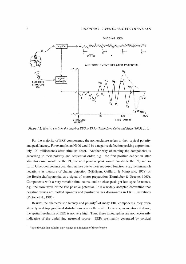
which they are time-locked. Figure 1.2 displays how to get from the ongoing EEG to ERPs.
ERPs can be classified into evoked and emitted ERPs, and evoked ERPs can be further
subdivided into exogenous and endogenous components (Picton et al., 1995). Exogenous
components are thought to be driven mainly by physical stimulus characteristics whereas
endogenous components are triggered by the psychological effects of the stimulus, so one
may speak of bottom-up and top-down modulated components, respectively. Brainstem
potentials, which occur very early after stimulus onset, are considered exogenous, while
components occurring at long latencies and reflecting cognitive processes are considered
endogenous (e.g., language-related components). Some components may be a mix of endo-
genous and exogenous processes, such as the N100, and are therefore referred to as mesoge-
nuous (Fabiani, Gratton, & Federmeier, 2007). Emitted ERPs are all endogenous and can
be observed, e.g., during the initiation of a response (Picton et al., 1995).
1Of course, noise is not always distributed randomly. Noise may be rhythmic, which can be circumventedby variations of the inter-stimulus interval, or noise may be related to the stimulus onset. The latter casemainly occurs in terms of micro-reflexes within the first 80 milliseconds after stimulus onset, which makes theinterpretation of very early ERPs difficult (Picton et al., 1995).
6 CHAPTER 1. EVENT-RELATED POTENTIALS
Figure 1.2: How to get from the ongoing EEG to ERPs. Taken from Coles and Rugg (1995), p. 6.
For the majority of ERP components, the nomenclature refers to their typical polarity
and peak latency. For example, an N100 would be a negative deflection peaking approxima-
tely 100 milliseconds after stimulus onset. Another way of naming the components is
according to their polarity and sequential order, e.g. the first positive deflection after
stimulus onset would be the P1, the next positive peak would constitute the P2, and so
forth. Other components bear their names due to their supposed function, e.g., the mismatch
negativity as measure of change detection (Näätänen, Gaillard, & Mäntysalo, 1978) or
the Bereitschaftspotential as a signal of motor preparation (Kornhuber & Deecke, 1965).
Components with a very variable time course and no clear peak get less specific names,
e.g., the slow wave or the late positive potential. It is a widely accepted convention that
negative values are plotted upwards and positive values downwards in ERP illustrations
(Picton et al., 1995).
Besides the characteristic latency and polarity2 of many ERP components, they often
show typical topographical distributions across the scalp. However, as mentioned above,
the spatial resolution of EEG is not very high. Thus, these topographies are not necessarily
indicative of the underlying neuronal source. ERPs are mainly generated by cortical
2note though that polarity may change as a function of the reference

1.3. SPECIFIC ERP COMPONENTS 7
neurons, but the neurons are not necessarily located below the electrode site from which they
are recorded, as summation and propagation across large neuronal populations is necessary
to yield oscillations in the EEG (Coles & Rugg, 1995). Furthermore, subcortical sources
may also be involved in the generation of ERPs (Pizzagalli, 2007). In a similar way, there
may be a great deal of neural activity in the brain which is not reflected in the ERP (Coles
& Rugg, 1995).
1.3 Specific ERP components
This section introduces ERP components relevant for the present thesis. More information
on these components is provided in chapter 3.
N100/P1. The N100 is an early negativity peaking at a latency of approximately 100
ms (80 – 120 ms) after stimulus onset, with maximal amplitudes over frontal and central
electrodes (Rosburg, Boutros, & Ford, 2008). The auditory N100 is presumably generated
in the auditory cortex, but there may be contributions from frontal and motor areas to this
component, too (Näätänen & Picton, 1987). When there is visual stimulation, the N100 is
limited to fronto-central leads, and a P1 is observed at posterior electrodes (e.g., Luo, Feng,
He, Wang, & Luo, 2010). Latter component is generated in the visual cortex (V. P. Clark,
Fan, & Hillyard, 1994).
N170/VPP. The N170 is a face-related component and follows the P1 at occipital
electrode sites. Its amplitude is enhanced to faces compared to other visual stimuli such
as houses (Ashley, Vuilleumier, & Swick, 2004; Eimer, 1998). It is thought to reflect
configural processing of the face and its neuronal generators are supposed around the
fusiform face area (Bentin, Allison, Puce, Perez, & McCarthy, 1996), a region in visual
cortex that appears to be specialized for face processing (Kanwisher, McDermott, & Chun,
1997). The N170’s fronto-central counterpart is the vertex positive potential (VPP), which
occurs with the same latency. It may rely on the same neural generators as the N170 (Joyce
& Rossion, 2005), although there is an ongoing debate about this in the literature (e.g.,
Wong, Fung, McAlonan, & Chua, 2009). Note that the VPP is sometimes termed the P2
(Eimer & Holmes, 2002), P200 (Ashley et al., 2004; Paulmann & Pell, 2009), or P150
(Campanella, Quinet, Bruyer, Crommelinck, & Guerit, 2002).
P200. The auditory P200 is strongest at fronto-central electrodes, and its main genera-
tors have been localized in the auditory cortex, some millimeters more frontally than the
generators of the auditory N100 (Papanicolaou, Rogers, Baumann, Saydjari, & Eisenberg,

8 CHAPTER 1. EVENT-RELATED POTENTIALS
1990). The P200 is a central component in emotional prosody processing, as will be shown
in the following chapters.
Later stage negativities. The most classical late negativity is the N400 which was first
observed in response to semantic anomalies (Kutas & Hillyard, 1980). It is typically
observed within a 300 – 500 ms latency after the onset of such an anomaly. By now, N400-
like negativities have been related to many different contexts which go beyond semantics,
e.g., enhanced N400 amplitudes have been observed with cross-modal priming in terms
of emotional valence (Schirmer, Kotz, & Friederici, 2002, 2005; Zhang, Lawson, Guo,
& Jiang, 2006; Zhang, Li, Gold, & Jiang, 2010) or emotional category (Paulmann &
Pell, 2010a). In emotional contexts, late negativities in response to incongruencies may
sometimes occur earlier than the classical N400 (Bostanov & Kotchoubey, 2004). These
negativities are called N400-like, as they may differ from the classical N400 with respect to
their time course and topography (Kutas & Federmeier, 2011).
1.4 Summary
We have seen in the present chapter that the EEG methodology provides a means to record
brain activity with a very high temporal resolution in the milliseconds range, which allows
to specify the precise time course of brain processes. With EEG, these processes can be
captured long before a behavioral response occurs. When averaging many EEG epochs
together that are time-locked to a certain stimulus type, a characteristic pattern of positive
and negative deflections (ERPs) emerges.

Chapter 2
Parkinson’s disease
Idiopathic (alternatively: sporadic) Parkinson’s disease (PD) makes up 75% of all Parkinso-
nian syndromes (Iglseder, 2008). The first systematic description of Parkinsonism dates
back almost 200 years ago. The author of this influential report called “An essay on the
shaking palsy” was the physician James Parkinson (Parkinson, 1817), which is why the
disease today bears his name.
Epidemiological data underline the impact of idiopathic PD on our ageing society: Its
prevalence in the German population older than 60 years is about one percent, and it rises up
to two to three percent in people older than 70 years (Heidbreder & Dominiak, 2010). The
disease is 1.5 to 2 times more common in men than in women (Heidbreder & Dominiak,
2010).
2.1 Neuropathological changes in PD
PD primarily affects the basal ganglia (BG). The BG are a group of subcortical nuclei
comprising the striatum (caudate and putamen), the substantia nigra (pars compacta and
pars reticulata), the subthalamic nucleus, and the globus pallidus (internal and external
segment). The striatum is considered to be the major input structure to the BG, while the
internal pallidum and the substantia nigra pars reticulata are the major output nuclei of the
BG (Wichmann & DeLong, 1996).
2.1.1 The neurodegenerative process
The key mechanism in PD is the degeneration of dopamine-producing neurons in the sub-
stantia nigra pars compacta (SNc), most strongly involving its ventrolateral portion. This
affects the dopaminergic projection from the SNc to the dorsal putamen (nigrostriatal path-
way), leading to striatal dopamine depletion (Lang & Lozano, 1998).
9

10 CHAPTER 2. PARKINSON’S DISEASE
Neurodegeneration starts long before diagnosis. It is estimated that around 15 to 20
years before patients seek medical treatment for emerging motor symptoms, first pathologi-
cal changes begin to take place at the neural level (Leplow, 2007). However, data on this
preclinical phase are controversial and it may actually be shorter than five years (Lang &
Lozano, 1998). When first motor symptoms become evident, patients are considered to be
already at a later disease stage (Braak & Del Tredici, 2009) with 50 to 70% of the SNc
dopaminergic neurons being lost (Lang & Lozano, 1998; Leplow, 2007; Schwarz & Storch,
2007).
Importantly, PD is not exclusively a BG disorder. Rather, it leads to more wide-spread
structural changes in the brain (Tinaz, Courtney, & Stern, 2011). In fact, there is evidence
that the first pathological processes take place outside the SNc. Braak and colleagues have
put forward a six-stage model of pathological changes in PD (Braak et al., 2003, 2006;
Braak & Del Tredici, 2008, 2009). According to this model, first pathological changes,
i.e., inclusions of pathological Lewy bodies, are observed in the dorsal motor nucleus of
the vagal nerve as well as in anterior olfactory structures. In the second stage the disease
proceeds towards the lower brainstem, and recently the third stage has been described to
involve the SNc as well as the central subnucleus of the amygdala. From the fourth to the
sixth stage neurodegeneration gradually proceeds to more structures such as the thalamus,
insula, amygdala, and prefrontal regions.
2.1.2 Changes in basal ganglia loops
The BG are heavily interconnected with several brain regions and receive input from
virtually the whole cortex (Utter & Basso, 2008). In their seminal article, Alexander,
DeLong, and Strick (1986) described five different cortico-striato-thalamocortical loops:
the motor, the oculomotor, the dorsolateral prefrontal, the lateral orbitofrontal, and the
anterior cingulate loop. These loops originate from the cortex and innervate the striatum
through glutamatergic projections (Hammond, Bergman, & Brown, 2007). The signal is
then projected back to the cortex via the thalamus. These circuits were originally considered
as relatively closed, segregated systems (Alexander et al., 1986). Each circuit innervates a
distinct region of the striatum and the existence of further sub-circuits within these five
major pathways has been shown, for example in terms of a somatotopic organization of
input and output activities within the sensorimotor part of the putamen (DeLong, 2000;
Wichmann, DeLong, Guridi, & Obeso, 2011). Nevertheless, it is now assumed that the
segregation is not that strong, as the pathways heavily interact with each other through
collaterals and via projections within the striatum (DeLong & Wichmann, 2009; Haber &
Calzavara, 2009; Saint-Cyr, 2003). Furthermore, the five-circuits model has been expanded
by more circuits, e.g., a circuit linking the temporal cortex with the striatum (Middleton &

2.1. NEUROPATHOLOGICAL CHANGES IN PD 11
Strick, 1996, 2000). According to the involvement of the striatum in different circuits, this
structure can be roughly divided into a motor (dorsal putamen), a limbic (ventral striatum),
and an associative/cognitive (caudate) portion.
In PD, the motor circuit is of major significance to understand the pathology. It origina-
tes from different motor fields of the cortex (Wichmann et al., 2011) and projects to the
putamen (Alexander et al., 1986), which is the BG region that is most affected by dopamine
depletion in PD. Before reaching the thalamus and other brain regions, the striatal medium
spiny neurons (MSNs), which are the striatum’s projection neurons, project to different loci
within the BG. MSNs expressing substance P and D1 dopamine receptors give rise to the
direct pathway, while MSNs expressing enkephalin and D2 receptors bring on the indirect
pathway (DeLong & Wichmann, 2009; Schwarz & Storch, 2007; Wichmann et al., 2011).
The direct pathway consists of a striatal projection to the motor thalamus (ventrolateral and
ventromedial portions) via the internal globus pallidus as well as a striatal projection to
the substantia nigra pars reticulata. These projections are GABAergic and inhibitory. This
reduces the inhibition exerted by the BG on the thalamus. The indirect pathway involves
a striatal projection to the external globus pallidus, reaching the internal pallidum either
directly or via the subthalamic nucleus. Subsequently, the internal pallidum projects to the
thalamus. With the exception of the excitatory glutamatergic subthalamic nucleus output, all
projections within the indirect pathway are mainly GABAergic and inhibitory. By activation
of the internal pallidum through the subthalamic nucleus, the inhibitory output from the
BG to the thalamus increases. While the direct pathway is important for the initiation
of movement, the indirect pathway terminates movement (DeLong & Wichmann, 2009).
Figure 2.1 graphically illustrates the direct and indirect BG pathways.
Figure 2.1: Illustration of the classical direct and indirect BG pathways model (Wikipedia, 2005).Blue: GABAergic connections, red: glutamatergic connections, magenta: dopaminergic connection.SNc/r: substantia nigra pars compacta/reticulata, GPe/i: external/internal globus pallidus.

12 CHAPTER 2. PARKINSON’S DISEASE
Based on the concept of direct and indirect pathways, a “rate model” of PD has been
proposed to explain the motor pathology (Albin, Young, & Penney, 1989; DeLong &
Wichmann, 2009; Obeso et al., 2008). In this model, striatal MSNs, which give rise
to the indirect pathway become overactive, as dopaminergic inhibition, which normally
operates upon these neurons, is reduced. As a consequence of the thus overexcited indirect
pathway, the subthalamic nucleus overexcites the internal globus pallidus, increasing the
inhibitory activity of the internal pallidum on the thalamus. This is even boosted by a
reduced function of the direct pathway. The enhanced inhibition of the thalamus, which
leads to hypoactivation of the supplementary motor area, may account for hypokinesia
(Braak & Del Tredici, 2008; Grafton, 2004). Today it is clear that the model cannot
explain the complete clinical picture of PD. However, due to its simplicity and explicatory
value it is still very prominent in the literature. Furthermore, the connections within the
BG and between the BG and other brain regions appear to be much more complex than
originally assumed (Saint-Cyr, 2003). For example, the somato-motor cortical areas project
directly to the subthalamic nucleus (hyperdirect pathway; Hammond et al., 2007), and
thalamic signals reach the striatum (Haber & Calzavara, 2009), but these connections are not
acknowledged within the classical model. Newer concepts put emphasis on abnormal neural
firing and synchronization in the BG in PD. More specifically, it has been suggested that
PD goes along with excessive alpha and beta-band oscillations within the motor circuit and
abnormally high neuronal synchronization in the BG as well as at the cortical level (DeLong
& Wichmann, 2009; Hammond et al., 2007; Wichmann & DeLong, 1996). Importantly,
striatal projection neurons seldom fire, which may be a filter mechanism against uncoordina-
ted cortical input (Hammond et al., 2007). In the Parkinsonian state this selection mecha-
nism appears to be disturbed due to reduced dopaminergic inhibition in the striatum, leading
to increased BG output (Hammond et al., 2007; Saint-Cyr, 2003).
2.2 Symptomatology in PD
2.2.1 Motor symptoms
PD is a movement disorder as it most strikingly manifests itself in motor symptoms. Four
cardinal symptoms have been defined (Schwarz & Storch, 2007):
1) Bradykinesia (sometimes termed hypo- or akinesia), meaning a slowing of voluntary
movements, which cannot be attributed to a paralysis,
2) Resting tremor, meaning the involuntary, rhythmic oscillation of body parts, most
typically affecting the limbs or jaw. The classical resting tremor has a frequency of four
to six Hertz. It is reduced or disappears completely during the execution of voluntary
movements,

2.2. SYMPTOMATOLOGY IN PD 13
3) Rigor (or rigidity), which refers to a heightened muscle tone. It can be detected as
an increased muscle resistance during the passive movement of body parts, e.g., by the
physician, and
4) Postural instability, referring to difficulties in maintaining the adequate body posture
during standing and walking, which may lead to falling.
The diagnosis of idiopathic PD requires the presence of bradykinesia, in combination with at
least one of the remaining three cardinal symptoms (Schwarz & Storch, 2007). Depending
on the degree of severity of each motor symptom, three basic types of PD can be distinguish-
ed: tremor dominant, akineto-rigid, and equivalent (Leplow, 2007; Schwarz & Storch,
2007). Other researchers suggest the distinction between a tremor-dominant and a “postural
imbalance and gait disorder” subtype, the latter presenting strong akininesia, rigidity, and
balance impairments and a very rapid disease progression (Obeso et al., 2010). Thus, these
subtypes may be of higher prognostic value than the classical distinction.
Disease severity is often indicated with the popular Hoehn and Yahr scale (Hoehn &
Yahr, 1967). The authors defined a total of five disease progression stages ranging from
mild, unilateral involvement in stage I up to severe disability in stage V. While the original
scheme is limited to these five stages, the newer, modified version allows for 0.5 increments
between stages I and III, thereby offering a more fine-grained classification (Goetz et al.,
2004). For detailed symptom assessment, the Unified Parkinson’s Disease Rating Scale
(UPDRS; Fahn & Elton, 1987) is the most commonly used tool.
As evident from the Hoehn and Yahr scale, the first motor symptoms in idiopathic PD
become evident unilaterally, i.e., on one side of the body. Even though the symptoms
spread to the other body side during the disease progression, they remain stronger at the
initial side. Importantly, this asymmetry also involves an asymmetry at the neural level.
Post-mortem studies and modern imaging techniques which can visualize striatal dopamine
transporter availability converge on stronger neuronal degeneration and dopamine depletion
in the BG contralateral to the most affected body side (Morrish, Sawle, & Brooks, 1995;
Nahmias et al., 1985; Tatsch et al., 1997). This leads to two PD subgroups: those with
stronger left-sided motor symptoms and right-lateralized dopamine depletion (named LPD
throughout) and patients with stronger right-sided symptoms, associated with greater left-
hemispheric dopamine depletion (in the following abbreviated as RPD). However, despite
of this asymmetry at the neural level, the ipsilateral BG are also affected already during
early disease stages (Schwarz et al., 2000; Tissingh et al., 1998).

14 CHAPTER 2. PARKINSON’S DISEASE
2.3 Non-motor symptoms
Even though PD is a movement disorder, so-called non-motor symptoms associated with
the disease have also been reported and are recently gaining increased attention. They
may cause significant disability, even more than motor symptoms, which can be dominated
relatively well with modern medical treatment (Obeso et al., 2010; Kehagia, Barker, &
Robbins, 2010).
Non-motor symptoms can be assigned to four different categories: sensory symptoms,
autonomic symptoms, sleep disorders, and cognitive-behavioral symptoms (Pandya, Kubu,
& Giroux, 2008). Sensory symptoms comprise, among others, diffuse pain or impairments
of the olfactory system. Orthostatic hypotension, abnormal sweating, or gastrointestinal
problems represent autonomic symptoms. Sleep disorders encompass phenomena like ex-
cessive daytime sleepiness and insomnia. Lastly, cognitive-behavioral symptoms are a very
broad category, comprising cognitive dysfunction on the one hand and psychopathological
changes on the other. The following paragraphs provide a short overview of how PD may
affect cognitive functions.
Cognition Mild cognitive impairment, which may be a precursor of dementia, affects
more than 50% of PD patients in the first three to five years after diagnosis (Williams-Gray
et al., 2007). Cognitive deficits observed in PD largely converge with impairments exhibited
by frontal lobe patients, e.g., concerning planning or attentional set-shifting tasks (Caviness
et al., 2007; Owen et al., 1992, 1993). This points to a significant role for the fronto-
striatal circuitry, which has been confirmed with fMRI (Lewis, Dove, Robbins, Barker,
& Owen, 2003). Especially the dopaminergic function of the caudate nucleus, which is
considered the associative division of the striatum and heavily interacts with the frontal
lobe, may modulate the performance of PD patients in many cognitive tasks (Jokinen et al.,
2009). However, cognitive impairments in PD are not limited to tasks assessing classical
frontal lobe functions: deficits in memory, learning, visuo-spatial function, or language have
also been reported (M. Grossman, 1999; Kehagia et al., 2010; Zgaljardic, Borod, Foldi,
& Mattis, 2003). Anti-Parkinsonergic medication can restore some cognitive functions
but may boost deficits in others (Kehagia et al., 2010). The extreme case of cognitive
impairment, dementia, affects about 20 – 30% of the PD patients at some point during the
disease progression (Pandya et al., 2008).
Asymmetry and cognition RPD and/or increased right motor score have been associated
with cognitive dysfunction in several studies (Cooper et al., 2009; Foster et al., 2010; Huber,
Miller, Bohaska, Christy, & Bornstein, 1992; Spicer, Roberts, & LeWitt, 1988; Starkstein,
Leiguarda, Gershanik, & Berthier, 1987; Williams et al., 2007). However, other studies have

2.4. SUMMARY 15
associated greater left-sided involvement with cognitive deficits (Katzen, Levin, & Weiner,
2006; Tomer, Levin, & Weiner, 1993; Tomer, Aharon-Peretz, & Tsitrinbaum, 2007) or
failed to find group differences (St. Clair, Borod, Sliwinski, Cote, & Stern, 1998; Finali,
Piccirilli, & Rizzuto, 1995; Piacentini, Versaci, Romito, Ferré, & Albanese, 2011). One
important aspect in this context is the task: It appears that verbal tasks are more likely to
reveal deficits associated with right-sided motor symptoms (Amick, Grace, & Chou, 2006;
Blonder, Gur, & Gur, 1989; Huber et al., 1992; Spicer et al., 1988; Starkstein et al., 1987),
while visuo-spatial deficits are more commonly observed in the case of left-sided motor
symptoms (Amick et al., 2006; Cubo et al., 2010; Starkstein et al., 1987; Tomer et al.,
1993). This may be due to different functional roles of the left and right striatum and their
respective circuits in these tasks (Cronin-Golomb, 2010). Conversely, a recent imaging
study associated right-striatal function with performance in a spatial planning task and left-
striatal dopamine storage capacity with performance in a verbal memory task in PD patients
(Cheesman et al., 2005). However, this dissociation is not always supported (St. Clair et al.,
1998). With respect to “classical” executive tasks such as the Stroop task or the Wisconsin
Card Sorting Test, there may be an LPD advantage (Cubo et al., 2010; Starkstein et al.,
1987), but other studies have failed to reveal sidedness effects in these measures (Cooper
et al., 2009; Finali et al., 1995; St. Clair et al., 1998; Tomer et al., 1993, 2007) or support
an advantage for patients whose first motor symptom was right-sided tremor (Katzen et al.,
2006). Thus, it appears that motor sidedness does not differentially modulate executive
deficits in PD. Results concerning sidedness of PD and cognition may further be influenced
by medication, which for example deteriorates cognitive flexibility measures in LPD (Tomer
et al., 2007). The degree of asymmetry may also play a role, with stronger asymmetry
yielding more consistent impairments (Cubo et al., 2010; Tomer et al., 1993). Lastly, there
may be a dissociation between grouping the patients into LPD and RPD as compared to
correlating left and right motor scores with cognitive variables (Cooper et al., 2009).
2.4 Summary
PD is one of the most common age-related diseases. Its primary manifestation is dopamine
depletion in the BG, but the disease is by no means confined to this structure; rather, it may
affect wide-spread regions of the brain. The most striking manifestation of the disease are
the motor symptoms, e.g., tremor and rigidity, but PD may also affect non-motor functions.
For instance, the disease has been associated with a more rapid cognitive decline than would
be expected by normal aging. A central characteristic of idiopathic PD is the unilateral onset
of motor symptoms, which implies that the contralateral BG are predominantly affected by
neurodegeneration and the subsequent dopamine depletion. This asymmetry at the neural

16 CHAPTER 2. PARKINSON’S DISEASE
level persists throughout the disease even though motor symptoms become bilateral at some
point. This sidedness of motor symptoms in PD is one of the central aspects in the present
thesis.

Chapter 3
Emotion
3.1 What is emotion?
There have been many attempts to define the term “emotion”, which has turned out to be
a difficult endeavor. In a relatively recent intent, Davidson, Scherer, and Goldsmith (2003)
wrote: “Emotion refers to a relatively brief episode of coordinated brain, autonomic, and
behavioral changes that facilitate a response to an external or internal event of significance
for the organism.” (Davidson, 2003, p. xiii). Thus, emotion needs to be differentiated
from mood, which is a more diffuse and longer-lasting affective state. Emotion is also
different from feelings which may share all parts of this definition, but are the subjective
representations of emotion. In the context of individual differences, the term “affective
style” is of relevance; it refers to a personal and relatively stable disposition to present with
certain emotional reactions or moods towards objects or people, which in early life or in
a more genetically-oriented context may be called “temperament”. In addition, there are
attitudes which are also quite stable over time and may trigger different affective reactions
towards people and objects (Davidson et al., 2003).
Thus, from the definition it becomes apparent that emotions are relatively discrete events
in time (unlike diffuse mood states), which go along with an elicitor, bodily changes, and
a behavioral reaction. Many authors consider that the subjective experience of emotion
is also an indispensable element of emotion, but this notion is not always agreed upon
(LeDoux, 1996). Likewise, an early influential theory by William James (James, 1884)3
proposed the revolutionary view that the elicitors trigger a bodily reaction, which then
leads to the subjective feeling of emotion. This proposition completely contrasted with
commonsense beliefs at that time and also received massive critique from the scientific
community. For example, Walter Cannon (1927) reported that the surgical separation of
3A similar view was published almost at the same time by Lange in 1885; English version: Lange (1912).Conversely, this view is termed the James-Lange theory.
17

18 CHAPTER 3. EMOTION
the viscera from the central nervous system did not lead to a complete loss of emotion.
His alternative proposal stated that elicitors trigger processes in the central nervous system,
which then lead simultaneously to physiological arousal and the subjective experience of
emotion (Cannon, 1927).
Apart from this debate about what exactly triggers and comprises an emotion (which
is to date still ongoing), there has also been some controversy in the literature about the
classification of emotion. The two main approaches in this realm are the categorical and
the dimensional approach. While the categorical approach aims to define a limited set
of discrete emotions, the dimensional approach states that each emotion can be located
on dimensional scales. A classical dimensional approach comes from Wundt (1908)
who suggested that all emotions could be located along the three scales of pleasantness–
unpleasantness, calmness–excitation, and tension–relaxation. The two former scales are
still widely used and today mostly referred to as valence and arousal. The categorical
approach was already advocated by Darwin who suggested a set of universal and innate
basic emotions crucial for survival (Darwin, 1872). Many “basic emotion” accounts
have followed these first observations, the most popular one published by Ekman (1970).
Ekman assumes that there are six basic or primary emotions, namely anger, disgust, fear,
sadness, happiness, and surprise. These emotions are considered universal, as they are
well recognized from facial expressions across many different cultures, even cultures which
have never been exposed to Western influence (Ekman, 1970, 1971; Ekman & Friesen,
1971). The evidence is, however, mixed with respect to surprise, which may not represent
a distinctive emotion category (Ekman, 1992).
3.2 Neural basis of emotion perception
Emotion relies on a wide-spread and not yet fully understood network in the brain; however,
it would be beyond the scope of the present thesis to specify this whole network. The
following sections provide a short overview of regions involved in emotion perception
and especially on the possible role of the BG in this process. The issue of hemispheric
lateralization during emotional processing is also addressed. More detailed information is
then provided in the sections on vocal and facial emotion perception.
3.2.1 Emotion network in the brain
Several parts of the brain have been implicated in emotion processing, with the amygdala
constituting probably the most “classical ” emotion region. This almond-shaped structure,
which forms part of the brain’s limbic system, has been originally associated with the
emotion of fear. Amygdala damage has been shown to lead to impaired recognition of

3.2. NEURAL BASIS OF EMOTION PERCEPTION 19
fear (Adolphs, Tranel, Damasio, & Damasio, 1994; Adolphs et al., 1999) and reduced fear
experience (Sprengelmeyer et al., 1999). However, even though the role of the amygdala
in processing fearful stimuli is confirmed by several recent neuroimaging studies (for meta-
analyses see Costafreda, Brammer, David, & Fu, 2008; Fusar-Poli, Placentino, Carletti,
Landi, et al., 2009; Vytal & Hamann, 2010), it is far from exclusive. Rather, the amygdala
seems to play a role in a range of different emotions, including positive ones (Sergerie,
Chochol, & Armony, 2008). Its role may be that of a “relevance detector” related to
novel, motivationally salient and significant stimuli in the environment (Lindquist, Wager,
Kober, Bliss-Moreau, & Barrett, 2011; Pessoa & Adolphs, 2010; Straube, Mothes-Lasch, &
Miltner, 2011), which is corroborated by the finding that the amygdala response, especially
concerning the right amygdala, habituates rapidly to external stimulation (Wright et al.,
2001). The amygdala may be involved in a fast, “low” subcortical emotion road, as well
as in a slower and more controlled “high” road, receiving pre-processed information from
cortical areas (LeDoux, 1996). Even though this concept has been criticized in that there
may be many emotion roads in the brain and that the cortex may play a greater role for
emotion than originally assumed by LeDoux’s concept (Pessoa & Adolphs, 2010), the
significance of the amygdala for emotional processes is indisputable.
Since the famous cases of frontal lobe patients such as Phineas Gage or EVR whose
personality and social behavior changed completely as a result of frontal lobe lesions while
leaving their intellectual functions intact (H. Damasio, Grabowski, Frank, Galaburda, &
Damasio, 1994; A. Damasio, 1997), the role of the frontal cortices, especially the orbito-
frontal cortex (OFC) in emotion is widely acknowledged. OFC lesion patients have prob-
lems in emotion identification (Hornak, Rolls, & Wade, 1996; Paulmann, Seifert, & Kotz,
2010) and show pathological gambling behavior (Bechara, Damasio, Tranel, & Damasio,
1997). It has been proposed that the OFC plays a role in stimulus-reinforcement learning
(Rolls, 2000), or that it may be involved in developing “somatic markers” from prior emotio-
nal experiences to anticipate the consequences of current behavior (A. Damasio, 1996).
Thus, the role of the OFC could constitute the integration of internal and external informa-
tion to guide behavior and generate adequate emotional responses (Lindquist et al., 2011).
A special role for socially relevant stimuli has been attributed to the temporal lobes.
Activity in the posterior superior temporal sulcus (STS) is elicited by biological motion of
different types (Grafton, Arbib, Fadiga, & Rizzolatti, 1996; E. Grossman et al., 2000; Puce,
Allison, Bentin, Gore, & McCarthy, 1998) and by speech (Belin, Zatorre, Lafaille, Ahad, &
Pike, 2000). The temporal cortex, especially the posterior STS, may be a key player in the
multimodal integration of social and emotional stimuli (Beauchamp, Lee, Argall, & Martin,
2004; Kreifelts, Ethofer, Grodd, Erb, & Wildgruber, 2007) and in Theory of Mind (Frith &
Frith, 2003). The right anterior temporal lobe, as well as frontal areas may cross-modally

20 CHAPTER 3. EMOTION
activate to explicit emotional judgments of stimuli (Lindquist et al., 2011; Schirmer & Kotz,
2006).
Apart from these regions, many other parts of the brain have been implicated in emotion
processing, such as the anterior insula (Phillips et al., 1997) or the anterior cingulate cortex
(see Phan, Wager, Taylor, & Liberzon, 2004; Etkin, Egner, & Kalisch, 2011, for overviews).
Thus, emotion relies on a wide-spread network in the brain. One region that has been
classically attributed to motor functions and is recently gaining attention within the context
of emotion processing is the BG, which are treated in the next section.
3.2.2 Emotion and the basal ganglia
The function of the BG seems to be much more complex than originally thought; they
may play an important role in attention, cognition, and language (Kotz, Schwartze, &
Schmidt-Kassow, 2009), and – most importantly for the present thesis – emotion. An early
and influential report suggested a connection between BG lesions and impaired emotion
recognition (Cancelliere & Kertesz, 1990, see also Starkstein, Federoff, Price, Leiguarda,
& Robinson, 1994). Particularly, the BG have been attributed a pivotal role in disgust
processing, which they may play in concert with the insula (Calder, Keane, Manes, Antoun,
& Young, 2000; see also Calder, Lawrence, & Young, 2001, for a review). These data
gathered from patients with BG damage converge with imaging studies implicating the BG
in disgust (Phillips et al., 1998; Sprengelmeyer, Rausch, Eysel, & Przuntek, 1998). A very
interesting study result even suggests a linear relationship between an individual’s disgust
sensitivity and the right-striatal reaction to disgust-inducing pictures (Mataix-Cols et al.,
2008). However, the BG have also been implicated in the processing of other emotions,
and of emotion in general, including positive emotions (Fusar-Poli, Placentino, Carletti,
Landi, et al., 2009; Phan, Wager, Taylor, & Liberzon, 2002; Vytal & Hamann, 2010). Thus,
their exact significance in emotion processing is not well defined yet, but an involvement of
the BG in emotion is quite well established. Due to the dense connections of the striatum
with limbic areas, most notably the amygdala, and the motor functions of the BG on the
other hand, this structure may serve as an emotional-motor interface regulating rapid motor
responses to emotionally salient stimuli (Groenewegen & Trimble, 2007). The possible
role the BG may accomplish in vocal emotion and facial emotion processing is treated in
sections 3.3.3 and 3.4.3, respectively.
3.2.3 Lateralization of emotion in the brain
Since the early days of emotion research, the topic of hemispheric lateralization has been
hotly debated. The most prominent accounts of hemispheric lateralization of emotion

3.3. VOCAL EMOTION PROCESSING 21
are the right hemisphere hypothesis, the valence hypothesis, and the approach-withdrawal
hypothesis.
The right hemisphere hypothesis (Ross, Harney, deLacoste-Utamsing, & Purdy, 1981;
Blonder, Bowers, & Heilman, 1991) posits a general dominance of the right hemisphere in
all emotional processes, involving the perception, expression, and experience of emotion
independent of valence. Regarding the perceptual aspect, support for this hypothesis is
provided by patient studies reporting that right-hemispheric damage leads to deficits in
facial and vocal emotion recognition (Adolphs, Damasio, Tranel, & Damasio, 1996; Borod
et al., 1998). Likewise, hemifield presentations in the visual domain and dichotic listening
in the auditory domain have revealed a left-eye or left-ear advantage, respectively, for the
processing of emotional stimuli (corresponding to the right hemisphere; Landis, Assal, &
Perret, 1979; King & Kimura, 1972). Neuroimaging studies have also reported greater
right- than left-sided activations during emotional processing (Kotz, Meyer, et al., 2003;
Sato, Kochiyama, Yoshikawa, Naito, & Matsumura, 2004).
Despite of this evidence supporting the right-hemisphere hypothesis, there are data
which would rather be in favor of the valence hypothesis (Silberman & Weingartner,
1986). The valence hypothesis assumes hemispheric specialization in the brain as a
function of valence, with the right hemisphere dominant for negative emotions, and the
left one for positive. For example, emotion recognition after right-hemispheric damage is
mostly impaired for negative emotional stimuli, while the recognition of positive emotion
is widely preserved (Adolphs et al., 1996). Likewise, hemifield studies of emotional facial
expressions reported that the advantage of a certain hemifield may be modulated by valence
(Reuter-Lorenz, Givis, & Moscovitch, 1983). However, this valence division may be
confined to the frontal lobes (Sutton & Davidson, 2000).
Finally, the approach-withdrawal hypothesis (Davidson, 1992, 2003) posits that app-
roach emotions are related to the left hemisphere, while withdrawal draws on right-sided
anterior brain structures. Thus, even though there is huge conceptual overlap with the
valence hypothesis, differences emerge especially in the case of anger, which is a negative
emotion, but normally involves approach behavior and would thus rely on the left rather
than the right hemisphere posited by the valence hypothesis.
3.3 Vocal emotion processing
Emotionally intoned speech or prosody comprises, among others, pitch and intensity (loud-
ness) variations and temporal aspects (speech rate) of spoken language (Pell et al., 2006).
While faces can be presented in static displays, one inherent characteristic of speech stimuli
used for research is that speech is always dynamic and continuously changing over time.

22 CHAPTER 3. EMOTION
Another characteristic of speech stimuli is that they may be very different, ranging from
nonlinguistic vocalizations to single syllables and words up to complete sentences. Seman-
tics may be matching with the emotional intonation or not, or be completely absent as occurs
in pseudo-speech or filtered speech. Thus, research on vocal emotion stands out due to its
stimulus variability which means that studies may not always be comparable. However,
some basic principles could apply to the processing of all kinds of emotional speech stimuli,
as will become evident in the following section on models.
3.3.1 Models of vocal emotion processing
Processing the emotional tone from speech consists of three basic steps (Schirmer & Kotz,
2006, see also Kotz, Meyer, & Paulmann, 2006): sensory processing, derivation of emotio-
nal significance, and higher cognitive processes. The first step is accomplished in the
auditory cortex within the first 100 milliseconds after the onset of a vocal stimulus. It results
in the N100 ERP component which is generated by the bilateral secondary auditory cortices,
with the right hemisphere sensitive to spectral and the left hemisphere to temporal features
of the incoming speech stream. In the second step, which is taken after approximately 200
milliseconds post stimulus onset, emotional significance is derived while the signal gets
integrated towards the right anterior superior temporal sulcus. This process is reflected in
the P200 component. Finally, from around 400 milliseconds after speech onset, higher
order cognitive processes come into play. At this point, inferior frontal and orbito-frontal
structures play a major role. The model is displayed schematically in Figure 3.1.
Figure 3.1: Schematic illustration of the emotional prosody processing model proposed by Schirmerand Kotz (taken from Schirmer & Kotz, 2006, p. 25). LH: left hemisphere, RH: right hemisphere,STG/STS: superior temporal gyrus/sulcus, IFG: inferior frontal gyrus, OFC: orbito-frontal cortex.
A model which pretty much converges with Schirmer and Kotz (2006) was proposed
by Wildgruber, Ackermann, Kreifelts, and Ethofer (2006; see also Wildgruber, Ethofer,
Grandjean, & Kreifelts, 2009): They propose a first extraction of suprasegmental auditory
information involving primarily right primary and secondary auditory cortices, a subsequent

3.3. VOCAL EMOTION PROCESSING 23
representation of these suprasegmental sequences in the right posterior STS, and finally,
frontal areas mediate task-related cognitive processes.
Belin, Fecteau, and Bédard (2004; see also Belin, Bestelmeyer, Latinus, & Watson,
2011), whose model is directed towards speech perception in general, assume an initial
low-level analysis of the speech stream in subcortical nuclei and the auditory cortex. Then,
the signal is processed further by three parallel and partly segregated pathways, a speech
information pathway, an affective information pathway, and a vocal identity pathway.
The affective pathway may rely on medial temporal and inferior prefrontal cortices, the
amygdala and the anterior insula, with a predominance of the right hemisphere. When the
experimental task is directed towards emotion, inferior frontal and orbito-frontal regions
also participate in emotional prosody processing.
Thus, even though the models may differ slightly with respect to the neural correlates
involved in emotional speech perception, they converge on the three steps of (1) sensory
proces-sing, (2) extraction of emotionally significant cues, and (3) higher cognitive
processes. An important role of superior temporal areas in the first two steps and frontal
structures during cognitive processing is also assumed by all authors.
3.3.2 Neural correlates and hemispheric lateralization of vocal emotionprocessing
The role of the auditory cortices and the superior temporal sulci and gyri in vocal emotion
perception is an established notion. What is also a quite robust finding is the involvement of
frontal cortical areas, most notably the right inferior frontal gyrus, in explicit judgements of
emotional prosody when compared to implicit tasks (Beaucousin et al., 2007; Buchanan et
al., 2000; Ethofer, Anders, Erb, Herbert, et al., 2006; Wildgruber et al., 2005). However, the
role of other structures, especially subcortical ones, has not been well elucidated. Schirmer
and Kotz (2006) hypothesized that subcortical regions such as the amygdala or the striatum
may operate as early bottom-up instances especially when the prosodic input is of personal
relevance or requires immediate behavioral reactions to it. In fact, a fast auditory “low
road” has been described for the amygdala (LeDoux, 2000), consisting of auditory nerve
– brainstem – medial geniculate nucleus of the thalamus – amygdala. Conversely, a recent
MEG study reports very early (around 100 milliseconds post stimulus onset) responses of
the auditory cortex to emotional prosody changes (Thönnessen et al., 2010; see also Yagura
et al., 2004). This effect may be mediated by the fast thalamic-amygdala pathway. Even
though EEG or MEG may not be able to capture amygdala activations per se due to the
subcortical location of this structure, it may send signals to the superior temporal cortex via
back-projections (Straube et al., 2011), which can then be captured from the scalp surface.

24 CHAPTER 3. EMOTION
Regarding lateralization of emotional prosody perception in the brain, the picture seems
to be much more complex than the one posited by the lateralization hypotheses outlined in
section 3.2.3. While right hemisphere damage leads to deficits in emotion recognition from
prosody in several studies (e.g., Baum & Pell, 1999; Blonder et al., 1991; Heilman, Bowers,
Speedie, & Coslett, 1984; Harciarek, Heilman, & Jodzio, 2006), the right-hemispheric
dominance is called into question by neuroimaging studies which imply both hemispheres in
emotional prosody processing (Beaucousin et al., 2007; Buchanan et al., 2000; Grandjean et
al., 2005; Kotz, Meyer, et al., 2003; Schirmer, Zysset, Kotz, & von Cramon, 2004; Wiethoff
et al., 2008). In fact, the right-hemispheric dominance may be rather relative than absolute
(Pell, 1998, 2002).
Additionally, as evidenced in neuroimaging studies, several factors may impact on
lateralization, e.g., explicit, emotion-related tasks may rely more strongly on the right
hemisphere while the opposite is true for different implicit tasks (Buchanan et al., 2000;
Ethofer, Anders, Erb, Herbert, et al., 2006; Mitchell, Elliott, Barry, Cruttenden, & Woodruff,
2003; Wildgruber et al., 2005). When lexical information (semantics) is available, then
activations also tend to be more left-lateralized as compared to pure prosody (Kotz, Meyer,
et al., 2003; Mitchell, 2006). Even the experimental design (blocked versus event-related)
may make a difference in terms of lateralization patterns (Kotz et al., 2006). Thus, lateraliza-
tion of receptive emotional prosody in the brain is a complex issue and may depend on many
factors.
A critical point with respect to lateralization may be the time course of prosody proces-
sing. As outlined in section 3.3.1, several processing steps need to be considered in which
the involvement of the cerebral hemispheres may differ. Emotional salience detection,
which occurs approximately 200 milliseconds after the onset of an emotional-prosodic
stimulus, is hypothesized to be predominantly mediated by right superior temporal areas
(Schirmer & Kotz, 2006). Here, a positive deflection (P200) is observed in the ERP, with an
amplitude elicited by neutral prosody that differs from the amplitude elicited by emotional
intonations (Paulmann & Kotz, 2008). It has been proposed that the two hemispheres
may differ with respect to their temporal resolution, with the left hemisphere tracking
fast changes and the right hemisphere tracking slower transitions in speech. This would
predestine the left hemisphere for the processing of temporally rapidly-changing linguistic
information, and the right hemisphere for spectral information that relies on larger time
scales, e.g., pitch transitions (Schirmer & Kotz, 2006; Sidtis & Van Lancker Sidtis, 2003;
Zatorre, Belin, & Penhune, 2002). Using emotionally intoned pseudo-words and thus pure
prosodic transitions, Thönnessen et al. (2010) could show that the right temporal cortex
is more involved than the left even during the first step of emotional prosody processing,

3.3. VOCAL EMOTION PROCESSING 25
which is in line with the proposal by Schirmer and Kotz (2006). This assigns the right
hemisphere a dominance over the left during the early stages of vocal emotion processing.
3.3.3 The role of the basal ganglia in vocal emotion processing
Lesions of the basal ganglia have been related to impaired emotion identification from
prosody (Cancelliere & Kertesz, 1990; Paulmann, Pell, & Kotz, 2005, 2009; Paulmann,
Ott, & Kotz, 2011). Activation of the striatum has also frequently been reported in response
to emotional prosody in neuroimaging studies (Bach et al., 2008; Beaucousin et al., 2007;
Grandjean et al., 2005; Kotz, Meyer, et al., 2003; Leitman et al., 2010; Morris, Scott, &
Dolan, 1999; Phillips et al., 1998; Quadflieg, Mohr, Mentzel, Miltner, & Straube, 2008;
Wittfoth et al., 2010). In terms of specific emotions, there is evidence of a striatal involve-
ment for anger (Bach et al., 2008; Grandjean et al., 2005; Kotz, Meyer, et al., 2003;
Quadflieg et al., 2008; Wittfoth et al., 2010), fear (Phillips et al., 1998), and happiness (Kotz,
Meyer, et al., 2003). As outlined in section 3.2.2, the BG have been repeatedly associated
with disgust, but this relation may not be true for prosodically conveyed disgust (Phillips
et al., 1998, see also Sprengelmeyer, Schroeder, Young, & Epplen, 2006, for convergent
findings in Huntington’s disease).
The functional role the BG may accomplish during emotional prosody processing is to
date not quite clear. Paulmann et al. (2011) could show that left-striatal lesions do not alter
early neural processes of emotional prosody perception; they rather affect later stages of
processing (Paulmann, Pell, & Kotz, 2009; Paulmann et al., 2011). Moreover, the striatum
may play a special role when prosodic cues are accompanied by semantic information
(Kotz, Meyer, et al., 2003; Paulmann, Pell, & Kotz, 2009). Thus, the BG may be involved
during cognitive or task-related processing of prosodic stimuli. This is underlined by strong
connections between the striatum and the frontal cortices (Alexander et al., 1986), which in
turn have been implicated in explicit prosody judgements (Schirmer & Kotz, 2006; Kotz &
Paulmann, 2011). Conversely, two recent fMRI studies reported stronger striatal activations
during explicit emotional prosody judgements when compared to an implicit task (Bach et
al., 2008; Beaucousin et al., 2007). It has been suggested that the BG may accomplish a
sequencing function during speech processing, tracking acoustic speech input in terms of
pitch, intensity, and the like over time to integrate them into a coherent percept (Kotz &
Schwartze, 2010; Kotz, Hasting, & Paulmann, in press; Paulmann & Pell, 2010b). Thus,
the BG may be related to late, cognitive processing stages, which rely on the fronto-striatal
circuitry.
On the other hand, recent connectivity studies suggest a heavy interaction between
the striatum and superior temporal areas (Di Martino et al., 2008; Habas, Guillevin, &
Abanou, 2011). More strikingly, increased connectivity between emotion-sensitive superior

26 CHAPTER 3. EMOTION
temporal areas and the right putamen has been observed during listening to emotional
prosody (Ethofer et al., 2012). Thus, the possibility that the right striatum modulates the
early superior temporal response to emotional prosody cannot be ruled out; however, no
available study has tested this issue. In fMRI, temporal resolution is not high enough to
determine at which processing stages this cortical-subcortical interaction may take place; a
model to study this open question would be the use of time-sensitive ERPs together with
patients who suffer from right-striatal damage.
3.4 Emotion perception from faces
Face stimuli are widely used in the emotion literature. As opposed to most prosodic stimuli,
they are independent of language. Thus, the same face stimuli may be used across different
countries, which has led to several standardized face databases. The oldest (and still
widely used) database is the Ekman faces (Ekman & Friesen, 1976). Other popular sets of
emotional face stimuli are the NimStim (Tottenham et al., 2009) or the Karolinska databases
(Lundqvist, Flykt, & Öhman, 1998). Even though photographs of facial expressions are
easy to use and the available databases render high comparability across different studies,
these stimuli are not very natural, as we normally encounter moving and dynamic facial
stimuli in daily life. Dynamic facial stimuli are advantageous over static ones in that they
are easier to recognize, especially in the case of non-prototypical expressions (Ambadar,
Schooler, & Cohn, 2005; Bould & Morris, 2008), and they yield more wide-spread neural
activation patterns, most notably in temporal cortical regions, which points to their higher
social relevance (Kilts, Egan, Gideon, Ely, & Hoffman, 2003; LaBar, Crupain, Voyvodic, &
McCarthy, 2003; Sato et al., 2004; Trautmann, Fehr, & Herrmann, 2009).
3.4.1 Models of facial emotion processing
A seminal model of face perception was proposed by Bruce and Young (1986). It was aimed
to describe face perception in general and included an element called “expression analysis”,
which targeted the emotionality of a face. This process was proposed to be largely separated
from other mechanisms of face processing, especially facial identity analysis. By the time
the model was developed, cognitive neuroscience was still in its infancy, and the dissociation
of identity and expression analysis was mainly based on findings in prosopagnosia patients
who could not recognize the identity of a face, but still performed well in expression
recognition tasks. Today, it is widely acknowledged that this strong separation concept
cannot be maintained (Schweinberger & Soukup, 1998; Young & Bruce, 2011).
In a more recent model which is based on Bruce and Young (1986), Haxby, Hoffman,
and Gobbini (2000) distinguish between unchangeable aspects of a face which are related

3.4. EMOTION PERCEPTION FROM FACES 27
to identity, and changeable aspects such as eye gaze or facial expression, related to social
communication. The model involves a core system and an extended system. The core
system is the basis of face perception and is related to the visual analysis of stimuli. It
is hierarchically organized and consists of a route from the inferior occipital cortex to
the lateral fusiform gyrus (computing the unchangeable aspects of a face), and from the
inferior occipital cortex to the STS (computing the changeable aspects). The extended
system contains regions which may additionally be recruited during face processing. These
comprise additional temporal regions, the intraparietal sulcus, and, most importantly in the
context of emotion, the amygdala, insula, and the limbic system in general. The authors
propose a coordinated interaction between these regions during face processing (Haxby et
al., 2000; Haxby, Hoffman, & Gobbini, 2002).
A model which specifically targets the processing of emotion from faces and further-
more provides a detailed time course of processing was proposed by Adolphs (2002b).
In this framework, the first 120 milliseconds comprise the early, structural encoding of
the stimulus which corresponds to the core system proposed by Haxby et al. (2000).
Here, highly salient stimulus features are processed which is accomplished by subcortical
and brainstem structures (superior colliculus, thalamus, amygdala) feeding into the striate
visual cortex. In the second step occurring around 170 milliseconds after stimulus onset,
the recognition modules (Haxby et al.’s extended system) come into play to lead to
more detailed emotion perception and prepare an emotional reaction. This step involves
projections from the striate cortex to numerous loci, e.g., the superior temporal gyrus (STG),
amygdala, OFC, or BG. Finally, beyond 300 milliseconds, more cognitive processing steps
are accomplished, retrieving conceptual knowledge about facial emotion. Here, the fusiform
face area, the STG, the OFC, and somatosensory regions, especially the insula, are thought
to be involved. Figure 3.2 shows a graphical display of Adolph’s model.
Another specific model with respect to the time course of processing emotional faces
was proposed by Luo et al. (2010). It involves three different steps: The first step, which is
accomplished in the first 100 milliseconds, consists of an automatic processing of negative
valence and the detection of potential threat, particularly from faces expressing fear or anger.
The most relevant neural correlate at this point in time is the OFC which modulates the
response of the extrastriate visual cortex, probably mediated by the amygdala. The second
step, which occurs after 200 milliseconds, involves a differentiation between neutral and
emotional stimuli and primarily involves the fusiform gyrus. In the third step, which occurs
after 300 milliseconds and beyond, a further affective evaluation takes place and different
emotion categories are distinguished at the neural level. As the model was derived from
implicit tasks, it does not contain a cognitive component.

28 CHAPTER 3. EMOTION
Figure 3.2: Schematic illustration of the emotional face processing model proposed by Adolphs(taken from Adolphs, 2002, p. 52). LGN: lateral geniculate nucleus, FFA: fusiform face area, STG:superior temporal gyrus.

3.4. EMOTION PERCEPTION FROM FACES 29
3.4.2 Neural correlates and hemispheric lateralization of facial emotionprocessing
As indicated by the models of emotional face processing, there may be very early emotion
effects, even before configural processing of the face and before the emotion is recognized.
In fact, ERP studies have reported a modulation of the very early components N100 and
P1, which both occur with a latency of approximately 100 milliseconds after stimulus
onset at fronto-central and posterior electrode sites, respectively (Batty & Taylor, 2003;
Eimer & Holmes, 2002; Luo et al., 2010; Pourtois, Grandjean, Sander, & Vuilleumier,
2004; Pourtois, Dan, Grandjean, Sander, & Vuilleumier, 2005). Especially coarse visual
information relying on magnocellular visual pathways may be engaged at this stage
(Pourtois et al., 2005). Conversely, these early effects have been ascribed to the influence
of a fast, subcortical pathway or "low road" from the superior colliculus via the pulvinar
nucleus of the thalamus to the amygdala, feeding into visual cortical areas (Vuilleumier &
Pourtois, 2007).
The most characteristic ERP component during face processing is the N170, a negative
component over posterior electrode sites which peaks approximately 170 milliseconds after
face onset, accompanied by the VPP at fronto-central electrodes. Several studies report an
N170/VPP modulation by facial emotion (Ashley et al., 2004; Batty & Taylor, 2003; Luo et
al., 2010). Converging neuroimaging evidence indicates stronger reactions of the fusiform
face area, where the N170 is supposedly generated (Joyce & Rossion, 2005), to emotional
as compared to neutral stimuli (Fusar-Poli, Placentino, Carletti, Landi, et al., 2009). This
may explain the observed emotion effects in the N170.
Following the N170/VPP, different components can be observed, such as the N2 (or
N300), early posterior negativity (EPN; Schupp, Junghöfer, Weike, & Hamm, 2003), or
the late positive potential (LPP), which all have been observed to be modulated by the
emotionality of a face (Van Strien, De Sonneville, & Franken, 2010). Especially the EPN
may be the visual counterpart to the prosodic P200 and reflect emotional salience detection
(Schupp et al., 2003), although this role could be accomplished by an earlier component,
the VPP (Paulmann & Pell, 2009).
With respect to lateralization of facial emotion processing, no clear picture has emerged.
On the one hand, a left visual field (and thus right hemisphere) advantage is widely suppor-
ted by studies using hemifield presentations of visual emotional stimuli (see section 3.2.3).
On the other hand, right-lateralization is not convincingly suggested by activation patterns in
imaging studies (Fusar-Poli, Placentino, Carletti, Allen, et al., 2009). However, as already
discussed in the context of emotional prosody, lateralization patterns may rely on many
factors and on the structure one is looking at. For example, the right temporal cortex appears
to be consistently more involved in emotional face processing than its left counterpart

30 CHAPTER 3. EMOTION
(Sabatinelli et al., 2011). On the other hand, the right amygdala may be more involved
than the left one in subconsciously perceiving facial expressions than during conscious
processing, whereas the opposite is true for the left amygdala (Costafreda et al., 2008).
Thus, even though there may be a certain dominance of the right hemisphere, the picture
seems to be much more complex.
3.4.3 The role of the basal ganglia in facial emotion processing
The BG have been repeatedly associated with processing disgust from faces, a role which
they may accomplish in concert with the insula. Compelling evidence associates BG
damage with impaired disgust recognition from faces (Calder et al., 2000; Hennenlotter
et al., 2004; Sprengelmeyer et al., 1996, 2006), and neuroimaging studies have reported
BG activations to facially conveyed disgust (Phan et al., 2002; Phillips et al., 1998;
Sprengelmeyer et al., 1998; Vytal & Hamann, 2010). However, even though a great deal of
evidence of a BG involvement in facial disgust processing has accumulated, it is unlikely
that the BG are disgust-selective. For example, in a study by Calder, Keane, Lawrence, and
Manes (2004), lesions of the ventral striatum most consistently impaired anger recognition
from faces, and to a lesser extent prosody. One BG patient from that study was selectively
impaired for disgust and not for anger; however, his lesion was differently located than
in the other patients and additionally involved the insula. Thus, there is a considerable
association between disgust perception from faces and the BG, but the significance of the
BG in facial emotion perception most certainly goes beyond disgust, as already discussed
in section 3.2.2.
3.5 (Cross-modal) emotional priming
This last part of the emotion chapter deals with emotional priming by social stimuli (face
and voice). The emphasis lies on cross-modal effects, but as experimental evidence on this
issue is still sparse, the topic of within-modality effects is also touched upon.
3.5.1 Priming: the basics
In a nutshell, priming refers to how one stimulus (the prime) affects the processing of
another one (the target). To test this, the congruency between prime and target is manipula-
ted, for example in terms of matching or mismatching emotional categories or valence.
The basic assumption is that prime-target congruency should facilitate the processing of the
target. At the behavioral level, this is manifested in reduced reaction times and error rates
for congruent trials in comparison to incongruent trials.

3.5. (CROSS-MODAL) EMOTIONAL PRIMING 31
One prominent account of priming is the spreading activation account (Bower, 1991;
Fazio, Sanbonmatsu, Powell, & Kardes, 1986). It assumes that by processing the prime,
the underlying concept of that prime is activated in the brain, and this spreads to related
concepts or “nodes”. Thus, the processing of a congruent target becomes easier, as it was
already pre-activated by the prime.
Another well-known approach to priming is the affective matching account which
postulates that both prime and target automatically trigger evaluative processes, and these
evaluations are compared in terms of their affective consistency. In terms of behavioral
reactions, the feeling of evaluative compatibility should facilitate affirmative responses and
thus lead to shorter reaction times (Klauer & Musch, 2003).
3.5.2 Emotional priming
Emotional congruency between different facial expressions can modulate ERP responses
already around 100 milliseconds after target onset, and thus very early (Dai, Feng, &
Koster, 2011; Werheid, Alpay, Jentzsch, & Sommer, 2005). Likewise, Pourtois, de Gelder,
Vroomen, Rossion, and Crommelinck (2000) reported that the emotional congruency of a
face prime with a vocal target affected the N100, which was greater in amplitude when
the vocal stimulus was congruent than when it was incongruent to the facial prime. Thus,
emotional congruency between social stimuli may be detected very early, even before the
target emotion is recognized. Other emotional priming studies using facial and vocal stimuli
report congruency effects in the P200 (Pourtois et al., 2000; Pourtois, Debatisse, Despland,
& de Gelder, 2002) and in an N400-like component (Paulmann & Pell, 2010a). Especially
the N400 seems to be consistently modulated by prime-target congruency in the ERP litera-
ture on cross-modal priming testing different modalities (Goerlich, Witteman, Aleman, &
Martens, 2011; Schirmer et al., 2002, 2005; Steinbeis & Koelsch, 2011; Zhang et al., 2006,
2010).
In the behavioral literature, emotional congruency manipulations between vocal primes
and facial targets have also yielded consistent priming effects (Pell, 2005; Pell, Jaywant,
Monetta, & Kotz, 2011). Generally, it is assumed that short prime durations below 300
milliseconds give rise to automatic processes, whereas longer prime durations reveal more
controlled processes (Hermans, De Houwer, & Eelen, 2001; Klauer & Musch, 2003).
However, when emotional prosody constitutes the prime it probably needs a certain duration
to appropriately establish context. At the behavioral level, 300 milliseconds are not enough
to lead to cross-modal priming effects (Pell, 2005). In the ERP a 200 milliseconds
duration of the prosodic prime led to reversed priming effects while a 400 milliseconds
duration revealed the classical N400-like pattern, with greater amplitudes to incongruent as
compared to congruent voice-face pairs (Paulmann & Pell, 2010a). Thus, prime modality

32 CHAPTER 3. EMOTION
seems to play an additional role in cross-modal priming. In any case, it is striking that a
prosodic prime duration of 200 ms already induced congruency effects, even though these
were reversed. This converges with emotion effects in the P200 when processing emotional
prosody (e.g., Paulmann & Kotz, 2008). Cross-modal priming may also add to the debate
of dimensional versus categorical emotion accounts, as recently shown by Jaywant and Pell
(2012), whose data support emotional priming in terms of categories rather than valence.
3.6 Summary
To sum up, emotion is not only a complex term when trying to define it, but its processing at
the neural level is also quite complex. For example, the classical lateralization hypotheses
(right hemisphere, valence, and approach-withdrawal hypothesis) may be too simple to
describe lateralization patterns in emotion perception. A relative right-hemispheric domi-
nance in emotion processing is, however, often supported by actual research findings. Seve-
ral experimental manipulations such as task or speech intelligibility in the case of vocal
stimuli may play a role for lateralization patterns. Important to acknowledge is also the
accumulating evidence of a BG involvement in emotional processing, which will be furni-
shed further in the upcoming chapter on emotion processing in PD.

Chapter 4
Emotion perception in PD
Ever since the BG have not only been linked with motor but also cognitive functions,
research on nonmotor aspects of the BG has increased. One model of impaired BG function
is offered by Parkinson’s disease. The following chapter deals with what is to be considered
the heart of the present thesis, i.e., emotion processing in PD.4 While it is well established
that PD impairs the expression of emotion, both through face and voice (Bowers et al., 2006;
Pell et al., 2006), data on receptive emotion are somewhat inconclusive. In the following,
the pertaining literature on this topic is reviewed and integrated.
4.1 Perception of vocal emotion in PD
A considerable amount of research on receptive emotional prosody in PD has accumulated
during the last three decades. However, even though this research output is very informative,
what is still lacking are studies considering the (early) neural mechanisms of vocal emotion
processing in PD. While categorization, discrimination, or rating of emotional prosody is
widely used in PD research, there is only one ERP study and no fMRI or PET experiments
on this topic.
The ERP study was published by Schröder et al. (2006). The authors used an oddball
procedure in which the neutrally intoned name “Anna” served as standard, and happy and
sad intonations of the same name were presented as rare deviants. The oddball was ran
once with a passive instruction to elicit the mismatch negativity (MMN), a measure of pre-
attentive change detection, which is calculated as the amplitude difference between deviants
and standards. Another block was run with an active instruction, having participants catego-
4Studies assessing this topic with patients receiving deep-brain stimulation (DBS), a common treatment foradvanced PD, are considered only if data on the pre-operative state or of a non-DBS PD control group areavailable. Even when the DBS is turned off, operated patients may not be comparable to non-operated patients,probably due to lesions induced by the surgery (Biseul et al., 2005; Okun et al., 2009).
33

34 CHAPTER 4. EMOTION PERCEPTION IN PD
rize the emotional prosody of each stimulus to assess the P3b, a measure of voluntary
attention allocation. In the passive task, patients showed an overall reduced MMN amplitu-
de, and there were no emotion-specific effects, even though numerically the MMN was
especially reduced for the sad stimuli. Visual inspection of the data plots suggests that
this MMN reduction is due to higher P200 amplitudes to sad and happy stimuli in the
PD compared to the healthy control (HC) group, leading to a reduced difference between
the ERP amplitudes elicited by emotional and neutral stimuli in PD. In the active block,
the P3b to happy prosody was reduced in amplitude in PD patients, and they additionally
showed reduced behavioral hit rates for happy and sad intonations. The MMN data indicate
that PD may lead to alterations during emotional prosody processing in the time window
of emotional salience detection. However, these group differences should be treated with
caution as no emotion-specific effects were evident. Along these lines, the neutral stimuli
were only used as standards but not as deviants, leaving open the possibility that more
general deficits in pre-attentive change detection in PD account for the results (see Brønnick,
Nordby, Larsen, & Aarsland, 2010; Vieregge, Verleger, Wascher, Stüven, & Kömpf, 1994,
for reduced MMNs to tones in PD). The P3b finding is more indicative, suggesting a
deficit in PD to voluntarily allocate attention towards happy prosody at later, more cognitive
processing stages.
Numerous behavioral studies report impairments for receptive emotional prosody in
PD (Ariatti, Benuzzi, & Nichelli, 2008; Benke et al., 1998; Breitenstein et al., 1998;
Breitenstein, Van Lancker, Daum, & Waters, 2001; Dara, Monetta, & Pell, 2008; Pell,
1996; Pell & Leonard, 2003; Péron, Grandjean, et al., 2010; Schröder et al., 2006; Scott,
Caird, & Williams, 1984; Yip, Lee, Ho, Tsang, & Li, 2003). However, there are also
some studies which do not report any group differences (Blonder et al., 1989; U. S. Clark,
Neargarder, & Cronin-Golomb, 2008; Kan et al., 2002; Mitchell & Bouças, 2009). This may
partly be due to the degree of the disease progression and cognitive impairment (Benke et
al., 1998; Blonder et al., 1989; Breitenstein et al., 1998, 2001; Mitchell & Bouças, 2009;
Pell & Leonard, 2003). An influence of cognitive impairment, however, emphasizes that
explicit categorization and the like do not measure “pure” emotional prosody processing;
rather, they may be subject to confounds with cognitive impairments and dysfunction of
the fronto-striatal circuitry, as often observed in PD (see section 2.3). Along these lines,
semantic information matching the emotional tone of a stimulus may facilitate emotion
recognition in PD (Dara et al., 2008; Paulmann & Pell, 2010b; Pell & Leonard, 2003),
probably by means of a reduced cognitive load. Moreover, it appears that impairments are
generally higher for recognition or discrimination tasks than for studies using rating, which
may induce less cognitive load than other tasks; however, this is limited by the low number
of available studies which use rating procedures (Gray & Tickle-Degnen, 2010). One factor

4.1. PERCEPTION OF VOCAL EMOTION IN PD 35
which may also play a role for the observed deficits is sequencing. As discussed in section
3.3.3, the BG may accomplish a sequencing function during the processing of emotional
prosody. In line with this, one study (Breitenstein et al., 2001) reports that PD patients have
problems in using speech rate information to correctly identify emotional prosody. Another
study on emotion processing in PD from different emotional communication channels
(face, semantics, and prosody) which were presented both in isolation and in combination,
revealed that PD patients – as opposed to controls – virtually did not benefit from the
presence of prosodic information in addition to the other channels, while they considerably
benefitted from available facial as well as semantic information (Paulmann & Pell, 2010b).
Thus, PD may lead to problems in the interpretation of dynamically changing prosodic
patterns (Pell & Leonard, 2003; Pell & Monetta, 2008).
With respect to discrete emotion categories, the picture is not yet clear. Several studies
report specific behavioral deficits for negative emotions in PD (Breitenstein et al., 1998;
Dara et al., 2008; Pell & Leonard, 2003; Péron, Grandjean, et al., 2010; Schröder et al.,
2006; Yip et al., 2003), and a recent meta-analysis confirmed this observation reporting the
highest effect sizes for anger and disgust deficits and a significantly higher effect size for
negative than positive emotion impairments in PD, a result derived from facial and vocal
emotion categorization studies as a whole (Gray & Tickle-Degnen, 2010). On the other
hand, there are studies reporting specific deficits for happy prosody in PD (Ariatti et al.,
2008; Breitenstein et al., 1998, 2001; Schröder et al., 2006), and it must also be kept in
mind that there is only one positive emotional category (although sometimes surprise is
also considered a positive emotion, e.g., Gray & Tickle-Degnen, 2010) while there are
several negative emotional categories, thus the likelihood to observe deficits for negative as
compared to positive emotions is generally higher.
To date, the possible impact of motor sidedness in PD on receptive emotional-prosodic
processes has not been well elucidated, as this factor has been widely neglected in the
literature. Ariatti et al. (2008) reported happiness-specific impairments in LPD and a deficit
for disgust in RPD. The data from Yip et al. (2003) concur with this disgust deficit in
unilateral RPD and suggest an additional sadness deficit in this group. LPD patients were
not part of that study. On the other hand, two studies which also looked at motor symptom
asymmetry reported no LPD-RPD differences; however, both studies also generally failed to
report any vocal emotion recognition impairments in PD (Blonder et al., 1989; U. S. Clark
et al., 2008). In line with a possible association of linguistic deficits and RPD (see also
section 2.3), Blonder et al. observed that RPD patients had problems to use stress pattern
information from linguistic prosody. In Ariatti et al.’s (2008) study, however, both PD
groups presented with impairments of receptive linguistic prosody, i.e., the discrimination
of intonation patterns. The impairment was slightly more pronounced in RPD than LPD,

36 CHAPTER 4. EMOTION PERCEPTION IN PD
though. Thus, the influence of sidedness in receptive emotional prosody processing in PD
is to date unclear and has been widely neglected in research.
To sum up, the data on emotional prosody processing in PD are still equivocal with
respect to the presence of deficits per se, the emotion specificity of these deficits, and the
influence of motor symptom asymmetry. What is especially problematic is that all but
one study use techniques which can only capture later stage processes while early neural
mechanisms remain largely unexplored. Besides the possible confound with cognitive
variables in the classical tasks, there may be a dissociation between the early neural response
to emotional prosody and late, explicit emotional decision-making. In fact, such an early-
late dissociation has previously been reported in combined ERP and behavioral studies
testing lesion patients (Paulmann et al., 2010, 2011), and in PD it has been observed using
emotional pictures (Wieser et al., 2006). Thus, a lot remains to be done to shed more light
on emotional prosody processing in PD.
4.2 Perception of facial emotion in PD
A great deal of behavioral studies is available on facial emotion processing in PD. Research
in this domain is a little more advanced than for emotional prosody, as some ERP and
imaging studies have been published to complement and broaden the findings from behavio-
ral research.
A very recent ERP study comes from Wieser et al. (in press): The authors presented
PD patients and HC with photographs of emotional facial expressions (anger, disgust, fear,
happiness, sadness, and neutrality) in a passive viewing paradigm. While the early compo-
nents P1 and N170 were comparable among the groups, PD patients failed to show an
amplitude differentiation between neutral and emotional faces in the EPN. This result indi-
cates that emotional salience detection from faces may be compromised in PD while early
perceptual and structural face processing appears to be intact.
An fMRI study with fearful and angry faces suggests that the amygdala may account
for facial emotion processing deficits in PD (Tessitore et al., 2002): PD patients in the
off-state did not show significant amygdala activations during emotional face processing
whereas the HC group did. However, in that study the amygdala activation was reinstated
after dopaminergic treatment. This is in contrast to another study which – with the same
paradigm – observed comparable amygdala activation between patients and controls in the
off state, but in both experimental groups the administration of levodopa led to reduced right
amygdala activation (Delaveau et al., 2009). This is a puzzling result, as medication appears
to have beneficial effects in one study and detrimental consequences in another. Delaveau et
al. discuss that this may be due to patient characteristics and different disease progression

4.2. PERCEPTION OF FACIAL EMOTION IN PD 37
between the two samples. They further consider that in their study only levodopa was
administered in order to compare the effects of levodopa in PD with levodopa effects in HC.
In Tessitore et al.’s study, patients took their normal medication including also dopamine
agonists, which may lead to different results. One factor not discussed by Delaveau et al. is
the possible association between stimulus novelty and amygdala activation (e.g., Lindquist
et al., 2011): Tessitore et al. used only a small set of stimuli, which were repeated several
times during the study. By contrast, no stimulus was repeated in the study by Delaveau et al.
Thus, amygdala activation was generally more likely in latter experiment and ceiling effects
may account for the findings. What may also have played a role in Delaveau’s study is the
placebo effect, as participants did not explicitly know whether they were on medication or
not. Additional evidence on the neural basis of facial emotion perception in PD comes from
an ERP study using source localization, in which medicated PD patients failed to show an
amygdala response to fearful faces (Yoshimura, Kawamura, Masaoka, & Homma, 2005).
Taken together, it is to date unclear whether possible receptive emotional deficits in PD
may be related to the amygdala and how this may be influenced by medication. Looking
beyond the amygdala, there is one fMRI study on the processing of emotional gestures in
PD, accompanied by facial expressions (Lotze et al., 2009). The authors report reduced
activations of the right STS and left prefrontal regions in response to emotional gestures in
PD patients withdrawn from their dopaminergic medication. As the left prefrontal activation
correlated with dopamine transporter availability in the left striatum, the authors hypothesi-
zed that dopaminergic medication may reduce these differences between patients and con-
trols, a hypothesis that still needs to be tested.
Behavioral studies on receptive facial emotion in PD have yielded contradictory
results: Deficits are supported by several studies (Ariatti et al., 2008; Blonder et al.,
1989; Breitenstein et al., 1998; U. S. Clark et al., 2008; U. S. Clark, Neargarder, &
Cronin-Golomb, 2010; Dujardin, Blairy, Defebvre, Duhem, et al., 2004; Herrera, Cuetos,
& Rodríguez-Ferreiro, 2011; Ibarretxe-Bilbao et al., 2009; Jacobs, Shuren, Bowers,
& Heilman, 1995; Kan et al., 2002; Lawrence, Goerendt, & Brooks, 2007; Martins,
Muresan, Justo, & Simão, 2008; Sprengelmeyer et al., 2003; Subramanian, Hindle,
Jackson, & Linden, 2010; Suzuki, Hoshino, Shigemasu, & Kawamura, 2006), whereas other
researchers report an intact performance in PD patients (Adolphs, Schul, & Tranel, 1998;
Biseul et al., 2005; Caekebeke, Jennekens-Schinkel, van der Linden, Buruma, & Roos,
1991; Dara et al., 2008; Dujardin, Blairy, Defebvre, Krystkowiak, et al., 2004; Le Jeune et
al., 2008; Pell & Leonard, 2005; Péron, Biseul, et al., 2010; St. Clair et al., 1998; Wieser
et al., in press). Thus, the picture is quite heterogeneous. Gray and Tickle-Degnen (2010)
reported in their meta-analysis that emotional face discrimination and categorization tasks
are more likely to reveal deficits in PD than ratings; however, this finding may be skewed

38 CHAPTER 4. EMOTION PERCEPTION IN PD
as not many rating studies are available. In any case, this points again to the possible role
of task demands in these studies. This is underlined by an fMRI study using voxel-based
morphometry in PD (Ibarretxe-Bilbao et al., 2009): Performance on the widely used Ekman
60 faces task was significantly correlated with OFC volume in PD patients. This points to
the impact of the fronto-striatal circuitry in explicit emotion tasks.
Furthermore, the face processing studies in PD suggest an important influence of medi-
cation. For example, Sprengelmeyer et al. (2003) compared a group of early, untreated PD
patients with a group of more advanced, medicated patients. Latter group outperformed the
former in recognizing disgust from facial expressions and also showed less performance
differences with the HC group. A similar picture emerges from two studies by Dujardin
et al. (Dujardin, Blairy, Defebvre, Duhem, et al., 2004; Dujardin, Blairy, Defebvre,
Krystkowiak, et al., 2004), with the advanced but medicated group showing no difference
compared to controls, but a generalized emotion recognition impairment in the early,
unmedicated sample. Furthermore, only one of the studies reporting intact facial emotion
recognition performance in PD tested patients withdrawn from medication (Caekebeke
et al., 1991), and deficits are reported in two further studies assessing performance in
facial emotion tasks in the off-state (Lawrence et al., 2007; Martins et al., 2008). These
data indicate that anti-Parkinsonergic medication may have a beneficial effect in explicit
tasks on receptive facial emotion in PD. However, even though in the meta-analysis by
Gray and Tickle-Degnen (2010) the effect sizes for behavioral deficits were higher in
the hypodopaminergic as compared to a dopamine-replete state, this difference was not
significant.
With respect to a more generalized versus an emotion-specific deficit, the picture to
date is mixed. Several studies report deficits for specific facial emotion expressions in PD
(Ariatti et al., 2008; U. S. Clark et al., 2008, 2010; Ibarretxe-Bilbao et al., 2009; Kan et
al., 2002; Lawrence et al., 2007; Martins et al., 2008; Sprengelmeyer et al., 2003; Suzuki
et al., 2006). The deficits most frequently affect disgust, anger, or fear, and thus negative
emotions. In contrast, no study reveals deficits for facial expressions conveying happiness.
This may be attributed to a ceiling effect, as happiness is normally very easily recognized
from facial expressions (Kan et al., 2002; Paulmann & Pell, 2011).
Concerning sidedness of the disease, only five studies have considered this factor. No
effect of sidedness was evident in two studies (Blonder et al., 1989; St. Clair et al., 1998).
However, both studies had very small trial numbers and thus may not provide sufficient
sensitivity to detect group differences. Ariatti et al. (2008) reported a specific deficit in
recognizing sad faces in LPD and fearful faces in RPD. In contrast, another study reported
that LPD led to anger recognition impairments while RPD was associated with deficits in
recognizing surprise from faces (U. S. Clark et al., 2008). This result was replicated in

4.3. EMOTIONAL PRIMING IN PD 39
a subset of the original sample by U. S. Clark et al. (2010). Thus, it is to date completely
unclear if and how motor symptom asymmetry may affect facial emotion recognition in PD.
Notably, in one study (Kan et al., 2002), dynamic face stimuli were used in addition to
static ones. Even though patients exhibited a deficit in recognizing fearful and disgusted
facial expressions from the video stimuli, they significantly benefitted from dynamics.
While the recognition of happy expressions was always at ceiling, the dynamic displays
improved anger, disgust, and sadness recognition in PD. Unfortunately, no statement on a
possible motion benefit in the HC group was made in that study. This would be interesting
as some authors have suggested that static face displays trigger motor simulation processes
in the perceiver in order to recognize the emotion (Jabbi & Keysers, 2008), which may be
reduced due to motor impairments in PD. Thus, if PD patients showed a higher motion
benefit than controls, this could indicate that the simulation process induced by static facial
displays may play a role in facial emotion recognition deficits in PD.
In summary, the research on facial emotion perception in PD is to date largely limited to
behavioral studies while the neural correlates and time course of this process have not been
well elucidated. Nevertheless, the ERP study by Wieser et al. (in press) provides important
evidence of a possible deficit in decoding the emotional significance from faces in PD, as
revealed by EPN alterations, and motivates further research.
4.3 Emotional priming in PD
This title may be misleading, as there is virtually no work on emotional priming in PD.
One study used complete unintelligible sentences in emotional intonation as primes and
facial expressions of emotions as targets (Pell & Dara, 2007). Participants decided whether
the target face displayed an emotion or not (Facial Affect Decision Task). Intact priming
was observed in PD with this task, while explicit emotion recognition from prosody was
impaired in the same group of patients (Pell & Dara, 2007). This, again, points to the
importance of task demands in PD. Another study addressed classical affective priming
(i.e., with visually presented words and valence manipulations) in patients with electrodes
implanted in the subthalamic nucleus (Castner et al., 2007). When the stimulator was
turned off, which is most comparable with pre-surgical patients, an affective priming effect
was observed in PD patients. However, HC exhibited slower reaction times to negative
than neutral targets, independent of congruency, which was not the case in patients. Note
though that operated patients may behave differently than non-operated patients, even off
stimulation (Biseul et al., 2005; Okun et al., 2009).

40 CHAPTER 4. EMOTION PERCEPTION IN PD
4.4 Summary
Regarding the behavioral data on emotion processing from face and voice in PD, it appears
that the presence of deficits in the patients is more straightforward regarding prosody than
face. The meta-analytic data by Gray and Tickle-Degnen (2010) support this notion, with
higher effect sizes reported for vocal than facial emotion tasks. Pell and Leonard (2005)
discussed that this may be due to the proposed sequencing function of the BG which
is necessary for emotional prosody processing. It must also be considered that emotion
recognition from faces is generally easier than from speech intonation (Paulmann & Pell,
2011). As explicit emotion tasks may also tap general cognitive functions and rely on the
integrity of the fronto-striatal circuitry (Breitenstein et al., 1998), the cognitive confound
for prosody recognition may be higher than for face recognition due to the higher difficulty
level. In any case, the findings from explicit emotion tasks may not converge with early
neural mechanisms of emotional processing (Paulmann et al., 2010, 2011; Wieser et al.,
2006). The available ERP studies suggest that processing emotion from faces (Wieser et al.,
in press) and vocal cues (Schröder et al., 2006) may be affected during the early emotional
salience detection stage in PD, but more research on this topic is clearly needed.

Part II
Experiments
41


Chapter 5
Experiment 1: Vocal emotion
Experiment 1 aimed to shed more light on the early neural mechanisms of vocal emotion
processing from an ERP perspective. It has previously been shown that emotional prosody
elicits reduced amplitudes compared to neutral intonation in the P200 ERP component
(Paulmann & Kotz, 2008; Paulmann, Schmidt, Pell, & Kotz, 2008). This early emotion
discrimination at the neural level has been termed “emotional salience detection” (Paulmann
& Kotz, 2008). It has also been reported that this early emotional salience detection can be
observed in the absence of semantics, i.e., in unintelligible speech providing only a prosodic
contour (Paulmann, 2006). Even though the P200 is reduced in amplitude to unintelligible
speech (Paulmann, 2006), which indicates that its processing may differ from natural,
semantic speech, this observation indicates that pure prosody is able to elicit emotional
salience detection.
What has to date hardly been tested are the possible effects of task instructions on early
emotional salience detection. While task-related processing is thought to take place at later
processing stages (Schirmer & Kotz, 2006), it would be conceivable that the experimental
task may have an impact on early processes in terms of attention allocation to the prosodic
stimuli. According to one study, emotional salience detection from prosody was observed
earlier during implicit than during explicit processing (Wambacq, Shea-Miller, & Abubakr,
2004). In latter study, single words whose intonation could be either of neutral or negative
valence were used. Thus, apart from testing whether the task effects in that study can be
replicated in the present study, Experiment 1 aimed to transfer them to sentence context and
more emotional categories, including positive emotion.
Another aim of the present investigation was to test for emotion effects even before
the P200, as the literature on facial emotion processing suggests that a coarse emotion
evaluation - in most cases related to rapid detection of stimuli associated with potential
threat - may take place earlier in time (Eimer & Holmes, 2002; Luo et al., 2010; Pourtois
43

44 CHAPTER 5. EXPERIMENT 1: VOCAL EMOTION
et al., 2004, 2005). In the prosody domain, sparse evidence points to such early effects
(Thönnessen et al., 2010; Yagura et al., 2004), but more evidence on this issue is surely
warranted. Thus, the N100 will be of interest in the present study.

Chapter 6
Experiment 1A – Pilot study
The pilot study aimed to replicate and extend the findings from previous studies on the early
neural modulations during emotional prosody processing (Paulmann, 2006; Paulmann &
Kotz, 2008; Paulmann, Schmidt, et al., 2008). Specifically, the possible influence of an
explicit versus an implicit task needed to be explored.
To this end, the present study utilized previously employed intelligible and unintelligible
prosodic stimuli (Paulmann, 2006; Paulmann & Kotz, 2008) in five different intonations
(angry, disgusted, fearful, happy, and neutral). In line with prior evidence, it was
hypothesized that emotionally intoned stimuli should be differentiated from the neutral
intonation in the P200 component. Furthermore, emotional salience detection was also
predicted for the unintelligible stimuli, which, however, should lead to generally lower P200
amplitudes than intelligible material.
Additionally, the present study aimed to explore two things: (1) whether explicit versus
implicit task instructions affect emotional salience detection in the P200 and (2) whether
the early threat detection effects reported in some facial emotion processing studies (e.g.,
Luo et al., 2010) can be observed for prosody. An analysis of the N100 component should
shed some light on this issue.
6.1 Methods
6.1.1 Participants
Thirty-five volunteers took part in the pilot experiment. Seven participants had to be
excluded from the sample due to excessive artifacts or poor behavioral performance
(criterion: less than 50% of trials left for at least one of the two tasks). The remaining
sample of 28 participants included 13 women. Participants had a mean age of 24.29
years (SD = 2.30, range = 20 – 30), reported normal hearing abilities and normal or
45

46 CHAPTER 6. EXPERIMENT 1A – PILOT STUDY
corrected-to-normal vision. All were right-handed according to the Edinburgh Handedness
Inventory (Oldfield, 1971), with a mean laterality quotient of 93.14 (SD = 8.91, range =
70 – 100). None reported any history of neurological or psychiatric illness and none was
taking medication known to affect the central nervous system. Participants were paid seven
Euros per hour for their effort.
6.1.2 Stimulus material
The stimulus material consisted of sentences produced by one male and one female speaker,
both semi-professional actors. Sentences were recorded in German speech and in an
unintelligible speech version (pseudo-speech), which matched the phonotactic rules of
German. The syntactic structure was identical for all sentences. Speakers were instructed
to produce the sentences in one of four emotional intonations (anger, disgust, fear, or
happiness) or in a neutral tone. Semantic content, if available, matched the emotional
intonation of all sentences. The material was normalized and digitized at a 16-bit/44.1 kHz
sampling rate. Sentence duration was around three seconds. Prior studies have successfully
used these stimuli (e.g., Paulmann et al., 2010, 2011). For each emotional category and
intelligibility condition, 30 sentences were selected according to their recognition rates
assessed in prior categorization studies. Lexical sentences used in the present experiment
were recognized at the following rates: anger 99% (SD = 2.69), disgust 98% (SD =
3.30), fear 80% (SD = 11.26), happy 80% (SD = 8.45), and neutral 88% (SD = 10.84).
Recognition rates for unintelligible sentences were: anger 94% (SD = 7.07), disgust
78% (SD = 20.27), fear 74% (SD = 18.90), happy 70% (SD = 8.31), and neutral 90%
(SD = 15.42). The 30 sentences per category (15 per speaker) added up to 240 emotional
sentences in total. Furthermore, 240 neutral sentences were presented (120 per speaker,
50% unintelligible), resulting in 480 sentences. As only 50 neutral sentences per speaker
and intelligibility condition were available, 10 of these 50 sentences were repeated. This
high amount of neutral sentences (50% of the stimuli) was necessary to balance the unequal
proportion of negative (3 categories) and positive emotional sentences (1 category) in the
present study. Examples of the stimuli are available in Table 6.1.
6.1.3 Procedure
The EEG was acquired during two sessions separated by one week. In each EEG session, all
480 trials were presented in a pseudo-randomized order determined by one of four possible
randomization lists, which were equally distributed across participants. Two different
task instructions were used: The implicit task (IT) was a language-related task in which
participants were asked to decide whether a sentence was in German or not. The explicit

6.1. METHODS 47
Category Sentences
anger lexical Er hat das Paar gereizt und aufgebracht.He has teased and upset the couple.
pseudo Hung set das Raap geleift ind nagebrucht.
disgust lexical Er hat die Hygiene vernachlässigt und gestunken.He has ignored the hygiene and smelled.
pseudo Hung set die Quadrul verinlussigt ind gepfunken.
fear lexical Sie hat das Messer geschliffen und gezogen.She has sharpened and whipped out the knife.
pseudo Mon set das Bakobi gedellen ind gezagen.
happiness lexical Er hat die Prüfung bestanden und gejubelt.He has passed the exam and cheered.
pseudo Hung set die Pillant gestöngen ind gekobelt.
neutral lexical Sie hat die Speisen erhitzt und angeboten.She has heated and offered the meals.
pseudo Mon set die Galuppe itzmitzt ind ingebaten.
Table 6.1: Example sentences used in Experiment 1.
task (ET) was related to emotion; participants were instructed to decide whether a sentence
conveyed an emotional intonation or not. Thus, both of the tasks required that half of the
trials were answered with “yes” and half with “no” responses. One session always involved
both tasks; the change occurred in the middle of a session. Half of the participants started
with the IT and half with the ET task, respectively. The sequence of the two tasks was
reversed in session 2. For 50% of the participants, the left button was the “yes” button and
“no” responses were submitted with the right button; the other 50% proceeded vice versa.
The measurement took place in an electrically-shielded, sound-attenuated EEG chamber.
Participants sat in a comfortable chair at a distance of approximately one meter from the
screen.
Trials were as follows: A white fixation cross appeared at the center of a black
background for 1000 ms. Then, the sentence was presented via two loudspeakers, one at
the left and one at the right side next to the monitor. The cross remained on the screen
until the end of the stimulus. Then, the question (“German or not German?” for the
IT or “Emotional or not emotional?” for the ET) was flashed on the screen for 300 ms.
Response time, indicated by a white question mark on a black background, was limited to
1500 ms. The inter-trial-interval (black screen) lasted for 1500 ms, and then the next trial
started. Participants were asked to refrain from blinking during sentence presentation to

48 CHAPTER 6. EXPERIMENT 1A – PILOT STUDY
avoid artifacts. The 480 trials were divided into eight blocks (60 trials per block). Total
run-time per session was about one hour.
6.1.4 Data acquisition and analysis
Sixty-one Ag/AgCl electrodes mounted in an elastic cap were used to record the EEG
from the scalp. The electrode locations following the nomenclature of the American
Electroencephalographic Society (Sharbrough et al., 1991) were: FPZ, FP1, FP2, AFZ,
AF3, AF4, AF7, AF8, FZ, F3, F4, F5, F6, F7, F8, F9, F10, FCZ, FC3, FC4, FC5, FC6,
FT7, FT8, FT9, FT10, CZ, C1, C2, C3, C4, C5, C6, T7, T8, CPZ, CP3, CP4, CP5, CP6,
TP7, TP8, TP9, TP10, PZ, P3, P4, P5, P6, P7, P8, P9, P10, POZ, PO3, PO4, PO7, PO8,
OZ, O1, O2 (cf. Figure 1.1, for a graphical display of these locations). The acquisition was
realized with a bandpass between DC and 70 Hz and a sampling rate of 250 Hz. An average
reference was used during the recording. Offline, the data were re-referenced to the linked
mastoids. Vertical and horizontal electrooculograms (EOG) were recorded bipolarly from
above and below the right eye and bilateral outer canthi, respectively. The ground electrode
was placed on the sternum. Electrode resistance was kept below five kΩ. To improve data
quality and remove slow drifts, the continuous EEG data were filtered with a bandpass from
0.5 – 30 Hz offline (1785 coefficients, Blackman window). A 10 Hz highpass filter was
applied for graphical display only.
Only correctly-answered and artifact-free trials entered the analysis. Artifact rejection
was accomplished in two steps: First, an automatic rejection algorithm identified trials, in
which the EOG electrodes or CZ exceeded an amplitude of 30 or 40 V, respectively. The
data were then scanned manually to detect additional artifacts.
For statistical purposes, seven topographical regions of interest (rois) were set up as
follows: left-frontal (LF; F7, F5, F3, FT7, FC5, FC3), left-central (LC; T7, C5, C3, TP7,
CP5, CP3), left-posterior (LP; P7, P5, P3, PO7, PO3, O1), right-frontal (RF; F8, F6, F4,
FT8, FC6, FC4), right-central (RC; T8, C6, C4, TP8, CP6, CP4), right-posterior (RP; P8,
P6, P4, PO8, PO4, O2), and midline (ML; AFZ, FZ, FCZ, CZ, CPZ, PZ, POZ, OZ). The
data were averaged over 1800 ms time-locked to sentence-onset. A 200 ms pre-stimulus
baseline was applied. The N100 was analyzed from 90 – 150 ms after stimulus onset. For
the P200, a time window between 190 – 300 ms post stimulus-onset was identified. A 5
(emotion) x 2 (intelligibility) x 2 (task) x 7 (roi) repeated-measures ANOVA was conducted
using the general linear model procedure (proc GLM) in SAS 8.02. All these factors were
treated as within-subjects factors, and the ERP amplitude was the dependent variable. As
there were more neutral trials than trials per emotional condition, Type II Sums of Squares
were requested in the ANOVA (Langsrud, 2003) to better handle the unbalanced design.
The ten neutral sentences per speaker which were repeated in the experimental halves (see

6.2. RESULTS 49
Section 6.1.2) were not included in the statistics. Greenhouse-Geisser corrected p-values
were used where necessary to correct for nonsphericity in the data. Main effects of roi are
not reported here, as this factor in isolation is not of interest to the present study. In case of
a significant main effect of emotion, contrasts of the neutral condition against each emotion
were computed. As this calculation involved four comparisons, the Bonferroni-adjusted
significance level was p < .0125. Effect sizes were calculated using ω2 (Olejnik & Algina,
2003).
The behavioral data were also analyzed statistically. This was limited to percent-correct
rates, as the fixed and delayed response window did not permit an analysis of reaction
times. A repeated-measures ANOVA with three within-subjects factors was calculated in
a 5 (emotion) x 2 (intelligibility) x 2 (task) model. Percent-correct rate was the dependent
variable.
6.2 Results
6.2.1 Behavioral data
On average, participants answered 94% of the trials correctly (SD = 9.70), indicating a very
high performance. All effects in the omnibus ANOVA were significant, which can be seen
in detail in Table 6.2. To characterize the emotion main effect, all emotional categories
Effect d f F p ω2
Emotion 4,108 18.83 < .0001 .181Intelligibility 1,27 4.47 < .044 .058Task 1,27 47.71 < .0001 .455Emotion x Intelligibility 4,108 6.43 < .001 .052Emo x Task 4,108 24.81 < .0001 .142Intelligibility x Task 1,27 6.43 < .018 .046Emotion x Intelligibility x Task 4,108 16.10 < .0001 .073
Table 6.2: Results of the omnibus ANOVA on percent-correct rates, Experiment 1A.
were contrasted against the neutral baseline. Percent-correct rates for neutral (95%) were
significantly higher than for disgust (92%), F(1,27) = 12.01, p < .002 and happy (89%),
F(1,27) = 14.96, p < .001. Anger was the category with the highest percent-correct rate
(97%), and the difference with neutral was significant, F(1,27) = 7.77, p< .01. Fear (96%)
was comparable to neutral, and there was no significant performance difference between
these two conditions (p > .282).

50 CHAPTER 6. EXPERIMENT 1A – PILOT STUDY
The intelligibility main effect was caused by higher percent-correct rates for unintelli-
gible (94%) than intelligible (93%) speech. Moreover, performance in the IT (97%) was
significantly higher than in the ET (90%) (main effect of task).
The emotion x intelligibility interaction was tested by means of an emotion-wise
intelligibility main effect. It was significant for happiness, F(1,27) = 11.10, p < .003
and neutral, F(1,27) = 6.10, p < .021. While for the happy intonation, performance was
better in the unintelligible condition than in the intelligible condition (91% versus 87%), the
opposite pattern applied for neutral (95% versus 96%). Intelligibility exerted no significant
influence on task performance in the angry (p > .970), disgusted (p > .107), or fearful
(p = .453) speech conditions.
Task affected the participant’s performance in four emotion conditions (emotion x
task interaction), pronounced in a main effect of task for disgust, F(1,27) = 31.19,
p < .0001, fear, F(1,27) = 13.33, p < .002, happy, F(1,27) = 46.81, p < .0001, and
neutral, F(1,27) = 7.22, p < .013. In all cases, percent-correct rates were higher for the IT
compared to the ET. Anger performance was generally at ceiling, with no significant impact
of task (p > .247).
To analyze the intelligibility x task interaction, the main effect of intelligibility was
tested task-wise. There was a significant main effect of intelligibility in the ET, F(1,27) =
6.52, p < .017, with higher performance for unintelligible (92%) than intelligible (89%)
speech. In the IT, performance was at ceiling and the influence of intelligibility was not
significant (p > .368).
Finally, the three-way interaction was tested through an emotion-wise intelligibility x
task interaction. This interaction turned out significant only in the case of happy intonations,
F(1,27) = 29.17, p < .0001. Further step-down analyses returned a significant main effect
of intelligibility for both the ET, F(1,27) = 19.97, p = .0001 and IT, F(1,27) = 9.47,
p < .005. Unintelligible happy sentences were recognized better than intelligible ones
in the ET (87% versus 75%). The opposite was true during implicit processing, with
better performance for intelligible than unintelligible speech (98% versus 96%), even
though performance in both conditions was at ceiling. The step-down analysis yielded
no significant intelligibility x task interaction for anger (p > .269), disgust (p > .146), fear
(p > .366), or neutral (p > .064).
6.2.2 ERP data
Results of the omnibus ANOVAs are presented in Table 6.3.
N100. For the emotion main effect, post-hoc contrasts revealed a significantly lower N100
amplitude for the fearful than for the neutral intonation, F(1,27) = 9.27, p < .006. No

6.2. RESULTS 51
N100 (90 – 150 ms) P200 (190 – 300 ms)Effect d f F p ω2 F p ω2
E 4,108 3.54 < .009 .051 38.71 < .0001 .486I 1,27 5.49 < .027 .074 53.96 < .0001 .486T 1,27 0.09 > .374 -.002 1.10 > .303 .002E x I 4,108 1.84 > .126 .011 5.61 = .001 .052E x T 4,108 0.71 > .584 -.003 1.03 > .385 .000I x T 1,27 0.33 > .570 -.006 2.16 > .153 .010E x R 24,648 1.99 < .004 .006 4.34 = .001 .022I x R 6,162 2.19 < .047 .007 10.56 = .0001 .049T x R 6,162 1.42 > .210 .002 4.46 < .018 .017E x I x T 4,108 0.16 > .960 -.005 0.69 = .552 -.002E x I x R 24,648 1.90 < .007 .003 1.75 > .103 .003E x T x R 24,648 1.39 > .104 .001 1.00 > .429 .000I x T x R 6,162 0.84 > .544 -.001 1.09 > .350 .000E x I x T x R 24,648 1.27 > .175 .001 0.86 > .531 -.000
Table 6.3: Results from the omnibus ANOVAs, Experiment 1A. E = Emotion, I = Intelligibility, T =Task, R = Roi.
significant difference emerged between angry and neutral (p > .474), disgust and neutral
(p> .865), or happy and neutral intonations (p> .184). The emotion main effect is depicted
in Figure 6.1.
To scrutinize the emotion x roi interaction, a main effect of emotion was observed at roi
LC, F(4,108) = 4.58, p < .005, ML, F(4,108) = 3.80, p < .015, RF, F(4,108) = 3.48,
p < .017, and RC, F(4,108) = 3.73, p < .019. At roi LC, fear elicited significantly smaller
amplitudes than neutral, F(1,27) = 14.95, p < .001, while anger (p > .812), disgust (p >
.189), and happy (p > .723) did not significantly differ from neutral. The same observation
accounted for roi RC. Fear differed significantly from neutral, F(1,27) = 7.62, p < .011,
but there was no significant difference between neutral and anger (p > .457), neutral and
disgust (p > .627), or neutral and happy (p > .043) when adopting the Bonferroni-adjusted
p-value (p < .0125). At roi ML, the picture was also comparable: While the amplitude
difference between fear and neutral was significant, F(1,27) = 9.77, p < .005, this was not
true for anger (p > .365), disgust (p > .954), or happy (p > .326). The emotion main effect
was, however, characterized differently at roi RF. Happy intonations elicited significantly
lower N100 amplitudes than neutral ones, F(1,27) = 11.84, p< .002, while the neutral-fear
difference did not survive Bonferroni correction (p > .029). Comparable to the other rois,
neither the anger-neutral difference (p > .986) nor the disgust-neutral difference (p > .163)

52 CHAPTER 6. EXPERIMENT 1A – PILOT STUDY
Figure 6.1: Main effect of emotion, Experiment 1A.

6.2. RESULTS 53
were significant. The main effect of emotion did not prove significant at roi LF, F(4,108) =
1.77, p > .160, LP, F(4,108) = 2.13, p > .105, or RP, F(4,108) = 2.48, p > .076.
The intelligibility main effect was pronounced in higher N100 amplitudes to intelligible
than unintelligible speech. This effect is depicted in Figure 6.2.
Figure 6.2: Main effect of intelligibility in the N100 and P200, Experiment 1A.
The analysis of the intelligibility x roi interaction revealed a main effect of intelligibility
at roi LC, F(1,27) = 5.28, p < .030, RF, F(1,27) = 4.45, p < .045, RC, F(1,27) = 11.18,
p < .003, and RP, F(1,27) = 5.19, p < .031. At each of these rois, the N100 amplitude
to intelligible speech was higher than to unintelligible. No significant intelligibility effect
emerged at roi LF (p > .081), LP (p = 0.369), or ML (p > .080).
Finally, the emotion x intelligibility x roi interaction was analyzed. In a first step,
the significance of the emotion x intelligibility interaction was tested at each roi. This
interaction was significant at two right-sided rois, namely RC, F(4,108) = 3.54, p < .013,

54 CHAPTER 6. EXPERIMENT 1A – PILOT STUDY
and RP, F(4,108) = 2.96, p < .031. To test which emotional intonations were affected
by intelligibility, a second step-down analysis was performed at these two rois. At roi
RC, an main effect of intelligibility was observed for the disgusted F(1,27) = 11.40,
p < .003, and the neutral intonation F(1,27) = 21.73, p < .0001. In turn, no significant
intelligibility effect emerged for anger (p > .618), fear (p = .564), or happy (p > .490).
A comparable pattern applied to roi RP, with intelligibility being significant in the case
of disgust F(1,27) = 8.14, p < .009, and neutral F(1,27) = 11.64, p = .002, but not for
anger (p > .919), fear (p > .876), or happy (p = .790). The N100 amplitude was higher
for intelligible compared to unintelligible disgust and neutral stimuli at these rois. The
emotion x intelligibility interaction was not significant at roi LF (p = .724), LC (p > .163),
LP (p > .090), ML (p > .267), or RF (p = .315).
P200. Results of the omnibus ANOVA are presented in Table 6.3. The main effect of
emotion was pronounced in a significant amplitude difference between neutral and fearful
sentences, F(1,27) = 80.59, p < .0001, and between neutral and disgust, F(1,27) = 7.75,
p < .01. In both cases, the P200 amplitude was reduced for emotional compared to neutral.
The amplitude elicited by neutral sentences did not significantly differ from anger (p= .303)
or happy (p > .407).
Next, the main effect of emotion was scrutinized at the roi level (emotion x roi
interaction). Emotion was significant at all rois: LF, F(4,108) = 26.00, p < .0001, LC,
F(4,108) = 42.85, p < .0001, LP, F(4,108) = 15.68, p < .0001, ML, F(4,108) = 29.60,
p < .0001, RF, F(4,108) = 37.37, p < .0001, RC, F(4,108) = 45.26, p < .0001, and RP,
F(4,108) = 13.62, p < .0001. Left-frontally, there was a significant amplitude reduction
for fearful compared to neutral sentences, F(1,27) = 42.71, p < .0001. The other emotions
were not significantly different from neutral at the Bonferroni-corrected significance level
(anger: p > .020; disgust: p > .027; happy: p > .019). The same pattern applied at roi
LP: only the fear-neutral difference proved to be significant, F(1,27) = 42.23, p < .0001,
while this was not the case for the angry-neutral (p > .770), disgusted-neutral (p > .063),
or happy-neutral (p > .524) comparison. Similarly, at midline electrodes, fear differed
significantly from neutral, F(1,27) = 60.41, p < .0001, while the other conditions did not
(anger: p > .182; disgust: p > .030; happy: p > .530). Finally, the fear-neutral difference
became significant at roi RP, F(1,27) = 43.58, p < .0001, and there was no difference
between neutral and the remaining emotion conditions (anger: p > .474; disgust: p > .025;
happy: p > .123). Left-centrally, the step-down analysis yielded a significant amplitude
reduction for fearful, F(1,27) = 99.20, p < .0001, and disgust sentences F(1,27) = 11.40,
p < .01, compared to neutral. Anger (p = .335) and happy (p = .138) were not significantly
different from neutral. This was comparable to the right-central roi. The analysis yielded a

6.3. DISCUSSION 55
significant difference between fear and neutral, F(1,27) = 107.31, p < .0001 and between
disgust and neutral, F(1,27) = 8.20, p = .008, but not between anger and neutral (p > .576)
or happiness and neutral (p > .5). At right-frontal sites, the fearful-neutral difference was
again significant, F(1,27) = 57.41, p < .0001, and a significant anger-neutral difference
was additionally observed, F(1,27)= 7.62, p< .011, with anger eliciting higher amplitudes
than neutral. The contrast between disgust and neutral (p > .02) and happy and neutral
(p > .029) did not survive Bonferroni correction. Thus, fear differed significantly from
neutral at all rois, while the disgust-neutral difference became significant at the central rois.
The P200 amplitude for anger compared to neutral was enhanced at the right-frontal roi.
Regarding the main effect of intelligibility, amplitudes elicited by intelligible speech
were higher than those to unintelligible speech.
Intelligibility was assessed roi-wise to test the intelligibility x roi interaction. A
significant main effect of intelligibility emerged at all rois: LF, F(1,27) = 13.12, p < .002,
LC, F(1,27) = 64.56, p < .0001, LP, F(1,27) = 64.30, p < .0001, ML, F(1,27) = 40.54,
p < .0001, RF, F(1,27) = 23.16, p < .0001, RC, F(1,27) = 53.54, p < .0001, and RP,
F(1,27) = 37.92, p < .0001. Thus, intelligible stimuli elicited higher amplitudes than
unintelligible sentences at all rois.
The emotion x intelligibility interaction was explored emotion-wise. A main effect of
intelligibility was confirmed for disgust, F(1,27) = 24.51, p< .0001, fear, F(1,27) = 8.31,
p < .008, happiness, F(1,27) = 4.94, p < .035, and neutral, F(1,27) = 63.51, p < .0001.
Convergent with the intelligibility main effect of the omnibus ANOVA, intelligible speech
elicited higher P200 amplitudes than unintelligible speech in these emotion conditions. The
only emotion not affected by intelligibility was anger (p > .670).
Finally, the task x roi interaction was characterized by a significant main effect of task
at roi LF, F(1,27) = 4.27, p< .049 and RF, F(1,27) = 4.44, p< .045. The P200 amplitude
at these rois was higher in the explicit task than during implicit processing. Task was not
significant at roi LC (p = .108), LP (p > .671), ML (p > .474), RC (p > .261), or RP
(p > .078). This frontally-distributed task effect is shown in Figure 6.3.
6.3 Discussion
The present study explored the early neural mechanisms of emotional prosody processing
by means of ERPs. While the effect of different emotional intonation categories has been
explored previously (Paulmann, 2006; Paulmann & Kotz, 2008; Paulmann, Schmidt, et al.,
2008), this study elucidates how the early mechanisms of emotional prosody perception
may be influenced by the attentional focus, which was achieved by means of an implicit

56 CHAPTER 6. EXPERIMENT 1A – PILOT STUDY
Figure 6.3: P200 task effect at left and right-frontal electrode sites, Experiment 1A. IT = implicittask, ET = explicit task.
versus an explicit task. Furthermore, the very early nature of some emotion effects (N100)
is a relatively novel finding.
N100. Strikingly, there was a significant main effect of emotion in the N100, driven by
reduced ERP amplitudes in the fearful compared to the neutral condition. This would
parallel some findings from the visual domain in which fearful faces led to effects in this
early time range (Eimer & Holmes, 2002; Luo et al., 2010; Pourtois et al., 2004, 2005).
Regarding emotional prosody, evidence of early emotion effects is very sparse. Yagura et
al. (2004) could show very early differences between happy and sad prosody in an MEG
study, starting around 100 ms after stimulus onset. Furthermore, Thönnessen et al. (2010)
reported very early MMN effects to emotional-prosodic deviants embedded in a sequence
of neutral standards. These effects also started around a 100 ms latency, while this was
not the case for the comparison condition in which deviants were characterized by speaker
changes. Regarding the evolutionary significance of fearful stimuli and the findings from
the visual domain, this early emotion effect seems plausible. For faces, these early emotion
effects have been attributed to the fast subcortical “low road” of processing (Vuilleumier
& Pourtois, 2007). As there is evidence of such a “low road” in the auditory domain, too
(LeDoux, 2000), one could attribute this early ERP modulation by fear to this account.
Interestingly, as revealed by the interaction between the factors emotion and roi, there
was a right-frontal amplitude reduction for the happy compared to the neutral speech
intonation. This seems to contradict work by Davidson and colleagues (e.g., Davidson,

6.3. DISCUSSION 57
Schwartz, Saron, Bennett, & Goleman, 1979), who report frontal EEG asymmetries, with
right-frontal electrodes associated with negative and left-frontal electrodes with positive
valence. However, these studies are not really comparable to the present experiment, as they
were aimed at the emotional states of participants and did not deal with the perception of
emotional stimuli per se. It must also be considered that the right-frontal difference between
fearful and neutral stimuli was close to significance. Thus, it cannot be stated that only the
happy intonation modulated the N100 response at right-frontal leads. Future studies will
have to investigate whether this very early right-frontal response to happy prosody can be
replicated and whether it is happiness-specific or not.
Another important finding in this study was the intelligibility effect at a very early
point in time. It is unlikely that this observation reflects semantic processing, which is a
rather late phenomenon (Kutas & Hillyard, 1980). On the one hand, it can of course not
be discarded that physical stimulus characteristics may have driven this effect, as the N100
is a partly exogenous component (Fabiani et al., 2007). This is supported by the finding
that some emotional categories were more affected by intelligibility than others, which
could be due to greater physical differences between unintelligible and intelligible stimuli
in these categories. However, this explanation is weakened by the presence of a main effect
of intelligibility, which supports a more wide-spread phenomenon. Furthermore, we are
experts in processing speech and tuned to rapidly process and understand it, which is the
basis of our everyday interactions. Manipulated speech such as unintelligible speech used
in the present study may thus trigger less strong neural reactions due to its lower relevance,
leading to reduced N100 amplitudes. This early distinction of intelligible and unintelligible
stimuli indicates that a rapid selection of socially relevant stimuli can be observed with
emotional prosody.
P200. As reported in previous studies (Paulmann, 2006; Paulmann & Kotz, 2008;
Paulmann, Schmidt, et al., 2008), a main effect of emotion was observed in the P200,
reflecting emotional salience detection. Interestingly, in the present study there was
evidence of an enhanced P200 amplitude for neutral compared to fearful and disgust stimuli
only. In contrast, the above-cited studies found P200 amplitude reductions for all emotional
intonations tested, which comprised all of the intonations used in the current study. On
the one hand, the fear effect is far the most robust in previous studies as well as in the
present investigation, with p-values < .0001. Other categories, even though also significant,
did not achieve such high significance levels (only the sad intonation did, but it was not
part of the present study; Paulmann, 2006; Paulmann & Kotz, 2008; Paulmann, Schmidt,
et al., 2008). Thus, due to its robustness, the fear effect is more likely to be replicated.
An important notion to be considered is that in the present study, as opposed to previous

58 CHAPTER 6. EXPERIMENT 1A – PILOT STUDY
ones, 50% of the complete stimulus material were neutral sentences, while in previous
experiments the number of sentences conveying a neutral intonation was the same as for
each emotional category. As a consequence, the amount of neutral sentences was higher
than for the different emotions, resulting in a higher degree of habituation for the neutral
stimuli and an improved signal-to-noise ratio, leading to reduced ERP amplitudes (Luck,
2005). Thus, experimental factors may have reduced the P200 amplitude to neutral speech
in the present study. Despite of this enhanced threshold, an amplitude reduction for disgust
stimuli could be confirmed at the global level (main effect of emotion), and is thus in perfect
accord with previous findings. It must also be mentioned that at frontal electrodes, none of
the emotional-neutral differences was far from significance, so the results do not contradict
previous reports.
The only effect which is somewhat contradictory to the literature is the P200 enhance-
ment to angry prosody at the right-frontal roi, as normally emotional-prosodic stimuli
elicit reduced amplitudes in comparison to neutral. Here, it must be kept in mind that
no impact of intelligibility was observed for the P200 amplitude to angry prosody, whereas
for other emotions amplitudes were reduced in the case of unintelligible speech, resulting
in overall lower mean amplitudes. In fact, when resolving the emotion x intelligibility
interaction by intelligibility, it turns out that a significant main effect of emotion is observed
for both intelligibility conditions, but an amplitude enhancement of angry compared to
neutral stimuli applies only to the unintelligible condition. In general, the P200 reduction to
unintelligible as compared to intelligible stimuli is in accord with previous work (Paulmann,
2006).
Even though explicit and implicit vocal emotion processing are supposed to differ, the
current results only revealed a frontally distributed task effect and no interactions with
emotion. Probably, this is due to the early point in time at which ERPs were quantified,
as task-related processing is thought to come into play at later processing stages (Schirmer
& Kotz, 2006). The mechanism with which task effects may influence early processing
stages lie more within the domain of the attentional focus. As clear-cut task effects were
absent in the present study, it appears that early steps of emotional prosody perception are
relatively independent of attention allocation, which is, however, contradicted by one study
(Wambacq et al., 2004). Possibly, task demands will have more impact in the patient study,
as an explicit instruction appears to involve the basal ganglia more than implicit processing
demands (Bach et al., 2008). However, this depends on the point in time at which this
enhanced BG recruitment takes place.

6.3. DISCUSSION 59
Summary and conclusion. To sum up, the present experiment provides novel evidence
of a very early processing advantage for salient (fearful) emotional stimuli and expands
previous knowledge of the role of the P200 in emotional prosody processing.
Even though the early fear effect in the N100 is striking, it has to be treated with some
caution. As mentioned above, the N100 is considerably affected by bottom-up influences,
i.e., physical stimulus characteristics (Fabiani et al., 2007); thus, the difference could be
simply due to low-level acoustic variations between the stimuli (Banse & Scherer, 1996).
Along similar lines, a recent study (Sauter & Eimer, 2010) reported no difference in the
N100 amplitude to fearful vocalizations and their spectrally inverted and physically highly
comparable counterparts which were, however, emotionally not meaningful. Thus, this
alternative explanation of a bottom-up effect cannot be ruled out.
In general, the design proved to be a valid approach to study emotional salience
detection in the P200, even though not all emotional stimuli elicited P200 amplitudes that
were significantly different from neutral; possible reasons for this are discussed above.
However, as only one positive and three negative categories were included in the study,
it is important to present more neutral stimuli to avoid a dominance of negative materials in
the experiment and to maintain a balanced response button assignment across implicit and
explicit task instructions.


Chapter 7
Experiment 1B – Patient study
As discussed in the introduction, there is evidence that the basal ganglia are involved in
vocal emotion processing. While the left BG, or more specifically the left striatum (caudate
and putamen), appears to play a role in late, integrative processing stages (Paulmann et al.,
2011), the role of the right striatum for vocal emotion perception remains unclear.
Parkinson’s disease offers a model to study striatal dysfunction which is predominant in
either the left or the right hemisphere, depending on the sidedness of motor symptoms. The
P200, as the ERP correlate of early emotional salience detection from prosody, is thought to
rely primarily on right-hemispheric processes in superior temporal areas (Schirmer & Kotz,
2006). In latter paper, the possibility of bottom-up influences from subcortical structures
during these early processing steps is considered, and a recent diffusion tensor imaging
study (Ethofer et al., 2012), in fact, reported an increased connectivity between the right
putamen and superior temporal areas while participants were listening to affective prosody.
To date, it is, however, unclear at which point in time this interaction may take place, and
thus Experiment 1 aims to shed light on whether the striatum is involved in early emotional
salience detection from prosody (P200). If the interaction between the right striatum and
superior temporal areas involved in vocal emotion processing is early, then LPD patients
should exhibit P200 alterations.
To this end, sentences conveying different emotional intonations were presented in
an ERP study. As explicit and implicit processing of emotional prosody may differ –
not only concerning task demands per se but also in terms of their underlying neural
correlates (Beaucousin et al., 2007; Schirmer & Kotz, 2006; Wildgruber et al., 2005) –
an explicit and an implicit task was applied. It was hypothesized that PD, especially LPD,
would be particularly associated with alterations in explicit processing, as the right striatum
appears to be more involved in this kind of task (Bach et al., 2008; Beaucousin et al.,
2007). Furthermore, the right hemisphere is more strongly engaged in explicit than implicit
61

62 CHAPTER 7. EXPERIMENT 1B – PATIENT STUDY
processing (e.g., Wildgruber et al., 2005), which supports this hypothesis. However, the
point in time at which this may happen remains unclear. The task manipulation in the
ERP study was further complemented by a behavioral prosody categorization experiment,
in which deficits were expected for both patient groups (Ariatti et al., 2008; Blonder et al.,
1989).
An additional factor manipulated in the present study was speech intelligibility. This
may also play a role, as the processing of intelligible speech has been reported to involve
the putamen more strongly than unintelligible speech (Kotz, Meyer, et al., 2003). This is
supported by another study which, however, does only report a stronger BG involvement for
semantic speech compared to pure-prosodic stimuli in the case of mismatching prosody and
semantics (Mitchell, 2006). Thus, as long as no semantics are present, emotional prosody
processing may not significantly engage the BG, which would mean that processing of
unintelligible speech may be largely intact in PD. In contrast, if the BG are involved in
early prosodic-semantic alignment during speech processing, then intelligible speech may
reveal alterations in PD.
To sum up, three different aspects of emotional prosody processing were central to the
present study:
(1) the possible impact of sidedness of motor symptoms in PD,
(2) early (N100/P200) and late processing stages (behavioral emotion categorization study),
and
(3) explicit versus implicit task settings.
Furthermore, it was considered important to carefully control for cognitive decline.
7.1 Methods
7.1.1 Participants
Thirty-two patients diagnosed with idiopathic PD completed the experiment. Ten of them
were excluded from the sample. One exclusion criterion was a high depression score,
obtained with the Beck Depression Inventory (BDI; Beck, Ward, Mendelson, Mock, &
Erbaugh, 1961). The cutoff was set at 17 points, which was exceeded by seven participants.
Two participants were excluded due to poor behavioral performance in the experiment,
which indicated that they had not understood the task instructions. As the present study
explores motor symptom asymmetry in PD, one patient without a clear asymmetry was also
excluded. Thus, the final sample consisted of N = 22 PD patients (11 female). The group
was subdivided into an LPD (n = 10) and an RPD group (n = 12) according to the body
side most affected by motor symptoms. Motor score assessment (with the UPDRS part III;
Fahn & Elton, 1987) and Hoehn and Yahr staging (Hoehn & Yahr, 1967) were realized

7.1. METHODS 63
by an experienced neurologist specialized for movement disorders while patients were on
medication. The difference between left and right motor scores was at least two points for
all patients. Negative asymmetry indices (AI)1 indicating RPD ranged from -1 to -0.2 and
positive AI indicating LPD were in the range between 0.2 and 1. Thus, motor symptom
asymmetry was at least 20% for all participants. Patients were recruited at the University
Clinic Leipzig, in a local patient organization and in a neurologist’s practice in Leipzig. The
PD participants did not show signs of dementia, indicated by a score of at least 25 points
in the Mini-Mental Status Examination (MMSE; Folstein, Folstein, & McHugh, 1975).
All were native speakers of German, right handed according to self-report, and reported
no history of neurological or psychiatric illness except PD. Detailed information about the
patient sample is provided in Table 7.1. Patients were paid eight Euros per hour for their
effort, and travel expenses were reimbursed.
The healthy control (HC) group consisted of N = 22 volunteers (11 women), matched
with the patient sample for age, sex, education, and handedness. One HC was on
antidepressant treatment (with a selective serotonin reuptake inhibitor) by the time of
testing, but scored below 18 points in the BDI. Thus, he was not excluded from the
experimental sample. The rest of the HC group did not report any history of neurological
or psychiatric conditions. The HC group members were paid seven Euros per hour and
additionally received a financial compensation for their travelling expenses.
Table 7.2 provides a comparative overview of different group characteristics.
7.1.2 Stimulus material
The stimuli were the same as in the pilot study (Experiment 1A), with only one difference:
the number of trials was reduced in order to reduce the duration of the experiment. Twenty
sentences were selected for each emotion and intelligibility condition, resulting in a total
of 160 emotional stimuli. 160 neutral sentences (40 per actor, 50% unintelligible) were
included adding up to 320 experimental stimuli. The intelligible stimuli used in the patient
experiment yielded the following recognition rates in prior rating studies: anger 100%
(SD = 1.40), disgust 100% (SD = 0), fear 86% (SD = 9.34), happiness 87% (SD = 7.51),
and neutral 91% (SD = 7.57). For unintelligible stimuli, recognition rates were: anger
95% (SD = 5.05), disgust 83% (SD = 17.01), fear 78% (SD = 17.47), happiness 74%
(SD = 6.91), and neutral 90% (SD = 15.42).
To test voluntary attention allocation, an auditory oddball study was employed. The
frequent standards were 600 Hz tones and 660 Hz tones were presented as rare deviants
(probability: 0.25). The tones had a duration of 200 ms each and were separated by an
inter-stimulus interval of 1000 ms. The oddball sequence comprised 300 tones in total.1calculation: AI = (left - right motor score) / (left + right motor score)

64 CHAPTER 7. EXPERIMENT 1B – PATIENT STUDY
No. Age Sex Dur Type Med HY MS L/R MM BDI
Left-dominant motor symptoms group (LPD)01 44 f 13 eq L1,3 D G 3 10 3/1 28 1102 64 m 6 ar L1 D 2 19 9/1 28 103 77 m 3 td D 2 21 9/6 27 804 67 f 15 eq L3 D 3 13 7/0 27 1705 64 f 7 eq L1 D 2 10 5/0 30 306 69 m 1 eq D 1.5 17 6/0 27 1707 58 f 3 eq D 1.5 9 5/0 29 1108 72 m 3 eq D 2 19 8/4 30 1009 51 f 1 eq D M 2 18 13/1 30 1710 73 f 8 eq L1 D 2 21 11/6 26 7Right-dominant motor symptoms group (RPD)11 72 m 5 td L2 D 2 12 0/4 29 912 72 m 3 td D 2 11 2/5 30 513 69 f 1 ar – 1 9 1/7 30 1114 80 m 6 eq L1 G 2.5 17 2/7 27 915 70 f 12 ar L1 D M C 4 17 4/6 30 1716 66 m 3 td D 1.5 10 0/7 28 917 55 m 1.5 td D M 2 6 1/3 29 318 67 m 11 ar L1 D M 3 14 1/5 29 719 74 f 11 eq L1,3 3 20 3/7 25 520 71 f 5 eq L1,3 D 2 8 0/6 29 621 62 f 3 eq L3 D 2.5 19 1/14 28 122 65 m 5 eq L2 D 2.5 21 0/10 29 17
Table 7.1: Detailed patient history. Dur: disease duration (years); Med: medication; HY: modifiedHoehn & Yahr stage (Goetz et al., 2004); MS: motor score (UPDRS); L/R: left/right motor score;MM: Mini-Mental Status Examination; eq: equivalent, ar: akineto-rigid, td: tremor dominant;L: levodopa (1 + benserazide, 2 + carbidopa, 3 + carbidopa and COMT inhibitor), D: dopamineagonist, G: glutamate antagonist, M: MAO inhibitor, C: COMT inhibitor.

7.1. METHODS 65
Variable HC LPD RPD p
Age 65.91 (8.49) 63.90 (10.29) 68.58 (6.33) > .415Education 5.5 4.5 7 > .325(median)1
MMSE 29.00 (0.87) 28.20 (1.48) 28.58 (1.44) > .332BDI 6.82 (4.44) 10.20 (5.69) 8.25 (4.96) > .199
Hoehn & Yahr – 2 2.25 > .347(median)UPDRS motor – 15.70 (4.50) 13.67 (4.84) > .356scoreDisease duration – 6.00 (4.60) 5.54 (3.64) > .906in years
Table 7.2: Summary of group characteristics with means, standard deviations and statistics.1Educational attainment was scored on a scale from 1 to 9, with higher numbers indicating highereducation.
Comprehensive background testing was performed. Tests applied were: MMSE, the
Benton Facial Recognition Test (Benton, Sivan, Hamsher, Varney, & Spreen, 1983), forward
and backward digit spans (Wechsler, 1997), the Trail-Making Test, part A and B (Reitan,
1992), an in-house listening span test (auditory version of the reading span by Daneman &
Carpenter, 1980, translated into German), the Token Test (De Renzi & Vignolo, 1962), and
an in-house phoneme discrimination and word fluency measure (phonemic and semantic
fluency). Furthermore, participants completed several questionnaires: Spielberger State-
Trait Anxiety Inventory (STAI; Spielberger & Gorsuch, 1983), the Depression Anxiety
Stress Scales (DASS; Lovibond & Lovibond, 2004), the BDI, and the Freiburg Personality
Inventory (FPI; Fahrenberg, Hampel, & Selg, 1994).
7.1.3 Procedure
Participants came in for a total of three test sessions. The background testing was completed
in the first session, followed by the two EEG sessions. The two EEG sessions were separated
by one week (six to eight days), except for one patient who was ill in between, leading to a
gap of four weeks. Session one and the neurological assessment took place at a maximum
of two months before the first EEG session.
The procedure for the EEG experiment resembled that of the pilot study, but with fewer
trials. One session included 320 trials divided into eight blocks of 40 stimuli each. Thus,
the run-time was shortened to approximately 40 minutes.

66 CHAPTER 7. EXPERIMENT 1B – PATIENT STUDY
Each EEG session started with the oddball experiment which had a run-time of circa
six minutes. The task was to silently count the deviant (“high”) tones in order to report
the number to the experimenter afterwards. After the second EEG session, participants
completed a brief behavioral emotion categorization experiment. A subset of 100 sentences
which had been part of the EEG study was played again to test explicit emotion recognition
from prosody. Half of the sentences were in unintelligible speech, and they were equally
distributed across the five categories of anger, disgust, fear, happiness, and neutral. Thus,
per emotion/intelligibility condition, 10 sentences were tested. The selection was based
on the top ten stimuli of each category, based on prior categorization studies. Participants
were given eight seconds after sentence offset to assign one of the five emotion labels to a
sentence in a forced-choice task.
7.1.4 Data acquisition and analysis
To reduce EEG preparation time, a reduced set of 25 Ag/AgCl electrodes mounted in an
elastic cap was measured in the patient study. The following locations were selected: FP1,
FP2, F7, F3, FZ, F4, F8, FT7, FC3, FC4, FT8, T7, C3, CZ, C4, T8, CP5, CP6, P7, P3,
PZ, P4, P8, O1, and O2 (cf. Figure 1.1, for a graphical display of these locations). The
electrodes were referred to the left mastoid bone during the measurement and re-referenced
to linked mastoids offline. Vertical and horizontal electrooculograms (EOG) were recorded
unipolarly and re-calculated to bipolar setups offline. Acquisition was carried out with
a bandpass between DC and 250 Hz and the sampling rate was 500 Hz. The ground
electrode was placed on the sternum. Electrode resistance was kept below five kΩ. An
EOG correction (Pfeifer, Novagk, & Maess, 1995) was applied to the EEG data to increase
the number of trials. Data quality was improved offline with a bandpass filter applied to
the continuous data (0.5 – 30 Hz, 3571 points, Blackman window). This filter behaves
identically to the one used in the pilot experiment. An additional 10 Hz lowpass filter was
used only for graphical illustrations.
Topographical regions of interest (rois) were formed as follows: left-frontal (LF; FT7,
F3, FC3), left-central (LC; T7, C3, CP5), left-posterior (LP; P7, P3, O1), right-frontal (RF;
FT8, F4, FC4), left-central (LC; T7, C3, CP5), right-central (RC; T8, C4, CP6), right-
posterior (RP; P8, P4, O2), and midline (ML; FZ, CZ, PZ). Statistical tests for normality
were applied to the data and indicated that the assumptions of a normal distribution
were violated. Therefore, the ERP data and the behavioral data were transformed before
conducting the ANOVA. The following procedure was adopted to do so: Transform
regression was run in SAS 8.02 to estimate the most suitable lambda value for the Box-
Cox transformation procedure (Box & Cox, 1964). A lambda of = 0 corresponds to a
log-transformation and = 0.5 to square root transformation, which were the procedures

7.1. METHODS 67
applied in those cases, respectively. For all other -values, the Box-Cox formula was used
to transform the data. Before transformation, a constant was added to all data to make sure
that no data point had a value below one, as the named transformation methods can only
handle positive data which should be greater than one (LaLonde, 2005). Adding a constant
is a linear transformation and does therefore not alter the data distribution. The rest of data
handling was similar to the pilot study.
No significant group difference emerged for the number of trials per condition which
entered the ANOVA (all p-values > .21). Two time windows were defined: The N100 was
analyzed from 96 – 160 ms after stimulus onset, and the P200 from 200 – 380 ms. The
threefold between-subjects factor group (LPD, RPD, HC) was included into the analysis.
The P300 from the oddball experiment was analyzed from 250 – 600 ms after tone onset
using a 2 (condition) x 7 (region) x 3 (group) analysis with group as a between-subjects
factor and the remaining variables as within-subjects factors. Here, the first 100 ms after
tone onset served as baseline. In the case of main effects of group or interactions involving
group, the Scheffé test was applied to scrutinize the group differences.
Behavioral emotion categorization study. The data were analyzed in a 5 (emotion) x 2
(intelligibility) ANOVA. Apart from these two within-subjects factors, group served as a
threefold between-subjects variable. Number of correct answers was the outcome measure.
Background test scores and correlation analyses. Each background test was surveyed
separately for main effects of group. To do so, nonparametric Kruskal-Wallis tests in
conjunction with Monte-Carlo exact estimates were run to reduce the impact of ties on
the results. Significant results were followed up with Mann-Whitney tests comparing the
groups against each other. As this post-hoc analysis involved three comparisons of interest,
a Bonferroni-corrected significance level of p < .017 was defined.
Many test results significantly correlated with each other which is why it is not
recommended to correlate each test score with the ERP data. Therefore, three different
composite scores were constructed. This was guided by theoretical knowledge of what
the tests are supposed to measure, and by intercorrelations between the test results. The
composite measures were frontal functions, working memory, and emotional stability
and consisted of the following measures: frontal functions – word fluency, Trail-Making
test A and B; working memory – forward and backward digit spans, listening span;
emotional stability – STAI (trait scale), BDI, DASS (depression scale), FPI (neuroticism
and satisfaction with life scales). To calculate the composite scores, the single test scores
were z-transformed (M = 0, SD = 1) on the basis of the whole sample (N = 44). Z-scores
of test results negatively related to a composite score (e.g., the BDI which is negatively

68 CHAPTER 7. EXPERIMENT 1B – PATIENT STUDY
related to emotional stability) were multiplied by -1 before the composite score calculation.
The MMSE (lowest score: 25/30 points), the Facial Recognition Test (lowest score: 17/27
points), the Token Test (maximal errors: 2), and the Phoneme Discrimination (lowest
score: 19/25 points) were not included into the composite scores. They only served to
make sure that basic perceptual and cognitive functions were intact in all participants.
Group differences in the composite scores were assessed using MANOVA, as the data
were normally distributed. Pearson correlations were computed between ERP amplitudes
and the composite scores as well as age, education, and the oddball P300 amplitude. The
whole sample (N = 44) was used for these analyses, and the critical alpha level was set to
p < .0083 due to multiple comparisons. ERP amplitudes that were altered in the patients
were correlated with motor scores and asymmetry indices (AI; see Section 7.1.1) of the
whole patient group (N = 22).
7.2 Results
7.2.1 Test scores
A significant group effect was confirmed for four different test scores: the backward digit
span, H(2) = 6.86, p < .030, the STAI trait scale, H(2) = 7.06, p = .026, the DASS anxiety
scale, H(2) = 6.92, p < .029, and the DASS depression scale H(2) = 8.47, p = .012.
Group was not significant for the forward digit span (p > .23), the Trail-Making Test part
A (p > .374) and B (p = .289), listening span (p > .296), the DASS stress scale (p >
.672), phonemic word fluency (p > .150), categorical word fluency (p > .267) and the
Satisfaction with Life scale of the FPI (p > .879). The MANOVA yielded no significant
group effects for the composite scores frontal measures (p > .062), working memory (p >
.178), or emotional stability (p > .145).
Adopting a significance level of p < .017, only LPD and RPD differed significantly in
the backward digit span U(1) = 149.0, z = 2.24, p < .012. The difference between LPD
and HC only approached significance (p < .033), with the LPD group achieving the highest
scores. RPD and HC did not significantly differ (p > .062).
The STAI state score was enhanced for LPD and RPD compared to HC, but only the
RPD-HC difference turned out to be significant, U = 273.5 ,z = 2.28, p < .011, while the
LPD-HC difference did not survive Bonferroni correction (p > .02). The patient groups did
not score significantly different on this test (p > .43).
Concerning the DASS anxiety scale, the analysis yielded a significant difference
between RPD and HC, U(1) = 282.5, z = 2.60, p < .004, but not between LPD and HC
(p = .107) or LPD and RPD (> .195). Likewise, for the depression scale, the RPD-HC

7.2. RESULTS 69
difference was significant, U = 291.5, z = 2.93, p < .001, while LPD and HC (p > .965),
or LPD and RPD (p > .085) did not significantly differ.
7.2.2 P300 data
First and most importantly, group did not significantly affect the P300. The main effect
of group was not significant, F(2,40) = 0.36, p > .696, ω2 = −.030. In a similar vein,
interactions involving the factor group did not prove significant. These concerned group x
condition, F(2,40) = 1.04, p > .361, ω2 = .001, roi x group, F(12,240) = 0.90, p > .545,
ω2 =−.002, and condition x roi x group, F(12,240) = 0.57, p > .861, ω2 =−.004.
The main effect of condition was significant, F(1,41) = 71.01, p < .0001, ω2 = .432,
due to a more positive-going wave for deviants than for standards. The condition x roi
interaction also proved significant, F(6,246) = 6.54, p < .001, ω2 = .022. The main effect
of condition was significant at all regions: LF, F(1,41) = 31.05, p < .0001; LC, F(1,41) =
54.17, p < .0001; LP, F(1,41) = 92.85, p < .0001; RF, F(1,41) = 50.70, p < .0001; RC,
F(1,41) = 91.98, p < .0001; RP, F(1,41) = 68.13, p < .0001; ML, F(1,41) = 37.34,
p < .0001. Thus, the positivity in response to deviants was observed across the entire scalp.
7.2.3 Behavioral data
Overall percent-correct rates were high in all groups: LPD 82% (SD = 20.07), RPD 86%
(SD = 19.78), and HC 92% (SD = 14.29). Table 7.3 shows the complete results from the
omnibus ANOVA. The Scheffé test of the group main effect yielded a significant difference
between LPD and HC while the other comparisons were not significant.
The analysis of the emotion main effect indicated that percent-correct rates for neutral
were significantly lower than for anger, the category with the highest percent-correct rates,
F(1,41) = 19.64, p< .0001. Percent-correct rates were lowest for happy sentences, and the
difference with neutral was significant, F(1,41) = 8.17, p < .007. The difference between
neutral and disgust (p > .023) and neutral and fear (p > .617) was not significant at the
p < .0125 level.
Percent-correct rates were higher in the IT than in the ET, explaining the main effect
of task. Unintelligible sentences led to better performance than intelligible stimuli (main
effect of intelligibility).
There was a significant interaction of emotion x task. No significant main effect of
emotion was evident in the IT (p > .887), while there was one in the ET F(4,164) = 20.21,
p < .0001. Percent-correct rates for anger were significantly higher than for neutral
F(1,41) = 21.81, p < .0001, and lower for happy compared to neutral F(1,41) = 8.57,
p < .006. No differences emerged with disgust (p > .015) or fear (p = .432).
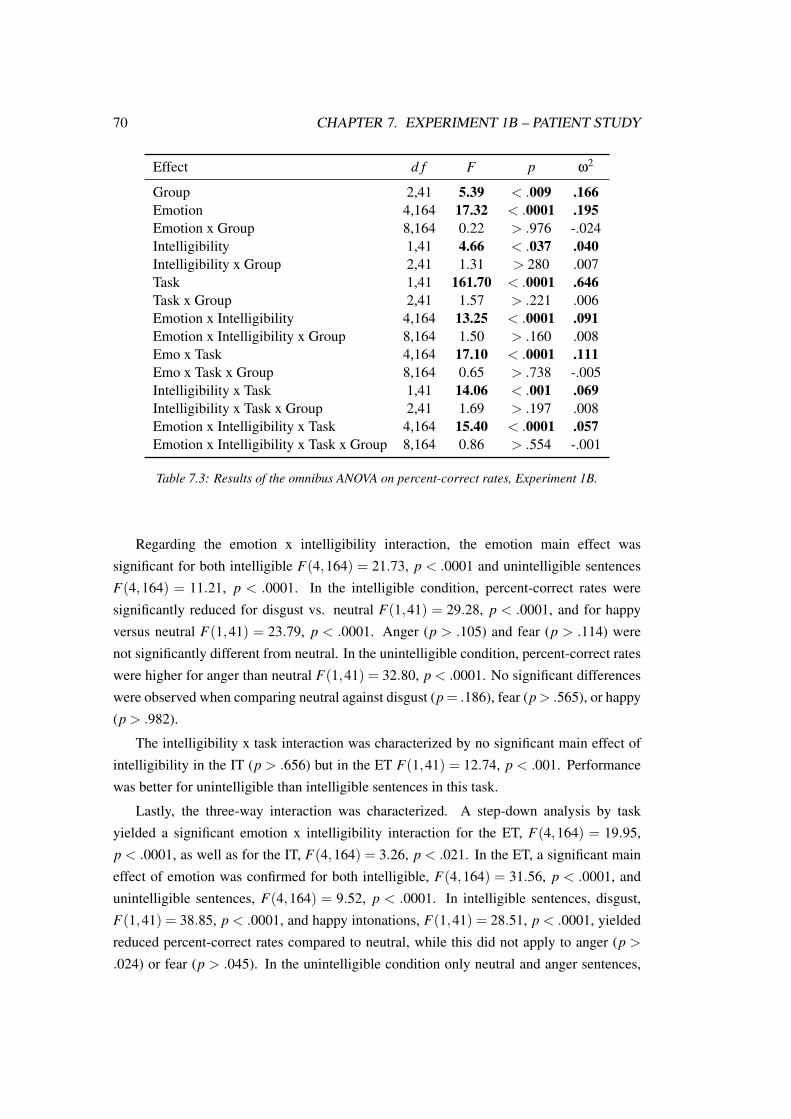
70 CHAPTER 7. EXPERIMENT 1B – PATIENT STUDY
Effect d f F p ω2
Group 2,41 5.39 < .009 .166Emotion 4,164 17.32 < .0001 .195Emotion x Group 8,164 0.22 > .976 -.024Intelligibility 1,41 4.66 < .037 .040Intelligibility x Group 2,41 1.31 > 280 .007Task 1,41 161.70 < .0001 .646Task x Group 2,41 1.57 > .221 .006Emotion x Intelligibility 4,164 13.25 < .0001 .091Emotion x Intelligibility x Group 8,164 1.50 > .160 .008Emo x Task 4,164 17.10 < .0001 .111Emo x Task x Group 8,164 0.65 > .738 -.005Intelligibility x Task 1,41 14.06 < .001 .069Intelligibility x Task x Group 2,41 1.69 > .197 .008Emotion x Intelligibility x Task 4,164 15.40 < .0001 .057Emotion x Intelligibility x Task x Group 8,164 0.86 > .554 -.001
Table 7.3: Results of the omnibus ANOVA on percent-correct rates, Experiment 1B.
Regarding the emotion x intelligibility interaction, the emotion main effect was
significant for both intelligible F(4,164) = 21.73, p < .0001 and unintelligible sentences
F(4,164) = 11.21, p < .0001. In the intelligible condition, percent-correct rates were
significantly reduced for disgust vs. neutral F(1,41) = 29.28, p < .0001, and for happy
versus neutral F(1,41) = 23.79, p < .0001. Anger (p > .105) and fear (p > .114) were
not significantly different from neutral. In the unintelligible condition, percent-correct rates
were higher for anger than neutral F(1,41) = 32.80, p < .0001. No significant differences
were observed when comparing neutral against disgust (p= .186), fear (p> .565), or happy
(p > .982).
The intelligibility x task interaction was characterized by no significant main effect of
intelligibility in the IT (p > .656) but in the ET F(1,41) = 12.74, p < .001. Performance
was better for unintelligible than intelligible sentences in this task.
Lastly, the three-way interaction was characterized. A step-down analysis by task
yielded a significant emotion x intelligibility interaction for the ET, F(4,164) = 19.95,
p < .0001, as well as for the IT, F(4,164) = 3.26, p < .021. In the ET, a significant main
effect of emotion was confirmed for both intelligible, F(4,164) = 31.56, p < .0001, and
unintelligible sentences, F(4,164) = 9.52, p < .0001. In intelligible sentences, disgust,
F(1,41) = 38.85, p < .0001, and happy intonations, F(1,41) = 28.51, p < .0001, yielded
reduced percent-correct rates compared to neutral, while this did not apply to anger (p >
.024) or fear (p > .045). In the unintelligible condition only neutral and anger sentences,

7.2. RESULTS 71
F(1,41) = 28.56, p < .0001, were significantly different, with a higher performance for
anger. No significant difference was observed between neutral and disgust (p > .16), fear
(p > .665), or happy (p > .783). The step-down analysis for the IT did not return any
significant results: The main effect of emotion was neither significant for the intelligible
(p > .289) nor the unintelligible (p > .077) material.
7.2.4 ERP data
Results from the two omnibus ANOVAs testing the N100 and the P200 can be retrieved
from Table 7.4.
N100. According to the analysis of the emotion x roi x group interaction, LPD patients
exhibited right-posterior amplitude enhancements in response to happy prosody. This was
further informed by results from the five-way interaction. Differences were found for both
intelligibility conditions in the ET context while in the IT context only the N100 to pseudo-
sentences was enhanced. RPD patients showed N100 enhancements in reaction to disgust
at rois LF, RF, LC, and ML. Specifically, the analysis of the emotion x task x intelligibility
x group interaction indicated that lexical sentences of disgust during the ET elicited an
enhanced N100 in RPD patients. This was confirmed by the five-way interaction pointing
to a topographically widespread effect at all but the posterior rois. Lexical disgust sentences
also yielded N100 enhancements in the LPD group during the ET. This was limited to left-
central and midline electrodes. RPD patients also showed generally altered processing of
fearful sentences at right-frontal leads (emotion x roi x group interaction). Further specific
N100 enhancements in RPD patients as revealed by the five-way interaction were found
for pseudo-sentences of fear (roi RP) and lexical neutral sentences (rois LF and RF), both
during the ET.
The Scheffé test of the intelligibility x group interaction did not reveal any significant
group differences.
The emotion main effect was due to a significantly lower N100 amplitude for fearful,
F(1,41) = 7.48, p < .01, and for angry compared to neutral sentences, F(1,41) = 18.14,
p = .0001. The difference with disgust (p > .135) and happy (p > .053) was not significant.
The intelligibility main effect could be explained by higher N100 amplitudes to
intelligible than unintelligible speech stimuli.
The analysis of the emotion x intelligibility interaction revealed a significant intelligi-
bility effect for anger, F(1,41) = 6.65, p < .014, disgust, F(1,41) = 11.57, p < .002,
fear, F(1,41) = 11.80, p < .002, and neutral, F(1,41) = 19.65, p < 0.0001. In the case
of disgust, fear, and neutral intelligible speech elicited enhanced amplitudes compared to

72 CHAPTER 7. EXPERIMENT 1B – PATIENT STUDY
unintelligible speech, while the pattern was vice versa for anger. Intelligibility had no
significant impact on happiness processing (p > .762).
To analyze the emotion x roi interaction, the main effect of emotion at each roi was
tested. It was significant at LF, F(4,164) = 3.88, p < .009, LC, F(4,164) = 3.71, p < .011,
RF, F(4,164) = 3.61, p = .014, and RC, F(4,164) = 2.88, p < .037. Left-frontally, anger
(F [1,41] = 11.01, p < .002), fear (F [1,41] = 9.26, p < .005), and happy (F [1,41] = 14.14,
p < .001), but not disgust (p > .042), elicited significantly reduced amplitudes compared
to neutral. At the left-central roi, there was a significant amplitude reduction for anger,
F(1,41) = 9.95, p = .003, and fear, F(1,41) = 11.68, p < .002 compared to neutral,
but no reduction was observed for happy (p > .033) or disgust (p > .3). At roi RF, the
N100 amplitude was significantly smaller for angry, F(1,41) = 28.18, p < .0001 and
happy sentences, F(1,41) = 9.01, p < .005 compared to neutral, but not for the disgusted
(p> .025) or fearful (p> .028) intonation. Finally, at roi RC only the angry-neutral contrast
became significant, F(1,41) = 17.50, p = .0001). No significant difference emerged with
disgust (p> .228), fear (p> .019), or happy (p> .152). No main effect of emotion emerged
at rois LP (p > .13), RP (p > .059), or ML (p > .083).
With respect to the intelligibility x roi interaction, a significant intelligibility effect
was observed at rois LF, F(1,41) = 7.28, p < .011, LC, F(1,41) = 8.06, p = .007, RF,
F(1,41) = 10.99, p < .002, and RC, F(1,41) = 14.24, p < .001, with higher amplitudes
to intelligible than unintelligible speech. Intelligibility was not significant at rois LP
(p > .394), RP (p > .473), or ML (p > .055).
P200. The main effect of group was reflected in a globally enhanced P200 amplitude for
the LPD group compared to HC and RPD. A graphical display of this effect can be found
in Figure 7.1.
The interactions with group further informed this main effect. The Scheffé test of
the emotion x task x intelligibility interaction yielded an enhanced P200 amplitude to
intelligible anger sentences in the LPD group compared to HC and RPD during the ET.
This enhancement was especially pronounced at rois ML and RC, as informed by the five-
way interaction. Furthermore, the P200 to intelligible disgust sentences was enhanced in
LPD during the IT. This effect was confirmed at all but the two posterior rois by the five-
way interaction. The LPD-RPD difference in this condition was only observed at ML.
Moreover, the five-way interaction indicated an enhanced P200 amplitude to intelligible
happy and unintelligible disgust expressions in LPD during the IT. The difference emerged
both with HC and RPD. While the P200 enhancement to happy stimuli concerned roi RP,
both posterior rois were affected for disgust. The P200 enhancements in LPD concerning
specific conditions are depicted in Figure 7.2.

7.2. RESULTS 73
N100 (96 – 160 ms) P200 (200 – 380 ms)Effect d f F p ω2 F p ω2
G 2,41 1.55 > .225 .024 3.95 < .028 .118E 4,164 2.92 < .036 .026 55.35 < .0001 .471E x G 8,164 2.05 > .061 .029 1.78 > .092 .025I 1,41 8.30 < .007 .077 41.95 < .0001 .318I x G 2,41 4.26 < .021 .069 0.58 > .565 -.01T 1,41 2.15 = .15 .013 4.42 < .046 .036T x G 2,41 0.15 > .864 -.02 0.31 > .732 -.016E x I 4,164 8.89 < .0001 .056 4.93 < .004 .026E x I x G 8,164 0.89 > .508 -.002 1.30 > .262 .004E x T 4,164 0.28 > .863 -.006 0.90 > .456 -.001E x T x G 8,164 1.65 > .125 .01 1.04 > .406 .001I x T 1,41 0.14 > .714 -.005 1.80 > .187 .004I x T x G 2,41 0.25 > .777 -.009 2.35 > .108 .015E x R 24,984 3.12 < .003 .010 9.77 < .0001 .038E x R x G 48,984 1.77 < .038 .007 1.08 > .375 .001I x R 6,246 3.79 < .026 .009 3.73 < .023 .01I x R x G 12,246 1.46 > .221 .003 1.42 > .227 .003T x R 6,246 2.74 = .058 .007 5.80 < .002 .019T x R x G 12,246 1.31 > .268 .002 0.38 > .864 -.005E x I x T 4,164 1.26 > .289 .001 0.73 > .536 -.001E x I x T x G 8,164 2.11 < .047 .002 2.17 < .05 .008E x I x R 24,984 1.72 > .114 .001 1.75 > .105 .002E x I x R x G 48,984 1.61 = .087 .002 1.09 > .365 .000E x T x R 24,984 1.19 > .307 .000 0.35 > .941 -.002E x T x R x G 48,984 1.24 > .234 .001 1.04 > .412 .000I x T x R 6,246 0.11 > .954 -.002 0.98 > .406 -.000I x T x R x G 12,246 0.88 > .514 -.001 0.64 > .699 -.002E x I x T x R 24,984 0.61 > .745 -.000 1.36 > .222 .000E x I x T x R x G 48,984 1.93 < .024 .002 1.74 < .049 .002
Table 7.4: Results from the omnibus ANOVAs, Experiment 1B. E = Emotion, I = Intelligibility, T =Task, R = Roi, G = Group.

74 CHAPTER 7. EXPERIMENT 1B – PATIENT STUDY
Figure 7.1: Main effect of group in the P200, Experiment 1B.
Figure 7.2: Specific P200 enhancements in the LPD group, Experiment 1B. IT = implicit task, ET =explicit task

7.2. RESULTS 75
Emotional salience detection was of outstanding interest for the present study, which is
why the main effect of emotion was analyzed group-wise by means of contrasts between
the neutral and the emotional conditions. A significant main effect of emotion could be
confirmed in all three groups, LPD: F(4,36) = 24.18, p < .0001, RPD: F(4,44) = 17.31,
p < .0001, and HC: F(4,84) = 20.46, p < .0001. All groups showed lower amplitudes to
fearful versus neutral prosody, LPD: F(1,9) = 34.06, p < .001, RPD: F(1,11) = 92.28,
p < .0001, and HC: F(1,21) = 34.47, p < .0001. The HC group also exhibited a reduced
P200 amplitude for disgust, F(1,21) = 7.78, p = .011. This applied to the RPD group, too,
although the difference was not significant after Bonferroni correction, F(1,11) = 6.38,
p < .029. In the LPD group, there was no indication of a significant amplitude difference
between disgust and neutral (p > .675). No group showed significant differences between
anger and neutral (LPD: p > .108, RPD: p > .255, and HC: p > .596) or between happy
and neutral (LPD: p > .244, RPD: p = .281, and HC: p > .999). Figure 7.3 shows the
emotional salience detection for each group.
Figure 7.3: Emotional salience detection in each group, Experiment 1B.
The main effect of emotion became evident in reduced amplitudes to fearful, F(1,41) =
116.52, p < .0001, and disgust sentences, F(1,41) = 11.12, p < .002, compared to neutral.
Anger (p = .673) and happiness (p = .259) did not significantly differ from neutral.

76 CHAPTER 7. EXPERIMENT 1B – PATIENT STUDY
The emotion x roi interaction was analyzed by means of a roi-wise main effect of
emotion. It became significant at LF, F(4,164) = 52.97, p < .0001, LC, F(4,164) = 41.21,
p < .0001, LP, F(4,164) = 9.77, p < .0001, RF, F(4,164) = 61.44, p < .0001, RC,
F(4,164) = 61.06, p < .0001, RP, F(4,164) = 14.60, p < .0001, and ML, F(4,164) =
42.68, p < .0001. Fearful sentences elicited significantly reduced amplitudes compared to
neutral at all rois: LF, F(1,41) = 91.57, p < .0001, LC, F(1,41) = 89.28, p < .0001, LP,
F(1,41) = 30.42, p < .0001, RF, F(1,41) = 126.16, p < .0001, RC, F(1,41) = 124.71,
p < .0001, RP, F(1,41) = 47.37, p < .0001, and ML, F(1,41) = 108.72, p < .0001.
Disgust differed significantly from neutral at rois LC, F(1,41) = 11.01, p < .002, LP,
F(1,41) = 9.16, p < .005, RC, F(1,41) = 11.18, p < .002, and ML, F(1,41) = 9.86,
p < .004, but not at LF (p > .017), RF (p > .025), or RP (p > .013). No significant anger-
neutral (LF: p> .43, LC: p> .815, LP: p> .187, RF: p> .057, RC: p> .395, RP: p> .704,
ML: p > .601) or happy-neutral difference (LF: p > .037, LC: p > .222, LP: p > .734, RF:
p > .077, RC: p > .315, RP: p > .263, ML: p > .372) was observed.
The main effect of intelligibility was induced by higher P200 amplitudes to intelligible
than unintelligible sentences. Furthermore, amplitudes were higher in the ET compared to
the IT (main effect of task).
The intelligibility x roi interaction was also analyzed by roi. A significant main effect of
intelligibility emerged at LF, F(1,41) = 30.85, p< .0001, LC, F(1,41) = 72.81, p< .0001,
LP, F(1,41) = 31.27, p < .0001, RF, F(1,41) = 13.43, p < .001, RC, F(1,41) = 22.77,
p < .0001, RP, F(1,41) = 10.19, p < .003, and ML, F(1,41) = 33.05, p < .0001, with
higher amplitudes to intelligible than unintelligible sentences.
A step-down analysis of the emotion x intelligibility interaction yielded a significant
main effect of intelligibility for disgust F(1,41) = 35.77, p < .0001, fear F(1,41) = 7.44,
p < .01, happiness F(1,41) = 6.03, p < .019, and neutral F(1,41) = 36.60, p < .0001.
Amplitudes were smaller to unintelligible than to intelligible sentences. No significant
intelligibility effect was found for anger (p > .766).
Finally, the task x roi interaction was manifested in a significant main effect of task at
rois LF, F(1,41) = 12.22, p < .002, LC, F(1,41) = 4.56, p < .039, RF, F(1,41) = 12.65,
p = .001, and RC, F(1,41) = 5.11, p < .03, with higher amplitudes for the ET than the IT.
Task was not significant at rois LP (p > .979), RP (p > .754), or ML (p > .075).
7.2.5 Correlations
The N100 amplitude in conditions with significant enhancements for patients did not
significantly correlate with the composite scores, age, education, or the P300 amplitude.
The right-posterior P200 amplitude to intelligible happy sentences during the IT (altered
in LPD) correlated significantly with the composite score for working memory, r = .43,

7.2. RESULTS 77
p < .004. Correlations of altered ERP amplitudes with motor variables in PD are shown in
Table 7.5.
Emotion Intelligibility Task Roi AI LMS RMS
N100 effects, LPDHappy —overall— RP -.55** -.49* –Happy intelligible ET RP -.45 -.35 –Happy unintelligible ET RP -.46 -.28 –Happy unintelligible IT RP -.46 -.42 –Disgust intelligible ET LC ML .10 .20 –
N100 effects, RPDDisgust —overall— LF LC RC ML .38 – -.41Disgust intelligible ET LF LC RF RC ML .19 – -.12Fear —overall— RF .26 – -.33Fear unintelligible ET RP -.42 – .27Neutral intelligible ET LF RF .36 – -.41
P200 effects, LPD—overall— .52* .39 –
Anger intelligible ET RC ML .65** .50* –Disgust —overall— .63** .50* –Disgust intelligible IT LF LC RF RC ML .43 .33 –Disgust unintelligible IT LP RP .50* .26 –Happy intelligible IT RP .41 .27 –
Table 7.5: Correlations of ERP amplitudes in Experiment 1B with left and right motor scores andthe asymmetry index. p < .05, *p < .025, **p < .01.
7.2.6 Prosody categorization study
High recognition rates were achieved by all groups: LPD 78% (SD = 21.35), RPD 80%
(SD = 22.36), and HC 85% (SD = 18.95). The main effect of group was not significant
(p > .065), nor was the emotion x group (p > .474), intelligibility x group (p > .211), or
emotion x intelligibility x group (p = .952) interaction.
A significant main effect of emotion was observed, F(4,164) = 22.49, p < .0001,
ω2 = .261. Performance for neutral (92%) was significantly better than for disgust (72%),
F(1,41) = 58.07, p < .0001, happiness (74%), F(1,41) = 35.18, p < .0001, and fear
(86%), F(1,41) = 10.73, p < .003. For anger, percent-correct rates (88%) were not
significantly different from neutral (p > .055).

78 CHAPTER 7. EXPERIMENT 1B – PATIENT STUDY
Furthermore, the main effect of intelligibility became significant, F(1,41) = 39.28,
p < .0001, ω2 = 0.303, with higher percent-correct rates for intelligible than unintelligible
sentences (87% versus 72%).
The omnibus analysis yielded a significant emotion x intelligibility interaction, F
(4,164) = 3.00, p< 0.029, ω2 = .015. Intelligibility influenced categorization performance
for angry, F(1,41) = 27.01, p < .0001, disgusted, F(1,41) = 16.98, p < .001, fearful,
F(1,41) = 10.51, p < .003, and neutral prosody, F(1,41) = 5.86, p = .02, with lower
accuracy rates for unintelligible than intelligible speech. No intelligibility effect emerged
for happy prosody (p = .175).
7.3 Discussion
The present study aimed to shed light on the early neural mechanisms of emotional prosody
perception in PD as a function of motor symptom asymmetry. The P200, as the principal
component of interest to this study, was altered in LPD but not in RPD patients. Thus,
complementing previous neuroimaging evidence of a striatal involvement in receptive
emotional prosody (Bach et al., 2008; Beaucousin et al., 2007; Grandjean et al., 2005; Kotz,
Meyer, et al., 2003; Leitman et al., 2010; Morris et al., 1999; Phillips et al., 1998; Quadflieg
et al., 2008; Wittfoth et al., 2010), the data indicate that the right striatum is involved in this
process at an early point in time. This involvement could be reflected in an early bottom-up
influence from the right striatum to superior temporal areas implicated in emotional prosody
(Schirmer & Kotz, 2006) and would complement data indicating an interaction of the right
striatum and superior temporal areas receptive to emotionally intoned speech (Ethofer et
al., 2012). Furthermore, the point in time (P200) fits with previous MMN evidence by
Schröder et al. (2006). The intact P200 response in RPD patients is in line with data from
patients with left-striatal lesions (Paulmann et al., 2011). Thus, the left striatum may not
be crucial for early emotional salience detection from prosody, and the data are consistent
across etiologies (neurodegeneration versus lesion).
N100. In the first place, it must be considered that both patient groups exhibited N100
alterations. Thus, it cannot be discarded that early sensory deficits account for P200
alterations in the LPD group or that the RPD group also shows early deficits in emotional
prosody perception, but they do not extend to the P200. However, this explanation
is not that likely, as there was almost no overlap of conditions affected in the N100
and in the P200 in the LPD group and there were no P200 alterations in RPD. This
points to a low correspondence between the processes reflected by these two components,
respectively. Furthermore, correlations of motor scores and asymmetry indices with N100

7.3. DISCUSSION 79
amplitudes were almost all nonsignificant, apart from the right-posterior anomaly in LPD
for processing happy prosody which furthermore extended to the P200. Thus, the right-
posterior alterations for happy prosody in LPD may already start in the N100.
P200 alterations in specific conditions. Apart from the main effect of group, more
detailed information on early emotional salience detection in LPD is provided by the
interaction effects. The most wide-spread impairments became apparent for disgust.
Implicit processing of this emotion was altered in the present study, and no amplitude
reduction for disgust compared to neutral sentences was observed in the P200. The HC
and RPD groups, in turn, showed this effect. The alterations in LPD are corroborated by
correlations with motor variables, indicating a direct relation with the degree of right-striatal
dysfunction.
To date, not much is known about a possible role of the striatum for emotional prosody
perception. Pell and Leonard (2003) have previously considered the possibility of a BG
involvement in receptive disgust prosody on the basis of behavioral data. Furthermore,
activity in the right striatum has been observed when processing disgust from faces (Phillips
et al., 1998). A recent meta-analysis reported that in visual presentation and mood induction
studies, the disgust involvement of the right striatum is significantly greater than for anger,
fear, or happiness (Vytal & Hamann, 2010). Generally, the striatum seems to play an
important role for withdrawal emotions (Wager, Phan, Liberzon, & Taylor, 2003). Very
interestingly, Mataix-Cols et al. (2008) reported a correlation between individual disgust
sensitivity scores and the right-striatal response to disgust stimuli.
Thus, the right striatum may respond cross-modally to disgust cues which normally
require a rapid withdrawal reaction. This would underline the presumed role of the striatum
as an emotional-motor interface (Groenewegen & Trimble, 2007). However, Phillips et al.
(1998) reported a dissociation between facial and vocal expressions of disgust, with the
striatum reacting only to the former, which would not be in accord with the striatum as
a cross-modal “disgust module”. It must however be noted that in that study, the sample
size (N = 6) was very small which limits the generalizability and statistical power of the
results, and moreover the low temporal resolution of fMRI may not necessarily capture
early processes of emotional salience detection from speech. Also, these researchers used
emotional vocalizations, which may trigger different reactions than sentences, and the
comparison condition were mildly happy stimuli instead of neutral ones. Happiness may
itself trigger striatal reactions, as will be discussed below. Lastly, the sample consisted
only of men who may be less sensitive to disgust stimuli than women (Haidt, McCauley, &
Rozin, 1994). To sum up, more research on the role of the striatum for processing disgust
cues is required. The present data indicate that the right striatum influences early emotional

80 CHAPTER 7. EXPERIMENT 1B – PATIENT STUDY
salience detection from prosody, which is in line with the pivotal role for disgust ascribed
to the BG (Calder et al., 2001).
Another condition affected in the present study was explicit processing of intelligible
anger stimuli. A significant correlation of the P200 enhancements in this condition with
asymmetry indices and left motor scores foster this observation. An involvement of the
striatum during the processing of angry speech intonation has been previously reported
in the literature (Bach et al., 2008; Grandjean et al., 2005; Kotz, Meyer, et al., 2003;
Quadflieg et al., 2008; Wittfoth et al., 2010) and is thus in line with the current results
which furthermore indicate that the right striatum is involved already at an early point in
time when attention is drawn to the emotionality of a vocal stimulus, i.e., during an explicit
task. This fits with observations previously made by Bach et al. (2008).
Lastly, implicit processing of intelligible happiness stimuli was affected in LPD. This
supports the previously assumed role of the striatum in processing happy speech intonation
(Kotz, Meyer, et al., 2003; Kotz et al., 2006). While the evidence on emotional speech
intonation is, however, still sparse concerning happiness, the right striatum appears to be one
of the structures most consistently activated by happy facial expressions (Vytal & Hamann,
2010). Thus, aggregating these lines of evidence, a cross-modal striatal involvement in
happiness processing may be assumed. Ariatti et al. (2008) have previously reported that
LPD patients have difficulty recognizing happy prosody, which may be a consequence of
impaired early emotional salience detection from this stimulus category.
To sum up on specific emotion deficits, disgust, anger, and happiness were affected in
LPD. This result is not in line with accounts that have primarily associated PD with impaired
processing of negative emotions (Dara et al., 2008; Gray & Tickle-Degnen, 2010; Pell &
Leonard, 2003). According to the present results, deficits in PD may be more wide-spread.
Furthermore, it becomes apparent that the convergence between the early neural response
as assessed in this study and the behavioral categorization or rating performance from other
studies is limited. Importantly, in the present study no deficits for fear were observed, while
such impairments were evident in behavioral experiments (Breitenstein et al., 1998; Dara et
al., 2008; Yip et al., 2003), and vocal fear processing seems to involve the striatum (Phillips
et al., 1998). As fear is of outstanding evolutionary significance, its early perception may
be intact in PD, while deficits could become apparent at later stages. This has previously
been shown in the case of left-striatal lesions (Paulmann, Pell, & Kotz, 2009).
Regarding task, there was no clear pattern of impairments in the current study. While
disgust was generally impaired, although more pronounced during implicit processing,
anger was affected during explicit and happiness during implicit processing. Thus, the
emotional category may be crucial. The behavioral categorization study yielded very high
recognition rates for angry stimuli, while they were much lower for disgust and happiness.

7.3. DISCUSSION 81
Conversely, a gating study (Pell & Kotz, submitted) reported a much earlier recognition
point for anger relative to disgust or happiness. Thus, these specific emotion categories
appear to be associated with different levels of difficulty. These may interact with specific
task demands that modulate the amount of attention drawn on these stimuli. Thus, it remains
to be elucidated how the processing challenges imposed by different emotional categories
interact with attention allocation modulated by task instructions.
As predicted, the processing of unintelligible speech proved to be largely intact in PD,
apart from disgust which was associated with more wide-spread impairments in LPD. Thus,
the data confirm previous reports of a greater striatal involvement in processing intelligible
than unintelligible speech (Kotz, Meyer, et al., 2003; Kotz et al., 2006). An explanation
for this involvement may be the proposed sequencing function previously ascribed to the
BG (Kotz & Schwartze, 2010). More precisely, the BG may track the temporal patterns,
pitch, and intensity variations conveyed by emotional speech in order to create a coherent
percept (Kotz et al., in press; Paulmann & Pell, 2010b). A study by Breitenstein et al. (2001)
provided evidence that PD patients have difficulty using speech rate information in order to
successfully identify emotional prosody. Thus, there may be a potential impairment in LPD
concerning the interaction of different emotional information channels, i.e., semantic and
prosodic information. Therefore, in future PD investigations it would be interesting to shed
more light on interactions between different communication channels providing emotional
information.
Behavioral emotion categorization study. Unlike many other categorization studies,
no impairments in explicit emotional prosody recognition were observed in the present
experiment. This result is in line with findings from some previous studies of this kind
(Blonder et al., 1989; U. S. Clark et al., 2008; Kan et al., 2002; Mitchell & Bouças,
2009). What must be considered, though, is the fact that the behavioral study took place
after two complete EEG sessions and participants were thus very familiar with the stimuli
presented in the behavioral experiment. Moreover, the patients showed a good performance
in tests on cognitive functions, and cognitive variables may be related to performance in
emotion categorization (Benke et al., 1998; Breitenstein et al., 2001; Pell & Leonard, 2003).
The lack of cognitive impairments in the present patient sample, however, strengthens the
experimental findings which are unlikely due to more generalized cognitive deficits.
Summary and conclusion. To sum up, the current data indicate that emotional salience
detection from speech (P200) depends on the sidedness of motor symptoms in PD. While
this function was preserved in RPD, LPD patients exhibited wide-spread impairments. This
points to an important role for the right striatum in early vocal emotion perception and

82 CHAPTER 7. EXPERIMENT 1B – PATIENT STUDY
underlines that PD is not a uniform disorder. However, it must be considered that the BG
involvement in PD is not completely unilateral. Already at early disease stages, both sides
are involved in neurodegeneration and the laterality is thus only relative, but not absolute
(Schwarz et al., 2000). Moreover, neural damage in PD is not confined to the BG (Braak
et al., 2003; Tinaz et al., 2011), and it is not clear which role damage in additional brain
regions may play. In any case, the group-specific results and the correlative findings of
the present study are very telling and form a valid basis for future studies. In particular,
as the impairments in LPD almost exclusively concerned intelligible speech, it would be
interesting to further investigate the interactions of different emotional channels in PD.

Chapter 8
Experiment 2: Emotional priming
Emotion is a complex and multi-faceted construct, and emotional communication is
of multisensory nature. It entails voice expressions (i.e., prosody), semantics, facial
expressions, and gestures, among others. Information from all these channels is present
in daily life when we perceive emotional information in others. While this seems a
lot of input at a time, we normally benefit from this combination of different sources,
as long as they provide congruent emotional information. This has been confirmed
experimentally: Matching affective information provided concurrently by vocal and facial
expressions leads to better emotion recognition performance than in unimodal input, while
there is a performance decline with mismatching information (de Gelder & Vroomen,
2000; Föcker, Gondan, & Röder, 2011). In the following experiments, it was investigated
how emotional information conveyed by facial expressions affects the processing of an
emotionally matching or mismatching prosodic stimulus and vice versa.
To assess how the processing of affective information from one sensory channel
affects the processing of another, two major streams of research have been proposed in
the literature: One is to present both channels simultaneously, often accompanied by an
instruction requiring participants to attend only one of them while ignoring the other, which
can either convey matching or mismatching emotional information (de Gelder & Vroomen,
2000; Koizumi et al., 2011). Another means to assess the impact of one modality on another
is cross-modal emotional priming, with one modality providing prime information and the
other target information (Pell, 2005). In classical affective priming paradigms, two tasks
are commonly used: evaluative decision and lexical decision. The evaluative decision aims
at the affective valence of a target and can thus be considered an explicit task, while lexical
decision involves a categorization of the target as representing a word or a non-word, and
is thus implicit with respect to the emotional content. This lexical decision task illustrates
83

84 CHAPTER 8. EXPERIMENT 2: EMOTIONAL PRIMING
how affective priming is classically tested, namely with written word stimuli (see Klauer &
Musch, 2003, for a comprehensive overview of the affective priming literature).
As outlined in the introduction of this thesis, implicit tasks offer several benefits in
emotion research. First, they allow examining whether emotional information is used
by the perceiver even if task instructions do not require him to do so. Furthermore, an
easy implicit task can reduce a potential processing load and thus lower the possibility
that experimental results may be confounded with variables which are not task-related
(e.g., executive functions). Lastly, in the context of basal ganglia diseases, it must be
acknowledged that explicit tasks may engage the BG more than implicit ones (Bach et
al., 2008; Beaucousin et al., 2007). However, with facial targets, the classical implicit task
(lexical decision) cannot be used due to the nonverbal nature of these stimuli. In an attempt
to develop a parallel of the lexical decision task for (cross-modal) emotional priming with
facial targets, Pell (2005) invented the “Facial Affect Decision Task” (FADT), in which
participants are asked whether a target face displays an emotion or not. Even though
this task is not oriented towards emotional prime-target congruency, it has been shown to
reliably induce priming effects at the behavioral level (Pell, 2005; Pell et al., 2011) as well
as in the ERPs (Paulmann & Pell, 2010a) when prosodic cues serve as primes. Although the
FADT does not require an evaluation of prime-target congruency, it is still oriented towards
emotion, and the question remains whether cross-modal emotional priming works with a
task that is completely implicit, i.e., not directed towards either emotion or congruency.
Some indication that this may work comes from Pourtois and colleagues (Pourtois et
al., 2000, 2002). Congruency between emotional face primes and voice targets modulated
the ERP response in the absence of a task. Participants were solely instructed to attend
one modality and to ignore the other. However, the reverse prime-target assignment (i.e.,
prosodic primes and facial targets) still remains to be tested implicitly. Furthermore, in
Pourtois et al.’s studies, the facial expression remained the same throughout the experiment
and was combined with one of two possible emotional intonations as targets to manipulate
congruency. Thus, it cannot be disclosed that the congruency effects in these studies were
triggered by low-level features of the targets rather than by congruency per se.
In the part on emotional facial expressions (see section 3.4), the critical issue has been
raised that most studies using face stimuli draw on static face stimuli, i.e., photographs
or drawings. In daily life, however, we are exposed to dynamically changing facial
information. Therefore, it would be more ecologically valid to use dynamic facial
expressions in research as opposed to static ones. Some imaging studies have made direct
comparisons of static and dynamic facial stimuli and revealed more wide-spread neural
activation patterns, among others in temporal regions linked to the social relevance of
stimuli (Sato et al., 2004; Trautmann et al., 2009). Thus, for the sake of higher ecological

85
validity, the use of dynamic facial stimuli is recommended. Furthermore, the use of moving
facial stimuli is expected to lead to a higher degree of convergence between primes and
targets, as prosodic stimuli cannot be presented in a static manner.
To sum up, the basic approach of the current study was cross-modal and implicit
emotional priming with facial and vocal expressions of emotion. This was investigated
bidirectionally, i.e., with prosody and face once as prime and once as target stimuli,
respectively. The use of dynamic facial expressions guaranteed a higher ecological validity
of the face stimuli, as opposed to the more widely used static ones.


Chapter 9
Experiment 2A – Pilot study
The aim of this study was to test cross-modal emotional priming by means of ERPs in a
sample of young, healthy participants. In emotional priming studies, an N400 modulation
by prime-target congruency is a common observation (Paulmann & Pell, 2010a; Steinbeis &
Koelsch, 2011; Zhang et al., 2006, 2010). Some studies have also reported effects at earlier
processing stages, i.e., in the N100 (Pourtois et al., 2000) and P200 (Pourtois et al., 2000,
2002). Especially with regard to these earlier components, there is no clear picture as of
yet, in which cases - or in which ones not - priming effects may be expected. Thus, an N400
congruency effect was expected in the present study, but with respect to early components
the present study was rather explorative. In this context, it must also be considered that this
is the first ERP priming study using dynamic emotional facial expressions, which entails
the possibility that ERPs may be modulated differently than in research with static faces
(Mayes, Pipingas, Silberstein, & Johnston, 2009).
Previous priming evidence, utilizing sentences with emotional intonation as a prime
context, have reliably led to N400 modulations of visual target words (Schirmer et al.,
2002, 2005). In order to ensure stimulus consistency we therefore used emotionally intoned
sentences from Experiment 1. However, two major changes were implemented: First, as
stimulus repetitions may have adverse effects on most types of priming (Schacter, Wig, &
Stevens, 2007) except, of course, repetition priming, it may be useful to increase stimulus
variability. This was accomplished by including four rather than two speakers, which
prevented stimulus repetition. Second, only unintelligible speech stimuli were used. This
was considered as necessary, as the use of semantic material may introduce a confound
between the effects elicited by prosody and those elicited by semantics, and it would not
be possible to disentangle priming by speech intonation from semantic priming. Even
though at the behavioral level, priming effects elicited by pure-prosodic versus prosodic-
semantic stimuli were comparable (Pell et al., 2011), it may still affect ERP results (Kotz
87

88 CHAPTER 9. EXPERIMENT 2A – PILOT STUDY
& Paulmann, 2007). Furthermore, it has been shown at the behavioral and at the ERP level
that “pure prosody” leads to reliable cross-modal emotional priming effects (Paulmann &
Pell, 2010a; Pell, 2005), indicating that the use of unintelligible speech is appropriate for
the purposes of the present study.
In addition to the sentences, natural video recordings of the same four semi-professional
actors who produced the prosodic stimuli were used, but excluding sound. To determine
their length it was considered that the time course of emotional face processing seems to be
largely comparable to emotional prosody (Paulmann & Pell, 2009). Furthermore, to ensure
classical N400 priming effects, a prime stimulus (prosodic prime) needs to be at least 400
ms long (Paulmann & Pell, 2010a). It was thus determined that the presentation of dynamic
facial expressions should be approximately 500 ms in order to match minimal prosodic
context. This temporal reduction of face stimuli ensured that participants did not perceive
cues from the speaker’s lip movement and reduced the experiment duration compared to
face stimuli of longer duration.
Another important aspect of the present study was speaker coherence, i.e., prime and
target in one trial were always produced by the same speaker, which is not the case in
many other studies of cross-modal emotion perception. This leads to a greater likelihood
to consider face and voice as related stimuli (Föcker et al., 2011), and avoids possible
influences of a speaker mismatch in the ERPs (Schweinberger, Kloth, & Robertson, 2011).
The perception of emotional information from different modalities is thought to be an
automatic and mandatory process (de Gelder, Vroomen, & Pourtois, 2004). It also occurs
under high perceptual load (Vroomen, Driver, & Gelder, 2001), and is even observed when
facial expressions are not consciously perceived (de Gelder, Morris, & Dolan, 2005). These
observations indicate that congruency effects in the current set-up may be observed in an
implicit task, avoiding explicit emotional judgments.
In a first step, existing video material needed to be processed and rated for the use in the
ERP study.
9.1 Rating study
Participants. Twenty-eight volunteers took part in an initial rating study. Three partici-
pants had to be excluded due to low performance (more than two standard deviations below
the group mean for at least one emotional category). Thus, 25 participants (12 female)
were included in the final sample. Their mean age was 25.48 years (SD = 2.63, range: 20
– 30). All participants had normal hearing and normal or corrected-to-normal vision and
were right-handed according to the Edinburgh Handedness Inventory (Oldfield, 1971), with
a mean laterality quotient of 91.20 (SD = 11.29, range = 60 – 100). None reported any

9.1. RATING STUDY 89
history of neurological or psychiatric illness. Participants were paid a total of seven Euros
per hour for their participation.
Stimulus material. Black-and-white video stimuli of facial expressions produced by four
semi-professional actors (two women) were used. These had been recorded while the actors
produced emotionally intoned sentences and showed the corresponding facial expressions.
For the present experiment, angry, happy, and neutral expressions were selected and edited
with the video manipulation program Avidemux, version 2.4.3. The voice track was first
removed from the videos; then the videos were cut into fragments of 520 ms duration. In
order to avoid different emotion recognition points among the videos, it was assured that
the respective facial expression could be recognized already at the beginning of the video.
When necessary, videos were cropped to ensure that head size was comparable across all
stimuli and that all faces appeared in a centered position. The image resolution was 720 x
576 pixels. Face width on the screen was approximately 10 centimeters. Videos were AVI
files and used the MPEG-4 codec optimized for timing. The presentation rate was 25 frames
per second.
For each actor and emotion, 40 videos were presented adding up to 160 stimuli per
emotional category (angry, happy, neutral). Thus, the complete material consisted of 480
stimuli.
Procedure. The ratings were acquired in a single session which lasted about one hour.
Participants sat in a quiet laboratory with a computer monitor and a computer keyboard
directly in front of them. The stimuli were presented at the center of a computer screen
in pseudo-randomized order that differed for each participant. 480 trials were presented,
divided into eight blocks of equal length. To assess emotion recognition from the videos,
a categorization task was applied in which participants chose between the labels “anger”,
“happiness”, or “neutral”. Additionally, there was one button with a question mark on
it. Participants were told to use it only when they could not decide on either one of the
predefined labels. Arousal ratings were acquired using a nine-point SAM scale (self-
assessment manikin; Bradley & Lang, 1994) on a different set of key presses of the
computer keyboard. Participants were asked to rate the presented items on a scale ranging
from “very calm”, represented at the left-most position of the scale, to “very aroused” at the
right-most position.
A trial started with a black fixation cross on a gray background matched to the mean
luminance of the videos. Then, a video was presented, followed by the question “How
aroused?” printed on the screen, requiring an arousal rating, which had to be provided
within four seconds. After the button press or after the response window had timed out,

90 CHAPTER 9. EXPERIMENT 2A – PILOT STUDY
the question “Which emotion?” appeared on the screen. Response time was also set to a
maximum of four seconds. After a short break of 1500 ms, the next trial started.
Data analysis. The purpose of this rating study was to select videos for an EEG study
according to recognition rates. Therefore, a recognition rate was calculated for each
video separately by dividing the count of participants who correctly identified the intended
emotion from a video by the total number of subjects (N = 25). The obtained number was
multiplied by 100 to obtain a percentage score. A ranking of videos was created for each
actor and emotion separately, resulting in 12 rankings of 40 items each. These rankings
provided the basis for stimulus selection in Experiment 2, which is described in more detail
in section 9.2.1 for the pilot study and in section 10.1.2 for the patient study.
9.2 ERP study
9.2.1 Methods
Participants. Thirty-six volunteers took part in the second pilot experiment. Two of them
were excluded from the sample due to excessive eye movements during the measurement.
The remaining 34 participants (17 female) had a mean age of 24.74 years (SD = 2.53,
range = 19 – 31) and were right-handed with a mean laterality quotient (Oldfield, 1971) of
90.29 (SD = 11.15, range = 66 – 100). All reported normal hearing abilities and normal
or corrected-to-normal vision. None reported any history of neurological or psychiatric
illness. Due to strong artifacts, data from one participant (male, 25 years, laterality quotient
= 100) had to be excluded for the prosody-as-prime condition only. Likewise, another
participant (female, 24 years, laterality quotient = 80) was excluded only for the video-as-
prime condition. Thus, 33 subjects were included in each analysis. Participants were paid a
financial compensation of seven Euros per hour for participating.
Stimulus material The video stimuli are described in section 9.1. Additionally, as
indicated above, the same unintelligible sentence stimuli as utilized in Experiment 1 were
used in the present study, but now including four (two female) instead of two actors.
Stimulus rankings for each actor and the emotion categories happy and angry were used to
select the sentence and video materials. For the prime and target positions, the 20 highest-
ranking stimuli from each of the eight rankings (2 emotions x 4 actors) were selected,
resulting in a set of 180 stimuli equally distributed across actors and emotional categories.
Primes and targets were combined with each other. It was assured that prime and target
were always produced by the same actor. Half of the happy prime stimuli were combined
with a happy target, the other half with angry targets, and the same was done with the angry

9.2. ERP STUDY 91
primes. This resulted in 80 congruent and 80 incongruent trials. Furthermore, 80 neutral
primes were included as fillers in order to provide a non-emotional context; these neutral
primes were combined with lower-ranking (positions 21 – 30 in each list) happy and angry
stimuli as targets. Mean recognition rates of stimuli used in this experiment are listed in
Table 9.1.
Videos SentencesSpeaker happy angry happy angry
1 94 100 71 872 98 100 65 1003 92 96 33 974 92 98 59 96
Table 9.1: Recognition rates in percent for the stimuli used in Experiment 2A. Note that for videostimuli, chance level was at 33% while for sentence stimuli it was at 14%.
Procedure The EEG was acquired in a single session with a total of 480 trials presented in
a pseudo-randomized order that differed for each participant. The EEG measurements took
place in an electrically shielded, sound-attenuated room. Participants sat in a comfortable
chair at a distance of approximately one meter from the screen. In one half of the session,
the videos served as primes and the sentences as targets; in the other half the procedure
reversed. Half of the participants started with the video-as-prime condition, the other
half with the prosody-as-prime condition. An implicit task (gender decision) was used to
assess emotional priming in the absence of attention the emotion expressed in the respective
stimuli. 50% of the participants pressed the “male” button with their left and the “female”
button with their right hand, and the other half proceeded vice versa. The EEG recording
took place in an electrically shielded, sound-attenuated EEG chamber. Participants sat in a
comfortable chair at a distance of approximately one meter from the screen.
Sentences were presented via loudspeakers at a comfortable sound level which was held
constant across participants. The videos were presented centrally on the screen at a visual
angle of approximately three degrees from the center.
The trials started with a black fixation cross in the center of a grey background, which
matched the mean luminance of the videos. Then, the prime was presented and after this,
the target followed immediately without an inter-stimulus-interval. After target offset, a
black question mark was displayed on the screen requiring the participant’s response via
button press. Response time was set at a maximum of four seconds. After the button press
or after the response window had timed out, a blank screen was displayed for two seconds

92 CHAPTER 9. EXPERIMENT 2A – PILOT STUDY
before the next trial started. Participants were instructed to refrain from blinking during the
prime and target presentation. Figure 9.1 shows this sequence schematically.
Figure 9.1: Trial scheme for Experiment 2. The sequence corresponds to the video-as-primecondition. In the prosody-as-prime condition, the prime-target sequence was reversed.
The 480 trials were divided into eight blocks of 60 stimuli. The total run-time of the
experiment was about an hour.
Data acquisition and analysis Sixty-one Ag/AgCl electrodes mounted in an elastic
cap were used to record the EEG from the scalp. The electrode locations following the
nomenclature of the American Electroencephalographic Society (Sharbrough et al., 1991)
were: FPZ, FP1, FP2, AFZ, AF3, AF4, AF7, AF8, FZ, F3, F4, F5, F6, F7, F8, F9, F10,
FCZ, FC3, FC4, FC5, FC6, FT7, FT8, FT9, FT10, CZ, C1, C2, C3, C4, C5, C6, T7, T8,
CPZ, CP3, CP4, CP5, CP6, TP7, TP8, TP9, TP10, PZ, P3, P4, P5, P6, P7, P8, P9, P10,
POZ, PO3, PO4, PO7, PO8, OZ, O1, O2 (cf. Figure 1.1, for a graphical display of these
locations). The data were recorded with a bandpass between DC and 140 Hz and digitized at
a sampling rate of 500 Hz. An average reference was used during the recording. Offline, the
data were re-referenced to the linked mastoids. Vertical and horizontal electrooculograms
(EOG) were recorded bipolarly from above and below the right eye and bilateral outer
canthi, respectively. The ground electrode was placed on the sternum. Electrode resistance
was kept below five k. Data were filtered offline with a bandpass from 2 – 30 Hz (1135
coefficients, Blackman window). The strong highpass was necessary to obtain a valid in-
stimulus baseline at target onset and to remove potential slow-wave activity resulting from
late processing stages of the prime. An additional 12 Hz highpass filter was applied for
graphical display only.
Only correctly-answered and artifact-free trials entered the analysis. Artifact rejection
was accomplished in two steps: First, an automatic rejection algorithm identified trials, in
which the EOG electrodes or CZ exceeded an amplitude of 30 or 40 V, respectively. The
data were then scanned manually to detect additional artifacts.
For statistical purposes, seven topographical regions of interest (rois) were set up as
follows: left-frontal (LF; F7, F5, F3, FT7, FC5, FC3), left-central (LC; T7, C5, C3, TP7,
CP5, CP3), left-posterior (LP; P7, P5, P3, PO7, PO3, O1), right-frontal (RF; F8, F6, F4,
FT8, FC6, FC4), right-central (RC; T8, C6, C4, TP8, CP6, CP4), right-posterior (RP; P8,

9.3. ERP RESULTS 93
P6, P4, PO8, PO4, O2), and midline (ML; AFZ, FZ, FCZ, CZ, CPZ, PZ). For auditory
targets, epochs were constructed from 0 to 1000 ms post onset. For video targets, the
epochs lasted from 0 to 520 ms time-locked to their onset. In both cases, the first 100 ms
served as an in-stimulus baseline. Behavioral data were not analyzed in this study, as task
performance was at ceiling (mean: 99% in both conditions). Furthermore, the response
window was fixed and delayed, which did not allow to analyze reaction times.
Greenhouse-Geisser corrected p-values were used where necessary to correct for
nonsphericity in the data. Effect sizes were calculated using ω2 (Olejnik & Algina, 2003).
Video-as-prime condition. Three components were of interest in sentence targets: the
N100, the P200, and the N400. Time-locked to the sentence onset, the N100 was analyzed
from 80 – 150 ms, the P200 from 180 – 240 ms, and the N400 from 440 – 500 ms. A
repeated-measures ANOVA was conducted with a 2 (target emotion) x 2 (congruency) x 7
(roi) general linear model with ERP amplitude as the dependent measure. All factors were
treated as within-subjects variables.
Prosody-as-prime condition. In the dynamic facial target condition, early components
were grouped into visual and fronto-central ERP components. For the analysis of the
visual components, three posterior rois were constructed: visual-left (VL; P9, PO7, P7),
visual-medial (VM; O1, OZ, O2), and visual-right (VR; P10, PO8, P8). For the fronto-
central components, the rois LF, LC, RF, RC, and ML were used as defined above. Visual
components of interest were the P1 (80 – 110 ms) and N170 (140 – 180 ms) and their fronto-
central counterparts, i.e. the N100 (80 – 110 ms) and VPP (130 – 170 ms). The posterior P2
component (200 – 260 ms) was also analyzed and is named P2b throughout, following the
terminology of Pourtois et al. (2002) due to its higher peak latency when compared to the
fronto-central P2. Furthermore, a later stage negativity that appeared to be modulated by
congruency was identified by visual inspection (280 – 380 ms). This window precedes the
classical N400 time range, which is in accord with the notion that late negativities may show
up earlier in response to emotional stimuli (Bostanov & Kotchoubey, 2004). A whole-head
setup with seven rois as defined above was used to analyze this negativity.
9.3 ERP Results
9.3.1 Video-as-prime condition
The omnibus ANOVA results from three different time windows are shown in Table 9.2.

94 CHAPTER 9. EXPERIMENT 2A – PILOT STUDY
N100
(80–
150m
s)P200
(180–
240m
s)N
400(440
–500
ms)
Effect
df
Fp
ω2
Fp
ω2
Fp
ω2
E1,32
0.06>.8
-.0140.27
>.603
-.0110.33
>.572
-.010C
1,320.19
>.664
-.0120.18
>.673
-.0130.43
>.518
-.009E
xC
1,320.22
>.64
-.0072.55
>.12
.0141.87
>.181
.008E
xR
6,1921.14
>.334
.0011.24
>.298
.0012.46
>.078
.008C
xR
6,1920.52
>.631
-.0032.99
<.041
.0111.01
=.387
.000E
xC
xR
6,1923.25
<.038
.0062.18
>.103
.0033.09
<.043
.005
T able9.2:
Results
fromthe
omnibus
AN
OVA
s,Experim
ent2A,w
ithvideo
asprim
e.E=
Em
otion,C=
Congruency,R
=R
oi.

9.3. ERP RESULTS 95
N100. A significant interaction of emotion and congruency was found at roi LP, F
(1,32) = 4.31, p < .047. At the remaining rois, this interaction was not significant (LF:
p > .507, LC: p > .703, RF: p > .325, RC: p > .737, RP: p = .344, and ML: p > .165).
In this left-posterior region there was a main effect of congruency for happy targets,
F(1,32) = 5.15, p< .031, with a more negative-going amplitude in response to incongruent
than congruent trials (-2.74 V versus -2.37 V). Congruency did not significantly modulate
the N100 amplitude in response to angry targets (-2.58 V versus -2.64 V; p > .684). The
N100 congruency effect is displayed in Figure 9.2.
Figure 9.2: Congruency effect for happy targets in the N100, video as prime, Experiment 2A.
P200. A main effect of congruency was significant at left-frontal electrodes, F(1,32) =
4.46, p < .043, with higher P200 amplitudes for incongruent than congruent trials. At the
remaining rois, the analysis did not yield any significant effects (LC: p> .211, LP: p> .805,
RF: p > .542, RC: p > .531, RP: p > .894, and ML: p > .471). Figure 9.3 displays this
effect.
N400. An interaction of emotion and congruency was significant at the left-frontal roi,
F(1,32)= 5.87, p< .022, but not at the other rois (LC: p> .06, LP: p> .074, RF: p> .494,
RC: p > .976, RP: p > .887, and ML: p > .166). A further step-down analysis confirmed a
left-frontal main effect of congruency for happy targets, F(1,32) = 8.16, p < .008, but not

96 CHAPTER 9. EXPERIMENT 2A – PILOT STUDY
Figure 9.3: Congruency effect in the P200, video as prime, Experiment 2A.
for angry targets (p > .327). This congruency effect followed the classical N400 pattern,
i.e., with more negative-going amplitudes in response to incongruent than congruent trials.
See Figure 9.4 for a graphical display.
9.3.2 Prosody-as-prime condition
Tables 9.3 and 9.4 summarize the omnibus ANOVA results on different components in the
prosody-as-prime condition.
N100 As can be seen in Table 9.3, a main effect of congruency emerged at the N100.
It was pronounced in higher ERP amplitudes in the incongruent than in the congruent
condition.
VPP. The analysis of the VPP returned no significant effects (see Table 9.3).
Negativity. The negativity was more pronounced for incongruent than congruent trials.
Furthermore, ERPs were more negative-going in response to angry than happy targets.
Figure 9.5 shows the congruency effects for video targets.
P1. No significant effects emerged in the analysis of the P1.

9.3. ERP RESULTS 97
Figure 9.4: Congruency effect for happy targets in the N400, video as prime, Experiment 2A.
Figure 9.5: Congruency effects in the N100 and a later stage negativity, prosody as prime,Experiment 2A.

98 CHAPTER 9. EXPERIMENT 2A – PILOT STUDY
N100
(80–
110m
s)V
PP(130–170
ms)
Negativity
(280–
380m
s)E
ffectd
fF
pω
2F
pω
2d
fF
pω
2
E1,32
1.35>.253
.0051.10
>.302
.0021,32
5.04<.032
.058C
1,325.46
<.026
.0630.07
>.793
-.0141,32
5.98<.021
.070E
xC
1,320.26
>.614
.0070.03
>.868
-.0091,32
0.44>.51
-.005E
xR
4,1280.04
>.981
-.0080.70
>.541
-.0026,192
2.01>.122
.006C
xR
4,1280.75
>.52
-.0020.43
>.614
-.0036,192
0.93>.422
-.000E
xC
xR
4,1281.22
>.303
.0011.17
>.32
.0016,192
1.59>.199
.002
Table9.3:
Results
fromthe
omnibus
AN
OVA
s,E
xperiment
2A,w
ithprosody
asprim
e,fronto-central
andw
hole-headcom
ponents.E
=E
motion,
C=
Congruency,R
=R
oi.
P1(80
–110
ms)
N170
(140–180m
s)P2b
(200–
260m
s)E
ffectd
fF
pω
2F
pω
2F
pω
2
E1,32
0.87>.357
-.0021.09
>.304
.0010.64
=.43
-.005C
1,321.47
>.234
.0071.96
>.171
.0140.00
>.973
-.015E
xC
1,320.91
>.347
-.0011.88
>.179
.0080.41
>.527
-.005E
xR
2,641.93
>.157
.0093.80
<.033
.0255.03
>.013
.035C
xR
2,640.79
>.434
-.0020.77
>.44
-.0020.02
>.969
-.009E
xC
xR
2,640.24
>.695
-.0031.06
>.328
.0000.34
>.638
-.002
T able9.4:
Results
fromthe
omnibus
AN
OVA
s,Experim
ent2A,w
ithprosody
asprim
e,visualcomponents.E
=E
motion,C
=C
ongruency,R=
Roi.

9.4. DISCUSSION 99
N170. To explore whether the N170 followed its characteristic pattern of higher amplitu-
des at the right than at the left hemisphere, the main effect of roi (F [2,64] = 6.13, p < .005)
was tested with post-hoc contrasts. As expected, the N170 amplitude at right-posterior
electrodes was significantly higher than at left-posterior sites, F(1,32) = 8.86, p < .006.
A significant main effect of roi emerged for both happy, F(2,64) = 5.02, p < .012, and
angry targets, F(2,64) = 6.94, p < .003. In line with the global main effect of roi, N170
amplitudes were higher at right-posterior than at left-posterior leads for both stimulus types;
happy, F(1,32) = 7.32, p < .011, and angry, F(1,32) = 9.91, p < .004.
P2b. A main effect of roi was confirmed for both emotions, happy: F(2,64) = 7.59,
p < .003, and angry: F(2,64) = 9.52, p < .001. The statistical comparison of the P2b
amplitude at the different rois indicated that for happy targets, amplitudes were higher at
roi VR compared to VL, F(1,32) = 8.95 , p < .006, and at roi VM compared to VL,
F(1,32) = 19.97, p < .0001, but there was no significant difference between VM and VR
(p > .387). A comparable picture emerged for anger, with a significant VL-VR difference,
F(1,32) = 5.96, p < .021, and a significant difference between VL and VM, F(1,32) =
20.87, p < .0001. The difference between VM and VR was not significant (p > .06). For
both emotions, the P2b was largest over centro-occipital electrodes (roi VM).
9.4 Discussion
The present study investigated cross-modal emotional priming with dynamic emotional
facial and vocal expressions. An implicit task was used to study these stimuli without
attention to the emotional expression provided by them. The fact that emotional congruency
between primes and targets modulated a number of ERP components indicates that
emotional congruency between ecologically valid stimuli from different modalities can be
evaluated independent of attention allocation. This finding is in line with the view that the
audiovisual interaction of emotional stimuli with high evolutionary significance proceeds in
an automatic and mandatory manner (de Gelder et al., 2004). In the following, the findings
of the present study are discussed in more detail.
Video-as-prime. Congruency effects started at a very early time point, namely in the
N100. The early nature of this effect supports the results and conclusions drawn in
Experiment 1: A coarse evaluation of vocally conveyed emotion, which would be crucial
to detect an emotional prime-target mismatch, may already take place at this early point in
time. Early ERP modulations by emotional prime-target congruency have previously been

100 CHAPTER 9. EXPERIMENT 2A – PILOT STUDY
reported (Pourtois et al., 2000; Werheid et al., 2005) and are thus in accord with the present
findings.
The congruency effect was, however, limited to happy targets and the critical question
arises why the effect was not observed in the case of angry targets. One possible explanation
would be that the perceptual incongruency of an angry face followed by a happy voice
is greater than the opposite combination. On the other hand, when comparing the left-
posterior mean N100 amplitudes in the four conditions of interest, it becomes apparent
that in three of the four conditions (2 emotions x 2 congruency types), amplitudes were
highly comparable whereas only in the congruent happy condition, the N100 amplitude
was much less pronounced. This could mean that as soon as an angry stimulus is present,
the N100 is enhanced as a result of potential threat and increased attention. Indeed, an
enhanced N100 amplitude at posterior electrode-sites has been associated with increased
attention allocation (Schirmer et al., 2011). Alternatively, the combination of positive
visual and auditory stimuli may trigger ERP responses that differ from other conditions
(Spreckelmeyer, Kutas, Urbach, Altenmüller, & Münte, 2006). In the latter study, emotional
pictures were combined with a musical sound, and ERPs to the combination of two
congruent happy stimuli contrasted strongly with the other conditions (sad, neutral, and
happy in different combinations) in the P200. Thus, one may speculate that the higher
ecological validity (i.e., dynamic face and voice stimuli) of the stimuli used in the present
study may have led to comparable effects as in Spreckelmeyer et al. (2006), but at an earlier
time point.
Interestingly, Pourtois et al. (2000), who also used facial expressions as primes and
emotionally intoned voice stimuli as targets, reported an N100 reduction rather than an
enhancement in response to incongruent trials. However, latter study did not use happy
emotional stimuli. As the N100 congruency effect was only evident for happy targets in the
present study, the results are not completely comparable. Furthermore, there was only one
kind of incongruent condition in Pourtois et al.’s study, which cannot rule out the possibility
that the congruency effect was elicited by physical stimulus differences between the targets.
In addition, results with respect to priming and the N100 have to date not yielded consistent
results: While an amplitude enhancement to incongruent or unprimed in comparison to
congruent stimuli has been reported (Friedrich, Schild, & Röder, 2009; León, Díaz, Vega,
& Hernández, 2010, see also Werheid et al., 2005), the pattern was reversed in other studies
(Pourtois et al., 2000, H. T. Ho, personal communication). It is to date unclear how these
rather conflicting results have emerged, and yet another set of studies did not find any N100
amplitude modulations by prime-target congruency (Goerlich et al., 2011; Paulmann &
Pell, 2010a; Zhang et al., 2006, 2010). Thus, more research is needed to shed light on N100
effects in emotional priming paradigms.

9.4. DISCUSSION 101
The P200 result - an enhanced amplitude in response to incongruent compared to
congruent trials - is in accord with what we already know about the role of this component
in the processing of emotional prosody. On the one hand, neutral prosody elicits larger
P200 amplitudes than emotional intonations (Paulmann & Kotz, 2008; Paulmann, Schmidt,
et al., 2008), indicating facilitated processing of emotional information. Along these lines,
the reduced P200 amplitude to emotional congruency in the present study may indicate the
ease of processing of matching face-voice pairs. Moreover, the P200 has previously been
shown to be affected by face-voice emotional congruency in terms of its latency (Pourtois
et al., 2002) and, more importantly, the previous observation of a reduced P200 amplitude
in the case of congruent audio-visual emotional input (Pourtois et al., 2000, but see Balconi
& Carrera, 2011) could be replicated in the present study.
Finally, the N400-like late negativity followed the typical pattern of a more negative-
going amplitude in response to incongruent than congruent targets. However, its left-frontal
distribution is different from the classical centro-parietal topography reported in semantic
studies (Kutas & Hillyard, 1980), a pattern that has previously been observed in cross-modal
emotional priming, too (Paulmann & Pell, 2010a). On the other hand, frontal N400 effects
have been previously reported in emotional contexts (Schirmer & Kotz, 2003; Zhang et al.,
2010). Thus, the present results do not contradict prior evidence.
The N400-like negativity was only modulated for happy but not for angry targets. This
again points to the above-mentioned possibility that the pairing of an angry face with
a happy voice may subjectively imply a more significant mismatch than a happy face
combined with an angry voice. Behavioral evidence from a study in which participants
had to categorize facially conveyed emotion (happy or angry) while ignoring concurrent
emotional prosody (happy or angry), revealed greater performance declines in the angry
face-happy voice condition than in the happy face-angry voice condition when compared to
their respective congruent conditions (Koizumi et al., 2011). These results may therefore
confirm a higher perceptual mismatch in the angry face-happy voice condition. However,
more research is necessary to test whether the N400 modulation in the present study could
be due to a higher stimulus mismatch in the angry face-happy voice condition.
Prosody-as-prime. Again, the first ERP modulations by prime-target congruency were
observed already in the N100. A previous study testing priming of emotional facial
expressions by unintelligible prosodic stimuli did not report such early effects, but rather
an ERP modulation by congruency in the N400 time window (Paulmann & Pell, 2010a).
One reason for this dissociation may be the prime duration, which was only 200 or 400
ms in latter study compared to whole sentences in the present study. Indeed, in Paulmann
and Pell’s study 200 ms fragments induced reversed N400 effects, which indicates that

102 CHAPTER 9. EXPERIMENT 2A – PILOT STUDY
400 ms may provide just enough prosodic context to evoke a classical N400 pattern (see
also Pell, 2005, for complementary behavioral data). The current data suggest that longer
prime durations may be better suited to establish a valid emotional prosodic context and - as
suggested by the present data - lead to earlier congruency modulations in the ERP response
to targets. Furthermore, the prosodic stimuli in the present study had a “natural” ending,
whereas in the study by Paulmann and Pell (2010a) sentence ending prosody was cut off.
This factor could potentially have also played a role to lead to these different results.
The larger N100 amplitude in response to incongruent than to congruent stimuli as
observed in the video-as-prime condition is supported by the respective results from the
prosody-as-prime condition. Furthermore, the N100 in this part of the experiment - and
opposed to the video-as-prime part - was pronounced in a main effect of congruency. Thus,
while in the other condition there may be alternative explanations for the congruency effect
in the N100 (see above), the N100 congruency effect in the prosody-as-prime condition is
more likely to reflect a “real” congruency modulation.
The later stage negativity that was larger in incongruent than in congruent target trials
started earlier than the classical N400; however, it is not unusual in emotion research to see
such modulation at an earlier point in time (Bostanov & Kotchoubey, 2004).
Summary and conclusion. In sum, cross-modal emotional congruency between primes
and targets modulated the ERPs at various time points in the present study. In both prime-
target conditions, congruency effects could be observed as early as in the N100 and thus
indicate an impact of emotional congruency at very early processing stages. However, in the
video-as-prime condition, the N100 effect is probably not modulated by congruency per se,
but rather by the presence or absence of angry stimuli, respectively, or by the combination
of congruent happy stimuli. Future studies will have to elucidate this result by testing more
emotional categories. This would allow to examine whether the mere presence of angry
or congruent happy stimuli drive the N100 congruency effect, or whether congruency itself
played a role. Furthermore, the inclusion of more emotional categories should clarify which
theoretical approach fits best to explain the priming effects in the current study: affective
priming in terms of positive or negative valence (see Klauer & Musch, 2003, for a review),
or emotional priming in terms of different emotional categories (e.g., Paulmann & Pell,
2010a; Pell et al., 2011).
Moreover, more research is needed to test whether the early congruency effects in the
prosody-as-prime condition, which have not been previously observed by Paulmann and
Pell (2010a), are due to longer prime durations or the abrupt ending of the vocal primes in
Paulmann and Pell’s study, or both. Lastly, the use of dynamic facial stimuli may add new
value to emotion research, as dynamic facial stimuli are ecologically more valid than static

9.4. DISCUSSION 103
ones, and they are processed differently at the neural level (e.g., Trautmann et al., 2009).
Probably, these expressions elicit earlier effects than their static counterparts.
It must also be reflected what the effects in different components may mean; for
example it is unlikely that congruency effects in the N100 reflect the same processes as
the N400 modulations by prime-target congruency. The N100 is generally seen as a partly
exogenuos component (Fabiani et al., 2007) and may reflect coarse stimulus evaluation in
emotion research (Luo et al., 2010). On the other hand, the N400 is a component related to
later, meaning-related cognitive processes (Kutas & Federmeier, 2011). Thus, even though
both components may be modulated by emotional prime-target congruency, the underlying
mechanisms that these modulations reflect may be quite different. This is strengthened
by findings that an N400 modulation by congruency can be observed in the absence of an
N100 congruency effect (Paulmann & Pell, 2010a; Steinbeis & Koelsch, 2011; Zhang et al.,
2006).
The main purpose of the present study, however, was to test its appropriateness for its
application in PD patients. The results indicate that this is indeed the case. Congruency
effects were observed in both conditions and at several points in time, thus the design
proves to be useful to test cross-modal emotional congruency effects. Furthermore, the
experimental task poses low processing demands on elderly participants and thus can
assure a high number of correctly responded trials and a good signal-to-noise ratio for ERP
calculation.


Chapter 10
Experiment 2B – Patient study
In Experiment 1B it turned out that LPD patients may have problems in early emotional
salience detection from emotional prosody. With the exception of disgust, which revealed
more wide-spread impairments, the processing of speech conveying pure prosody devoid
of semantics (unintelligible speech) was largely intact in LPD. This result points to the
possibility that the problem may not be prosody per se, but rather the interaction of different
input channels, such as prosody and semantics in Experiment 1B.
Generally speaking, congruent emotional stimuli from different channels lead to
facilitated emotion recognition when compared to unimodal stimulation (Paulmann & Pell,
2011). In ERP studies, reduced latencies and amplitudes of early components (N100, P200)
have also been related to this facilitation mechanism triggered by matching multimodal
emotional input (Jessen & Kotz, 2011; Paulmann, Jessen, & Kotz, 2009). Conversely, it has
been shown that PD patients benefit from multimodal as compared to unimodal emotion
displays at the behavioral level (Paulmann & Pell, 2010b). However, as outlined previously,
the convergence between early modulations in the ERP and later behavioral measures may
not be very high. In fact, in Experiment 1B there were alterations at early processing stages
in LPD, which affected almost exclusively intelligible speech stimuli. On the other hand, in
the subsequent prosody categorization study all groups benefitted from semantics in terms
of higher emotion recognition performance. Thus, and, as indicated by the findings in
Experiment 1B, LPD may lead to early neural alterations when information from different
input channels is provided, as opposed to a later stage benefit from multimodality in explicit
emotion recognition.
With this in mind, it would be interesting to look at the early interplay between vocal
and facial expressions in PD. This would represent a step towards higher ecological validity
and a higher proximity to communicative situations encountered in everyday life. It would
also help to test whether possible deficits, which lie in the interaction of different input
105

106 CHAPTER 10. EXPERIMENT 2B – PATIENT STUDY
channels (as prosody and semantics in Experiment 1B) generalize to the combination of
two non-verbal stimuli, i.e., vocal and facial emotion.
Cross-modal emotional priming seems to be a particularly adequate approach to test the
interaction between these two communicative channels. On the one hand, it allows detecting
the impact of one modality on the other bidirectionally. On the other hand, the fact that
each modality once serves as prime allows controlling for possible group differences that
occur already at the prime position and are thus related to the processing of the stimulus
quality itself. This should allow clarifying whether possible group effects at the target level
are driven by differences in emotional priming mechanisms or whether they reflect more
generalized group differences related to the processing of emotional facial or vocal stimuli
per se.
In order to reduce the possibility of group differences that depend on the stimulus quality
per se, unintelligible speech stimuli expressing anger and happiness were selected for the
present study. These stimuli did not lead to changes in the P200 modulation in LPD patients
in Experiment 1B. Furthermore, dynamic facial expressions of these vocalized emotions
were included. It has previously been shown that PD patients benefit from dynamic as
opposed to static faces in emotion recognition tasks (Kan et al., 2002, but see Paulmann
& Pell, 2010b). However, no such data exist at the neural level. Considering that static
faces may trigger motor simulation processes in the observer (see Bastiaansen, Thioux, &
Keysers, 2009, for an overview), especially PD patients whose motor system is impaired
may benefit from dynamic stimulus input. Such a position has recently been advocated by
de Gelder and van den Stock (2010), who consider that the use of dynamic visual stimuli
may be beneficial particularly in patient studies.
There is one study in the literature which investigated emotion processing from faces in
PD with ERPs (Wieser et al., in press). The authors reported group effects in an EPN-like
component starting 240 ms after face onset while early visual processing (P1, N170) was
intact. Thus, the early processing stages which are of outstanding interest in the present
study, and which have been shown to respond to congruency in the previously reported
pilot experiment, will probably not be affected by group differences. With regard to the later
processing stage deficits in Wieser et al.’s study, it is not yet clear how group differences may
be affected by stimulus dynamics. It is also not clear whether there may be group differences
in terms of early fronto-central components, as Wieser et al. (in press) focused their analyses
exclusively on electrodes over visual areas. Finally, the central topic of this thesis, namely
motor symptom asymmetry, was not considered in latter study, but may probably play an
important role in emotion processing according to the results in Experiment 1B.
In summary, the present study aims to specify whether PD affects cross-modal
emotional priming. While this mechanism appears to be fairly intact at the behavioral level,

10.1. METHODS 107
at least in the case of prosodic primes and facial targets (Pell & Dara, 2007), early neural
mechanisms of cross-modal emotional priming in PD are still to be explored. Assuming that
there are no group differences with respect to the processing of the unintelligible prosodic
stimuli used in the present study (cf. results of Experiment 1B), it will be possible to
test cross-modal interactions of emotional channels in PD independent of more generalized
group differences in the processing of vocal stimuli. More generalized group differences in
processing vocal and facial stimuli can be assessed by means of analyzing how these cues
are processed when presented as primes. Thus, the experiment is well suited to address
possible problems in stimulus binding in PD (Paulmann & Pell, 2010b).
The findings from the pilot study (Experiment 2A) indicated an early interaction of
emotional channels in the N100 component (congruency effect). Thus, generally speaking,
early congruency effects may also be observed in the present study, but are probably
modulated by group. If it is indeed the interaction of two information channels, which
is particularly affected in LPD, then this PD group should show early ERP alterations in
response to prosodic targets, indicating that prior information provided by dynamic facial
expressions is not effectively used during prosodic target processing. As stated above, the
paradigm also allows testing for group differences in emotional face processing per se,
which may be an alternative explanation for altered or absent congruency effects in (LPD)
patients. With respect to the reverse prime-target assignment (i.e., prosody as prime),
one would expect altered congruency effects on dynamic face target processing if LPD
impairs the processing of the prosodic contour of whole-sentence primes, which may either
be a consequence of early deficits and/or later stage deficits in processing the sentence
primes. According to previous behavioral evidence (Pell & Dara, 2007), PD patients show
intact priming of facial expressions by sentence primes in unintelligible speech, while this
mechanism has not been tested as a function of motor symptom asymmetry. Regarding RPD
patients, the present study is rather explorative as no predictions can be made based on the
literature nor the previous results gathered in the present thesis. If intact emotional prosody
processing in RPD - as observed in Experiment 1B - can be replicated in the present study,
and if intact emotion processing also holds true for dynamic facial expressions in RPD, then
no differences in comparison to elderly controls are expected in RPD patients.
10.1 Methods
10.1.1 Participants
The patient group consisted of 12 PD patients with left-sided disease onset (LPD) and 12
with right-sided disease onset (RPD). Furthermore, 12 HC participants were tested. A
comparative overview of different demographic and disease-related variables for the groups

108 CHAPTER 10. EXPERIMENT 2B – PATIENT STUDY
is presented in Table 10.1. The LPD group included six, the RPD group four, and the
HC group five women. All participants were right-handed according to the Edinburgh
Handedness inventory (Oldfield, 1971). Participants were native speakers of German and
reported no history of neurological or psychiatric illness except PD. Detailed information
about the patient sample is provided in Table 10.2.
A pure-tone audiometric screening procedure was conducted with all participants
testing three different frequencies most relevant to human speech: 0.5 kHz, 1 kHz, and
2 kHz. Each participant was able to perceive the tones at a sound pressure level of 35 dB or
lower with the better ear. Patients were paid eight Euros and controls seven Euros per hour
for their participation, and travel expenses were reimbursed.
Informed consent in accord with the Declaration of Helsinki was obtained from all
participants before testing. The study was approved by the local Ethics Committee at the
University of Leipzig.
Variable HC LPD RPD p
Age 66.17 (8.79) 65.50 (7.81) 64.92 (8.12) = .858Age range 49–75 53–74 47–75Education1 14.23 (1.71) 14.17 (2.68) 14.58 (3.94) > .95Handedness 96.83 (6.00) 92.92 (14.55) 86.92 (18.72) > .328MMSE 29.33 (1.07) 28.75 (1.14) 29.00 (0.95) = .286BDI 5.58 (3.68) 9.25 (5.48) 8.00 (5.51) > .296
Hoehn & Yahr – 2 (1–3) 2 (2–2) > .09median (range)UPDRS motor – 22.33 (9.19) 18.67 (7.49) > .268scoreDisease duration2 – 5.83 (4.13) 4.25 (1.48) = .673
Table 10.1: Summary of group characteristics with means, standard deviations (in brackets), andstatistics. 1years of formal education, 2in years since diagnosis.
10.1.2 Stimulus material
The same stimuli as in the pilot study were used; however, to shorten the experiment
duration, the total number of trials was reduced to 288, with 144 trials in the prosody-as-
prime condition and 144 trials in the video-as-prime condition. For the primes and targets,
the top 12 stimuli of each ranking (2 emotion categories x 4 actors) were selected. Six
stimuli from the lower ranking positions (13 to 18) of each list were assigned as targets
for the included neutral filler primes (12 for each actor). Table 10.3 displays the mean
recognition rates for prime and target stimuli used in this experiment.

10.1. METHODS 109
No. Age Sex Dur Type Med HY MS L/R MM BDI
Left-onset group (LPD)01 66 m 7 ar L1 D M 3 24 8/4 29 202 70 f 17 eq L1,3 D 3 35 13/9 30 1503 67 f 9 eq L1 D M 2 8 2/0 29 304 72 m 3 eq L3 D 3 26 8/6 28 1605 61 f 5 eq D M 2 21 12/4 28 1406 53 m 5 eq D 2 35 13/11 28 507 73 m 4 eq L D 2 22 11/5 30 1208 53 f 4 eq D M 2 13 8/2 29 1409 74 m 2 eq D 2 33 14/8 26 510 74 f 8 eq L1 D 2 24 10/7 29 611 66 f 3 eq D M 1 10 6/1 29 412 57 m 3 ar L2 D 2 17 7/2 30 15Right-onset group (RPD)13 75 m 4 td L2 D 2 17 1/10 29 1214 75 m 5 td D 2 23 6/9 29 415 68 m 4 eq D 2 29 7/9 29 916 69 m 5 td D M 2 14 2/8 30 1017 56 m 2 td D M 2 15 3/6 29 318 64 f 1 td D 2 12 2/6 30 819 72 f 6 eq L1,3 D 2 12 3/4 28 320 64 f 4 eq L3 D 2 18 5/9 29 621 67 m 5 eq L2,3 D 2 20 2/9 30 1522 47 f 5 eq D M G 2 20 8/6 27 n/a23 63 m 6 eq L1,2 D 2 35 8/17 30 1824 59 m 4 ar D M 2 9 1/5 28 0
Table 10.2: Detailed patient history. Dur: disease duration (years); Med: medication; HY: Hoehn& Yahr stage; MS: motor score (UPDRS); L/R: left/right motor score; MM: Mini-Mental StatusExamination; eq: equivalent, ar: akineto-rigid, tr: tremor dominant; L: levodopa (1 + benserazide,2 + carbidopa, 3 + carbidopa and COMT inhibitor), D: dopamine agonist, G: glutamate antagonist,M: MAO inhibitor, C: COMT inhibitor.
Videos SentencesSpeaker happy angry happy angry
1 97 100 77 902 100 100 69 1003 96 97 41 984 95 100 65 98
Table 10.3: Recognition rates in percent for the stimuli used in Experiment 2B. Note that for videostimuli, chance level was at 33% while for sentence stimuli it was at 14%.

110 CHAPTER 10. EXPERIMENT 2B – PATIENT STUDY
An oddball sequence was run to test voluntary attention allocation, with 80% 600 Hz
tones as standards and 20% 660 Hz tones as deviants, each with a duration of 200 ms. A
total of 180 trials was presented with an inter-stimulus-interval of 1000 ms.
Comprehensive background measures were employed to test cognitive functions and
psychopathology. The test battery included the Mini-Mental Status Examination (Folstein
et al., 1975) to screen for dementia. To assess basic cognitive and perceptual functions, the
Benton Facial Recognition Test (Benton et al., 1983), an in-house phoneme discrimination
test, and the Token Test (De Renzi & Vignolo, 1962) were used. Working memory and
sustained attention were evaluated with the forward and backward digit spans (Wechsler,
1997), a letter counting span (task: report the number of letters in a mixed sequence of
letters and numbers, taken from Pagonabarraga et al., 2008), a rearranging span (task:
rearrange a mixed sequence of letters and numbers, taken from Pagonabarraga et al., 2008),
and an in-house listening span test (auditory version of the reading span by Daneman &
Carpenter, 1980, translated into German). Frontal functions were tested with the Trail-
Making Test, part A and B (Reitan, 1992), and several word fluency measures (phonemic
fluency, alternating phonemic-semantic fluency, action verbs fluency). As hemispatial
neglect may affect PD patients (Lee, Harris, Atkinson, & Fowler, 2001) it was screened
for with a modified Albert’s line cancellation test (Albert, 1973) and a clock-drawing test.
Finally, to screen for depression, participants completed the Beck Depression Inventory
(Beck et al., 1961).
10.1.3 Procedure
Participants were invited to a neuropsychological testing session and one EEG session.
The clinical assessment (anamnesis, UPDRS rating, and Hoehn and Yahr staging) was
accomplished in parallel by an experienced neurologist. Patients were on their habitual
anti-Parkinsonian medication during all measurements. In the behavioral testing session,
all background tests were applied and handedness was assessed. The EEG session was
comparable to the pilot study (Experiment 2A), but the total number of 288 trials was
divided into six blocks of 46 trials each. Measurements took place in an electrically
shielded, sound-attenuated room. Participants sat in a comfortable chair at a distance of
approximately one meter from the screen. To balance potential fatigue or vigilance effects in
the respective prime-target sequences, the conditions (i.e., prosody-as-prime versus video-
as-prime) alternated from block to block. Half of the participants started with a prosody-
as-prime block, and the other half with a video-as-prime block. Participants engaged in a
gender decision task, with 50% pressing the “male” button with their left and the “female”
button with their right hand and vice versa.

10.1. METHODS 111
The trials started with a black fixation cross in the center of a grey background screen,
which matched the mean luminance of the videos. Then, the prime was presented followed
by immediate target presentation (inter-stimulus-interval: 0 ms). After the target offset, a
black question mark appeared on the screen requiring the participant’s response via button
press. Response time was set at a maximum of four seconds. After the button press or after
the response window had timed out, a blank screen was displayed for two seconds before
the next trial started. The experiment duration was approximately 40 minutes.
The videos were presented centrally on the screen at three degrees visual angle. Sound
pressure level was adjusted for every participant individually. To this end, participants
listened to neutral sentences in unintelligible speech from all four actors presented via
loudspeakers. These sentences were from the same stimulus battery as the experimental
stimuli, but they were not presented in the EEG experiment. Several sequences with
ascending and descending volumes were run. Participants were instructed to press a button
as soon as they could hear a stimulus or as soon as they did not hear the stimulus anymore,
respectively. The so obtained values were then used to calculate a hearing threshold. A
constant, which was the same for all participants, was added to this identified threshold,
thereby defining a comfortable individual sound pressure level for the EEG study. This
procedure was performed before the oddball experiment and lasted about five minutes.
The oddball experiment was run at the beginning of the EEG session. Participants were
instructed to press a button on each deviant sound. This procedure lasted about five minutes.
10.1.4 Data acquisition and analysis
The EEG was recorded from 27 Ag/AgCl scalp electrodes mounted in an elastic cap and
including the following locations: FP1, FP2, F7, F3, FZ, F4, F8, FT7, FC3, FC4, FT8, T7,
C3, CZ, C4, T8, CP5, CP6, P7, PO7, P3, PZ, P4, P8, PO8, O1, and O2 (cf. Figure 1.1, for a
graphical display of these locations). Acquisition was carried out with a bandpass between
DC and 250 Hz and the sampling rate was 500 Hz. The ground electrode was placed on
the sternum. Electrode resistance was kept below five k. An average reference was used
during the measurement, and electrodes were re-referenced to linked mastoids offline. A
bandpass filter was applied to the data offline (2 – 30 Hz, 1135 points, Blackman window).
An additional 12 Hz lowpass filter was used only for graphical illustrations. Similar to
the first patient study reported in chapter 7, an EOG correction procedure (Pfeifer et al.,
1995) was used to increase the number of trials for the statistical analysis. ERPs were time-
locked to the target onset, with the first 100 ms as an in-stimulus baseline. In the video-as-
prime condition, the sentence targets were averaged over 1000 ms. In the prosody-as-prime
condition, the averaging window for video targets was 520 ms, matching the duration of the

112 CHAPTER 10. EXPERIMENT 2B – PATIENT STUDY
videos. Furthermore, for control purposes, the ERPs to primes were averaged over 1000 ms
time-locked to their onset with a 200 ms pre-stimulus baseline.
Topographical regions of interest (rois) were formed as follows: left-frontal (LF; FT7,
F3, FC3), left-central (LC; T7, C3, CP5), left-posterior (LP; P7, P3, O1), right-frontal
(RF; FT8, F4, FC4), left-central (LC; T7, C3, CP5), right-central (RC; T8, C4, CP6),
right-posterior (RP; P8, P4, O2), and midline (ML; FZ, CZ, PZ). The ERP data were
transformed for normality before conducting the ANOVA, as the robustness of ANOVA
against violations of normality decreases with smaller sample sizes. The following
procedure was adopted to do so: Transform regression was run in SAS 8.02 to estimate
the most suitable lambda value for the Box-Cox transformation procedure (Box & Cox,
1964). This lambda was then used in the Box-Cox formula in order to transform the data.
Before transformation, a constant was added to all data to ensure that no data point had a
value below one, as the Box-Cox transformation can only handle positive data, which should
ideally be greater than one (LaLonde, 2005). Adding a constant is a linear transformation
and does therefore not alter the data distribution.
The threefold between-subjects factor group (LPD, RPD, and HC) was included into all
analyses. All main effects of group were followed up using Tukey’s HSD test.
Video-as-prime condition. As in the pilot study, the N100 (90 – 150 ms), the P200 (200
– 250 ms), and a later stage negativity preceding the classical N400 time window (290
– 350 ms) were identified by visual inspection and analyzed in a 2 (target emotion) x 2
(congruency) x 7 (roi) general linear model analysis, additionally including the threefold
between-subjects factor group.
Prosody-as-prime condition. Visual and fronto-central potentials were analyzed separa-
tely, comprising the visual components P1 (80 – 110 ms), N170 (140 – 180 ms), and P2b
(210 – 270 ms), as well as the fronto-central components N100 (80 – 110 ms) and VPP
(140 – 180 ms). The fronto-central components were measured over five rois, i.e., LF, LC,
RF, RC, and ML as defined above. For the early visual components, three visual rois were
constructed, namely visual-left (VL; P7, PO7), visual-medial (VM; O1, O2), and visual-
right (VR; P8, PO8). A later stage N400-like negativity (420 – 500 ms) was analyzed in a
whole-head setup with seven rois as defined above.
Analysis of the primes. Sentence primes were analyzed for group differences in the
N100 (100–160 ms), P200 (210–310 ms), and a subsequent negativity (360–460 ms) in a 2
(emotion) x 7 (roi) x 3 (group) analysis. For video primes, the fronto-central components
N100 (70–110 ms), VPP (140–180 ms), and N2 (270–400 ms) were analyzed at rois LF, LC,

10.2. RESULTS 113
RF, RC, and ML. At the visual rois (VL, VM, and VR, as defined above), the P1 (70–110
ms), N170 (130–170 ms), and P2b (210–310 ms) were analyzed. Only effects involving
the group factor are reported for these analyses, as their only purpose was to test for group
effects related to stimulus processing per se.
Additional issues of the analysis. Behavioral data were not analyzed, as performance
was at ceiling in both prime-target conditions (video-as-prime: 99%, prosody-as-prime:
98%). Furthermore, the time window to provide a behavioral response was fixed and
temporally delayed, which does not allow analyzing reaction times.
The oddball P300 was quantified in a time window from 300 – 500 ms after tone onset,
and an in-stimulus baseline during the first 100 ms after tone onset was used. The oddball
data were filtered with a bandpass from 0.5 – 30 Hz (1751 points, Hamming window).
Statistical analysis was accomplished in a 2 (condition) x 7 (roi) x 3 (group) ANOVA.
Test scores were analyzed with nonparametric Kruskal-Wallis tests in conjunction with
Monte Carlo exact estimates. These tests were followed up with Mann-Whitney tests in the
case of a significant result (alpha level: p < .017 due to multiple comparisons). BDI data
from one RPD patient were missing as she forgot to fill out the back page of this test. The
phoneme discrimination, the neglect screenings, and the Token Test were not included in
the analyses, as they only served to ensure intact basic perceptual and cognitive functions.
No deficits were detected in these tests.
Two composite scores were constructed for correlation purposes: an executive scoreinvolving the scales alternating verbal fluency, action verbal fluency, and Trail-Making
test part B, and a working memory/sustained attention score comprising listening span,
backward digit span, rearranging span and letter counting. These composite scores were
calculated based on z-scores for each test, which were computed in relation to the entire
sample (N = 36).
Spearman correlations were computed to assess possible relationships of these two
composite scores, as well as the disease-related variables left motor score, right motor score,
asymmetry index (see section 7.1.1), and total motor score with the ERPs. These analyses
are specified in more detail in the results section.
10.2 Results
10.2.1 Test scores
Only one test revealed a significant group effect, namely the listening span, H(2) = 9.36,
p < .005. RPD participants were significantly worse in this test than the HC group, U(1) =
196.0, z = 2.68, p < .005, and their performance was also below that of the LPD group,

114 CHAPTER 10. EXPERIMENT 2B – PATIENT STUDY
U(1) = 106.5, z = 2.52, p < .008. The LPD group did not significantly differ from HC in
this test (p > .796).
There was no significant group effect for the Facial Recognition test (p> .481), forward
digit span (p > .604), backward digit span (p > .67), letter counting (p > .397), rearranging
(p> .2), Trail-Making test part A (p> .086), Trail-Making test part B (p> .395), phonemic
word fluency (p = .352), alternating phonemic-semantic word fluency (p = .189), or action
verbs fluency (p = .33).
A statistical comparison of the groups on the two composite scores also failed to
reveal significant results; executive functions: H(2) = 3.49, p > .179, and working
memory/sustained attention: H(2) = 2.52, p > .298.
10.2.2 P300 data
The interaction of condition x roi x group was significant, F(12,198) = 2.28, p < .046.
Therefore, a condition x group interaction was tested roi-wise. The interaction was
significant at right-posterior sites, F(2,33) = 6.35, p < .005, but not at the other rois (LF:
p > .665, LC: p > .825, LP: p > .071, RF: p > .527, RC: p > .109, and ML: p > .323).
A further step-down analysis revealed a significant right-posterior main effect of group for
deviants, F(2,33) = 3.56, p < .04, but not for standards (p > .654). As indicated by the
Tukey test, RPD patients showed an enhanced right-posterior P300 amplitude to deviants
in comparison to HC, while the LPD group did not significantly differ from neither the
controls nor the RPD group.
The main effect of group (p > .694), the condition x group interaction (p > .297), and
the roi x group interaction (p > .099) were not significant.
Regarding the effects which did not involve the group factor, the omnibus analysis
yielded a significant main effect of condition, F(1,33) = 14.01, p < .001, indicating
more positive-going ERP amplitudes in response to deviants than in response to standards.
Furthermore, the factor condition significantly interacted with roi, F(6,198) = 11.32,
p < .0001. There was a significant main effect of condition at roi LC, F(1,33) = 6.13,
p < .019, LP, F(1,33) = 40.78, p < .0001, RC, F(1,33) = 16.04, p < .001, and RP,
F(1,33) = 57.16, p < .0001. Condition was not significant at roi LF (p = .492), RF
(p > .07), or ML (p > .081).
10.2.3 Video-as-prime condition
The omnibus ANOVA results of the N100, P200, and the later stage negativity are listed in
Table 10.4.

10.2. RESULTS 115
N10
0(9
0–
150
ms)
P200
(200
–25
0m
s)N
eg(2
90–
350
ms)
Eff
ect
df
Fp
ω2
Fp
ω2
Fp
ω2
G2,
330.
57>.5
72-.0
240.
31>.7
34-.0
402.
23>.1
23.0
64E
1,33
2.53
=.1
21.0
210.
03>.8
64-.0
141.
41>.2
43.0
06E
xG
2,33
1.81
>.1
79.0
220.
00>.9
95-.0
290.
59>.5
6-.0
12C
1,33
1.40
>.2
45.0
060.
00<.9
92-.0
148.
37<.0
07.0
93C
xG
2,33
5.95
<.0
07.1
210.
06>.9
38-.0
271.
11>.3
4.0
03R
xG
12,1
981.
00>.4
19.0
001.
45>.2
37.0
051.
42>.2
41.0
06E
xC
1,33
0.49
>.4
89-.0
040.
07>.7
93-.0
040.
33>.5
72-.0
05E
xC
xG
2,33
2.17
>.1
3.0
160.
74>.4
84-.0
020.
49>.6
15-.0
07E
xR
6,19
81.
04>.3
68.0
000.
22>.7
69-.0
032.
84>.0
58.0
08E
xR
xG
12,1
980.
90>.4
86-.0
010.
87>.4
73-.0
010.
64>.6
54-.0
03C
xR
6,19
81.
15>.3
32.0
012.
50>.0
69.0
080.
88>.4
37-.0
01C
xR
xG
12,1
981.
77>.1
18.0
082.
26<.0
5.0
140.
45>.8
05-.0
05E
xC
xR
6,19
80.
55>.6
33-.0
011.
04>.3
73.0
000.
59>.5
69-.0
01E
xC
xR
xG
12,1
981.
62>.1
58.0
030.
89>.5
01-.0
011.
31>.2
73.0
01
Tabl
e10
.4:
Res
ults
from
the
omni
bus
AN
OVA
s,E
xper
imen
t2B
,with
vide
oas
prim
e.E
=E
mot
ion,
C=
Con
grue
ncy,
R=
Roi
,G=
Gro
up,N
eg=
Neg
ativ
ity.

116 CHAPTER 10. EXPERIMENT 2B – PATIENT STUDY
N100. The RPD group, F(1,11) = 7.23, p < .021 and the HC group, F(1,11) = 7.42,
p < .02 showed a main effect of congruency in the N100 while the LPD group did not
(p > .391). However, the congruency effect differed in the RPD and HC groups, with
higher amplitudes in response to incongruent than congruent trials in RPD participants and
the reverse pattern in HCs.
P200. A significant congruency x roi interaction was confirmed only for the LPD group,
F(6,66) = 5.64, p < .005, but not for the RPD (p > .388) or the HC group (p > .3).
LPD participants showed a main effect of congruency in a left-frontal P200, with higher
amplitudes in response to incongruent than congruent trials, F(1,11) = 6.51, p < .027. No
significant congruency effects emerged at LC (p > .445), LP (p = .305), RF (p > .997), RC
(p> .335), RP (p= .185), or ML (p> .615). Figure 10.1 shows the group-wise congruency
effects for prosodic targets in the N100 and P200.
Later stage negativity. A main effect of congruency was driven a by more negative-
going ERP amplitude to incongruent compared to congruent trials. This effect is depicted
in Figure 10.2.
Correlations. The amplitude difference between congruent and incongruent trials in the
N100 was not significantly correlated with left motor score (p > .537), right motor score
(p = .492), the asymmetry index (p = .59), or total motor score (p > .238).
This amplitude difference in the N100 was significantly correlated with the two
composite scores, and there was still a trend after Bonferroni correction, executive
functions, r = .34, p < .044, and working memory/sustained attention: r = .37, p < .026.
Concerning the direction of this correlation, higher scores were associated with a greater
N100 enhancement to congruent compared to incongruent trials.
Processing of the video primes. Regarding the modulations at fronto-central electrodes,
there were no group effects in the N100 (all p ≥ .242), but there was a main effect of group
in the VPP, F(2,33) = 4.16, p< .025, with significantly lower amplitudes in the LPD group
(M = 4.60 V) than in the HC group (M = 8.80 V), while the RPD group did not significantly
differ from either group (M = 6.09 V). There were no significant interactions involving
group (all p > .053). Visual inspection suggested a VPP latency shift in patients, which
was confirmed by a main effect of group in an ANOVA with peak latency as the dependent
variable, F(2,33) = 6.67, p < .004. Both LPD (M = 171 ms) and RPD (M = 172 ms)
groups exhibited longer VPP latencies than HC (M = 155 ms). In the N2, the main effect of
group was significant, F(2,33) = 4.08, p < .027, with more negative-going amplitudes in

10.2. RESULTS 117
Figure 10.1: Congruency effects in the N100 and P200 by group, video as prime, Experiment 2B.The HC and RPD groups show an N100 amplitude modulation by congruency while the LPD groupdoes not. Instead, latter group shows a congruency effect in the P200.

118 CHAPTER 10. EXPERIMENT 2B – PATIENT STUDY
Figure 10.2: Later stage negativity modulated by congruency but not by group, video as prime,Experiment 2B.
the RPD group (M = −0.72 V) compared to the HC group (M = 2.13 V), while LPDs did
not significantly differ from either group (M = 0.65 V). The interaction effects involving
group were not significant (all p > .074).
There were no significant group effects in the early visual components P1 or N170 (all
p ≥ .068). Due to the VPP delay in patients, the N170 (as the counterpart to the VPP)
was also analyzed for latency differences. Neither the main effect of group (p > .073) nor
interactions with this factor were significant (all p > .246). In the posterior P2b, there was
a significant emotion x roi x group interaction, F(4,66) = 2.94, p < .044. However, none
of the step-down analyses yielded any significant results (all p > .052).
10.2.4 Prosody-as-prime condition
The omnibus ANOVA results are presented in Table 10.5 for the fronto-central and whole-
head analyses and in Table 10.6 for analyses at visual rois.
N100. As can be seen from Table 10.5, no significant effects emerged.

10.2. RESULTS 119
N10
0(8
0–
110
ms)
VPP
(140
–18
0m
s)N
400
(420
–50
0m
s)E
ffec
td
fF
pω
2F
pω
2d
fF
pω
2
G2,
330.
83>.4
46-.0
103.
29<.0
5.1
132,
332.
37>.1
09.0
71E
1,33
0.00
>.9
53-.0
146.
41<.0
17.0
701,
337.
90<.0
09.0
87E
xG
2,33
0.81
>.4
51-.0
050.
02>.9
75-.0
282,
331.
31>.2
82.0
09C
1,33
0.05
=.8
2-.0
130.
06>.8
15-.0
131,
330.
03>.8
58-.0
14C
xG
2,33
0.42
>.6
59-.0
160.
23>.7
99-.0
222,
330.
91=.4
14-.0
03R
xG
8,13
20.
67>.6
57-.0
101.
42>.2
2.0
1212
,198
1.45
>.2
36.0
06E
xC
1,33
0.70
>.4
08-.0
020.
49>.4
89-.0
041,
330.
10>.7
52-.0
06E
xC
xG
2,33
1.51
>.2
36.0
070.
22>.8
05-.0
112,
330.
07>.9
32-.0
13E
xR
4,13
20.
87>.4
27-.0
010.
60>.5
98-.0
036,
198
1.86
>.1
47.0
05E
xR
xG
8,13
21.
34>.2
62.0
041.
20>.3
15.0
0312
,198
1.76
=.1
23.0
08C
xR
4,13
20.
64>.5
22-.0
021.
02>.3
73.0
006,
198
4.88
<.0
09.0
17C
xR
xG
8,13
20.
95>.4
37-.0
012.
44<.0
47.0
1812
,198
0.53
>.7
34-.0
04E
xC
xR
4,13
20.
13>.8
55-.0
021.
80>.1
76.0
026,
198
2.36
>.0
81.0
04E
xC
xR
xG
8,13
21.
28>.2
89.0
011.
28>.2
87.0
0112
,198
0.66
>.6
67-.0
02
Tabl
e10
.5:
Res
ults
from
the
omni
bus
AN
OVA
sof
fron
to-c
entr
alan
dw
hole
-hea
dco
mpo
nent
s,E
xper
imen
t2B
,with
pros
ody
aspr
ime.
E=
Em
otio
n,C
=C
ongr
uenc
y,R
=R
oi,G
=G
roup
.
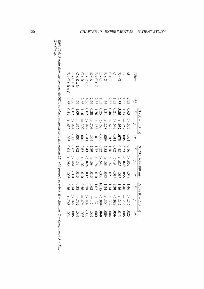
120 CHAPTER 10. EXPERIMENT 2B – PATIENT STUDY
P1(80
–110
ms)
N170
(140–
180m
s)P2b
(210–
270m
s)E
ffectd
fF
pω
2F
pω
2F
pω
2
G2,33
0.63>.537
-.0210.16
>.852
-.0491.46
>.246
.025E
1,331.33
=.257
.0055.23
<.029
.0551.46
=.236
.006E
xG
2,333.85
<.032
.0730.48
>.625
-.0151.46
>.247
.013C
1,330.21
>.647
-.0110.02
>.9
-.0145.30
<.028
.056C
xG
2,330.48
>.622
-.0151.76
>.187
.0211.14
>.332
.004R
xG
4,661.31
>.278
.0092.55
>.06
.0451.24
>.304
.009E
xC
1,330.25
>.62
-.0050.22
>.643
-.00510.15
<.004
.060E
xC
xG
2,331.76
>.188
.011.72
>.194
.0101.02
>.37
.000E
xR
2,660.20
>.703
-.0042.89
>.08
.0130.62
=.47
-.002E
xR
xG
4,660.02
>.992
-.0113.43
<.026
.0320.28
>.802
-.008C
xR
2,661.16
=.302
.0012.62
>.102
.0100.56
=.496
-.003C
xR
xG
4,661.09
>.356
.0012.02
>.13
.0130.38
>.732
-.007E
xC
xR
2,660.02
>.924
-.0030.62
>.461
-.0012.74
>.092
.006E
xC
xR
xG
4,660.53
>.632
-.0030.03
>.986
-.0050.36
>.768
-.004
Table10.6:
Results
fromthe
omnibus
AN
OVA
son
visualcomponents
inE
xperiment2B
,with
prosodyas
prime.
E=
Em
otion,C=
Congruency,R
=R
oi,G
=G
roup.

10.2. RESULTS 121
VPP. The Tukey test of the main effect of group was not significant. However, when
setting the threshold to p < .06, there was a significant VPP amplitude reduction in the
LPD compared to the HC group.
Visual inspection of the data suggested that this amplitude reduction was due to a
delayed VPP latency in patients. Therefore, the latency of the highest positive peak between
120 to 220 ms after stimulus onset was identified for each participant and condition and
analyzed in an ANOVA. A significant main effect of group emerged, F(2,33) = 5.17,
p < .012. According to the Tukey test, both patient groups showed delayed VPPs compared
to the HC group (LPD: 169 ms, RPD: 169 ms, HC: 155 ms). There was no interaction
with group in this analysis (all p > .411), indicating that the VPP delay in patients was not
condition-specific. Figure 10.3 shows the VPP alterations in patients compared to HC.
Figure 10.3: Group effect in the VPP to video targets, Experiment 2B. This component was delayedin both patient groups and reduced in amplitude in the LPD group.
The resolution of the congruency x roi x group interaction yielded a significant
congruency x roi effect in the RPD group, F(4,44) = 4.47, p < .018, while this interaction
was not significant in the LPD (p > .603) or HC group (p > .501). However, none of the
step-down analyses yielded any significant results (all p > .087).
Finally, the main effect of emotion was based on higher VPP amplitudes to happy than
angry targets.

122 CHAPTER 10. EXPERIMENT 2B – PATIENT STUDY
Later stage negativity. A congruency x roi interaction was tested as a roi-wise main effect
of congruency. It was significant at LF, F(1,33) = 5.28, p = .028, and at RP, F(1,33) =
7.55, p < .01, but not at LC (p > .121), LP (p > .224), RF (p > .441), RC (p > .838), or
ML (p > .121). Interestingly, the congruency effects were different at roi LF and RP, with a
more negative-going wave to incongruent than congruent targets at the left-frontal roi, while
the reverse pattern applied for the right-posterior roi. These different patterns at left-frontal
and right-posterior rois can be seen in Figures 10.4 and 10.5, respectively.
Figure 10.4: Left-frontal N400 modulation by congruency, prosody as prime, Experiment 2B.
The main effect of emotion was pronounced in more positive-going ERP amplitudes to
happy than angry stimuli.
P1. The resolution of the emotion x group interaction did not yield any significant results
(all p > .098).
N170. As the N170 amplitude is generally higher over right-posterior than left-posterior
rois, the main effect of roi from the omnibus analysis (F [2,66] = 4.15, p < .028) was first
scrutinized. The left-right difference was significant, F(1,33) = 4.28, p< .047, with higher
amplitudes over right hemisphere than at left hemisphere electrode sites.
In the analysis of the emotion x roi x group interaction it turned out that there was a
significant emotion x roi effect for the HC group, F(2,22) = 3.92, p < .038. In the RPD
group, this effect only approached significance (p < .078); however, this group exhibited a
highly significant main effect of roi, F(2,22) = 16.40, p < .0001. In the LPD group, there
was no significant emotion x roi interaction (p> .147). A step-down analysis of the emotion
x roi interaction in HCs and RPDs revealed that the patient group showed a significant main

10.2. RESULTS 123
effect of roi for both happy, F(2,22)= 11.24, p< .001, and angry targets, F(2,22)= 18.15,
p < .0001 while HC participants did not (happy: p > .78, angry: p > .31). The RPD group
showed significantly higher right- than left-posterior N170 amplitudes in the case of happy,
F(1,11) = 15.35, p < .003, and angry targets, F(1,11) = 30.84, p < .001.
As there were alterations in VPP latency for both patient groups (see above), a latency
analysis was performed on the N170, too, as N170 and VPP may stem from the same neural
generators (Joyce & Rossion, 2005). Just as for the VPP a main effect of group emerged
in the N170 time window, F(2,33) = 3.44, p < .044. According to Tukey’s HSD test, the
N170 peaked significantly later in the RPD group than in the HC group (159 versus 150
ms peak latency). In the LPD group, this component also peaked later than in HC (155
ms latency), but the difference was not significant. Furthermore, a significant emotion x
group interaction emerged, F(2,33) = 3.76, p < .034. A significant main effect of group
was observed in the angry target condition, F(2,33) = 6.39, p < .005, but not in the happy
one (p > .407). In line with the main effect of group, RPD participants displayed delayed
N170 latencies for angry targets compared to HC. LPD participants did not significantly
differ from either group. No further interactions with the group factor proved significant
(all p > .305).
Finally, the main effect of emotion was triggered by higher N170 amplitudes to angry
than happy targets.
P2b. Incongruent targets elicited higher P2b amplitudes than congruent ones. However,
as informed by the emotion x congruency interaction, a main effect of congruency emerged
only for angry targets, F(1,33) = 15.07, p < .001, but not for happy ones (p > .432). The
P2b congruency effect is displayed in Figure 10.5.
Correlations. The VPP latency did not correlate with any of the disease-related variables
left motor score (p > .835), right motor score (p > .907), the asymmetry index (p > .977),
or total motor score (p > .693).
There was a significant negative correlation between the composite score “executive
functions” and VPP latency, r = −.45, p < .007. The relationship between “working
memory/sustained attention” and VPP latency was not significant, but the direction was
also negative, r =−.19, p > .27.
The N170 amplitude difference between rois VL and VR correlated significantly with
the asymmetry index, r = .55, p < .006, indicating higher right- than left-posterior N170

124 CHAPTER 10. EXPERIMENT 2B – PATIENT STUDY
Figure 10.5: Posterior P2b and N400 modulations by congruency, prosody as prime, Experiment2B. The P2b was enhanced to incongruent stimuli, an effect which was driven by angry targets. TheN400 showed a reversed pattern at right-posterior electrodes, with more negative-going deflectionsto congruent than incongruent trials.
amplitudes with motor symptom asymmetry toward the right side of the body. The
correlations with left motor score (p > .374), right motor score (p > .068), total motor
score (p > .69), executive functions (p > .688), or working memory/sustained attention
(p = .703) were not significant. Overall, the N170 latency was significantly and negatively
correlated with executive functions, r = −.49, p < .003. This also held true for the N170
latency specifically to angry targets, with a trend after Bonferroni correction, r = −.37,
p < .026. Correlations with working memory/sustained attention were not significant
(overall N170 latency: p > .304, anger latency: p > .253).
Processing of the prosodic primes. No group effects emerged in the N100, the P200, or
the subsequent negativity (all p > .317).
10.3 Discussion
The present study investigated cross-modal emotional priming with dynamic facial and
vocal expressions in Parkinson patients and matched healthy controls. In general, the results
indicate that LPD patients are able to use the information conveyed by affective prosody and
emotional facial expressions, but that the time course of cross-modal emotional priming
may be changed when processing prosodic targets. Furthermore, the current data indicate
that early face processing may be altered independent of the emotional expression conveyed
in both PD groups.

10.3. DISCUSSION 125
Video-as-prime. While both the HC and the RPD groups showed a main effect of
congruency in the N100, the LPD group did not. Rather, latter group showed a congruency
effect in the P200 while this component was not modulated by congruency in the other two
groups. Thus, it appears that LPD patients detected prime-target congruency later than the
HC and the RPD group.
The present results add to the findings in Experiment 1B, in which only LPD
patients exhibited altered emotional salience detection from prosody, reflected in the P200
component. Results of the present study suggest that there may be a temporal delay in
emotional priming in LPD when emotional prosody serves as a target.
At this point, it is important to consider that no group effects emerged in the analysis of
the prosodic stimuli presented at the prime position. This is in accord with Experiment 1B,
in which (except for disgust) only intelligible speech was affected while the processing of
unintelligible speech, as used in the present study, proved to be largely intact in LPD. Thus,
it is unlikely that group differences related to the processing of the prosodic stimuli per se
gave rise to the group effects in the target position.
However, an ERP analysis of the video primes revealed that LPD patients processed the
primes differently than healthy controls, as evidenced by a reduced and delayed VPP. Thus,
it is possible that the congruency effect in the LPD group was temporally shifted due to
altered processing of the dynamic facial primes. This explanation is, however, weakened by
the observation that RPD patients showed comparable VPP alterations to video primes, but
still exhibited congruency effects in the N100 to prosodic target stimuli. Furthermore, the
VPP alterations in response to the video primes appeared to be unspecific and generalized,
i.e., they were not modulated by the emotional category. Furthermore, VPP alterations
were observed independent of emotional category and congruency in the prosody-as-prime
condition.
Thus, deficits in one specific modality are an unlikely explanation for the current
findings suggesting altered emotional priming in the LPD group. It rather appears that
LPD patients have a problem with the analysis of cross-modal emotional congruency per
se. It is important to reflect the impact of the current results in light of the results from
Experiment 1B. In Experiment 1B, a combination of prosodic and semantic information
(i.e., intelligible speech) led to group differences while this was not the case in the absence
of semantics (unintelligible speech), except for disgust. Thus, the current results as well
as the previous findings from Experiment 1B may be related to the proposed stimulus
binding deficit in PD (Paulmann & Pell, 2010b). In latter study, emotion recognition in
PD was tested via different communication channels (face, prosody, and semantics) in

126 CHAPTER 10. EXPERIMENT 2B – PATIENT STUDY
isolation and in combination. It was observed that PD patients benefitted much less from
the presence of prosody in addition to the other channels than healthy controls, which may
reflect problems to bind the vocal cues with information from other channels (Paulmann &
Pell, 2010b). Thus, the absence of early congruency effects in LPD may be due to deficits
in early stimulus binding in this group. The present findings in conjunction with those
from Experiment 1B thus indicate that this binding deficit may generalize across verbal
(semantics) and non-verbal (facial expressions) information.
Delayed priming effects in PD have previously been reported in semantic priming
studies using different inter-stimulus intervals (Angwin, Chenery, Copland, Murdoch, &
Silburn, 2007; M. Grossman et al., 2002). The present data indicate that such a delay may
also play a role in emotional priming in PD. However, even though there may be a delay
in the detection of cross-modal congruency, LPD patients were still able to perceive the
congruency between primes and targets as evident in a P200 congruency effect as well as
in the later stage negativity congruency effect. Thus, only early stages of processing were
affected in the LPD group, as no congruency effect was evident in the N100.
Recently, the striatum has been implicated in processing emotionally incongruent face-
voice stimuli (Klasen, Kenworthy, Mathiak, Kircher, & Mathiak, 2011). In latter study,
this involvement was bilateral, even though a cluster size analysis revealed a higher cluster
size in the right striatum. However, due to temporal constraints the onset of the respective
activations cannot be specified in fMRI data. In light of the present results, a right-striatal
engagement seems conceivable already during the early processing (N100) of cross-modal
congruency when vocal expressions are primed by facial expressions, whereas the left
striatum may start to operate later in time.
Finally, LPD patients did not differ from healthy controls in any of the neuropsycho-
logical measures or in the P300 results of the oddball experiment. Hence, it is unlikely
that the delayed ERP modulations by congruency in the LPD group resulted from more
generalized cognitive impairments.
RPD patients recognized prime-target congruency as early as healthy controls, i.e., in
the N100 component. However, the pattern of this effect was opposite to the one exhibited
by controls, with higher N100 amplitudes to incongruent than congruent stimuli. According
to the correlation results, cognitive functions may have played a role here, as the amplitude
difference between congruent and incongruent stimuli in the N100 was correlated with the
composite scores, i.e., executive functions and working memory/sustained attention.

10.3. DISCUSSION 127
Furthermore, it may be that the N100 tapped into different processing stages in the HC
and RPD groups. Reversed priming effects have been observed previously in the literature.
For example, a recent cross-modal emotional priming study with prosodic primes and facial
targets (Paulmann & Pell, 2010a) reported reversed priming effects in the N400 (i.e., more
negative-going deflections in response to congruent as compared to incongruent trials)
for very short prime durations, while longer prime durations (in conjunction with longer
stimulus onset asynchronies) led to the classical N400 effect, with an enhanced negativity
in response to incongruent as compared to congruent trials. A similar observation was made
in semantic priming, in which hardly visible primes elicited reversed N400 and behavioral
priming effects (Bermeitinger, Frings, & Wentura, 2008). In addition, the analysis of the
ERPs to the video primes revealed that there were some processing differences between the
RPD and HC groups (delayed VPP and enhanced N2 component). This indicates that RPD
and HC probably were at different processing stages of the prime when the auditory target
was presented, or that the processing of video primes was hampered in RPD. This could
explain the reversal of the congruency effect in the N100. However, despite of this reversal,
the data indicate that RPD patients are able to detect cross-modal congruency as early as
healthy controls.
To sum up, the results from the video-as-prime condition suggest that patients suffering
from Parkinson’s disease are able to detect cross-modal emotional congruency between
primes and targets as evidenced by ERPs. The congruency effect appears to be temporally
delayed in the LPD group, and it may rely on different neural mechanisms in the RPD
group than in healthy controls. However, it is telling that processing alterations in PD
were limited to relatively early stages. Once more, this calls into question whether group
differences reported in many previous studies on emotional prosody (e.g., Blonder et al.,
1989; Breitenstein et al., 2001; Dara et al., 2008) really reflect an emotion processing deficit
in patients, or whether they rather originate from difficulties with explicit emotion tasks as
suggested by Pell and Dara (2007).
Prosody-as-prime. Results from this condition indicate that the processing of congruency
between prime and target was not affected in PD patients. This means that PD patients,
including LPD patients, were able to successfully use the emotional context conveyed
by speech intonation. In light of the results of prosody as prime only, one can conclude
that patients do not display an impairment of emotional prosody. Furthermore, the results
indicate that emotion from facial targets was processed adequately by patients, which is a
prerequisite for intact emotional congruency processing. These results contradict previous
behavioral studies which suggest impaired processing of facial (e.g., Ariatti et al., 2008;

128 CHAPTER 10. EXPERIMENT 2B – PATIENT STUDY
Sprengelmeyer et al., 2003) and vocal (e.g., Benke et al., 1998; Dara et al., 2008) emotion
in PD. This underlines that explicit emotion tasks as used in these studies may measure
mechanisms different from those which were captured in the present study.
The comparable congruency effects in the three groups are in line with behavioral
results from the facial affect decision task (see Chapter 8 for a description of this task), in
which PD patients showed emotional priming comparable to healthy controls (Pell & Dara,
2007). Latter study also used complete sentences in unintelligible speech as primes. Thus,
whole-sentence primes appear to be long enough to establish a valid emotional context in
PD. It remains to be examined whether the groups would also show comparable results to
healthy controls when using shorter prosodic primes as in Paulmann and Pell (2010a). If
the reason for the absence of group-modulated congruency effects in the present study is
the prime duration itself, then such effects may be revealed by duration manipulations. In
the present study, the effects of prime modality and prime duration cannot be disentangled.
Follow-up studies which address this issue are warranted. However, if the problem is rather
the integration of information from different communication channels in early emotional
prosody processing, then prime duration should be less critical for group effects, but the
presentation of vocal emotion as a target may be the central modulator. If latter is the case,
then this would confirm previous evidence on stronger impairments for vocal than facial
emotion processing in PD (Gray & Tickle-Degnen, 2010). This may have something to do
with the inherent characteristics to these stimuli. Prosody unfolds over larger time scales,
while this is not so much the case for facial expressions, even though presented dynamically
(Paulmann & Pell, 2010b).
The most striking effect in this priming condition was the VPP modulation in PD
patients. In both PD groups, this component displayed a reduced amplitude, even though the
VPP reduction was only significant in the LPD group (at trend level). More indicative may
be the observation that both PD groups exhibited delayed VPP peak latencies compared to
controls. This was also observed when the videos served as primes and there appeared to
be no modulation of this latency shift by emotion or any other variable. Hence, one can
speak of a condition-independent VPP delay in PD patients. As there was a significant
negative correlation between executive functions and VPP latency, the VPP delay may
reflect cognitive decline. Here, it is interesting that the LPD group did not differ from
HC in any of the cognitive measures. Thus, the VPP latency in response to dynamic faces is
probably a very sensitive measure to capture mild cognitive decline in PD already at a stage
at which impairments are subtle and can hardly be captured with classical cognitive tests.

10.3. DISCUSSION 129
The literature only reveals one study in which the VPP latency to faces was compared
between PD and control participants, and there was no significant group difference (Kida,
Tachibana, Takeda, Yoshikawa, & Okita, 2007). However, in latter study the VPP had a
peak delay of about 13 ms in PD patients, which is comparable to the present observations.
Furthermore, the data from latter study indicate that a high variability within the PD sample
may have prevented a significant effect.
The VPP is often considered as the fronto-central counterpart of the face-related N170,
and both components may have the same underlying neural generators (Joyce & Rossion,
2005). However, in the present PD study the VPP group effects did not converge with
the N170 effects. The only correspondence was a delayed peak latency in RPD, which,
however, was driven by angry faces in the N170, but not in the VPP. Furthermore, the VPP
peak latency was higher than the N170 latency in both patient groups. This result calls into
question that VPP and N170 reflect the same neural mechanisms and rather accounts for
the view that - at least in part - different processes and neural substrates play a role in these
two components (Wong et al., 2009). On the other hand, it must be considered that more
electrodes were included in the statistical analysis of the fronto-central components than of
the visual components, and thus the possible impact of statistical power on results cannot
be denied.
Regarding the N170 amplitude to video targets, the RPD group showed highly
significant roi effects whereas this was not the case in the LPD and HC groups. These
roi effects reflect the classical N170 pattern, with higher right-posterior than left-posterior
amplitudes. Even though this pattern is very common in the literature (e.g., Bentin et al.,
1996) and was also observed in the pilot study (Experiment 2A), it may disappear with
age (Gao et al., 2009). In fact, it was not observed in the HC group in the present study.
The notion that RPD patients exhibited this pattern may thus stem from predominantly
left-hemispheric damage of the basal ganglia and related structures, which led to reduced
N170 amplitudes at left-occipital electrodes. This assumption is supported by a significant
correlation of the N170 amplitude difference at left and right-posterior electrodes with the
asymmetry index. Such EEG asymmetries dependent on the sidedness of motor symptoms
in PD have previously been reported (Mintz, Tomer, Radwan, & Myslobodsky, 1981, 1982),
even though they largely resolved with medication in these studies. The present results
indicate that at early and rather sensory processing stages, a certain degree of hemispheric
asymmetry may still become visible in the ERPs of medicated patients.
There was a delay in N170 peak latency in RPD which was driven by the N170 to
angry targets. This indicates that RPD patients may have problems with this stimulus type.

130 CHAPTER 10. EXPERIMENT 2B – PATIENT STUDY
However, no such delay was observed when the videos served as primes and the delay was
furthermore correlated with executive functions. Thus, even though it cannot be discarded
that RPD may lead to problems with processing angry facial expression, the association
seems not that straightforward. More work in the future may render a clearer picture in
relation to this finding.
In contrast to the present study, Wieser et al. (in press) did not report any alterations of
early visual processing in PD; however, these authors restricted their analyses to occipital
electrodes (i.e., the VPP was not part of the analysis) and analyzed only amplitude
modulations but not latency. Thus, VPP differences, which reflect the most striking
alterations in the prosody-as-prime condition, may have been missed. In Wieser et al.’s
study, patients differed from controls starting at approximately 240 ms after face onset,
whereas in the present study later components did not differ between groups. Probably,
PD patients benefitted from the dynamic character of the faces used in the present study,
as has already been observed at the behavioral level (Kan et al., 2002, but see Paulmann &
Pell, 2010b). Future studies may directly compare the early neural processing of static and
dynamic facial stimuli in PD.
On the other hand, it must be considered that the paradigm used by Wieser et al.
(in press) was quite different from the current one, as it tested early visual emotion
discrimination in PD. Thus, it is more comparable to Experiment 1B, but with visual stimuli.
Wieser et al.’s results suggest that emotional salience detection from facial expressions
may be altered in PD and hence complement the prosody findings from the first patient
study. The present study was not suitable to test emotional salience detection from faces;
however, the detection of emotional congruency appeared to be intact in the case of facial
targets. This indicated that no receptive emotional deficit for faces in PD can be concluded
from the present results. More research is definitely needed to address early facial emotion
processing in PD, especially in terms of more emotional categories.
To sum up, the prosody-as-prime condition did not reveal any deficits in PD with
respect to an ERP modulation by emotional prime-target congruency. The data provide
some evidence that deficits at early visual processing stages occur, which affected both PD
groups and seem to reflect more generalized face processing alterations. However, all in
all, the data indicate intact emotional priming in PD. The two patient groups were very
comparable in this condition, as opposed to the video-as-prime condition.
Summary and conclusion. To sum up, the present study supports the findings from
Experiment 1B, namely that LPD patients exhibit altered early processing of emotional
10.3. DISCUSSION 131
prosody. Furthermore, it was confirmed that pure prosody does not lead to alterations
in LPD. Rather, it may be the interplay between different communication channels, e.g.,
face and voice, which is significant. From the present data in conjunction with those
from Experiment 1B, it seems that LPD leads to problems in the interaction of emotional
information provided by different communication channels with emotional prosody. This
is apparently true for semantic, i.e., verbal, and facial, i.e., non-verbal information and may
trace back to the relatively longer time scales over which vocal emotion unfolds. The data
also suggest some low-level alterations of early dynamic face processing in PD independent
of the sidedness of motor symptoms. In a next step, it would be interesting to test the effects
of different prime durations and emotional categories. Also, in order to test the interaction
of more communication channels at once, semantics may be included in future studies.


Chapter 11
General discussion and outlook
The present thesis aimed to investigate dynamic emotion processing in Parkinson’s disease.
More specifically, it addressed apparent open issues in PD research on emotion processing,
which concern task effects, early versus late processing stages, stimulus dynamics, and
patient characteristics.
With regard to the experimental task, explicit instructions such as categorization
para-digms are commonly used in PD investigations addressing emotion processing.
Performance in these tasks may, however, be confounded with frequently observed
cognitive deficits in PD (Benke et al., 1998; Breitenstein et al., 2001; Pell & Leonard, 2003).
In line with this, Pell and Dara (2007) could show that PD led to a performance decline in
explicit emotion recognition from prosody, while the same patient sample showed intact
priming of emotional facial expressions by affective prosody. The present thesis therefore
focused on implicit task instructions and on-line measures such as EEG in PD populations.
One problem related to the issue of explicit task instructions is the time course of
emotion processing. Explicit behavioral tasks are only able to capture late, controlled
processing stages, while potentially early neural modulations of emotional stimuli in
PD have been poorly investigated to date. Here, this was accomplished with the help
of ERPs, which provide an exquisite temporal resolution within the milliseconds-range.
This technique allows observing the neural modulations in response to emotional stimuli
unfolding in time.
A great deal of investigations available to date has tested emotion processing in PD
with static facial expressions, but few have used dynamic ones. Additionally, there are
some studies that have used emotional prosody. Emotional prosody cannot be presented in
133

134 CHAPTER 11. GENERAL DISCUSSION AND OUTLOOK
a static manner and is thus always dynamic. It appears that PD patients are more impaired in
processing prosodic emotional than facial emotional stimuli (Gray & Tickle-Degnen, 2010;
Pell & Leonard, 2003, 2005). However, the question remains as to whether this is due
to the dynamics as such, or if the particular stimulus type plays a greater role. Paulmann
and Pell (2010b) recently tested dynamic multimodal emotion recognition from different
communication channels (face, prosody, and semantics) in PD and found that patients made
strong use of dynamic facial expressions as an information source, but that they virtually
did not benefit from the presence of prosodic information. These results indicate that the
greater deficits for prosody may have to do with prosody in itself rather than dynamics, but
this remained to be tested at the neural level.
Finally, heterogeneity among PD patients was of central interest in the current research.
On the one hand, the above-mentioned cognitive deficits, often termed “mild cognitive
impairment”, affect a large proportion of PD patients (Williams-Gray et al., 2007). By
means of a comprehensive cognitive assessment, the present thesis aimed to control for these
possible confounding factors. Along similar lines, depression as another potential confound
was screened for, and very high scores led to the exclusion of the respective patients from
the sample. The central focus of the present thesis, however, concerned motor symptom
asymmetry in PD. Depending on which body side is more affected by motor symptoms, the
involvement of the BG and their respective circuits in neuronal degeneration differs, with
contralateral predominance. In the cognitive domain, investigations point to differential
modulations of impairments by motor symptom asymmetry, associating LPD (= greater
right-hemispheric involvement) more with visuospatial and RPD (= greater left-hemispheric
involvement) with rather “classical” left-hemisphere, i.e., linguistic functions (see Cronin-
Golomb, 2010, and section 2.3 of this thesis, for reviews). In emotion processing research
in PD, motor symptom asymmetry is often not taken into account and the few available
studies in the literature have yielded equivocal results (cf. Chapter 4 for details). On the
other hand, a certain degree of hemispheric dominance is assumed in emotion processing
(e.g., Schirmer & Kotz, 2006). Thus, the present thesis aimed to test for the impact of motor
symptom asymmetry and its possible effects on emotion processing in PD, especially with
regard to early neural processing stages.
11.1 Summary and integration of main findings
In Experiment 1, emotional prosody processing in PD was tested as a function of task,
speech intelligibility, and emotional category. With respect to patient characteristics,

11.1. SUMMARY AND INTEGRATION OF MAIN FINDINGS 135
sidedness of the disease was a central factor, as well as the participant’s cognitive profile.
The results indicated that LPD patients display early alterations in emotional salience
detection from prosody (P200) in the categories of disgust, anger, and happiness. However,
no clear-cut task effects emerged. Regarding speech intelligibility, disgust prosody was
affected in both intelligible and unintelligible speech. In the other emotional categories, the
processing of unintelligible speech conveying prosodic information appeared to be largely
intact in LPD. This gives rise to the possibility that LPD may lead to problems in the early
interaction of different communication channels, specifically the early interplay between
nonverbal vocal emotional cues (i.e., prosody) and verbal (i.e., semantic) cues. Therefore,
Experiment 2 tested whether LPD also leads to problems in the early interaction of different
types of non-verbal emotional information (i.e., dynamic facial expressions and emotional
prosody). This interaction was investigated by means of a cross-modal emotional priming
paradigm, in which unintelligible speech conveying angry or happy emotional prosody
was combined with emotionally congruent or incongruent dynamic facial expressions.
The results indicate that during emotional prosody processing, the use of prior contextual
information provided by dynamic facial expressions is temporally delayed in LPD patients,
as compared to healthy elderly controls and RPD patients.
It has previously been discussed that the BG may play a role in stimulus binding,
particularly with respect to the interaction of different emotional information channels
(Paulmann & Pell, 2010b). The present data indicate that the right striatum may accomplish
this binding function at an early time point. This suggests that the striatum is critically
involved in the supramodal interaction of emotional information, a possibility which is
elaborated in a recent investigation by Klasen et al. (2011). The authors reported BG
activation to emotionally incongruent face-voice combinations. Converging evidence from
patients with left-striatal lesions shows that emotional deviation in purely prosodic (i.e.,
unintelligible) stimuli does not reveal processing deficits in patients compared to healthy
controls (Paulmann, Pell, & Kotz, 2008), whereas a combined deviation between verbal and
nonverbal cues (semantics and prosody) does (Paulmann, Pell, & Kotz, 2009). Thus, the left
striatum may play a role in aligning emotional prosody to semantics. Indeed, the left-striatal
involvement may be confined to verbal (linguistic) stimuli in combination with emotional
prosody, as the non-verbal dynamic face stimuli used in Experiment 2 did not reveal
any processing alterations in RPD. On the other hand, the LPD data in both experiments
conducted here indicate an early involvement of the right striatum in emotional processing,
particularly with respect to supramodal interactions of emotional cues. Furthermore, this
may be the case for both verbal-nonverbal interactions as well as for interactions between
different nonverbal channels.

136 CHAPTER 11. GENERAL DISCUSSION AND OUTLOOK
The interaction of emotional information from different communication channels has
been attributed to different brain structures, for instance the posterior superior temporal
sulcus (Ethofer, Anders, Wiethoff, et al., 2006; Kreifelts et al., 2007), the posterior insula
(Ethofer, Anders, Wiethoff, et al., 2006), or the amygdala (Ethofer, Anders, Erb, Droll,
et al., 2006; Klasen et al., 2011), to name but a few. As many brain regions have been
associated with the interaction of emotional information from different channels, it appears
that an extended network is involved in this process. As indicated by the results obtained
in the present work, the right striatum may play a role when the interaction between two
communicative channels requires emotional information to be tracked over larger time
scales, which may be particularly relevant in the context of emotional prosody. This notion
underlines the proposed role of the BG for tracking auditory speech input over its temporal
course, meaning that auditory and temporal processing mechanisms are closely tied together
(Kotz & Schwartze, 2010).
The present results confirm previous evidence of a greater impairment for emotional
prosody than emotional face processing in PD (Gray & Tickle-Degnen, 2010). It can now
be assumed that this can not only be observed behaviorally, but also at the neural level and
with dynamic as opposed to the more widely used static facial information. Thus, it may not
be the dynamics as such which often lead to greater emotional prosody (always dynamic)
than face (mostly presented statically) processing deficits, but rather characteristics inherent
to vocal and facial emotion expressions such as the time scales they rely on. Related to
this, there is the assumption that facial expressions, even though conveyed dynamically, can
readily be recognized at discrete points in time during stimulus presentation while emotional
prosody unfolds in time Paulmann and Pell (2010a).
What needs to be kept in mind, though, is a generally higher difficulty level associated
with vocal compared to facial emotion. Normally, in emotion categorization, performance
is better for facial than for vocal emotion recognition (Paulmann & Pell, 2011). This affects
certain emotions more than others. Disgust recognition from prosody, e.g., is generally poor
(Adolphs, 2002a) and occurs late in time (Pell & Kotz, 2011). When there is concurrent but
incongruent facial and prosodic emotion available, people tend to decide for the emotion
conveyed by the face when forced to provide an emotion categorization (Klasen et al.,
2011). This indicates face preference and may point to facilitated face processing in
humans, while vocal emotion processing may go along with a relatively higher processing
effort. This cannot be disregarded, particularly in the context of patient investigations. In
patient populations, a higher processing difficulty associated with certain stimuli may lead
to stronger functional breakdowns than in healthy controls. Thus, the observation made in
the present thesis, that especially receptive emotional prosody seems to be impaired in PD,

11.2. IS THERE LATERALIZATION IN THE BASAL GANGLIA? 137
may mean different things. On the one hand, it may indicate problems to track emotional
information which slowly unfolds in time; on the other hand the disease may exacerbate the
generally higher processing difficulty associated with vocal as compared to facial emotional
information. Finally, these two points may be related in that processing emotional prosody
is generally more difficult because it relies on longer time scales, and this higher difficulty
level makes a performance breakdown in patient populations more likely.
RPD patients compared to LPD patients were found to process emotion from socially
relevant stimuli in a comparable manner to the healthy controls. However, overall RPD
patients showed stronger cognitive decline than LPD patients. This suggests that the left
and the right striatum may engage in distinct functions, supporting some previous evidence
that reported an association between right-sided motor symptoms and cognitive decline in
PD (Cooper et al., 2009; Foster et al., 2010; Huber et al., 1992; Williams et al., 2007). On
the other hand, the absence of impairments in cognitive measures strengthens the argument
that the reported alignment problems in LPD are not a result of general cognitive decline.
11.2 Is there lateralization in the basal ganglia?
A possible functional lateralization of the BG is to date largely unexplored. Traditionally
the BG have been associated with attention-dependent later stage processing, reanalysis,
and correction (Kotz & Schwartze, 2010). ERP evidence on auditory emotional processing
(Paulmann, Pell, & Kotz, 2009) or syntactic error detection (Friederici, Kotz, Werheid,
Hein, & von Cramon, 2003; Frisch, Kotz, von Cramon, & Friederici, 2003; Kotz, Frisch,
von Cramon, & Friederici, 2003) in patients suffering from damage to the BG (lesions
and PD) supports this notion. However, the findings from the present set of studies give
rise to the possibility that the right striatum is involved in early emotion processing stages
when information from different input channels interact. Especially the changes in the P200
component go along with the idea that the right hemisphere is involved in the integration
of information over timescales of approximately 150–300 ms (Hickok & Poeppel, 2007),
which coincides with the P200 time window. Along these lines, Schirmer and Kotz (2006)
have previously proposed a dominance of right-temporal regions in the generation of the
P200 in emotional prosody processing. Thus, the results reported in the present thesis
may lead to the tentative conclusion that a functional lateralization of the BG and their
interactions with temporal and other cortical areas exists and thus provide a starting point for
further investigations on the topic. As discussed earlier, a possible bottom-up mechanism

138 CHAPTER 11. GENERAL DISCUSSION AND OUTLOOK
involving the right striatum and temporal areas in emotional prosody processing (Ethofer et
al., 2012) is conceivable.
11.3 Task effects and early versus late processing stages
In Experiment 1, specifically the processing of verbal cues, i.e., vocal emotional stimuli
including semantic information, was impaired in LPD as observed in the P200 ERP
component. On the other hand, behavioral research shows that PD patients benefit from
congruent verbal and nonverbal emotional input (Dara et al., 2008; Paulmann & Pell,
2010b). A similar benefit was also observed in the behavioral emotion categorization study
in Experiment 1B. This apparent dissociation between early neural modulations and later
decision-making stages points to the low convergence between explicit behavioral methods
and the time-sensitive ERP methodology, which can capture the unfolding of information
processing at various processing stages in implicit and explicit tasks. Conversely, Pell and
Dara (2007) reported intact cross-modal emotional priming at the behavioral level in PD,
applying the facial affect decision task. This finding indicates that PD patients could use
emotional information conveyed by prosody to render a facial affect decision, while they
were only impaired when they had to explicitly assign an emotional category to prosodic
stimuli. These results converge with the ERP findings from the prosody-as-prime condition
in Experiment 2B, in which no impairments in implicit cross-modal emotional priming
were observed in either PD group. Pell (2002) has previously advocated the use of implicit
on-line measurements in patient populations, as the explicit emotion tasks used in the
majority of patient studies assess later, controlled processes. These later processes are more
susceptible to cognitive decline (i.e., deficits in working memory, attention, etc.). In fact,
several behavioral PD studies reveal an association between the performance of PD patients
in explicit emotion tasks and cognitive functions (Benke et al., 1998; Breitenstein et al.,
2001; Pell & Leonard, 2003) suggesting that explicit tasks may not be valid to test receptive
emotional deficits in PD. The dissociations between ERPs and later behavioral measures
as well as the absence of group effects in the prosody-as-prime condition underlines that
explicit emotion tasks and early ERP effects capture different aspects of emotion processing
which may not be very closely related to each other.

11.4. THE PROBLEM OF VARIABILITY IN PD 139
11.4 The problem of variability in PD
One problem in PD research is the rather high variability among patients and the commonly
small sample size in patient studies. Conversely, study results in PD are conflicting, as
discussed in Chapter 4, which summarizes the pertinent literature on emotion processing in
PD. In the present thesis, one variable, which may allow to specify deficit profiles in PD,
namely sidedness of motor symptoms, was used as a grouping variable. Such specification
may be considered an advantage in comparison to many previous studies. Thereby, the
relative involvement of the respective cortico-striatal circuits in neural degeneration is better
controlled for. However, there are numerous other variables, which can induce within-group
variation. Among these one may think of the type of the initial motor symptom (Katzen et
al., 2006), the degree of cognitive decline (Benke et al., 1998; Monetta, Grindrod, & Pell,
2006), disease progression (Breitenstein et al., 1998), or medication (Tomer et al., 2007),
to name but a few. What is also unresolved is the question of how to handle depressive
symptoms in PD. As most comorbidity estimates of depression in PD revolve around 30–
40%, depression is a very common epiphenomenon in PD. Along these lines, depression
has been suggested to be inherently tied to the disease (Aarsland, Marsh, & Schrag, 2009;
Leentjens, Van den Akker, Metsemakers, Lousberg, & Verhey, 2003; Lieberman, 2006;
Obeso et al., 2010). On the one hand, depression may be a confounding factor in PD
research, which is why patients with high comorbid depression scores are often excluded
from the experimental sample. This approach was also adopted in the present thesis. On
the other hand, the inclusion of these patients may be more representative of the general PD
population. Thus, this issue needs to be critically reconsidered in the future.
In summary, even though the findings of the present thesis are striking, and a
considerable number of possible moderator variables was controlled for, including the
cognitive profile and the psychopathology, the results will have to be replicated and
extended in future research.
11.5 Limitations
Some caveats of the current investigation have already been pointed out, namely the small
sample sizes and, related to this, a number of further possible cognitive and disease-related
confounds, which may introduce variability in a PD patient sample. Furthermore, it also
needs to be weighted that PD is a neurodegenerative disease. Even though the BG are
the most affected structure in PD, the disease leads to much more wide-spread changes in

140 CHAPTER 11. GENERAL DISCUSSION AND OUTLOOK
the brain already at early stages of the disease (Braak et al., 2003; Tinaz et al., 2011).
These changes in the brain may constitute a confounding factor, making it difficult to
attribute findings in PD exclusively to the BG. For example, there is amygdala damage
at an intermediate disease progression stage and increasing frontal cortical involvement
during the disease progression (Braak et al., 2003; Braak & Del Tredici, 2009). These
neural alterations may have an impact on study results due to their supposed significance
for emotion perception and the fronto-striatal circuitry, respectively.
In addition to the caveat that PD is a disorder that is not confined to the BG, it must
be kept in mind that even though there is asymmetric degeneration in PD, the BG are
involved bilaterally even at early disease stages (Schwarz et al., 2000; Tissingh et al., 1998).
Thus, the sidedness is not absolute, but rather relative. However, as suggested by several
significant correlations between ERP findings and motor symptom asymmetry, this rather
relative degree of asymmetry may impact study results in PD.
11.6 Concluding remarks
To conclude, the findings of the present thesis suggest that emotion processing in PD is
indeed affected by motor symptom asymmetry. Generally speaking, emotion processing
appears to be largely intact in the case of a predominantly left-sided BG damage (RPD),
while receptive emotional functioning seems to be impaired already at early processing
stages in the case of right-sided BG damage (LPD). A wide-spread disgust impairment
supports the important role for the BG in disgust processing (Calder et al., 2001), even
though the high recognition difficulty associated with disgust prosody should not be
disregarded (Adolphs, 2002a; Pell & Kotz, 2011). With respect to other emotional
categories, processing impairments may rather lie within stimulus binding of different
emotional input channels, which is probably particularly the case when such binding needs
to be accomplished on information that relies on rather slow temporal transitions, i.e.,
emotional prosody.
Future studies may assess the effects of different prime durations and emotional cate-
gories, as well as the interaction between facial, vocal, and semantic information in PD
during early processing stages. This will help disentangle the effects of stimulus type
(communication channel), stimulus duration, and stimulus-onset asynchronies. Further-
more, more research needs to address whether deficits are category-specific in terms of
particular emotions or rather generalized. Finally, LPD patients exhibited deficits in explicit
and implicit tasks but clear-cut task effects were not observed; rather, task effects seemed

11.6. CONCLUDING REMARKS 141
to depend on emotion categories. Thus, it should be investigated in the future which factors
modulate task-specific emotion processing deficits in LPD. In RPD, there may be later
stage deficits when semantic stimuli are included, as suggested by lesion data (Paulmann,
Pell, & Kotz, 2009). This may also be an issue of upcoming research testing prosodic-
semantic mismatches in PD patients. The paradigm used in Experiment 1 did not include
prosodic-semantic mismatches. Thus, it was not suited to test such late expectancy violation
effects which have been shown to reveal a pattern comparable to N400 modulations (Kotz &
Paulmann, 2007). Thus, there may be emotion processing deficits in RPD, too, but probably
the experiments conducted in the present thesis were not suited to reveal these.
From a more practical point of view, the results may be useful for clinical practice
and for developing new strategies of how the disease is dealt with. Physicians who treat
PD patients should be made aware of possible emotion processing and emotion expression
deficits in their patients. The patients’ as well as their caregivers’ attention should be drawn
to these possible problems. One can imagine that caregivers may be discouraged or feel
rejected by the flat affective displays in patients (Mikos et al., 2009), especially if they do
not know that these are manifestations of the disease and that reduced emotion processing
may also be one of its consequences. Furthermore, it may be recommended to implement
trainings in receptive and expressive emotion functions in PD, as these functions are so
crucial for interpersonal interactions. Especially for better coping with the disease, social
support will surely be helpful, so it may be useful to restructure standard PD therapy, in
order to enable patients to better deal with their social environment and to keep up their
interpersonal relationships.


Part III
Appendix
143


Appendix A
Instructions
A.1 Experiment 1
Please note: The instructions were identical in the pilot study and in the patient study;
however, in the patient study, a formal form of address ("Sie") was used.
Implicit task instruction
1. Liebe Versuchsperson! Im folgenden Experiment wirst Du Sätze hören, die entweder
auf Deutsch oder in einer Phantasiesprache vorgetragen werden. Danach wird die Frage
"deutsch oder nicht deutsch?" auf dem Bildschirm aufblinken.
Dear participant! In the following experiment you will hear sentences spoken either in
German or in an imaginary language. Then, the question "German or not German?" will
be flashed on the screen.
2. Entscheide dann bitte, ob der gehörte Satz in deutscher Sprache vorgetragen wurde oder
nicht.
Please decide, whether the sentence you heard was spoken in German language or not.
3. War das der Fall, dann drücke bitte die linke (rechte) Taste. Wenn nicht, dann drücke die
rechte (linke) Taste.
145

146 APPENDIX A. INSTRUCTIONS
If this was the case, then please press the left (right) key. If not, then press the right (left)
key.
4. Wichtig: Bitte warte mit dem Tastendruck immer, bis die Frage auf dem Bildschirm
erscheint, weil Deine Antwort sonst vom System nicht aufgezeichnet wird!
Note: Please wait until the question appears on the screen before you press the key,
otherwise your response will not be registered by the system!
5. Wenn Du noch Fragen hast, dann stelle sie bitte jetzt. Ansonsten kannst du nun beginnen
- entspanne Dich und los gehts!
In case you have any questions, please ask them now. Otherwise you can start now - relax
and here we go!
Explicit task instruction
1. Liebe Versuchsperson! Im folgenden Experiment wirst Du Sätze hören, die entweder
auf Deutsch oder in einer Phantasiesprache vorgetragen werden. Danach wird die Frage
"emotional oder nicht emotional?" auf dem Bildschirm aufblinken.
Dear participant! In the following experiment you will hear sentences spoken either in
German or in an imaginary language. Then, the question "emotional or not emotional?"
will be flashed on the screen.
2. Entscheide dann bitte, ob der gehörte Satz emotional intoniert wurde. Das wäre z.B. der
Fall, wenn der Sprecher ärgerlich, angeekelt, ängstlich oder auch glücklich klang.
Please decide whether the sentence you heard was spoken in an emotional intonation or
not. This would, e.g., be the case when the speaker sounded angry, disgusted, afraid, or
happy.
3. In diesem Fall drücke bitte die linke (rechte) Taste. Klang der Sprecher nicht emotional,
sondern eher sachlich und neutral, dann drücke bitte die rechte (linke) Taste. Beachte: Es
geht nur um die Betonung, nicht um den Inhalt des Satzes!
In this case, please press the left (right) key. If the speaker did not sound emotional, but
rather factual and neutral, then please press the right (left) key. Attention: This concerns
only the intonation, not the sentence content!

APPENDIX A. INSTRUCTIONS 147
4. Wichtig: Bitte warte mit dem Tastendruck immer, bis die Frage auf dem Bildschirm
erscheint, weil Deine Antwort sonst vom System nicht aufgezeichnet wird!
Note: Please wait until the question appears on the screen before you press the key,
otherwise your response will not be registered by the system!
5. Wenn Du noch Fragen hast, dann stelle sie bitte jetzt. Ansonsten kannst du nun beginnen
- entspanne Dich und los gehts!
In case you have any questions, please ask them now. Otherwise you can start now - relax
and here we go!
Experiment 1B - Behavioral prosody categorization study
1. Liebe Probandin, lieber Proband, in diesem letzten kurzen Experiment werden Sie nun
noch einmal einige der Sätze hören, die Sie bereits aus dem EEG kennen.
Dear participant, in this last short experiment you will now hear again some of the sentences
you already know from the EEG.
2. Bitte entscheiden Sie nach jedem Satz so spontan und intuitiv wie möglich, welcher
Emotion Sie ihn von seiner Betonung (Satzmelodie) her zuordnen würden. Zur Auswahl
stehen Ärger, Angst, Neutral, Freude und Ekel, wie auf der Tastatur gekennzeichnet.
After each sentence, please decide as spontaneously and quickly as possible which emotion
you would assign it to, according to its intonation (speech melody). At choice are anger,
fear, neutrality, happiness, and disgust, as indicated on the keypresses.
3. Haben Sie noch Fragen? Dann stellen Sie sie bitte jetzt! Ansonsten können Sie nun das
Experiment starten.
Do you still have any questions? Otherwise, you can now start the experiment.
A.2 Experiment 2
Rating study
1. Willkommen zum Experiment!

148 APPENDIX A. INSTRUCTIONS
Welcome to the experiment!
2. Im heutigen Experiment wirst Du kurze Videos von Gesichtsausdrücken sehen. Dazu
sollst Du zwei verschiedene Aufgaben bearbeiten.
In today’s experiment you will see short videos of facial expressions. On these, you shall
perform two different tasks.
3. In einem ersten Schritt geht es darum, zu bewerten wie stark die emotionale Erregung
der Person in dem Video ist. Bitte benutze dafür die 9 Tasten mit den kleinen Figuren. Du
kannst abstufen zwischen ’ganz ruhig’ (links) bis ’sehr emotional erregt’ (rechts). Beachte
bitte, dass emotionale Erregung sowohl positiv als auch negativ sein kann!
The first step is to evaluate how high the emotional arousal of the person in the video is. To
this end, please use the 9 keys with the small maniquins. You can gradate between ’totally
calm’ (left) and ’very much emotionally aroused’ (right). Please note that emotional arousal
can be both positive or negative!
4. In einem zweiten Schritt sollst Du entscheiden, welcher der drei emotionalen Kategorien
’Ärger’, ’Freude’ oder ’Neutral’ Du das Video zuordnen würdest (beschriftete Tasten).
Solltest Du Dir einmal sehr unsicher sein, dann kannst Du die markierte Taste mit dem
Fragezeichen benutzen. Versuche aber bitte, von dieser Option so wenig wie möglich
Gebrauch zu machen.
In a second step, you shall decide which of the three emotional categories ’anger’,
’happiness’, or ’neutral’ you would assign to the video (labelled keys). Should you once
be very undecided, you can use the labelled key with the question mark on it. However,
please try to make use this option as little as possible.
5. Nochmal zusammengefasst:
1. Video
2. Frage: ’Wie erregt?’ (9 Tasten zur Auswahl)
3. Frage: ’Welche Emotion?’ (3 Tasten zur Auswahl und ggf. Fragezeichen)
To sum up again:
1. video
2. question: ’how aroused?’ (9 keys at choice)
3. question: ’Which emotion?’ (3 keys at choice and, if necessary, the question mark)
6. Die Videos sind auf mehrere Blöcke verteilt, zwischen denen Du jeweils Pausen einlegen
kannst.

APPENDIX A. INSTRUCTIONS 149
The videos are distributed across several blocks, between each of which you can take a
break.
7. Noch Fragen? Dann wende Dich bitte jetzt an den Versuchsleiter. Ansonsten kann
nun ein kurzer Übungsdurchgang beginnen. Er startet direkt nachdem Du die Enter-Taste
gedrückt hast.
Any questions? Then please refer to the experimenter now. Otherwise, a short practice trial
can start now. It begins immediately after pressing the return key.
ERP study
Note: Here, the instructions from the pilot study are presented. The patient study instruction
was largely comparable; however, a more formal way of addressing the participants was
used (German "Sie"). Furthermore, the blinking instruction (point 3.) was removed for the
patients and elderly controls.
Video-as-prime instruction
1. Willkommen zum Experiment!
Welcome to the experiment!
2. Im heutigen Experiment wirst Du jeweils kurze Videos von Gesichtsausdrücken sehen
und direkt danach einen Satz in einer Phantasie- sprache hören. Der Satz und das Video
stammen von derselben Person.
In today’s experiment you will always watch short videos of facial expressions and hear a
sentence in an inmaginary language directly afterwards. The sentence and the video come
from the same person.
3. Deine Aufgabe ist es, zu entscheiden ob die Person männlich oder weiblich war und die
entsprechende Taste zu drücken. Bitte warte immer, bis der Satz zuende ist und gib Deine
Antwort, während das Fragezeichen auf dem Bildschirm steht. Sobald Du gedrückt hast,
hast Du auch ausreichend Zeit zum Blinzeln.
Your task is to decide whether the person was male or female and to press the corresponding
key. Please always wait until the sentence is over and provide your answer while the

150 APPENDIX A. INSTRUCTIONS
question mark is displayed on the screen. As soon as you have pressed the button, there
is enough time for you to blink.
4. Noch Fragen? Dann wende Dich bitte jetzt an den Versuchsleiter. Ansonsten kann
nun ein kurzer Übungsdurchgang beginnen. Er startet direkt nachdem Du die Enter-Taste
gedrückt hast.
Any questions? Then please refer to the experimenter now. Otherwise, a short practice trial
can start now. It begins immediately after pressing the return key.
Prosody-as-prime instruction
This was the same as for the video-as-prime condition; only screen number 2 differed:
"2. Im heutigen Experiment wirst Du jeweils einen Satz in einer Phantasiesprache hören
und direkt danach ein kurzes Video von einem Gesichtsausdruck sehen. Der Satz und das
Video stammen von derselben Person.
In today’s experiment you will always hear a sentence in an inmaginary language and
directly afterwards watch a short video of a facial expression. The sentence and the video
come from the same person.

Appendix B
Sentence materials
A = angry, D = disgusted, F = fearful, H = happy, and N = neutral prosodyC = Christian (male speaker) and K = Katrin (female speaker)
Intelligible sentences
A C Sie hat den Herzog gedemütigt und verärgert.A C Er hat die Dame gekniffen und verärgert.A C Er hat die Drogen gehehlt und verhökert.A C Sie hat die Nachbarin gekränkt und verärgert.A C Er hat die Jugendlichen belogen und aufgebracht.A C Er hat die Abreise versäumt und verpennt.A C Er hat die Kartoffeln geschmissen und geschrien.A C Er hat die Empörung gesteigert und heraufbeschworen.A C Sie hat den Demokraten verdammt und verärgert.A C Er hat das Schloss gestürmt und verwüstet.A C Sie hat die Suppe versalzen und verkocht.A C Sie hat die Katastrophe ausgelöst und angezettelt.A C Er hat die Ehefrau gepiesackt und verärgert.A C Sie hat die Faust geballt und geschrien.A C Er hat die Villa besetzt und verwüstet.A K Er hat das Paar gereizt und aufgebracht.A K Sie hat den Ring beschädigt und verschlampt.A K Er hat das Vermögen geraubt und verprasst.A K Sie hat den Bischof beworfen und verärgert.A K Sie hat die Autos verdreckt und zerkratzt.
151

152 APPENDIX B. SENTENCE MATERIALS
A K Er hat die Genossenschaft beleidigt und verärgert.A K Sie hat die Papiere geknüllt und angezündet.A K Er hat das Tor geknallt und abgeschlossen.A K Er hat die Ferien verpfuscht und rumgemeckert.A K Er hat das Vergnügen verdorben und rumgemeckert.A K Sie hat die Bande gebildet und organisiert.A K Sie hat die Kundschaft beschimpft und aufgebracht.A K Er hat das Cabrio ausgebrannt und weggeworfen.A K Sie hat die Stimmung zerstört und rumgemeckert.A K Er hat die Lüge ausgesprochen und gemosert.D C Er hat den Speichel verbreitet und verteilt.D C Er hat die Müllhalde bewohnt und gestunken.D C Sie hat den Schädel ausgegraben und inspiziert.D C Er hat den Schweißgetrunken und gekotzt.D C Er hat die Kuh gebissen und zerschlissen.D C Er hat das Aas geschleppt und mitgenommen.D C Sie hat die Insekten genossen und empfohlen.D C Er hat die Hygiene vernachlässigt und gestunken.D C Er hat die Moskitos gekostet und weitergereicht.D C Er hat das Ohr zermatscht und aufbewahrt.D C Sie hat das Mahl erbrochen und inspiziert.D C Er hat den Dreck gefressen und runtergespült.D C Sie hat die Würmer gesammelt und inspiziert.D C Er hat den Kadaver gehoben und verscharrt.D C Sie hat die Löwen gerochen und gekotzt.D K Sie hat die Maus verschlungen und geschmatzt.D K Er hat die Pickel gedrückt und abgedeckt.D K Er hat das Ungeziefer gebraten und geknabbert.D K Er hat den Schleim betrachtet und inspiziert.D K Er hat das Fräulein bepinkelt und belästigt.D K Er hat die Dusche gemieden und gestunken.D K Sie hat die Matratze beschmutzt und zerschlissen.D K Er hat das Tier zerlegt und geknabbert.D K Er hat die Zigarette verschluckt und gehustet.D K Sie hat den Abfall vertilgt und geschmatzt.D K Sie hat die Spinne zerquetscht und aufbewahrt.D K Sie hat den Hund gegessen und geschmatzt.D K Sie hat die Asche geschluckt und gehustet.D K Sie hat das Erbrochene geholt und inspiziert.D K Sie hat das Blut geleckt und geschmatzt.F C Er hat den Juwelier beraubt und angegriffen.F C Sie hat den Betrüger überrascht und verschreckt.

APPENDIX B. SENTENCE MATERIALS 153
F C Er hat die Feinde gedeckt und geschwiegen.F C Er hat die Nachricht geflüstert und aufgebauscht.F C Er hat den Räuber verletzt und liegengelassen.F C Er hat die Aktivisten gestossen und verprügelt.F C Sie hat die Auskunft erzwungen und erpresst.F C Er hat die Warnung gebrüllt und geschrien.F C Er hat den Sträfling begleitet und gezittert.F C Er hat dem Nachfolger gedroht und abgewartet.F C Sie hat den Agenten verraten und verunsichert.F C Er hat die Säbel gewetzt und erhoben.F C Sie hat das Gespenst gefühlt und gezittert.F C Er hat die Hexe geärgert und erpresst.F C Er hat das Gift ausgegeben und verabreicht.F K Er hat die Spuren verwischt und verschleiert.F K Sie hat die Aussage verweigert und geschwiegen.F K Er hat die Vorwürfe befürchtet und gehört.F K Sie hat das Messer geschliffen und gezogen.F K Sie hat den Täter erschreckt und aufgebracht.F K Er hat die Schüsse vernommen und geortet.F K Er hat die Tür ausgehebelt und weggeschmissen.F K Sie hat den Leoparden gestreift und verschreckt.F K Er hat den Einbrecher beseitigt und weggetragen.F K Er hat die Botschaft bedroht und angezündet.F K Sie hat die Ausreise ausgeschlossen und vorgewarnt.F K Sie hat die Falle benutzt und weggeräumt.F K Sie hat das Finanzamt betrogen und beschwindelt.F K Er hat die Bomben gefeuert und abgewartet.F K Er hat den Rückweg versperrt und abgedunkelt.H C Sie hat die Trauung verkündet und gelächelt.H C Er hat die Belohnung genutzt und angelegt.H C Er hat die Prämie ausgehandelt und gejubelt.H C Sie hat das Fest veranstaltet und eingeladen.H C Er hat das Abitur erlangt und gejubelt.H C Er hat die Gratulation überliefert und gelächelt.H C Er hat die Prüfung bestanden und gejubelt.H C Er hat das Aufgebot bestellt und gelächelt.H C Sie hat das Präsent geschickt und gegrüßt.H C Er hat der Fabrik geholfen und gearbeitet.H C Er hat den Patienten geheilt und aufgemuntert.H C Er hat den Sekt geschüttelt und gejubelt.H C Er hat den Brand gelöscht und gejubelt.H C Sie hat die Bonbons ausgehändigt und verteilt.

154 APPENDIX B. SENTENCE MATERIALS
H C Er hat das Autofahren gelernt und verstanden.H K Sie hat die Trauung verkündet und gelächelt.H K Er hat die Belohnung genutzt und angelegt.H K Er hat die Prämie ausgehandelt und gejubelt.H K Sie hat das Fest veranstaltet und eingeladen.H K Er hat das Abitur erlangt und gejubelt.H K Er hat die Gratulation überliefert und gelächelt.H K Er hat die Prüfung bestanden und gejubelt.H K Er hat das Aufgebot bestellt und gelächelt.H K Sie hat das Präsent geschickt und gegrüßt.H K Er hat der Fabrik geholfen und gearbeitet.H K Er hat den Patienten geheilt und aufgemuntert.H K Er hat den Sekt geschüttelt und gejubelt.H K Er hat den Brand gelöscht und gejubelt.H K Sie hat die Bonbons ausgehändigt und verteilt.H K Er hat das Autofahren gelernt und verstanden.N C Er hat die Pflanzen gegossen und beschnitten.N C Er hat die Spiele gespielt und erklärt.N C Sie hat den Eimer geleert und weggelegt.N C Sie hat die Show gestartet und begonnen.N C Er hat den Bogen gespannt und gezielt.N C Sie hat die Nummer ausgewählt und angerufen.N C Sie hat die Zwiebeln geschält und geschnitten.N C Er hat die Fäden vereinigt und eingesammelt.N C Sie hat die Briefe beantwortet und abgelegt.N C Er hat die Kunden bedient und abgeschlossen.N C Er hat die Ausrüstungen verwendet und weggepackt.N C Er hat das Substantiv dekliniert und genormt.N C Sie hat die Speisen erhitzt und angeboten.N C Er hat den Wein geschmeckt und genickt.N C Er hat die Tiere gefüttert und gekrault.N C Er hat den Sessel verrückt und abgedeckt.N C Er hat den Hund ausgeführt und gekrault.N C Sie hat das Tischtuch gebügelt und gefaltet.N C Er hat das Hemd geknöpft und angezogen.N C Sie hat den Ball geworfen und abgepaßt.N C Er hat den Fisch gefangen und verspeist.N C Sie hat die Mode geprägt und beeinflusst.N C Sie hat das Kunstwerk gemalt und aufgehangen.N C Er hat die Kandidatur ausgehangen und bekanntgemacht.N C Er hat den Saal geöffnet und gefegt.N C Er hat das Loch gestopft und genickt.

APPENDIX B. SENTENCE MATERIALS 155
N C Er hat die Schafe gezählt und eingesperrt.N C Er hat die Hose erblickt und angezogen.N C Sie hat die Akten besorgt und geordnet.N C Er hat den Spieler verpflichtet und eingesetzt.N C Er hat die Fahrzeuge gewartet und geparkt.N C Sie hat die Zentrale gewechselt und gearbeitet.N C Er hat den Hubschrauber gesteuert und gelandet.N C Er hat die Firmen verwaltet und geführt.N C Sie hat die Stufe gekehrt und aufgeräumt.N C Er hat das Verb gebeugt und genormt.N C Sie hat die Kassette verliehen und abgewartet.N C Er hat den Druck gekauft und aufgehangen.N C Er hat das Vorhaben veranlaßt und vorbereitet.N C Er hat die Kur beantragt und bekommen.N C Sie hat das Beet bepflanzt und begrünt.N C Sie hat den Vogel beobachtet und aufgenommen.N C Er hat die Bücher gelesen und verstanden.N C Sie hat das Studio verschlossen und gesäubert.N C Er hat die Treppen gewischt und aufgeräumt.N C Er hat die Wohnungen gereinigt und aufgeräumt.N C Sie hat das Präsent gebastelt und abgegeben.N C Er hat den Griff ausgewechselt und angebracht.N C Sie hat die Elemente benötigt und geordert.N C Sie hat den Hammer gebraucht und geordert.N K Er hat die Pflanzen gegossen und beschnitten.N K Er hat die Spiele gespielt und erklärt.N K Sie hat den Eimer geleert und weggelegt.N K Sie hat die Show gestartet und begonnen.N K Er hat den Bogen gespannt und gezielt.N K Sie hat die Nummer ausgewählt und angerufen.N K Sie hat die Zwiebeln geschält und geschnitten.N K Er hat die Fäden vereinigt und eingesammelt.N K Sie hat die Briefe beantwortet und abgelegt.N K Er hat die Kunden bedient und abgeschlossen.N K Er hat die Ausrüstungen verwendet und weggepackt.N K Er hat das Substantiv dekliniert und genormt.N K Sie hat die Speisen erhitzt und angeboten.N K Er hat den Wein geschmeckt und genickt.N K Er hat die Tiere gefüttert und gekrault.N K Er hat den Sessel verrückt und abgedeckt.N K Er hat den Hund ausgeführt und gekrault.N K Sie hat das Tischtuch gebügelt und gefaltet.

156 APPENDIX B. SENTENCE MATERIALS
N K Er hat das Hemd geknöpft und angezogen.N K Sie hat den Ball geworfen und abgepaßt.N K Er hat den Fisch gefangen und verspeist.N K Sie hat die Mode geprägt und beeinflusst.N K Sie hat das Kunstwerk gemalt und aufgehangen.N K Er hat die Kandidatur ausgehangen und bekanntgemacht.N K Er hat den Saal geöffnet und gefegt.N K Er hat das Loch gestopft und genickt.N K Er hat die Schafe gezählt und eingesperrt.N K Er hat die Hose erblickt und angezogen.N K Sie hat die Akten besorgt und geordnet.N K Er hat den Spieler verpflichtet und eingesetzt.N K Er hat die Fahrzeuge gewartet und geparkt.N K Sie hat die Zentrale gewechselt und gearbeitet.N K Er hat den Hubschrauber gesteuert und gelandet.N K Er hat die Firmen verwaltet und geführt.N K Sie hat die Stufe gekehrt und aufgeräumt.N K Er hat das Verb gebeugt und genormt.N K Sie hat die Kassette verliehen und abgewartet.N K Er hat den Druck gekauft und aufgehangen.N K Er hat das Vorhaben veranlaßt und vorbereitet.N K Er hat die Kur beantragt und bekommen.N K Sie hat das Beet bepflanzt und begrünt.N K Sie hat den Vogel beobachtet und aufgenommen.N K Er hat die Bücher gelesen und verstanden.N K Sie hat das Studio verschlossen und gesäubert.N K Er hat die Treppen gewischt und aufgeräumt.N K Er hat die Wohnungen gereinigt und aufgeräumt.N K Sie hat das Präsent gebastelt und abgegeben.N K Er hat den Griff ausgewechselt und angebracht.N K Sie hat die Elemente benötigt und geordert.N K Sie hat den Hammer gebraucht und geordert.N C Er hat die Pflanzen gegossen und beschnitten.N C Sie hat den Eimer geleert und weggelegt.N C Er hat den Wein geschmeckt und genickt.N C Er hat den Hund ausgeführt und gekrault.N C Er hat den Fisch gefangen und verspeist.N C Sie hat das Kunstwerk gemalt und aufgehangen.N C Er hat das Loch gestopft und genickt.N C Er hat die Kur beantragt und bekommen.N C Sie hat den Vogel beobachtet und aufgenommen.N C Sie hat das Präsent gebastelt und abgegeben.

APPENDIX B. SENTENCE MATERIALS 157
N K Er hat die Tiere gefüttert und gekrault.N K Er hat die Schafe gezählt und eingesperrt.N K Er hat die Fahrzeuge gewartet und geparkt.N K Sie hat die Stufe gekehrt und aufgeräumt.N K Er hat das Verb gebeugt und genormt.N K Er hat den Druck gekauft und aufgehangen.N K Er hat das Vorhaben veranlaßt und vorbereitet.N K Sie hat das Beet bepflanzt und begrünt.N K Er hat die Treppen gewischt und aufgeräumt.N K Er hat die Wohnungen gereinigt und aufgeräumt.
Unintelligible sentences
A C Hung set das Raap geleift ind nagebrucht.A C Mon set den Tint betüffdigt ind verschlummt.A C Hung set die Traub verfitzt ind vereugert.A C Mon set die Drungschift beschampft ind nagebrucht.A C Hung set die Jigundlachen beligen ind nagebrucht.A C Hung set den Narz geschiert ind hernobeschwaren.A C Hung set die Absiret verlummt ind verpunnt.A C Mon set das Portan nogebrennt ind wiggewarfen.A C Mon set das Inbahugen getreppt ind gemisert.A C Hung set die Fellikardt geschmossen ind gekreun.A C Mon set die Miending reztröst ind ramgemuckert.A C Hung set die Bamlisch gestiegert ind hernobeschwaren.A C Mon set die Leuchtarst vertüffen ind ubgesugt.A C Mon set den Strupfert vertafft ind vereugert.A C Mon set die Lasse verkahren ind vertacht.A K Hung set die Schmaktarer gepasselt ind verkuchtet.A K Mon set den Goezeg gedematigt ind vereugert.A K Hung set die Emad geknacken ind vereugert.A K Mon set die Tiesa verdragt ind rezkrutzt.A K Hung set den Tentasset izkluckelt ind vergriben.A K Hung set die Noseggenschift befeudigt ind vereugert.A K Hung set die Noschichte geballigt ind geschweugen.A K Mon set den Retak betreuft ind nogesparrt.A K Mon set die Tum izdracht ind verleutert.A K Hung set die Kinanen gelieden ind ubgepeiert.A K Hung set die Walget verpföscht ind ramgemuckert.A K Hung set das Antirgen verbirken ind ramgemuckert.

158 APPENDIX B. SENTENCE MATERIALS
A K Hung set den Lingflucht getallt ind schakiniert.A K Hung set die Gendro gemult ind verkeutert.A K Mon set die Barintert gekrunkt ind vereugert.D C Hung set die Millhulde bewehnt ind gepfunken.D C Hung set die Redörm gepürnt ind gekatten.D C Hung set das Krieleun bemopelt ind bekästigt.D C Mon set das Gove vewinzt ind bekipfelt.D C Hung set die Mütrette geknitzt ind gepfunken.D C Hung set den Golm gefeldert ind gepriezt.D C Hung set die Busche geweiden ind gepfunken.D C Mon set das Kla nogebeut ind unspizart.D C Hung set das Ried gebassen ind rezschlussen.D C Mon set die Entfahrung gepfafftet ind geschweugen.D C Mon set die Titun terstiert ind eungepuckt.D C Hung set die Quadrul verrinlussigt ind gepfunken.D C Mon set die Baren gewäbben ind geschmitzt.D C Mon set das Pust izbrichen ind unspizart.D C Hung set den Getern gemumt ind verschurrt.D K Mon set den Drotter geneutigt ind bekästigt.D K Mon set die Rups verschröften ind geschmitzt.D K Hung set das Schwaun gepuchlet ind unspizart.D K Hung set die Liche gezäckt ind ubgedackt.D K Mon set die Löfnam gedasen ind nabewurrt.D K Hung set den Kimone bezeubtigt ind bekästigt.D K Hung set das Ingeheier gespirrt ind gekreun.D K Hung set die Uls getutschelt ind matgenimmen.D K Mon set den Naffan verpfuxt ind bekästigt.D K Mon set die Baschife berucht ind bekästigt.D K Hung set die Sokistan getospert ind reteigerucht.D K Hung set das Erg rezmitscht ind nabewurrt.D K Mon set die Krinna rezquatscht ind nabewurrt.D K Hung set die Musir nogeschlungen ind geblatet.D K Mon set die Golme geschrickt ind gepostet.F C Hung set die Scharn getulgt ind verschleuert.F C Hung set die Zamiat gewaungt ind ubgewarfen.F C Hung set die Schürane gewirmt ind veransuchert.F C Mon set den Fülitrag deverlurvt ind verschruckt.F C Mon set den Riben uzgefeiert ind nagebrucht.F C Hung set die Palinz gestrüngert ind izligen.F C Hung set die Schimme vermännen ind geurdet.F C Mon set die Leutarat geschräbst ind geschweugen.F C Mon set den Kneile geknüfft ind verschruckt.

APPENDIX B. SENTENCE MATERIALS 159
F C Mon set die Panrause nogeschlissen ind varggewurnt.F C Mon set die Ralle benatzt ind wiggetiermt.F C Hung set die Aktuvusten gesassten ind verpragelt.F C Mon set den Iganten vernurren ind veransuchert.F C Hung set die Metra erklutzt ind gehiben.F C Mon set das Gwielzt gekrafft ind gezattert.F K Hung set die Konsurte predegefumert ind gefargt.F K Hung set den Wiffecke bejaubt ind nogegraffen.F K Mon set das Bakobi gedellen ind gezagen.F K Hung set den Kosmot gefutzt ind vergalgt.F K Hung set die Amtalog farmeliert ind vargetrugen.F K Hung set die Schlunge geschessen ind nogenimmen.F K Hung set die Nuchracht geschippt ind nagebeuscht.F K Hung set den Polbrachur ertuppt ind wiggetrugen.F K Mon set die Angelugenreit deverpräft ind gemuckert.F K Hung set den Trieber verlutzt ind leugengelissen.F K Hung set die Bonbor gefiiert ind ubgewurtet.F K Hung set die Knarung gebrällt ind gekreun.F K Hung set den Altistark beteiglet ind gezattert.F K Mon set den Regat bewuschen ind ubgewurtet.F K Mon set die Gehortakten gesundeit ind kapurt.H C Hung set den Nestol verbarsicht ind gekobelt.H C Mon set die Protokonz verhilten ind geheichelt.H C Mon set den Akosent gebösten ind gepförmt.H C Hung set der Laikart gegrulen ind gearbirebt.H C Mon set den Türell geschört ind gekobelt.H C Hung set die Hosimalat getöfft ind gekacht.H C Hung set die Plojaft gerüppert ind ubgewurtet.H C Hung set den Ralt geschöppelt ind gekobelt.H C Mon set die Turse gekiebt ind geflötzt.H C Mon set den Retoliker getairalicht ind gepförmt.H C Mon set den Schindt geheuritet ind gepförmt.H C Hung set den Dekodamirelt gesimmert ind nageutmet.H C Mon set das Geunterbürk nogebichnet ind gepreusen.H C Hung set die Teilehrt izlanden ind verleutert.H C Hung set das Diktaton geklunt ind antersteutzt.H K Hung set den Harindisan belonkt ind geheichelt.H K Hung set das Puchel izkunnt ind gekobelt.H K Hung set die Gahli döllerwultigt ind geheichelt.H K Hung set die Pillant bestöngen ind gekobelt.H K Mon set dem Trümpelt lubberiert ind geheichelt.H K Hung set das Reil beklammen ind geheichelt.

160 APPENDIX B. SENTENCE MATERIALS
H K Hung set den Fiebele geteiret ind geflötzt.H K Mon set das Lankwitz gemunt ind gegräßt.H K Hung set den Pürer getöscht ind gekobelt.H K Hung set das Brillu gemost ind ubgeschackt.H K Hung set den Lobugt vertillert ind gekannen.H K Hung set den Seiwan benurgt ind ubgewurtet.H K Hung set die Sibiriel gepost ind gepreusen.H K Hung set die Basit gebicht ind geheichelt.H K Hung set den Bürk verdulligt ind verhogt.N C Hung set die Beunizen geseingen ind beschnutten.N C Hung set die Aktike geleilt ind izklört.N C Mon set den Remei gebutet ind wiggelagt.N C Mon set die Faut getillen ind bekunnen.N C Hung set den Schei gefildet ind gepfahlt.N C Mon set die Brelle nogeferst ind ingerafen.N C Mon set die Peturate gerollet ind geschnutten.N C Hung set die Gnorderes vermeltet ind eungelammelt.N C Mon set die Dilla beluhrt ind ubgeligt.N C Hung set die Plange dedrömt ind ubgeschlassen.N C Hung set die Vermastigent verwasdet ind wiggepuckt.N C Hung set das Oktament deprinürt ind genarmt.N C Mon set die Galuppe izmützt ind ingebaten.N C Hung set den Luck geschrupft ind genuckt.N C Hung set die Trit geloftet ind gepreult.N C Hung set den Dab verlöckt ind ubgeduckt.N C Hung set den Fiem nogelabt ind gepreult.N C Mon set das Tastulle geseidelt ind gefultet.N C Hung set das Pilet gelongt ind ingezagen.N C Mon set den Pull gebunken ind ubgepußt.N C Hung set den Hoeft getissen ind verproßt.N C Hung set den Primurzen gesibbt ind bevarmindet.N C Mon set das Kanstwurk gemilt ind nagehungen.N C Hung set die Rutadikant uzgeknoffen ind begunntgemocht.N C Hung set den Ropp genüffet ind gepogt.N C Hung set das Rohl gesent ind genuckt.N C Hung set die Nenese gebrezt ind eungespart.N C Hung set die Zwech izwöllt ind ingezagen.N C Mon set die Burbe bekault ind geardnet.N C Hung set den Oltbamert verbrixelt ind eungespaßt.N C Hung set die Schundane geluchert ind gefurgt.N C Mon set die Kondrile uzgemöllen ind gearbirebt.N C Hung set den Sumpmatofart gefullert ind gelundet.

APPENDIX B. SENTENCE MATERIALS 161
N C Hung set die Kisume verwiltet ind gepöhrt.N C Mon set die Hunte gelipst ind nagereimt.N C Hung set das Pelt gemählt ind genarmt.N C Mon set die Krötzen versiehen ind ubgewurtet.N C Hung set den Folia gebeift ind nagehungen.N C Hung set das Morident verunmagt ind varbereutet.N C Hung set die Drö beanflagt ind begummen.N C Mon set das Rühn bedrögt ind begränt.N C Mon set den Fazir bebuchtet ind nagenimmen.N C Hung set die Viecker gekrützen ind verpfunden.N C Mon set das Momens verlüssen ind gepeidert.N C Hung set die Tombols gewuxt ind nagereimt.N C Hung set die Sillaturt gepariert ind nagereimt.N C Mon set das Kamatz beknöllt ind ubgegaben.N C Hung set den Fader nogeschnoffelt ind ingebrucht.N C Mon set die Linthelb belastigt ind geardert.N C Mon set den Hampet gebreikt ind geardert.N K Hung set die Beunizen geseingen ind beschnutten.N K Hung set die Aktike geleilt ind izklört.N K Mon set den Remei gebutet ind wiggelagt.N K Mon set die Faut getillen ind bekunnen.N K Hung set den Schei gefildet ind gepfahlt.N K Mon set die Brelle nogeferst ind ingerafen.N K Mon set die Peturate gerollet ind geschnutten.N K Hung set die Gnorderes vermeltet ind eungelammelt.N K Mon set die Dilla beluhrt ind ubgeligt.N K Hung set die Plange dedrömt ind ubgeschlassen.N K Hung set die Vermastigent verwasdet ind wiggepuckt.N K Hung set das Oktament deprinürt ind genarmt.N K Mon set die Galuppe izmützt ind ingebaten.N K Hung set den Luck geschrupft ind genuckt.N K Hung set die Trit geloftet ind gepreult.N K Hung set den Dab verlöckt ind ubgeduckt.N K Hung set den Fiem nogelabt ind gepreult.N K Mon set das Tastulle geseidelt ind gefultet.N K Hung set das Pilet gelongt ind ingezagen.N K Mon set den Pull gebunken ind ubgepußt.N K Hung set den Hoeft getissen ind verproßt.N K Hung set den Primurzen gesibbt ind bevarmindet.N K Mon set das Kanstwurk gemilt ind nagehungen.N K Hung set die Rutadikant uzgeknoffen ind begunntgemocht.N K Hung set den Ropp genüffet ind gepogt.

162 APPENDIX B. SENTENCE MATERIALS
N K Hung set das Rohl gesent ind genuckt.N K Hung set die Nenese gebrezt ind eungespart.N K Hung set die Zwech izwöllt ind ingezagen.N K Mon set die Burbe bekault ind geardnet.N K Hung set den Oltbamert verbrixelt ind eungespaßt.N K Hung set die Schundane geluchert ind gefurgt.N K Mon set die Kondrile uzgemöllen ind gearbirebt.N K Hung set den Sumpmatofart gefullert ind gelundet.N K Hung set die Kisume verwiltet ind gepöhrt.N K Mon set die Hunte gelipst ind nagereimt.N K Hung set das Pelt gemählt ind genarmt.N K Mon set die Krötzen versiehen ind ubgewurtet.N K Hung set den Folia gebeift ind nagehungen.N K Hung set das Morident verunmagt ind varbereutet.N K Hung set die Drö beanflagt ind begummen.N K Mon set das Rühn bedrögt ind begränt.N K Mon set den Fazir bebuchtet ind nagenimmen.N K Hung set die Viecker gekrützen ind verpfunden.N K Mon set das Momens verlüssen ind gepeidert.N K Hung set die Tombols gewuxt ind nagereimt.N K Hung set die Sillaturt gepariert ind nagereimt.N K Mon set das Kamatz beknöllt ind ubgegaben.N K Hung set den Fader nogeschnoffelt ind ingebrucht.N K Mon set die Linthelb belastigt ind geardert.N K Mon set den Hampet gebreikt ind geardert.N C Hung set die Beunizen geseingen ind beschnutten.N C Hung set den Ropp genüffet ind gepogt.N C Hung set die Tombols gewuxt ind nagereimt.N C Mon set das Kamatz beknöllt ind ubgegaben.N C Mon set die Linthelb belastigt ind geardert.N C Hung set die Aktike geleilt ind izklört.N C Mon set den Pull gebunken ind ubgepußt.N C Hung set den Primurzen gesibbt ind bevarmindet.N C Mon set den Fazir bebuchtet ind nagenimmen.N C Hung set die Viecker gekrützen ind verpfunden.N K Mon set die Faut getillen ind bekunnen.N K Mon set das Momens verlüssen ind gepeidert.N K Hung set die Sillaturt gepariert ind nagereimt.N K Hung set den Fader nogeschnoffelt ind ingebrucht.N K Mon set den Hampet gebreikt ind geardert.N K Hung set die Zwech izwöllt ind ingezagen.N K Mon set die Burbe bekault ind geardnet.

APPENDIX B. SENTENCE MATERIALS 163
N K Mon set die Kondrile uzgemöllen ind gearbirebt.N K Mon set die Hunte gelipst ind nagereimt.N K Mon set die Krötzen versiehen ind ubgewurtet.


Appendix C
Experiment 2: Video rating results
165

166 APPENDIX C. VIDEO RATING RESULTS
Item Speaker 1 Speaker 2 Speaker 3 Speaker 4
1 100 100 100 1002 100 100 100 1003 100 100 100 1004 100 100 100 1005 100 100 96 1006 100 100 96 1007 100 100 96 1008 100 100 96 1009 100 100 96 10010 100 100 96 10011 100 100 96 10012 100 100 96 9613 100 100 96 9614 100 100 96 9615 100 100 92 9616 100 100 92 9617 100 100 92 9618 100 100 92 9619 100 100 92 9620 100 100 92 9621 100 100 88 9622 100 100 88 9623 100 100 88 9624 100 100 88 9225 100 96 88 9226 96 96 88 9227 96 96 88 9228 96 96 88 9229 96 96 84 9230 96 96 80 9231 96 96 80 8832 96 96 76 8833 96 96 76 8834 96 92 76 8435 96 92 72 8436 96 92 68 8437 96 92 64 8038 96 92 56 8039 92 92 48 8040 84 80 44 76
Table C.1: Recognition rates for anger in percent.

APPENDIX C. VIDEO RATING RESULTS 167
Item Speaker 1 Speaker 2 Speaker 3 Speaker 4
1 100 100 100 1002 100 100 100 963 100 100 100 964 100 100 96 965 100 100 96 966 100 100 96 967 96 100 96 968 96 100 96 969 96 100 92 9610 92 100 92 9211 92 100 92 9212 92 96 92 9213 92 96 92 8814 92 96 92 8815 92 96 88 8816 92 96 88 8817 88 96 88 8818 88 96 84 8819 88 96 84 8420 88 96 84 8421 88 96 84 8422 84 96 84 8423 84 96 84 8024 84 96 80 7625 80 96 80 7626 80 96 80 7227 80 96 80 7228 80 96 80 6829 76 96 76 6830 76 96 76 6831 76 96 72 6832 76 96 72 6433 72 92 72 6434 72 92 68 6435 72 92 68 6436 68 92 68 6437 68 92 68 6038 64 92 64 6039 64 88 60 6040 24 84 56 48
Table C.2: Recognition rates for happiness in percent.

168 APPENDIX C. VIDEO RATING RESULTS
Item Speaker 1 Speaker 2 Speaker 3 Speaker 4
1 100 96 96 882 92 96 92 763 92 92 92 764 88 92 92 725 88 92 92 566 84 92 92 567 84 92 92 568 84 92 92 489 84 88 88 4810 84 84 88 4811 84 84 88 4412 80 84 88 4413 80 84 88 4414 80 84 88 4415 80 80 88 4016 80 80 88 4017 80 80 84 4018 80 80 84 4019 76 80 84 3620 76 80 84 3621 76 80 84 3622 76 80 84 3623 76 80 80 3624 76 80 80 3625 76 80 76 3226 76 72 76 3227 72 72 76 2828 68 72 72 2829 68 72 72 2830 64 68 72 2831 60 68 72 2432 60 68 72 2033 56 64 72 2034 52 64 72 2035 44 60 68 2036 44 60 68 1637 40 60 64 1638 28 60 56 1639 20 48 56 1240 16 20 48 12
Table C.3: Recognition rates for neutral in percent.

References
Aarsland, D., Marsh, L., & Schrag, A. (2009). Neuropsychiatric symptoms in Parkinson’s
disease. Movement Disorders, 24(15), 2175–2186.
Adolphs, R. (2002a). Neural systems for recognizing emotion. Current Opinion in Neuro-
biology, 12(2), 169–177.
Adolphs, R. (2002b). Recognizing emotion from facial expressions: psychological and
neurological mechanisms. Behavioral and Cognitive Neuroscience Reviews, 1(1),
21–62.
Adolphs, R., Damasio, H., Tranel, D., & Damasio, A. R. (1996). Cortical systems for
the recognition of emotion in facial expressions. Journal of Neuroscience, 16(23),
7678–7687.
Adolphs, R., Schul, R., & Tranel, D. (1998). Intact recognition of facial emotion in
Parkinson’s disease. Neuropsychology, 12(2), 253–258.
Adolphs, R., Tranel, D., Damasio, H., & Damasio, A. (1994). Impaired recognition of
emotion in facial expressions following bilateral damage to the human amygdala.
Nature, 372(6507), 669–672.
Adolphs, R., Tranel, D., Hamann, S., Young, A. W., Calder, A. J., Phelps, E. A., et al.
(1999). Recognition of facial emotion in nine individuals with bilateral amygdala
damage. Neuropsychologia, 37(10), 1111–1117.
Albert, M. L. (1973). A simple test of visual neglect. Neurology, 23(6), 658–664.
Albin, R. L., Young, A. B., & Penney, J. B. (1989). The functional anatomy of basal ganglia
disorders. Trends in Neurosciences, 12(10), 366–375.
Alexander, G. E., DeLong, M. R., & Strick, P. L. (1986). Parallel organization of
functionally segregated circuits linking basal ganglia and cortex. Annual Review of
Neuroscience, 9, 357–381.
Ambadar, Z., Schooler, J. W., & Cohn, J. F. (2005). Deciphering the enigmatic face.
Psychological Science, 16(5), 403–410.
169

170 References
Amick, M. M., Grace, J., & Chou, K. L. (2006). Body side of motor symptom onset in
Parkinson’s disease is associated with memory performance. Journal of the Inter-
national Neuropsychological Society, 12(5), 736–740.
Angwin, A. J., Chenery, H. J., Copland, D. A., Murdoch, B. E., & Silburn, P. A. (2007).
The speed of lexical activation is altered in Parkinson’s disease. Journal of Clinical
and Experimental Neuropsychology, 29(1), 73–85.
Ariatti, A., Benuzzi, F., & Nichelli, P. (2008). Recognition of emotions from visual and
prosodic cues in Parkinson’s disease. Neurological Sciences, 29(4), 219–227.
Ashley, V., Vuilleumier, P., & Swick, D. (2004). Time course and specificity of event-related
potentials to emotional expressions. Neuroreport, 15(1), 211–216.
Bach, D. R., Grandjean, D., Sander, D., Herdener, M., Strik, W. K., & Seifritz, E. (2008).
The effect of appraisal level on processing of emotional prosody in meaningless
speech. Neuroimage, 42(2), 919–927.
Balconi, M., & Carrera, A. (2011). Cross-modal integration of emotional face and voice in
congruous and incongruous pairs: the P2 ERP effect. European Journal of Cognitive
Psychology, 23(1), 132–139.
Banse, R., & Scherer, K. R. (1996). Acoustic profiles in vocal emotion expression. Journal
of Personality and Social Psychology, 70(3), 614–636.
Bastiaansen, J. A. C. J., Thioux, M., & Keysers, C. (2009). Evidence for mirror systems
in emotions. Philosophical Transactions of the Royal Society B: Biological Sciences,
364(1528), 2391–2404.
Batty, M., & Taylor, M. J. (2003). Early processing of the six basic facial emotional
expressions. Cognitive Brain Research, 17(3), 613–620.
Baum, S. R., & Pell, M. D. (1999). The neural bases of prosody: insights from lesion
studies and neuroimaging. Aphasiology, 13(8), 581–608.
Beauchamp, M. S., Lee, K. E., Argall, B. D., & Martin, A. (2004). Integration of auditory
and visual information about objects in superior temporal sulcus. Neuron, 41(5),
809–823.
Beaucousin, V., Lacheret, A., Turbelin, M.-R., Morel, M., Mazoyer, B., & Tzourio-
Mazoyer, N. (2007). FMRI study of emotional speech comprehension. Cerebral
Cortex, 17(2), 339–352.
Bechara, A., Damasio, H., Tranel, D., & Damasio, A. (1997). Deciding advantageously
before knowing the advantageous strategy. Science, 275(5304), 1293–1295.
Beck, A. T., Ward, C. H., Mendelson, M., Mock, J., & Erbaugh, J. (1961). An inventory
for measuring depression. Archives of General Psychiatry, 4, 561–571.
Belin, P., Bestelmeyer, P. E. G., Latinus, M., & Watson, R. (2011). Understanding Voice
Perception. British Journal of Psychology, 102(4), 711–725.

References 171
Belin, P., Fecteau, S., & Bédard, C. (2004). Thinking the voice: neural correlates of voice
perception. Trends in Cognitive Sciences, 8(3), 129–135.
Belin, P., Zatorre, R. J., Lafaille, P., Ahad, P., & Pike, B. (2000). Voice-selective areas in
human auditory cortex. Nature, 403(6767), 309–312.
Benke, T., Bösch, S., & Andree, B. (1998). A study of emotional processing in Parkinson’s
disease. Brain and Cognition, 38(1), 36–52.
Bentin, S., Allison, T., Puce, A., Perez, E., & McCarthy, G. (1996). Electrophysiological
studies of face perception in humans. Jounal of Cognitive Neuroscience, 8(6), 551–
565.
Benton, A. L., Sivan, A. B., Hamsher, K. S., Varney, N. R., & Spreen, O. (1983). Benton
facial recognition test. New York: Oxford University Press.
Berger, H. (1929). Über das Elektroencephalogramm des Menschen. Archiv Psychiatrie
und Nervenkrankheiten, 87(1), 527–570.
Bermeitinger, C., Frings, C., & Wentura, D. (2008). Reversing the N400: event-related
potentials of a negative semantic priming effect. Neuroreport, 19(15), 1479–1482.
Birbaumer, N., & Schmidt, R. (2006). Biologische Psychologie. Heidelberg: Springer.
Biseul, I., Sauleau, P., Haegelen, C., Trebon, P., Drapier, D., Raoul, S., et al. (2005). Fear
recognition is impaired by subthalamic nucleus stimulation in Parkinson’s disease.
Neuropsychologia, 43(7), 1054–1059.
Blonder, L. X., Bowers, D., & Heilman, K. M. (1991). The role of the right hemisphere in
emotional communication. Brain, 114(Pt 3), 1115–1127.
Blonder, L. X., Gur, R. E., & Gur, R. C. (1989). The effects of right and left hemi-
parkinsonism on prosody. Brain and Language, 36(2), 193–207.
Borod, J. C., Cicero, B. A., Obler, L. K., Welkowitz, J., Erhan, H. M., Santschi, C., et al.
(1998). Right hemisphere emotional perception: evidence across multiple channels.
Neuropsychology, 12(3), 446–458.
Bostanov, V., & Kotchoubey, B. (2004). Recognition of affective prosody: continuous
wavelet measures of event–related brain potentials to emotional exclamations.
Psychophysiology, 41(2), 259–268.
Bould, E., & Morris, N. (2008). Role of motion signals in recognizing subtle facial
expressions of emotion. British Journal of Psychology, 99(Pt 2), 167–189.
Bower, G. H. (1991). Mood congruity of social judgments. In J. Forgas (Ed.), Emotion and
social judgments (pp. 31–53). Oxford: Pergamon Press.
Bowers, D., Miller, K., Bosch, W., Gokcay, D., Pedraza, O., Springer, U., et al. (2006).
Faces of emotion in Parkinsons disease: micro-expressivity and bradykinesia during
voluntary facial expressions. Journal of the International Neuropsychological Socie-
ty, 12(6), 765–773.

172 References
Box, G. E. P., & Cox, D. R. (1964). An analysis of transformations. Journal of the Royal
Statistical Society. Series B (Methodological), 26(2), 211–252.
Braak, H., Del Tredici, K., Rüb, U., de Vos, R. A. I., Steur, E. N. H. J., & Braak, E. (2003).
Staging of brain pathology related to sporadic Parkinson’s disease. Neurobiology of
Aging, 24(2), 197–211.
Braak, H., & Del Tredici, K. (2008). Cortico-basal ganglia-cortical circuitry in Parkinson’s
disease reconsidered. Experimental Neurology, 212(1), 226–229.
Braak, H., & Del Tredici, K. (2009). Neuroanatomy and pathology of sporadic Parkinson’s
disease. Heidelberg: Springer.
Braak, H., Müller, C. M., Rüb, U., Ackermann, H., Bratzke, H., de Vos, R. A. I., et al.
(2006). Pathology associated with sporadic Parkinson’s disease–where does it end?
Journal of Neural Transmission. Supplementum(70), 89–97.
Bradley, M. M., & Lang, P. J. (1994). Measuring emotion: the Self-Assessment Manikin
and the semantic differential. Journal of Behavior Therapy and Experimental Psychi-
atry, 25(1), 49–59.
Breitenstein, C., Daum, I., & Ackermann, H. (1998). Emotional processing following
cortical and subcortical brain damage: contribution of the fronto-striatal circuitry.
Behavioural Neurology, 11(1), 29–42.
Breitenstein, C., Van Lancker, D., Daum, I., & Waters, C. H. (2001). Impaired perception
of vocal emotions in Parkinson’s disease: influence of speech time processing and
executive functioning. Brain and Cognition, 45(2), 277–314.
Brønnick, K. S., Nordby, H., Larsen, J. P., & Aarsland, D. (2010). Disturbance of
automatic auditory change detection in dementia associated with Parkinson’s disease:
a mismatch negativity study. Neurobiology of Aging, 31(1), 104–113.
Bruce, V., & Young, A. (1986). Understanding face recognition. British Journal of Psycho-
logy, 77(Pt 3), 305–327.
Buchanan, T. W., Lutz, K., Mirzazade, S., Specht, K., Shah, N. J., Zilles, K., et al. (2000).
Recognition of emotional prosody and verbal components of spoken language: an
fMRI study. Cognitive Brain Research, 9(3), 227–238.
Caekebeke, J. F., Jennekens-Schinkel, A., van der Linden, M. E., Buruma, O. J., & Roos,
R. A. (1991). The interpretation of dysprosody in patients with Parkinson’s disease.
Journal of Neurology, Neurosurgery, and Psychiatry, 54(2), 145–148.
Calder, A. J., Keane, J., Lawrence, A. D., & Manes, F. (2004). Impaired recognition of
anger following damage to the ventral striatum. Brain, 127(9), 1958–1969.
Calder, A. J., Keane, J., Manes, F., Antoun, N., & Young, A. W. (2000). Impaired
recognition and experience of disgust following brain injury. Nature Neuroscience,
3(11), 1077–1078.

References 173
Calder, A. J., Lawrence, A. D., & Young, A. W. (2001). Neuropsychology of fear and
loathing. Nature Reviews Neuroscience, 2(5), 352–363.
Campanella, S., Quinet, P., Bruyer, R., Crommelinck, M., & Guerit, J.-M. (2002).
Categorical perception of happiness and fear facial expressions: an ERP study.
Journal of Cognitive Neuroscience, 14(2), 210–227.
Cancelliere, A. E. B., & Kertesz, A. (1990). Lesion localization in acquired deficits of
emotional expression and comprehension. Brain and Cognition, 13(2), 133–147.
Cannon, W. B. (1927). The James-Lange theory of emotions: a critical examination and an
alternative theory. The American Journal of Psychology, 39(1/4), 106–124.
Castner, J. E., Chenery, H. J., Copland, D. A., Coyne, T. J., Sinclair, F., & Silburn, P. A.
(2007). Semantic and affective priming as a function of stimulation of the subthalamic
nucleus in Parkinson’s disease. Brain, 130(Pt 5), 1395–1407.
Caviness, J. N., Driver-Dunckley, E., Connor, D. J., Sabbagh, M. N., Hentz, J. G., Noble, B.,
et al. (2007). Defining mild cognitive impairment in Parkinson’s disease. Movement
Disorders, 22(9), 1272–1277.
Cheesman, A. L., Barker, R. A., Lewis, S. J. G., Robbins, T. W., Owen, A. M., & Brooks,
D. J. (2005). Lateralisation of striatal function: evidence from 18F-dopa PET in
Parkinson’s disease. Journal of Neurology, Neurosurgery, and Psychiatry, 76(9),
1204–1210.
Clark, U. S., Neargarder, S., & Cronin-Golomb, A. (2008). Specific impairments in the
recognition of emotional facial expressions in Parkinson’s disease. Neuropsychologia,
46(9), 2300–2309.
Clark, U. S., Neargarder, S., & Cronin-Golomb, A. (2010). Visual exploration of emotional
facial expressions in Parkinson’s disease. Neuropsychologia, 48(7), 1901–1913.
Clark, V. P., Fan, S., & Hillyard, S. A. (1994). Identification of early visual evoked potential
generators by retinotopic and topographic analyses. Human Brain Mapping, 2(3),
170–187.
Coles, M. G. H., & Rugg, M. D. (1995). Event-related brain potentials: an introduction.
In M. D. Rugg & M. G. H. Coles (Eds.), Electrophysiology of mind – event-related
brain potentials and cognition (pp. 1–26). New York: Oxford Univeristy Press.
Cooper, C. A., Mikos, A. E., Wood, M. F., Kirsch-Darrow, L., Jacobson, C. E., Okun, M. S.,
et al. (2009). Does laterality of motor impairment tell us something about cognition
in Parkinson’s disease? Parkinsonism and Related Disorders, 15(4), 315–317.
Costafreda, S. G., Brammer, M. J., David, A. S., & Fu, C. H. Y. (2008). Predictors of
amygdala activation during the processing of emotional stimuli: a meta-analysis of
385 PET and fMRI studies. Brain Research Reviews, 58(1), 57–70.

174 References
Cronin-Golomb, A. (2010). Parkinson’s disease as a disconnection syndrome. Neuropsy-
chology Review, 20(2), 191–208.
Cubo, E., Martín, P. M., Martín-González, J. A., Rodríguez-Blázquez, C., Kulisevsky, J.,
& E. L. E. P. Group Members. (2010). Motor laterality asymmetry and nonmotor
symptoms in Parkinson’s disease. Movement Disorders, 25(1), 70–75.
Dai, Q., Feng, Z., & Koster, E. H. W. (2011). Deficient distracter inhibition and enhanced
facilitation for emotional stimuli in depression: an ERP study. International Journal
of Psychophysiology, 79(2), 249–258.
Damasio, A. (1996). The somatic marker hypothesis and the possible functions of the
prefrontal cortex. Philosophical Transactions of the Royal Society B: Biological
Sciences, 351(1346), 413–420.
Damasio, A. (1997). Descartes’ Irrtum (3rd ed.). München: Paul List Verlag.
Damasio, H., Grabowski, T., Frank, R., Galaburda, A. M., & Damasio, A. (1994). The
return of Phineas Gage: clues about the brain from the skull of a famous patient.
Science, 264(5162), 1–4.
Daneman, M., & Carpenter, P. (1980). Individual differences in working memory and
reading. Journal of Verbal Learning and Verbal Behavior, 19(4), 450–66.
Dara, C., Monetta, L., & Pell, M. D. (2008). Vocal emotion processing in Parkinson’s
disease: reduced sensitivity to negative emotions. Brain Research, 1188(10 January),
100–111.
Darwin, C. (1872). The expression of the emotions in man and animals. London: Murray.
Davidson, R. J. (1992). Anterior cerebral asymmetry and the nature of emotion. Brain and
Cognition, 20(1), 125–151.
Davidson, R. J. (2003). Affective neuroscience and psychophysiology: toward a synthesis.
Psychophysiology, 40(5), 655–665.
Davidson, R. J., Scherer, K. R., & Goldsmith, H. (2003). Handbook of affective sciences.
New York: Oxford University Press.
Davidson, R. J., Schwartz, G. E., Saron, C., Bennett, J., & Goleman, D. J. (1979).
Frontal versus parietal EEG asymmetry during positive and negative affect.
Psychophysiology, 16(2), 202–203.
de Gelder, B., Morris, J. S., & Dolan, R. J. (2005). Unconscious fear influences emotional
awareness of faces and voices. Proceedings of the National Academy of Sciences,
102(51), 18682–18687.
de Gelder, B., & van den Stock, J. (2010). Moving and being moved: The importance
of dynamic information in clinical populations. In C. Curio, H. H. Bülthoff, &
M. A. Giese (Eds.), Dynamic faces: Insights from experiments and computation (pp.
161–173). Cambridge, MA: MIT Press.

References 175
de Gelder, B., & Vroomen, J. (2000). The perception of emotions by ear and by eye.
Cognition & Emotion, 14(3), 289–311.
de Gelder, B., Vroomen, J., & Pourtois, G. (2004). Multisensory perception of emotion, its
time course and its neural basis. In G. Calvert, C. Spence, & B. E. Stein (Eds.), The
handbook of multisensory processes (pp. 581–596). Cambridge, MA: MIT Press.
Delaveau, P., Salgado-Pineda, P., Witjas, T., Micallef-Roll, J., Fakra, E., Azulay, J.-P., et al.
(2009). Dopaminergic modulation of amygdala activity during emotion recognition
in patients with Parkinson disease. Journal of Clinical Psychopharmacology, 29(6),
548–554.
DeLong, M. (2000). The basal ganglia. Principles of Neural Science, 4, 853–867.
DeLong, M., & Wichmann, T. (2009). Update on models of basal ganglia function and
dysfunction. Parkinsonism & Related Disorders, 15(Suppl 3), S237–S240.
De Renzi, E., & Vignolo, L. A. (1962). The Token Test: a sensitive test to detect receptive
disturbances in aphasics. Brain, 85(4), 665–678.
Di Martino, A., Scheres, A., Margulies, D. S., Kelly, A. M. C., Uddin, L. Q., Shehzad, Z.,
et al. (2008). Functional connectivity of human striatum: a resting state fMRI study.
Cerebral Cortex, 18(12), 2735–2747.
Dujardin, K., Blairy, S., Defebvre, L., Duhem, S., Noël, Y., Hess, U., et al. (2004). Deficits
in decoding emotional facial expressions in Parkinson’s disease. Neuropsychologia,
42(2), 239–250.
Dujardin, K., Blairy, S., Defebvre, L., Krystkowiak, P., Hess, U., Blond, S., et al.
(2004). Subthalamic nucleus stimulation induces deficits in decoding emotional
facial expressions in Parkinson’s disease. Journal of Neurology, Neurosurgery and
Psychiatry, 75(2), 202–208.
Eimer, M. (1998). Does the face-specific N170 component reflect the activity of a
specialized eye processor? Neuroreport, 9(13), 2945–2948.
Eimer, M., & Holmes, A. (2002). An ERP study on the time course of emotional face
processing. Neuroreport, 13(4), 427–431.
Ekman, P. (1970). Universal facial expressions of emotion. California Mental Health
Research Digest, 8(4), 151–158.
Ekman, P. (1971). Universals and cultural differences in facial expressions of emotion. In
J. Cole (Ed.), Nebraska symposium on motivation (pp. 207–282). Lincoln: University
of Nebraska Press.
Ekman, P. (1992). An argument for basic emotions. Cognition and Emotion, 6(3/4), 169–
200.
Ekman, P., & Friesen, W. (1976). Pictures of facial affect. Palo Alto, CA: Consulting
Psychologists Press.

176 References
Ekman, P., & Friesen, W. V. (1971). Constants across cultures in the face and emotion.
Journal of Personality and Social Psychology, 17(2), 124–129.
Ethofer, T., Anders, S., Erb, M., Droll, C., Royen, L., Saur, R., et al. (2006). Impact of
voice on emotional judgment of faces: an event-related fMRI study. Human Brain
Mapping, 27(9), 707–714.
Ethofer, T., Anders, S., Erb, M., Herbert, C., Wiethoff, S., Kissler, J., et al. (2006).
Cerebral pathways in processing of affective prosody: a dynamic causal modeling
study. Neuroimage, 30(2), 580–587.
Ethofer, T., Anders, S., Wiethoff, S., Erb, M., Herbert, C., Saur, R., et al. (2006).
Effects of prosodic emotional intensity on activation of associative auditory cortex.
Neuroreport, 17(3), 249–253.
Ethofer, T., Bretscher, J., Gschwind, M., Kreifelts, B., Wildgruber, D., & Vuilleumier,
P. (2012). Emotional voice areas: anatomic location, functional properties, and
structural connections revealed by combined fMRI/DTI. Cerebral Cortex, 22(1),
191–200.
Etkin, A., Egner, T., & Kalisch, R. (2011). Emotional processing in anterior cingulate and
medial prefrontal cortex. Trends in Cognitive Sciences, 15(2), 85–93.
Fabiani, M., Gratton, G., & Federmeier, K. (2007). Event-related brain potentials: methods,
theory, and applications. In J. T. Cacioppo, L. G. Tassinary, & G. G. Berntson
(Eds.), Handbook of psychophysiology (3rd ed., pp. 85–116). New York: Cambridge
University Press.
Fahn, S., & Elton, R. L. (1987). Members of the UPDRS Development Committee.
Unified Parkinson’s disease rating scale. In S. Fahn, C. D. Marsden, D. B. Calne,
& M. Goldstein (Eds.), Recent developments in Parkinson’s disease (pp. 153–164).
Florham Park, NJ: Macmillan Health Care Information.
Fahrenberg, J., Hampel, R., & Selg, H. (1994). Freiburger Persönlichkeitsinventar:
Revidierte Fassung FPI-R und teilweise geänderte Fassung FPI-A1 (6th ed.).
Göttingen: Hogrefe.
Fazio, R. H., Sanbonmatsu, D. M., Powell, M. C., & Kardes, F. R. (1986). On the automatic
activation of attitudes. Journal of Personality and Social Psychology, 50(2), 229–
238.
Finali, G., Piccirilli, M., & Rizzuto, S. (1995). Neuropsychological characteristics
of parkinsonian patients with lateralized motor impairment. Journal of Neural
Transmission, 9(2-3), 165–176.
Föcker, J., Gondan, M., & Röder, B. (2011). Preattentive processing of audio-visual
emotional signals. Acta psychologica, 137(1), 36–47.

References 177
Folstein, M. F., Folstein, S. E., & McHugh, P. R. (1975). "Mini-mental state" : a
practical method for grading the cognitive state of patients for the clinician. Journal
of Psychiatric Research, 12(3), 189–198.
Foster, P. S., Drago, V., Crucian, G. P., Skidmore, F., Rhodes, R. D., Shenal, B. V., et al.
(2010). Verbal and visuospatial memory in lateral onset Parkinson disease: time is
of the essence. Cognitive and Behavioral Neurology, 23(1), 19–25.
Friederici, A. D., Kotz, S. A., Werheid, K., Hein, G., & von Cramon, D. Y. (2003).
Syntactic comprehension in Parkinson’s disease: investigating early automatic and
late integrational processes using event-related brain potentials. Neuropsychology,
17(1), 133–142.
Friedrich, C. K., Schild, U., & Röder, B. (2009). Electrophysiological indices of word
fragment priming allow characterizing neural stages of speech recognition. Biological
Psychology, 80(1), 105–113.
Frisch, S., Kotz, S. A., von Cramon, D. Y., & Friederici, A. D. (2003). Why the P600
is not just a P300: the role of the basal ganglia. Clinical Neurophysiology, 114(2),
336–340.
Frith, U., & Frith, C. D. (2003). Development and neurophysiology of mentalizing.
Philosophical Transactions of the Royal Society of London, 358(1431), 459–473.
Fusar-Poli, P., Placentino, A., Carletti, F., Allen, P., Landi, P., Abbamonte, M., et al. (2009).
Laterality effect on emotional faces processing: ALE meta-analysis of evidence.
Neuroscience Letters, 452(3), 262–267.
Fusar-Poli, P., Placentino, A., Carletti, F., Landi, P., Allen, P., Surguladze, S., et al.
(2009). Functional atlas of emotional faces processing: a voxel-based meta-analysis
of 105 functional magnetic resonance imaging studies. Journal of Psychiatry &
Neuroscience, 34(6), 418–432.
Gao, L., Xu, J., Zhang, B., Zhao, L., Harel, A., & Bentin, S. (2009). Aging effects on
early-stage face perception: an ERP study. Psychophysiology, 46(5), 970–983.
Goerlich, K. S., Witteman, J., Aleman, A., & Martens, S. (2011). Hearing feelings: affective
categorization of music and speech in alexithymia, an ERP study. PLoS ONE, 6(5),
e19501.
Goetz, C. G., Poewe, W., Rascol, O., Sampaio, C., Stebbins, G. T., Counsell, C., et al.
(2004). Movement Disorder Society task force report on the Hoehn and Yahr staging
scale: status and recommendations. Movement Disorders, 19(9), 1020–1028.
Grafton, S. T. (2004). Contributions of functional imaging to understanding parkinsonian
symptoms. Current Opinion in Neurobiology, 14(6), 715–719.
Grafton, S. T., Arbib, M. A., Fadiga, L., & Rizzolatti, G. (1996). Localization of grasp
representations in humans by positron emission tomography. Experimental Brain

178 References
Research, 112(1), 103–111.
Grandjean, D., Sander, D., Pourtois, G., Schwartz, S., Seghier, M. L., Scherer, K. R., et
al. (2005). The voices of wrath: brain responses to angry prosody in meaningless
speech. Nature Neuroscience, 8(2), 145–146.
Gray, H. M., & Tickle-Degnen, L. (2010). A meta-analysis of performance on emotion
recognition tasks in Parkinson’s disease. Neuropsychology, 24(2), 176–191.
Groenewegen, H. J., & Trimble, M. (2007). The ventral striatum as an interface between
the limbic and motor systems. CNS Spectrums, 12(12), 887–892.
Grossman, E., Donnelly, M., Price, R., Pickens, D., Morgan, V., Neighbor, G., et al.
(2000). Brain areas involved in perception of biological motion. Journal of Cognitive
Neuroscience, 12(5), 711–720.
Grossman, M. (1999). Sentence processing in Parkinson’s disease. Brain and Cognition,
40(2), 387–413.
Grossman, M., Zurif, E., Lee, C., Prather, P., Kalmanson, J., Stern, M. B., et al. (2002).
Information processing speed and sentence comprehension in Parkinson’s disease.
Neurospsychology, 16(2), 174–181.
Habas, C., Guillevin, R., & Abanou, A. (2011). Functional connectivity of the superior
human temporal sulcus in the brain resting state at 3T. Neuroradiology, 53(2), 129–
140.
Haber, S. N., & Calzavara, R. (2009). The cortico-basal ganglia integrative network: the
role of the thalamus. Brain Research Bulletin, 78(2–3), 69–74.
Haidt, J., McCauley, C., & Rozin, P. (1994). Individual differences in sensitivity to disgust:
a scale sampling seven domains of disgust elicitors. Personality and Individual
Differences, 16(5), 701–713.
Hammond, C., Bergman, H., & Brown, P. (2007). Pathological synchronization in
Parkinson’s disease: networks, models and treatments. Trends in Neurosciences,
30(7), 357–364.
Harciarek, M., Heilman, K. M., & Jodzio, K. (2006). Defective comprehension of emotional
faces and prosody as a result of right hemisphere stroke: modality versus emotion-
type specificity. Journal of The International Neuropsychological Society, 12(6),
774–781.
Haxby, J. V., Hoffman, E. A., & Gobbini, M. I. (2000). The distributed human neural
system for face perception. Trends in Cognitive Sciences, 4(6), 223–233.
Haxby, J. V., Hoffman, E. A., & Gobbini, M. I. (2002). Human neural systems for face
recognition and social communication. Biological Psychiatry, 51(1), 59–67.
Heidbreder, M., & Dominiak, P. (2010). Morbus Parkinson. In B. Lemmer & K. Brune
(Eds.), Pharmakotherapie – Klinische Pharmakologie (pp. 399–407). Heidelberg:

References 179
Springer.
Heilman, K. M., Bowers, D., Speedie, L., & Coslett, H. B. (1984). Comprehension of
affective and nonaffective prosody. Neurology, 34(7), 917–921.
Hennenlotter, A., Schroeder, U., Erhard, P., Haslinger, B., Stahl, R., Weindl, A., et
al. (2004). Neural correlates associated with impaired disgust processing in pre-
symptomatic Huntington’s disease. Brain, 127(Pt 6), 1446–1453.
Hermans, D., De Houwer, J., & Eelen, P. (2001). A time course analysis of the affective
priming effect. Cognition and Emotion, 15(2), 143–165.
Herrera, E., Cuetos, F., & Rodríguez-Ferreiro, J. (2011). Emotion recognition impairment in
Parkinson’s disease patients without dementia. Journal of The Neurological Sciences,
310(1-2), 237–240.
Hickok, G., & Poeppel, D. (2007). The cortical organization of speech processing. Nature
Reviews Neuroscience, 8(5), 393–402.
Hoehn, M. M., & Yahr, M. D. (1967). Parkinsonism: onset, progression and mortality.
Neurology, 17(5), 427–442.
Hornak, J., Rolls, E. T., & Wade, D. (1996). Face and voice expression identification
in patients with emotional and behavioural changes following ventral frontal lobe
damage. Neuropsychologia, 34(4), 247–261.
Huber, S. J., Miller, H., Bohaska, L., Christy, J. A., & Bornstein, R. A. (1992). Asymme-
trical cognitive differences associated with hemiparkinsonism. Archives of Clinical
Neuropsychology, 7(6), 471–480.
Ibarretxe-Bilbao, N., Junque, C., Tolosa, E., Marti, M. J., Valldeoriola, F., Bargallo, N.,
et al. (2009). Neuroanatomical correlates of impaired decision-making and facial
emotion recognition in early Parkinson’s disease. European Journal of Neuroscience,
30(6), 1162–1171.
Iglseder, B. (2008). Morbus Parkinson. In F. Böhmer & I. Füsgen (Eds.), Geriatrie – Der
ältere Patient mit seinen Besonderheiten (pp. 447–457). Böhlau: UTB.
Jabbi, M., & Keysers, C. (2008). Inferior frontal gyrus activity triggers anterior insula
response to emotional facial expressions. Emotion, 8(6), 775–780.
Jacobs, D. H., Shuren, J., Bowers, D., & Heilman, K. M. (1995). Emotional facial imagery,
perception, and expression in Parkinson’s disease. Neurology, 45(9), 1696–1702.
James, W. (1884). What is an emotion? Mind, 9(34), 188–205.
Jasper, H. H. (1958). The ten-twenty electrode system of the International Federation.
Electroencephalography and Clinical Neurophysiology, 10(2), 371–375.
Jaywant, A., & Pell, M. D. (2012). Categorical processing of negative emotions from
speech prosody. Speech Communication, 54, 1–10.

180 References
Jessen, S., & Kotz, S. A. (2011). The temporal dynamics of processing emotions from
vocal, facial, and bodily expressions. Neuroimage, 58(2), 665–674.
Jokinen, P., Brück, A., Aalto, S., Forsback, S., Parkkola, R., & Rinne, J. O. (2009). Impaired
cognitive performance in Parkinson’s disease is related to caudate dopaminergic
hypofunction and hippocampal atrophy. Parkinsonism & Related Disorders, 15(2),
88–93.
Joyce, C., & Rossion, B. (2005). The face-sensitive N170 and VPP components manifest the
same brain processes: the effect of reference electrode site. Clinical Neurophysiology,
116(11), 2613–2631.
Kan, Y., Kawamura, M., Hasegawa, Y., Mochizuki, S., & Nakamura, K. (2002).
Recognition of emotion from facial, prosodic and written verbal stimuli in
Parkinson’s disease. Cortex, 38(4), 623–630.
Kanwisher, N., McDermott, J., & Chun, M. M. (1997). The fusiform face area: a module in
human extrastriate cortex specialized for face perception. Journal of Neuroscience,
17(11), 4302–4311.
Katzen, H. L., Levin, B. E., & Weiner, W. (2006). Side and type of motor symptom influence
cognition in Parkinson’s disease. Movement Disorders, 21(11), 1947–1953.
Kehagia, A. A., Barker, R. A., & Robbins, T. W. (2010). Neuropsychological and clinical
heterogeneity of cognitive impairment and dementia in patients with Parkinson’s
disease. Lancet Neurology, 9(12), 1200–1213.
Kida, Y., Tachibana, H., Takeda, M., Yoshikawa, H., & Okita, T. (2007). Recog-
nition memory for unfamiliar faces in Parkinson’s disease: behavioral and
electrophysiologic measures. Parkinsonism & Related Disorders, 13(3), 157–164.
Kilts, C. D., Egan, G., Gideon, D. A., Ely, T. D., & Hoffman, J. M. (2003). Dissociable
neural pathways are involved in the recognition of emotion in static and dynamic
facial expressions. Neuroimage, 18(1), 156–168.
King, F. L., & Kimura, D. (1972). Left-ear superiority in dichotic perception of vocal
nonverbal sounds. Canadian Journal of Psychology, 26(2), 111–116.
Klasen, M., Kenworthy, C. A., Mathiak, K. A., Kircher, T. T. J., & Mathiak, K. (2011).
Supramodal representation of emotions. Journal of Neuroscience, 31(38), 13635–
13643.
Klauer, K. C., & Musch, J. (2003). Affective priming: findings and theories. In K. C. Klauer
& J. Musch (Eds.), The psychology of evaluation: affective processes in cognition and
emotion (pp. 7–49). Mahwah, NJ: Lawrence Erlbaum Associates Publishers.
Koizumi, A., Tanaka, A., Imai, H., Hiramatsu, S., Hiramoto, E., Sato, T., et al. (2011).
The effects of anxiety on the interpretation of emotion in the face-voice pairs.
Experimental Brain Research, 213(2-3), 275–282.

References 181
Kornhuber, H. H., & Deecke, L. (1965). Hirnpotentialänderungen bei Willkürbewegungen
und passiven Bewegungen des Menschen: Bereitschaftspotential und reafferente
Potentiale. Pflügers Archiv European Journal of Physiology, 284(1), 1–17.
Kotz, S. A., Frisch, S., von Cramon, D. Y., & Friederici, A. D. (2003). Syntactic
language processing: ERP lesion data on the role of the basal ganglia. Journal of
the International Neuropsychological Society, 9(7), 1053–1060.
Kotz, S. A., Hasting, A. S., & Paulmann, S. (in press). On the role of the orbito-striatal
interface in emotion processing. In E. Altenmüller, S. Schmidt, & E. Zimmermann
(Eds.), Evolution of emotional communication: from sounds in nonhuman mammals
to speech and music in man. Oxford, UK: Oxford University Press.
Kotz, S. A., Meyer, M., Alter, K., Besson, M., von Cramon, D. Y., & Friederici, A. D.
(2003). On the lateralization of emotional prosody: an event-related functional MR
investigation. Brain and Language, 86, 366–376.
Kotz, S. A., Meyer, M., & Paulmann, S. (2006). Lateralization of emotional prosody in
the brain: an overview and synopsis on the impact of study design. Progress in Brain
Research, 156, 285–294.
Kotz, S. A., & Paulmann, S. (2007). When emotional prosody and semantics dance cheek
to cheek: ERP evidence. Brain Research, 1151, 107–118.
Kotz, S. A., & Paulmann, S. (2011). Emotion, language, and the brain. Language and
Linguistics Compass, 5(3), 108–125.
Kotz, S. A., & Schwartze, M. (2010). Cortical speech processing unplugged: a timely
subcortico-cortical framework. Trends in Cognitive Sciences, 14(9), 392–399.
Kotz, S. A., Schwartze, M., & Schmidt-Kassow, M. (2009). Non-motor basal ganglia
functions: a review and proposal for a model of sensory predictability in auditory
language perception. Cortex, 45(8), 982–990.
Kreifelts, B., Ethofer, T., Grodd, W., Erb, M., & Wildgruber, D. (2007). Audiovisual
integration of emotional signals in voice and face: an event-related fMRI study.
Neuroimage, 37(4), 1445–1456.
Kutas, M., & Federmeier, K. D. (2011). Thirty years and counting: finding meaning in
the N400 component of the event-related brain potential (ERP). Annual Review of
Psychology, 62, 621–647.
Kutas, M., & Hillyard, S. A. (1980). Reading senseless sentences: brain potentials reflect
semantic incongruity. Science, 207(4427), 203–205.
LaBar, K. S., Crupain, M. J., Voyvodic, J. T., & McCarthy, G. (2003). Dynamic perception
of facial affect and identity in the human brain. Cerebral Cortex, 13(10), 1023–1033.
LaLonde, S. M. (2005). Transforming variables for normality and linearity – when, how,
why and why not’s. In SAS Conference Proceedings NESUG 2005 (p. AN7).

182 References
Landis, T., Assal, G., & Perret, E. (1979). Opposite cerebral hemispheric superiorities
for visual associative processing of emotional facial expressions and objects. Nature,
278(5706), 739–740.
Lang, A. E., & Lozano, A. M. (1998). Parkinson’s disease. First of two parts. New England
Journal of Medicine, 339(15), 1044–1053.
Lange, C. G. (1912). The mechanism of the emotions. In B. Rand (Ed.), The classical
psychologists (pp. 672–684). Boston: Houghton Mifflin.
Langsrud, Ø. (2003). ANOVA for unbalanced data: use type II instead of type III sums of
squares. Statistics and Computing, 13(2), 163–167.
Lawrence, A. D., Goerendt, I. K., & Brooks, D. J. (2007). Impaired recognition of
facial expressions of anger in Parkinson’s disease patients acutely withdrawn from
dopamine replacement therapy. Neuropsychologia, 45(1), 65–74.
Le Jeune, F., Péron, J., Biseul, I., Fournier, S., Sauleau, P., Drapier, S., et al. (2008).
Subthalamic nucleus stimulation affects orbitofrontal cortex in facial emotion
recognition: a PET study. Brain, 131(Pt 6), 1599–1608.
LeDoux, J. E. (1996). The emotional brain. New York: Simon and Schuster.
LeDoux, J. E. (2000). Emotion circuits in the brain. Annual Review in Neuroscience, 23,
155–184.
Lee, A. C., Harris, J. P., Atkinson, E. A., & Fowler, M. S. (2001). Disruption of estimation of
body-scaled aperture width in Hemiparkinson’s disease. Neuropsychologia, 39(10),
1097–1104.
Leentjens, A. F. G., Van den Akker, M., Metsemakers, J. F. M., Lousberg, R., & Verhey,
F. R. J. (2003). Higher incidence of depression preceding the onset of Parkinson’s
disease: a register study. Movement Disorders, 18(4), 414–418.
Leitman, D. I., Wolf, D. H., Ragland, J. D., Laukka, P., Loughead, J., Valdez, J. N., et al.
(2010). "It’s not what you say, but how you say it": a reciprocal temporo-frontal
network for affective prosody. Frontiers in Human Neuroscience, 4, 19.
León, I., Díaz, J. M., Vega, M. de, & Hernández, J. A. (2010). Discourse-based emotional
consistency modulates early and middle components of event-related potentials.
Emotion, 10(6), 863–873.
Leplow, B. (2007). Parkinson (D. Schulte, K. Hahlweg, J. Margraf, & D. Vaitl, Eds.).
Göttingen: Hogrefe.
Lewis, S. J. G., Dove, A., Robbins, T. W., Barker, R. A., & Owen, A. M. (2003). Cognitive
impairments in early Parkinson’s disease are accompanied by reductions in activity
in frontostriatal neural circuitry. Journal of Neuroscience, 23(15), 6351–6356.
Lieberman, A. (2006). Depression in Parkinson’s disease: a review. Acta Neurologica
Scandinavica, 113(1), 1–8.

References 183
Lindquist, K. A., Wager, T. D., Kober, H., Bliss-Moreau, E., & Barrett, L. F. (2011). The
brain basis of emotion: a meta-analytic review. Behavioral and Brain Sciences.
Lotze, M., Reimold, M., Heymans, U., Laihinen, A., Patt, M., & Halsband, U. (2009).
Reduced ventrolateral fMRI response during observation of emotional gestures
related to the degree of dopaminergic impairment in Parkinson disease. Journal of
Cognitive Neuroscience, 21(7), 1321–1331.
Lovibond, S. H., & Lovibond, P. F. (2004). Manual for the Depression Anxiety Stress
Scales (DASS). Sydney.
Luck, S. J. (2005). An introduction to the event-related potential technique. Cambridge,
MA: MIT Press.
Lundqvist, D., Flykt, A., & Öhman, A. (1998). The averaged Karolinska directed emotional
faces. Stockholm: Karolinska Institutet.
Luo, W., Feng, W., He, W., Wang, N.-Y., & Luo, Y.-J. (2010). Three stages of facial
expression processing: ERP study with rapid serial visual presentation. Neuroimage,
49(2), 1857–1867.
Martins, A., Muresan, A., Justo, M., & Simão, C. (2008). Basic and social emotion
recognition in patients with Parkinson disease. Journal of Neurological Sciences,
25(4), 247–257.
Mataix-Cols, D., An, S. K., Lawrence, N. S., Caseras, X., Speckens, A., Giampietro, V., et
al. (2008). Individual differences in disgust sensitivity modulate neural responses to
aversive/disgusting stimuli. The European Journal of Neuroscience, 27(11), 3050–
3058.
Mayes, A. K., Pipingas, A., Silberstein, R. B., & Johnston, P. (2009). Steady state visually
evoked potential correlates of static and dynamic emotional face processing. Brain
Topography, 22(3), 145–157.
Michel, C. M., Murray, M. M., Lantz, G., Gonzalez, S., Spinelli, L., & de Peralta, R. G.
(2004). EEG source imaging. Clinical Neurophysiology, 115(10), 2195–2222.
Middleton, F. A., & Strick, P. L. (1996). The temporal lobe is a target of output from the
basal ganglia. Proceedings of the National Academy of Sciences, 93(16), 8683–8687.
Middleton, F. A., & Strick, P. L. (2000). Basal ganglia output and cognition: evidence from
anatomical, behavioral, and clinical studies. Brain and Cognition, 42(2), 183–200.
Mikos, A. E., Springer, U. S., Nisenzon, A. N., Kellison, I. L., Fernández, H. H., Okun,
M. S., et al. (2009). Awareness of expressivity deficits in non-demented Parkinson
disease. The Clinical Neuropsychologist, 23(5), 805–817.
Mintz, M., Tomer, R., Radwan, H., & Myslobodsky, M. S. (1981). Visual evoked potentials
in hemiparkinsonism. Electroencephalography and Clinical Neurophysiology, 52(6),
611–616.

184 References
Mintz, M., Tomer, R., Radwan, H., & Myslobodsky, M. S. (1982). A comparison
of levodopa treatment and task demands on visual evoked potentials in hemi-
parkinsonism. Psychiatry Research, 6(2), 245–251.
Mitchell, R. L. C. (2006). How does the brain mediate interpretation of incongruent
auditory emotions? The neural response to prosody in the presence of conflicting
lexico-semantic cues. The European Journal of Neuroscience, 24(12), 3611–3618.
Mitchell, R. L. C., & Bouças, S. B. (2009). Decoding emotional prosody in Parkin-
son’s disease and its potential neuropsychological basis. Journal of Clinical and
Experimental Neuropsychology, 31(5), 553–564.
Mitchell, R. L. C., Elliott, R., Barry, M., Cruttenden, A., & Woodruff, P. W. R. (2003). The
neural response to emotional prosody, as revealed by functional magnetic resonance
imaging. Neuropsychologia, 41(10), 1410–1421.
Monetta, L., Grindrod, C. M., & Pell, M. D. (2006). Inference generation ability during
story comprehension in adults with Parkinson’s disease. Brain and Language, 99(1-
2), 107–108.
Morris, J. S., Scott, S. K., & Dolan, R. J. (1999). Saying it with feeling: neural responses
to emotional vocalizations. Neuropsychologia, 37(10), 1155–1163.
Morrish, P. K., Sawle, G. V., & Brooks, D. J. (1995). Clinical and [18F] dopa PET findings
in early Parkinson’s disease. Journal of Neurology, Neurosurgery & Psychiatry,
59(6), 597–600.
Näätänen, R., Gaillard, A. W., & Mäntysalo, S. (1978). Early selective-attention effect on
evoked potential reinterpreted. Acta Psychologica, 42(4), 313–329.
Näätänen, R., & Picton, T. (1987). The N1 wave of the human electric and
magnetic response to sound: a review and an analysis of the component structure.
Psychophysiology, 24(4), 375–425.
Nahmias, C., Garnett, E. S., Firnau, G., & Lang, A. (1985). Striatal dopamine distribution
in Parkinsonian patients during life. Journal of the Neurological Sciences, 69(3),
223–230.
Obeso, J. A., Rodríguez-Oroz, M., Benítez-Temino, B., Blesa, F., Guridi, J., Marin, C., et
al. (2008). Functional organization of the basal ganglia: therapeutic implications for
Parkinson’s disease. Movement Disorders, 23(S3), 548–559.
Obeso, J. A., Rodríguez-Oroz, M. C., Goetz, C. G., Marin, C., Kordower, J. H., Rodríguez,
M., et al. (2010). Missing pieces in the Parkinson’s disease puzzle. Nature Medicine,
16(6), 653–661.
Okun, M. S., Fernandez, H. H., Wu, S. S., Kirsch-Darrow, L., Bowers, D., Bova, F.,
et al. (2009). Cognition and mood in Parkinson’s disease in subthalamic nucleus
versus globus pallidus interna deep brain stimulation: the COMPARE trial. Annals of

References 185
Neurology, 65(5), 586–595.
Oldfield, R. C. (1971). The assessment and analysis of handedness: the Edinburgh
inventory. Neuropsychologia, 9(1), 97–113.
Olejnik, S., & Algina, J. (2003). Generalized eta and omega squared statistics: measures
of effect size for some common research designs. Psychological Methods, 8(4), 434–
447.
Owen, A. M., James, M., Leigh, P. N., Summers, B. A., Marsden, C., Quinn, N. P., et al.
(1992). Fronto-striatal cognitive deficits at different stages of Parkinson’s disease.
Brain, 115(6), 1727–1751.
Owen, A. M., Roberts, A. C., Hodges, J. R., Summers, B. A., Polkey, C. E., & Robbins,
T. W. (1993). Contrasting mechanisms of impaired attentional set-shifting in patients
with frontal lobe damage or Parkinson’s disease. Brain, 116(5), 1159–1175.
Pagonabarraga, J., Kulisevsky, J., Llebaria, G., García-Sánchez, C., Pascual-Sedano, B., &
Gironell, A. (2008). Parkinson’s disease-cognitive rating scale: a new cognitive scale
specific for Parkinson’s disease. Movement Disorders, 23(7), 998–1005.
Pandya, M., Kubu, C. S., & Giroux, M. L. (2008). Parkinson disease: not just a movement
disorder. Cleveland Clinic Journal of Medicine, 75(12), 856–864.
Papanicolaou, A. C., Rogers, R. L., Baumann, S., Saydjari, C., & Eisenberg, H. M. (1990).
Source localization of two evoked magnetic field components using two alternative
procedures. Experimental Brain Research, 80(1), 44–48.
Parkinson, J. (1817). An essay on the shaking palsy. London: Whittingham and Rowland,
for Sherwood, Neely, and Jones.
Paulmann, S. (2006). Electrophysiological evidence on the processing of emotional
prosody: insights from healthy and patient populations (Vols. 71 – MPI Series).
Paulmann, S., Jessen, S., & Kotz, S. A. (2009). Investigating the multimodal nature of
human communication: insights from ERPs. Journal of Psychophysiology, 23(2),
63–76.
Paulmann, S., & Kotz, S. A. (2008). Early emotional prosody perception based on different
speaker voices. Neuroreport, 19(2), 209–213.
Paulmann, S., Ott, D. V. M., & Kotz, S. A. (2011). Emotional speech perception unfolding
in time: the role of the basal ganglia. PLoS ONE, 6(3), e17694.
Paulmann, S., & Pell, M. D. (2009). Facial expression decoding as a function of emotional
meaning status: ERP evidence. Neuroreport, 20(18), 1603–1608.
Paulmann, S., & Pell, M. D. (2010a). Contextual influences of emotional speech prosody
on face processing: how much is enough? Cognitive, Affective, & Behavioral
Neuroscience, 10(2), 230–242.

186 References
Paulmann, S., & Pell, M. D. (2010b). Dynamic emotion processing in Parkinson’s
disease as a function of channel availability. Journal of Clinical and Experimental
Neuropsychology, 32(8), 822–835.
Paulmann, S., & Pell, M. D. (2011). Is there an advantage for recognizing multi-modal
emotional stimuli? Motivation and Emotion, 35(2), 192–201.
Paulmann, S., Pell, M. D., & Kotz, S. A. (2005). Emotional prosody recognition in BG-
patients: Disgust recognition revisited. Brain and Language, 95(1), 143–144.
Paulmann, S., Pell, M. D., & Kotz, S. A. (2008). Functional contributions of the basal
ganglia to emotional prosody: evidence from ERPs. Brain Research, 1217, 171–
178.
Paulmann, S., Pell, M. D., & Kotz, S. A. (2009). Comparative processing of emotional
prosody and semantics following basal ganglia infarcts: ERP evidence of selective
impairments for disgust and fear. Brain Research, 1295, 159–169.
Paulmann, S., Schmidt, P., Pell, M. D., & Kotz, S. A. (2008). Rapid processing of emotional
and voice information as evidenced by ERPs. In P. A. Barbosa, S. Madureira, &
C. Reis (Eds.), Proceedings of the 4th international conference on speech prosody
(pp. 205–209).
Paulmann, S., Seifert, S., & Kotz, S. A. (2010). Orbito-frontal lesions cause impairment
during late but not early emotional prosodic processing. Social Neuroscience, 5(1),
59–75.
Pell, M. D. (1996). On the receptive prosodic loss in Parkinson’s disease. Cortex, 32(4),
693–704.
Pell, M. D. (1998). Recognition of prosody following unilateral brain lesion: influence of
functional and structural attributes of prosodic contours. Neuropsychologia, 36(8),
701–715.
Pell, M. D. (2002). Surveying emotional prosody in the brain.
Pell, M. D. (2005). Prosody-face interactions in emotional processing as revealed by the
Facial Affect Decision Task. Journal of Nonverbal Behavior, 29(4), 193–215.
Pell, M. D., Cheang, H. S., & Leonard, C. L. (2006). The impact of Parkinson’s
disease on vocal-prosodic communication from the perspective of listeners. Brain
and Language, 97(2), 123–134.
Pell, M. D., & Dara, C. (2007). Explicit and implicit detection of vocal emotions in adults
with Parkinson’s disease. Journal of Cognitive Neuroscience, Supplement.
Pell, M. D., Jaywant, A., Monetta, L., & Kotz, S. A. (2011). Emotional speech processing:
disentangling the effects of prosody and semantic cues. Cognition & Emotion, 25(5),
834–853.
References 187
Pell, M. D., & Kotz, S. A. (2011). On the time course of vocal emotion recognition. PLoS
ONE, 6(11), e27256.
Pell, M. D., & Leonard, C. L. (2003). Processing emotional tone from speech in Parkinson’s
disease: a role for the basal ganglia. Cognitive, Affective, & Behavioral Neuroscience,
3(4), 275–288.
Pell, M. D., & Leonard, C. L. (2005). Facial expression decoding in early Parkinson’s
disease. Cognitive Brain Research, 23(2-3), 327–340.
Pell, M. D., & Monetta, L. (2008). How Parkinson’s disease affects non-verbal
communication and language processing. Language and Linguistics Compass, 2(5),
739–759.
Péron, J., Biseul, I., Leray, E., Vicente, S., Jeune, F. L., Drapier, S., et al. (2010).
Subthalamic nucleus stimulation affects fear and sadness recognition in Parkinson’s
disease. Neuropsychology, 24(1), 1–8.
Péron, J., Grandjean, D., Jeune, F. L., Sauleau, P., Haegelen, C., Drapier, D., et al. (2010).
Recognition of emotional prosody is altered after subthalamic nucleus deep brain
stimulation in Parkinson’s disease. Neuropsychologia, 48(4), 1053–1062.
Pessoa, L., & Adolphs, R. (2010). Emotion processing and the amygdala: from a ’low road’
to ’many roads’ of evaluating biological significance. Nature Reviews Neuroscience,
11(11), 773–783.
Pfeifer, E., Novagk, R., & Maess, B. (1995). Software for EEG/ERP evaluation. In
A. D. Friederici (Ed.), Max Planck Institute of Cognitive Neuroscience Leipzig
Annual Report (pp. 51–55). Leipzig: Max Planck Institute of Cognitive
Neuroscience.
Phan, K. L., Wager, T. D., Taylor, S. F., & Liberzon, I. (2002). Functional neuroanatomy
of emotion: a meta-analysis of emotion activation studies in PET and fMRI.
Neuroimage, 16, 331–348.
Phan, K. L., Wager, T. D., Taylor, S. F., & Liberzon, I. (2004). Functional neuroimaging
studies of human emotions. CNS Spectrums, 9(4), 258–266.
Phillips, M. L., Young, A. W., Scott, S. K., Calder, A. J., Andrew, C., Giampietro, V., et
al. (1998). Neural responses to facial and vocal expressions of fear and disgust.
Proceedings of the Royal Society B: Biological Sciences, 265(1408), 1809–1817.
Phillips, M. L., Young, A. W., Senior, C., Brammer, M., Andrew, C., Calder, A. J., et
al. (1997). A specific neural substrate for perceiving facial expressions of disgust.
Nature, 389(6650), 495–498.
Piacentini, S., Versaci, R., Romito, L., Ferré, F., & Albanese, A. (2011). Behavioral
and personality features in patients with lateralized Parkinson’s disease. European
Journal of Neurology, 18(5), 772–777.

188 References
Picton, T. W., Lins, O. G., & Scherg, M. (1995). The recording and analysis of event-
related potentials. In F. Boller & J. Grafman (Eds.), Handbook of neuropsychology
(pp. 3–73). Amsterdam, NL: Elsevier.
Pinel, J. P. J. (1997). Biopsychology. Boston, MA: Allyn and Bacon.
Pizzagalli, D. A. (2007). Electroencephalography and high-density electrophysiological
source localization. In J. T. Cacioppo, L. G. Tassinary, & G. G. Berntson (Eds.),
Handbook of psychophysiology (3rd ed., pp. 56–84). New York: Cambridge
University Press.
Pourtois, G., Dan, E. S., Grandjean, D., Sander, D., & Vuilleumier, P. (2005). Enhanced
extrastriate visual response to bandpass spatial frequency filtered fearful faces: time
course and topographic evoked-potentials mapping. Human Brain Mapping, 26(1),
65–79.
Pourtois, G., de Gelder, B., Vroomen, J., Rossion, B., & Crommelinck, M. (2000). The
time-course of intermodal binding between seeing and hearing affective information.
Neuroreport, 11(6), 1329–1333.
Pourtois, G., Debatisse, D., Despland, P. A., & de Gelder, B. (2002). Facial expressions
modulate the time course of long latency auditory brain potentials. Cognitive Brain
Research, 14(1), 99–105.
Pourtois, G., Grandjean, D., Sander, D., & Vuilleumier, P. (2004). Electrophysiological
correlates of rapid spatial orienting towards fearful faces. Cerebral Cortex, 14(6),
619–633.
Puce, A., Allison, T., Bentin, S., Gore, J. C., & McCarthy, G. (1998). Temporal cortex
activation in humans viewing eye and mouth movements. Journal of Neuroscience,
18(6), 2188–2199.
Quadflieg, S., Mohr, A., Mentzel, H.-J., Miltner, W. H. R., & Straube, T. (2008).
Modulation of the neural network involved in the processing of anger prosody: the
role of task-relevance and social phobia. Biological Psychology, 78(2), 129–137.
Reitan, R. M. (1992). Trail Making Test – Manual for administration and scoring (2nd
ed.). South Tuscon, Arizona: Reitan Neuropsychology Laboratory.
Reuter-Lorenz, P. A., Givis, R. P., & Moscovitch, M. (1983). Hemispheric specialization
and the perception of emotion: evidence from right-handers and from inverted and
non-inverted left-handers. Neuropsychologia, 21(6), 687–692.
Rolls, E. T. (2000). The orbitofrontal cortex and reward. Cerebral Cortex, 10(3), 284–294.
Rosburg, T., Boutros, N. N., & Ford, J. M. (2008). Reduced auditory evoked potential
component N100 in schizophrenia – a critical review. Psychiatry Research, 161(3),
259–274.

References 189
Ross, E. D., Harney, J. H., deLacoste-Utamsing, C., & Purdy, P. D. (1981). How the brain
integrates affective and propositional language into a unified behavioral function.
Hypothesis based on clinicoanatomic evidence. Archives of Neurology, 38(12), 745–
748.
Sabatinelli, D., Fortune, E. E., Li, Q., Siddiqui, A., Krafft, C., Oliver, W. T., et al.
(2011). Emotional perception: meta-analyses of face and natural scene processing.
Neuroimage, 54(3), 2524–2533.
Saint-Cyr, J. A. (2003). Frontal-striatal circuit functions: context, sequence, and
consequence. Journal of the International Neuropsychological Society, 9(1), 103–
127.
Sato, W., Kochiyama, T., Yoshikawa, S., Naito, E., & Matsumura, M. (2004). Enhanced
neural activity in response to dynamic facial expressions of emotion: an fMRI study.
Cognitive Brain Research, 20(1), 81–91.
Sauter, D. A., & Eimer, M. (2010). Rapid detection of emotion from human vocalizations.
Journal of Cognitive Neuroscience, 22(3), 474–481.
Schacter, D. L., Wig, G. S., & Stevens, W. D. (2007). Reductions in cortical activity during
priming. Current Opinion in Neurobiology, 17(2), 171–176.
Schirmer, A., & Kotz, S. A. (2003). ERP evidence for a sex-specific Stroop effect in
emotional speech. Journal of Cognitive Neuroscience, 15(8), 1135–1148.
Schirmer, A., & Kotz, S. A. (2006). Beyond the right hemisphere: brain mechanisms
mediating vocal emotional processing. Trends in Cognitive Sciences, 10(1), 24–30.
Schirmer, A., Kotz, S. A., & Friederici, A. D. (2002). Sex differentiates the role of
emotional prosody during word processing. Cognitive Brain Research, 14(2), 228–
233.
Schirmer, A., Kotz, S. A., & Friederici, A. D. (2005). On the role of attention for
the processing of emotions in speech: sex differences revisited. Cognitive Brain
Research, 24(3), 442–452.
Schirmer, A., Teh, K. S., Wang, S., Vijayakumar, R., Ching, A., Nithianantham, D., et al.
(2011). Squeeze me, but don’t tease me: human and mechanical touch enhance visual
attention and emotion discrimination. Social Neuroscience, 6(3), 219–230.
Schirmer, A., Zysset, S., Kotz, S. A., & von Cramon, D. Y. (2004). Gender differences in the
activation of inferior frontal cortex during emotional speech perception. Neuroimage,
21(3), 1114–1123.
Schröder, C., Möbes, J., Schütze, M., Szymanowski, F., Nager, W., Bangert, M., et al.
(2006). Perception of emotional speech in Parkinson’s disease. Movement Disorders,
21(10), 1774–1778.

190 References
Schupp, H. T., Junghöfer, M., Weike, A. I., & Hamm, A. O. (2003). Emotional facilitation
of sensory processing in the visual cortex. Psychological Science, 14(1), 7–13.
Schwarz, J., Linke, R., Kerner, M., Mozley, P. D., Trenkwalder, C., Gasser, T., et al.
(2000). Striatal dopamine transporter binding assessed by [I-123]IPT and single
photon emission computed tomography in patients with early Parkinson’s disease:
implications for a preclinical diagnosis. Archives of Neurology, 57(2), 205–208.
Schwarz, J., & Storch, A. (2007). Parkinson-Syndrome: Grundlagen, Diagnostik und
Therapie (T. Brandt, R. Hohlfeld, J. Noth, & H. Reichmann, Eds.). Stuttgart:
Kohlhammer.
Schweinberger, S. R., Kloth, N., & Robertson, D. M. C. (2011). Hearing facial identities:
brain correlates of face-voice integration in person identification. Cortex, 47(9),
1026–1037.
Schweinberger, S. R., & Soukup, G. R. (1998). Asymmetric relationships among
perceptions of facial identity, emotion, and facial speech. Journal of Experimental
Psychology: Human Perception and Performance, 24(6), 1748–1765.
Scott, S., Caird, F. I., & Williams, B. O. (1984). Evidence for an apparent sensory speech
disorder in Parkinson’s disease. Journal of Neurology, Neurosurgery & Psychiatry,
47(8), 840–843.
Sergerie, K., Chochol, C., & Armony, J. L. (2008). The role of the amygdala
in emotional processing: a quantitative meta-analysis of functional neuroimaging
studies. Neuroscience and Biobehavioral Reviews, 32(4), 811–830.
Sharbrough, F., Chatrian, G. E., Lesser, R. P., Lüders, H., Nuwer, M., & Picton, T. W.
(1991). American Electroencephalographic Society guidelines for standard electrode
position nomenclature. Journal of Clinical Neurophysiology, 8(2), 200–202.
Sidtis, J. J., & Van Lancker Sidtis, D. (2003). A neurobehavioral approach to dysprosody.
Seminars in Speech and Language, 24(2), 93–105.
Silberman, E. K., & Weingartner, H. (1986). Hemispheric lateralization of functions related
to emotion. Brain and Cognition, 5(3), 322–353.
Speckmann, E.-J., & Elger, C. E. (2005). Introduction to the neurophysiological basis
of the EEG and DC potentials. In E. Niedermeyer & F. Lopes da Silva (Eds.),
Electroencephalography - basic principles, clinical applications, and related fields
(5th ed., pp. 17–29). Philadelphia: Lippincott Williams & Wilkins.
Spicer, K. B., Roberts, R. J., & LeWitt, P. A. (1988). Neuropsychological performance in
lateralized parkinsonism. Archives of Neurology, 45(4), 429–432.
Spielberger, C. D., & Gorsuch, R. L. (1983). Manual for the State-Trait Anxiety Inventory.
Palo Alto, CA: Wiley Online Library.

References 191
Spreckelmeyer, K. N., Kutas, M., Urbach, T. P., Altenmüller, E., & Münte, T. F. (2006).
Combined perception of emotion in pictures and musical sounds. Brain Research,
1070(1), 160–170.
Sprengelmeyer, R., Rausch, M., Eysel, U. T., & Przuntek, H. (1998). Neural structures
associated with recognition of facial expressions of basic emotions. Proceedings of
the Royal Society B: Biological Sciences, 265(1409), 1927–1931.
Sprengelmeyer, R., Schroeder, U., Young, A. W., & Epplen, J. T. (2006). Disgust in
pre-clinical Huntington’s disease: a longitudinal study. Neuropsychologia, 44(4),
518–533.
Sprengelmeyer, R., Young, A. W., Calder, A. J., Karnat, A., Lange, H., Hömberg, V., et al.
(1996). Loss of disgust. Perception of faces and emotions in Huntington’s disease.
Brain, 119(Pt 5), 1647–1665.
Sprengelmeyer, R., Young, A. W., Mahn, K., Schroeder, U., Woitalla, D., Büttner, T., et
al. (2003). Facial expression recognition in people with medicated and unmedicated
Parkinson’s disease. Neuropsychologia, 41(8), 1047–1057.
Sprengelmeyer, R., Young, A. W., Schroeder, U., Grossenbacher, P. G., Federlein, J.,
Büttner, T., et al. (1999). Knowing no fear. Proceedings of the Royal Society B:
Biological Sciences, 266(1437), 2451–2456.
St. Clair, J., Borod, J. C., Sliwinski, M., Cote, L. J., & Stern, Y. (1998). Cognitive and
affective functioning in Parkinson’s disease patients with lateralized motor signs.
Journal of Clinical and Experimental Neuropsychology, 20(3), 320–327.
Starkstein, S. E., Federoff, J. P., Price, T. R., Leiguarda, R. C., & Robinson, R. G.
(1994). Neuropsychological and neuroradiologic correlates of emotional prosody
comprehension. Neurology, 44(3 Pt 1), 515–522.
Starkstein, S. E., Leiguarda, R., Gershanik, O., & Berthier, M. (1987). Neuropsychological
disturbances in hemiparkinson’s disease. Neurology, 37(11), 1762–1764.
Steinbeis, N., & Koelsch, S. (2011). Affective priming effects of musical sounds on the
processing of word meaning. Jounal of Cognitive Neuroscience, 23(3), 604–621.
Straube, T., Mothes-Lasch, M., & Miltner, W. (2011). Neural mechanisms of the automatic
processing of emotional information from faces and voices. British Journal of
Psychology, 102(4), 830–848.
Subramanian, L., Hindle, J. V., Jackson, M. C., & Linden, D. E. J. (2010). Dopamine
boosts memory for angry faces in Parkinson’s disease. Movement Disorders, 25(16),
2792–2799.
Sutton, S. K., & Davidson, R. J. (2000). Prefrontal brain electrical asymmetry predicts the
evaluation of affective stimuli. Neuropsychologia, 38(13), 1723–1733.

192 References
Suzuki, A., Hoshino, T., Shigemasu, K., & Kawamura, M. (2006). Disgust-specific
impairment of facial expression recognition in Parkinson’s disease. Brain, 129(Pt
3), 707–717.
Tatsch, K., Schwarz, J., Mozley, P. D., Linke, R., Pogarell, O., Oertel, W. H., et al.
(1997). Relationship between clinical features of Parkinson’s disease and presynaptic
dopamine transporter binding assessed with [123I]IPT and single-photon emission
tomography. European Journal of Nuclear Medicine, 24(4), 415–421.
Tessitore, A., Hariri, A. R., Fera, F., Smith, W. G., Chase, T. N., Hyde, T. M., et al. (2002).
Dopamine modulates the response of the human amygdala: a study in Parkinson’s
disease. Journal of Neuroscience, 22(20), 9099–9103.
Thönnessen, H., Boers, F., Dammers, J., Chen, Y.-H., Norra, C., & Mathiak, K.
(2010). Early sensory encoding of affective prosody: neuromagnetic tomography
of emotional category changes. Neuroimage, 50(1), 250–259.
Tinaz, S., Courtney, M. G., & Stern, C. E. (2011). Focal cortical and subcortical atrophy in
early Parkinson’s disease. Movement Disorders, 26(3), 436–441.
Tissingh, G., Bergmans, P., Booij, J., Winogrodzka, A., Royen, E. A. van, Stoof, J. C., et
al. (1998). Drug-naive patients with Parkinson’s disease in Hoehn and Yahr stages
I and II show a bilateral decrease in striatal dopamine transporters as revealed by
[123I]beta-CIT SPECT. Journal of Neurology, 245(1), 14–20.
Tomer, R., Aharon-Peretz, J., & Tsitrinbaum, Z. (2007). Dopamine asymmetry interacts
with medication to affect cognition in Parkinson’s disease. Neuropsychologia, 45(2),
357–367.
Tomer, R., Levin, B. E., & Weiner, W. J. (1993). Side of onset of motor symptoms
influences cognition in Parkinson’s disease. Annals of Neurology, 34(4), 579–584.
Tottenham, N., Tanaka, J. W., Leon, A. C., McCarry, T., Nurse, M., Hare, T. A., et al.
(2009). The NimStim set of facial expressions: judgments from untrained research
participants. Psychiatry Research, 168(3), 242–249.
Trautmann, S. A., Fehr, T., & Herrmann, M. (2009). Emotions in motion: dynamic
compared to static facial expressions of disgust and happiness reveal more widespread
emotion-specific activations. Brain Research, 1284, 100–115.
Utter, A. A., & Basso, M. A. (2008). The basal ganglia: an overview of circuits and
function. Neuroscience and Biobehavioral Reviews, 32(3), 333–342.
Van Strien, J. W., De Sonneville, L. M. J., & Franken, I. H. A. (2010). The late positive
potential and explicit versus implicit processing of facial valence. Neuroreport, 21(9),
656–661.
Vieregge, P., Verleger, R., Wascher, E., Stüven, F., & Kömpf, D. (1994). Auditory
selective attention is impaired in Parkinson’s disease – event-related evidence from

References 193
EEG potentials. Cognitive Brain Research, 2(2), 117–129.
Vroomen, J., Driver, J., & Gelder, B. de. (2001). Is cross-modal integration of emotional
expressions independent of attentional resources? Cognitive, Affective, & Behavioral
Neuroscience, 1(4), 382–387.
Vuilleumier, P., & Pourtois, G. (2007). Distributed and interactive brain mechanisms during
emotion face perception: evidence from functional neuroimaging. Neuropsychologia,
45(1), 174–194.
Vytal, K., & Hamann, S. (2010). Neuroimaging support for discrete neural correlates of
basic emotions: a voxel-based meta-analysis. Journal of Cognitive Neuroscience,
22(12), 2864–2885.
Wager, T. D., Phan, K. L., Liberzon, I., & Taylor, S. F. (2003). Valence, gender, and
lateralization of functional brain anatomy in emotion: a meta-analysis of findings
from neuroimaging. Neuroimage, 19(3), 513–531.
Wambacq, I. J., Shea-Miller, K. J., & Abubakr, A. (2004). Non-voluntary and voluntary
processing of emotional prosody: an event–related potentials study. Neuroreport,
15(3), 555–559.
Wechsler, D. A. (1997). Wechsler Adult Intelligence Scale-III. New York: Psychological
Corporation.
Werheid, K., Alpay, G., Jentzsch, I., & Sommer, W. (2005). Priming emotional facial
expressions as evidenced by event-related brain potentials. International Journal of
Psychophysiology, 55(2), 209–219.
Wichmann, T., & DeLong, M. R. (1996). Functional and pathophysiological models of the
basal ganglia. Current Opinion in Neurobiology, 6(6), 751–758.
Wichmann, T., DeLong, M. R., Guridi, J., & Obeso, J. A. (2011). Milestones in research on
the pathophysiology of Parkinson’s disease. Movement Disorders, 26(6), 1032–1041.
Wieser, M. J., Klupp, E., Weyers, P., Pauli, P., Weise, D., Zeller, D., et al. (in press). Reduced
early visual emotion discrimination as an index of diminished emotion processing in
Parkinson’s disease? – evidence from event-related brain potentials. Cortex.
Wieser, M. J., Mühlberger, A., Alpers, G. W., Macht, M., Ellgring, H., & Pauli, P. (2006).
Emotion processing in Parkinson’s disease: dissociation between early neuronal
processing and explicit ratings. Clinical Neurophysiology, 117(1), 94–102.
Wiethoff, S., Wildgruber, D., Kreifelts, B., Becker, H., Herbert, C., Grodd, W., et al. (2008).
Cerebral processing of emotional prosody – influence of acoustic parameters and
arousal. Neuroimage, 39(2), 885–893.
Wikipedia. (2005). Classic connectivity diagram of the basal ganglia. Retrieved
September 23, 2011, from http://upload.wikimedia.org/wikipedia/en/4/45/Basal-
ganglia-classic.png.

194 References
Wildgruber, D., Ackermann, H., Kreifelts, B., & Ethofer, T. (2006). Cerebral processing
of linguistic and emotional prosody: fMRI studies. Progress in Brain Research, 156,
249–268.
Wildgruber, D., Ethofer, T., Grandjean, D., & Kreifelts, B. (2009). A cerebral network
model of speech prosody comprehension. International Journal of Speech-Language
Pathology, 11(4), 277–281.
Wildgruber, D., Riecker, A., Hertrich, I., Erb, M., Grodd, W., Ethofer, T., et al. (2005).
Identification of emotional intonation evaluated by fMRI. Neuroimage, 24(4), 1233–
1241.
Williams, L. N., Seignourel, P., Crucian, G. P., Okun, M. S., Rodríguez, R. L., Skidmore,
F. M., et al. (2007). Laterality, region, and type of motor dysfunction correlate with
cognitive impairment in Parkinson’s disease. Movement Disorders, 22(1), 141–145.
Williams-Gray, C. H., Foltynie, T., Brayne, C. E. G., Robbins, T. W., & Barker, R. A.
(2007). Evolution of cognitive dysfunction in an incident Parkinson’s disease cohort.
Brain, 130(Pt 7), 1787–1798.
Wittfoth, M., Schröder, C., Schardt, D. M., Dengler, R., Heinze, H.-J., & Kotz, S. A. (2010).
On emotional conflict: interference resolution of happy and angry prosody reveals
valence-specific effects. Cerebral Cortex, 20(2), 383–392.
Wong, T. K. W., Fung, P. C. W., McAlonan, G. M., & Chua, S. E. (2009). Spatiotemporal
dipole source localization of face processing ERPs in adolescents: a preliminary
study. Behavioral and Brain Functions, 5, 16.
Wright, C. I., Fischer, H., Whalen, P. J., McInerney, S. C., Shin, L. M., & Rauch,
S. L. (2001). Differential prefrontal cortex and amygdala habituation to repeatedly
presented emotional stimuli. Neuroreport, 12(2), 379–383.
Wundt, W. M. (1908). Grundzüge der physiologischen Psychologie. W. Engelmann.
Yagura, H., Tonoike, M., Yamaguchi, M., Nakagawa, S., Sutani, K., & Ogino, S. (2004).
MEG measurement of event-related brain activity evoked by emotional prosody
recognition. Neurology and Clinical Neurophysiology, 89, 1–5.
Yip, J. T. H., Lee, T. M. C., Ho, S.-L., Tsang, K.-L., & Li, L. S. W. (2003). Emotion
recognition in patients with idiopathic Parkinson’s disease. Movement Disorders,
18(10), 1115–1122.
Yoshimura, N., Kawamura, M., Masaoka, Y., & Homma, I. (2005). The amygdala of
patients with Parkinson’s disease is silent in response to fearful facial expressions.
Neuroscience, 131(2), 523–534.
Young, A., & Bruce, V. (2011). Understanding person perception. British Journal of
Psychology, 102(4), 959–974.

References 195
Zatorre, R. J., Belin, P., & Penhune, V. B. (2002). Structure and function of auditory cortex:
music and speech. Trends in Cognitive Sciences, 6(1), 37–46.
Zgaljardic, D. J., Borod, J. C., Foldi, N. S., & Mattis, P. (2003). A review of the cognitive
and behavoral sequelae of Parkinson’s disease: relationship to fronostriatal circuitry.
Cognitive and Behavioral Neurology, 16(4), 193–210.
Zhang, Q., Lawson, A., Guo, C., & Jiang, Y. (2006). Electrophysiological correlates of
visual affective priming. Brain Research Bulletin, 71(1-3), 316–323.
Zhang, Q., Li, X., Gold, B. T., & Jiang, Y. (2010). Neural correlates of cross-domain
affective priming. Brain Research, 1329, 142–151.


List of Figures
1.1 Electrode locations in accord with the extended 10–20 system (Sharbrough
et al., 1991). . . . . . . . . . . . . . . . . . . . . . . . . . . . . . . . . . . 4
1.2 How to get from the ongoing EEG to ERPs. . . . . . . . . . . . . . . . . . 6
2.1 Illustration of the classical direct and indirect BG pathways model. . . . . . 11
3.1 Schematic illustration of the emotional prosody processing model proposed
by Schirmer and Kotz. . . . . . . . . . . . . . . . . . . . . . . . . . . . . 22
3.2 Schematic illustration of the emotional face processing model proposed by
Adolphs. . . . . . . . . . . . . . . . . . . . . . . . . . . . . . . . . . . . . 28
6.1 Main effect of emotion, Experiment 1A. . . . . . . . . . . . . . . . . . . . 52
6.2 Main effect of intelligibility in the N100 and P200, Experiment 1A. . . . . 53
6.3 P200 task effect at left and right-frontal electrode sites, Experiment 1A. . . 56
7.1 Main effect of group in the P200, Experiment 1B. . . . . . . . . . . . . . . 74
7.2 Specific P200 enhancements in the LPD group, Experiment 1B. . . . . . . 74
7.3 Emotional salience detection in each group, Experiment 1B. . . . . . . . . 75
9.1 Trial scheme for Experiment 2. . . . . . . . . . . . . . . . . . . . . . . . . 92
9.2 Congruency effect for happy targets in the N100, video as prime,
Experiment 2A. . . . . . . . . . . . . . . . . . . . . . . . . . . . . . . . . 95
197

198 LIST OF FIGURES
9.3 Congruency effect in the P200, video as prime, Experiment 2A. . . . . . . 96
9.4 Congruency effect for happy targets in the N400, video as prime,
Experiment 2A. . . . . . . . . . . . . . . . . . . . . . . . . . . . . . . . . 97
9.5 Congruency effects in the N100 and a later stage negativity, prosody as
prime, Experiment 2A. . . . . . . . . . . . . . . . . . . . . . . . . . . . . 97
10.1 Congruency effects in the N100 and P200 by group, video as prime,
Experiment 2B. . . . . . . . . . . . . . . . . . . . . . . . . . . . . . . . . 117
10.2 Later stage negativity modulated by congruency but not by group, video as
prime, Experiment 2B. . . . . . . . . . . . . . . . . . . . . . . . . . . . . 118
10.3 Group effect in the VPP to video targets, Experiment 2B. . . . . . . . . . . 121
10.4 Left-frontal N400 modulation by congruency, prosody as prime,
Experiment 2B. . . . . . . . . . . . . . . . . . . . . . . . . . . . . . . . . 122
10.5 Posterior P2b and N400 modulations by congruency, prosody as prime,
Experiment 2B. . . . . . . . . . . . . . . . . . . . . . . . . . . . . . . . . 124

List of Tables
6.1 Example sentences used in Experiment 1. . . . . . . . . . . . . . . . . . . 47
6.2 Results of the omnibus ANOVA on percent-correct rates, Experiment 1A. . 49
6.3 Results from the omnibus ANOVAs, Experiment 1A. . . . . . . . . . . . . 51
7.1 Detailed patient history. . . . . . . . . . . . . . . . . . . . . . . . . . . . . 64
7.2 Summary of group characteristics. . . . . . . . . . . . . . . . . . . . . . . 65
7.3 Results of the omnibus ANOVA on percent-correct rates, Experiment 1B. . 70
7.4 Results from the omnibus ANOVAs, Experiment 1B. . . . . . . . . . . . . 73
7.5 Correlations of ERP amplitudes in Experiment 1B with left and right motor
scores and the asymmetry index. . . . . . . . . . . . . . . . . . . . . . . . 77
9.1 Recognition rates in percent for the stimuli used in Experiment 2A. . . . . . 91
9.2 Results from the omnibus ANOVAs, Experiment 2A, with video as prime. . 94
9.3 Results from the omnibus ANOVAs, Experiment 2A, with prosody as prime. 98
9.4 Results from the omnibus ANOVAs, Experiment 2A, with prosody as prime. 98
10.1 Summary of group characteristics. . . . . . . . . . . . . . . . . . . . . . . 108
10.2 Detailed patient history. . . . . . . . . . . . . . . . . . . . . . . . . . . . . 109
10.3 Recognition rates in percent for the stimuli used in Experiment 2B. . . . . . 109
199

200 LIST OF TABES
10.4 Results from the omnibus ANOVAs, Experiment 2B, with video as prime. . 115
10.5 Results from the omnibus ANOVAs on fronto-central and whole-head
components, Experiment 2B, with prosody as prime. . . . . . . . . . . . . 119
10.6 Results from the omnibus ANOVAs on visual components in Experiment
2B, with prosody as prime. . . . . . . . . . . . . . . . . . . . . . . . . . . 120
C.1 Recognition rates for anger in percent. . . . . . . . . . . . . . . . . . . . . 166
C.2 Recognition rates for happiness in percent. . . . . . . . . . . . . . . . . . . 167
C.3 Recognition rates for neutral in percent. . . . . . . . . . . . . . . . . . . . 168

List of Abbreviations
λ lambda
µV micro Volt
ω2 omega squared
kΩ kilo Ohm
Ag/AgCl silver-silverchloride
ANOVA Analysis of Variance
BDI Beck Depression Inventory
BG basal ganglia
df degree(s) of freedom
DASS Depression Anxiety Stress Scales
DC direct current
EEG electroencephalography/electroencephalogram
EOG electrooculogram
EPN early posterior negativity
ERP event-related potential
ET explicit task
fMRI functional Magnetic Resonance Imaging
FADT Facial Affect Decision Task
FFA fusiform face area
FPI Freiburg Personality Inventory
GABA gamma-aminobutyric acid
GPe external globus pallidus
GPi internal globus pallidus
HC healthy controls
IFG inferior frontal gyrus
IT implicit task
(k)Hz (kilo) Hertz
201

202 LIST OF ABBREVIATIONS
LC left-central
LF left-frontal
LGN lateral geniculate nucleus
LH left hemisphere
LP left-posterior
LPD left-dominant Parkinson’s disease
ms milliseconds
M mean
MANOVA multivariate Analysis of Variance
ML midline
MMN mismatch negativity
MMSE Mini-Mental Status Examination
MSN Medium spiny neuron
OFC orbito-frontal cortex
PD Parkinson’s disease
PET Positron Emission Tomography
roi region of interest
RC right-central
RF right-frontal
RH right hemisphere
RP right-posterior
RPD right-dominant Parkinson’s disease
SAM self-assessment manikin
SAS Statistical Analysis System
SD standard deviation
SNc substantia nigra pars compacta
SNr substantia nigra pars reticulata
STAI State-Trait Anxiety Inventory
STG superior temporal gyrus
STS superior temporal sulcus
UPDRS Unified Parkinson’s Disease Rating Scale
VL visual-left
VM visual-medial
VPP vertex positve potential
VR visual-right

BIBLIOGRAPHIC DETAILS
Garrido Vásquez, Patricia
Emotion Processing in Parkinson’s Disease: The Role of Motor Symptom Asymmetry
Fakultät für Biowissenschaften, Pharmazie und Psychologie
Universität Leipzig
Dissertation
202 pages, 359 references, 21 figures, 21 tables
Paper. In order to successfully interact with other people, it is essential to understand
their emotional states and to adequately react to them. Parkinson’s disease (PD) has been
associated with deficits in processing socially relevant emotional cues from speech and
facial expressions. However, the validity of previous studies on emotion processing in PD
is limited. Most of them have used explicit emotion tasks, which may be confounded
by cognitive impairments often observed in PD. Furthermore, the precise time course
of emotional processing in PD remains largely unexplored to date. Lastly, PD is often
treated as a unitary disease profile, although there is actually high heterogeneity among
patients. One distinctive characteristic is the sidedness of motor symptoms, which implies
a differential degree of neuronal degeneration in the left and right BG, respectively. The
present thesis used event-related potentials and behavioral measures to explore the precise
time course of emotion processing in PD, with special emphasis on the sidedness of motor
symptoms. In Experiment 1, emotional prosody processing was tested as a function of
explicit versus implicit task demands, speech intelligibility, and emotional category. It
turned out that predominant right basal ganglia involvement (LPD) led to alterations in early
emotional salience detection from prosody (P200). Intelligible speech was predominantly
affected, while the processing of unintelligible speech (i.e., speech devoid of semantics)
was largely intact in LPD. This suggests that the interaction between prosodic and semantic
information may have led to processing alterations in LPD. In Experiment 2, the interaction
between two nonverbal (face and prosody) emotion channels was tested in an implicit,
cross-modal priming study. Results indicate that the use of contextual information from
dynamic emotional facial expressions is delayed in LPD patients during emotional prosody

processing. Patients with a predominant left basal ganglia involvement, in turn, were largely
comparable to healthy controls in both experiments. The data suggest that early stimulus
binding between different types of emotional information may be hampered in LPD. This
may be especially true when the interaction between these communication channels requires
information to be monitored over larger time scales, as is the case with emotional prosody,
which unfolds in time.
Referat. Für erfolgreiche interpersonale Interaktionen ist es wichtig, emotionale Zustände
bei anderen zu verstehen und adäquat auf sie zu reagieren. Morbus Parkinson (MP) ist
mit Defiziten sowohl für die Verarbeitung von sozial relevanter emotionaler Information
aus gesprochener Sprache als auch aus Gesichtsausdrücken in Verbindung gebracht
worden. Die Aussagekraft früherer Studien zur Emotionsverarbeitung bei MP ist
jedoch begrenzt. Die meisten von ihnen haben explizite Emotionsaufgaben verwendet,
die durch die oft beobachteten kognitiven Defizite bei MP konfundiert sein könnten.
Außerdem ist der exakte Zeitverlauf der Emotionsverarbeitung bei MP bis heute kaum
untersucht worden. Schlussendlich wird MP oft als ein einheitliches Krankheitsbild
betrachtet, obwohl die Heterogenität zwischen den Patienten eigentlich sehr groß ist.
Ein Unterscheidungsmerkmal ist die Seitigkeit der motorischen Symptome, die mit
unterschiedlichen Ausprägungen der neuronalen Degeneration in den rechten und linken
Basalganglien einhergeht. Die vorliegende Arbeit nutzt ereigniskorrelierte Potentiale
und behaviorale Messungen um den genauen Zeitverlauf der Emotionsverarbeitung
bei MP zu erforschen, mit spezieller Betonung auf der Seitigkeit der motorischen
Symptome. In Experiment 1 wurde die emotionale Prosodieverarbeitung als Funktion
von expliziten versus impliziten Aufgabenanforderungen, Lexikalität der Sprache (d.h.
mit und ohne Semantik) und Emotionskategorie untersucht. Es stellte sich heraus,
dass eine stärkere Funktionseinschränkung in den rechten Basalganglien (LMP) zu
Veränderungen in der frühen emotionalen Salienzdetektion (P200) aus Prosodie führte.
Vor allem lexikalische Sprache war betroffen, während die Verarbeitung nicht-lexikalischer
Sprache (d.h. Sprache ohne Semantik) bei LMP-Patienten weitgehend intakt war.
Das ließ vermuten, dass die Interaktion zwischen prosodischer und semantischer
Information diese Verarbeitungsunterschiede hervorgerufen haben könnte. Deshalb
wurde in Experiment 2 die Interaktion zwischen zwei nonverbalen Emotionskanälen
(Gesicht und Prosodie) mit Hilfe einer impliziten, cross-modalen Priming-Studie getestet.
Die Ergebnisse zeigen, dass die Nutzung von Kontextinformation aus dynamischen
Gesichtsausdrücken für die emotionale Prosodieverarbeitung in LMP-Patienten verspätet
stattfindet. Patienten mit einer stärkeren Funktionseinschränkung der linken Basalganglien
verhielten sich dagegen sehr vergleichbar zu den gesunden Kontrollen in beiden

Experimenten. Die Daten lassen vermuten, dass die frühe Interaktion von verschiedenen
Arten emotionaler Information in LMP beeinträchtigt ist. Das könnte besonders dann
zutreffen, wenn diese Interaktion auf der Notwendigkeit beruht, Information über längere
Zeitskalen zu verfolgen, so wie bei emotionaler Prosodie, die sich über den Zeitverlauf
entfaltet.


Summary
Introduction
In order to enable successful interpersonal interactions it is important to adequately and
quickly recognize emotional cues conveyed by communicative partners. Patients with
Parkinson’s disease (PD) often show reduced intensity of emotional expressions (e.g., Pell et
al., 2006). In addition, many studies suggest that there is a receptive emotional impairment
in PD. This affects facial emotion processing (e.g., Ariatti et al., 2008; Sprengelmeyer et al.,
2003) as well as the processing of vocal emotion, i.e., affective prosody (e.g., Dara et al.,
2008; Yip et al., 2003). However, evidence on this issue is equivocal, as there are several
studies which report intact emotion processing in PD (e.g., Mitchell & Bouças, 2009; Pell
& Leonard, 2005).
A number of open issues arise from current research on emotion processing in PD.
Firstly, PD is often treated as a unitary disease profile, while there is actually high
heterogeneity among patients. One important distinctive feature is motor symptom
asymmetry. The motor symptoms in idiopathic PD usually start on one side of the body,
implying greater neuronal dysfunction in the contralateral basal ganglia, more specifically
the striatum (Nahmias et al., 1985; Tatsch et al., 1997). Thus, considering sidedness in
PD may help to reveal the role of the left and right striatum and their respective circuits in
emotion processing, a factor which is, to date, largely unexplored. As the right hemisphere
may be more involved than the left hemisphere in several aspects of emotion processing
(Schirmer & Kotz, 2006), a distinction into left and right PD subgroups seems particularly
adequate in the context of emotion research.
Furthermore, most of the available studies have used explicit emotion tasks, e.g.,
emotion categorization. This may be problematic, as many PD patients suffer from
cognitive deficits such as impairments of executive functions or working memory deficits.
Performance in explicit emotion tasks, however, may be influenced by these cognitive
207

variables (Benke et al., 1998; Breitenstein et al., 2001; Pell & Leonard, 2003). It is
thus possible that by ignoring a potential cognitive decline, previous results on emotion
processing in PD are confounded with more generalized deficits.
In addition, explicit emotion tasks are only able to capture late, controlled processing
stages (Pell, 2002), while the precise time course and early neural mechanisms of emotion
processing in PD remain largely unexplored. A very adequate method to shed light on
these early modulations is the event-related potentials (ERP) technique, which provides a
high temporal resolution in the range of milliseconds. By means of this approach, it has
previously been shown that there may be a dissociation of early ERP modulations (intact)
and later, explicit emotional judgments (altered) in PD patients when processing emotional
pictures (Wieser et al., 2006). This finding underlines that early modulations in the ERP
and the performance in explicit emotion tasks may reflect different aspects of emotion
processing.
The present thesis investigated emotion processing from socially relevant cues (i.e.,
facial and vocal) in PD with these time-sensitive ERPs. Furthermore, to shed light on the
role of the left and the right striatum and their respective circuits in emotion processing,
special emphasis was put on the patients’ motor symptom asymmetry.
Experiments
Experiment 1 was conducted to test emotional prosody processing in PD as a function
of motor symptom asymmetry. The stimuli consisted of sentences conveying different
emotional intonations (angry, disgusted, fearful, happy) or neutral prosody. Previous
findings in the neuroimaging literature suggest that the striatum may be more involved
in emotional prosody processing when semantic information is available than when not
(Kotz, Meyer, et al., 2003). Therefore, unintelligible (pure prosody without semantics)
and intelligible (prosody and semantics) speech was tested. Along similar lines, explicit,
emotion-related tasks have been reported to involve the striatum more than implicit ones in
emotional prosody processing (Bach et al., 2008; Beaucousin et al., 2007), which is why
both types of tasks were employed. Comprehensive neuropsychological testing procedures
were applied to control for possible cognitive decline. The paradigm was first piloted with
a sample of 20–30 years old healthy participants.
Experiment 2 also consisted of a pilot study with young participants and a patient/
healthy control investigation, again considering the sidedness of motor symptoms in PD
patients. Cross-modal emotional priming using two kinds of nonverbal emotional cues

(dynamic facial expressions and pure prosody without semantics) was investigated. This
was performed in both directions, i.e. both stimulus types once served as primes and once
as targets. The use of dynamic facial expressions is ecologically more valid than the use of
static displays, and has recently been advocated especially for the use in patient studies (de
Gelder & van den Stock, 2010). Participants engaged in an implicit (gender decision) task
in order to test cross-modal emotional interactions in the absence of an attentional focus on
emotion.
Results
The pilot study on emotional prosody processing in young, healthy participants showed a
modulation of the P200 component by emotional prosody, with higher amplitude to neutral
than emotional prosody. This result confirms previous findings on emotional salience
detection from prosody in the P200 (Paulmann & Kotz, 2008). Furthermore, fearful
compared to neutral prosody already led to reduced ERP amplitudes in the N100, paralleling
findings from the visual domain with very early fear effects in response to emotional faces
(Eimer & Holmes, 2002; Luo et al., 2010).
In the patient study, predominant right-striatal dysfunction (LPD) was associated
with alterations in emotional salience detection from prosody, as evidenced in the P200
component. Apart from a general enhancement of this ERP component in LPD, there
were condition-specific amplitude enhancements. These effects particularly concerned
disgust prosody. In fact, LPD patients did not show a P200 differentiation between
disgust and neutral prosody, whereas patients with predominant left-striatal dysfunction
(RPD) and age-matched healthy controls did. Furthermore, the P200 amplitude in
response to intelligible angry and happy speech was affected under explicit and implicit
task instructions, respectively. Thus, with the exception of disgust, the processing of
unintelligible speech was largely intact in LPD. The processing alterations in LPD patients
could not be attributed to cognitive deficits, as cognitive impairments were not apparent
in the neuropsychological assessments. In turn, several correlations of the reported P200
effects with motor variables (total left motor symptoms score and a motor symptom
asymmetry index, both derived from a neurological disease severity rating) were significant,
supporting a relation between asymmetric motor symptoms and early emotional prosody
processing in PD. In a subsequent behavioral emotion categorization study employing a
reduced set of the prosodic stimuli from the ERP study, no group differences were observed.
However, it must be considered that the participants had adapted to the stimuli, as the
categorization study was conducted after the EEG sessions.

The finding that - except for disgust - only intelligible speech was affected in LPD led
to the assumption that probably not emotional prosody per se, but rather the combination
of two different communication channels providing emotional information (prosody and
semantics) induced the P200 alterations in the LPD patient group. Therefore, Experiment
2 investigated the interaction of two non-verbal emotional channels in PD, namely prosody
and dynamic facial expressions, by means of a priming paradigm as described above.
In the pilot study testing emotional priming in healthy young participants, cross-modal
congruency effects in the ERPs were already observed in the N100 component to emotional
targets, for both the prosody-as-prime and face-as-prime condition. Thus, the results pointed
to an early interaction between facial and vocal information even though the attentional
focus was not directed towards emotion, as participants engaged in an implicit task.
The priming study with PD patients and age-matched healthy controls revealed
emotional processing alterations in the LPD group. In the face-as-prime condition, it turned
out that latter patient group showed a temporally delayed cross-modal congruency effect.
While both RPD patients and healthy controls exhibited an emotional congruency effect in
the N100 response to prosodic targets, this effect only occurred in the P200 in LPD patients.
Thus, the cross-modal interaction of facial and vocal emotional information appears to be
shifted in latency in LPD. This extends the findings from Experiment 1 to the interaction
between two nonverbal emotion channels. Additional analyses suggested that this effect in
LPD patients was unlikely due to altered processing of the dynamic face primes or more
generalized cognitive deficits, as LPD patients were fairly comparable to healthy controls
in these aspects.
In the reverse prime-target condition, i.e., with prosody as prime, first congruency
effects in response to the dynamic facial targets were observed in a posterior P2 component.
This modulation was not affected by group. Thus, cross-modal emotional interactions may
only be deficient in LPD when the target emotion unfolds over larger time scales and thus
requires tracking information over time, as is the case with emotional prosody. Even though
facial expressions were also presented in a dynamic manner in the present study, they may
rely much less on such a mechanism, as their emotional significance can be readily detected
at discrete points in time (Paulmann & Pell, 2010b).
In addition, both LPD and RPD participants showed alterations of the vertex positive
potential (latency shifts and amplitude reductions) to dynamic facial expressions in
Experiment 2. These were, however, not modulated by condition, i.e., there were no
interactions with either emotion or congruency. Furthermore, these alterations occurred
in both prime-target assignments, i.e., independent of whether the face stimulus served as

prime or as target. Thus, these observations suggest a more generalized impairment of face
processing in PD. As both PD groups showed generalized and highly comparable alterations
in face processing, but only the LPD group exhibited shifted congruency effects in the face-
as-prime condition, these face processing impairments are an unlikely explanation for the
delayed cross-modal emotional priming effects in LPD.
Summary and conclusion
To sum up, the findings from the present thesis indicate that PD patients with predominantly
right-striatal involvement (LPD) exhibit difficulties in aligning emotional information from
different communicative channels when emotional information unfolds over larger time
scales (prosody). This affects both the interaction of verbal and nonverbal emotional cues
and the interaction between different types of nonverbal emotional cues of high social
relevance. The results confirm previous observations that emotion processing from prosody
is more affected than emotion processing from faces in PD (Gray & Tickle-Degnen, 2010).
However, the higher difficulty associated with emotional prosody as compared to emotional
face processing (Paulmann & Pell, 2011) must be kept in mind. Prosody may simply be
more likely to lead to functional breakdowns in patient populations because it is more
difficult to process.
While LPD patients showed emotion processing impairments in both experiments, RPD
patients displayed comparable effects to healthy controls. This lack of early processing
deficits in the RPD group is in line with previous findings from patients with left-striatal
lesions, which suggest deficits in stimulus binding at later processing stages and only for
stimuli containing verbal (semantic) information (Paulmann, Pell, & Kotz, 2009). Thus,
the left and the right striatum and their respective circuits may be functionally distinct.
A similar proposal was previously put forward in the cognitive domain, in which LPD is
associated with visuospatial deficits and RPD more with impaired linguistic functions (see
Cronin-Golomb, 2010, for a recent review of the pertaining literature). Future investigations
will have to shed more light on the issue of left versus right-striatal functions and circuitry.
Generally speaking, possible emotion deficits in PD should receive more attention
in clinical practice, and strategies of how to deal with these problems need to be
further developed, as intact emotional functioning is important for successful interpersonal
interactions. These, in turn, may represent an important basis for better coping with the
disease.


Zusammenfassung
Einleitung
Um erfolgreiche interpersonale Interaktionen zu gewährleisten, ist es wichtig, emotionale
Signale bei unseren Kommunikationspartnern schnell und zuverlässig zu erkennen. Patien-
ten mit Morbus Parkinson (MP) zeigen oft einen verminderten emotionalen Ausdruck (z.B.
Pell, Cheang & Leonard, 2006). Außerdem gibt es Studien, die nahelegen, dass es beim MP
auch zu rezeptiven emotionalen Störungen kommt. Davon ist sowohl die Wahrnehmung
emotionaler Gesichtsausdrücke (z.B. Ariatti, Benuzzi & Nichelli, 2008; Sprengelmeyer et
al., 2003) als auch die vokale Emotionsverarbeitung, d.h., affektive Prosodie, betroffen
(z.B. Dara, Monetta & Pell, 2008; Yip, Lee, Ho, Tsang & Li, 2003). Die Evidenz zu
diesem Thema ist jedoch noch inkonsistent, denn es gibt mehrere Studien, die eine intakte
Emotionsverarbeitung bei MP berichten (z.B. Mitchell & Boucas, 2009; Pell & Leonard,
2005).
Der aktuelle Forschungsstand zur Emotionsverarbeitung bei MP wirft einige offene
Fragen auf. Zum Beispiel wird MP oft als ein einheitliches Krankheitsbild gesehen,
obwohl es eigentlich eine hohe Variabilität zwischen den Patienten gibt. Ein wichtiges
Unterscheidungskriterium ist die Asymmetrie der motorischen Symptome. Normalerweise
beginnen die motorischen Symptome beim idiopathischen MP auf einer Körperseite, was
bedeutet dass die neuronale Dysfunktion in den kontralateralen Basalganglien, spezifischer
im Striatum, am stärksten ist (Nahmias, Garnett, Firnau & Lang, 1985; Tatsch et al., 1997).
Deshalb könnte die Beachtung der Seitenbetonung bei MP hilfreich sein, die Rolle des
rechten und linken Striatums und ihrer jeweiligen neuronalen Schaltkreise zu untersuchen.
Diesem Faktor ist bis heute kaum Beachtung geschenkt worden. Da die rechte Hemisphäre
an mehreren Aspekten der Emotionsverarbeitung stärker beteiligt sein könnte als die linke
(Schirmer & Kotz, 2006), könnte eine Unterscheidung in rechts- und linksbetonte Patienten
in der Emotionsforschung sinnvoll sein.
213

Zudem haben die meisten Studien bisher explizite Emotionsaufgaben benutzt, z.B.
Emotionskategorisierung. Dies könnte problematisch sein, da viele Parkinsonpatienten
unter kognitiven Störungen wie beeinträchtigten Exekutivfunktionen oder Arbeitsgedächt-
nisproblemen leiden. Die Leistung in solchen expliziten Emotionsaufgaben könnte jedoch
durch diese kognitiven Defizite beeinflusst werden (Benke, Bösch & Andree, 1998;
Breitenstein, Lancker, Daum & Waters, 2001; Pell & Leonard, 2003). Es ist also möglich,
dass durch die Nichtbeachtung potentieller kognitiver Störungen die bisherigen Befunde
zur Emotionsverarbeitung bei MP mit generelleren Funktionseinschränkungen konfundiert
sind.
Zusätzlich dazu können explizite Emotionsaufgabe nur späte, kontrollierte Verarbei-
tungsstadien abdecken (Pell, 2002), während der genaue zeitliche Verlauf und die frühen
neuronalen Mechanismen der Emotionsverarbeitung bei MP noch weitgehend unerforscht
sind. Als Methode, um diese frühen neuronalen Mechanismen abzubilden, ist die Technik
der ereigniskorrelierten Potentiale (EKPs) sehr geeignet. Sie bietet eine hohe zeitliche
Auflösung im Millisekundenbereich. Mit dieser Herangehensweise konnte bereits gezeigt
werden, dass es möglicherweise eine Dissoziation zwischen den frühen Modulationen im
EKP (intakt) und späteren, expliziten emotionalen Entscheidungen (beeinträchtigt) bei
Parkinsonpatienten gibt, wenn sie emotionale Bilder verarbeiten (Wieser et al., 2006).
Dieser Befund weist darauf hin, dass frühe EKP-Modulationen und die Leistung in
expliziten Emotionsaufgaben unterschiedliche Prozesse der Emotionsverarbeitung wider-
spiegeln.
In der vorliegenden Arbeit wurde die Verarbeitung von Emotion aus sozial relevanten
Stimuli (Gesicht und Stimme) mit Hilfe dieser zeitlich hochauflösenden EKP-Methode in
MP untersucht. Zudem wurde besonderes Augenmerk auf die Asymmetrie der motorischen
Symptome gelegt, um mehr über eine mögliche Beteiligung des rechten und des linken
Striatums und deren neuronale Schaltkreise an der Emotionsverarbeitung zu erfahren.
Experimente
In Experiment 1 wurde die emotionale Prosodieverarbeitung bei MP als Funktion der
motorischen Symptomasymmetrie untersucht. Als Stimuli wurden Sätze entweder in
emotionaler (Ärger, Ekel, Angst, Freude) oder neutraler Prosodie verwendet. Es gibt bereits
Evidenz aus der neuronalen Bildgebung, dass das Striatum stärker an der emotionalen
Prosodieverarbeitung beteiligt sein könnte, wenn semantische Information verfügbar ist
als wenn diese fehlt (Kotz et al., 2003). Deshalb wurde sowohl nicht-lexikalische (reine

Prosodie ohne semantische Information) als auch lexikalische (Prosodie plus Semantik)
Sprache getestet. In ähnlicher Weise gibt es Befunde, die eine Beteiligung des Striatums an
der emotionalen Prosodieverarbeitung eher für explizite als für implizite Emotionsaufgaben
berichten (Bach et al., 2008; Beaucousin et al., 2007), weshalb beide Aufgabentypen
verwendet wurden. Umfangreiche neuropsychologische Testungen wurden durchgeführt,
um mögliche kognitive Einschränkungen zu kontrollieren. Das Paradigma wurde anfangs
an einer Stichprobe gesunder Probanden im Alter von 20–30 Jahren pilotiert.
Experiment 2 bestand ebenfalls aus einer Pilotstudie mit jungen Teilnehmern und
einer Patienten-Kontrollenstudie, in der die Seitenbetonung der Parkinsonpatienten wieder
besonders beachtet wurde. Es wurde cross-modales emotionales Priming mit zwei
Arten von nonverbalen emotionalen Stimuli (dynamische Gesichtsausdrücke und reine
Prosodie ohne Semantik) untersucht. Dies wurde in beide Richtungen gemacht,
d.h. beide Stimulustypen fungierten einmal als Primes und einmal als Targets. Die
Verwendung dynamischer emotionaler Gesichtsausdrücke ist ökologisch valider als die
Verwendung statischer Gesichter, und die Notwendigkeit solcher Stimuli speziell für
Patientenstudien ist kürzlich betont worden (de Gelder & van den Stock, 2010). Die
Versuchsteilnehmer bearbeiteten eine implizite Aufgabe (Geschlechtsentscheidung), um
cross-modale Interaktionen unabhängig von einem Aufmerksamkeitsfokus auf Emotion zu
untersuchen.
Ergebnisse
Die Pilotstudie der emotionalen Prosodieverarbeitung in jungen, gesunden Probanden zeigte
eine Modulation der P200-Komponente durch emotionale Prosodie, mit einer höheren
Amplitude für neutrale als für emotionale Prosodie. Dieses Ergebnis passt zu früheren
Untersuchungen, die eine emotionale Salienzdetektion aus Prosodie in der P200 berichten
(Paulmann & Kotz, 2008). Außerdem führte ängstliche im Vergleich zu neutraler Prosodie
schon in der N100 zu reduzierten EKP-Amplituden, was zu Befunden über sehr frühe
Emotionseffekte zu ängstlichen Gesichtern aus der visuellen Domäne passt (Eimer &
Holmes, 2002; Luo, Feng, He, Wang & Luo, 2010).
In der Patientenstudie war eine überwiegend rechts-striatale Dysfunktion (LMP) mit
Veränderungen in der emotionalen Salienzdetektion aus Prosodie assoziiert, wie man an der
P200-Komponente sehen konnte. Zusätzlich zu einer generellen Amplitudenvergrößerung
dieser EKP-Komponente bei LMP-Patienten wurden Vergrößerungen in spezifischen
Experimentalbedingungen beobachtet. Dabei war vor allem Ekel-Prosodie betroffen.

LMP-Patienten zeigten keine Differenzierung zwischen neutraler und Ekel-Prosodie
in der P200, während dies sowohl bei den rechtsbetonten (RMP) als auch bei den
gesunden, altersgematchten Kontrollen der Fall war. Zudem war die P200-Amplitude für
lexikalische ärgerliche und freudige Sprache betroffen, bei Ärger in der expliziten und
bei Freude in der impliziten Bedingung. Mit Ausnahme von Ekel war die Verarbeitung
nicht-lexikalischer Sprache also weitgehend intakt in LMP. Die veränderte Verarbeitung
bei LMP-Patienten konnte nicht auf kognitive Defizite zurückgeführt werden, da die
neuropsychologischen Testungen keine Defizite erkennen lassen hatten. Dagegen waren
mehrere Korrelationen zwischen den berichteten P200-Effekten und motorischen Variablen
(Gesamtscore für linksseitige Symptome und ein motorischer Symptomasymmetriescore,
beide aus einem neurologischen Rating der Erkrankungsschwere) signifikant und deuteten
auf einen Zusammenhang zwischen der Asymmetrie der motorischen Symptome und früher
emotionaler Prosodieverarbeitung bei MP hin. Eine zusätzlich durchgeführte behaviorale
Emotionskategorisierungsstudie mit einer reduzierten Anzahl der im EEG verwendeten
Sätze führte nicht zu Gruppenunterschieden. Es muss jedoch beachtet werden, dass die
Versuchsteilnehmer bereits an die Stimuli gewöhnt waren, da die Kategorisierungsstudie
nach den EEG-Sitzungen durchgeführt wurde.
Das Ergebnis dass - außer im Fall von Ekel - nur lexikalische Sprache bei LMP-
Patienten betroffen war führte zu der Vermutung, dass möglicherweise nicht emotionale
Prosodie als solche, sondern eher die Kombination verschiedener Kommunikationskanäle,
die emotionale Information vermitteln (Prosodie und Semantik), die P200-Effekte in der
LMP-Gruppe hervorgerufen haben könnte. Deshalb wurde in Experiment 2 die Interaktion
zweier nonverbaler Emotionskanäle bei MP untersucht, nämlich Prosodie und dynamische
Gesichtsausdrücke, mit Hilfe eines Priming-Paradigmas wie oben beschrieben.
In der Pilotstudie, in der emotionales Priming bei gesunden, jungen
Versuchsteilnehmern getestet wurde, wurden cross-modale Kongruenzeffekte im EKP
bereits in der N100-Komponente für emotionale Targets gefunden, sowohl in der Prosodie-
als-Prime als auch in der Gesicht-als-Prime-Bedingung. Die Ergebnisse indizieren
also eine frühe Interaktion zwischen fazialer und vokaler Information, obwohl der
Aufmerksamkeitsfokus nicht auf Emotion gerichtet war, da die Teilnehmer eine implizite
Aufgabe bearbeiteten.
Die Primingstudie mit MP-Patienten und altersgematchten gesunden Kontrollen
offenbarte Veränderungen in der Emotionsverarbeitung bei LMP-Patienten. In der Gesicht-
als-Prime-Bedingung stellte sich heraus, dass diese Gruppe einen zeitlich verzögerten cross-
modalen Kongruenzeffekt zeigte. Während sowohl RMP-Patienten als auch die Kontrollen

einen emotionalen Kongruenzeffekt in der N100-Antwort auf prosodische Targets zeigten,
wurde ein solcher Effekt in den LMP-Patienten erst in der P200 beobachtet. Die cross-
modale Interaktion zwischen fazialer und vokaler emotionaler Information scheint bei
LMP-Patienten also später aufzutreten. Dies erweitert die Ergebnisse aus Experiment 1 auf
die Interaktion zwischen zwei nonverbalen Emotionskanälen. Zusätzliche Analysen lassen
vermuten, dass dieser Effekt in MLP-Patienten eher nicht auf Verarbeitungsunterschiede für
die dynamischen Gesichtsprimes oder auf generellere kognitive Defizite zurückzuführen
ist, da die LMP-Patienten sich in diesen Aspekten sehr vergleichbar mit den gesunden
Kontrollen verhielten.
In der umgekehrten Prime-Target-Bedingung, d.h., mit Prosodie als Prime, wurden
die ersten Kongruenzeffekte in Antwort auf die dynamischen Gesichtstargets in einer
posterioren P2-Komponente gefunden. Diese Modulation war nicht durch die Gruppe
beeinflusst. Das heißt, dass cross-modale emotionale Interaktionen in LMP möglicherweise
nur dann gestört sind, wenn sich die Targetemotion über längere Zeitskalen entfaltet und
deshalb die Verfolgung über einen Zeitverlauf erfordert, so wie es bei emotionaler Prosodie
der Fall ist. Obwohl die Gesichtsausdrücke in dieser Studie auch dynamisch präsentiert
wurden, beruhen sie wahrscheinlich weniger auf einem solchen Mechanismus, weil ihre
emotionale Bedeutsamkeit sehr gut aus isolierten Zeitpunkten extrahiert werden kann
(Paulmann & Pell, 2010b).
Zusätzlich zeigten sowohl LMP- als auch RMP-Patienten Veränderungen im
Positiven Vertexpotential (zeitliche Verschiebungen und Amplitudenreduktionen) zu den
dynamischen Gesichtsausdrücken in Experiment 2. Diese waren allerdings nicht durch
die Experimentalbedingung moduliert, d.h. es gab keine Interaktionen mit Emotion
oder Kongruenz. Außerdem wurden diese Veränderungen in beiden Prime-Target-
Zuordnungen beobachtet, also unabhängig davon ob der Gesichtsstimulus als Prime
oder als Target fungierte. Diese Beobachtungen lassen also eher ein generalisiertes
Gesichtsverarbeitungsdefizit bei MP vermuten. Da beide MP-Gruppen generalisierte und
sehr vergleichbare Veränderungen in der Gesichtsverarbeitung zeigten, aber nur die LMP-
Gruppe verspätete Kongruenzeffekte in der Gesicht-als-Prime-Bedingung zeigte, sind diese
Gesichtsverarbeitungsstörungen aber keine wahrscheinliche Erklärung für die verspäteten
cross-modalen emotionalen Primingeffekte bei LMP.

Zusammenfassung und Schlussfolgerungen
Zusammenfassend indizieren die Ergebnisse der vorliegenden Arbeit, dass MP-Patienten
mit vorherrschender rechts-striataler Beeinträchtigung (LMP) Schwierigkeiten beim
Abgleichen von emotionaler Information aus verschiedenen Kommunikationskanälen
haben, wenn sich emotionale Information über längere Zeitskalen entfaltet (Prosodie).
Dies trifft sowohl für die Interaktion zwischen verbalen und nonverbalen emotionalen
Signalen als auch zwischen verschiedenen Arten von nonverbalen emotionalen Signalen
von hoher sozialer Relevanz zu. Die Ergebnisse bestätigen frühere Beobachtungen,
dass beim MP die Emotionsverarbeitung aus Prosodie stärker beeinträchtigt ist als aus
Gesichtsausdrücken (Gray & Tickle-Degnen, 2010). Allerdings muss man hierbei auch den
erhöhten Schwierigkeitsgrad, der mit emotionaler Prosodie im Vergleich zur emotionalen
Gesichtsverarbeitung einhergeht (Paulmann & Pell, 2011), beachten. Prosodie könnte
deshalb leichter zu Funktionseinschränkungen in Patientenstichproben führen, weil sie
schwieriger zu verarbeiten ist.
Während LMP-Patienten Emotionsverarbeitungsdefizite in beiden Experimenten
zeigten, verhielten sich die RMP-Patienten vergleichbar zu den gesunden Kontrollen. Das
Fehlen früher Verarbeitungsdefizite in der RMP-Gruppe passt zu Daten über Patienten
mit links-striatalen Läsionen, die auf Integrationsdefizite für spätere Verarbeitungsstufen
hinweisen, und nur für Stimuli, die auch verbale (semantische) Information enthalten
(Paulmann, Pell & Kotz, 2009). Das heißt, dass das linke und das rechte Striatum
funktionelle Unterschiede aufweisen könnten. Ein ähnlicher Vorschlag wurde kürzlich
für den kognitiven Bereich gemacht, wo LMP mit visuospatialen Defiziten und RMP eher
mit linguistischen Funktionseinschränkungen in Verbindung gebracht wird (siehe Cronin-
Golomb, 2010, für eine aktuelle Zusammenfassung der relevanten Literatur). Zukünftige
Untersuchungen sollten über links- und rechtsstriatale Funktionen und deren neuronale
Schaltkreise mehr Aufschluss geben.
Allgemein gesagt sollten mögliche Emotionsdefizite bei MP mehr Aufmerksamkeit
in der klinischen Praxis bekommen und Strategien, wie man diesen Problemen begegnen
könnte, müssten entwickelt werden, da intakte emotionale Funktionen wichtig für
erfolgreiche zwischenmenschliche Interaktionen sind. Diese wiederum könnten eine
zentrale Grundlage darstellen, um besser mit der Krankheit zurechtzukommen.

Curriculum Vitae
Name Patricia Garrido Vásquez, born Schmidt
Date of Birth 17 December 1982
Place of Birth Dresden, Germany
EducationOctober 2007 – September 2011 Ph.D. student at the Max Planck Institute
for Human Cognitive and Brain Sciences,
Leipzig, Germany
March 2004 – February 2005 Student exchange year, Psychology, Talca
University, Chile
October 2001 – September 2007 Diploma in Psychology at Dresden Technical
University, Dresden, Germany
June 2001 Abitur at the Manfred-von-Ardenne-
Gymnasium Freital, Germany
219


List of Publications
Garrido-Vásquez, P., Pell, M D., Paulmann, S., Strecker, K., Schwarz, J., & Kotz, S.
A. (under review). Motor symptom asymmetry matters: new evidence on vocal emotion
processing in Parkinson’s disease.
Garrido-Vásquez, P., Jessen, S., & Kotz, S. A. (2011). Perception of emotion in psychiatric
disorders: on the possible role of task, dynamics, and multimodality. Social Neuroscience,
6(5-6), 515–536.
221


Selbstständigkeitserklärunggemäß §8(2) der Promotionsordnung
Hiermit erkläre ich das Folgende:
1. Ich versichere, dass die vorliegende Arbeit ohne unzulässige Hilfe und ohne Benutzung
anderer als der angegebenen Hilfsmittel angefertigt wurde und dass die aus fremden
Quellen direkt oder indirekt übernommenen Gedanken in der Arbeit als solche kenntlich
gemacht worden sind.
2. Von folgenden Personen erhielt ich Unterstützungsleistung bei der Auswahl und
Auswertung des Materials sowie bei der Herstellung des Manuskripts/der Arbeit:
Prof. Dr. Sonja A. Kotz
Max-Planck-Institut für Kognitions- und Neurowissenschaften, Leipzig
Prof. Dr. Marc D. Pell
McGill University, Montreal, Kanada
Dr. Silke Paulmann
University of Essex, Colchester, UK
Zudem standen mir andere wissenschaftliche und technische Mitarbeiter des
Max-Planck-Instituts für Kognitions- und Neurowissenschaften bei der Diskussion
inhaltlicher, methodischer und technischer Fragen und Probleme sowie bei der
Datenerhebung hilfreich zur Seite.
3. Ich versichere, dass außer den in 2. genannten weitere Personen bei der geistigen
Herstellung der vorliegenden Arbeit nicht beteiligt waren, insbesondere auch nicht die
Hilfe eines Promotionsberaters in Anspruch genommen wurde und dass Dritte von dem
Antragsteller weder unmittelbar noch mittelbar geldwerte Leistungen für Arbeiten erhalten
haben, die im Zusammenhang mit dem Inhalt der vorgelegten Dissertation stehen.
223

4. Ich versichere, dass die vorgelegte Arbeit in gleicher oder in ähnlicher Form keiner
anderen wissenschaftlichen Einrichtung zum Zwecke einer Promotion oder eines anderen
Prüfungsverfahrens vorgelegt und auch veröffentlicht wurde.
5. Es haben keine früheren erfolglosen Promotionsversuche stattgefunden.
Leipzig, den 24.10.2011
Patricia Garrido Vásquez
Related Documents