RESEARCH Open Access Elevated HDAC activity and altered histone phospho-acetylation confer acquired radio- resistant phenotype to breast cancer cells Asmita Sharda 1,2 , Mudasir Rashid 1,2 , Sanket Girish Shah 1,2 , Ajit Kumar Sharma 1,3 , Saurav Raj Singh 4 , Poonam Gera 5 , Murali Krishna Chilkapati 2,4 and Sanjay Gupta 1,2* Abstract Background: Poor-responsiveness of tumors to radiotherapy is a major clinical problem. Owing to the dynamic nature of the epigenome, the identification and targeting of potential epigenetic modifiers may be helpful to curb radio-resistance. This requires a detailed exploration of the epigenetic changes that occur during the acquirement of radio-resistance. Such an understanding can be applied for effective utilization of treatment adjuncts to enhance the efficacy of radiotherapy and reduce the incidence of tumor recurrence. Results: This study explored the epigenetic alterations that occur during the acquirement of radio-resistance. Sequential irradiation of MCF7 breast cancer cell line up to 20 Gy generated a radio-resistant model. Micrococcal nuclease digestion demonstrated the presence of compact chromatin architecture coupled with decreased levels of histone PTMs H3K9ac, H3K27 ac, and H3S10pK14ac in the G 0 /G 1 and mitotic cell cycle phases of the radio-resistant cells. Further investigation revealed that the radio-resistant population possessed high HDAC and low HAT activity, thus making them suitable candidates for HDAC inhibitor–based radio-sensitization. Treatment of radio-resistant cells with HDAC inhibitor valproic acid led to the retention of γH2AX and decreased H3S10p after irradiation. Additionally, an analysis of 38 human patient samples obtained from 8 different tumor types showed variable tumor HDAC activity, thus demonstrating inter-tumoral epigenetic heterogeneity in a patient population. Conclusion: The study revealed that an imbalance of HAT and HDAC activities led to the loss of site-specific histone acetylation and chromatin compaction as breast cancer cells acquired radio-resistance. Due to variation in the tumor HDAC activity among patients, our report suggests performing a prior assessment of the tumor epigenome to maximize the benefit of HDAC inhibitor–based radio-sensitization. Keywords: Breast cancer, Chromatin, Histone deacetylase, Histone post-translational modifications, Radio-resistance, Radiotherapy, Valproic acid Background Breast cancer is the most common cancer among women in India and worldwide, with an annual increase in both incidence and mortality rate [1, 2]. Surgery and radiotherapy are the mainstay for loco-regional control, along with an additional chemo/hormone therapy to tackle possible distant metastasis [3]. Radiotherapy post mastectomy has been associated with an increased over- all survival, decreased mortality and recurrence rate of lymph node–positive breast cancer patients [4]. The major aspects that govern tumor response to radiation (also called the 4R’s of radiobiology) are repair, redistri- bution, repopulation, and reoxygenation. Yet what still intrigues clinicians is tumor recurrence or poor treat- ment response to radiotherapy. This elusive phenomenon, called radio-resistance, has been termed as the 5th R of radiobiology [5]. © The Author(s). 2020 Open Access This article is distributed under the terms of the Creative Commons Attribution 4.0 International License (http://creativecommons.org/licenses/by/4.0/), which permits unrestricted use, distribution, and reproduction in any medium, provided you give appropriate credit to the original author(s) and the source, provide a link to the Creative Commons license, and indicate if changes were made. The Creative Commons Public Domain Dedication waiver (http://creativecommons.org/publicdomain/zero/1.0/) applies to the data made available in this article, unless otherwise stated. * Correspondence: [email protected] 1 Epigenetics and Chromatin Biology Group, Gupta Lab, Cancer Research Institute, Advanced Centre for Treatment, Research and Education in Cancer (ACTREC), Tata Memorial Centre (TMC), Kharghar, Navi Mumbai, MH 410210, India 2 Homi Bhabha National Institute, Training School Complex, Anushakti Nagar, Mumbai, MH 400085, India Full list of author information is available at the end of the article Sharda et al. Clinical Epigenetics (2020) 12:4 https://doi.org/10.1186/s13148-019-0800-4

Welcome message from author
This document is posted to help you gain knowledge. Please leave a comment to let me know what you think about it! Share it to your friends and learn new things together.
Transcript
RESEARCH Open Access
Elevated HDAC activity and altered histonephospho-acetylation confer acquired radio-resistant phenotype to breast cancer cellsAsmita Sharda1,2, Mudasir Rashid1,2, Sanket Girish Shah1,2, Ajit Kumar Sharma1,3, Saurav Raj Singh4, Poonam Gera5,Murali Krishna Chilkapati2,4 and Sanjay Gupta1,2*
Abstract
Background: Poor-responsiveness of tumors to radiotherapy is a major clinical problem. Owing to the dynamicnature of the epigenome, the identification and targeting of potential epigenetic modifiers may be helpful to curbradio-resistance. This requires a detailed exploration of the epigenetic changes that occur during the acquirementof radio-resistance. Such an understanding can be applied for effective utilization of treatment adjuncts to enhancethe efficacy of radiotherapy and reduce the incidence of tumor recurrence.
Results: This study explored the epigenetic alterations that occur during the acquirement of radio-resistance.Sequential irradiation of MCF7 breast cancer cell line up to 20 Gy generated a radio-resistant model. Micrococcalnuclease digestion demonstrated the presence of compact chromatin architecture coupled with decreased levels ofhistone PTMs H3K9ac, H3K27 ac, and H3S10pK14ac in the G0/G1 and mitotic cell cycle phases of the radio-resistantcells. Further investigation revealed that the radio-resistant population possessed high HDAC and low HAT activity,thus making them suitable candidates for HDAC inhibitor–based radio-sensitization. Treatment of radio-resistantcells with HDAC inhibitor valproic acid led to the retention of γH2AX and decreased H3S10p after irradiation.Additionally, an analysis of 38 human patient samples obtained from 8 different tumor types showed variabletumor HDAC activity, thus demonstrating inter-tumoral epigenetic heterogeneity in a patient population.
Conclusion: The study revealed that an imbalance of HAT and HDAC activities led to the loss of site-specifichistone acetylation and chromatin compaction as breast cancer cells acquired radio-resistance. Due to variation inthe tumor HDAC activity among patients, our report suggests performing a prior assessment of the tumorepigenome to maximize the benefit of HDAC inhibitor–based radio-sensitization.
Keywords: Breast cancer, Chromatin, Histone deacetylase, Histone post-translational modifications, Radio-resistance,Radiotherapy, Valproic acid
BackgroundBreast cancer is the most common cancer amongwomen in India and worldwide, with an annual increasein both incidence and mortality rate [1, 2]. Surgery andradiotherapy are the mainstay for loco-regional control,
along with an additional chemo/hormone therapy totackle possible distant metastasis [3]. Radiotherapy postmastectomy has been associated with an increased over-all survival, decreased mortality and recurrence rate oflymph node–positive breast cancer patients [4]. Themajor aspects that govern tumor response to radiation(also called the 4R’s of radiobiology) are repair, redistri-bution, repopulation, and reoxygenation. Yet what stillintrigues clinicians is tumor recurrence or poor treat-ment response to radiotherapy. This elusivephenomenon, called radio-resistance, has been termedas the 5th R of radiobiology [5].
© The Author(s). 2020 Open Access This article is distributed under the terms of the Creative Commons Attribution 4.0International License (http://creativecommons.org/licenses/by/4.0/), which permits unrestricted use, distribution, andreproduction in any medium, provided you give appropriate credit to the original author(s) and the source, provide a link tothe Creative Commons license, and indicate if changes were made. The Creative Commons Public Domain Dedication waiver(http://creativecommons.org/publicdomain/zero/1.0/) applies to the data made available in this article, unless otherwise stated.
* Correspondence: [email protected] and Chromatin Biology Group, Gupta Lab, Cancer ResearchInstitute, Advanced Centre for Treatment, Research and Education in Cancer(ACTREC), Tata Memorial Centre (TMC), Kharghar, Navi Mumbai, MH 410210,India2Homi Bhabha National Institute, Training School Complex, Anushakti Nagar,Mumbai, MH 400085, IndiaFull list of author information is available at the end of the article
Sharda et al. Clinical Epigenetics (2020) 12:4 https://doi.org/10.1186/s13148-019-0800-4
Attempts to elucidate the genetic, proteomic andtranscriptomic determinants of radio-resistance re-vealed altered gene expression patterns and protein–protein interaction networks [6–9]. However, thesestudies have a major caveat of not taking the cellcycle phase–specific alterations in gene expressionand protein profile into consideration. A radio-sensitivity signature, comprising of DNA repair andcell cycle–related genes (identified in breast cancercell lines and validated in an independent patientdataset) was successful in predicting local recurrence[10]. Another study reported an increase in expres-sion of genes involved in epithelial-to-mesenchymal(EMT) transition, angiogenesis, and proliferation in ahighly radio-resistant tumor, glioblastoma [11]. Also,a study of genomic alterations in breast cancer pa-tients revealed an association of phosphatidylinositol-4, 5-bisphosphate 3-kinase catalytic subunit alpha(PIK3CA) mutation with loco-regional recurrence incase of breast cancer [12]. Thus, identification of spe-cific radio-resistance-associated signature patterns canbe of immense utility to develop novel treatmentstrategies or introduce “customized” radio-sensitizersto prevent recurrence [13].Major biological factors like the tumor microenviron-
ment [14], cell cycle phase [15] and DNA repair path-ways [16, 17] strongly govern the outcome ofradiotherapy. Carefully coordinated epigenetic eventsthat lead to changed histone post-translational modifica-tion (PTMs) levels are crucial during the initiation, re-pair as well as the termination stages of the DNAdamage response (DDR) [18–20]. Alterations of histonePTMs during DDR could possibly affect the cellular re-sponse to radiation, thereby influencing the acquirementof radio-resistance. Indeed, studies on human skin fibro-blasts, 3D cultures of A549 human lung carcinoma cellline, and salt-based solid-phase chromatin manipulationhave shown an increase in heterochromatin content tobe associated with radio-resistance [21–23]. Human lungcarcinoma cells exposed to linear energy transfer (LET)radiation displayed an overall increase in heterochroma-tinization and higher levels of H3K9me3 [24]. Loss ofH4K20me3 was also observed in the thymus of mice ex-posed to low-dose total body irradiation [25]. Addition-ally, our group has previously demonstrated theassociation of H3S10p dephosphorylation and deacetyla-tion of residues H3K9ac, H3K14ac, and H3K56ac duringradiation-induced DNA damage [26]. This evidencestrongly points towards the possibility of a distinct epi-genetic signature that develops during radiotherapy.Epigenome-modifying enzymes that influence gene ex-pression (by altering histone PTM levels) may also regu-late resistance towards radiation. Small moleculeinhibitors like valproic acid (VPA), trichostatin A, and
suberoylanilide hydroxamic acid against histone deacety-lases (HDACs) have sparked considerable interest due totheir potent radio-sensitization ability [27–31]. Thus, anunderstanding of the epigenetic alterations during radio-therapy may be useful in combating radio-resistance.In light of the available reports, in this study, radio-
resistant MCF7 and MDA-MB231 breast cancer sub-cell lines were developed to investigate the epigeneticalterations that occur during radiotherapy. Changes inthe epigenome pointed towards a more condensedchromatin architecture, significant alterations in his-tone H3 acetylation and phosphorylation as well ashistone acetyl transferase (HAT) and HDAC activities.These radio-resistant cells were effective targets forsensitization using HDAC inhibitor VPA. We alsoprovide evidence of variable HDAC activity in humantumor samples. Finally, we propose the necessity ofpatient categorization (based on epigenetic back-ground) to maximize the effectiveness of epi-drugtherapy and reduce the rate of tumor recurrence.
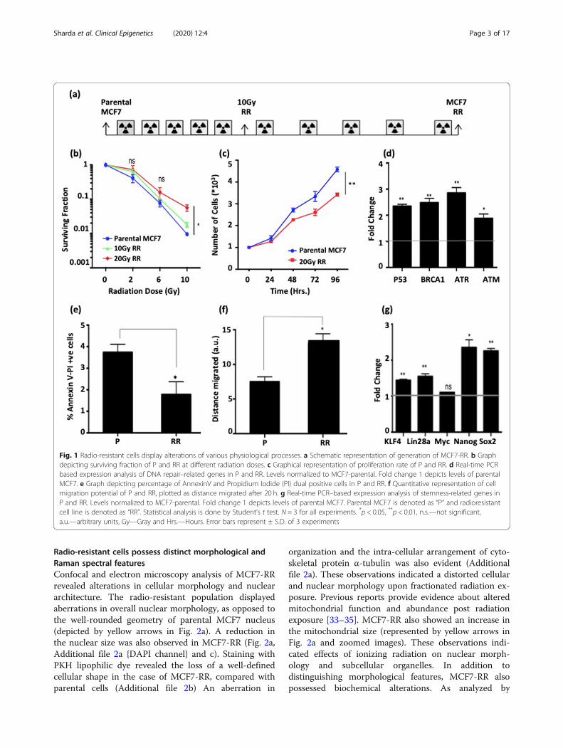
ResultsRadio-resistant cells display alterations of variousphysiological processesMCF7 breast cancer cell line was subjected to radi-ation (10 rounds of 2 Gy each) and a radio-resistantsub-cell line (designated as MCF7-RR) was generated(Fig. 1a). Similarly, another radio-resistant sub-cellline of MDA-MB231 breast cancer cell line (desig-nated as 231RR) was generated. Clonogenic assay re-vealed an enhanced dose-dependent survival for bothMCF7-RR (Fig. 1b, Additional file 1a and b) and231RR (Additional file 3a and b). MCF7-RR cellsdemonstrated a decrease in their proliferation capacitycompared with parental cells in a time-dependentmanner (Fig. 1c). Also, an increase in the expressionof DNA repair-related genes was observed in MCF7-RR, which might contribute to enhanced radio-resistance potential of these cells (Fig. 1d). A com-parative analysis revealed decreased levels ofAnnexinV-PI-positive population (depicting late apop-totic cells) in MCF7-RR (1.36%) compared with par-ental MCF7 (3.48%) (Fig. 1e and Additional file 1c).Further, an assessment of cell migration potential re-vealed an enhanced migration capacity of MCF7-RR(Fig. 1f and Additional file 1d). This corroborated aprevious study that reported an epithelial-to-mesenchymal transition (EMT) to be induced inbreast cancer cells as an effect of radiation [32].MCF7-RR also showed an increase in expression oftranscription factors KLF4, SOX2, and Lin28A gene,which might be responsible for conferring enhancedmigration capacity of MCF7-RR (Fig. 1g).
Sharda et al. Clinical Epigenetics (2020) 12:4 Page 2 of 17
Radio-resistant cells possess distinct morphological andRaman spectral featuresConfocal and electron microscopy analysis of MCF7-RRrevealed alterations in cellular morphology and nucleararchitecture. The radio-resistant population displayedaberrations in overall nuclear morphology, as opposed tothe well-rounded geometry of parental MCF7 nucleus(depicted by yellow arrows in Fig. 2a). A reduction inthe nuclear size was also observed in MCF7-RR (Fig. 2a,Additional file 2a {DAPI channel} and c). Staining withPKH lipophilic dye revealed the loss of a well-definedcellular shape in the case of MCF7-RR, compared withparental cells (Additional file 2b) An aberration in
organization and the intra-cellular arrangement of cyto-skeletal protein α-tubulin was also evident (Additionalfile 2a). These observations indicated a distorted cellularand nuclear morphology upon fractionated radiation ex-posure. Previous reports provide evidence about alteredmitochondrial function and abundance post radiationexposure [33–35]. MCF7-RR also showed an increase inthe mitochondrial size (represented by yellow arrows inFig. 2a and zoomed images). These observations indi-cated effects of ionizing radiation on nuclear morph-ology and subcellular organelles. In addition todistinguishing morphological features, MCF7-RR alsopossessed biochemical alterations. As analyzed by
Fig. 1 Radio-resistant cells display alterations of various physiological processes. a Schematic representation of generation of MCF7-RR. b Graphdepicting surviving fraction of P and RR at different radiation doses. c Graphical representation of proliferation rate of P and RR. d Real-time PCRbased expression analysis of DNA repair–related genes in P and RR. Levels normalized to MCF7-parental. Fold change 1 depicts levels of parentalMCF7. e Graph depicting percentage of AnnexinV and Propidium Iodide (PI) dual positive cells in P and RR. f Quantitative representation of cellmigration potential of P and RR, plotted as distance migrated after 20 h. g Real-time PCR–based expression analysis of stemness-related genes inP and RR. Levels normalized to MCF7-parental. Fold change 1 depicts levels of parental MCF7. Parental MCF7 is denoted as “P” and radioresistantcell line is denoted as “RR”. Statistical analysis is done by Student’s t test. N = 3 for all experiments. *p < 0.05, **p < 0.01, n.s.—not significant,a.u.—arbitrary units, Gy—Gray and Hrs.—Hours. Error bars represent ± S.D. of 3 experiments
Sharda et al. Clinical Epigenetics (2020) 12:4 Page 3 of 17
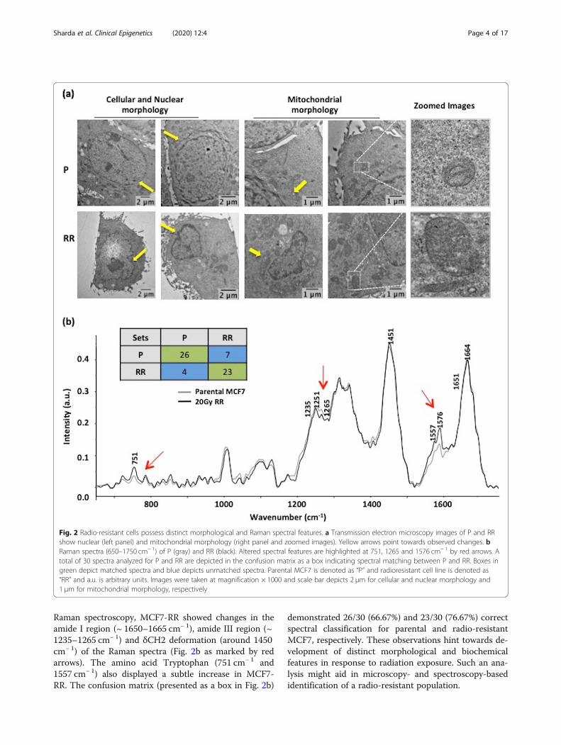
Raman spectroscopy, MCF7-RR showed changes in theamide I region (~ 1650–1665 cm− 1), amide III region (~1235–1265 cm− 1) and δCH2 deformation (around 1450cm− 1) of the Raman spectra (Fig. 2b as marked by redarrows). The amino acid Tryptophan (751 cm− 1 and1557 cm− 1) also displayed a subtle increase in MCF7-RR. The confusion matrix (presented as a box in Fig. 2b)
demonstrated 26/30 (66.67%) and 23/30 (76.67%) correctspectral classification for parental and radio-resistantMCF7, respectively. These observations hint towards de-velopment of distinct morphological and biochemicalfeatures in response to radiation exposure. Such an ana-lysis might aid in microscopy- and spectroscopy-basedidentification of a radio-resistant population.
Fig. 2 Radio-resistant cells possess distinct morphological and Raman spectral features. a Transmission electron microscopy images of P and RRshow nuclear (left panel) and mitochondrial morphology (right panel and zoomed images). Yellow arrows point towards observed changes. bRaman spectra (650–1750 cm− 1) of P (gray) and RR (black). Altered spectral features are highlighted at 751, 1265 and 1576 cm− 1 by red arrows. Atotal of 30 spectra analyzed for P and RR are depicted in the confusion matrix as a box indicating spectral matching between P and RR. Boxes ingreen depict matched spectra and blue depicts unmatched spectra. Parental MCF7 is denoted as “P” and radioresistant cell line is denoted as“RR” and a.u. is arbitrary units. Images were taken at magnification × 1000 and scale bar depicts 2 μm for cellular and nuclear morphology and1 μm for mitochondrial morphology, respectively
Sharda et al. Clinical Epigenetics (2020) 12:4 Page 4 of 17
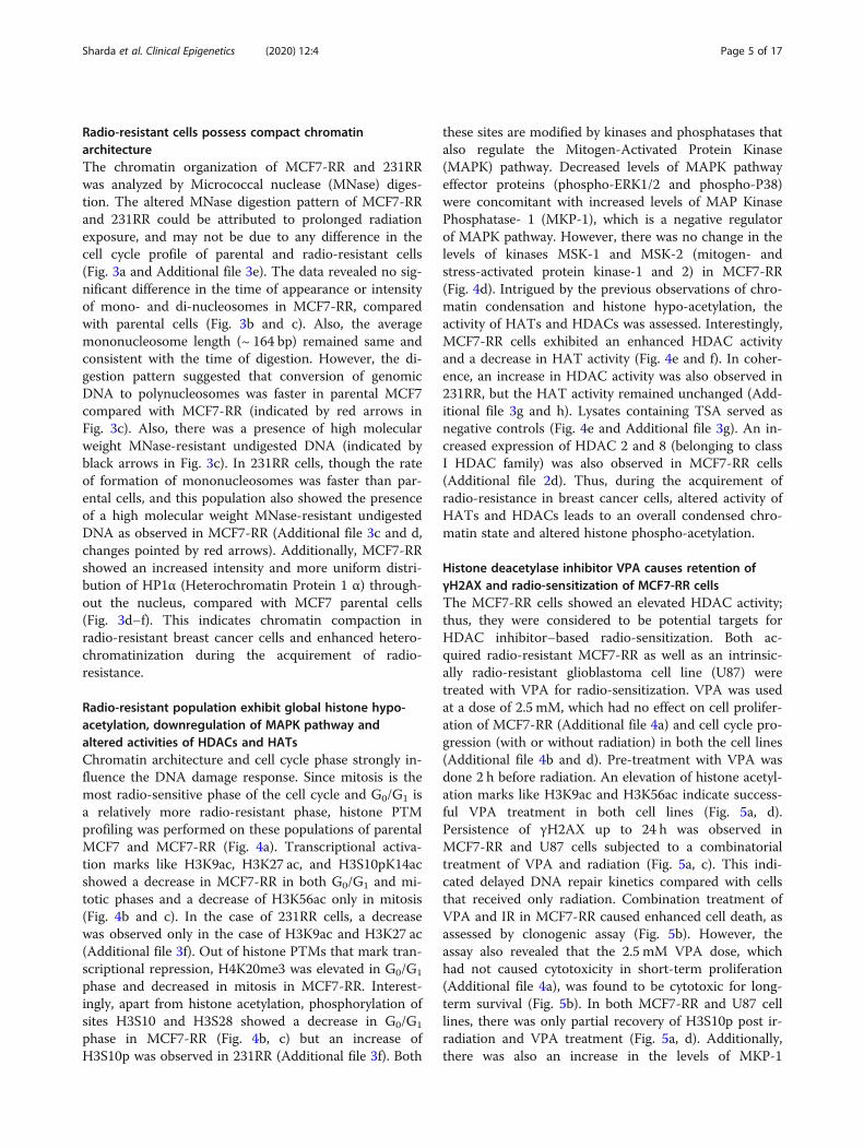
Radio-resistant cells possess compact chromatinarchitectureThe chromatin organization of MCF7-RR and 231RRwas analyzed by Micrococcal nuclease (MNase) diges-tion. The altered MNase digestion pattern of MCF7-RRand 231RR could be attributed to prolonged radiationexposure, and may not be due to any difference in thecell cycle profile of parental and radio-resistant cells(Fig. 3a and Additional file 3e). The data revealed no sig-nificant difference in the time of appearance or intensityof mono- and di-nucleosomes in MCF7-RR, comparedwith parental cells (Fig. 3b and c). Also, the averagemononucleosome length (~ 164 bp) remained same andconsistent with the time of digestion. However, the di-gestion pattern suggested that conversion of genomicDNA to polynucleosomes was faster in parental MCF7compared with MCF7-RR (indicated by red arrows inFig. 3c). Also, there was a presence of high molecularweight MNase-resistant undigested DNA (indicated byblack arrows in Fig. 3c). In 231RR cells, though the rateof formation of mononucleosomes was faster than par-ental cells, and this population also showed the presenceof a high molecular weight MNase-resistant undigestedDNA as observed in MCF7-RR (Additional file 3c and d,changes pointed by red arrows). Additionally, MCF7-RRshowed an increased intensity and more uniform distri-bution of HP1α (Heterochromatin Protein 1 α) through-out the nucleus, compared with MCF7 parental cells(Fig. 3d–f). This indicates chromatin compaction inradio-resistant breast cancer cells and enhanced hetero-chromatinization during the acquirement of radio-resistance.
Radio-resistant population exhibit global histone hypo-acetylation, downregulation of MAPK pathway andaltered activities of HDACs and HATsChromatin architecture and cell cycle phase strongly in-fluence the DNA damage response. Since mitosis is themost radio-sensitive phase of the cell cycle and G0/G1 isa relatively more radio-resistant phase, histone PTMprofiling was performed on these populations of parentalMCF7 and MCF7-RR (Fig. 4a). Transcriptional activa-tion marks like H3K9ac, H3K27 ac, and H3S10pK14acshowed a decrease in MCF7-RR in both G0/G1 and mi-totic phases and a decrease of H3K56ac only in mitosis(Fig. 4b and c). In the case of 231RR cells, a decreasewas observed only in the case of H3K9ac and H3K27 ac(Additional file 3f). Out of histone PTMs that mark tran-scriptional repression, H4K20me3 was elevated in G0/G1
phase and decreased in mitosis in MCF7-RR. Interest-ingly, apart from histone acetylation, phosphorylation ofsites H3S10 and H3S28 showed a decrease in G0/G1
phase in MCF7-RR (Fig. 4b, c) but an increase ofH3S10p was observed in 231RR (Additional file 3f). Both
these sites are modified by kinases and phosphatases thatalso regulate the Mitogen-Activated Protein Kinase(MAPK) pathway. Decreased levels of MAPK pathwayeffector proteins (phospho-ERK1/2 and phospho-P38)were concomitant with increased levels of MAP KinasePhosphatase- 1 (MKP-1), which is a negative regulatorof MAPK pathway. However, there was no change in thelevels of kinases MSK-1 and MSK-2 (mitogen- andstress-activated protein kinase-1 and 2) in MCF7-RR(Fig. 4d). Intrigued by the previous observations of chro-matin condensation and histone hypo-acetylation, theactivity of HATs and HDACs was assessed. Interestingly,MCF7-RR cells exhibited an enhanced HDAC activityand a decrease in HAT activity (Fig. 4e and f). In coher-ence, an increase in HDAC activity was also observed in231RR, but the HAT activity remained unchanged (Add-itional file 3g and h). Lysates containing TSA served asnegative controls (Fig. 4e and Additional file 3g). An in-creased expression of HDAC 2 and 8 (belonging to classI HDAC family) was also observed in MCF7-RR cells(Additional file 2d). Thus, during the acquirement ofradio-resistance in breast cancer cells, altered activity ofHATs and HDACs leads to an overall condensed chro-matin state and altered histone phospho-acetylation.
Histone deacetylase inhibitor VPA causes retention ofγH2AX and radio-sensitization of MCF7-RR cellsThe MCF7-RR cells showed an elevated HDAC activity;thus, they were considered to be potential targets forHDAC inhibitor–based radio-sensitization. Both ac-quired radio-resistant MCF7-RR as well as an intrinsic-ally radio-resistant glioblastoma cell line (U87) weretreated with VPA for radio-sensitization. VPA was usedat a dose of 2.5 mM, which had no effect on cell prolifer-ation of MCF7-RR (Additional file 4a) and cell cycle pro-gression (with or without radiation) in both the cell lines(Additional file 4b and d). Pre-treatment with VPA wasdone 2 h before radiation. An elevation of histone acetyl-ation marks like H3K9ac and H3K56ac indicate success-ful VPA treatment in both cell lines (Fig. 5a, d).Persistence of γH2AX up to 24 h was observed inMCF7-RR and U87 cells subjected to a combinatorialtreatment of VPA and radiation (Fig. 5a, c). This indi-cated delayed DNA repair kinetics compared with cellsthat received only radiation. Combination treatment ofVPA and IR in MCF7-RR caused enhanced cell death, asassessed by clonogenic assay (Fig. 5b). However, theassay also revealed that the 2.5 mM VPA dose, whichhad not caused cytotoxicity in short-term proliferation(Additional file 4a), was found to be cytotoxic for long-term survival (Fig. 5b). In both MCF7-RR and U87 celllines, there was only partial recovery of H3S10p post ir-radiation and VPA treatment (Fig. 5a, d). Additionally,there was also an increase in the levels of MKP-1
Sharda et al. Clinical Epigenetics (2020) 12:4 Page 5 of 17
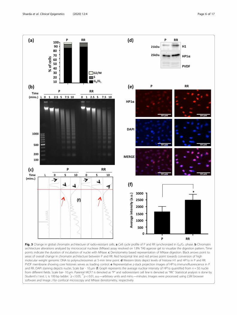
Fig. 3 Change in global chromatin architecture of radio-resistant cells. a Cell cycle profile of P and RR synchronized in G0/G1 phase. b Chromatinarchitecture alterations analyzed by micrococcal nuclease (MNase) assay resolved on 1.8% TAE-agarose gel to visualize the digestion pattern. Timepoints indicate the duration of incubation of nuclei with MNase. c Densitometry based representation of MNase digestion. Black arrows point toareas of overall change in chromatin architecture between P and RR. Red horizontal line and red arrows point towards conversion of highmolecular weight genomic DNA to polynucleosomes at 5-min time point. d Western blots depict levels of histone H1 and HP1α in P and RR.PVDF membrane showing core histones serves as loading control. e Representative z-stack projection images of HP1α immunofluorescence in Pand RR. DAPI staining depicts nuclei. Scale bar - 10 μm (f) Graph represents the average nuclear intensity of HP1α quantified from n = 50 nucleifrom different fields. Scale bar- 10 μm. Parental MCF7 is denoted as “P” and radioresistant cell line is denoted as “RR.” Statistical analysis is done byStudent’s t test. L is 100 bp ladder. *p < 0.05, **p < 0.01, a.u.—arbitrary units and mins.—minutes. Images were processed using LSM browsersoftware and Image J for confocal microscopy and MNase densitometry, respectively
Sharda et al. Clinical Epigenetics (2020) 12:4 Page 6 of 17
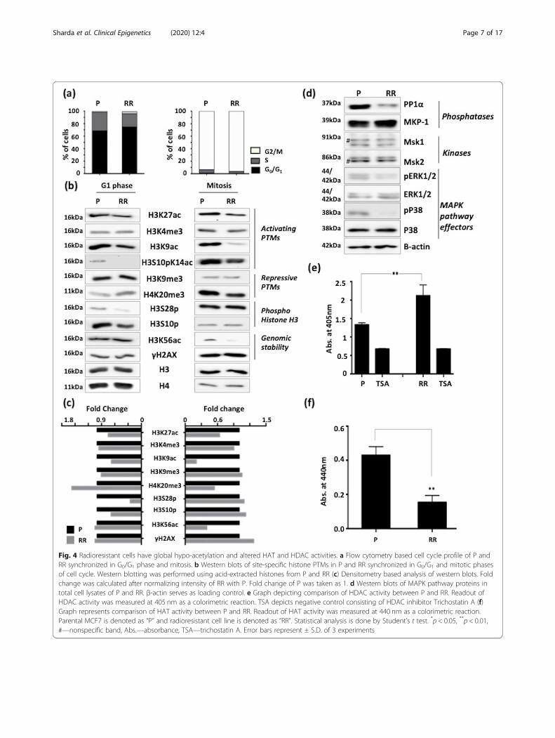
Fig. 4 Radioresistant cells have global hypo-acetylation and altered HAT and HDAC activities. a Flow cytometry based cell cycle profile of P andRR synchronized in G0/G1 phase and mitosis. b Western blots of site-specific histone PTMs in P and RR synchronized in G0/G1 and mitotic phasesof cell cycle. Western blotting was performed using acid-extracted histones from P and RR (c) Densitometry based analysis of western blots. Foldchange was calculated after normalizing intensity of RR with P. Fold change of P was taken as 1. d Western blots of MAPK pathway proteins intotal cell lysates of P and RR. β-actin serves as loading control. e Graph depicting comparison of HDAC activity between P and RR. Readout ofHDAC activity was measured at 405 nm as a colorimetric reaction. TSA depicts negative control consisting of HDAC inhibitor Trichostatin A (f)Graph represents comparison of HAT activity between P and RR. Readout of HAT activity was measured at 440 nm as a colorimetric reaction.Parental MCF7 is denoted as “P” and radioresistant cell line is denoted as “RR”. Statistical analysis is done by Student’s t test. *p < 0.05, **p < 0.01,#—nonspecific band, Abs.—absorbance, TSA—trichostatin A. Error bars represent ± S.D. of 3 experiments
Sharda et al. Clinical Epigenetics (2020) 12:4 Page 7 of 17
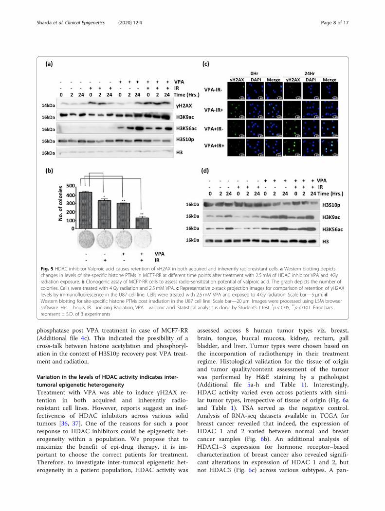
phosphatase post VPA treatment in case of MCF7-RR(Additional file 4c). This indicated the possibility of across-talk between histone acetylation and phosphoryl-ation in the context of H3S10p recovery post VPA treat-ment and radiation.
Variation in the levels of HDAC activity indicates inter-tumoral epigenetic heterogeneityTreatment with VPA was able to induce γH2AX re-tention in both acquired and inherently radio-resistant cell lines. However, reports suggest an inef-fectiveness of HDAC inhibitors across various solidtumors [36, 37]. One of the reasons for such a poorresponse to HDAC inhibitors could be epigenetic het-erogeneity within a population. We propose that tomaximize the benefit of epi-drug therapy, it is im-portant to choose the correct patients for treatment.Therefore, to investigate inter-tumoral epigenetic het-erogeneity in a patient population, HDAC activity was
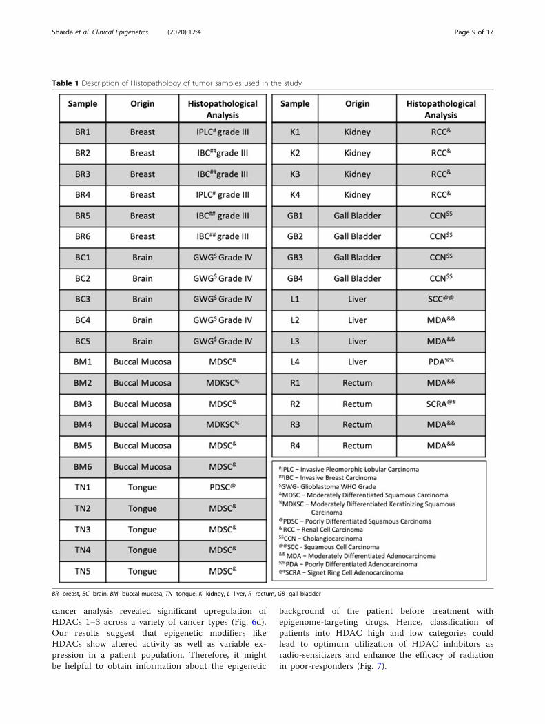
assessed across 8 human tumor types viz. breast,brain, tongue, buccal mucosa, kidney, rectum, gallbladder, and liver. Tumor types were chosen based onthe incorporation of radiotherapy in their treatmentregime. Histological validation for the tissue of originand tumor quality/content assessment of the tumorwas performed by H&E staining by a pathologist(Additional file 5a-h and Table 1). Interestingly,HDAC activity varied even across patients with simi-lar tumor types, irrespective of tissue of origin (Fig. 6aand Table 1). TSA served as the negative control.Analysis of RNA-seq datasets available in TCGA forbreast cancer revealed that indeed, the expression ofHDAC 1 and 2 varied between normal and breastcancer samples (Fig. 6b). An additional analysis ofHDAC1–3 expression for hormone receptor–basedcharacterization of breast cancer also revealed signifi-cant alterations in expression of HDAC 1 and 2, butnot HDAC3 (Fig. 6c) across various subtypes. A pan-
Fig. 5 HDAC inhibitor Valproic acid causes retention of γH2AX in both acquired and inherently radioresistant cells. a Western blotting depictschanges in levels of site-specific histone PTMs in MCF7-RR at different time points after treatment with 2.5 mM of HDAC inhibitor VPA and 4Gyradiation exposure. b Clonogenic assay of MCF7-RR cells to assess radio-sensitization potential of valproic acid. The graph depicts the number ofcolonies. Cells were treated with 4 Gy radiation and 2.5 mM VPA. c Representative z-stack projection images for comparison of retention of γH2AXlevels by immunofluorescence in the U87 cell line. Cells were treated with 2.5 mM VPA and exposed to 4 Gy radiation. Scale bar—5 μm. dWestern blotting for site-specific histone PTMs post irradiation in the U87 cell line. Scale bar—20 μm. Images were processed using LSM browsersoftware. Hrs.—hours, IR—ionizing Radiation, VPA—valproic acid. Statistical analysis is done by Student’s t test. *p < 0.05, **p < 0.01. Error barsrepresent ± S.D. of 3 experiments
Sharda et al. Clinical Epigenetics (2020) 12:4 Page 8 of 17
cancer analysis revealed significant upregulation ofHDACs 1–3 across a variety of cancer types (Fig. 6d).Our results suggest that epigenetic modifiers likeHDACs show altered activity as well as variable ex-pression in a patient population. Therefore, it mightbe helpful to obtain information about the epigenetic
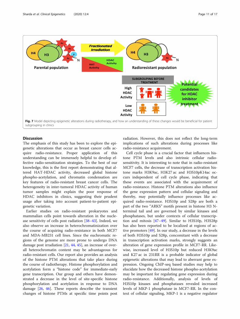
background of the patient before treatment withepigenome-targeting drugs. Hence, classification ofpatients into HDAC high and low categories couldlead to optimum utilization of HDAC inhibitors asradio-sensitizers and enhance the efficacy of radiationin poor-responders (Fig. 7).
Table 1 Description of Histopathology of tumor samples used in the study
BR -breast, BC -brain, BM -buccal mucosa, TN -tongue, K -kidney, L -liver, R -rectum, GB -gall bladder
Sharda et al. Clinical Epigenetics (2020) 12:4 Page 9 of 17
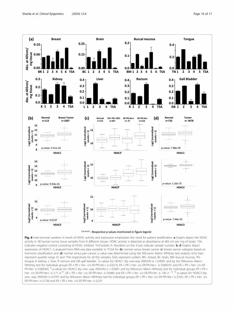
Fig. 6 Inter-tumoral variation in levels of HDAC activity and expression emphasizes the need for patient stratification. a Graphs depict the HDACactivity in 38 human tumor tissue samples from 8 different tissues. HDAC activity is depicted as absorbance at 405 nm per mg of lysate. TSAindicates negative control consisting of HDAC inhibitor Trichostatin A. Numbers on the X-axis indicate sample number. b–d Graphs depictexpression of HDAC1–3 analyzed from RNA-seq data available in TCGA for (b) normal versus breast cancer (c) breast cancer subtypes based onhormone classification and (d) normal versus pan-cancer. p value was determined using the Wilcoxon–Mann–Whitney test analysis. Error barsrepresent quartile range 25 and 75% respectively for all the samples. Dots represent outliers. BR—breast, BC–brain, BM–buccal mucosa, TN–tongue, K–kidney, L–liver, R–rectum and GB–gall bladder. $p values for HDAC1 (by one-way ANOVA) is < 0.0001 and by the Wilcoxon–Mann–Whitney test for individual groups ER + PR + Her- v/s ER-PR-Her+ is 0.0274, ER + PR + Her- v/s ER-PR-Her+ is 0.000191 and ER + PR + Her- v/s ER-PR-Her- is 0.000305. # p-values for HDAC2 (by one- way ANOVA) is < 0.0001 and by Wilcoxon–Mann–Whitney test for individual groups ER + PR +Her- v/s ER-PR-Her+ is 2.71 e-07, ER + PR + Her- v/s ER-PR-Her+ is 0.0485 and ER + PR + Her- v/s ER-PR-Her- is 1.85 e− 23. % p-values for HDAC3 (byone- way ANOVA) is 0.4797 and by Wilcoxon–Mann–Whitney test for individual groups ER + PR + Her- v/s ER-PR-Her+ is 0.591, ER + PR + Her- v/sER-PR-Her+ is 0.738 and ER + PR + Her- v/s ER-PR-Her- is 0.231
Sharda et al. Clinical Epigenetics (2020) 12:4 Page 10 of 17
DiscussionThe emphasis of this study has been to explore the epi-genetic alterations that occur as breast cancer cells ac-quire radio-resistance. Proper application of thisunderstanding can be immensely helpful to develop ef-fective radio-sensitization strategies. To the best of ourknowledge, this is the first report demonstrating that al-tered HAT-HDAC activity, decreased global histonephospho-acetylation, and chromatin condensation arekey features of radio-resistant breast cancer cells. Theheterogeneity in inter-tumoral HDAC activity of humantumor samples might explain the poor response ofHDAC inhibitors in clinics, suggesting their prudentusage after taking into account patient-to-patient epi-genetic variation.Earlier studies on radio-resistant prokaryotes and
mammalian cells point towards alteration in the nucle-ase sensitivity of cells post radiation [38–43]. Indeed, wealso observe an increase in heterochromatinization overthe course of acquiring radio-resistance in both MCF7and MDA-MB231 cell lines. Since the euchromatic re-gions of the genome are more prone to undergo DNAdamage post irradiation [21, 44, 45], an increase of over-all heterochromatin content may be advantageous forradio-resistant cells. Our report also provides an analysisof the histone PTM alterations that take place duringthe course of radiotherapy. Histone phosphorylation andacetylation form a “histone code” for immediate-earlygene transcription. Our group and others have demon-strated a decrease in the level of site-specific histonephosphorylation and acetylation in response to DNAdamage [26, 46]. These reports describe the transientchanges of histone PTMs at specific time points post
radiation. However, this does not reflect the long-termimplications of such alterations during processes likeradio-resistance acquirement.Cell cycle phase is a crucial factor that influences his-
tone PTM levels and also intrinsic cellular radio-sensitivity. It is interesting to note that in radio-resistantMCF7 cells, the decrease of transcription activation his-tone marks H3K9ac, H3K27 ac and H3S10pK14ac oc-curs independent of cell cycle phase, indicating thatthese events are associated with the acquirement ofradio-resistance. Histone PTM alterations also influencethe gene expression pattern and cellular signaling andthereby, may potentially influence processes like ac-quired radio-resistance. H3S10p and S28p are both apart of the two “ARKS” motifs present in histone H3 N-terminal tail and are governed by similar kinases andphosphatases, but under contexts of cellular transcrip-tion and mitosis [47–49]. Similar to H3S10p, H3S28phas also been reported to be localized at regions of ac-tive promoters [49]. In our study, a decrease in the levelsof both H3S10p and S28p, concomitant with a decreasein transcription activation marks, strongly suggests analteration of gene expression profile in MCF7-RR. Like-wise, increased level of H3S10p but reduced H3K9acand K27 ac in 231RR is a probable indicator of globalepigenetic alterations that may lead to aberrant gene ex-pression. Ongoing ChIP-seq based studies may help toelucidate how the decreased histone phospho-acetylationmay be important for regulating gene expression duringradio-resistance. Additionally, analysis of levels ofH3S10p kinases and phosphatases revealed increasedlevels of MKP-1 phosphatase in MCF7-RR. In the con-text of cellular signaling, MKP-1 is a negative regulator
Fig. 7 Model depicting epigenetic alterations during radiotherapy, and how an understanding of these changes would be beneficial for patientsubgrouping in clinics
Sharda et al. Clinical Epigenetics (2020) 12:4 Page 11 of 17
of the MAPK pathway [50, 51]. We observe an increasein levels of MKP-1, thus explaining the decreased phos-phorylation of ERK1/2 and P38. Upregulation of MKP-1is also implicated to be a poor prognostic factor forbreast cancer and mediates therapy resistance to Her2-positive breast tumors [52, 53]. Thus, our report sug-gests that alteration in levels of histone PTMs and theirrespective modifying enzymes during radiotherapy mightinfluence gene expression and functioning of key cellularpathways.Dysregulation of HDACs is implicated in various can-
cers and plays an important role during DDR [54, 55].We observed an altered HDAC/HAT activity pattern inradio-resistant breast cancer cells, with an increase inHDAC activity for both MCF7-RR and 231RR. This ex-plains the previously described global histone hypo-acetylation as well as chromatin condensation. Treat-ment of this target population by HDAC inhibitorscould lead to potential sensitization towards radiation.Advantages of using HDAC inhibitor VPA include FDAapproval, low cellular toxicity, increased half-life, andthe ability to cross the blood–brain barrier. To comparethe mechanism of action of VPA, both an acquired(MCF7-RR) and inherently radio-resistant (U87) celllines were used. Interestingly, we observed a reductionin the levels of H3S10p post VPA and radiation combin-ation treatment in both types of cells. Our group haspreviously reported that H3S10p undergoes dephosphor-ylation and subsequent re-phosphorylation in responseto DNA damage, both of which are crucial for effectiveDNA repair and cell survival post radiation [56, 57].Since HDACs can act on both histone and non-histonesubstrates, their mechanism of action is varied [27].Treatment with HDAC inhibitor TSA decreasesphospho-histone H3 due to non-localization of kineto-chore protein BubR1, potentially attributed to impaireddeacetylation of non-histone substrates of HDACs [58].Also, since histone PTMs function as a “histone code”[59], persistence of histone hyper-acetylation post VPAtreatment could affect the phosphorylation of H3S10p.A study suggests that hypo-acetylated histone tails arepreferred substrates for Aurora Kinase B [60]. We ob-serve an induction in the levels of MKP-1 phosphatasepost VPA treatment, concomitant with a previous study[61]. But there have been no reports about histone ki-nases as substrates of HDACs. Hence, the reduced levelsof H3S10p after VPA treatment may be attributed toboth histone hyper-acetylation and the effect of VPA onnon-histone substrates, which may affect survival of cellspost-radiation.Despite showing promising results in hematological
malignancies, the utility of HDAC inhibitors in solid tu-mors has been limited due to toxicity, poor pharmaco-kinetics as well as low half-life. Our group has recently
demonstrated HDAC activity in serum of human normaland tumor counterparts [62]. This suggests that moni-toring HDAC/HAT activities during treatment may pro-vide “real-time” information about treatment response.In this study, we observe an increase in HDAC activityduring acquired radio-resistance. Therefore, we proposethat an evaluation of tumor HDAC activity be carriedout before beginning with radiotherapy, which may helpto identify suitable candidates for HDAC inhibitor–based radio-sensitization. This is based on our observa-tion of epigenetic heterogeneity in terms of HDAC activ-ity status across 38 tumor samples. It is very interestingthat even in our small sample size, tumors of the samehistopathological type display variation in HDAC activ-ity, for example, BR3 and BR6 are both invasive breastcarcinoma grade III cancers, yet have more than two-fold difference in HDAC activity. This points towardsthe extensive inter-tumor heterogeneity in a population,emphasizing that not all patients may respond similarlyto HDAC inhibitor therapy. Additionally, we were ableto check the HDAC activity of as little as 1 mg of tumortissue, thereby excluding the limitation of sampleamount and availability from a biopsy. Therefore, sub-grouping of patients into low and high HDAC activitygroups, and then treatment of the correct subgroup mayhelp to greatly increase the efficacy of HDAC inhibitoras a radio-sensitizer. According to our analysis, patientshaving tumors with high HDAC activity are the suitablegroup for HDAC inhibitor–based radio-sensitization.However, altered HDAC activity may not be the onlymeasure for poor response towards radiation. In such ascenario, it is possible that other factors like epigenetic/genetic determinants, tumor microenvironment andstemness might impart radio-sensitivity [14–17]. There-fore, studies are required to understand the cause ofpoor response to radiation in cancer patients. Such anunderstanding of epigenetic alterations during radiother-apy may help in preventing episodes of recurrence dueto radio-resistance. Therefore, the need of the hour is torealize the tremendous potential of epi-drug therapy andutilize strategies like patient subgrouping for better dis-ease management.
ConclusionTo summarize, our study demonstrates that epigeneticalterations are an important player during radiotherapyand may be responsible for acquired radio-resistance.Radio-resistant cells show high HDAC activity–lowHAT activity that explains the compact chromatin archi-tecture and altered histone phospho-acetylation. Also,epigenetic variation across human tumor samples interms of the tumor HDAC activity indicates that priorstratification of patients is important before beginningany HDAC inhibitor–based treatment.
Sharda et al. Clinical Epigenetics (2020) 12:4 Page 12 of 17
MethodsCell culture, synchronization, and irradiationThe cell lines used in this study are epithelial originbreast cancer (MCF7 and MDA-MB231) and glioblast-oma (U87) cell lines. All cell lines were cultured inDMEM media (Invitrogen) supplemented with 2 mMglutamine (Sigma), 10% fetal bovine serum (FBS) (Gibco)and antibiotic antimycotic solution (Himedia). Cellswere maintained at 37 °C and 5% CO2. Synchronizationin G0/G1 and mitotic phase of the cell cycle was done byserum starvation using 0.02% serum for 72 h and 18 hincubation with 200 ng/ml nocodazole (Sigma), respect-ively. Cells were irradiated using Co-60 radioactivesource–based machine Bhabhatron-II (Panacea MedicalTechnologies Ltd. and Bhabha Atomic Research Centre(BARC), India) installed at the Department of RadiationOncology, ACTREC. Field size was 25 cm × 25 cm,source-to-skin distance (SSD) as 80 cm, and gantry wasangled at 180° to the specimen.
Generation of radio-resistant cell lineFractionated Irradiation (FIR) was used to generateradio-resistant cells. Ten fractions of 2 Gy each wereused to generate MCF7-RR and 231RR, respectively.Cells were maintained up to 60% confluency at the timeof irradiation and exposed to 2-Gy radiation dose/frac-tions for 10 cycles (Dose rate 0.75 Gy/min). Cells wereallowed to recover and gain 80–85% confluency beforesub-culturing for the next dose. Parental cells were shamirradiated during the process. Cells were allowed to growfor 21 days after the final dose of 20 Gy. The radio-resistant populations are designated as parental MCF7-RR and 231RR.
Clonogenic assayCells were irradiated, allowed to recover for 6 h,counted, seeded and maintained for 14 days. Colonieswere fixed with 4% paraformaldehyde (Sigma) for 20 minfollowed by washing with phosphate buffered saline(PBS). Staining was performed using 0.5% crystal violet.Colonies were counted under a light microscope andonly colonies containing > 50 cells were considered to beviable. The plating efficiency was calculated as men-tioned [63] and surviving fraction of each cell line wascalculated. The D0 value of each cell line was calculatedusing the survival fraction and the doses 2 Gy and 4 Gy.
Cell migration assayCells were seeded in a 6-well plate, allowed to grow for24 h and then serum starved in 0.02% FBS containingmedia for 72 h. At the time of the experiment, 3 woundswere made in the 100% confluent cell monolayer. Debrisof dislodged cells was washed off using PBS and 0.02%serum-containing media was added. Cells were
incubated in a CO2 chamber at 37 °C and monitored formigration using live-cell microscopy for 20 h.
Cell proliferation assayA total of 1000 cells of parental MCF7 and MCF7-RRwere seeded in a 96-well plate and growth was analyzedfor 96 h. A 3-(4,5-dimethylthiazol-2-yl)-2,5-diphenyltet-razolium bromide (MTT) reagent (5 mg/ml in PBS,Sigma) was added to each well at 1/10th medium vol-ume and incubated for 4 h. Formazan crystals were solu-bilized using MTT solubilization buffer (10% sodiumdodecyl sulfate (SDS), 0.01M HCl) and incubated for 24h in dark. Absorption at 570 nm was measured usingSpectrostar Nano Biotek LabTech 96-well plate reader.
Flow cytometry–based cell cycle analysisCell cycle analysis was carried out using propidium iod-ide (PI)-based DNA content analysis as described previ-ously [26]. DNA content analysis was carried out usingfluorescence-activated cell sorting (FACS) Calibur flowcytometer (Becton Dickinson) and analysis done usingMODFIT software by Verity house.
AnnexinV/PI stainingThe apoptotic population analysis was done by Annex-inV- Fluorescein isothiocyanate (FITC) apoptosis detec-tion kit (Sigma), strictly following manufacturerinstructions. Fluorescence was immediately measuredusing FACS Calibur flow cytometer (Becton Dickinson)and analysis done using CELLQUEST software.
Transmission Electron microscopyParental MCF7 and MCF7-RR cell pellets were fixedwith 3% glutaraldehyde. Post-fixation was performedwith 1% osmium tetraoxide. Alcoholic uranyl acetatetreatment for 1 min and lead citrate treatment for 30 swas done for grid contrasting and then observed underCarl Zeiss LIBRA120 EFTEM.
Raman spectroscopyFresh cell pellets of parental MCF7 and MCF7-RR werefixed using 1% Paraformaldehyde (PFA) at 4 °C for 10min followed by two washes of saline at 5000 rpm for 5min at 4 °C. A total of 30 spectra were analyzed for bothparental and radio-resistant MCF7. Raman spectra wererecorded using a commercial Raman micro spectroscope(WITec alpha300RS, λX-532 nm, 10mW, 600 grooves/mm). Preprocessed Raman spectra (smoothening, fifthpoint, baseline, fifth order, and vector normalization) in650–1750 cm− 1 were subjected to PCA and PCA-LDAusing commercial Unscrambler® X software.
Sharda et al. Clinical Epigenetics (2020) 12:4 Page 13 of 17
Micrococcal nuclease digestion assayMNase digestion was performed as described previously[26]. The DNA pellet was dissolved in 50 μl Tris-EDTA(TE) buffer and samples were resolved on 1.8% 1× tris-EDTA-acetic acid (TAE) agarose gel containing 0.5 μg/ml ethidium bromide. The image was analyzed usingImage J software.
Histone isolationHistone isolation was performed as described earlier[26]. The chromatin bound histones were extractedusing acid extraction method. The histones obtained inthe final pellet was resuspended in 0.1% β-mercaptoethanol and stored in − 20 °C.
Western blottingHistones and total cell lysates were resolved on 18% and10% SDS-poly-acrylamide gel electrophoresis (SDS-PAGE) respectively, transferred on PVDF membraneand subjected to western blotting. Antibodies and theirdilution used are as provided in Additional file 6.
Quantitative PCRRNA extraction from parental and MCF7-RR was doneby Trizol method, followed by DNaseI treatment (Fer-mentas) and cDNA synthesis using random hexamerprimers (Revert-Aid cDNA Synthesis Kit, Thermo Scien-tific), strictly as per manufacturer’s instructions. Real-time PCR was performed using gene-specific primers(Additional file 7) using amplification conditions of 30 sat 94 °C, 1 min at 60 °C, and 1min at 72 °C for 30 cyclesfollowed by 10 min of final extension. SYBR-Green fromApplied Biosystems was used for real-time PCR. The re-actions were performed and monitored using Quant-Studio 12 K Flex Real-Time PCR System. Fold changewas calculated using the ΔΔCt method. The expressionlevels of MCF7-RR were plotted as fold change normal-ized to MCF7 parental cell line.
Immunofluorescence microscopyImmunofluorescence was performed as described previ-ously [26]. The specific dilutions used for antibodies aredescribed in Additional document 4. PKH staining(Sigma)–based live-cell staining for cell morphologyvisualization was performed according to manufacturer’sinstruction. Imaging was done using Zeiss 510 meta con-focal microscope.
Human tissue sample collectionApproval from Institute Ethics Committee III (Projectnumber 164) was obtained for working on human tumorsamples, collected retrospectively with an approved wai-ver of consent. Samples were collected from the Biore-pository of ACTREC-TMC. Histopathological analysis of
the samples confirmed tumor status and quality. Sam-ples were stored in − 80 °C and used as required.
HDAC and HAT activity assayAssays were performed using the colorimetric HDACand HAT activity assay kits from BioVision (BioVisionResearch Products, USA) as per manufacturer instruc-tions. A total of 100 μg of cell lysate was used for the as-says. The HDAC activity of human tumor samples wasassessed from tissue samples powdered in liquid nitro-gen. Fifty micrograms of tissue powder was dissolved in300 μl RIPA buffer (250 mM sucrose, 50 mM Tris-Cl,pH 7.5, 25 mM KCl, 5 mM MgCl2, 0.2 mM PMSF, 50mM NaHSO3, 45 mM sodium butyrate, 10 mM β-ME,0.2% TritonX-100) and incubated on ice for 30 min,followed by centrifugation at 5000 rpm for 10 min. Thepellet obtained was sonicated (5 s using 30% amplitude)and centrifuged at 15000 rpm for 30min. Supernatantequivalent to 1 mg tissue (6 μl) was used for HDACassay. Each tissue sample had its respective blank, i.e.,without lysine developer. Absorbance was estimatedusing a 96-well plate reader at 405 nm and 440 nm forHDAC and HAT assays, respectively.
In silico analysis in TCGA pan-cancer datasetTo analyze the expression levels of HDAC1, HDAC2,and HDAC3 in normal and tumor samples of pan-cancer, TCGA PANCAN normalized RSEM counts wereobtained from the UCSC cancer genome browser. TheTCGA PANCAN study includes multiple cancers, listedas follows: acute myeloid leukemia, adreno-cortical car-cinoma, bladder urothelial carcinoma, lower grade gli-oma, breast invasive carcinoma, cervical squamous cellcarcinoma and endo-cervical adenocarcinoma, cholan-giocarcinoma, colorectal adenocarcinoma, esophagealcarcinoma, glioblastoma multiforme, head and necksquamous cell carcinoma, kidney chromophobe, kidneyrenal clear cell carcinoma, kidney renal papillary cell car-cinoma, liver hepatocellular carcinoma, lung adenocar-cinoma, lung squamous cell carcinoma, lymphoidneoplasm diffuse large B-cell lymphoma, mesothelioma,ovarian serous cystadeno-carcinoma, pancreatic adeno-carcinoma, pheochromocytoma and paraganglioma,prostate adenocarcinoma, sarcoma, skin cutaneous mel-anoma, stomach adenocarcinoma, testicular germ celltumors, thyroid carcinoma, uterine carcino-sarcoma,uterine corpus endometrial carcinoma, and uveal melan-oma. These counts were transformed in (Log2 + 1) valuesand represented between normal (n = 677) and tumorsamples (n = 9078). R3.3.3 software (http://www.R-pro-ject.org/) was used for boxplot representation. p valuewas determined using Wilcoxon–Mann–Whitney testanalysis and one-way ANOVA (for hormone-based sub-grouping of breast cancer).
Sharda et al. Clinical Epigenetics (2020) 12:4 Page 14 of 17
Statistical analysisAll numerical data were expressed as average of valuesobtained ± standard deviation (SD). Statistical signifi-cance was determined by conducting a Student’s t test.
Supplementary informationSupplementary information accompanies this paper at https://doi.org/10.1186/s13148-019-0800-4.
Additional file 1. (a) Clonogenic assay depicting enhanced cell survivalof parental MCF7, 10Gy and 20Gy radioresistant cells at different radiationdoses. (b) Graph depicting D0 values of MCF7 parental, 10Gy and 20Gyradioresistant populations. (c) Representative images of flow cytometrybased analysis of AnnexinV and Propidium Iodide positive population. (d)Representative images of changes in cell migration potential ofradioresistant MCF7 and MCF7-RR, assessed by live cell microscopy. Par-ental MCF7 is denoted as “P” and radioresistant cell line is denoted as“RR”. Statistical analysis is done by student’s t-test. n = 3 for all experi-ments. *p < 0.05, **p < 0.01. n.s.- not significant. Error bars represent ± S.D.of 3 experiments.
Additional file 2. (a) Representative z-stack projection images for im-munofluorescence analysis of P and RR depicting changes in organizationof α-tubulin. Magnification – 40x, scale bar- 10 μm. (b) Representative z-stack projection images for immunofluorescence analysis of P and RRdepicting change in cellular morphology by PKH staining. Magnification– 40x, scale bar- 10 μm. (c) Graph depicting comparison of nuclear areabetween P and RR. Area was quantified from n = 50 DAPI stained nuclei.(d) Real time PCR based analysis depicts alteration in expression of differ-ent HDAC genes. Expression normalized to MCF7-parental. Fold change 1depicts levels of parental MCF7. Images were processed using LSMbrowser software. Parental MCF7 is denoted as “P” and radioresistant cellline is denoted as “RR”. Statistical analysis is done by student’s t-test. n = 3for all experiments. *p < 0.05, **p < 0.01 and a.u.- arbitrary units. Error barsrepresent ± S.D. of 3 experiments.
Additional file 3. (a) Clonogenic assay depicting enhanced cell survivalof 231P and 231RR at different radiation doses. (b) Graph depictingnumber of colonies obtained after subjecting parental MDA-MB231 and231RR to 4Gy and 8Gy radiation. (c) Chromatin architecture alterations an-alyzed by Micrococcal Nuclease (MNase) assay visualized on 1.8% TAE-agarose gel. Time points indicate the duration of incubation of nucleiwith MNase. (d) Densitometry based representation of MNase digestion.Red arrows point to areas of overall change in chromatin architecture be-tween 231P and 231RR. (e) Flow cytometry based cell cycle profile of231P and 231RR, representative of cell cycle profile for all subsequent ex-periments. (f) Western blots depict levels of histone PTMs in 231P and231RR. Western blotting was performed using acid extracted histonesfrom P and RR (g) Graph depicting comparison of HDAC activity between231P and 231RR. Readout of HDAC activity was measured at 405 nm as acolorimetric reaction. TSA depicts negative control consisting of HDAC in-hibitor Trichostatin A (h) Graph represents comparison of HAT activity be-tween 231P and 231RR. Readout of HAT activity was measured at 440 nmas a colorimetric reaction. 231P and 231RR represents parental and radio-resistant MDA-MB231 cells, respectively. Statistical analysis is done by stu-dent’s t-test. *p < 0.05, **p < 0.01, Abs. – absorbance, TSA – Trichostatin A.Error bars represent ± S.D. of 3 experiments.
Additional file 4. (a) Graph represents change in percentage growth ofP and RR after 48 hours of dose dependent VPA treatment. (b) Graphicalrepresentation of changes in cell cycle profile of RR at different timepoints upon IR (4Gy) and VPA (2.5 mM) treatment. (c) Western blotsdepict levels of H3S10p modifying kinases and phosphatases at differenttime points post VPA treatment and IR exposure in radioresistant cell line.(d) Graph depicts cell cycle profile of U87 cell line at different time pointspost radiation and VPA treatment. UT- Untreated and Hrs. = Hours.Parental MCF7 is denoted as “P” and radioresistant cell line is denoted as“RR”.
Additional file 5. (a-h) Representative images of Hematoxylin and Eosinstaining of human tumor samples, assessed by pathologist for tumor
content. Samples were derived from tumors of eight different tissueorigins. Magnification – 40X, Scale bar- 20 μm.
Additional file 6. Details of the antibodies used in the study.
Additional file 7. Sequence of primers used in the study.
AbbreviationsDDR: DNA damage response; EMT: Epithelial to mesenchymal transition;FIR: Fractionated irradiation; Gy: Gray; HATs: Histone acetyl transferases;HDACs: Histone deacetylases; LET: Linear energy transfer; MNase: Micrococcalnuclease; PIK3CA: Phosphatidylinositol-4, 5-bisphosphate 3-kinase catalyticsubunit alpha; PTMs: Post-translational modifications; SSD: Source to skindistance; VPA: Valproic acid
AcknowledgementsThe authors would like to thank the Department of Radiation Oncology andFlow cytometry at ACTREC. We are also thankful to Dr. Santosh KumarSandur (BARC, India) for providing the MCF7 breast cancer cell line. Dr.Narendra Joshi and Dr. Neelam Shirsat, ACTREC, provided MDA-MB231 andU87 cell lines, respectively. We are thankful to Dr. Sorab Dalal and Dr. JyotiKode, ACTREC, for providing α-tubulin antibody and PKH staining reagent,respectively.
Authors’ contributionsSG and AS contributed to the conception and design of experiments. ASperformed the experiments. AKS (former graduate student at Guptalaboratory, currently at the University of Alberta) performedimmunofluorescence of γH2AX in the U87 cell line. SRS performed theRaman spectroscopy studies. MKC validated Raman spectroscopy dataanalysis. MR and SS collected human tissue samples, performed H&E stainingand TCGA data analysis. PG performed histopathological analysis. AS and SGwrote the manuscript. The paper was critically read by all authors andapproved for publication.
FundingThe authors would like to thank the Advanced Centre for TreatmentResearch and Education in Cancer (ACTREC) – India and Department ofScience and Technology (DST) – India, for funding Gupta Laboratory. Theauthors are also thankful to Tata Memorial Hospital for funding the projectno. 164 by Intra-mural Research Grant. AS was supported by the UniversityGrants Commission (UGC, India) for her doctoral fellowship. MR and SS aresupported by ACTREC for their doctoral fellowship.
Availability of data and materialsThe protocols are detailed in the manuscript for scientists wishing to usethem for their research work. Also, any supporting data will be madeavailable to editors and peer-reviewers, if required for the purpose of evaluat-ing the manuscript.
Ethics approval and consent to participateThe protocol was reviewed and approved by the Institutional Review Boardand Ethics Committee of Tata Memorial Centre, ACTREC, Navi Mumbai(Project Number 164). And the waiver of consent was granted for theproposed study as the samples were collected retrospectively fromBiorepository, ACTREC. Therefore, consent for publication is not applicable toour study.
Competing interestsThe authors declare that they have no competing interests.
Author details1Epigenetics and Chromatin Biology Group, Gupta Lab, Cancer ResearchInstitute, Advanced Centre for Treatment, Research and Education in Cancer(ACTREC), Tata Memorial Centre (TMC), Kharghar, Navi Mumbai, MH 410210,India. 2Homi Bhabha National Institute, Training School Complex, AnushaktiNagar, Mumbai, MH 400085, India. 3Department of Oncology, Faculty ofMedicine and Dentistry, University of Alberta, 11560 University Avenue,Edmonton, Alberta T6G 1Z2, Canada. 4Chilkapati Laboratory, AdvancedCentre for Treatment, Research and Education in Cancer (ACTREC), TataMemorial Centre (TMC), Kharghar, Navi Mumbai, MH 410210, India.5Biorepository, Advanced Centre for Treatment, Research and Education in
Sharda et al. Clinical Epigenetics (2020) 12:4 Page 15 of 17
Cancer (ACTREC), Tata Memorial Centre (TMC), Kharghar, Navi Mumbai, MH410210, India.
Received: 22 May 2019 Accepted: 23 December 2019
References1. Malvia S, Bagadi SA, Dubey US, Saxena S. Epidemiology of breast cancer in
Indian women. Asia Pac J Clin Oncol. 2017;13(4):289–95.2. Ghoncheh M, Pournamdar Z, Salehiniya H. Incidence and mortality and
epidemiology of breast cancer in the world. Asian Pac J Cancer Prev. 2016;17(S3):43–6.
3. Jameel JKA, Rao VSR, Cawkwell L, Drew PJ. Radioresistance in carcinoma ofthe breast. Breast. 2004;13(6):452–60.
4. EBCTCG (Early Breast Cancer Trialists’ Collaborative Group), McGale P, TaylorC, Correa C, Cutter D, Duane F, et al. Effect of radiotherapy aftermastectomy and axillary surgery on 10-year recurrence and 20-year breastcancer mortality: meta-analysis of individual patient data for 8135 women in22 randomised trials. Lancet (London, England). 2014;383(9935):2127–35.
5. Steel GG, McMillan TJ, Peacock JH. The 5Rs of radiobiology. Int J Radiat Biol.1989;56(6):1045–8.
6. Fujimori H, Sato A, Kikuhara S, Wang J, Hirai T, Sasaki Y, et al. Acomprehensive analysis of radiosensitization targets; functional inhibition ofDNA methyltransferase 3B radiosensitizes by disrupting DNA damageregulation. Sci Rep. 2015;5:18231.
7. Hodzic J, Dingjan I, Maas MJ, van der Meulen-Muileman IH, de Menezes RX,Heukelom S, et al. A cell-based high-throughput screening assay forradiation susceptibility using automated cell counting. Radiat Oncol. 2015;10:55.
8. Guo L, Xiao Y, Fan M, Li JJ, Wang Y. Profiling Global Kinome Signatures ofthe Radioresistant MCF-7/C6 Breast Cancer Cells Using MRM-based TargetedProteomics. J Proteome Res. 2015;14(1):193–201.
9. Hou D-L, Chen L, Liu B, Song L-N, Fang T. Identification of common genenetworks responsive to radiotherapy in human cancer cells. Cancer GeneTher. 2014;21:542.
10. Speers C, Zhao S, Liu M, Bartelink H, Pierce LJ, Feng FY. Development andValidation of a Novel Radiosensitivity Signature in Human Breast Cancer.Clin Cancer Res. 2015;21(16):3667–77.
11. Doan NB, Nguyen HS, Alhajala HS, Jaber B, Al-Gizawiy MM, Ahn E-YE, et al.Identification of radiation responsive genes and transcriptome profiling viacomplete RNA sequencing in a stable radioresistant U87 glioblastomamodel. Oncotarget. 2018;9(34):23532–42.
12. Bernichon E, Vallard A, Rancoule C, Magné N, Pissaloux D, Wang Q, et al.Genomic alterations and radioresistance in breast cancer: an analysis of theProfiLER protocol. Ann Oncol. 2017;28(11):2773–9.
13. Bristow RG, Berlin A, Dal Pra A. An arranged marriage for precisionmedicine: hypoxia and genomic assays in localized prostate cancerradiotherapy. Br J Radiol. 2014;87(1035):20130753.
14. Barker HE, Paget JTE, Khan AA, Harrington KJ. The tumourmicroenvironment after radiotherapy: mechanisms of resistance andrecurrence. Nat Rev Cancer. 2015;15(7):409–25.
15. Pawlik TM, Keyomarsi K. Role of cell cycle in mediating sensitivity toradiotherapy. Int J Radiat Oncol. 2004;59(4):928–42.
16. Lomax ME, Folkes LK, O’Neill P. Biological consequences of radiation-induced DNA damage: relevance to radiotherapy. Clin Oncol. 2013;25(10):578–85.
17. Weichselbaum RR, Schmit A, Little JB. Cellular repair factors influencingradiocurability of human malignant tumours. Br J Cancer. 1982;45(1):10–6.
18. Polo SE, Almouzni G. Chromatin dynamics after DNA damage: The legacy ofthe access-repair-restore model. DNA Repair (Amst). 2015;36:114–21.
19. Paull TT, Rogakou EP, Yamazaki V, Kirchgessner CU, Gellert M, Bonner WM. Acritical role for histone H2AX in recruitment of repair factors to nuclear fociafter DNA damage. Curr Biol. 2000;10(15):886–95.
20. Pai C-C, Deegan RS, Subramanian L, Gal C, Sarkar S, Blaikley EJ, et al. Ahistone H3K36 chromatin switch coordinates DNA double-strand breakrepair pathway choice. Nat Commun. 2014;5:4091.
21. Takata H, Hanafusa T, Mori T, Shimura M, Iida Y, Ishikawa K, et al. Chromatincompaction protects genomic DNA from radiation damage. PLoS One.2013;8(10):e75622.
22. Falk M, Lukášová E, Kozubek S. Chromatin structure influences the sensitivityof DNA to γ-radiation. Biochim Biophys Acta. 2008;1783(12):2398–414.
23. Storch K, Eke I, Borgmann K, Krause M, Richter C, Becker K, et al. Three-dimensional cell growth confers radioresistance by chromatin densitymodification. Cancer Res. 2010;70(10):3925–34.
24. Wang P, Yuan D, Guo F, Chen X, Zhu L, Zhang H, et al. Chromatin remodelingmodulates radiosensitivity of the daughter cells derived from cell populationexposed to low- and high-LET irradiation. Oncotarget. 2017;8(32):52823–36.
25. Pogribny I, Koturbash I, Tryndyak V, Hudson D, Stevenson SML, SedelnikovaO, et al. Fractionated low-dose radiation exposure leads to accumulation ofDNA damage and profound alterations in DNA and histone methylation inthe murine thymus. Mol Cancer Res. 2005;3(10):553–61.
26. Sharma AK, Bhattacharya S, Khan SA, Khade B, Gupta S. Dynamic alterationin H3 serine 10 phosphorylation is G1-phase specific during ionizationradiation induced DNA damage response in human cells. Mutat Res MolMech Mutagen. 2015;773:83–91.
27. Groselj B, Sharma NL, Hamdy FC, Kerr M, Kiltie AE. Histone deacetylaseinhibitors as radiosensitisers: effects on DNA damage signaling and repair.Br J Cancer. 2013;108(4):748–54.
28. Lee J-H, Choy ML, Ngo L, Foster SS, Marks PA. Histone deacetylase inhibitorinduces DNA damage, which normal but not transformed cells can repair.Proc Natl Acad Sci U S A. 2010;107(33):14639–44.
29. Chinnaiyan P, Cerna D, Burgan WE, Beam K, Williams ES, Camphausen K,et al. Postradiation sensitization of the histone deacetylase inhibitor valproicacid. Clin Cancer Res. 2008;14(17):5410–5.
30. Harikrishnan KN, Karagiannis TC, Chow MZ, El-Osta A. Effect of valproic acidon radiation-induced DNA damage in euchromatic and heterochromaticcompartments. Cell Cycle. 2008;7(4):468–76.
31. Camphausen K, Burgan W, Cerra M, Oswald KA, Trepel JB, Lee M-J, et al.Enhanced radiation-induced cell killing and prolongation of γH2AX fociexpression by the histone deacetylase inhibitor MS-275. Cancer Res. 2004;64(1):316–21.
32. Kim R-K, Kaushik N, Suh Y, Yoo K-C, Cui Y-H, Kim M-J, et al. Radiation drivenepithelial-mesenchymal transition is mediated by Notch signaling in breastcancer. Oncotarget. 2016;7(33):53430–42.
33. Yamamori T, Sasagawa T, Ichii O, Hiyoshi M, Bo T, Yasui H, et al. Analysis ofthe mechanism of radiation-induced upregulation of mitochondrialabundance in mouse fibroblasts. J Radiat Res. 2017;58(3):292–301.
34. Leach JK, Van Tuyle G, Lin P-S, Schmidt-Ullrich R, Mikkelsen RB. Ionizingradiation-induced, mitochondria-dependent generation of reactive oxygen/nitrogen. Cancer Res. 2001;61(10):3894–901.
35. Shimura T, Sasatani M, Kawai H, Kamiya K, Kobayashi J, Komatsu K, et al. Acomparison of radiation-induced mitochondrial damage between neuralprogenitor stem cells and differentiated cells. Cell Cycle. 2017;16(6):565–73.
36. Schrump DS. Cytotoxicity mediated by histone deacetylase inhibitors in cancer cells:mechanisms and potential clinical implications. Clin Cancer Res. 2009;15(12):3947–57.
37. Ceccacci E, Minucci S. Inhibition of histone deacetylases in cancer therapy:lessons from leukaemia. Br J Cancer. 2016;114(6):605–11.
38. Rajab NF, McKenna DJ, Diamond J, Williamson K, Hamilton PW, McKelvey-Martin VJ. Prediction of radiosensitivity in human bladder cell lines usingnuclear chromatin phenotype. Cytom Part A. 2006;69A(10):1077–85.
39. Lieber A, Leis A, Kushmaro A, Minsky A, Medalia O. Chromatin organizationand radio resistance in the bacterium Gemmata obscuriglobus. J Bacteriol.2009;191(5):1439–45.
40. Suciu D. Nuclear volume and chromatin organization in some radiosensitiveand radioresistant mammalian cells. J Theor Biol. 1985;117(4):587–96.
41. Nackerdien Z, Michie J, Böhm L. Chromatin decondensed by acetylationshows an elevated radiation response. Radiat Res. 1989;117(2):234–44.
42. Terry SYA, Vallis KA. Relationship between chromatin structure andsensitivity to molecularly targeted auger electron radiation therapy. Int JRadiat Oncol Biol Phys. 2012;83(4):1298–305.
43. Sharma K, Kumar A, Chandna S. Constitutive hyperactivity of histonedeacetylases enhances radioresistance in lepidopteran Sf9 insect cells.Biochim Biophys Acta - Gen Subj. 2016;1860(6):1237–46.
44. Cowell IG, Sunter NJ, Singh PB, Austin CA, Durkacz BW, Tilby MJ. γH2AX fociform preferentially in euchromatin after ionising-radiation. PLoS One. 2007;2(10):e1057.
45. Vasireddy RS, Karagiannis TC, El-Osta A. γ-radiation-induced γH2AXformation occurs preferentially in actively transcribing euchromatic loci. CellMol Life Sci. 2010;67(2):291–4.
46. Tjeertes JV, Miller KM, Jackson SP. Screen for DNA-damage-responsivehistone modifications identifies H3K9Ac and H3K56Ac in human cells. EMBOJ. 2009;28(13):1878–89.
Sharda et al. Clinical Epigenetics (2020) 12:4 Page 16 of 17
47. Walter W, Clynes D, Tang Y, Marmorstein R, Mellor J, Berger SL. 14–3-3interaction with histone H3 involves a dual modification pattern ofphosphoacetylation. Mol Cell Biol. 2008;28(8):2840–9.
48. Strelkov IS, Davie JR. Ser-10 phosphorylation of histone H3 and immediateearly gene expression in oncogene-transformed mouse fibroblasts. CancerRes. 2002;62(1):75–8.
49. Sun J-M, Chen HY, Espino PS, Davie JR. Phosphorylated serine 28 of histoneH3 is associated with destabilized nucleosomes in transcribed chromatin.Nucleic Acids Res. 2007;35(19):6640–7.
50. Kinney CM, Chandrasekharan UM, Yang L, Shen J, Kinter M, McDermott MS,et al. Histone H3 as a novel substrate for MAP kinase phosphatase-1. Am JPhysiol Cell Physiol. 2009;296(2):C242–9.
51. Kondoh K, Nishida E. Regulation of MAP kinases by MAP kinasephosphatases. Biochim Biophys Acta - Mol Cell Res. 2007;1773(8):1227–37.
52. Rojo F, González-Navarrete I, Bragado R, Dalmases A, Menéndez S, Cortes-Sempere M, et al. Mitogen-activated protein kinase phosphatase-1 inhuman breast cancer independently predicts prognosis and is repressed bydoxorubicin. Clin Cancer Res. 2009;15(10):3530–9.
53. Candas D, Li JJ. MKP1 mediates resistance to therapy in HER2-positivebreast tumors. Mol Cell Oncol. 2015;2(4):e997518.
54. Glozak MA, Seto E. Histone deacetylases and cancer. Oncogene. 2007;26:5420.
55. Miller KM, Tjeertes JV, Coates J, Legube G, Polo SE, Britton S, et al. HumanHDAC1 and HDAC2 function in the DNA-damage response to promoteDNA nonhomologous end-joining. Nat Struct Mol Biol. 2010;17(9):1144–51.
56. Sharma AK, Bhattacharya S, Khan SA, Khade B, Gupta S. Mutation research /fundamental and molecular mechanisms of mutagenesis dynamic alterationin H3 serine 10 phosphorylation is G1-phase specific during ionizationradiation induced DNA damage response in human cells. Mutat Res -Fundam Mol Mech Mutagen. 2015;773:83–91.
57. Sharma AK, Khan SA, Sharda A, Reddy DV, Gupta S. Mutation research /fundamental and molecular mechanisms of mutagenesis MKP1phosphatase mediates G1-specific dephosphorylation of H3Serine10P inresponse to DNA damage. Mutat Res - Fundam Mol Mech Mutagen. 2015;778:71–9.
58. Dowling M, Voong KR, Kim M, Keutmann MK, Harris E, Kao GD. Mitoticspindle checkpoint inactivation by trichostatin a defines a mechanism forincreasing cancer cell killing by microtubule-disrupting agents. Cancer BiolTher. 2005;4(2):197–206.
59. van Attikum H, Gasser SM. The histone code at DNA breaks: a guide torepair? Nat Rev Mol Cell Biol. 2005;6:757.
60. Li Y, Kao GD, Garcia BA, Shabanowitz J, Hunt DF, Qin J, et al. A novelhistone deacetylase pathway regulates mitosis by modulating Aurora Bkinase activity. Genes Dev. 2006;20(18):2566–79.
61. Chuang Y-F, Yang H-Y, Ko T-L, Hsu Y-F, Sheu J-R, Ou G, et al. Valproic acidsuppresses lipopolysaccharide-induced cyclooxygenase-2 expression viaMKP-1 in murine brain microvascular endothelial cells. Biochem Pharmacol.2014;88(3):372–83.
62. Reddy D, Khade B, Pandya R, Gupta S. A novel method for isolation ofhistones from serum and its implications in therapeutics and prognosis ofsolid tumours. Clin Epigenetics. 2017;9:30.
63. Franken NA, Rodermond HM, Stap J, Haveman J, van Bree C. Clonogenicassay of cells in vitro. Nat Protoc. 2006;1(5):2315–9.
Publisher’s NoteSpringer Nature remains neutral with regard to jurisdictional claims inpublished maps and institutional affiliations.
Sharda et al. Clinical Epigenetics (2020) 12:4 Page 17 of 17
Related Documents