Optica Applicata, Vol. XXXVI, No. 1, 2006 Efficient residual error reduction in complex spectral optical coherence tomography with arbitrary or unknown phase MACIEJ SZKULMOWSKI * , TOMASZ BAJRASZEWSKI, ANNA SZKULMOWSKA, PIOTR TARGOWSKI, ANDRZEJ KOWALCZYK Institute of Physics, Nicolaus Copernicus University, ul. Grudziądzka 5/7, 87-100 Toruń, Poland * Corresponding author: M. Szkulmowski, [email protected] Complex spectral optical coherence tomography (CSOCT) produces images free of parasitic mirror components. This effectively doubles the measurement range. Complete removal of mirror components from CSOCT tomograms requires exact knowledge of the phase shifts introduced, which is usually difficult to achieve. The method presented effectively removes the mirror image, even without precise knowledge of the phases and is applicable to any variation of CSOCT. “Mirror-image-free” tomograms of a human anterior chamber in vivo obtained with the aid of this approach are presented. Keywords: optical coherence tomography (OCT), spectral interferometry, complex spectral optical coherence tomography (CSOCT), numerical data analysis. 1. Introduction Optical coherence tomography (OCT) is a well-established, optical interferometric method used in medical imaging. The method analyses back-scattering of light from an object penetrated by a beam of low temporal but high spatial coherence. As a result, the in-depth scattering profile of the object investigated is obtained. There are two implementations of OCT. Historically, the first was time-domain OCT (TdOCT), whereas the second – so-called spectral OCT (SOCT) – was developed rather later. In TdOCT [1], a single in-depth scattering profile is obtained by changing the light path in the reference arm of the interferometer. In such a system, both the depth and transversal directions have to be scanned mechanically in order to register a two-dimensional cross-sectional map of the region of interest within the object. The method has successfully been applied for visualization of retinal and corneal anatomy and pathology [2–5], as well as for dermal [6] and gastroenterological

Welcome message from author
This document is posted to help you gain knowledge. Please leave a comment to let me know what you think about it! Share it to your friends and learn new things together.
Transcript
Optica Applicata, Vol. XXXVI, No. 1, 2006
Efficient residual error reduction in complex spectral optical coherence tomography with arbitrary or unknown phase
MACIEJ SZKULMOWSKI*, TOMASZ BAJRASZEWSKI, ANNA SZKULMOWSKA, PIOTR TARGOWSKI, ANDRZEJ KOWALCZYK
Institute of Physics, Nicolaus Copernicus University, ul. Grudziądzka 5/7, 87-100 Toruń, Poland
*Corresponding author: M. Szkulmowski, [email protected]
Complex spectral optical coherence tomography (CSOCT) produces images free of parasitic mirrorcomponents. This effectively doubles the measurement range. Complete removal of mirrorcomponents from CSOCT tomograms requires exact knowledge of the phase shifts introduced,which is usually difficult to achieve. The method presented effectively removes the mirror image,even without precise knowledge of the phases and is applicable to any variation of CSOCT.“Mirror-image-free” tomograms of a human anterior chamber in vivo obtained with the aid of thisapproach are presented.
Keywords: optical coherence tomography (OCT), spectral interferometry, complex spectral opticalcoherence tomography (CSOCT), numerical data analysis.
1. Introduction
Optical coherence tomography (OCT) is a well-established, optical interferometricmethod used in medical imaging. The method analyses back-scattering of light froman object penetrated by a beam of low temporal but high spatial coherence. As a result,the in-depth scattering profile of the object investigated is obtained. There are twoimplementations of OCT. Historically, the first was time-domain OCT (TdOCT),whereas the second – so-called spectral OCT (SOCT) – was developed rather later.
In TdOCT [1], a single in-depth scattering profile is obtained by changing the lightpath in the reference arm of the interferometer. In such a system, both the depthand transversal directions have to be scanned mechanically in order to registera two-dimensional cross-sectional map of the region of interest within the object.The method has successfully been applied for visualization of retinal and cornealanatomy and pathology [2–5], as well as for dermal [6] and gastroenterological
148 M. SZKULMOWSKI et al.
imaging [7]. It has also been implemented commercially (StratusOCT – Carl ZeissMeditech Inc., OCT-Ophthalmoscope – Ophthalmic Technologies Inc.).
An alternative to TdOCT is spectral optical coherence tomography (SOCT) [8, 9].This is about 100-fold faster and significantly more sensitive [10–14] than traditionaltime-domain OCT, which makes it particularly useful in ophthalmology [9, 10, 15].In SOCT, light leaving the interferometer is analysed by a spectrometer and detectedas a function of wavelength by a CCD camera. The image reconstruction is performedvia Fourier transformation of recorded spectra. As the spectrum is real, its Fouriertransform contains two “mirror-reversed” images. In order to obtain a usefultomogram, the two images should not overlap. For thin objects (like the retina ofthe eye), this is achieved by separating the images in Fourier space. Unfortunately,this simple procedure halves the imaging range, making the SOCT method difficult toapply to imaging of thick objects, such as the anterior chamber of the eye.
The full available imaging range is attained if the Fourier transformation is appliedto the complex interferometric signal. The phase information required to reconstructsuch a signal from intensity measurements is obtained from recordings of two to fivespectra collected for a constant penetrating beam, but with the reference mirror beingshifted by various fractions of wavelength of the light used [16–19]. The phase shiftcan also be introduced by using a reference beam with a tilted wavefront [20], or byoptic couplers [21]. These methods are called complex spectral optical coherencetomography (CSOCT). If the complex signal is precisely recovered, its Fouriertransform is free of the “mirror” component.
In practice, such a result is difficult to achieve. Most of the methods [16–18] requireshifting the reference mirror by a precise fraction of the wavelength. If a relativelybroadband light source is used, this leads to so-called polychromatic error, since thiscondition is fulfilled for one wavelength only. We have overcome this drawback withan algorithm calculating phase shifts for every wavelength from known mirrordisplacements [19]. Even this method is not sufficient for in vivo measurements, sinceinvoluntary movements of the eye examined introduce phase noise, leading to residual“mirror-reversed” images (“ghosts”). This effect may be eliminated numerically byfine adjustments of the phases used to recover the complex signal [22]. This procedureis effective, although it requires significant computation time.
In this contribution, we present a simple and fast method avoiding thesedisadvantages which can be used to remove faint mirror images in any kind of CSOCTtomogram.
2. Theory
The interferometric component retrieved from the whole signal registered by SOCTspectrography can be expressed as follows:
(1)G' k( ) S k( ) 2 IR Ij kzj( )cosj∑=
Efficient residual error reduction in complex spectral optical coherence tomography... 149
where S (k ) is the spectrum of the light source, IR is the intensity of returning light inthe reference arm of the interferometer, Ij is the intensity of scattered light returningfrom the j-th reflecting interface inside the sample, and zj is the optical path differencebetween the light reflected from the mirror in the reference arm and the light scatteredat the j-th interface in the sample. Since the mirror is held in fixed position, thesenumbers are equivalent to the positions of the scattering interfaces. Other componentsof the SOCT signal (useless for SOCT analysis and not appearing in Eq. (1)) areeffectively removed by numerical procedures, described in detail in [23].
In order to recover positions zj, one has to calculate the Fourier transform of G'(k )
(2)
where s (z ) is the Fourier transform of S (k ) and ⊗ denotes convolution. The aboveexpression describes several peaks located at the positions of the scattering interfacesconvolved with s (z ). Unfortunately in Eq. (2) every interface in the sample shows uptwice at two different locations (z ± zj). Due to this fact, SOCT tomograms alwayscomprise two “mirror-reversed” images. In order to achieve the full possible imagingrange, it is necessary to create a complex interferometric signal, instead of a real one(Eq. (1)):
(3)
Several methods have so far been proposed to achieve this goal. All of themrequire a number of additional measurements conducted in the same place in the samplewith phase shift introduced in the reference arm. The methods work well only whenthe shifts are set to integral numbers of λ /2n for n shifts, or the exact values of arbitraryshifts are known. Otherwise, one of the images is not completely removed, and a faint“ghost” remains. In this contribution, we propose a two-step procedure to eliminatethe “ghost” mirror image.
In the first step, one of the mirror images must be suppressed as much as possiblein order to introduce asymmetry in the image. Sufficient suppression occurs in all ofthe known CSOCT methods to fulfil this condition.
In the second step, every line of the tomogram (A-scan) obtained in the first stepis reversed and, after scaling, subtracted from the original A-scan. The scaling factorf is calculated from intensities of peaks that are well isolated:
(4)
where IM, IG, IN are the intensities of the main image, the “ghost” image andthe background noise level, respectively.
g z( ) FT G' k( )( ) s z( ) 2 IR Ij δ z zj±( )j∑⊗= =
G'' k( ) S k( ) 2 IR Ij ikzj( )expj∑=
fIG IN–
IM IN–---------------------=
150 M. SZKULMOWSKI et al.
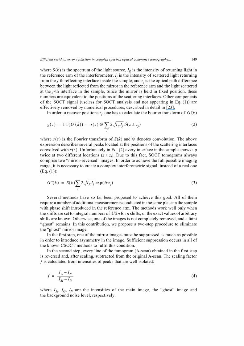
The method is computationally effective, as it consists only of a few multiplicationsand subtractions. The total procedure is presented in a pictorial way in Fig. 1.
The scaling factor f is computed separately for every A-scan. In practice, f iscalculated by minimaization of a fuction R( f ):
where Ak is the intensity at the k-th point of a smoothed A-scan obtained with complexmethod, AN – k is the intensity in its mirror image, and the function R( f ) representsthe level of correlation between the clean A-scan Vn and its mirror image VN – n.The function R( f ) reaches minimum if the ghosts are completely suppressed.
The main drawback of the method is a slight decrease in the signal-to-noise ratio.The signal-to-noise ratio of the main image before applying the method is given by:
(5)
R f( ) Vn VN n–n∑=
Vk Ak f AN k––=
SN
-------
uncorr
IM IN–
σ---------------------=
Fig. 1. Schematic of the second step of the proposed method. a – an A-scan obtained via Fouriertransformation of complex interferometric signal (see Eq. (3)). This procedure introduces asymmetryof intensity magnitudes in the main (IM ) and ‘mirror-reversed’ (IG ) images. The background noise levelis labeled IN. b – the scaled (Eq. (4)) and reversed A-scan from (a). c – the effect of subtraction of (b)from (a).
a
b
c
Efficient residual error reduction in complex spectral optical coherence tomography... 151
where σ is the variance of the noise. After applying the method, the signal-to-noiseratio decreases, and can be described as:
(6)
The above equation may be rewritten in the more convenient form:
(7)
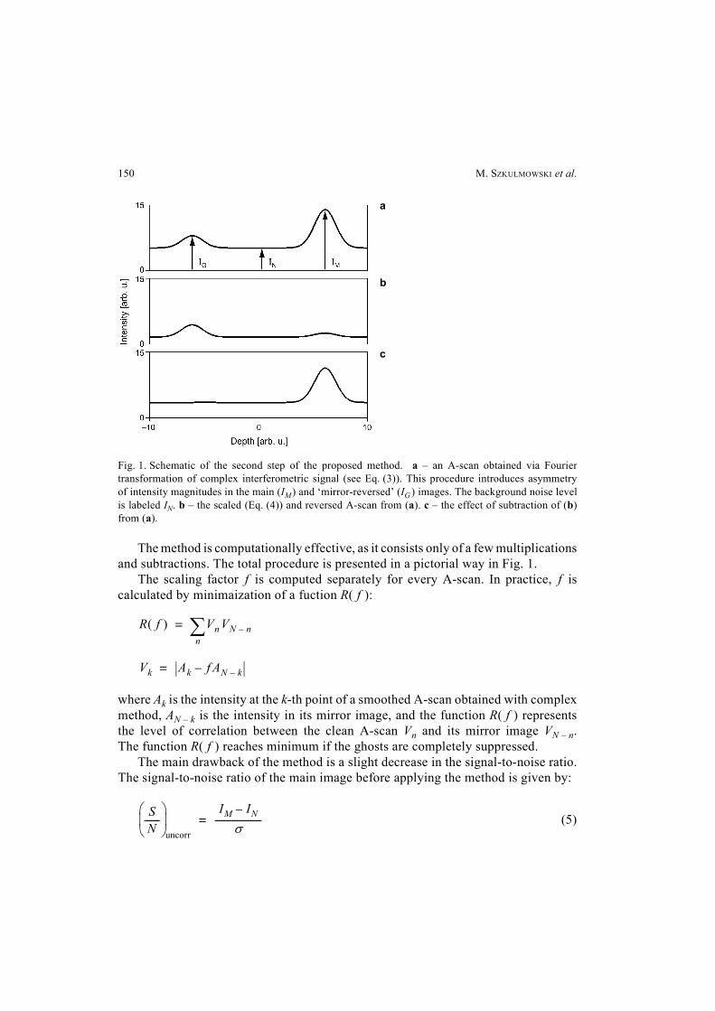
The value of (S/N)corr is a function of the scaling factor f, see Fig. 2.In order to obtain a high signal-to-noise ratio, one should try to attain as great
an intensity difference between the main image and the “ghost” as possible. It canhowever be seen that even when the scaling factor f is equal to 0.4, the decrease ofthe signal-to-noise ratio is only about 1 dB. Because the signal-to-noise ratioachievable by the SOCT technique is about 90 dB, [24] and tomograms are plotted ona logarithmic scale, this decrease of signal-to-noise ratio does not noticeably affectimage quality.
In order for the method to work properly, “mirror-reversed” images may differonly in amplitude: the shapes of the two images (defined by s (z), Eq. (2)) have to beidentical. If this is not the case, simple subtraction does not remove the “ghost” image.Therefore, the method cannot be applied to CSOCT tomograms if numeric dispersioncompensation has been applied [14].
SN
-------
corr
IM f IG– IN 1 f–( )–
σ 1 f2+
------------------------------------------------------=
SN
-------
corr
SN
-------
uncorr
1 f2–
1 f2+
-----------------------=
Fig. 2. Decrease of the signal-to-noise ratio of the CSOCT tomogram after applying the “ghost” imagereduction technique, as a function of the scaling factor f (Eq. (7)). The dashed lines show the –1 and–3 dB levels.
152 M. SZKULMOWSKI et al.
3. Results and discussion
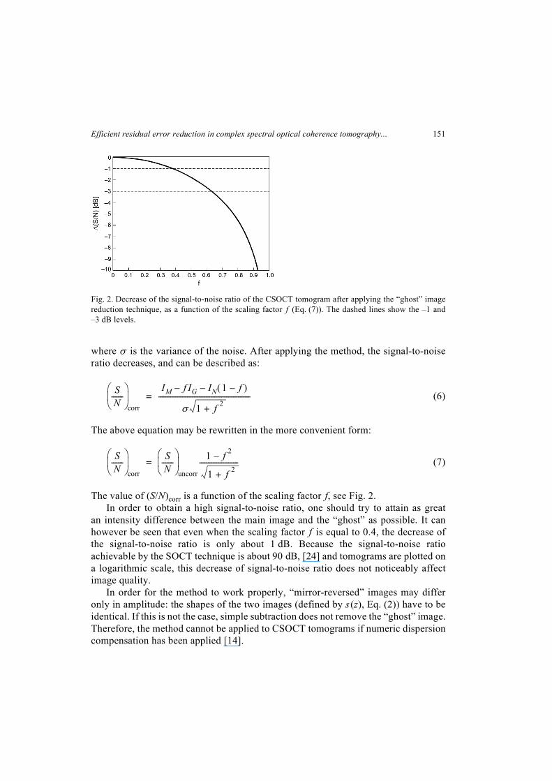
All results have been obtained with our in-house SOCT instrument based on an opticalfiber Michelson interferometer set-up (Fig. 3). The superluminescent diode with highspatial, but low temporal coherence is used as a light source (Superlum, Russia, centralwavelength 811 nm, full width at half maximum 20 nm). The light is split by the fibercoupler into reference and object arms. The mirror in the reference arm is fixed tothe piezotranslator (Physik Instrumente, Germany), which enables introduction ofpreselected delays. The object arm consists of a collimator, transversal scannersand custom designed interface optics, which allows for imaging the anterior part ofthe eye. The narrow beam of light penetrates the object, scatters from the elementsof its structure, then is collected by the same optics back to the coupler. Then,it interferes with the light returning from the reference arm and the spectrum ofthe interference signal is detected by a 12-bit line scan CCD camera and transferredto a personal computer.
In the work reported in this contribution, we used a two-frame method [18]. In thisapproach, two spectra G1(k ) and G2(k ), taken at the same point of the object, arecombined into a complex signal using the following relationship:
G'(k ) = G1(k ) + iG2(k ) (8)
The modulus of the inverse Fourier transform of G'(k ) gives a “ghost”-freetomogram providing the phase shift between G1(k ) and G2(k ) is exactly equal to π/2for every k and the phase error remains small. We tested a case where these conditionswere deliberately not fulfilled – for a phase shift between the two measurements setto 2π/3, so that a faint “ghost” image was present in the tomogram.
Fig. 3. Diagram of the instrument: LS – superluminescent light source, IO – fiber isolator, FC – fibercoupler, PCO – polarization controller, RM – reference mirror, PT – piezotranslator, X-Y – galvoscanners,DG – diffraction grating, CCD – line scan camera, PC – personal computer.
Efficient residual error reduction in complex spectral optical coherence tomography... 153
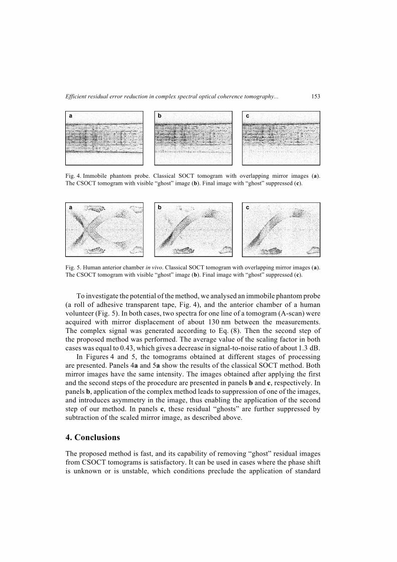
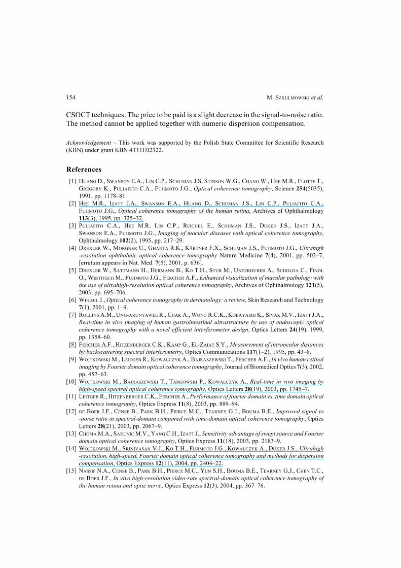
To investigate the potential of the method, we analysed an immobile phantom probe(a roll of adhesive transparent tape, Fig. 4), and the anterior chamber of a humanvolunteer (Fig. 5). In both cases, two spectra for one line of a tomogram (A-scan) wereacquired with mirror displacement of about 130 nm between the measurements.The complex signal was generated according to Eq. (8). Then the second step ofthe proposed method was performed. The average value of the scaling factor in bothcases was equal to 0.43, which gives a decrease in signal-to-noise ratio of about 1.3 dB.
In Figures 4 and 5, the tomograms obtained at different stages of processingare presented. Panels 4a and 5a show the results of the classical SOCT method. Bothmirror images have the same intensity. The images obtained after applying the firstand the second steps of the procedure are presented in panels b and c, respectively. Inpanels b, application of the complex method leads to suppression of one of the images,and introduces asymmetry in the image, thus enabling the application of the secondstep of our method. In panels c, these residual “ghosts” are further suppressed bysubtraction of the scaled mirror image, as described above.
4. Conclusions
The proposed method is fast, and its capability of removing “ghost” residual imagesfrom CSOCT tomograms is satisfactory. It can be used in cases where the phase shiftis unknown or is unstable, which conditions preclude the application of standard
Fig. 4. Immobile phantom probe. Classical SOCT tomogram with overlapping mirror images (a).The CSOCT tomogram with visible “ghost” image (b). Final image with “ghost” suppressed (c).
a b c
Fig. 5. Human anterior chamber in vivo. Classical SOCT tomogram with overlapping mirror images (a).The CSOCT tomogram with visible “ghost” image (b). Final image with “ghost” suppressed (c).
a b c
154 M. SZKULMOWSKI et al.
CSOCT techniques. The price to be paid is a slight decrease in the signal-to-noise ratio.The method cannot be applied together with numeric dispersion compensation.
Acknowledgement – This work was supported by the Polish State Committee for Scientific Research(KBN) under grant KBN 4T11E02322.
References
[1] HUANG D., SWANSON E.A., LIN C.P., SCHUMAN J.S, STINSON W.G., CHANG W., HEE M.R., FLOTTE T.,GREGORY K., PULIAFITO C.A., FUJIMOTO J.G., Optical coherence tomography, Science 254(5035),1991, pp. 1178–81.
[2] HEE M.R., IZATT J.A., SWANSON E.A., HUANG D., SCHUMAN J.S., LIN C.P., PULIAFITO C.A.,FUJIMOTO J.G., Optical coherence tomography of the human retina, Archives of Ophthalmology113(3), 1995, pp. 325–32.
[3] PULIAFITO C.A., HEE M.R, LIN C.P., REICHEL E., SCHUMAN J.S., DUKER J.S., IZATT J.A.,SWANSON E.A., FUJIMOTO J.G., Imaging of macular diseases with optical coherence tomography,Ophthalmology 102(2), 1995, pp. 217–29.
[4] DREXLER W., MORGNER U., GHANTA R.K., KÄRTNER F.X., SCHUMAN J.S., FUJIMOTO J.G., Ultrahigh-resolution ophthalmic optical coherence tomography Nature Medicine 7(4), 2001, pp. 502–7,[erratum appears in Nat. Med. 7(5), 2001, p. 636].
[5] DREXLER W., SATTMANN H., HERMANN B., KO T.H., STUR M., UNTERHUBER A., SCHOLDA C., FINDL
O., WIRTITSCH M., FUJIMOTO J.G., FERCHER A.F., Enhanced visualization of macular pathology withthe use of ultrahigh-resolution optical coherence tomography, Archives of Ophthalmology 121(5),2003, pp. 695–706.
[6] WELZEL J., Optical coherence tomography in dermatology: a review, Skin Research and Technology7(1), 2001, pp. 1–9.
[7] ROLLINS A.M., UNG-ARUNYAWEE R., CHAK A., WONG R.C.K., KOBAYASHI K., SIVAK M.V., IZATT J.A.,Real-time in vivo imaging of human gastrointestinal ultrastructure by use of endoscopic opticalcoherence tomography with a novel efficient interferometer design, Optics Letters 24(19), 1999,pp. 1358–60.
[8] FERCHER A.F., HITZENBERGER C.K., KAMP G., EL-ZAIAT S.Y., Measurement of intraocular distancesby backscattering spectral interferometry, Optics Communications 117(1–2), 1995, pp. 43–8.
[9] WOJTKOWSKI M., LEITGEB R., KOWALCZYK A., BAJRASZEWSKI T., FERCHER A.F., In vivo human retinalimaging by Fourier domain optical coherence tomography, Journal of Biomedical Optics 7(3), 2002,pp. 457–63.
[10] WOJTKOWSKI M., BAJRASZEWSKI T., TARGOWSKI P., KOWALCZYK A., Real-time in vivo imaging byhigh-speed spectral optical coherence tomography, Optics Letters 28(19), 2003, pp. 1745–7.
[11] LEITGEB R., HITZENBERGER C.K., FERCHER A., Performance of fourier domain vs. time domain opticalcoherence tomography, Optics Express 11(8), 2003, pp. 889–94.
[12] DE BOER J.F., CENSE B., PARK B.H., PIERCE M.C., TEARNEY G.J., BOUMA B.E., Improved signal-to-noise ratio in spectral-domain compared with time-domain optical coherence tomography, OpticsLetters 28(21), 2003, pp. 2067–9.
[13] CHOMA M.A., SARUNIC M.V., YANG C.H., IZATT J., Sensitivity advantage of swept source and Fourierdomain optical coherence tomography, Optics Express 11(18), 2003, pp. 2183–9.
[14] WOJTKOWSKI M., SRINIVASAN V.J., KO T.H., FUJIMOTO J.G., KOWALCZYK A., DUKER J.S., Ultrahigh-resolution, high-speed, Fourier domain optical coherence tomography and methods for dispersioncompensation, Optics Express 12(11), 2004, pp. 2404–22.
[15] NASSIF N.A., CENSE B., PARK B.H., PIERCE M.C., YUN S.H., BOUMA B.E., TEARNEY G.J., CHEN T.C.,DE BOER J.F., In vivo high-resolution video-rate spectral-domain optical coherence tomography ofthe human retina and optic nerve, Optics Express 12(3), 2004, pp. 367–76.
Efficient residual error reduction in complex spectral optical coherence tomography... 155
[16] WOJTKOWSKI M., KOWALCZYK A., LEITGEB R., FERCHER A.F., Autocorrelation free spectral OCTtechniques in eye imaging, Proceedings of the SPIE 4431, 2001, pp. 46–51.
[17] WOJTKOWSKI M., KOWALCZYK A., LEITGEB R., FERCHER A.F., Full range complex spectral opticalcoherence tomography technique in eye imaging, Optics Letters 27(16), 2002, pp. 1415–7.
[18] LEITGEB R., BAJRASZEWSKI T., HITZENBERGER C.K., FERCHER A.F., Novel phase-shifting algorithm toachieve high-speed long-depth range probing by frequency domain optical coherence tomography,Proceedings of the SPIE 4956, 2003, pp. 101–8.
[19] TARGOWSKI P., WOJTKOWSKI M., KOWALCZYK A., BAJRASZEWSKI T., SZKULMOWSKI M., GORCZYNSKA I.,Complex spectral OCT in human eye imaging in vivo, Optics Communications 229(1–6), 2004,pp. 79–84.
[20] YASUNO Y., MAKITA S., ENDO T., AOKI G., SUMIMURA H., ITOH M., YATAGI T., One-shot-phase-shifting Fourier domain optical coherence tomography by reference wavefront tilting, OpticsExpress 12(25), 2004, pp. 6184–91.
[21] CHOMA M.A., YANG C., IZATT J.A., Instantaneous quadrature low-coherence interferometry with3*3 fiber-optic couplers, Optics Letters 28(22), 2003, pp. 2162–4.
[22] TARGOWSKI P., GORCZYŃSKA I., SZKULMOWSKI M., WOJTKOWSKI M., KOWALCZYK A., Improvedcomplex spectral domain OCT for in vivo eye imaging, Optics Communications 249(1–3), 2005,pp. 357–62.
[23] SZKULMOWSKI M., WOJTKOWSKI M., BAJRASZEWSKI T., GORCZYŃSKA I., TARGOWSKI P., WASILEWSKI W.,KOWALCZYK A., RADZEWICZ C., Quality improvement for high resolution in vivo images by spectraldomain optical coherence tomography with supercontinuum source, Optics Communications246(4–6), 2005, pp. 569–78.
[24] SZKULMOWSKA A., WOJTKOWSKI M., GORCZYŃSKA I., BAJRASZEWSKI T., SZKULMOWSKI M.,TARGOWSKI P., KOWALCZYK A., KAŁUŻNY J.J., Coherent noise-free ophthalmic imaging by spectraloptical coherence tomography, Journal of Physics D: Applied Physics 38(15), 2005, pp. 2606–11.
Received June 23, 2005in revised form August 29, 2005
Related Documents