Effects of smoking cessation on lung function and airway inflammation in smokers with asthma Rekha Chaudhuri l , Eric Livingston l , Alex D McMahon 2 , Jane Lafferty l , Iona Fraser 3 , Mark Spears l , Charles P McSharry 3 , Neil C Thomson l Departments of Respiratory Medicine l and Immunology 3 , University of Glasgow and Robertson Centre for Biostatistics 2 , Glasgow, UK Correspondence: Professor Neil C Thomson Department of Respiratory Medicine Division of Immunology, Infection and Inflammation University of Glasgow and Western Infirmary Glasgow, G11 6NT Scotland, UK Telephone: 44-141-211-3241 Fax: 44-141-211-3464 E- mail: [email protected] Funded by: Asthma UK, Scottish Council for Postgraduate Medical and Dental Education and Chief Scientist Office, Chest Heart and Stroke, Scotland. Running head: Smoking cessation in smokers with asthma Descriptor number: 64 Word count: 3035 "This article has an online data supplement for the methods section, which is accessible from this issue's table of content online at www.atsjournals.org" AJRCCM Articles in Press. Published on April 27, 2006 as doi:10.1164/rccm.200510-1589OC Copyright (C) 2006 by the American Thoracic Society.

Welcome message from author
This document is posted to help you gain knowledge. Please leave a comment to let me know what you think about it! Share it to your friends and learn new things together.
Transcript
Effects of smoking cessation on lung function and airway inflammation
in smokers with asthma
Rekha Chaudhuri l, Eric Livingston l, Alex D McMahon 2, Jane Lafferty l,
Iona Fraser 3, Mark Spears l, Charles P McSharry 3, Neil C Thomson l
Departments of Respiratory Medicine l and Immunology 3, University of Glasgow
and Robertson Centre for Biostatistics2, Glasgow, UK
Correspondence:
Professor Neil C Thomson
Department of Respiratory Medicine
Division of Immunology, Infection and Inflammation
University of Glasgow and Western Infirmary
Glasgow, G11 6NT Scotland, UK
Telephone: 44-141-211-3241 Fax: 44-141-211-3464
E- mail: [email protected]
Funded by: Asthma UK, Scottish Council for Postgraduate Medical and Dental
Education and Chief Scientist Office, Chest Heart and Stroke, Scotland.
Running head: Smoking cessation in smokers with asthma
Descriptor number: 64
Word count: 3035
"This article has an online data supplement for the methods section, which is accessible
from this issue's table of content online at www.atsjournals.org"
AJRCCM Articles in Press. Published on April 27, 2006 as doi:10.1164/rccm.200510-1589OC
Copyright (C) 2006 by the American Thoracic Society.
1
ABSTRACT
Rationale: Active smoking in asthma is associated with worsening of symptoms, accelerated
decline in lung function and impaired response to corticosteroids.
Objectives: To examine the short-term effects of smoking cessation on lung function, airway
inflammation and corticosteroid responsiveness in smokers with asthma.
Methods and measurements: Smokers with asthma were given the option to quit or continue
smoking. Both groups underwent spirometry and induced sputum at baseline and at one, three
and six weeks. Cutaneous vasoconstrictor response to topical beclometasone, airway response to
oral prednisolone and sensitivity of peripheral blood lymphocytes to corticosteroids were
measured before smoking cessation and at six weeks.
Main results: Of 32 subjects recruited, 11 opted to continue smoking (smoking controls).
Twenty-one subjects opted for smoking cessation of whom ten quit smoking for six weeks (quit
group). Comparing quitters with controls at six weeks, the mean (Cl) difference in FEV1 was
407 ml (21,793), p=0.040; and the proportion of sputum neutrophils reduced by -29 (-51, -8),
p=0.039. Total cutaneous vasoconstrictor response score to topical beclometasone improved
following smoking cessation with a mean (CI) difference of 3.56 (0.84, 6.28), p=0.042
comparing quitters with control smokers. There was no change in airway corticosteroid
responses after smoking cessation.
Conclusions: Six weeks after smoking cessation, smokers with asthma achieved considerable
improvement in lung function and a fall in sputum neutrophil count compared to smokers who
continued to smoke. These findings highlight the importance of smoking cessation in asthma.
Word count: 236
Key words: Smoking cessation; smoking; asthma; lung function; airway inflammation
2
INTRODUCTION
Active cigarette smoking has detrimental effects on asthma morbidity. Compared to non-
smokers with asthma, smokers have more severe symptoms (1, 2), increased rates of
hospitalization (3), accelerated decline in lung function (4, 5) and impaired therapeutic responses
to inhaled (6, 7) and oral corticosteroids (8). Furthermore, the prevalence rates for cigarette
smoking in individuals with asthma are similar to that of the general population (9), and in many
developed countries over 20% of adults with asthma are active smokers (1, 2, 9-12). Particularly
high rates have been noted in adults presenting to hospital emergency departments with acute
asthma (3). There is limited information on the effects of cigarette smoking on airway
inflammation in asthma (13). Sputum neutrophil counts are reported to be increased in heavy
smokers with mild asthma compared to non-smokers with asthma (14), and sputum
concentrations of cytokines such as IL-8 are raised (14) and others such as IL-18 are suppressed
(15) in smokers with asthma.
Despite its importance, there is limited published data on the effects of smoking cessation on
symptoms, lung function and corticosteroid responsiveness in smokers with asthma. In an
uncontrolled study, seven subjects who quit smoking for a week showed improvements in
symptoms and lung function (16). In a longer term study, a prospective cohort of smokers with
asthma reported improvements in symptoms and bronchial hyperreactivity after four months of
smoking cessation (17). In ten subjects with asthma who were ex-smokers for at least a year, the
therapeutic response to oral corticosteroids was midway between that of current smokers and
never smokers; suggesting that smoking cessation may partially restore corticosteroid
responsiveness (8).
3
The effect of smoking cessation on airway inflammation in healthy smokers, shows a dose-
dependent relationship between smoking and airway inflammation (18). In these subjects, two
months smoking reduction lead to a significant fall in bronchoalveolar lavage neutrophil and
macrophage counts (19). In contrast to the improvement in airway inflammation found in
healthy smokers after smoking cessation, there is minimal change in airway inflammation in
patients with COPD after quitting smoking (20-22), and the effect of smoking cessation on
airway inflammation in smokers with asthma is not known.
Our hypothesis is that smoking cessation improves lung function, reduces airway inflammation
and restores corticosteroid responsiveness in smokers with asthma. The aim of the study was to
prospectively study smokers with asthma who successfully quit smoking and compare outcome
measures with asthmatic smokers who continue to smoke; measurements included lung
function, asthma control score, induced sputum cell counts and mediator levels. A secondary
aim was to assess the airway, cutaneous and lymphocyte sensitivity to corticosteroids after
smoking cessation. Some of the results have been reported in abstract form (23).
METHODS
Subjects
Smokers with asthma [ATS criteria, (24)] aged 18-60 years, baseline FEV1 ≤85% predicted, ≥
15% reversibility of FEV1 after nebulised albuterol, a smoking history of ≥ 10 pack years and
currently smoking ≥ 10 cigarettes/day were recruited. The study was approved by the West
Glasgow Ethics Committee.
Study design
This was a prospective, controlled study. Airway corticosteroid sensitivity to oral prednisolone,
40 mg daily for 14 days, was assessed in all subjects by change in pre-albuterol FEV1, asthma
4
control score, morning and evening PEF, daily morning and night symptoms and use of β-
agonist inhalers. Subjects were recalled to discuss smoking cessation, 2-12 months after the
corticosteroid trial. Spirometry, induced sputum, exhaled nitric oxide (FENO), exhaled carbon
monoxide (CO), asthma control score, skin vasoconstrictor response to beclometasone, serum
cotinine and peripheral blood lymphocyte proliferation assay (LP) were performed. All subjects
were given the option to quit or continue smoking. Both groups were issued diary cards. Visits
were arranged one, three and six weeks later. Following the six-week visit, both groups
received a second course of oral prednisolone 40mg daily for 14 days, with similar end-points
used to compare results. Spirometry, exhaled gases and asthma control score were recorded at
all visits; induced sputum at three and six weeks; cutaneous vasoconstrictor test and LP at six
weeks. Patient adherence to smoking cessation was accepted by patient history.
Measurements
ATS asthma impairment score (25) and a validated asthma control questionnaire (26) were
recorded. Patients maintained a validated diary card (27), recording morning and night PEF
(Mini-Wright, Clement Clarke, Harlow, UK), daytime symptoms, night awakenings, use of
inhaled rescue medication and study tablet consumption. Spirometry was measured with a dry
spirometer (Vitalograph Ltd, Buckingham, UK). Sputum was induced as previously described
(14, 28, 29). FENO and CO were measured using a chemiluminescence analyzer (Logan
Research Ltd, Rochester, UK) (30) and serum cotinine by enzyme immunoassay (Cozart
Bioscience Ltd, Abingdon, UK). Treatment compliance was assessed by tablet count.
Cutaneous vasoconstrictor response to topical beclometasone was measured as described
previously (31, 32). Concentrations of 0, 1, 3, 10, 30, 100, 300 and 1000 µg/ml were applied to
the skin in a random double-blind manner and the degree of blanching assessed visually after 18
hours by a single trained observer. Blanching at each concentration was graded according to a 4-
5
point scale [0-3] and a total score calculated [0-24]. Sensitivity of peripheral blood T-
lymphocytes to glucocorticoids was assessed in a functional assay as described previously (33).
The percentage suppression at the final concentration of corticosteroid used was defined as the
maximum inhibitory dose [Imax %].
Statistical analysis
Baseline characteristics were compared by chi-squared, Wilcoxon and t-tests. Response to
smoking cessation on lung function, diary data, induced sputum, mediator levels, skin
vasoconstrictor response and oral prednisolone trials for smokers versus never smokers with
asthma was assessed by Analysis of Covariance models that adjusted each factor by its baseline
measure. Correlations were assessed by Spearman’s rank correlations. Only those subjects who
completed the study up to the six week visit were included in the analysis. Significance at a level
of 5% was considered for the primary end-point; the change in FEV1 at six weeks. Tests were
performed using SAS version 9.0 (Cary, NC, USA).
RESULTS
Recruitment
The flow of participants through the study is depicted in Figure 1. 11/32 subjects opted to
continue smoking (smoking controls). Of the 21 who opted to attempt quitting smoking, 14
achieved 1 week off and 10 completed six weeks of smoking cessation.
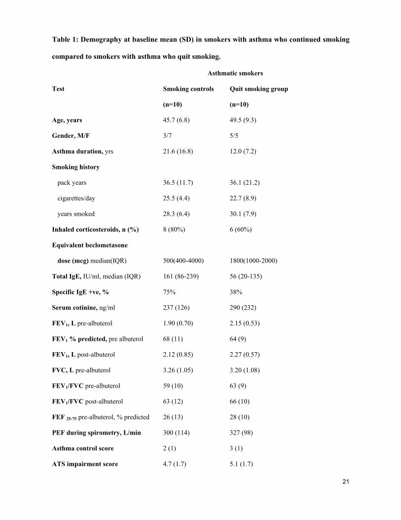
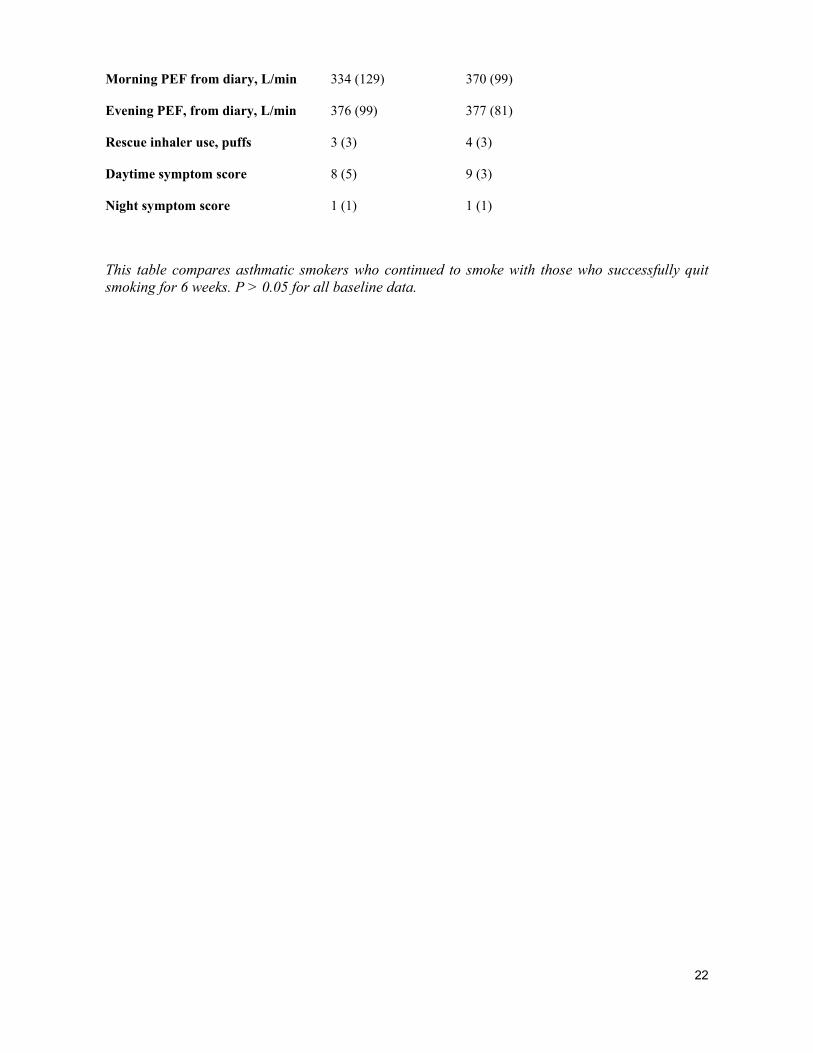
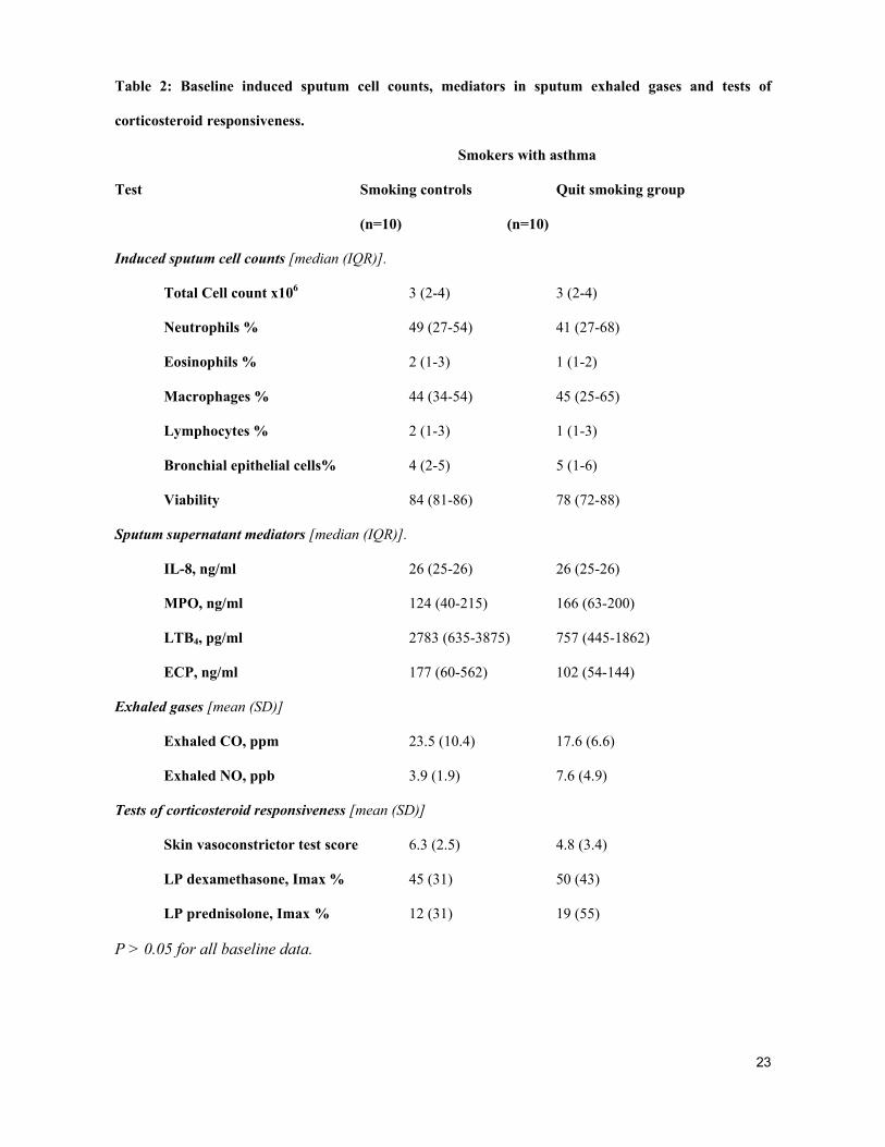
Baseline characteristics
There were no differences in age, duration of asthma, equivalent dose of inhaled beclometasone,
asthma control score, ATS impairment score, IgE levels, spirometry, skin vasoconstrictor test
6
score, inhibitory dose of dexamethasone maximally suppressing lymphocyte proliferation in
vitro, induced sputum cell counts and inflammatory mediators between the smoking control and
quit group. Mean (SD) baseline FEV1 percentage predicted was 68 (11) in the quit group and 64
(94) in the controls [Tables 1 and 2]. Baseline reversibility of FEV1 to inhaled albuterol [mean%
(SD)] was 25 (11) in the quit group and 19 (6) in the control group. Age, gender distribution,
atopic status, smoking history, duration of asthma and serum cotinine levels were similar in the
successful quitters and those subjects who opted for the quit group but were unable to achieve a
week off smoking (n=7). In the 10 subjects who successfully quit smoking and completed the
study, five used nicotine patches, one subject used acupuncture and four quit without any aid.
Change in lung function, asthma control and airway inflammatory markers with smoking
cessation
Lung function: The mean (SD) change in FEV1, ml in the quit group was 356 (278) at 1 week,
390 (311) at 3 weeks, and 450 (471) at 6 weeks of smoking cessation, p values 0.015, 0.009 and
0.031 respectively. There was no difference in the equivalent FEV1 measures in the smoking
control group. Comparing quitters with control smokers at six weeks’ cessation, there was a
mean improvement of 407 ml in FEV1, 15.2% in FEV1% predicted and 93L/min in PEF (Figure
2, Table 3). There was no correlation with the improvement in FEV1 and age, gender, atopy,
duration of smoking, pack years smoked, dose of inhaled corticosteroids and sputum cell counts.
Asthma control: The mean (CI) asthma control score showed an improvement at six weeks after
smoking cessation of -1.2 (-2.2,-0.2), p=0.021 (Table 3).
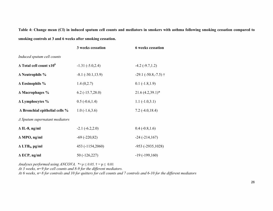
Induced sputum: Quitters showed a decrease in proportion of sputum neutrophils compared to
controls at six weeks [mean % difference (CI) -29 (-51, -8), p=0.013; with no change in
inflammatory mediator levels (Table 4). The mean (CI) difference for absolute neutrophil
count in sputum at three weeks was -52 (-141, 36), p=0.224 and at six weeks cessation it was
-121 (-215, -26), p=0.017. The degree of improvement in FEV1 was not related to the
7
baseline neutrophil level [r= 0.24, p=0.248] or the change in neutrophil proportion [r= -0.11,
p=0.819]. There was no difference in mediator response in those quitters who used nicotine
replacement therapy compared to those who did not.
Exhaled gases: Compared to the control smokers the quit group showed a reduction in exhaled
CO at one, three and six weeks. (Table 3). There was no change in exhaled NO levels.
Response to corticosteroids
Airway response: There was no difference in the FEV1 response to oral prednisolone while
smoking or at eight weeks after smoking cessation in the control smokers with asthma compared
to quitters. The mean (CI) change in FEV1, ml with the first prednisolone course when subjects
were smoking was 105 (-231,441), p=0.518 for the quit group versus smoking control group and
after eight weeks of smoking cessation, the difference was 190 (-121, 501), p=0.214.
Compliance with medication was 89% in controls and 100% in the quit group for the first
corticosteroid course and 89% in controls and 100% in quitters with the second course.
Cutaneous vasoconstrictor response to topical beclometasone: The mean (CI) total cutaneous
vasoconstrictor response score to topical beclometasone improved following smoking cessation
with a mean difference of 3.56 (0.84, 6.28), p=0.014; comparing quitters with control smokers.
Lymphocyte proliferation response: There was no difference in the lymphocyte proliferation
response after six weeks of smoking cessation. The mean difference (CI) Imax % for
dexamethasone suppression of lymphocyte proliferation was -9.3 (-28.4, 9.9), p=0.322 for
quitters compared to smokers and for prednisolone suppression was -4.0 (-17.1, 9.0); p=0.522.
DISCUSSION
Active cigarette smoking is known to worsen the severity of asthma (13). In this study we have
demonstrated that lung function of smokers with asthma improves by a considerable degree
8
within a week of stopping smoking. This improvement in FEV1 [mean (CI)] at one week was of
the order of 396 (129,664) ml compared to those that continued to smoke and this improvement
increased up to six weeks after smoking cessation. In addition, there was a fall in the sputum
neutrophil counts, evident at six weeks after smoking cessation.
Subjects in the quit group progressed through the study unless they admitted to smoking. This
method of assessment of smoking status is known to overestimate the number of subjects who
effectively quit smoking. Due to the short half-life, only daily measurements of carbon
monoxide or cotinine could have provided objective evidence for smoking cessation in between
clinic visits. Cotinine can be affected by nicotine replacement therapy used for cessation and
daily measurements for eight weeks were not feasible. However the reduction in exhaled carbon
monoxide in the quit group compared to controls at all visits provided objective evidence for
effective smoking cessation, at least for the previous 24-48 hours prior to each clinic
assessment. If some of the subjects in the quit group had not stopped smoking completely, the
results obtained may underestimate the extent of the improvement in lung function and
neutrophil count possible with smoking cessation.
The improvement in lung function seen following smoking cessation was clinically significant.
This demonstrates that there is a reversible component to the harmful effects of smoking on the
airways in asthma. The degree of improvement noted by smoking cessation far exceeds that of
high dose anti-inflammatory treatment, such as oral prednisolone 40 mg daily for 2 weeks,
which had no effect on lung function in smokers in our current study and in our previous work
(8). The improvement in lung function could be due to removal of the acute bronchoconstrictor
effects of cigarette smoke (34) or a reduction in the pro-inflammatory effects of cigarette smoke
on the airways (14). It is also possible that stopping smoking leads to a reduction in
corticosteroid insensitivity, as ex-smokers who had quit smoking for at least one year have a
9
better response to oral prednisolone compared to current smokers (8). Reduced histone
deacetylase activity is one of the possible mechanisms of corticosteroid insensitivity in smokers
(34), but there are no reports of measurement of HDAC levels following smoking cessation.
In asthma, there have been two previous studies looking at the effect of smoking cessation on
lung function. The first was a small uncontrolled study where 7 subjects quit smoking for a week
and showed improvements in PEF, symptoms and specific airways conductance (16). There was
a reduction in histamine airway responsiveness after 24 hours of smoking cessation (16). A
prospective cohort study compared the effects of smoking cessation, smoking reduction and
continuing smoking on asthma control and biomarkers of exposure to cigarette smoke (17). This
study showed a significant improvement in symptoms and bronchial hyperreactivity after four
months of smoking cessation, but no change in FEV1. The baseline FEV1 % predicted was much
higher than in our subjects and hence it is possible that scope for further improvement was
limited.
In our study, although the asthma control score improved, we did not obtain improvement in
symptoms in the diary card up to two months after cessation. In a study by Hillerdahl and
Rylander, two thirds of subjects with asthma who quit smoking reported no improvement or
worsening of symptoms after smoking cessation (35). A cross-sectional study of 3197
smokers, ex-smokers (stopped smoking for at least one year) and non-smokers with asthma,
reported that ex-smokers had significantly less cough, wheeze, night symptoms and sputum
production compared to current smokers, but a similar degree of shortness of breath (36). This
implies that many, but not all asthma symptoms can return to the level of never-smokers with
smoking cessation.
10
The raised sputum neutrophil count found in high intensity smokers with asthma (14, 37)
may be partly responsible for their reduced responsiveness to corticosteroids. Unlike
eosinophils, which are exquisitely sensitive to corticosteroids, neutrophils are poorly
responsive to corticosteroid therapy (37) and their survival and proliferation is promoted by
glucocorticoids (38). We demonstrated a reduction in induced sputum neutrophils with
smoking cessation, but no change in mediator levels. We are unaware of any previous
published studies that have measured airway inflammation in smokers with asthma after
smoking cessation. In healthy heavy smokers bronchoalveolar lavage neutrophil counts were
reduced two months after smoking reduction (19). In contrast, the effect of smoking cessation
on airway inflammation in COPD has shown that most inflammatory cells, including
neutrophils, persist in ex-smokers (20, 22, 39) and can even increase (40). The mechanisms
by which the neutrophil count is reduced following smoking cessation are unclear at present.
It seems unrelated to the continued presence of neutrophil chemo-attractants LTB4 and IL-8
in the airways, and may therefore be related to reduced diapedesis following the down-
regulatory effects of smoking cessation on adhesion molecules (41), perhaps in addition to
more efficient apoptosis of neutrophils by alveolar macrophages after smoking cessation (42,
43).
The short-term improvement in FEV1 was not related to the change in neutrophil count, which
might suggest that the removal of bronchoconstrictor effects of cigarette smoke is more
important than neutrophils themselves; but the small number of subjects studied may explain our
findings. The levels of the inflammatory mediators in induced sputum did not change at six
weeks following smoking cessation. This result is similar to the finding in COPD, where IL-8
levels in sputum were similar in smokers and ex-smokers (44) and chronic bronchitis, where
BAL levels of TNF-alpha were unaltered by smoking cessation (22). It is possible that clinical
improvement may precede changes in inflammation, as in the third national health and nutrition
11
examination survey (NHANES III), inflammatory markers (CRP, white cell count, fibrinogen
and albumin) resolved more slowly than traditional cardiovascular risk factors (45).
Smoking cessation did not improve corticosteroid responsiveness measured by changes in FEV1
and PEF. However, there was a large improvement in baseline lung function with smoking
cessation and it is possible that this reduced the scope for further improvement in lung function,
after corticosteroids. In a previous study, ex-smokers with asthma who had quit smoking for at
least a year showed an improvement in morning PEF, but not FEV1 after high dose oral
prednisolone compared to placebo (8).
The skin vasoconstrictor response to topical beclometasone (32, 46) is based on the ability of
corticosteroids to cause transient vasoconstriction and skin blanching, has been used as a
screening test to determine the relative anti-inflammatory potency of inhaled corticosteroids (47)
and as an index of systemic sensitivity to corticosteroids (31). Objective methods of detecting
glucocorticoid-induced skin blanching have been compared to the visual scoring system, but the
human eye has been found to be the most sensitive tool to measure dermal blanching (32). The
cutaneous vasoconstrictor test for corticosteroid sensitivity showed an improvement after six
weeks of cessation implying some restoration in peripheral corticosteroid sensitivity within this
period. The mechanism for skin vasoconstriction with corticosteroids is not known, but a
possible explanation is that glucocorticoids increase the sensitivity of vascular smooth muscle to
the vasoconstrictor effects of noradrenaline (48). The blanching is possibly mediated through
glucocorticoid receptors, since oral administration of RU 486, a glucocorticoid receptor
antagonist, can abolish this response (49). Smoking causes vascular dysfunction but the additive
effect of nicotine on corticosteroids has not been studied in skin vasculature. The lymphocyte
proliferative response did not alter with smoking cessation in our study. A lack of correlation
between different tests of tissue sensitivity to corticosteroids has been reported previously in
12
health human volunteers (50).
In conclusion, in smokers with asthma, improvement in lung function occurs as early as one
week after smoking cessation with a further improvement up to six weeks. There is a reduction
of sputum neutrophil percentage after six weeks of smoking cessation but no change in common
inflammatory mediator levels. These findings highlight the importance of smoking cessation in
asthma.
13
ACKNOWLEDGEMENTS
We are grateful to Kathy McFall, medical illustration department, for assistance with the
figures.
14
REFERENCES
1. Siroux V, Pin I, Oryszcyn MP, Le Moual N, Kauffmann F. Relationships of active
smoking to asthma and asthma severity in the EGEA study. Eur Respir J 2000;15:470-477.
2. Althuis M, Sexton M, Prybylski D. Cigarette smoking and asthma symptom severity
among adult asthmatics. J Asthma 1999;36(3):257-264.
3. Silverman RA, Boudreaux ED, Woodruff PG, Clark S, Camargo CA, Jr. Cigarette
smoking among asthmatic adults presenting to 64 emergency departments. Chest
2003;123(5):1472-1479.
4. Apostol G, Jacobs D, Tsai A, et al. Early life factors contribute to the decrease in lung
function between ages 18 and 40. Am J Respir Crit Care Med 2002;166:166-172.
5. Lange P, Parner J, Vestbo J, Schnohr P, Jensen G. A 15 year follow-up study of
ventilatory function in adults with asthma. N Engl J Med 1998;339:1194-1200.
6. Chalmers GW, Macleod KJ, Little SA, Thomson LJ, McSharry CP, Thomson NC.
Influence of cigarette smoking on inhaled corticosteroid treatment in mild asthma. Thorax
2002;57(3):226-230.
7. Tomlinson JEM, McMahon AD, Chaudhuri R, Thompson JM, Wood SF, Thomson
NC. Efficacy of low and high dose inhaled corticosteroid in smokers versus non-smokers
with mild asthma. Thorax 2005;60(4):282-287.
8. Chaudhuri R, Livingston E, McMahon AD, Thomson L, Borland W, Thomson NC.
Cigarette smoking impairs the therapeutic response to oral corticosteroids in chronic
asthma. Am J Respir Crit Care Med 2003;168(11):1308-1311.
9. Lemiere C, Boulet L-P. Cigarette smoking and asthma: A dangerous mix. Can Respir J
2005;12:79-80.
10. Sippel JM, Pedula KL, Vollmer WM, Buist AS, Osborne ML. Associations of smoking
with hospital-based care and quality of life in patients with obstructive airway disease.
15
Chest 1999;115(3):691-696.
11. Turner M, Noertjojo K, Vedal S, Bai T, Crump S, Fitzgerald J. Risk factors for near
fatal asthma A case-control study in hospitalized patients with asthma. Am J Respir Crit
Care Med 1998;157:1804-1809.
12. Walsh LJ, Wong CA, Cooper S, Guhan AR, Pringle M, Tattersfield AE. Morbidity
from asthma in relation to regular treatment: a community based study. Thorax
1999;54(4):296-300.
13. Thomson NC, Chaudhuri R, Livingston E. Asthma and cigarette smoking. Eur Respir J
2004;24(5):822-833.
14. Chalmers G, MacLeod K, Thomson L, Little S, McSharry C, Thomson N. Smoking
and airway inflammation in patients with mild asthma. Chest 2001;120(6):1917-1922.
15. McKay A, Komai-Koma M, MacLeod K, et al. Interleukin-18 levels in induced sputum
are reduced in asthmatic and normal smokers. Clin Exp Allergy 2004;34:904-910.
16. Fennerty A, Banks J, Ebden P, Bevan C. The effect of cigarette withdrawal on
asthmatics who smoke. Eur J Repir Dis 1987;71(5):395-399.
17. Tonnesen P, Pisinger C, Hvidberg S, et al. Effects of smoking cessation and reduction in
asthmatics. Nicotine & Tobacco Research 2005;7(1):139-48.
18. Kuschner W, D'Alessandro A, Wong H, al. e. Dose-dependent cigarette smoking-related
inflammatory responses in health adults. Eur Respir J 1996;9:1989-1944.
19. Rennard S, Daughton D, Fujita J, et al. Short-term smoking reduction is associated with
reduction in measures of lower respiratory tract inflammation in heavy smokers. Eur
Respir J 1990;3:752-759.
20. Domagala-Kulawik J, Maskey-Warzechowska M, Kraszewska I, Chazan R. The
cellular composition and macrophage phenotype in induced sputum in smokers and ex-
smokers. Chest 2003;123:1054-1059.
21. Willemse BWM, ten Hacken NHT, Rutgers B, Lesman-Leegte IGAT, Timens W,
16
Postma DS. Smoking cessation improves both direct and indirect airway
hyperresponsiveness in COPD. Eur Respir J 2004;24(3):391-396.
22. Turato G, Di Stefano P, Maestrelli P, et al. Effect of smoking cessation on airway
inflammation in chronic bronchitis. Am J Respir Crit Care Med 1995;152(4 I):1262-1267.
23. Chaudhuri R, Livingston E, McMahon A, et al. Smoking cessation improves lung
function in smokers with asthma. Proceedings of American Thoracic Society.
2005;2:A625.
24. ATS. Standards for the diagnosis and care of patients with chronic obstructive pulmonary
disease (COPD) and asthma. Am Rev Resp Dis 1987;136:225-234.
25. ATS. Guidelines for the evaluation of impairment/disability in patients with asthma. Am
Rev Resp Dis 1993;147:1056-1061.
26. Juniper E, O'Byrne P, Guyatt G, Ferrie P, King D. Development and validation of a
questionnaire to measure asthma control. Eur Respir J 1999;14:902-907.
27. Santanello N, Barber B, Reiss T, Friedman B, Juniper E, Zhang J. Measurement
characteristics of two asthma symptom diary scales for use in clinical trials. Eur Respir J
1997;10:646-651.
28. Marquette C, Saulnier F, Leroy O, et al. Long-term prognosis of near-fatal asthma. Am
Rev Resp Dis 1992;146:76-81.
29. Popov T, Gottschalk R, Kolendowicz R, Dolovich J, Powers P, Hargreave F. The
evaluation of a cell dispersion method of sputum examination. Clin Exp Allergy
1994;24:778-783.
30. Kharitonov S, Alving K, Barnes P. Exhaled and nasal nitric oxide measurements:
recommendations. Eur Respir J 1997;10:1683-1693.
31. Brown PH, Teelucksingh S, Matusiewicz SP, Greening AP, Crompton GK, Edwards
CRW. Cutaneous vasoconstrictor response to glucocorticoids in asthma. Lancet
1991;337:576-80.
17
32. Noon PJ, Evans CE, Haynes WG, Webb DJ, Walker BR. A comparison of techniques
to assess skin blanching following the topical application of glucocorticoids. Br J Dermatol
1996;134:837-842.
33. Corrigan C, Brown P, Barnes N, et al. Glucocorticoid resistance in chronic asthma:
Glucocorticoid pharmacokinetics, glucocorticoid receptor characteristics, and inhibition of
peripheral blood T cell proliferation by glucocorticoids in vitro. Am Rev Respir Dis
1991;144:1016-1025.
34. Barnes P. Reduced Histone Deacetylase in COPD: Clinical Implications. Chest
2006;129(1):151-155.
35. Hillerdahl G, Rylander R. Asthma and smoking cessation. Clin Allergy 1984;14:45-47.
36. Suzuki K, Tanaka H, Kaneko S, et al. Respiratory symptoms and cigarette smoking in
3,197 pulmonologist-based asthmatic patients with a highly prevalent use of inhaled
corticosteroid. J Asthma 2003;40(3):243-50.
37. Green RH, Brightling CE, Woltmann G, Parker D, Wardlaw AJ, Pavord ID. Analysis
of induced sputum in adults with asthma: identification of subgroup with isolated sputum
neutrophilia and poor response to inhaled corticosteroids. Thorax 2002;57(10):875-879.
38. Meagher L, Cousin J, Secki J, Haslett C. Opposing effects of glucocorticoids on the rate
of apoptosis in neutrophilic and eosinophilic granulocytes. J Immunol 1996;156:4422-
4428.
39. Willemse B, Postma DS, Timens W, ten Hacken N. The impact of smoking cessation on
respiratory symptoms, lung function, airway hyperresponsiveness and inflammation. Eur
Respir J 2004;23:464-476.
40. Willemse B, ten Hacken N, Rutgers B, Lesman-Leegte I, Postma D, Timens W. Effect
of 1-year smoking cessation on airway inflammation in COPD and asymptomatic smokers.
Eur Resp J 2005;26(5):835-845.
41. Palmer R, Stapleton J, Sutherland G, Coward P, Wilson R, Scott D. Effect of nicotine

18
replacement and quitting smoking on circulating adhesion molecule profiles (sICAM-1,
sCD44v5, sCD44v6). Eur J Clin Invest 2002;32:852-857.
42. Kotani N, Kushikata T, Hashimoto H, Sessler D, Muraoka M, Matsuki A. Recovery of
intraoperative microbicidal and inflammatory functions of alveolar immune cells after a
tobacco smoke-free period. Anesthesiology 2001;94:999-1006.
43. Kirkham P, Spooner G, Rahman I, Rossi A. Macrophage phagocytosis of apoptotic
neutrophils is compromised by matrix proteins modified by
cigarette smoke and lipid peroxidation products. Biochem Biophys Res Commun
2004;318:32-37.
44. Vernooy J, Kucukaycan M, Jacobs D, et al. Local and systemic inflammation in patients
wiht chronic obstructive pulmonary disease; soluble tumor necrosis factor receptors are
increased in sputum. Am J Respir Crit Care Med 2002;166:1218-1224.
45. Bakhru A, Erlinger T. Smoking cessation and cardiovascular disease risk factors: results
from the third national health and nutrition examination survey. PLoS Medicine/Public
Library of Science 2005;2(6 e160):528-536.
46. McKenzie A, Stoughton R. Method for comparing percutanous absorption of steroids.
Arch Dermatol 1962;86:608-610.
47. Johnson M. Development of fluticasone proprionate and comparison with other inhaled
corticosteroids. J Allergy Clinical Immunology. 1998;101(Number 4, Part 2):S434-S439.
48. Horvath G, Lieb T, Conner GE, Salathe M, Wanner A. Steroid Sensitivity of
Norepinephrine Uptake by Human Bronchial Arterial and Rabbit Aortic Smooth Muscle
Cells. Am J Respir Cell Mol Biol 2001;25(4):500-506.
49. Gaillard R, Poffet D, Riondel A, Saurat J. RU 486 inhibits peripheral effects of
glucocorticoids in humans. J Clin Endocrinol Metab 1985;61:1009-1011.
50. Hearing S, Norman M, Smyth C, Foy C, Dayan C. Wide variation in lymphocyte steroid
sensitivity among healthy human volunteers. J Clin Endocrinol Metab 1999;84(11):4149-
19
4154.
51. Juniper E, Svensson K, Mork A, Stahl E. Measurement properties and interpretation of
three shortened versions of the asthma control questionnaire. Resp Med 2005;99:553-558.
52. Johansson S, Hourihane JB, Bousquet J, et al. A revised nomenclature for allergy: An
EAACI position statement from the EACCI nomenclature task force. Allergy
2001;56:813-849.
20
Figure Legends
Figure 1
Flow of subjects through the study
Figure 2
Mean (95% CI) difference between quitters and control asthmatic smokers in change in FEV1 (ml)
compared to baseline.
21
Table 1: Demography at baseline mean (SD) in smokers with asthma who continued smoking
compared to smokers with asthma who quit smoking.
Asthmatic smokers
Test Smoking controls Quit smoking group
(n=10) (n=10)
Age, years 45.7 (6.8) 49.5 (9.3)
Gender, M/F 3/7 5/5
Asthma duration, yrs 21.6 (16.8) 12.0 (7.2)
Smoking history
pack years 36.5 (11.7) 36.1 (21.2)
cigarettes/day 25.5 (4.4) 22.7 (8.9)
years smoked 28.3 (6.4) 30.1 (7.9)
Inhaled corticosteroids, n (%) 8 (80%) 6 (60%)
Equivalent beclometasone
dose (mcg) median(IQR) 500(400-4000) 1800(1000-2000)
Total IgE, IU/ml, median (IQR) 161 (86-239) 56 (20-135)
Specific IgE +ve, % 75% 38%
Serum cotinine, ng/ml 237 (126) 290 (232)
FEV1, L pre-albuterol 1.90 (0.70) 2.15 (0.53)
FEV1 % predicted, pre albuterol 68 (11) 64 (9)
FEV1, L post-albuterol 2.12 (0.85) 2.27 (0.57)
FVC, L pre-albuterol 3.26 (1.05) 3.20 (1.08)
FEV1/FVC pre-albuterol 59 (10) 63 (9)
FEV1/FVC post-albuterol 63 (12) 66 (10)
FEF 25-75 pre-albuterol, % predicted 26 (13) 28 (10)
PEF during spirometry, L/min 300 (114) 327 (98)
Asthma control score 2 (1) 3 (1)
ATS impairment score 4.7 (1.7) 5.1 (1.7)
22
Morning PEF from diary, L/min 334 (129) 370 (99)
Evening PEF, from diary, L/min 376 (99) 377 (81)
Rescue inhaler use, puffs 3 (3) 4 (3)
Daytime symptom score 8 (5) 9 (3)
Night symptom score 1 (1) 1 (1)
This table compares asthmatic smokers who continued to smoke with those who successfully quit smoking for 6 weeks. P > 0.05 for all baseline data.
23
Table 2: Baseline induced sputum cell counts, mediators in sputum exhaled gases and tests of
corticosteroid responsiveness.
Smokers with asthma
Test Smoking controls Quit smoking group
(n=10) (n=10)
Induced sputum cell counts [median (IQR)].
Total Cell count x106 3 (2-4) 3 (2-4)
Neutrophils % 49 (27-54) 41 (27-68)
Eosinophils % 2 (1-3) 1 (1-2)
Macrophages % 44 (34-54) 45 (25-65)
Lymphocytes % 2 (1-3) 1 (1-3)
Bronchial epithelial cells% 4 (2-5) 5 (1-6)
Viability 84 (81-86) 78 (72-88)
Sputum supernatant mediators [median (IQR)].
IL-8, ng/ml 26 (25-26) 26 (25-26)
MPO, ng/ml 124 (40-215) 166 (63-200)
LTB4, pg/ml 2783 (635-3875) 757 (445-1862)
ECP, ng/ml 177 (60-562) 102 (54-144)
Exhaled gases [mean (SD)]
Exhaled CO, ppm 23.5 (10.4) 17.6 (6.6)
Exhaled NO, ppb 3.9 (1.9) 7.6 (4.9)
Tests of corticosteroid responsiveness [mean (SD)]
Skin vasoconstrictor test score 6.3 (2.5) 4.8 (3.4)
LP dexamethasone, Imax % 45 (31) 50 (43)
LP prednisolone, Imax % 12 (31) 19 (55)
P > 0.05 for all baseline data.
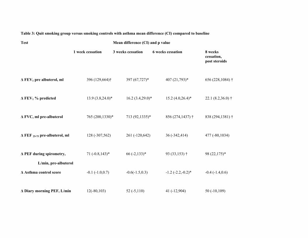
Table 3: Quit smoking group versus smoking controls with asthma mean difference (CI) compared to baseline
Test Mean difference (CI) and p value 1 week cessation 3 weeks cessation 6 weeks cessation 8 weeks cessation, post steroids
∆ FEV1 pre albuterol, ml 396 (129,664)† 397 (67,727)* 407 (21,793)* 656 (228,1084) †
∆ FEV1 % predicted 13.9 (3.8,24.0)* 16.2 (3.4,29.0)* 15.2 (4.0,26.4)* 22.1 (8.2,36.0) †
∆ FVC, ml pre-albuterol 765 (200,1330)* 713 (92,1335)* 856 (274,1437) † 838 (294,1381) †
∆ FEF 25-75 pre-albuterol, ml 128 (-307,562) 261 (-120,642) 36 (-342,414) 477 (-80,1034)
∆ PEF during spirometry, 71 (-0.8,143)* 66 (-2,133)* 93 (33,153) † 98 (22,175)*
L/min, pre-albuterol
∆ Asthma control score -0.1 (-1.0,0.7) -0.6(-1.5,0.3) -1.2 (-2.2,-0.2)* -0.4 (-1.4,0.6)
∆ Diary morning PEF, L/min 12(-80,103) 52 (-5,110) 41 (-12,904) 50 (-10,109)
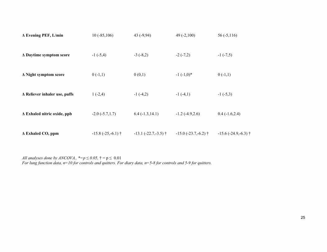
25
∆ Evening PEF, L/min 10 (-85,106) 43 (-9,94) 49 (-2,100) 56 (-5,116)
∆ Daytime symptom score -1 (-5,4) -3 (-8,2) -2 (-7,2) -1 (-7,5)
∆ Night symptom score 0 (-1,1) 0 (0,1) -1 (-1,0)* 0 (-1,1)
∆ Reliever inhaler use, puffs 1 (-2,4) -1 (-4,2) -1 (-4,1) -1 (-5,3)
∆ Exhaled nitric oxide, ppb -2.0 (-5.7,1.7) 6.4 (-1.3,14.1) -1.2 (-4.9,2.6) 0.4 (-1.6,2.4)
∆ Exhaled CO, ppm -15.8 (-25,-6.1) † -13.1 (-22.7,-3.5) † -15.0 (-23.7,-6.2) † -15.6 (-24.9,-6.3) †
All analyses done by ANCOVA.. *=p ≤ 0.05, † = p ≤ 0.01 For lung function data, n=10 for controls and quitters. For diary data, n=5-8 for controls and 5-9 for quitters.
26
Table 4: Change mean (CI) in induced sputum cell counts and mediators in smokers with asthma following smoking cessation compared to
smoking controls at 3 and 6 weeks after smoking cessation.
3 weeks cessation 6 weeks cessation
Induced sputum cell counts
∆ Total cell count x106 -1.31 (-5.0,2.4) -4.2 (-9.7,1.2)
∆ Neutrophils % -8.1 (-30.1,13.9) -29.1 (-50.8,-7.5) †
∆ Eosinophils % 1.4 (0,2.7) 0.1 (-1.8,1.9)
∆ Macrophages % 6.2 (-15.7,28.0) 21.6 (4.2,39.1)*
∆ Lymphocytes % 0.5 (-0.6,1.4) 1.1 (-1.0,3.1) ∆ Bronchial epithelial cells % 1.0 (-1.6,3.6) 7.2 (-4.0,18.4) ∆ Sputum supernatant mediators
∆ IL-8, ng/ml -2.1 (-6.2,2.0) 0.4 (-0.8,1.6)
∆ MPO, ng/ml -69 (-220,82) -24 (-214,167)
∆ LTB4, pg/ml 453 (-1154,2060) -953 (-2935,1028)
∆ ECP, ng/ml 50 (-126,227) -19 (-199,160)
Analyses performed using ANCOVA. *=p ≤ 0.05, † = p ≤ 0.01 At 3 weeks, n=9 for cell counts and 8-9 for the different mediators. At 6 weeks, n=8 for controls and 10 for quitters for cell counts and 7 controls and 6-10 for the different mediators
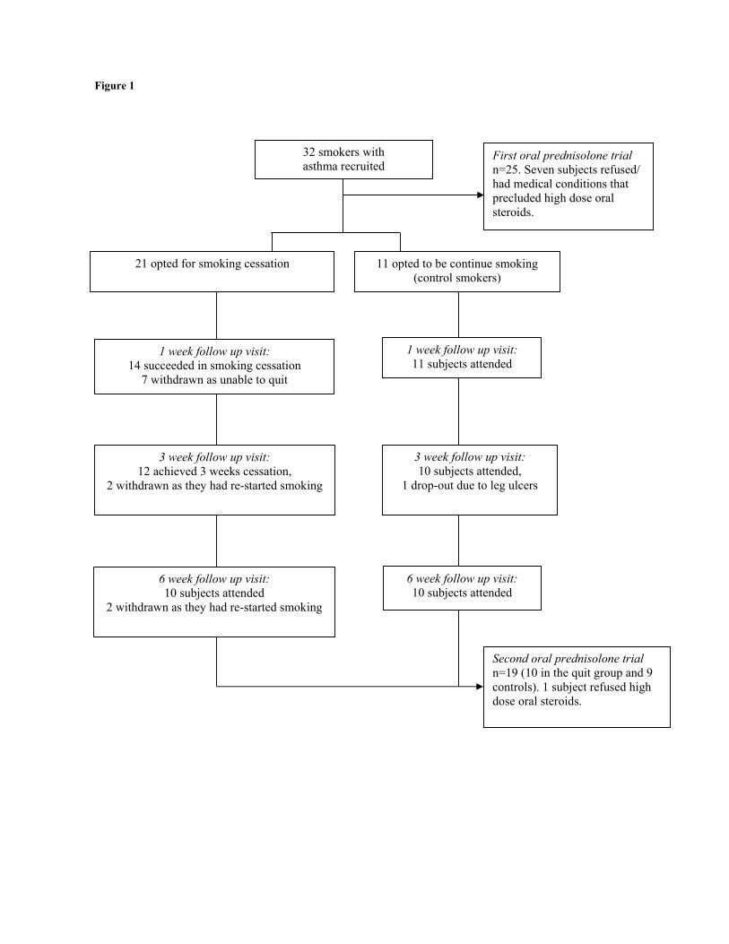
Figure 1
32 smokers with asthma recruited
First oral prednisolone trial n=25. Seven subjects refused/ had medical conditions that precluded high dose oral steroids.
21 opted for smoking cessation
1 week follow up visit: 11 subjects attended
11 opted to be continue smoking (control smokers)
3 week follow up visit: 12 achieved 3 weeks cessation,
2 withdrawn as they had re-started smoking
6 week follow up visit: 10 subjects attended
2 withdrawn as they had re-started smoking
1 week follow up visit: 14 succeeded in smoking cessation
7 withdrawn as unable to quit
Second oral prednisolone trial n=19 (10 in the quit group and 9 controls). 1 subject refused high dose oral steroids.
3 week follow up visit: 10 subjects attended,
1 drop-out due to leg ulcers
6 week follow up visit: 10 subjects attended
28
Figure 2
29
Effects of smoking cessation on lung function and airway inflammation in
smokers with asthma
Rekha Chaudhuri l, Eric Livingston l, Alex D McMahon 2, Jane Lafferty l,
Iona Fraser 3, Mark Spears l, Charles P McSharry 3, Neil C Thomson l
Online Data Supplement
30
METHODS
Subjects
Smokers with asthma aged 18-60 years were recruited. The study was approved by the West
Glasgow Ethics Committee and all participants gave written informed consent. Asthma was
diagnosed by ATS criteria (E1). Subjects had a baseline FEV1 ≤85% predicted and
demonstrated ≥ 15% reversibility of FEV1 after nebulised albuterol. Smokers with ≥ 10 pack
years smoking history and currently smoking ≥ 10 cigarettes/day were included.
Study design
This was a prospective, controlled study performed in two parts. Initially, smokers with asthma
received 40 mg of oral prednisolone for 14 days after performing baseline spirometry and
reversibility to nebulized albuterol. End-points used to assess airway corticosteroid sensitivity
were pre-albuterol FEV1, asthma control score, change in morning and evening PEF, daily
morning and night symptoms and reduction in the use of β-agonist inhalers. Subjects were then
recalled to discuss smoking cessation, two to twelve months after the corticosteroid trial.
Spirometry, induced sputum, exhaled nitric oxide, exhaled carbon monoxide, asthma control
score, cutaneous vasoconstrictor test were performed and peripheral blood taken for lymphocyte
proliferation functional assay. Serum cotinine was measured to confirm smoking status. All
subjects were given the option to quit smoking or continue smoking. The quitting and continued
smoking groups were issued diary cards to record morning and night-time peak flows,
symptoms and use of reliever inhaler. The smoking cessation group were allowed to choose any
method of support that would help them to quit (e.g. nicotine replacement, zyban or self-control
alone). Visits were arranged one, three and six weeks later. After the six weeks cessation visit,
both groups received oral prednisolone 40mg daily for 14 days, as in the initial phase of the
study, with similar end-points used to compare results. Spirometry, exhaled gases and asthma
31
control score were recorded at all visits. Induced sputum was performed at three and six weeks;
cutaneous vasoconstrictor test and blood lymphocyte proliferation were performed at six weeks.
Exhaled carbon monoxide was measured at all visits. Quitting smoking was accepted by self-
report from the patient. If a subject in the quit group had a lapse that lasted a day, they were
allowed to continue in the study, but their next appointment was postponed by a week. If the
smoking lapse lasted for two or more days, they were encouraged not to smoke, but were
withdrawn from the study.
Measurements
Asthma severity was scored according to the ATS asthma impairment score (E2) and asthma
control was measured using a validated questionnaire with a range from 0 to 6 for increasing
severity and a reduction of 0.5 as a minimal important difference (E3,E4). Patients maintained a
validated diary card (E5), recording morning and night PEF (Mini-Wright peak flow meter,
Clement Clarke, Harlow, UK), daytime symptoms (range 0-6, for increasing severity) and night
awakenings (range 0-3, for increasing severity), use of inhaled rescue medication and study
tablet consumption. Total serum IgE and specific IgE to house dust mite, grass pollen and cat
dander were measured by enzyme linked immunoassay (Unicap, Pharmacia Ltd, Milton Keynes,
UK) (E6). Spirometry was measured with a dry spirometer (Vitalograph Ltd, Buckingham, UK).
Sputum induction with 3% hypertonic saline was performed using a modification of the method
described by Pin et al (E7,E8,E9). Eosinophil cationic protein (ECP), interleukin-8 (IL-8),
myeloperoxidase (MPO) and leukotriene B4 (LTB4) were measured in the fluid-phase of induced
sputum samples. ECP was measured using a sandwich ELISA assay (Caltag Medsystems,
Buckinghamshire, UK) with a sensitivity of 0.125ng/ml, and an inter-assay variability <10%, an
intra-assay variability of <5%, and a recovery in spiked samples of 105+/-9%. LTB4 was
measured using a sandwich ELISA assay (R&D Systems, Oxon, UK) with a sensitivity of
5.63pg/ml, an intra-assay variability of 9.3%, and a recovery in spiked samples of 114.1%.
32
Cross-reactivity with other compounds was:- 6-trans-12-epi-LTB4 (5.5%), 6-trans-LTB4 (4.9%),
12-epi-LTB4 (0.94%), and < 0.2% for the following PGE2, PGF2, 20-OH-LTB4, 20-COOH-
LTB4, LTC4, LTD4, LTE4, 5(S)-HETE, 12(S)-HETE and 15(S)-HETE. MPO was measured by
ELISA (Metachem Diagnostics, Northampton, UK) with a sensitivity of 0.13ng/ml, an intra-
assay variability of <5.4%, an inter-assay variability of <8.8%, and a recovery in spiked sputum
samples of 94%. Cross reactivity with related human compounds was:- erythropoietin 1.1%,
elastase 0.2%, lactoferrin 0.2%, thyroid peroxidase < 0.1%, eosinophil-derived neurotoxin <
0.1%. IL-8 was measured by fluorescent-linked immunoassay (Biosource, Nivelles, Belgium) as
part of a multiplex kit (Human inflammatory 5-plex), with a sensitivity of 8 pg/ml, an intra-assay
variability of <5%, an inter-assay variability of <10%.
Exhaled nitric oxide and carbon monoxide were measured using a chemiluminescence analyzer
(Logan Research Ltd, Rochester, UK) (E10). Subjects who were smoking were asked to abstain
for an hour prior to their visit. Serum cotinine was measured using an enzyme immunoassay
(Cozart Bioscience Ltd, Abingdon, UK) to confirm the smoking status of all subjects.
Compliance was assessed by counting the numbers of tablets remaining after each treatment
period.
The cutaneous vasoconstrictor response to topical beclometasone was measured as described
previously (E11,E12), with minor modifications. Beclometasone dipropionate (Sigma, Poole,
UK) was dissolved in 95% ethanol to concentrations of 1, 3, 10, 30, 100, 300 and 1000 µg/ml. A
control solution of 95% ethanol was used. Solutions were stored at 4°C and used within 2
weeks of preparation. Test sites were outlined by the application of adhesive tape in which
holes, 2cm in diameter, had been cut. Solutions were randomly allocated a letter (A-H) by staff
not involved in the application of solutions or the reading of the test, and solutions applied in
order, A-H. The test was not unblinded until the completion of the study. After evaporation of
33
the diluent, the sites were occluded (for 16-18 hours) with plastic film to enhance percutaneous
absorption of the beclometasone. A tubular bandage (Tubigrip; Seton Healthcare Group plc,
Manchester, UK) was applied to the forearm to attenuate any changes in ambient temperature.
The following morning the adhesive tape and plastic film were removed, and the degree of
blanching assessed 1-2 hours later. The test sites were examined in standard lighting conditions
and given a blanching score by a single trained observer who did not know the order of the
applications of the solutions. Blanching at each concentration was graded according to a 4-point
scale: 0 = no blanching; 1 = faint blanching; 2 = obvious blanching not extending outwith the
test site; 3 = intense blanching extending over the margin of the test site. Addition of individual
concentration scores gave a total score [range 0-24]. A high score indicated a high degree of
corticosteroid sensitivity.
The sensitivity of peripheral blood T-lymphocytes to glucocorticoids was assessed in a
functional assay as described previously (E13) at the same two visits as the skin test. The
percentage suppression at the final concentration of corticosteroid used (dexamethasone or
prednisolone), termed Imax % in this study was compared between the two groups.
Statistical analysis
Baseline characteristics were compared by chi-squared tests, Wilcoxon tests and t-tests.
Diary data was analysed using the last three days of each period. The first three days of diary
card data were used as baseline values. The response to smoking cessation on lung function,
diary data, induced sputum, mediator levels, skin vasoconstrictor response and oral
prednisolone trials for smokers versus never smokers with asthma was assessed by Analysis
of Covariance models that adjusted each factor by its baseline measure. Correlations were
assessed by Spearman’s rank correlations. Only those subjects who completed the study up
to the six week visit were included in the analysis. Significance at a level of 5% was
34
considered for the primary end-point; the change in FEV1 at six weeks. A sample size of 10
in each group will have 80% power to detect a difference in means of 300.0 ml assuming
that the common standard deviation is 224.2 using a two group t-test with a 0.050 two-sided
significance level. Tests were performed using SAS version 9.0 (Cary, NC, USA).
35
REFERENCES
E1. ATS: Standards for the diagnosis and care of patients with chronic obstructive pulmonary
disease (COPD) and asthma. Am Rev Resp Dis. 1987;136:225-234.
E2. ATS: Guidelines for the evaluation of impairment/disability in patients with asthma. Am
Rev Resp Dis. 1993;147:1056-1061.
E3. Juniper E, O'Byrne P, Guyatt G, Ferrie P, King D. Development and validation of a
questionnaire to measure asthma control. Eur Respir J. 1999;14:902-907.
E4 . Juniper E, Svensson K, Mork A, Stahl E. Measurement properties and interpretation of
three shortened versions of the asthma control questionnaire. Resp Med. 2005;99:553-558.
E5. Santanello N, Barber B, Reiss T, Friedman B, Juniper E, Zhang J. Measurement
characteristics of two asthma symptom diary scales for use in clinical trials. Eur Respir J.
1997;10:646-651.
E6 . Johansson S, Hourihane JB, Bousquet J, Bruijnzeel-Koomen C, Dreborg S, Haahtela T et
al. A revised nomenclature for allergy: An EAACI position statement from the EACCI
nomenclature task force. Allergy. 2001;56:813-849
E7 . Chalmers G, MacLeod K, Thomson L, Little S, McSharry C, Thomson N. Smoking and
airway inflammation in patients with mild asthma. Chest. 2001;120(6):1917-1922.
E8. Marquette C, Saulnier F, Leroy O, Wallaer B, Chopin C, Demarcq JM, Durocher A,
Tonnel AB. Long-term prognosis of near-fatal asthma. Am Rev Resp Dis. 1992;146:76-81.
E9. Popov T, Gottschalk R, Kolendowicz R, Dolovich J, Powers P, Hargreave F. The
evaluation of a cell dispersion method of sputum examination. Clin Exp Allergy.
1994;24:778-783.
E10. Kharitonov S, Alving K, Barnes P. Exhaled and nasal nitric oxide measurements:
recommendations. Eur Respir J. 1997;10:1683-1693.
E11. Brown PH, Teelucksingh S, Matusiewicz SP, Greening AP, Crompton GK, Edwards
36
CRW. Cutaneous vasoconstrictor response to glucocorticoids in asthma. Lancet.
1991;337:576-80.
E12. Noon PJ, Evans CE, Haynes WG, Webb DJ, Walker BR. A comparison of techniques to
assess skin blanching following the topical application of glucocorticoids. Br J Dermatol.
1996;134:837-842.
E13. Corrigan C, Brown P, Barnes N, Szefler SJ, Tsai JJ, Frew AJ, Kay AB. Glucocorticoid
resistance in chronic asthma: Glucocorticoid pharmacokinetics, glucocorticoid receptor
characteristics, and inhibition of peripheral blood T cell proliferation by glucocorticoids in
vitro. Am Rev Respir Dis. 1991;144:1016-1025.
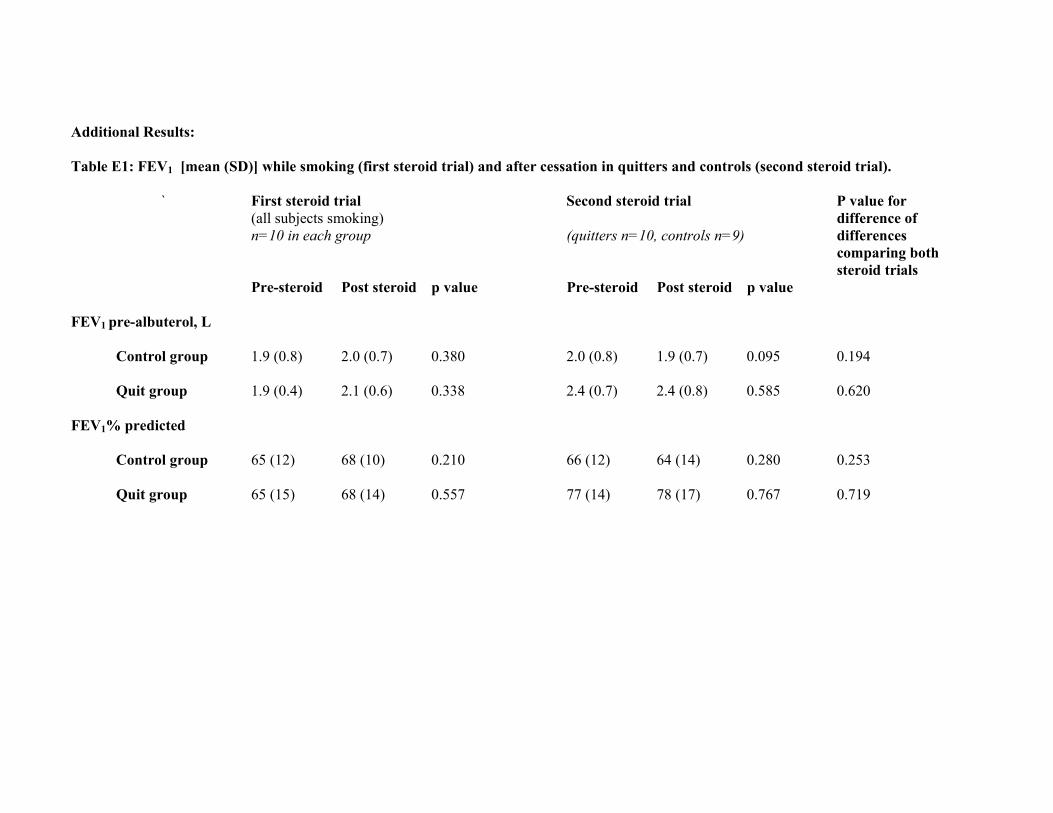
Additional Results: Table E1: FEV1 [mean (SD)] while smoking (first steroid trial) and after cessation in quitters and controls (second steroid trial). ` First steroid trial Second steroid trial P value for (all subjects smoking) difference of n=10 in each group (quitters n=10, controls n=9) differences comparing both steroid trials Pre-steroid Post steroid p value Pre-steroid Post steroid p value FEV1 pre-albuterol, L Control group 1.9 (0.8) 2.0 (0.7) 0.380 2.0 (0.8) 1.9 (0.7) 0.095 0.194 Quit group 1.9 (0.4) 2.1 (0.6) 0.338 2.4 (0.7) 2.4 (0.8) 0.585 0.620 FEV1% predicted Control group 65 (12) 68 (10) 0.210 66 (12) 64 (14) 0.280 0.253 Quit group 65 (15) 68 (14) 0.557 77 (14) 78 (17) 0.767 0.719
Related Documents