RESEARCH Open Access Effects of rapamycin on social interaction deficits and gene expression in mice exposed to valproic acid in utero Hiroko Kotajima-Murakami 1,2 , Toshiyuki Kobayashi 3 , Hirofumi Kashii 1,4 , Atsushi Sato 1,5 , Yoko Hagino 1 , Miho Tanaka 1,6 , Yasumasa Nishito 7 , Yukio Takamatsu 7 , Shigeo Uchino 1,2 and Kazutaka Ikeda 1* Abstract The mammalian target of rapamycin (mTOR) signaling pathway plays a crucial role in cell metabolism, growth, and proliferation. The overactivation of mTOR has been implicated in the pathogenesis of syndromic autism spectrum disorder (ASD), such as tuberous sclerosis complex (TSC). Treatment with the mTOR inhibitor rapamycin improved social interaction deficits in mouse models of TSC. Prenatal exposure to valproic acid (VPA) increases the incidence of ASD. Rodent pups that are exposed to VPA in utero have been used as an animal model of ASD. Activation of the mTOR signaling pathway was recently observed in rodents that were exposed to VPA in utero, and rapamycin ameliorated social interaction deficits. The present study investigated the effect of rapamycin on social interaction deficits in both adolescence and adulthood, and gene expressions in mice that were exposed to VPA in utero. We subcutaneously injected 600 mg/kg VPA in pregnant mice on gestational day 12.5 and used the pups as a model of ASD. The pups were intraperitoneally injected with rapamycin or an equal volume of vehicle once daily for 2 consecutive days. The social interaction test was conducted in the offspring after the last rapamycin administration at 5–6 weeks of ages (adolescence) or 10–11 weeks of age (adulthood). Whole brains were collected after the social interaction test in the adulthood, and microarray and Western blot analyses were performed. Mice that were exposed to VPA and treated with vehicle exhibited a decrease in social interaction compared with control mice that were treated with vehicle. Rapamycin treatment in VPA-exposed mice improved social deficits. Mice that were exposed to VPA and treated with vehicle exhibited the aberrant expression of genes in the mTOR signaling pathway, and rapamycin treatment recovered changes in the expression of some genes, including Fyb and A330094K24Rik. Rapamycin treatment suppressed S6 phosphorylation in VPA-exposed mice. Aberrant gene expression was associated with social interaction deficits in VPA-exposed mice. Rapamycin may be an effective treatment for non-syndromic ASD in adolescent and adult patients who present impairments in the mTOR signaling pathway. Keywords: Autism spectrum disorder, mTOR signaling pathway, Valproic acid, Rapamycin Introduction Autism spectrum disorder (ASD) is a neurodevelopmental disorder that is characterized by deficits in social inter- action and communication, repetitive behaviors, and re- stricted interests [1]. Autism spectrum disorder has several peripheral symptoms (e.g., aberrant sensitization, clumsiness of movement, and digestive system disease), but social interaction deficits are a core symptom [2]. The prevalence of ASD in school-aged children is > 1–2% [3–5], with a male/female ratio of approximately 4:1 [6]. Various genetic and/or environmental factors have been investigated in studies of ASD using animal models of syndromic ASD and non-syndromic ASD [7]. However, the pathophysiology of social interaction deficits in ASD and effective treatments have not been clarified. Mammalian target of rapamycin (mTOR) is a serine/threonine kinase that belongs to the family of phosphatidylinositol-3 kinase (PI3K)-related kinases * Correspondence: [email protected] 1 Addictive Substance Project, Tokyo Metropolitan Institute of Medical Science, 2-1-6 Kamikitazawa, Setagaya-ku, Tokyo, Japan Full list of author information is available at the end of the article © The Author(s). 2019 Open Access This article is distributed under the terms of the Creative Commons Attribution 4.0 International License (http://creativecommons.org/licenses/by/4.0/), which permits unrestricted use, distribution, and reproduction in any medium, provided you give appropriate credit to the original author(s) and the source, provide a link to the Creative Commons license, and indicate if changes were made. The Creative Commons Public Domain Dedication waiver (http://creativecommons.org/publicdomain/zero/1.0/) applies to the data made available in this article, unless otherwise stated. Kotajima-Murakami et al. Molecular Brain (2019) 12:3 https://doi.org/10.1186/s13041-018-0423-2

Welcome message from author
This document is posted to help you gain knowledge. Please leave a comment to let me know what you think about it! Share it to your friends and learn new things together.
Transcript

RESEARCH Open Access
Effects of rapamycin on social interactiondeficits and gene expression in miceexposed to valproic acid in uteroHiroko Kotajima-Murakami1,2, Toshiyuki Kobayashi3, Hirofumi Kashii1,4, Atsushi Sato1,5, Yoko Hagino1,Miho Tanaka1,6, Yasumasa Nishito7, Yukio Takamatsu7, Shigeo Uchino1,2 and Kazutaka Ikeda1*
Abstract
The mammalian target of rapamycin (mTOR) signaling pathway plays a crucial role in cell metabolism, growth, andproliferation. The overactivation of mTOR has been implicated in the pathogenesis of syndromic autism spectrumdisorder (ASD), such as tuberous sclerosis complex (TSC). Treatment with the mTOR inhibitor rapamycin improvedsocial interaction deficits in mouse models of TSC. Prenatal exposure to valproic acid (VPA) increases the incidenceof ASD. Rodent pups that are exposed to VPA in utero have been used as an animal model of ASD. Activation ofthe mTOR signaling pathway was recently observed in rodents that were exposed to VPA in utero, and rapamycinameliorated social interaction deficits. The present study investigated the effect of rapamycin on social interactiondeficits in both adolescence and adulthood, and gene expressions in mice that were exposed to VPA in utero. Wesubcutaneously injected 600 mg/kg VPA in pregnant mice on gestational day 12.5 and used the pups as a model ofASD. The pups were intraperitoneally injected with rapamycin or an equal volume of vehicle once daily for 2consecutive days. The social interaction test was conducted in the offspring after the last rapamycin administrationat 5–6 weeks of ages (adolescence) or 10–11 weeks of age (adulthood). Whole brains were collected after the socialinteraction test in the adulthood, and microarray and Western blot analyses were performed. Mice that wereexposed to VPA and treated with vehicle exhibited a decrease in social interaction compared with control micethat were treated with vehicle. Rapamycin treatment in VPA-exposed mice improved social deficits. Mice that wereexposed to VPA and treated with vehicle exhibited the aberrant expression of genes in the mTOR signalingpathway, and rapamycin treatment recovered changes in the expression of some genes, including Fyb andA330094K24Rik. Rapamycin treatment suppressed S6 phosphorylation in VPA-exposed mice. Aberrant geneexpression was associated with social interaction deficits in VPA-exposed mice. Rapamycin may be an effectivetreatment for non-syndromic ASD in adolescent and adult patients who present impairments in the mTORsignaling pathway.
Keywords: Autism spectrum disorder, mTOR signaling pathway, Valproic acid, Rapamycin
IntroductionAutism spectrum disorder (ASD) is a neurodevelopmentaldisorder that is characterized by deficits in social inter-action and communication, repetitive behaviors, and re-stricted interests [1]. Autism spectrum disorder hasseveral peripheral symptoms (e.g., aberrant sensitization,clumsiness of movement, and digestive system disease),
but social interaction deficits are a core symptom [2]. Theprevalence of ASD in school-aged children is > 1–2%[3–5], with a male/female ratio of approximately 4:1 [6].Various genetic and/or environmental factors have beeninvestigated in studies of ASD using animal models ofsyndromic ASD and non-syndromic ASD [7]. However,the pathophysiology of social interaction deficits in ASDand effective treatments have not been clarified.Mammalian target of rapamycin (mTOR) is a
serine/threonine kinase that belongs to the family ofphosphatidylinositol-3 kinase (PI3K)-related kinases
* Correspondence: [email protected] Substance Project, Tokyo Metropolitan Institute of MedicalScience, 2-1-6 Kamikitazawa, Setagaya-ku, Tokyo, JapanFull list of author information is available at the end of the article
© The Author(s). 2019 Open Access This article is distributed under the terms of the Creative Commons Attribution 4.0International License (http://creativecommons.org/licenses/by/4.0/), which permits unrestricted use, distribution, andreproduction in any medium, provided you give appropriate credit to the original author(s) and the source, provide a link tothe Creative Commons license, and indicate if changes were made. The Creative Commons Public Domain Dedication waiver(http://creativecommons.org/publicdomain/zero/1.0/) applies to the data made available in this article, unless otherwise stated.
Kotajima-Murakami et al. Molecular Brain (2019) 12:3 https://doi.org/10.1186/s13041-018-0423-2

(PIKKs) [8–10]. The mTOR signaling pathway plays acrucial role in protein synthesis and cell growth, pro-liferation, and metabolism [10]. Overactivation of themTOR signaling pathway causes diseases that are syn-dromic with ASD, such as tuberous sclerosis complex(TSC), neuronal fibromatosis 1 (NF1), and tensinhomolog deleted on chromosome 10 (PTEN) [11].Approximately 50% of child and adolescent patientswith TSC are diagnosed with ASD [12]. TSC1, TSC2,PTEN, and NF1 are negative regulators of mTORC1,and mice that possess a mutation of these genes areconsidered animal models of syndromic ASD [13].Tsc1+/− mice exhibit impairments in learning andmemory that depend on the hippocampus and a re-duction of social interaction compared with wild-typemice [14]. Disturbances of excitatory/inhibitory synap-tic balance are thought to be involved in the path-ology of ASD [15]. Hippocampal hyperexcitability hasbeen reported in Tsc1 knockout (KO) cultures, andTsc1 conditional KO mice exhibited elevations of S6phosphorylation in the hippocampus compared withwildtype mice [16]. PTEN KO and Nf1+/− mice alsoexhibit social interaction deficits [17–19]. Rapamycinimproved learning and memory deficits in Tsc2+/−
mice [20]. Rapamycin treatment also recovered socialinteraction deficits in Tsc1+/− and Tsc2+/− mice andrescued the levels of phosphorylated S6K, which isdownstream of the mTOR signaling pathway and in-volved in protein synthesis in Tsc2+/− mice [21].Treatment with rapamycin improved social interactiondeficits and spine pruning defects in Tsc2+/− mice[22]. Rapamycin administration in PTEN KO micealso attenuated anxiety-like behavior, attenuated soci-ability deficits, and increased the ratio of Akt phos-phorylation [23]. Furthermore, a recent clinical studyreported that everolimus, an mTOR inhibitor, amelio-rated autistic behavior scores in a patient with TSC[24]. These studies suggest that overactivation of themTOR signaling pathway is associated with ASD, andmTOR inhibition may be a potential therapeutic strat-egy for the treatment of syndromic ASD. However,unclear is whether rapamycin is effective for thetreatment of non-syndromic ASD.Valproic acid (VPA) is used as an anti-epileptic drug,
mood stabilizer, and treatment for migraine. However,pregnant mothers who are treated with VPA have beenreported to deliver children with fetal valproate syn-drome and a high incidence of ASD [25]. Valproic acidis used to model non-syndromic ASD in animals.Valproic acid-treated mice and rats exhibit impairmentsin motor function, aberrant sensitivity, and social inter-action deficits [26–28]. Valproic acid activated the PI3K/Akt/mTOR pathway in muscle in a mouse model of Du-chenne muscular dystrophy [29]. A reduction of PTEN
protein levels and a higher ratio of Akt phosphorylationwere found in VPA-exposed rat brains [30]. The block-ade of N-methyl-D-aspartate (NMDA) receptors, whichare upstream of the mTOR signaling pathway, attenu-ated social interaction deficits in VPA-exposed mice[31]. Nicolini et al. reported that the phosphorylation ofmTOR, Akt, and S6 decreased in the lateral temporalneocortex in VPA-exposed rats compared withsaline-treated rats [32]. A recent study reported thatrapamycin treatment suppressed hippocampal neuronapoptosis in VPA-exposed rats [33]. Furthermore, rapa-mycin treatment attenuated social interaction deficitsand the enhancement of mTOR and S6 phosphorylationin the cerebellum, prefrontal cortex, and hippocampusin VPA-exposed rats on postnatal day 33–35 (adoles-cence) [34]. These previous studies suggest that an aber-rant mTOR signaling pathway causes ASD-likebehaviors in VPA-exposed animals. However, the effectsof rapamycin on social interaction deficits have not beeninvestigated in adult mice that were exposed to VPA inutero. Moreover, gene expression analysis in wholebrains has not been performed in VPA-exposed orrapamycin-treated mice. Therefore, the present study in-vestigated the effect of rapamycin treatment on socialinteraction deficits in adolescent and adult mice thatwere exposed to VPA in utero. We also comprehensivelyanalyzed the gene expression including mTOR signalingpathway and S6 phosphorylation in mouse whole brains.
MethodsAnimals and VPA administrationPregnant female C57BL/6 J mice (CLEA, Tokyo, Japan)received a single subcutaneous injection of 600 mg/kgsodium valproate (Sigma-Aldrich, St. Louis, MO, USA)on day 12.5 after conception. Valproic acid was dissolvedin saline, and control mice received saline. All of themice were returned to their home cages immediatelyafter the injection. We used 10 dams. Eight to 10 pupswere obtained from VPA- and saline-treated pregnantfemale mice. The pups were culled to eight animals perlitter on P4. The number of mice per litter was normalcompared with control mice. In this experiment, we didnot observe postnatal mortality. The day of birth was re-corded as day 0, and all of the pups were labeled for in-dividual identification. The pups were weaned, sexed,and caged in groups of 3–5 mice of the same sex on post-natal day 26 (P26). All of the behavioral tests were con-ducted from 9:00 AM to 6:00 PM. The mice were housedon a 12 h/12 h light/dark cycle (lights on 8:00 AM to 8:00PM), and temperature was maintained at 22 °C. All of themice had ad libitum access to food and water. In thepresent study, we analyzed male mice only because theprevalence of ASD is higher in males than in females. Allof the animal experiments were performed in accordance
Kotajima-Murakami et al. Molecular Brain (2019) 12:3 Page 2 of 14

with the Guidelines for the Care of Laboratory Animals ofthe Tokyo Metropolitan Institute of Medical Science, andthe housing conditions were approved by the InstitutionalAnimal Care and Use Committee.
Behavioral testsFigure 1a shows a schedule of the experiments. The be-havioral tests were conducted in a sound-proof room,and the mice were given a 60 min habituation periodafter transportation to the behavioral room before thestart of each test. Body maturation was assessed bymeasuring body weight and eye-opening. Motor functionwas assessed by the righting reflex and hanging wiretests. The social interaction test was conducted in bothadolescence (5–6 weeks of age) and adulthood (10–11weeks of age).
Body weight and eye-opening testBody weight was recorded on P7, P11, P14, P18, P21,and P25. Body weight was also recorded when eachmouse underwent the social interaction test in bothadolescence and adulthood. Eye opening was observedonce daily from P14 to P18. The eye-opening scorewas the following: 0 = both eyes closed, 1 = one eyeopen, and 2 = both eyes open.
Righting reflex testEach mouse was placed on its back and gently held byits four limbs. The latency to right, such that all fourpaws were touching the surface, was recorded. The cut-off time was set at 20 s. Each mouse underwent therighting reflex test on P7, P9, and P11.
Hanging wire testThe hanging wire test (O’Hara & Co., Tokyo, Japan) wasconducted from P14 to P25. The mice were placed on agrid wire surface (150 mm× 150mm, divided into 10mm grid squares). The plane was inverted, and the la-tency to fall was recorded, with a maximum of 600 s.
Social interaction testWe conducted the social interaction test as previouslydescribed [21]. Each mouse was left alone in its homecage for 15 min for habituation. The home cage wasplaced in a sound-attenuating chamber. An unfamiliarC57BL/6 J mouse of the same sex and age was then in-troduced. The behavior of the test mouse wasvideo-recorded for 10 min and blindly scored for activesocial interaction, consisting of sniffing, allo-grooming,mounting, and following. A mouse, which went out ofits home cage during the 15 min habituation period, wasexcluded from analysis. Each mouse underwent the
Fig. 1 Schedule of experimental design and rapamycin treatment. (a) Body maturation and motor function were assessed from P7 to P25. Thesocial interaction test was conducted in both adolescence (5–6 weeks of age) and adulthood (10–11 weeks of age). The mice were scarified tosample whole brains. R, righting reflex test; Eye, eye-opening test; HW, hanging wire test; BW, body weight; Ra, rapamycin; Ve, vehicle; Social,social interaction test. (b) The mice were reared with their littermates. A mouse that was injected with vehicle in adolescence was injected withrapamycin in adulthood. Conversely, a mouse that was injected with rapamycin in adolescence was injected with vehicle in adulthood
Kotajima-Murakami et al. Molecular Brain (2019) 12:3 Page 3 of 14
social interaction test during both adolescence andadulthood. One of the 10 rapamycin-treated vehiclemice and one of the 11 rapamycin-treated VPA micewent out of their home cages for a habituation periodduring adolescence. The number of mice per group wasthe following: adolescent (n = 11 vehicle-treated controlmice, n = 13 vehicle-treated VPA-exposed mice, n = 9rapamycin-treated control mice, n = 10 rapamycin-treated VPA-exposed mice) and adult (n = 10vehicle-treated control mice, n = 11 vehicle-treatedVPA-exposed mice, n = 11 rapamycin-treated controlmice, n = 13 rapamycin-treated VPA-exposed mice).
Rapamycin treatmentRapamycin (LC Laboratories, Woburn, MA, USA) wasdissolved in 10% dimethyl sulfoxide diluted with saline.The mice received a 10 ml/kg rapamycin solution or anequal volume of vehicle intraperitoneally once daily for2 consecutive days. The dose of rapamycin was 10mg/kg. Brain levels of rapamycin remained sufficiently highto inhibit mTOR throughout the 48 h period after rapa-mycin administration (6 mg/kg, i.p.) in mice [35]. Sato etal. reported that 5 or 10 mg/kg rapamycin treatment ef-fectively attenuated social interaction deficits in Tsc1+/−
and Tsc2+/− mice [21]. Thus, we tested the dose of 10mg/kg rapamycin. The social interaction test was per-formed 24 h after the second administration. Eachmouse was randomly assigned to vehicle or rapamycinadministration in adolescence (Fig. 1b). A mouse that re-ceived vehicle in adolescence received rapamycin inadulthood. Conversely, a mouse that received rapamycinin adolescence received vehicle in adulthood.
Brain collection and RNA extractionWhole brains were collected after the end of the socialinteraction test in adulthood. Because the precise brainregions that are associated with ASD have not yet beenfully clarified, we examined whole brains in the presentstudy. Brains were frozen in liquid nitrogen and storedat − 80 °C until further processing. Total RNA that wasextracted from whole brains was homogenized inAmbion TRIzol Reagent (Thermo Fisher Scientific,Waltham, MA, USA) using a homogenizer. RNA wasisolated using chloroform and precipitated using isopro-pyl alcohol. The quality of RNA was assessed withNanodrop 1000 (Thermo Fisher Scientific). All of theRNA samples had an A260/280 ratio between 2.0 and 2.1and an A230/260 ratio between 2.2 and 2.3.
Microarray analysiscRNA targets were synthesized and hybridized using theWhole Mouse Genome Microarray according to themanufacturer’s instructions (Agilent Technologies, SantaClara, CA, USA). The array slides were scanned using a
SureScan Microarray Scanner (Agilent Technologies).Before analyzing gene expression, microarray data werenormalized and sorted using GeneSpring 14.5 software(Agilent Technologies). Each sample was normalized bya 75% percentile shift. Compromised probes wereremoved, and remaining probes with expression values< 20% were excluded. The probes were then filteredbased on expression levels for quality control. TheBenjamini and Hochberg false-discovery rate (FDR)was determined for the remaining probes and thosewith p < 0.05. Each group comparison was performedusing t-tests (p < 0.05). Each group consisted of fivemice (vehicle-treated control mice, rapamycin-treatedcontrol mice, vehicle-treated VPA-exposed mice, andrapamycin-treated VPA-exposed mice).
Mining of public databasesThe genomic data repositories in BaseSpace (illumina,https://www.nextbio.com/b/authentication/login.nb) wereused to analyze all differentially expressed genes with stat-istical significance from vehicle-treated VPA-exposed micevs. vehicle-treated control mice, rapamycin-treatedVPA-exposed mice vs. vehicle-treated VPA-exposed mice,and rapamycin-treated control mice vs. vehicle-treatedcontrol mice. The data were compared with curateddatasets that are available in BaseSpace to identify pub-lished studies of Diseases, Pharmaco, and Knockout miceusing the BaseSpace Diseases atlas application, BaseSpacePharmaco atlas application, and BaseSpace Knockout atlasapplication, respectively. Rank-based enrichment statisticswere employed to calculate BaseSpace scores for eachdisease, compound, and gene for knockout mice.MetaCore (Thomson Reuters, https://portal.genego.com)was used to build the network for two negative correlationgenes in vehicle-treated VPA-exposed mice and vehicle-treated control mice vs. rapamycin-treated VPA-exposedmice and vehicle-treated VPA-exposed mice.
Antibodies and Western blotRabbit anti-S6 antibodies and antiphospho-S6 (S235/236) antibodies (1:500) were purchased from Cell Signal-ing Technology (Danvers, MA, USA). We conductedprotein extraction and Western blot as previously de-scribed [21]. We prepared protein samples from thewhole brain because the precise brain regions that areassociated with ASD have not yet been fully clarified.Total protein from the frozen mouse brain was extractedfor Western blot. Whole brains were homogenized usinga tissue homogenizer in 1× sodium dodecyl sulfate(SDS) gel-loading buffer (50 mM Tris–HCl [pH 6.8], 2%SDS, and 10% glycerol). The supernatant was obtainedby centrifugation at 17,000×g. The protein concentrationwas determined using the Bio-Rad DC Protein Assay Kit(Bio-Rad Laboratories, Hercules, CA, USA). Equal
Kotajima-Murakami et al. Molecular Brain (2019) 12:3 Page 4 of 14
amounts of extracted protein were added to 5% mercap-toethanol and boiled. Proteins were resolved bySDS-polyacrylamide gel electrophoresis (PAGE), trans-ferred to a polyvinylidene fluoride membrane (Immobi-lon-P, Merck Millipore, Billerica, MA, USA), andblocked in 1% skim milk/Tris-buffered saline that con-tained 0.05% Tween 20 at room temperature for 2 h.The membranes were incubated with the primary anti-bodies at room temperature for 1 h. Protein bands weredetected using the EnVisiont Kit (Dako, Glostrup,Denmark) and ECL Western Blotting Detection System(GE Healthcare, Buckinghamshire, UK) and quantita-tively analyzed using ImageJ 1.45 software.
Statistical analysisThe results of the behavioral tests were analyzed usingStatistical Package for the Social Sciences 14.0 software(SPSS, Tokyo, Japan). The data were analyzed usingStudent’s t-test, the Mann-Whitney U test, and two-wayanalysis of variance (ANOVA). All of the data are pre-sented as mean ± standard error of the mean (SEM).
Values of p < 0.05 were considered statisticallysignificant.
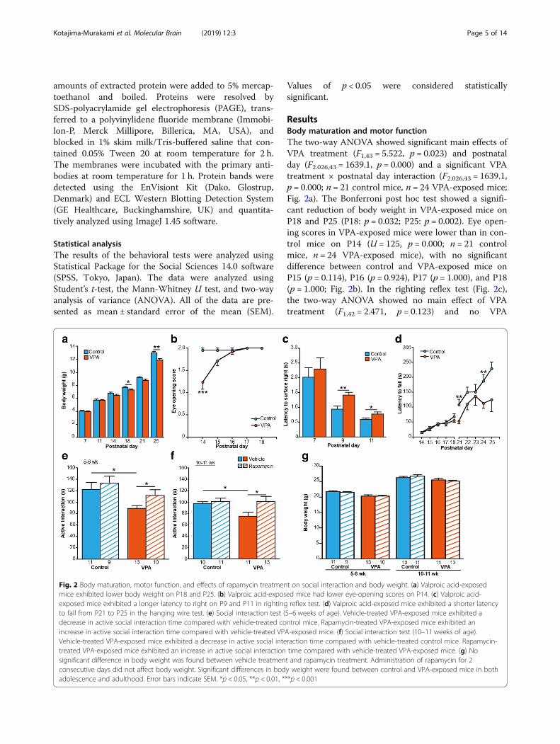
ResultsBody maturation and motor functionThe two-way ANOVA showed significant main effects ofVPA treatment (F1,43 = 5.522, p = 0.023) and postnatalday (F2.026,43 = 1639.1, p = 0.000) and a significant VPAtreatment × postnatal day interaction (F2.026,43 = 1639.1,p = 0.000; n = 21 control mice, n = 24 VPA-exposed mice;Fig. 2a). The Bonferroni post hoc test showed a signifi-cant reduction of body weight in VPA-exposed mice onP18 and P25 (P18: p = 0.032; P25: p = 0.002). Eye open-ing scores in VPA-exposed mice were lower than in con-trol mice on P14 (U = 125, p = 0.000; n = 21 controlmice, n = 24 VPA-exposed mice), with no significantdifference between control and VPA-exposed mice onP15 (p = 0.114), P16 (p = 0.924), P17 (p = 1.000), and P18(p = 1.000; Fig. 2b). In the righting reflex test (Fig. 2c),the two-way ANOVA showed no main effect of VPAtreatment (F1,42 = 2.471, p = 0.123) and no VPA
Fig. 2 Body maturation, motor function, and effects of rapamycin treatment on social interaction and body weight. (a) Valproic acid-exposedmice exhibited lower body weight on P18 and P25. (b) Valproic acid-exposed mice had lower eye-opening scores on P14. (c) Valproic acid-exposed mice exhibited a longer latency to right on P9 and P11 in righting reflex test. (d) Valproic acid-exposed mice exhibited a shorter latencyto fall from P21 to P25 in the hanging wire test. (e) Social interaction test (5–6 weeks of age). Vehicle-treated VPA-exposed mice exhibited adecrease in active social interaction time compared with vehicle-treated control mice. Rapamycin-treated VPA-exposed mice exhibited anincrease in active social interaction time compared with vehicle-treated VPA-exposed mice. (f) Social interaction test (10–11 weeks of age).Vehicle-treated VPA-exposed mice exhibited a decrease in active social interaction time compared with vehicle-treated control mice. Rapamycin-treated VPA-exposed mice exhibited an increase in active social interaction time compared with vehicle-treated VPA-exposed mice. (g) Nosignificant difference in body weight was found between vehicle treatment and rapamycin treatment. Administration of rapamycin for 2consecutive days did not affect body weight. Significant differences in body weight were found between control and VPA-exposed mice in bothadolescence and adulthood. Error bars indicate SEM. *p < 0.05, **p < 0.01, ***p < 0.001
Kotajima-Murakami et al. Molecular Brain (2019) 12:3 Page 5 of 14
treatment × postnatal day interaction (F2.026,42 = 0.294,p = 0.620; n = 21 control mice, n = 24 VPA-exposedmice). Both groups (i.e., exposed to VPA or saline)exhibited a progressive decline in the latency to righton P7, P9, and P11 (F2.026,42 = 0.294, p = 0.620). Thetwo-way ANOVA showed no main effect of VPAtreatment (F1,24 = 0.371, p = 0.548) and no VPA treatment× postnatal day interaction (F3.054,24 = 0.355, p = 0.790).The Bonferroni post hoc test showed a significantlylonger latency in VPA-exposed mice on P9 and P11(P9: p = 0.001; P11: p = 0.046). Both groups (i.e., ex-posed to VPA or saline) exhibited a progressive in-crease in the latency to fall on P14–18 (F3.054,24 =16.621, p = 0.000; Fig. 2d). The two-way ANOVA re-vealed a significant main effect of VPA treatment onthe latency to fall on P21–25 (F1,22 = 12.250, p =0.002), with no main effect of postnatal day (F2.835,22= 2.703, p = 0.056) and no VPA treatment × postnatalday interaction (F2.835,22 = 0.626, p = 0.592). The Bonfer-roni post hoc test showed a significantly shorter latency tofall in VPA-exposed mice on P21 and P24 (P21: p = 0.001;P24: p = 0.001).We assessed the effects of rapamycin treatment on so-
cial interaction in control and VPA-exposed mice duringboth adolescence and adulthood. In adolescence (Fig. 2e),vehicle-treated VPA-exposed mice exhibited a decrease inactive social interaction time compared with vehicle-treated control mice (t22 = 2.669, p = 0.014; n = 11vehicle-treated control mice, n = 13 vehicle-treated VPA-exposed mice). Active social interaction time inrapamycin-treated VPA-exposed mice significantlyincreased compared with vehicle-treated VPA-exposedmice (t21 = − 2.134, p = 0.045; n = 13 vehicle-treatedVPA-exposed mice, n = 10 rapamycin-treated VPA-ex-posed mice). No significant difference in active socialinteraction time was found between vehicle-treatedcontrol mice and rapamycin-treated control mice (t18= − 0.599, p = 0.557; n = 11 vehicle-treated control mice,n= 9 rapamycin-treated control mice). In adulthood (Fig.2f ), vehicle-treated VPA-exposed mice exhibited a de-crease in active social interaction time compared withvehicle-treated control mice (t19 = 2.586, p = 0.018; n = 10vehicle-treated control mice, n = 11 vehicle-treatedVPA-exposed mice). Rapamycin-treated VPA-exposedmice exhibited an increase in active social interactiontime compared with vehicle-treated VPA-exposedmice (t22 = − 2.284, p = 0.032; n = 11 vehicle-treatedVPA-exposed mice, n = 13 rapamycin-treatedVPA-exposed mice). No significant difference in activesocial interaction time was found between vehicle--treated control mice and rapamycin-treated control mice(t21 = − 0.415, p = 0.682; n = 10 vehicle-treated controlmice, n = 11 rapamycin-treated control mice). Body weightwas recorded immediately before the social interaction
test (Fig. 2g). A significant difference in body weight wasfound between control and VPA-exposed mice in ado-lescence (5–6 weeks of age; vehicle-treated controlmice vs. vehicle-treated VPA-exposed mice, t22 =3.226, p = 0.004; rapamycin-treated control mice vs.rapamycin-treated VPA-exposed mice, t19 = − 3.255, p= 0.004), with no significant difference between vehi-cle-treated control mice vs. rapamycin-treated controlmice (t19 = 0.610, p = 0.549) or between vehicle-treatedVPA-exposed mice vs. rapamycin-treated VPA-exposedmice (t22 = 0.617, p = 0.869). In adulthood (10–11 weeks ofage), rapamycin-treated VPA-exposed mice exhibitedlower body weight than rapamycin-treated control mice(t20.846 = − 2.941, p = 0.008). No significant difference inbody weight was found between vehicle-treated controlmice and vehicle-treated VPA-exposed mice (t19 = 1.005,p = 0.327). No significant difference in body weightwas found between vehicle-treated control mice vs.rapamycin-treated control mice (t19 =− 0.899, p=0.380) orbetween vehicle-treated VPA-exposed mice vs. rapamycin-treated VPA-exposed mice (t22 = 0.753, p=0.459).
Effects of VPA exposure in utero on gene expressionVehicle-treated VPA-exposed mice (n = 5) exhibited thedifferential expression of 5644 genes (2761 upregulatedgenes, 2883 downregulated genes) compared withvehicle-treated control mice (n = 5; Table 1; for informa-tion for each gene, see Additional file 1: Table S1a). Wedetected associations between the up- and downregu-lated genes in VPA-exposed mice and curated studies ofDiseases, Compounds, and Knockout mice in theBaseSpace database. As shown in Additional file 2:Table S2a, we found that 148 diseases were positivelycorrelated with upregulated genes using the BaseSpaceDiseases atlas application. Vitamin D deficiency,which is a Nutritional and Metabolic Disease, had thehighest score (96.94107042). Within the top five inthe score list, we found Developmental Disorder andDevelopmental Delay (score = 82.71872854). We foundthat 136 diseases were positively correlated withdownregulated genes using the BaseSpace Diseasesatlas application. Injury of the eye region, which is anEye Disorder, had the highest score (92.03862769).
Table 1 Number of genes with altered expression in eachgroup
Group Total alteredgenes
Upregulatedgenes
Downregulatedgenes
VPA + vehicle/Control + vehicle
5644 2761 2883
VPA + rapamycin/VPA + vehicle
23 7 16
Control + rapamycin/Control + vehicle
– – –
Kotajima-Murakami et al. Molecular Brain (2019) 12:3 Page 6 of 14
The BaseSpace Pharmaco atlas application showedthat 387 compounds were positively correlated withupregulated genes (Additional file 2: Table S2b).Glycidol had the highest score (100). The BaseSpacePharmaco atlas application showed that 446 com-pounds were positively correlated with downregulatedgenes. Ozagrel had the highest score (84.66459954).The BaseSpace Knockout atlas application showedthat 26 KO mice were positively correlated with up-regulated genes (Additional file 2: Table S2c). PRKG1had the highest score (93.20344653). The BaseSpaceKnockout atlas application showed that 25 KO micewere positively correlated with downregulated genes.PRKG1 had the highest score (79.58880595).
Expression of genes associated with mTOR signalingpathwayWe found 5644 significantly altered genes (2761 upregu-lated genes, 2883 downregulated genes) between vehi-cle-treated control mice (control + vehicle) andvehicle-treated VPA-exposed mice (VPA+ vehicle; Table 1).Twenty-three genes (seven upregulated genes, 16 downreg-ulated genes; for information for each gene, see Additionalfile 1: Table S1b) were differentially expressed inrapamycin-treated VPA-exposed mice (VPA+ rapamycin,n = 5) compared with vehicle-treated VPA-exposed mice(VPA+ vehicle, n = 5). No significantly altered genes werefound between rapamycin-treated control mice (control +rapamycin, n = 5) and vehicle-treated control mice (control+ vehicle, n = 5). Altered genes that are associated with themTOR signaling pathway between vehicle-treated controlmice and vehicle-treated VPA-exposed mice were extractedfrom Pathway Enrichment of BaseSpace (Table 2). mTOR,Mapk1, Rps6ka3, Akt2, and Pick3cg had fold changes > 1.2or < − 1.2. These genes in the mTOR pathway (Table 2)were unaltered between vehicle-treated VPA-exposed miceand rapamycin-treated VPA-exposed mice.
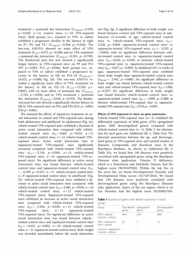
Characteristics of gene expression induced by rapamycintreatmentFigure 3 presents a Venn diagram of significantly alteredgene expression in vehicle-treated VPA-exposed mice(VPA + vehicle) vs. vehicle-treated control mice (control+ vehicle; 5644 genes) and rapamycin-treatedVPA-exposed mice (VPA + rapamycin) vs. vehicle-treated VPA-exposed mice (VPA + vehicle; 23 genes).We found 11 common genes (Msl1, Sly, LOC100504642,LOC380994, 4921509O09Rik, Gm14625, A330094K24Rik, Fyb, two genes that have no gene symbols, andSly that has two probes; Fig. 3). Msl1 expression in-creased in both vehicle-treated VPA-exposed mice vs.vehicle-treated control mice (VPA + vehicle/control +vehicle) and rapamycin-treated VPA-exposed mice vs.vehicle-treated VPA-exposed mice (VPA + rapamycin/
Table 2 Expression change in mTOR signaling pathway-associated genes in vehicle-treated VPA-exposed mice andvehicle-treated control mice
Gene EntrezGene ID Imported ID Fold change
Mtor 56,717 A_52_P67643 −1.5175
Mapk1 26,413 A_55_P1973339 −1.2254
Rheb 19,744 A_55_P2129034 −1.1928
Rps6kb1 72,508 A_55_P2019083 −1.173
Cab39 12,283 A_51_P230934 −1.163
Pld2 18,806 A_55_P2049572 −1.1607
Eif4a1 13,681 A_55_P2046398 −1.158
Yy1 22,632 A_55_P2107745 −1.1493
Cab39l 69,008 A_55_P2001948 −1.1442
Rheb 19,744 A_55_P2170139 −1.1396
Atg13 51,897 A_55_P2028394 −1.135
Tsc1 64,930 A_55_P2157695 −1.132
Pik3cd 18,707 A_52_P99848 −1.1195
Ppp2ca 19,052 A_55_P2288117 −1.1053
Raf1 110,157 A_55_P1973643 −1.09
Ulk1 22,241 A_55_P1977314 −1.084
Pik3r3 18,710 A_55_P2000158 −1.0813
Rhoa 11,848 A_55_P2008081 −1.0709
Rac1 19,353 A_55_P2076489 −1.0697
Ywhaq 22,630 A_55_P2142296 −1.0651
Fbxw11 103,583 A_51_P141860 −1.0642
Mapkap1 227,743 A_55_P2046363 1.0597
Map2k1 26,395 A_51_P241074 1.0611
Eif4g2 13,690 A_51_P306066 1.0807
Eif4g3 230,861 A_55_P2034372 1.0837
Rps6 20,104 A_55_P1955239 1.0851
Rac1 19,353 A_51_P513254 1.0907
Ywhaq 22,630 A_65_P16952 1.0955
Eif4g1 208,643 A_55_P2046378 1.1071
Rragc 54,170 A_51_P346826 1.1071
Stk11 20,869 A_55_P2166399 1.1071
Eif4b 75,705 A_51_P151271 1.1223
Clip1 56,430 A_55_P2006519 1.1358
Vegfa 22,339 A_52_P638895 1.1535
Vegfb 22,340 A_52_P436628 1.155
Eif4ebp1 13,685 A_51_P330428 1.1939
Vegfa 22,339 A_52_P249424 1.1976
Rps6ka3 110,651 A_55_P2404434 1.2021
Akt2 11,652 A_55_P2066523 1.2491
Pik3cg 30,955 A_51_P507832 1.3404
Kotajima-Murakami et al. Molecular Brain (2019) 12:3 Page 7 of 14

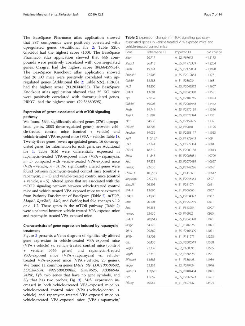
VPA + vehicle). Sly, LOC100504642, LOC380994, 4921509O09Rik, Gm14625, and two genes without genesymbols decreased in both vehicle-treated VPA-exposedmice vs. vehicle-treated control mice (VPA + vehicle /control + vehicle) and rapamycin-treated VPA-exposedmice vs. vehicle-treated VPA-exposed mice (VPA +rapamycin/VPA + vehicle). A330094K24Rik and Fybincreased in vehicle-treated VPA-exposed mice vs.vehicle-treated control mice (VPA + vehicle / control +vehicle) but decreased in rapamycin-treated VPA-ex-posed mice vs. vehicle-treated VPA-exposed mice(VPA + rapamycin / VPA + vehicle). Furthermore, weinvestigated networks of A330094K24Rik and Fyb using“build networks” in MetaCore. We were unable to de-tect networks for A330094K24Rik but found that Fybwas in networks that are associated with p70S6, which
is downstream of the mTOR signaling pathway (Fig. 4;Fyb is also referred to as SLAP-130 or ADAP).
Expression and phosphorylation levels of S6A previous study reported that rapamycin treatment inTsc1+/− and Tsc2+/− mice improved social interactiondeficits and decreased the protein level of activatedphospho-S6K [21]. The phosphorylation of S6K phos-phorylates S6, and this process is associated with proteinsynthesis [8]. A recent study showed that lymphoblas-toid cell lines from patients with idiopathic autism in-creased S6 phosphorylation compared with controls[36]. Therefore, we analyzed the phosphorylation of S6protein in whole mouse brains in each group (Fig. 5a).Vehicle-treated VPA-exposed mice exhibited an eleva-tion of phospho-S6 levels compared with vehicle-treated
Fig. 3 Effects of rapamycin treatment on gene expression. The expression of 5644 genes significantly changed between vehicle-treatedVPA-exposed mice and vehicle-treated control mice (VPA + vehicle / control + vehicle). The expression of 23 genes significantly changedbetween vehicle-treated VPA-exposed mice and rapamycin-treated VPA-exposed mice (VPA + rapamycin / VPA + vehicle). The expression of11 genes changed in both groups. The expression of A330094K24Rik and Fyb oppositely changed between the two groups
Kotajima-Murakami et al. Molecular Brain (2019) 12:3 Page 8 of 14
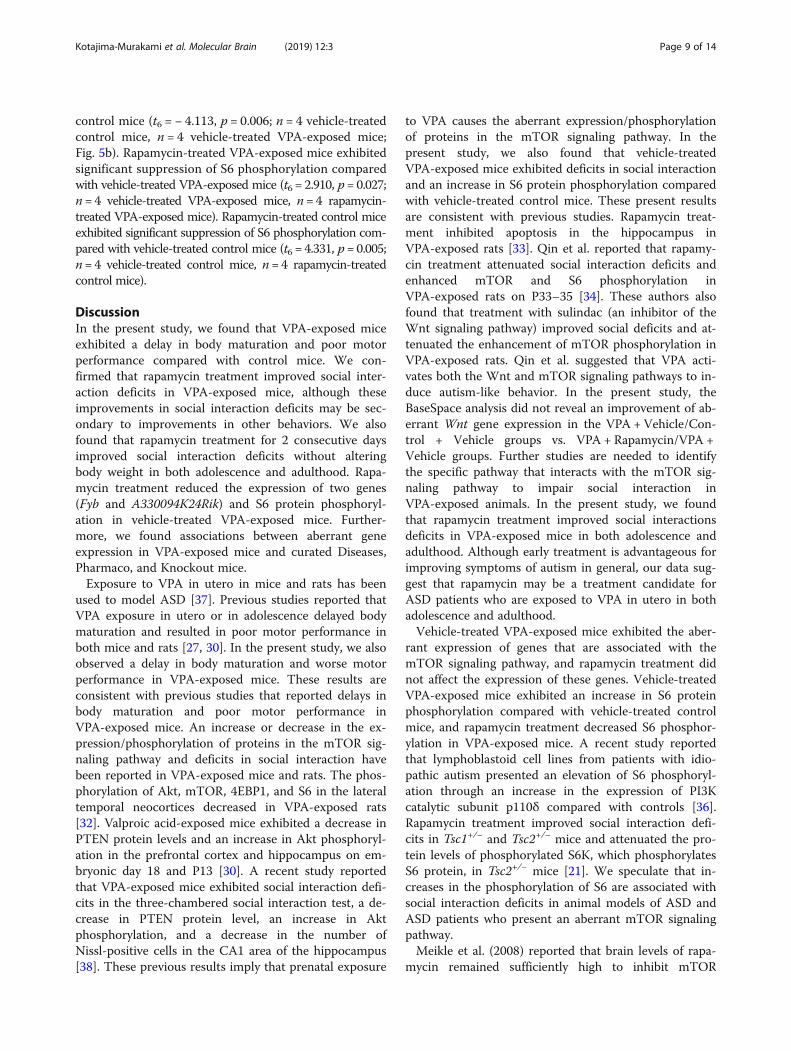
control mice (t6 = − 4.113, p = 0.006; n = 4 vehicle-treatedcontrol mice, n = 4 vehicle-treated VPA-exposed mice;Fig. 5b). Rapamycin-treated VPA-exposed mice exhibitedsignificant suppression of S6 phosphorylation comparedwith vehicle-treated VPA-exposed mice (t6 = 2.910, p= 0.027;n= 4 vehicle-treated VPA-exposed mice, n= 4 rapamycin-treated VPA-exposed mice). Rapamycin-treated control miceexhibited significant suppression of S6 phosphorylation com-pared with vehicle-treated control mice (t6 = 4.331, p= 0.005;n= 4 vehicle-treated control mice, n= 4 rapamycin-treatedcontrol mice).
DiscussionIn the present study, we found that VPA-exposed miceexhibited a delay in body maturation and poor motorperformance compared with control mice. We con-firmed that rapamycin treatment improved social inter-action deficits in VPA-exposed mice, although theseimprovements in social interaction deficits may be sec-ondary to improvements in other behaviors. We alsofound that rapamycin treatment for 2 consecutive daysimproved social interaction deficits without alteringbody weight in both adolescence and adulthood. Rapa-mycin treatment reduced the expression of two genes(Fyb and A330094K24Rik) and S6 protein phosphoryl-ation in vehicle-treated VPA-exposed mice. Further-more, we found associations between aberrant geneexpression in VPA-exposed mice and curated Diseases,Pharmaco, and Knockout mice.Exposure to VPA in utero in mice and rats has been
used to model ASD [37]. Previous studies reported thatVPA exposure in utero or in adolescence delayed bodymaturation and resulted in poor motor performance inboth mice and rats [27, 30]. In the present study, we alsoobserved a delay in body maturation and worse motorperformance in VPA-exposed mice. These results areconsistent with previous studies that reported delays inbody maturation and poor motor performance inVPA-exposed mice. An increase or decrease in the ex-pression/phosphorylation of proteins in the mTOR sig-naling pathway and deficits in social interaction havebeen reported in VPA-exposed mice and rats. The phos-phorylation of Akt, mTOR, 4EBP1, and S6 in the lateraltemporal neocortices decreased in VPA-exposed rats[32]. Valproic acid-exposed mice exhibited a decrease inPTEN protein levels and an increase in Akt phosphoryl-ation in the prefrontal cortex and hippocampus on em-bryonic day 18 and P13 [30]. A recent study reportedthat VPA-exposed mice exhibited social interaction defi-cits in the three-chambered social interaction test, a de-crease in PTEN protein level, an increase in Aktphosphorylation, and a decrease in the number ofNissl-positive cells in the CA1 area of the hippocampus[38]. These previous results imply that prenatal exposure
to VPA causes the aberrant expression/phosphorylationof proteins in the mTOR signaling pathway. In thepresent study, we also found that vehicle-treatedVPA-exposed mice exhibited deficits in social interactionand an increase in S6 protein phosphorylation comparedwith vehicle-treated control mice. These present resultsare consistent with previous studies. Rapamycin treat-ment inhibited apoptosis in the hippocampus inVPA-exposed rats [33]. Qin et al. reported that rapamy-cin treatment attenuated social interaction deficits andenhanced mTOR and S6 phosphorylation inVPA-exposed rats on P33–35 [34]. These authors alsofound that treatment with sulindac (an inhibitor of theWnt signaling pathway) improved social deficits and at-tenuated the enhancement of mTOR phosphorylation inVPA-exposed rats. Qin et al. suggested that VPA acti-vates both the Wnt and mTOR signaling pathways to in-duce autism-like behavior. In the present study, theBaseSpace analysis did not reveal an improvement of ab-errant Wnt gene expression in the VPA + Vehicle/Con-trol + Vehicle groups vs. VPA + Rapamycin/VPA +Vehicle groups. Further studies are needed to identifythe specific pathway that interacts with the mTOR sig-naling pathway to impair social interaction inVPA-exposed animals. In the present study, we foundthat rapamycin treatment improved social interactionsdeficits in VPA-exposed mice in both adolescence andadulthood. Although early treatment is advantageous forimproving symptoms of autism in general, our data sug-gest that rapamycin may be a treatment candidate forASD patients who are exposed to VPA in utero in bothadolescence and adulthood.Vehicle-treated VPA-exposed mice exhibited the aber-
rant expression of genes that are associated with themTOR signaling pathway, and rapamycin treatment didnot affect the expression of these genes. Vehicle-treatedVPA-exposed mice exhibited an increase in S6 proteinphosphorylation compared with vehicle-treated controlmice, and rapamycin treatment decreased S6 phosphor-ylation in VPA-exposed mice. A recent study reportedthat lymphoblastoid cell lines from patients with idio-pathic autism presented an elevation of S6 phosphoryl-ation through an increase in the expression of PI3Kcatalytic subunit p110δ compared with controls [36].Rapamycin treatment improved social interaction defi-cits in Tsc1+/− and Tsc2+/− mice and attenuated the pro-tein levels of phosphorylated S6K, which phosphorylatesS6 protein, in Tsc2+/− mice [21]. We speculate that in-creases in the phosphorylation of S6 are associated withsocial interaction deficits in animal models of ASD andASD patients who present an aberrant mTOR signalingpathway.Meikle et al. (2008) reported that brain levels of rapa-
mycin remained sufficiently high to inhibit mTOR
Kotajima-Murakami et al. Molecular Brain (2019) 12:3 Page 9 of 14

throughout the 48 h period after rapamycin administra-tion (6 mg/kg, i.p.) in mice. Immunoblot analyses ofbrain lysates that were collected 24 h after rapamycin ad-ministration (6 mg/kg, i.p.) showed a reduction of pS6 inTsc1 null-neuron mice [35]. In the present study, we treatedmice with 10 mg/kg rapamycin. We collected brains onthe same day after the end of the social interaction testin adulthood. The brains were immediately frozen in li-quid nitrogen after collection and stored at − 80 °C untilWestern blot analysis. Thus, we presumed that rapamy-cin continued to exert its effects in VPA-exposed mice.The rapamycin-induced improvements in social inter-
action deficits were transient in VPA-exposed mice, inwhich we found that the effects of rapamycin treatmentdisappeared within 5 weeks (see rapamycin-treatedVPA-exposed mice in Fig. 2e and vehicle-treatedVPA-exposed mice in Fig. 2f ). As mentioned above,brain levels of rapamycin remained sufficiently high toinhibit mTOR throughout the 48 h period after rapamy-cin administration in mice [35]. This previous study in-dicates that the effects of rapamycin are still evident 2days after administration. To our knowledge, no studieshave evaluated the effects of rapamycin on improving so-cial interaction deficits longer than 2 days after adminis-tration. A rat pharmacokinetic study showed that the T1/2
of rapamycin was 14.0 h for intravenous administration
and 33.4 h for oral administration [39]. In the presentstudy, the effects of rapamycin treatment disappearedwithin 5 weeks. Further studies are needed to clarify theeffects of rapamycin treatment on social interaction defi-cits by investigating specific treatment periods, doses, tim-ing, combined drugs and therapies, and the duration ofthe effects.Valproic acid is a histone deacetylase (HDAC) inhibi-
tor that plays a role in transcriptional regulation [40]. Itis used for the treatment of spinal muscular atrophy, inwhich it increases the survival motor neuron (SMN)protein volume [41]. Valproic acid is associated with theregulation of protein expression. Although VPA does nothave 1:1 reactivity with S6, the action of the HDAC in-hibitor may have influenced the expression of total S6 inthe present study.Overactivation of the mTOR signaling pathway elicits
the pathology of ASD. Previous studies and the presentdata demonstrate overactivation of the mTOR signalingpathway in ASD. To our knowledge, only one otherstudy has reported a decrease in mTOR signaling inVPA-exposed animals [32], but it investigated rats ratherthan mice, evaluated different brain regions, andassessed the animals at different ages. Future studiesshould delineate the specific brain regions that are asso-ciated with ASD symptoms, and mTOR signaling
Fig. 4 MetaCore network built from Fyb (SLAP-130 or ADAP). Fyb is also referred to as SLAP-130 or ADAP. The red circle shows Fyb (SLAP-130 orADAP). The networks that are associated with Fyb were built using “build networks” in MetaCore, and interactions were visualized. In thesenetworks, Fyb is indirectly related to S6K (p70 S6 kinase). Bcl-10, B-cell CLL/lymphoma 10; CARD11, caspase recruitment domain family member11; CBM complex, CARD11-Bcl-10-MALT1 signalosome complex; GSK3 alpha, glycogen synthase kinase 3α; GSK3 alpha/beta, glycogen synthasekinase 3α/β; GSK3 beta, glycogen synthase kinase 3β; IKK-gamma, inhibitor of nuclear factor κB kinase subunit γ; MALT1, MALT1 paracaspase; OGT(GlcNAc transferase), O-linked N-acetylglucosamine transferase; p70 S6 kinase, 70 KDa ribosomal protein S6 kinase; p70 S6 kinase 1, 70 KDaribosomal protein S6 kinase 1; p70 S6 kinase 2, 70 KDa ribosomal protein S6 kinase 2; PP2A regulatory, protein phosphatase 2A; PPP2R2C, proteinphosphatase 2 regulatory subunit Bγ; RNPC1, RNA binding motif protein 38 (RNPC1 is previous HGNC symbol for RBM38 gene); Securin, Securin isalso referred to as pituitary tumor-transforming 1 (PTTG1); TAK1 (MAP3K7), mitogen-activated protein kinase kinase kinase 7 (TAK1 is previousHGNC symbol for MAP3K7)
Kotajima-Murakami et al. Molecular Brain (2019) 12:3 Page 10 of 14
pathways can then be investigated in each of these brainregions.We found two negatively correlated genes, Fyb and
A330094K24Rik, between vehicle-treated VPA-exposedmice vs. vehicle-treated control mice and rapamycin-treated VPA-exposed mice vs. vehicle-treated VPA-ex-posed mice. Although A330094K24Rik is registered withMouse Genome Informatics (http://www.informatics.-jax.org/), there is no detected information forA330094K24Rik. Fyb is a FYN binding protein that actsas an adapter protein of FYN [42]. Fyb/SLP-associatedprotein is a negative regulator of interleukin 2 (IL-2)transcription [43]. IL-2 plays an important role in immu-noregulatory functions that are related to the centralnervous system and is reportedly associated with ASD[44]. Fyb is a candidate gene in bioinformatic analyses of
ASD [45]. Gera et al. reported that rapamycin treatmentdecreased the translational ratio of Fyb in LAPC-4 cellsin which AKT is highly phosphorylated by transfectionof myristoylated AKT [46]. In the present study, wefound that Fyb is in the network correlated with S6K,which is downstream of the mTOR signaling pathway(Fig. 4). Fyb in this network is associated with inflamma-tory cytokine and chemokine production [47]. Cytokinesand chemokines play a role in developmental cell death,synapse refinement, and phagocytosis during develop-ment [48]. The aberrant expression of cytokines andchemokines is associated with the pathology of ASD[49]. Fyb may be a key molecule in ASD with an aber-rant mTOR signaling pathway.Previous studies analyzed associations between
VPA-exposed mice and genes that are associated withASD [50, 51], but the characteristics of gene expres-sion in whole brains from VPA-exposed mice havenot yet been comprehensively investigated. Thepresent study is the first to comprehensively investi-gate gene expression in VPA-exposed mice. Thepresent study found positive correlations betweengene expression in VPA-exposed mouse brains andcurated studies of Diseases, Pharmaco, and Knockoutmice. Upregulated and downregulated genes haveshown positive correlations with 148 and 136 dis-eases, respectively (Additional file 2: Table S2a). Theseresults indicate that each up- or downregulated genein VPA-exposed mouse brains showed similarities toDiseases other than ASD. Scores for Vitamin D defi-ciency and Injury of the eye region were greater than90. Interestingly, vitamin D deficiency was assessed asa risk factor for ASD [52]. A recent clinical study re-ported that oral vitamin D supplementation in chil-dren for ASD improved Childhood Autism RatingScale scores compared with the placebo group [53].Moreover, vitamin D acts as an inhibitor of themTOR signaling pathway. Vitamin D activatesDNA-damage-inducible transcript 4 (DDIT4), whichfacilitates activation of the Tsc1/Tsc2 complex; there-fore, vitamin D treatment suppresses downstreammTOR activity [54]. Investigating associations be-tween ASD and the comorbid diseases may be neces-sary to clarify the pathogenesis of ASD.Upregulated genes showed a positive correlation
with 387 compounds, and downregulated genesshowed a positive correlation with 446 compounds.The top compounds with a positive correlation wereGlysidol and Ozagrel. Previous studies reported thatthese compounds were associated with neurogenesisand neurite outgrowth in the developmental mousebrains [55–58]. Although VPA is used to model ASDin animals, unknown is the way in which it causesASD-like symptoms [59]. Thus, analyzing associations
Fig. 5 Western blot of S6 protein phosphorylation. (a) Immunoblots ofphospho- and total S6. Control + Ve; vehicle-treated control mice;Control + Ra, rapamycin-treated control mice; VPA + Ve, vehicle-treatedVPA-exposed mice; VPA + Ra, rapamycin-treated VPA-exposed mice. (b)Densitometric analysis of S6 proteins. Error bars indicate SEM.*p < 0.05, **p < 0.01
Kotajima-Murakami et al. Molecular Brain (2019) 12:3 Page 11 of 14
between the compounds that are presented inAdditional file 2: Table S2b and ASD-like symptomsmay contribute to a better understanding of the pathogen-esis of ASD that is induced by VPA exposure.Alterations of the expression of genes (both upregulated
genes and downregulated genes) were similar to PRKG1KO mice (Additional file 2: Table S2c). PRKG1 KO miceare used as a mouse model of sleep disorders [60]. Inter-estingly, patients with ASD have a high incidence of sleepdisturbances [61]. Additionally, a previous study reportedthat VPA-exposed rats exhibited a disruption of normalsleep architecture and a reduction of the expression levelsof GAD65 and GAD67, which are involved in sleep/wake-fulness, in cortical tissue [62]. Investigations of ASD-likesymptoms in KO mice and other animal models of ASDwill contribute to a better understanding of the pathogen-esis of ASD and medication development.In the present study, we found that rapamycin treat-
ment improved impairments in social interaction in bothadolescence and adulthood. The expression of two genes(Fyb and A330094K24Rik) and S6 phosphorylation werereduced by rapamycin treatment. Altogether, these re-sults suggest that an aberrant mTOR signaling pathwayis associated with impairments in social interaction inVPA-exposed mice, and rapamycin may be an effectivetreatment for adolescent and adult patients with notonly particular syndromic ASD but also non-syndromicASD with an aberrant mTOR signaling pathway.
Additional Files
Additional file 1: Table S1. Gene expression in each group. Geneexpression in vehicle-treated VPA-exposed mice / vehicle-treated controlmice (a) and rapamycin-treated VPA-exposed mice / vehicle-treatedVPA-exposed mice (b). p values were produced by t-test comparisonsbetween VPA-treated and control sample (rapamycin-treated and non-treated samples) probeset intensity values. Ranks are based on foldchanges. Genes are arranged in descending order of fold change.(XLSX 403 kb)
Additional file 2: Table S2. Each atlas application score fromBaseSpace. Each individual tab represents Disease, Pharmaco, andKnockout atlas application. (a) Disease atlas application score. (b)Pharmaco atlas application score. (c) Knockout atlas application score.From BaseSpace. (XLSX 59 kb)
AbbreviationsASD: autism spectrum disorder; Fyb: FYN binding protein; KO: knockout;MAPK: mitogen-activated protein kinase; mTOR: mammalian/mechanistictarget of rapamycin; mTORC1: mTOR complex 1; mTORC2: mTOR complex 2;NF1: neuronal fibromatosis 1; NMDA: N-methyl-D-aspartate;PI3K: phosphatidylinositol-3 kinase; PIKK: phosphatidylinositol-3 kinase (PI3K)-related kinase; PTEN: tensin homolog deleted on chromosome 10;TSC: tuberous sclerosis complex; VPA: valproic acid
AcknowledgementsWe thank Michael Arends for assistance with editing the manuscript andEtsuko Kamegaya and Yukiko Matsushima for assistance with breedingVPA-exposed mice.
FundingThis research was supported by Grants-in-Aid for Scientific Research from theMinistry of Education, Culture, Sports, Science and Technology (MEXT)KAKENHI (26860836, 16 K15565, 16H06276, 15H01303) and Japan Agency forMedical Research and Development (AMED; JP16ek0109015).
Availability of data and materialsSupplemental data are available online. All other data are available from thecorresponding author upon request.
Authors’ contributionsHKM, KI, AS, and SU designed the experiments. HKM and KI wrote the paper.HKM performed the mouse behavioral testing and analyzed the behavioraldata. HKM, HK, and MT analyzed the gene expression data. TK performed theWestern blot analysis. YN and YT performed the microarray analysis. HKMand YH generated a mouse model of ASD by VPA injection and wereresponsible for breeding management.
Ethics approval and consent to participateAll of the animal experiments were performed in accordance with theGuidelines for the Care of Laboratory Animals of the Tokyo MetropolitanInstitute of Medical Science, and the housing conditions were approved bythe Institutional Animal Care and Use Committee.
Consent for publicationNot applicable.
Competing interestsThe authors declare that they have no competing interests.
Publisher’s NoteSpringer Nature remains neutral with regard to jurisdictional claims inpublished maps and institutional affiliations.
Author details1Addictive Substance Project, Tokyo Metropolitan Institute of MedicalScience, 2-1-6 Kamikitazawa, Setagaya-ku, Tokyo, Japan. 2Department ofBiosciences, School of Science and Engineering, Teikyo University, 1-1Toyosatodai, Utsunomiya-shi, Tochigi, Japan. 3Department of MolecularPathogenesis, Graduate School of Medicine, Juntendo University, 2-1-1Hongo, Bunkyo-ku, Tokyo, Japan. 4Department of Pediatrics, Graduate Schoolof Medicine, The University of Tokyo, 7-3-1 Hongo, Bunkyo-ku, Tokyo, Japan.5Department of Pediatrics, The University of Tokyo Hospital, 7-3-1 Hongo,Bunkyo-ku, Tokyo, Japan. 6Department of Developmental Disorders, NationalInstitute of Mental Health, National Center of Neurology and Psychiatry, 4-1-1Higashimachi, Kodaira-shi, Tokyo, Japan. 7Center for Basic TechnologyResearch, Tokyo Metropolitan Institute of Medical Science, 2-1-6Kamikitazawa, Setagaya-ku, Tokyo, Japan.
Received: 30 September 2018 Accepted: 25 December 2018
References1. American Psychiatric Association. Diagnostic and statistical manual of
mental disorders. 5th ed. Washington DC: American Psychiatric Publishing;2013. p. 50–9.
2. Constantino JN. The quantitative nature of autistic social impairment.Pediatr Res. 2011;69:55R–62R.
3. Kim YS, Leventhal BL, Koh YJ, Fombonne E, Laska E, Lim EC, et al.Prevalence of autism spectrum disorders in a total population sample. Am JPsychiatry. 2011;168:904–12.
4. Russell G, Rodgers LR, Ukoumunne OC, Ford T. Prevalence of parent-reported ASD and ADHD in the UK: findings from the millennium cohortstudy. J Autism Dev Disord. 2014;44:31–40.
5. Christensen DL, Baio J, Van Naarden Braun K, Bilder D, Charles J,Constantino JN, et al. Prevalence and characteristics of autism spectrumdisorder among children aged 8 years: autism and developmentaldisabilities monitoring network, 11 sites, United States, 2012. MMWR SurveillSumm. 2016;65:1–23.
6. Werling DM, Geschwind DH. Sex differences in autism spectrum disorders.Curr Opin Neurol. 2013;26:146–53.
Kotajima-Murakami et al. Molecular Brain (2019) 12:3 Page 12 of 14
7. Varghese M, Keshav N, Jacot-Descombes S, Warda T, Wicinski B, DicksteinDL, et al. Autism spectrum disorder: neuropathology and animal models.Acta Neuropathol. 2017;134:537–66.
8. Xiong Y, Sheen J. The role of target of rapamycin signaling networks inplant growth and metabolism. Plant Physiol. 2014;164:499–512.
9. Heitman J, Movva NR, Hall MN. Targets for cell cycle arrest by theimmunosuppressant rapamycin in yeast. Science. 1991;253:905–9.
10. Laplante M, Sabatini DM. mTOR signaling in growth control and disease.Cell. 2012;149:274–93.
11. Costa-Mattioli M, Monteggia LM. mTOR complexes in neurodevelopmentaland neuropsychiatric disorders. Nat Neurosci. 2013;16:1537–43.
12. de Vries PJ, Hunt A, Bolton PF. The psychopathologies of children andadolescents with tuberous sclerosis complex (TSC): a postal survey of UKfamilies. Eur Child Adolesc Psychiatry. 2007;16:16–24.
13. Silverman JL, Yang M, Lord C, Crawley JN. Behavioural phenotyping assaysfor mouse models of autism. Nat Rev Neurosci. 2010;11:490–502.
14. Goorden SM, van Woerden GM, van der Weerd L, Cheadle JP, Elgersma Y.Cognitive deficits in Tsc1+/− mice in the absence of cerebral lesions andseizures. Ann Neurol. 2007;62:648–55.
15. Rubenstein JL, Merzenich MM. Model of autism: increased ratio of excitation/inhibition in key neural systems. Genes Brain Behav. 2003;2:255–67.
16. Bateup HS, Johnson CA, Denefrio CL, Saulnier JL, Kornacker K, Sabatini BL.Excitatory/inhibitory synaptic imbalance leads to hippocampal hyperexcitabilityin mouse models of tuberous sclerosis. Neuron. 2013;78:510–22.
17. Page DT, Kuti OJ, Prestia C, Sur M. Haploinsufficiency for Pten and serotonintransporter cooperatively influences brain size and social behavior. Proc NatlAcad Sci U S A. 2009;106:1989–94.
18. Kwon CH, Luikart BW, Powell CM, Zhou J, Matheny SA, Zhang W, et al. Ptenregulates neuronal arborization and social interaction in mice. Neuron. 2006;50:377–88.
19. Molosh AI, Johnson PL, Spence JP, Arendt D, Federici LM, Bernabe C, et al.Social learning and amygdala disruptions in Nf1 mice are rescued byblocking p21-activated kinase. Nat Neurosci. 2014;17:1583–90.
20. Ehninger D, Han S, Shilyansky C, Zhou Y, Li W, Kwiatkowski DJ, et al.Reversal of learning deficits in a Tsc2+/− mouse model of tuberous sclerosis.Nat Med. 2008;14:843–8.
21. Sato A, Kasai S, Kobayashi T, Takamatsu Y, Hino O, Ikeda K, et al. Rapamycinreverses impaired social interaction in mouse models of tuberous sclerosiscomplex. Nat Commun. 2012;3:1292.
22. Tang G, Gudsnuk K, Kuo SH, Cotrina ML, Rosoklija G, Sosunov A, et al. Lossof mTOR-dependent macroautophagy causes autistic-like synaptic pruningdeficits. Neuron. 2014;83:1131–43.
23. Zhou J, Blundell J, Ogawa S, Kwon CH, Zhang W, Sinton C, et al.Pharmacological inhibition of mTORC1 suppresses anatomical, cellular, andbehavioral abnormalities in neural-specific Pten knock-out mice. J Neurosci.2009;29:1773–83.
24. Hwang SK, Lee JH, Yang JE, Lim CS, Lee JA, Lee YS, et al. Everolimusimproves neuropsychiatric symptoms in a patient with tuberous sclerosiscarrying a novel TSC2 mutation. Mol Brain. 2016;9:56.
25. Christensen J, Grønborg TK, Sørensen MJ, Schendel D, Parner ET, PedersenLH, et al. Prenatal valproate exposure and risk of autism spectrum disordersand childhood autism. JAMA. 2013;309:1696–703.
26. Schneider T, Przewłocki R. Behavioral alterations in rats prenatally exposedto valproic acid: animal model of autism. Neuropsychopharmacology. 2005;30:80–9.
27. Wagner GC, Reuhl KR, Cheh M, McRae P, Halladay AK. A newneurobehavioral model of autism in mice: pre- and postnatal exposure tosodium valproate. J Autism Dev Disord. 2006;36:779–93.
28. Kataoka S, Takuma K, Hara Y, Maeda Y, Ago Y, Matsuda T.Autism-like behaviours with transient histone hyperacetylationin mice treated prenatally with valproic acid. Int JNeuropsychopharmacol. 2013;16:91–103.
29. Gurpur PB, Liu J, Burkin DJ, Kaufman SJ. Valproic acid activates the PI3K/Akt/mTOR pathway in muscle and ameliorates pathology in a mouse model ofDuchenne muscular dystrophy. Am J Pathol. 2009;174:999–1008.
30. Yang EJ, Ahn S, Lee K, Mahmood U, Kim HS. Early behavioral abnormalitiesand perinatal alterations of PTEN/AKT pathway in valproic acid autismmodel mice. PLoS One. 2016;11:e0153298.
31. Kang J, Kim E. Suppression of NMDA receptor function in mice prenatallyexposed to valproic acid improves social deficits and repetitive behaviors.Front Mol Neurosci. 2015;8:17.
32. Nicolini C, Ahn Y, Michalski B, Rho JM, Fahnestock M. Decreased mTORsignaling pathway in human idiopathic autism and in rats exposed tovalproic acid. Acta Neuropathol Commun. 2015;3:3.
33. Zhang J, Zhang JX, Zhang QL. PI3K/AKT/mTOR-mediated autophagy in thedevelopment of autism spectrum disorder. Brain Res Bull. 2016;125:152–8.
34. Qin L, Dai X, Yin Y. Valproic acid exposure sequentially activates Wnt andmTOR pathways in rats. Mol Cell Neurosci. 2016;75:27–35.
35. Meikle L, Pollizzi K, Egnor A, Kramvis I, Lane H, Sahin M, et al. Response of aneuronal model of tuberous sclerosis to mammalian target of rapamycin(mTOR) inhibitors: effects on mTORC1 and Akt signaling lead to improvedsurvival and function. J Neurosci. 2008;(21):5422–32.
36. Poopal AC, Schroeder LM, Horn PS, Bassell GJ, Gross C. Increased expressionof the PI3K catalytic subunit p110δ underlies elevated S6 phosphorylationand protein synthesis in an individual with autism from a multiplex family.Mol Autism. 2016;7:3.
37. Mabunga DF, Gonzales EL, Kim JW, Kim KC, Shin CY. Exploring the validityof valproic acid animal model of autism. Exp Neurobiol. 2015;24:285–300.
38. Mahmood U, Ahn S, Yang EJ, Choi M, Kim H, Regan P, et al. Dendritic spineanomalies and PTEN alterations in a mouse model of VPA-induced autismspectrum disorder. Pharmacol Res. 2018;128:110–21.
39. Chen YW, Smith ML, Sheets M, Ballaron S, Trevillyan JM, Burke SE, et al.Zotarolimus, a novel sirolimus analogue with potent anti - proliferativeactivity on coronary smooth muscle cells and reduced potential forsystemic immunosuppression. J Cardiovasc Pharmacol. 2007;49:228–35.
40. Göttlicher M, Minucci S, Zhu P, Krämer OH, Schimpf A, Giavara S, et al.Valproic acid defines a novel class of HDAC inhibitors inducingdifferentiation of transformed cells. EMBO J. 2001;20:669–78.
41. Seo J, Howell MD, Singh NN, Singh RN. Spinal muscular atrophy: an updateon therapeutic progress. Biochim Biophys Acta. 2013;1832:2180–90.
42. Liu J, Kang H, Raab M, da Silva AJ, Kraeft SK, Rudd CE. FYB (FYN bindingprotein) serves as a binding partner for lymphoid protein and FYN kinasesubstrate SKAP55 and a SKAP55-related protein in T cells. Proc Natl Acad SciU S A. 1998;95:8779–84.
43. Musci MA, Hendricks-Taylor LR, Motto DG, Paskind M, Kamens J, Turck CW,et al. Molecular cloning of SLAP-130, an SLP-76-associated substrate of the Tcell antigen receptor-stimulated protein tyrosine kinases. J Biol Chem. 1997;272:11674–7.
44. Xu N, Li X, Zhong Y. Inflammatory cytokines: potential biomarkers ofimmunologic dysfunction in autism spectrum disorders. Mediat Inflamm.2015;2015:531518.
45. Yonan AL, Palmer AA, Smith KC, Feldman I, Lee HK, Yonan JM, et al.Bioinformatic analysis of autism positional candidate genes usingbiological databases and computational gene network prediction. GenesBrain Behav. 2003;2:303–20.
46. Gera JF, Mellinghoff IK, Shi Y, Rettig MB, Tran C, Hsu JH, et al. AKT activitydetermines sensitivity to mammalian target of rapamycin (mTOR) inhibitors byregulating cyclin D1 and c-myc expression. J Biol Chem. 2004;279:2737–46.
47. Rajasekaran K, Kumar P, Schuldt KM, Peterson EJ, Vanhaesebroeck B, Dixit V,et al. Signaling by Fyn-ADAP via the Carma1-Bcl-10-MAP3K7 signalosomeexclusively regulates inflammatory cytokine production in NK cells. NatImmunol. 2013;14:1127–36.
48. Deverman BE, Patterson PH. Cytokines and CNS development. Neuron.2009;64:61–78.
49. Young AM, Chakrabarti B, Roberts D, Lai MC, Suckling J, Baron-Cohen S.From molecules to neural morphology: understanding neuroinflammationin autism spectrum condition. Mol Autism. 2016;7:9.
50. Kolozsi E, Mackenzie RN, Roullet FI, deCatanzaro D, Foster JA. Prenatalexposure to valproic acid leads to reduced expression of synaptic adhesionmolecule neuroligin 3 in mice. Neuroscience. 2009;163:1201–10.
51. Kawanai T, Ago Y, Watanabe R, Inoue A, Taruta A, Onaka Y, et al. Prenatalexposure to histone deacetylase inhibitors affects gene expression ofautism-related molecules and delays neuronal maturation. Neurochem Res.2016;41:2574–84.
52. Máčová L, Bičíková M, Ostatníková D, Hill M, Stárka L, Vitamin D.neurosteroids and autism. Physiol Res. 2017;66(Suppl 3):S333–40.
53. Saad K, Abdel-Rahman AA, Elserogy YM, Al-Atram AA, El-Houfey AA,Othman HA, et al. Randomized controlled trial of vitamin Dsupplementation in children with autism spectrum disorder. J Child PsycholPsychiatry. 2018;59:20–9.
54. Lisse TS, Liu T, Irmler M, Beckers J, Chen H, Adams JS, et al.Gene targeting by the vitamin D response element binding protein
Kotajima-Murakami et al. Molecular Brain (2019) 12:3 Page 13 of 14
reveals a role for vitamin D in osteoblast mTOR signaling. FASEB J.2011;25:937–47.
55. Akane H, Shiraki A, Imatanaka N, Akahori Y, Itahashi M, Ohishi T, et al.Glycidol induces axonopathy by adult-stage exposure and aberration ofhippocampal neurogenesis affecting late-stage differentiation bydevelopmental exposure in rats. Toxicol Sci. 2013;134:140–54.
56. Kawashima M, Watanabe Y, Nakajima K, Murayama H, Nagahara R, Jin M, etal. Late effect of developmental exposure to glycidol on hippocampalneurogenesis in mice: loss of parvalbumin-expressing interneurons. ExpToxicol Pathol. 2017;69:517–26.
57. Aritake S, Oguro H, Iwasa K, Mitaki S, Takayoshi H, Abe S, Onoda K,Yamaguchi S. Clinical efficacy of ozagrel with or without edaravone in 156acute stroke patients. J Neurol Neuromed. 2017;2:16–9.
58. Sumimoto S, Muramatsu R, Yamashita T. Thromboxane A2 stimulatesneurite outgrowth in cerebral cortical neurons via mitogen activatedprotein kinase signaling. Brain Res. 2015;1594:46–51.
59. Mabunga DF, Gonzales EL, Kim JW, Kim KC, Shin CY. Exploring the validityof Valproic acid animal model of autism. Exp Neurobiol. 2015;24:285–300.
60. Feil R, Hölter SM, Weindl K, Wurst W, Langmesser S, Gerling A, et al. cGMP-dependent protein kinase I, the circadian clock, sleep and learning.Commun Integr Biol. 2009;2:298–301.
61. Moore M, Evans V, Hanvey G, Johnson C. Assessment of sleep in childrenwith autism spectrum disorder. Child Aust. 2017;4:72.
62. Cusmano DM, Mong JA. In utero exposure to valproic acid changes sleep injuvenile rats: a model for sleep disturbances in autism. Sleep. 2014;37:1489–99.
Kotajima-Murakami et al. Molecular Brain (2019) 12:3 Page 14 of 14
Related Documents


![Target of Rapamycin Signaling in Plant Stress …Update on Target of Rapamycin Signaling in Plant Stress Responses Target of Rapamycin Signaling in Plant Stress Responses1[OPEN] Liwen](https://static.cupdf.com/doc/110x72/5f05e4b57e708231d4153f1e/target-of-rapamycin-signaling-in-plant-stress-update-on-target-of-rapamycin-signaling.jpg)

