Differential Effects of Rapamycin and Dexamethasone in Mouse Models of Established Allergic Asthma Elizabeth M. Mushaben 1 , Eric B. Brandt 2 , Gurjit K. Khurana Hershey 2 , Timothy D. Le Cras 1 * 1 Division of Pulmonary Biology, Department of Pediatrics, Cincinnati Children’s Hospital, University of Cincinnati School of Medicine, Cincinnati, Ohio, United States of America, 2 Division of Asthma Research, Department of Pediatrics, Cincinnati Children’s Hospital, University of Cincinnati School of Medicine, Cincinnati, Ohio, United States of America Abstract The mammalian target of rapamycin (mTOR) plays an important role in cell growth/differentiation, integrating environmental cues, and regulating immune responses. Our lab previously demonstrated that inhibition of mTOR with rapamycin prevented house dust mite (HDM)-induced allergic asthma in mice. Here, we utilized two treatment protocols to investigate whether rapamycin, compared to the steroid, dexamethasone, could inhibit allergic responses during the later stages of the disease process, namely allergen re-exposure and/or during progression of chronic allergic disease. In protocol 1, BALB/c mice were sensitized to HDM (three i.p. injections) and administered two intranasal HDM exposures. After 6 weeks of rest/recovery, mice were re-exposed to HDM while being treated with rapamycin or dexamethasone. In protocol 2, mice were exposed to HDM for 3 or 6 weeks and treated with rapamycin or dexamethasone during weeks 4–6. Characteristic features of allergic asthma, including IgE, goblet cells, airway hyperreactivity (AHR), inflammatory cells, cytokines/ chemokines, and T cell responses were assessed. In protocol 1, both rapamycin and dexamethasone suppressed goblet cells and total CD4 + T cells including activated, effector, and regulatory T cells in the lung tissue, with no effect on AHR or total inflammatory cell numbers in the bronchoalveolar lavage fluid. Rapamycin also suppressed IgE, although IL-4 and eotaxin 1 levels were augmented. In protocol 2, both drugs suppressed total CD4 + T cells, including activated, effector, and regulatory T cells and IgE levels. IL-4, eotaxin, and inflammatory cell numbers were increased after rapamycin and no effect on AHR was observed. Dexamethasone suppressed inflammatory cell numbers, especially eosinophils, but had limited effects on AHR. We conclude that while mTOR signaling is critical during the early phases of allergic asthma, its role is much more limited once disease is established. Citation: Mushaben EM, Brandt EB, Hershey GKK, Le Cras TD (2013) Differential Effects of Rapamycin and Dexamethasone in Mouse Models of Established Allergic Asthma. PLoS ONE 8(1): e54426. doi:10.1371/journal.pone.0054426 Editor: Christian Taube, Leiden University Medical Center, The Netherlands Received September 18, 2012; Accepted December 11, 2012; Published January 17, 2013 Copyright: ß 2013 Mushaben et al. This is an open-access article distributed under the terms of the Creative Commons Attribution License, which permits unrestricted use, distribution, and reproduction in any medium, provided the original author and source are credited. Funding: This research was funded by National Institutes of Health Grants U19A170235 (GKH), HL097135 (TDLC and GKH), and NIEHS T32 ES010957 (EBB). The funders had no role in study design, data collection and analysis, decision to publish, or preparation of the manuscript. Competing Interests: The authors have declared that no competing interests exist. * E-mail: [email protected] Introduction Allergic asthma is a heterogeneous disease characterized by airway hyperreactivity (AHR), inflammation, goblet cell metapla- sia, and increases in Th2 cytokines and IgE [1,2,3,4]. Although current therapies such as glucocorticoids and bronchodilators are effective in suppressing symptoms in some patients, not all asthmatic patients respond to these therapies [1]. As the prevalence of asthma continues to rise, especially in children [1,5,6], it is imperative that the mechanisms underlying this disease be identified. For some patients, allergic asthma is an ongoing disease, but for others, asthma symptoms only develop when patients are exposed to seasonal allergens or are exposed to a stimulus that provokes their asthma symptoms. Asthma exacerbations are a major problem and account for a high proportion of emergency room visits, hospitalizations and healthcare related cost [7,8]. Prevention of these exacerbations or reversal of chronic, established allergic disease would help improve disease management and reduce both hospitalizations and deaths from acute asthma attacks. Mammalian target of rapamycin (mTOR) signaling occurs downstream of the PI3K-signaling cascade and is known to play a major role in growth/differentiation, cell metabolism, and survival in many different cell types [9,10]. More recent work has demonstrated an important role for mTOR in T cell proliferation and differentiation [11,12,13]. An inhibitor of mTOR, rapamycin, is already used clinically as an immunosuppressant to prevent organ rejection after transplantation [14,15]. In addition, the use of rapamycin in patients suffering from the destructive lung disease, lymphangioleiomyomatosis [16], has demonstrated prom- ise in its ability to reduce disease symptoms and stabilize lung function [17]. Previously, our lab demonstrated that inhibition of mTOR with rapamycin prevented allergic asthma in a mouse model induced by exposure to the allergen, house dust mite (HDM). In these studies, rapamycin prevented the allergic response and still suppressed many key asthma characteristics after allergic sensitization was established [18]. Although this study showed that mTOR inhibition could suppress allergic asthma early in the disease process, the role of mTOR during allergen re- exposure and chronic, established allergic disease remained unclear. The goal of this study was to determine whether inhibition of mTOR with rapamycin would attenuate key characteristics of allergic asthma in two models that addressed chronic/established PLOS ONE | www.plosone.org 1 January 2013 | Volume 8 | Issue 1 | e54426

Welcome message from author
This document is posted to help you gain knowledge. Please leave a comment to let me know what you think about it! Share it to your friends and learn new things together.
Transcript
Differential Effects of Rapamycin and Dexamethasone inMouse Models of Established Allergic AsthmaElizabeth M. Mushaben1, Eric B. Brandt2, Gurjit K. Khurana Hershey2, Timothy D. Le Cras1*
1 Division of Pulmonary Biology, Department of Pediatrics, Cincinnati Children’s Hospital, University of Cincinnati School of Medicine, Cincinnati, Ohio, United States of
America, 2 Division of Asthma Research, Department of Pediatrics, Cincinnati Children’s Hospital, University of Cincinnati School of Medicine, Cincinnati, Ohio, United
States of America
Abstract
The mammalian target of rapamycin (mTOR) plays an important role in cell growth/differentiation, integratingenvironmental cues, and regulating immune responses. Our lab previously demonstrated that inhibition of mTOR withrapamycin prevented house dust mite (HDM)-induced allergic asthma in mice. Here, we utilized two treatment protocols toinvestigate whether rapamycin, compared to the steroid, dexamethasone, could inhibit allergic responses during the laterstages of the disease process, namely allergen re-exposure and/or during progression of chronic allergic disease. In protocol1, BALB/c mice were sensitized to HDM (three i.p. injections) and administered two intranasal HDM exposures. After 6 weeksof rest/recovery, mice were re-exposed to HDM while being treated with rapamycin or dexamethasone. In protocol 2, micewere exposed to HDM for 3 or 6 weeks and treated with rapamycin or dexamethasone during weeks 4–6. Characteristicfeatures of allergic asthma, including IgE, goblet cells, airway hyperreactivity (AHR), inflammatory cells, cytokines/chemokines, and T cell responses were assessed. In protocol 1, both rapamycin and dexamethasone suppressed goblet cellsand total CD4+ T cells including activated, effector, and regulatory T cells in the lung tissue, with no effect on AHR or totalinflammatory cell numbers in the bronchoalveolar lavage fluid. Rapamycin also suppressed IgE, although IL-4 and eotaxin 1levels were augmented. In protocol 2, both drugs suppressed total CD4+ T cells, including activated, effector, and regulatoryT cells and IgE levels. IL-4, eotaxin, and inflammatory cell numbers were increased after rapamycin and no effect on AHR wasobserved. Dexamethasone suppressed inflammatory cell numbers, especially eosinophils, but had limited effects on AHR.We conclude that while mTOR signaling is critical during the early phases of allergic asthma, its role is much more limitedonce disease is established.
Citation: Mushaben EM, Brandt EB, Hershey GKK, Le Cras TD (2013) Differential Effects of Rapamycin and Dexamethasone in Mouse Models of Established AllergicAsthma. PLoS ONE 8(1): e54426. doi:10.1371/journal.pone.0054426
Editor: Christian Taube, Leiden University Medical Center, The Netherlands
Received September 18, 2012; Accepted December 11, 2012; Published January 17, 2013
Copyright: � 2013 Mushaben et al. This is an open-access article distributed under the terms of the Creative Commons Attribution License, which permitsunrestricted use, distribution, and reproduction in any medium, provided the original author and source are credited.
Funding: This research was funded by National Institutes of Health Grants U19A170235 (GKH), HL097135 (TDLC and GKH), and NIEHS T32 ES010957 (EBB). Thefunders had no role in study design, data collection and analysis, decision to publish, or preparation of the manuscript.
Competing Interests: The authors have declared that no competing interests exist.
* E-mail: [email protected]
Introduction
Allergic asthma is a heterogeneous disease characterized by
airway hyperreactivity (AHR), inflammation, goblet cell metapla-
sia, and increases in Th2 cytokines and IgE [1,2,3,4]. Although
current therapies such as glucocorticoids and bronchodilators are
effective in suppressing symptoms in some patients, not all
asthmatic patients respond to these therapies [1]. As the
prevalence of asthma continues to rise, especially in children
[1,5,6], it is imperative that the mechanisms underlying this
disease be identified.
For some patients, allergic asthma is an ongoing disease, but for
others, asthma symptoms only develop when patients are exposed
to seasonal allergens or are exposed to a stimulus that provokes
their asthma symptoms. Asthma exacerbations are a major
problem and account for a high proportion of emergency room
visits, hospitalizations and healthcare related cost [7,8]. Prevention
of these exacerbations or reversal of chronic, established allergic
disease would help improve disease management and reduce both
hospitalizations and deaths from acute asthma attacks.
Mammalian target of rapamycin (mTOR) signaling occurs
downstream of the PI3K-signaling cascade and is known to play a
major role in growth/differentiation, cell metabolism, and survival
in many different cell types [9,10]. More recent work has
demonstrated an important role for mTOR in T cell proliferation
and differentiation [11,12,13]. An inhibitor of mTOR, rapamycin,
is already used clinically as an immunosuppressant to prevent
organ rejection after transplantation [14,15]. In addition, the use
of rapamycin in patients suffering from the destructive lung
disease, lymphangioleiomyomatosis [16], has demonstrated prom-
ise in its ability to reduce disease symptoms and stabilize lung
function [17]. Previously, our lab demonstrated that inhibition of
mTOR with rapamycin prevented allergic asthma in a mouse
model induced by exposure to the allergen, house dust mite
(HDM). In these studies, rapamycin prevented the allergic
response and still suppressed many key asthma characteristics
after allergic sensitization was established [18]. Although this study
showed that mTOR inhibition could suppress allergic asthma
early in the disease process, the role of mTOR during allergen re-
exposure and chronic, established allergic disease remained
unclear.
The goal of this study was to determine whether inhibition of
mTOR with rapamycin would attenuate key characteristics of
allergic asthma in two models that addressed chronic/established
PLOS ONE | www.plosone.org 1 January 2013 | Volume 8 | Issue 1 | e54426
disease, namely allergen re-exposure and disease progression. In
addition to rapamycin, mice were also treated with the steroid,
dexamethasone, for comparison purposes since steroids are
currently a mainstay therapy for chronic asthma [1]. We
hypothesized that rapamycin and dexamethasone would suppress
asthma exacerbations during allergen re-exposure and suppress
progressive/ongoing allergic disease by inhibiting T cells. To test
this hypothesis, mice in protocol one, which was designed to mimic
the effects of allergen re-exposure in a previously sensitized
individual, were sensitized to HDM by i.p. injection and then
exposed to intranasal HDM to induce asthma. Then, after a 6
week rest/recovery period, mice were re-exposed to HDM while
being treated with rapamycin or dexamethasone. In protocol two,
to address the role of mTOR in chronic/established allergic
asthma, mice were exposed to HDM for 6 weeks and treated with
rapamycin or dexamethasone from weeks 4 to 6 of the exposure
period. Endpoints assessed included allergen specific IgE, AHR,
inflammatory cells, goblet cell metaplasia, cytokine/chemokine
levels, and T cell numbers.
Materials and Methods
Ethics Statement and Animal Treatment ProtocolsAnimal procedures and protocols were approved by the Animal
Care and Use Committee at the Cincinnati Children’s Hospital
Research Foundation (Cincinnati, OH) (Protocol Number:
1D02011). All procedures were performed under anesthesia to
minimize suffering. Female BALB/c mice were purchased at 6–8
weeks of age from Charles River Laboratories (Wilmington, MA).
The two different treatment protocols that were used in this study
are described below, and each study was performed twice.
Protocol 1: Allergen Re-Exposure ModelMice were sensitized to HDM by three intraperitoneal (i.p.)
injections (50 mg HDM in 100 ml saline; Greer Laboratories,
Lenoir, NC) at 7 day intervals (Figure 1A). Two intranasal (i.n.)
HDM exposures (50 mg HDM in 20 ml saline), 2 days apart, were
administered 7 days after the last i.p. injection. One group of mice
was sacrificed 48 hours after the last i.n. HDM exposure to
determine the disease phenotype at this time point (Group 1). The
remaining mice were allowed to rest from allergen exposure for 6
weeks, with no treatment. Mice from group 2 were sacrificed after
6 weeks of rest/recovery and before allergen re-exposure. Mice in
group 3, which were previously exposed to HDM and rested for 6
weeks, were re-exposed to i.n. HDM (50 mg HDM in 20 ml saline)
twice, 2 days apart. Rapamycin (4 mg/kg; LC Laboratories,
Woburn, MA), dexamethasone (1 mg/kg; Sigma Aldrich, St.
Louis, MO), or vehicle (0.25% PEG400, 0.25% Tween20 in
dH2O) was administered by i.p. injection one day prior to i.n.
HDM re-exposure and continued for 6 days for a total of 6 drug
treatments (Figure 1A). All mice re-exposed to HDM (group 3)
received vehicle 6 rapamycin or dexamethasone.
Protocol 2: Chronic Allergen/Reversal ModelMice were exposed to intranasal doses of HDM (50 mg in 20 ul
saline) or saline (0.9% NaCl, 20 ml; control group) 3 times a week
for 3 or 6 weeks (Figure 1B). Another group of mice were treated
with rapamycin (4 mg/kg) or dexamethasone (1 mg/kg) 6 days a
week by i.p. injection starting at week 4 through week 6 of HDM
exposure.
Allergic SensitizationTo assess allergic sensitization, HDM-specific IgE and IgG1
levels were measured in the serum by ELISA as previously
described [18,19]. Briefly, an ELISA plate was coated overnight
with 0.01% HDM in PBS. The next day, 200 ml of blocking
solution (1% BSA in PBS) was added to the plate for 1 hour. Next,
50 ml of sample was added to the plate for 1 hour, followed by
biotin-anti-mouse IgE or biotin-anti-mouse IgG1 (2.0 mg/ml,
Pharmingen) for 1 hour. To detect biotin-labeled IgE or IgG1,
streptavidin-HRP (1:100, R & D Systems, Minneapolis, MN) was
added to the plate for 30 minutes. A tetramethylbenzidine
substrate reagent set (1:1)(BD Biosciences, San Jose, CA) was
added to detect levels of IgE or IgG1.
Airway Hyperreactivity (AHR)All mice were anesthetized with a mixture of ketamine/xylaxin/
acepromazine (4:1:1) by i.p. injection 48 hours after the last HDM
exposure and before AHR was assessed. Airway resistance to
increasing doses of methacholine was measured as previously
described [18,20]. Briefly, the trachea of each mouse was
cannulated and connected to a flexiVent system (SCIREQ,
Montreal, QC, Canada). 16PBS (baseline) and increasing doses
of methacholine (12.5, 25, and 50 mg/ml; acetyl-b-methylcholine
chloride, Sigma, St. Louis, MO) were nebulized into the mouse
lungs via the tracheotomy to measure airway resistance. The
thoracic aorta was cut after lung mechanic measurements were
finished so that blood could be collected for IgE and IgG1
measurements.
Inflammatory Cells and Cytokine LevelsInflammatory cells were collected and assessed in the broncho-
alveolar lavage fluid (BALF) as previously described [18,20].
Briefly, lungs were lavaged with 1 ml of 16PBS containing BSA
(1%) and EDTA (2 mM). The BALF was centrifuged (5,000 rpm),
and the supernatant was collected and frozen at 280uC for
cytokine measurements. Next, cells were resuspended in red blood
cell lysis buffer (Sigma) to lyse any red blood cells. Cells were then
centrifuged again, supernatant was removed, and cells were
resuspended in 16PBS+BSA (1%) and EDTA (2 mM). Total
inflammatory BALF cells were counted using a hemacytometer
and differential cell counts were determined by staining cytospin
slides with Diff-Quick (Shandon Lipshaw, Pittsburgh, PA). Three
hundred cells per slide were counted and then the percentages and
total differential inflammatory cells numbers of macrophages,
lymphocytes, neutrophils, and eosinophils were calculated. A
multiplex biomarker panel (Cytokine/Chemokine Panel I) and
Luminex xMAP technology (Millipore, Billerica, MA) were used to
measure IL-4, IL-5, IL-13, and IL-17A cytokine levels as well as
the chemokine, eotaxin 1 in the BALF. IFN-c cytokine levels were
measured in the BALF by ELISA according to the manufacturer’s
instructions (Biolegend, San Diego, CA).
Western Blot AnalysisWestern blot analysis was performed on lung homogenates
using the following antibodies: chloride channel, calcium activat-
ed, family member 3 (CLCA3, 1:5,000; Abcam, Cambridge, MA)
[18,21,22], Phosphorylated S6 (P-S6, 1:1000; Cell Signaling,
Danvers, MA), Phosphorylated Akt (P-Akt, 1:1000; Cell Signaling,
Danvers, MA), and SAM-pointed domain-containing Ets-like
factor (SPDEF, 1:5,000; in house). To control for protein loading,
C4 Actin (1:40,000; Seven Hills Bioreagents, Cincinnati, OH)
levels were assessed and CLCA3, P-S6, and SPDEF levels were
normalized to Actin levels. P-Akt was normalized to total Akt
levels (1:1000; Cell Signaling, Danvers, MA). HRP-conjugated
secondary antibodies were goat anti-mouse and goat anti-rabbit
(1:10,000, Calbiochem). Chemiluminescence was detected using
Luminata Forte Western HRP Substrate (Millipore, Billerica,
Effects of Rapamycin and Dexamethasone on Asthma
PLOS ONE | www.plosone.org 2 January 2013 | Volume 8 | Issue 1 | e54426
MA). Data were imaged using an LAS4000 imaging system and
quantitated with Multi Gauge 3 software (Fujifilm, Tokyo, Japan).
Muc5AC and a-SMA immunohistochemistry. Lungs were
inflation fixed at a constant pressure (25 cmH2O) by tracheal
installation of 4% paraformaldehyde, transferred to 70% ethanol
after 24 hrs, and embedded in paraffin as previously described
[18,23,24]. Immunostaining for Mucin 5AC (Muc5AC) and a-
smooth muscle actin (a-SMA) were performed on 5 mm paraffin-
embedded sections as previously described [18,20,23]. Briefly, for
Muc5AC, slides were incubated with a primary Muc5AC mouse
monoclonal antibody (diluted 1:200, Thermo Scientific, Waltham,
MA) overnight at 4uC, followed by a goat a mouse IgG1
secondary antibody (diluted 1:200, Southern Biotech, Birming-
ham, AL). For a-SMA, slides were incubated with a primary a-
SMA antibody (diluted 1:20,000, Clone 1A4 Sigma, St.
Louis,MO) overnight at 4uC, followed by a goat a mouse IgG2a
secondary antibody (1:200, Southern Biotech, Birmingham, AL).
For both antibodies, signal was detected using the DAB method of
detection. Digital images of Muc5AC and a-SMA immunostaining
were obtained using a Zeiss Axioplan 2 microscope and camera
(Carl Zeiss Microimaging, Thornwood, NY).
Flow CytometryThe upper right lung lobe was minced and incubated at 37uC
for 25–30 minutes in 2 ml of RPMI 1640 containing Liberase DL
(0.5 mg/ml; Roche Diagnostics, Idianapolis, IN) and DNAse I
(0.5 mg/ml; Sigma, St Louis, MO). Lung cells were passed
through a 70 mm cell strainer and the strainer washed with 5 ml of
RPMI+DNAse I media. Cells were centrifuged and resuspended
in 2 ml of RPMI before counting with a hemacytometer and
viability confirmed by trypan blue exclusion. Approximately
500,000 lung cells were transferred to a 96 well plate with V
shaped bottom on ice, centrifuged and resuspended in 16PBS
containing FcBlock (2.4G2 mAb). Lung T cells were stained with
antibodies for CD4-FITC, CD69-PE, CD3e-PE/Cy7, Foxp3-
PerCP5.5, and CD44-PacificBlue (BioLegend, San Diego, CA).
Intracellular staining for Foxp3-PerCP5.5 was performed using the
classic protocol and reagents from eBioscience (San Diego,CA).
Lung cells were also stained with B220-FITC, CD62L-PE, CD4-
PEcy7, CD8b-PerCP5.5, CD3-AF700, and CD44-PB. Live and
dead cells were labeled with LIVE/DEAD Fixable Aqua Dead
Cell Stain Kit according to manufacturer’s instructions (Invitrogen
by Life Technologies, Carlsbad, CA). Acquisition was done on a
FACS Canto III (Becton Dickinson, Mountain View, CA) and
analyzed used FlowJo software (Tree Star, Ashland, OR).
Statistical AnalysisPrism 5 software (GraphPad Software, San Diego, CA) was used
to perform statistical analysis. Statistical tests used included
unpaired t tests, one-way ANOVA with the Bonferroni post hoc
test between selected columns, and two-way ANOVA tests with
the Bonferroni post hoc test. Statistically significant results were
reported when p values were ,0.05.
Results
Protocol 1: Re-Exposure StudyAllergic sensitization, inflammatory cell numbers, and
AHR. Allergic asthma is typically characterized by elevated IgE,
eosinophilia and AHR. HDM-specific IgE levels were increased
after the first round of HDM exposures (group 1) compared to
saline controls (Figure 2A). After 6 weeks of rest/recovery (group
2), IgE levels were not increased compared to saline controls. To
assess the impact of rapamycin and dexamethasone treatment on
sensitization, HDM-specific IgE and HDM-specific IgG1 titers
were assessed in the serum after HDM re-exposure (Figure 2A and
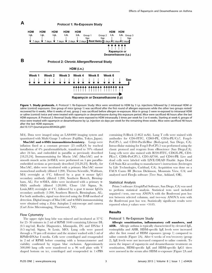
Figure 1. Study protocols. A: Protocol 1: Re-Exposure Study: Mice were sensitized to HDM by 3 i.p. injections followed by 2 intranasal HDM orsaline (control) exposures. One group of mice (group 1) was sacrificed after the first round of allergen exposures while the other two groups rested/recovered for 6 weeks. After 6 weeks of rest, group 2 was sacrificed before allergen re-exposure. Mice in group 3 were re-exposed to intranasal HDMor saline (control) twice and were treated with rapamycin or dexamethasone during this exposure period. Mice were sacrificed 48 hours after the lastHDM exposure. B: Protocol 2: Reversal Study: Mice were exposed to HDM intranasally 3 times per week for 3 or 6 weeks. Starting at week 4, groups ofmice were treated with rapamycin or dexamethasone by i.p. injection six days per week for the remaining three weeks. Mice were sacrificed 48 hoursafter the last HDM exposure.doi:10.1371/journal.pone.0054426.g001
Effects of Rapamycin and Dexamethasone on Asthma
PLOS ONE | www.plosone.org 3 January 2013 | Volume 8 | Issue 1 | e54426
Supplemental Figure 1A, respectively). Following allergen re-
exposure (group 3), HDM-specific IgE levels increased further.
Rapamycin treatment prevented this increase in IgE after HDM
re-exposure, but dexamethasone treatment did not. HDM-specific
IgG1 was increased in HDM exposed mice after 6 weeks of rest/
recovery (group 2). There was no further increase in IgG1 after
HDM re-exposure. Accordingly, neither rapamycin nor dexa-
methasone treatment suppressed HDM-specific IgG1 levels in the
serum (group 3).
To assess inflammatory cell numbers, BALF was collected from
the lungs. Total BALF cell numbers were increased after the first
round of HDM exposures (group 1) (Figure 2B), most notably
eosinophils (Figure 2C and D). After 6 weeks of rest (group 2), total
BALF cell numbers, including lymphocytes, neutrophils, and
eosinophils numbers were similar to saline controls (Figure 2B, C,
and D). Mice re-exposed to HDM after 6 weeks of rest (group 3)
demonstrated increases in total BALF cell numbers similar to those
seen after the initial HDM exposure period (group 1) (Figure 2B)
and this was mainly due to eosinophils (Figure 2C and D).
Rapamycin treatment during HDM re-exposure did not suppress
total BALF cell counts (Figure 2B). In particular, total eosinophil
numbers and percentages remained elevated after rapamycin
treatment (Figure 2C and D). There was a slight, but significant
decrease in total BALF cell counts in the dexamethasone treated
group that was mainly due to lower eosinophil numbers (Figure 2B,
C, and D). Finally, AHR to methacholine was increased (50 mg/
ml; 5.5 fold) in mice after the first round of HDM exposures (group
1) compared to saline controls (Figure 2E). In mice that rested/
recovered for 6 weeks after HDM exposure (group 2), AHR was
similar to saline controls. Upon allergen re-exposure (group 3),
AHR increased in mice (50 mg/mL; 6 fold) compared to saline
controls, but neither rapamycin nor dexamethasone treatment
attenuated this increase in AHR (Figure 2E).
Goblet cells and airway remodeling. Another hallmark of
asthma is increased airway mucus production. To assess changes
in mucus producing goblet cells, the calcium-activated chloride
channel 3 (CLCA3) protein, which associates with the mucin
granule membranes of goblet cells [22], was measured in lung
homogenates by Western blot analysis. In protocol 1, CLCA3
protein was not detectable in saline controls, but was readily
detectable after the first round of HDM exposures (group 1) and
with HDM re-exposure (group 3), but not after 6 weeks of rest
(group 2) (Figure 3A). Rapamycin treatment attenuated increases
in CLCA3 levels after HDM re-exposure, as did dexamethasone,
although rapamycin was more effective. CLCA3 protein levels in
both treatment groups were still higher than saline controls.
Goblet cells also express the transcription factor SAM-pointed
domain-containing Ets-like factor (SPDEF), which has been shown
to be both necessary and sufficient for their differentiation into
mucus producing goblet cells [25]. Similar to CLCA3, SPDEF
levels were increased after HDM re-exposure (group 3) (Figure 3B).
Rapamycin attenuated this increase, however, dexamethasone
treatment did not suppress SPDEF levels (Figure 3B). In addition
to CLCA3 and SPDEF, another goblet cell marker, Muc5AC, was
also assessed. Immunohistochemical staining of lung sections
showed increases in Muc5AC staining in the epithelial cells of
HDM exposed mice (group 1) compared to saline control, that was
attenuated after 6 weeks of rest (group 2) (Figure 3C). Muc5AC
staining increased again after HDM re-exposure (group 3), but
staining was reduced with rapamycin treatment (Figure 3C). Only
a slight reduction in Muc5AC staining was observed in
dexamethasone treated mice (Figure 3C). In addition to Muc5AC,
we also performed immunohistochemical staining for the smooth
muscle cell marker, a-smooth muscle actin (a-SMA), to assess
airway muscularization and remodeling. Similar levels of a-SMA
staining were observed between all groups (Supplemental
Figure 5A). This is consistent with there being no differences in
baseline airway resistance between the groups (Figure 2E).
Systemic and local cellularity. HDM re-exposure (group 3)
induced an increase in lung tissue cellular infiltrate (Supplemental
Figure 2B), associated with increased spleen weights (Supplemental
Figure 2A). Rapamycin and dexamethasone treatments both lead
to decreases in spleen weights and lung cellularity (Supplemental
Figure 2A and 2B) indicating broad anti-inflammatory impact. To
specifically address the impact of these treatments on lung
lymphocytes, flow cytometry was used to identify B and T cells.
Lung CD3+ T cells numbers were increased following HDM re-
exposure (Supplemental Figure 2C). Both rapamycin and dexa-
methasone treatment abrogated this influx of T cells into the lung
tissue (Supplemental Figure 2C). A trend towards a decrease in the
number of B cells after rapamycin treatment was observed;
however, this did not reach statistical significance (Supplemental
Figure 2D).
Lung T cells. Since mTOR has previously been shown to
play an important role in the growth and proliferation of T cells,
we focused on the effects of rapamycin and dexamethasone on
CD4+ T cells. In this study, we characterized T cell subsets after
mice were treated with HDM, rested/recovered for 6 weeks, and
then were re-exposed to HDM since our earlier studies [18] had
already assessed T cells in the early stages of HDM-induced
allergic airway disease. Similar to total CD3+ lung cells, total
CD4+ lung T cells were increased after HDM re-exposure (group
3) compared to saline controls (Figure 4A). Both rapamycin and
dexamethasone treatment suppressed this increase. Next we
assessed specific T cell populations in the lung tissue. CD69+
activated T cells were increased after HDM re-exposure compared
to saline controls (Figure 4B). Both rapamycin and dexamethasone
suppressed this response although rapamycin treatment was more
effective since the number of CD69+ T cells were still increased in
dexamethasone treated mice compared to saline controls
(Figure 4B and Supplemental Figure 7A). When CD69+ activated
T cells were assessed as a percentage of total CD4+ T cells, all
HDM re-exposed mice showed increases in the percentage of
CD69+ T cells compared to saline controls, but only rapamycin
treatment suppressed this response compared to HDM alone
(Figure 4C). CD44+ effector T cells were also increased after
HDM re-exposure compared to saline controls (Figure 4D).
Rapamycin and dexamethasone suppressed this response, howev-
er, the number of effector T cells was still increased in these groups
compared to saline controls. When the percentage of CD44+
effector T cells was determined, increases were observed in all
HDM re-exposed mice, but neither rapamycin nor dexametha-
sone suppressed this response (Figure 4E). Total Foxp3+CD25+
regulatory T cell numbers were also increased in HDM re-exposed
mice compared to saline controls (Figure 4F) and were completely
suppressed by rapamycin and dexamethasone treatment. When
regulatory T cells were assessed as a percentage of CD4+ T cells,
rapamycin, but not dexamethasone, suppressed regulatory T cells,
(Figure 4G). Finally, the ratio of regulatory T cells to CD44+
effector T cells was also determined and all HDM exposed mice
demonstrated decreased ratios compared to saline controls
(Figure 4H). Taken together, these results suggest that rapamycin
and dexamethasone treatment decrease many lung T cell
populations, however, it is unclear if the effects of these drugs
are specific to T cells since both drugs also decreased total lung
cells (Supplemental Figure 2B). In addition, there was a trend
towards a decrease in the number of B cells after rapamycin
Effects of Rapamycin and Dexamethasone on Asthma
PLOS ONE | www.plosone.org 4 January 2013 | Volume 8 | Issue 1 | e54426

Figure 2. Protocol 1- Allergic sensitization, inflammatory cell numbers in the BALF, and AHR after HDM re-exposure. A, HDM-specificIgE levels were increased in HDM exposed (group 1) and HDM re-exposed (group 3) mice compared to saline controls. Rapamycin (Rapa) attenuatedHDM-induced increases in IgE after allergen re-exposure, while dexamethasone (Dex) had no effect (n = 4–12 mice/group). *p,0.05 versus saline;,p,0.05 versus HDM Rest;ˆp,0.05 versus HDM re-exposure; #p,0.05 versus Dex. B, Total BALF cell numbers were increased in HDM exposed micein groups 1 and 3, but not in mice that rested for 6 weeks after HDM exposure (group 2) and unaltered by Rapa or Dex treatment (n = 4–12 mice/group). *p,0.05 versus saline; ,p,0.05 versus HDM Rest. C, Total numbers of macrophages and eosinophils were increased after HDM re-exposure(group 3), but not in HDM rest (group 2) animals in the BALF. Total neutrophil numbers in the BALF were slightly increased after HDM re-exposure inRapa treated mice. Rapa did not suppress HDM-induced increases in eosinophils. Eosinophil numbers were lower in Dex treated mice compared toHDM re-exposed and Rapa treated groups, but still higher then saline control (n = 4–12 mice/group). *p,0.05 versus saline;ˆp,0.05 versus HDM re-exposure; ,p,0.05 versus HDM Rest; #p,0.05 versus Dex. D, The percentage of eosinophils in the BALF was increased in all HDM exposed groupsexcept HDM rest mice, while the percentage of macrophages were reduced. (n = 4–12 mice/group). *p,0.05 versus saline;ˆp,0.05 versus HDM re-exposure; ,p,0.05 versus HDM Rest; #p,0.05 versus Dex. E, AHR was increased after the initial set of HDM exposures (group 1) compared to salinecontrols and was still increased after allergen re-exposure (group 3). Neither Rapa nor Dex suppressed HDM-induced increases in AHR after allergenre-exposure. AHR was similar to controls in HDM rest mice (group 2) (n = 6–14 mice/group). *p,0.05 versus saline.doi:10.1371/journal.pone.0054426.g002
Effects of Rapamycin and Dexamethasone on Asthma
PLOS ONE | www.plosone.org 5 January 2013 | Volume 8 | Issue 1 | e54426

Figure 3. Protocol 1- Goblet cell markers in HDM re-exposed mice. A, Western blot analysis of lung homogenates showed that the goblet cellprotein, CLCA3, was increased after the initial set of HDM exposures (group 1) and after HDM re-exposure (group 3). Both rapamycin (Rapa) anddexamethasone (Dex) treatment attenuated HDM-induced increases in CLCA3 (n = 3–5 mice/group). *p,0.05 versus saline; ,p,0.05 versus HDMRest;ˆp,0.05 versus HDM re-exposed; #p,0.05 versus Dex. B, HDM-induced increases in the transcription factor, SPDEF, were suppressed by Rapa,
Effects of Rapamycin and Dexamethasone on Asthma
PLOS ONE | www.plosone.org 6 January 2013 | Volume 8 | Issue 1 | e54426

but not Dex after allergen re-exposure (group 3). (n = 3 mice/group). *p,0.05 versus saline;ˆp,0.05 versus HDM re-exposed; #p,0.05 versus Dex. C,Muc5AC immunostaining was increased in lung epithelial cells after HDM exposure (group 1) compared to saline controls, but was attenuated after 6weeks of rest (group 2). After HDM re-exposure (Group 3), Muc5AC staining was increased again compared to saline controls and HDM rest (group 2).Rapamycin reduced Muc5AC staining in the lung, but staining was still elevated compared to saline controls. Dexamethasone treatment appeared tohave minimal effects on HDM-induced increases in Muc5AC staining.doi:10.1371/journal.pone.0054426.g003
Figure 4. Protocol 1- T cell populations in mice after HDM re-exposure. A, HDM-induced increases in total CD4+ lung cells after allergen re-exposure were suppressed by rapamycin (Rapa) and dexamethasone (Dex) (n = 4–12 mice/group). *p,0.05 versus saline; ˆp,0.05 versus HDM re-exposed. B, Activated T cells (CD69+Foxp32) were increased after HDM re-exposure and suppressed by Rapa and Dex (n = 4–12 mice/group). *p,0.05versus saline;ˆp,0.05 versus HDM re-exposed; #p,0.05 versus dex. C, CD69+Foxp32 activated T cells, as a percentage of total CD4+ T cells was alsoincreased after HDM re-exposure and suppressed by Rapa, but not Dex (n = 4–12 mice/group) *p,0.05 versus saline;ˆp,0.05 versus HDM re-exposed;#p,0.05 versus dex. D, Lung effector T cells (CD44+Foxp32) were increased after HDM re-exposure and attenuated by Rapa and Dex (n = 4–12 mice/group). *p,0.05 versus saline;ˆp,0.05 versus HDM re-exposed. E, CD44+Foxp32 effector cells, as a percentage of total CD4+ T cells were increased inall HDM re-exposed groups and not suppressed by Rapa or Dex (n = 4–12 mice/group). *p,0.05 versus saline. F, Total lung regulatory T cells(Foxp3+CD25+) were increased after HDM re-exposure and suppressed by Rapa and Dex (n = 4–12 mice/group). *p,0.05 versus saline;ˆp,0.05 versusHDM re-exposed. G, Foxp3+CD25+cells, as a percentage of total lung CD4+ T cells, were slightly reduced by Rapa treatment, but not Dex (n = 4–12mice/group). ˆp,0.05 versus HDM re-exposed. H, The ratio of Foxp3+CD25+ regulatory T cells/CD44+Foxp32 effector T cells was lower in HDM re-exposed mice compared to saline controls, as well as Rapa and Dex groups (n = 4–12 mice/group). *p,0.05 versus saline.doi:10.1371/journal.pone.0054426.g004
Effects of Rapamycin and Dexamethasone on Asthma
PLOS ONE | www.plosone.org 7 January 2013 | Volume 8 | Issue 1 | e54426

treatment; however, this did not reach statistical significance
(Supplemental Figure 2D).
Cytokines. Cytokines were assessed to identify mediators of
the allergic responses. Since mTOR inhibition has been previously
shown to affect T cell differentiation [11,12,13], Th1, Th2, and
Th17 cytokines were assessed in the BALF. After HDM re-
exposure, a trend for increased INF-c, a Th1 cytokine, was
observed in HDM exposed mice, but none of the groups were
significantly increased compared to saline controls (Figure 5A).
Similarly, although the IFN-c response appeared lower in the
rapamycin treated group, these data were not significant. The
levels of the Th17 cytokine, IL-17A, were not significantly
increased at this time point in any HDM exposed mice compared
to saline controls (Figure 5B). IL-17A levels were lower with
rapamycin treatment; however, this was not statistically different
from HDM re-exposed mice. Th2 cytokines, IL-4, IL-5, IL-13 and
the chemokine, eotaxin 1, were all increased in BALF after the first
round of HDM exposures (group 1) compared to saline controls
(Figure 5C–F). All of these cytokines returned to saline control
levels after 6 weeks of rest (group 2). After HDM re-exposure, IL-
4, IL-5, IL-13, and eotaxin 1 levels were slightly higher, however,
these values did not reach statistical significance compared to
saline controls. Neither rapamycin nor dexamethasone had any
suppressive effects on the levels of these mediators. In fact,
rapamycin treatment during HDM re-exposure augmented the
IL-4 and eotaxin 1 levels compared to saline controls and mice re-
exposed to HDM only (Figure 5D and F).
mTOR signaling: P-S6 and P-Akt. Phosphorylation of S6,
which is downstream of the rapamycin sensitive mTOR complex 1
(mTORC1), was measured by Western blot analysis on lung
homogenates to determine whether the dose of rapamycin used in
these studies was sufficient since rapamycin treatment had limited
effects on the asthmatic response during allergen re-exposure.
Phosphorylation of S6 was increased in HDM re-exposed mice
(group 3) compared to saline controls (Figure 6A). This increase
was completely blocked by rapamycin treatment, but not
dexamethasone. Phosphorylation of Akt (S473), which is down-
stream of mTOR complex 2 (mTORC2), was not suppressed by
rapamycin treatment (Figure 6B). These data suggest that the dose
of rapamycin used in this study was sufficient to block the
mTORC1 pathway, but not mTORC2.
Protocol 2: Reversal StudyAllergic sensitization, inflammatory cell numbers, and
AHR. Allergic sensitization was assessed by measuring HDM-
specific IgG1 and IgE levels in the serum of mice exposed to HDM
intranasally, 3 times a week for either 3 or 6 weeks. HDM-specific
IgG1 levels were increased after both 3 and 6 weeks of HDM
(Supplemental Figure 1B). Both rapamycin and dexamethasone
treatment attenuated these increases in HDM-specific IgG1, but
levels were still increased compared to saline controls. HDM-
specific IgE levels were also increased in mice exposed to HDM for
3 and 6 weeks (Supplemental Figure 3A), and reduced by
rapamycin and dexamethasone compared to mice exposed to
HDM alone for 3 or 6 weeks. Total BALF cell numbers were
increased after 3 and 6 weeks of HDM exposure compared to
saline controls (Supplemental Figure 3B). In rapamycin treated
mice, total BALF cell numbers were higher compared to mice
exposed to HDM alone for 6 weeks. The augmented total BALF
cell numbers in rapamycin treated mice were mainly due to an
increase in macrophages and eosinophils (Supplemental Figure 3C
and D). Dexamethasone treatment suppressed total BALF cell
numbers compared to mice exposed to HDM alone for 6 weeks
and this was mainly due to a decrease in eosinophils (Supplemental
Figure 3C and D). AHR was increased in all HDM exposed
groups at 50 mg/ml methacholine compared to saline controls.
Neither rapamycin nor dexamethasone treatment attenuated
HDM-induced AHR (Supplemental Figure 3E) at 50 mg/ml;
however, dexamethasone did suppress AHR at 25 mg/ml
compared to mice exposed to HDM for 3 or 6 weeks.
Goblet cells and airway remodeling. CLCA3 protein
levels in the lung were barely detectable in saline controls
(Supplemental Figure 4A), but were increased after 3 and 6 weeks
of chronic HDM exposure compared to controls. CLCA3 protein
levels were not reduced by rapamycin, but were reduced by
dexamethasone. SPDEF protein levels were increased in mice
exposed to HDM for 6 weeks. SPDEF levels were not altered with
rapamycin treatment and while there was a trend for lower
SPDEF levels with dexamethasone, these levels did not reach
statistical significance (Supplemental Figure 4B). Muc5AC staining
of lung tissue was also increased after both 3 and 6 weeks of HDM
exposure compared to saline controls (Supplemental Figure 4C).
Muc5AC staining was similar between mice exposed to HDM
alone for 6 weeks and mice treated with rapamycin. Muc5AC
staining appeared attenuated with dexamethasone treatment.
When airway muscularization and remodeling was assessed by
a-SMA immunohistochemistry staining, no noticeable differences
between the animal groups were observed (Supplemental
Figure 5B). This is consistent with there being no differences in
baseline airway resistance between the groups (Supplemental
Figure 3E).
Lung T Cells. Total lung CD4+ T cells were increased after 6
weeks of HDM exposure compared to saline controls, and both
rapamycin and dexamethasone treatment suppressed this response
(Figure 7A). Next, different T cell populations were assessed in the
lung. Total CD69+ activated CD4+ T cells were increased after 6
weeks of HDM exposures compared to saline controls (Figure 7B).
Rapamycin and dexamethasone treatment suppressed this re-
sponse. When CD69+ activated T cells were measured as a
percentage of total CD4+ T cells, animals exposed to HDM for 6
weeks showed an increase in the percentage of activated T cells
compared to saline controls (Figure 7C and Supplemental
Figure 7B). However, rapamycin and dexamethasone had a
limited impact on the percentage of activated T cells (Figure 7C
and Supplemental Figure 7B). Total CD44+ effector T cells were
also determined and were increased after HDM exposure and
decreased after rapamycin and dexamethasone treatment
(Figure 7D). Similarly, when CD44+ effector cells were assessed
as a percentage of total CD4+ T cells, effector T cells percentages
were increased with HDM exposure and partially decreased by
rapamycin and dexamethasone (Figure 7E). Total Foxp3+CD25+
regulatory T cell numbers were also increased in HDM exposed
mice compared to saline controls (Figure 7F), and both rapamycin
and dexamethasone suppressed this response. When regulatory T
cells were assessed as a percentage of all CD4+ T cells, only
dexamethasone suppressed regulatory T cells (Figure 7G). Finally,
the ratio of regulatory T cells to CD44+ effector T cells was also
determined and was decreased in all HDM exposed mice
(Figure 7H). Although rapamycin and dexamethasone appeared
to have specific suppressive effects on different T cell populations
in the lung tissue, spleen weights (Supplemental Figure 2E) were
also decreased by both drugs, suggesting that the effects of
rapamycin and dexamethasone may not be specific to T cells.
Finally, dexamethasone suppressed total lung cells and B cells and
although there was a trend towards a decrease in B cells after
rapamycin treatment, this did not reach statistical significance
(Supplemental Figure 2F and H).
Effects of Rapamycin and Dexamethasone on Asthma
PLOS ONE | www.plosone.org 8 January 2013 | Volume 8 | Issue 1 | e54426
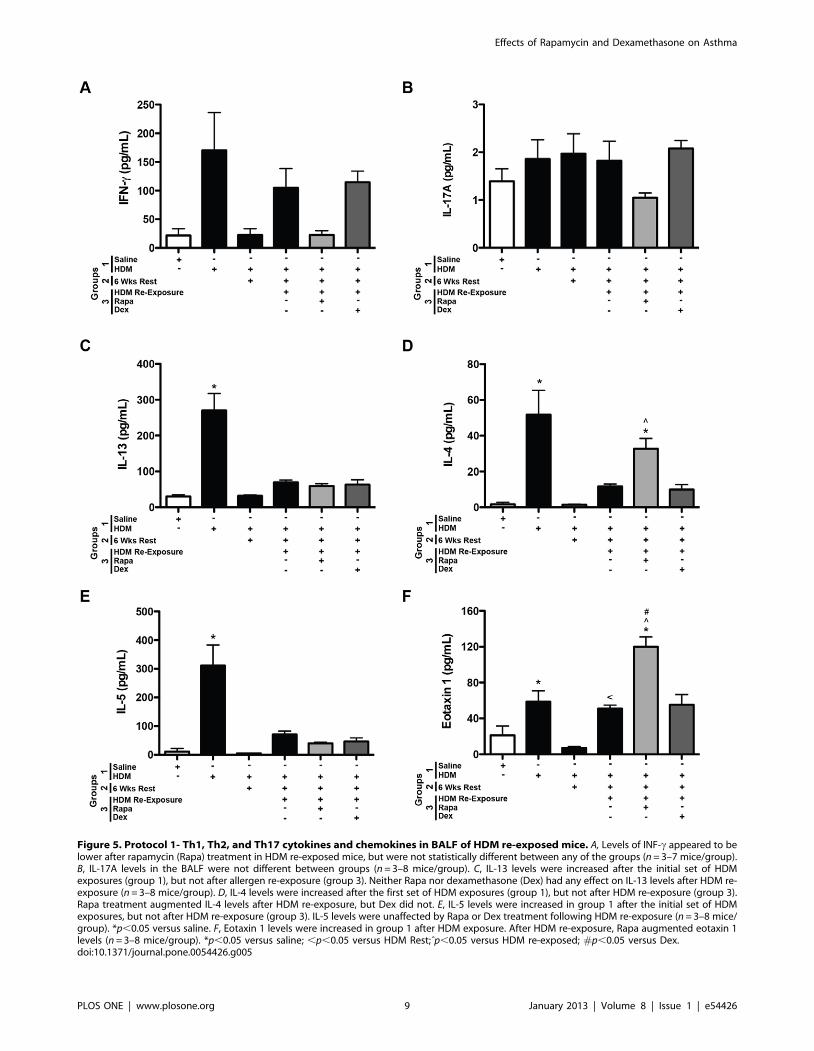
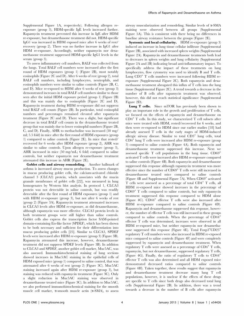
Figure 5. Protocol 1- Th1, Th2, and Th17 cytokines and chemokines in BALF of HDM re-exposed mice. A, Levels of INF-c appeared to belower after rapamycin (Rapa) treatment in HDM re-exposed mice, but were not statistically different between any of the groups (n = 3–7 mice/group).B, IL-17A levels in the BALF were not different between groups (n = 3–8 mice/group). C, IL-13 levels were increased after the initial set of HDMexposures (group 1), but not after allergen re-exposure (group 3). Neither Rapa nor dexamethasone (Dex) had any effect on IL-13 levels after HDM re-exposure (n = 3–8 mice/group). D, IL-4 levels were increased after the first set of HDM exposures (group 1), but not after HDM re-exposure (group 3).Rapa treatment augmented IL-4 levels after HDM re-exposure, but Dex did not. E, IL-5 levels were increased in group 1 after the initial set of HDMexposures, but not after HDM re-exposure (group 3). IL-5 levels were unaffected by Rapa or Dex treatment following HDM re-exposure (n = 3–8 mice/group). *p,0.05 versus saline. F, Eotaxin 1 levels were increased in group 1 after HDM exposure. After HDM re-exposure, Rapa augmented eotaxin 1levels (n = 3–8 mice/group). *p,0.05 versus saline; ,p,0.05 versus HDM Rest;ˆp,0.05 versus HDM re-exposed; #p,0.05 versus Dex.doi:10.1371/journal.pone.0054426.g005
Effects of Rapamycin and Dexamethasone on Asthma
PLOS ONE | www.plosone.org 9 January 2013 | Volume 8 | Issue 1 | e54426

Cytokines. IFN-c levels in the BALF were lower after 6
weeks of HDM and rapamycin treatment, but unaffected by
dexamethasone (Figure 8A). IL-17A levels appeared to be
increased after 3 and 6 weeks of HDM exposure and lower with
rapamycin and dexamethasone treatment; however, none of the
differences reached statistical significance (Figure 8B). Although
there was an upward trend, neither IL-13 nor IL-5 levels were
significantly increased in the BALF after 3 weeks of HDM
exposure (Figure 8 C and E). IL-4 and eotaxin 1 levels were
significantly increased after 3 weeks of HDM exposure (Figure 8D
and F). By six weeks of HDM exposure, although there were
upward trends for increases in IL-4, IL-5, and eotaxin 1 levels,
they were not significantly increased in mice compared to saline
controls. Interestingly, IL-4 and eotaxin 1 levels were augmented
with rapamycin treatment compared to saline controls (Figure 8 D
and F). Th2 cytokine levels were unaltered with dexamethasone
treatment at this time point.
mTOR signaling: P-S6 and P-Akt. Similar to the findings in
the re-exposure study, Western blot analysis demonstrated
increases in phosphorylated S6 (P-S6) in lung tissue after 3 weeks
of HDM exposure (Supplemental Figure 6A). P-S6 levels were also
increased after 6 weeks of HDM exposure and suppressed by
rapamycin and dexamethasone treatment. Phosphorylated Akt
(S473) levels were unaltered by rapamycin treatment (Supplemen-
tal Figure 6B). These data indicate that the dose of rapamycin used
in this study was sufficient to block mTORC1 signaling, but not
mTORC2.
Discussion
The goal of our study was to determine whether mTOR
inhibition with rapamycin would suppress the key features and
mediators of HDM-induced allergic asthma in established
asthmatic disease. In addition, we also compared rapamycin to
the steroid, dexamethasone, since steroids are currently a mainstay
Figure 6. Protocol 1- Western blot of P-S6 and P-Akt in lung homogenates of HDM re-exposed mice. A, P-S6, a downstream mediator ofmTOR complex 1 signaling, was increased in HDM re-exposed mice (group 3) and this was blocked by rapamycin (Rapa) treatment (n = 3–5 mice/group). *p,0.05 versus saline;ˆp,0.05 versus HDM re-exposed. B, P-Akt, a downstream mediator of mTOR complex 2 signaling, was increased afterallergen re-exposure in Rapa treated mice compared to saline controls (n = 3–5 mice/group), but unaffected by Dex. *p,0.05 versus saline; #p,0.05versus Rapa.doi:10.1371/journal.pone.0054426.g006
Effects of Rapamycin and Dexamethasone on Asthma
PLOS ONE | www.plosone.org 10 January 2013 | Volume 8 | Issue 1 | e54426
treatment for asthma. In the first protocol, we assessed whether
rapamycin or dexamethasone could suppress allergic disease
during allergen re-exposure. Although rapamycin suppressed IgE
levels, goblet cells, and total lung T cells, it had no effect on AHR
or BALF cellularity and IL-4 and eotaxin 1 levels were actually
augmented. Dexamethasone suppressed goblet cells and total lung
T cells, but had no effect on IgE or AHR and only slightly reduced
BALF eosinophilia. Our second protocol assessed whether
rapamycin or dexamethasone could reverse or inhibit the
progression of asthmatic responses during chronic allergic airway
disease. In this model, rapamycin did not suppress AHR or goblet
cells and actually augmented inflammatory cell numbers, IL-4 and
Figure 7. Protocol 2- T cell populations after chronic HDM exposure. A, Total CD4+ T cells were increased after 6 weeks of HDM andsuppressed by rapamycin (Rapa) and dexamethasone (Dex) (n = 4–12 mice/group). *p,0.05 versus saline;ˆp,0.05 versus HDM. B, Total activated Tcells (CD69+Foxp32) were increased in mice after 6 weeks of chronic HDM exposure and suppressed by Rapa and Dex (n = 6–8 mice/group). *p,0.05versus saline;ˆp,0.05 versus HDM. C, CD69+Foxp32 activated T cells, when assessed as a percentage of total CD4+ T cells, were increased after HDMexposure and unaffected by Rapa and Dex (n = 6–8 mice/group). *p,0.05 versus saline. D, Total effector T cells (CD44+Foxp32) were increased after 6weeks of HDM exposure and suppressed by Rapa and Dex (n = 6–8 mice/group). *p,0.05 versus saline;ˆp,0.05 versus HDM. E, CD44+Foxp32 effectorT cells, when expressed as a percentage of total CD4+ T cells, were increased after HDM exposure and attenuated by Rapa and Dex (n = 6–8 mice/group). *p,0.05 versus saline;ˆp,0.05 versus HDM. F, Total lung regulatory T cells (Foxp3+CD25+) were increased after chronic HDM exposure andsuppressed by Rapa and Dex (n = 6–8 mice/group). *p,0.05 versus saline;ˆp,0.05 versus HDM. G, Foxp3+CD25+ T cells, as a percentage of total lungCD4+ T cells, were reduced by Dex, but not by Rapa (n = 6–8 mice/group).ˆp,0.05 versus HDM; #p,0.05 versus Rapa. H, The ratio of regulatory T cellsFoxp3+CD25+ to CD44+Foxp32 effector T cells was decreased in HDM exposed mice compared to saline controls (n = 6–8 mice/group). *p,0.05versus saline.doi:10.1371/journal.pone.0054426.g007
Effects of Rapamycin and Dexamethasone on Asthma
PLOS ONE | www.plosone.org 11 January 2013 | Volume 8 | Issue 1 | e54426

Figure 8. Protocol 2- Th1, Th2, and Th17 cytokines and chemokines in the BALF after chronic HDM exposure. A, INF-c was suppressedafter 6 weeks of HDM exposure and in the rapamycin (Rapa) treated HDM group (n = 3–5 mice/group). *p,0.05 versus saline; #p,0.05 versus Dex. B,No significant differences in IL-17A levels were observed between animal groups, although there were trends for increased IL-17 in the HDM exposed
Effects of Rapamycin and Dexamethasone on Asthma
PLOS ONE | www.plosone.org 12 January 2013 | Volume 8 | Issue 1 | e54426

eotaxin 1 levels in the BALF. Dexamethasone had limited effects
on AHR, but did attenuate the inflammatory cell influx into the
BALF, especially eosinophils. Despite these limited effects, both
rapamycin and dexamethasone suppressed lung tissue lymphocyte
numbers and serum IgE levels.
Previous studies from our lab demonstrated that HDM-induced
allergic asthma could be prevented if rapamycin was administered
early and simultaneously with HDM. In this case, rapamycin
prevented HDM-induced AHR, inflammation, goblet cells, and
allergic sensitization [18]. Since allergic sensitization was sup-
pressed in our previous studies, we also determined whether
rapamycin could prevent allergic responses once sensitization had
already been established. To do this, mice were first sensitized
systemically to HDM by i.p. injection. During subsequent
intranasal HDM exposures, mice were treated with rapamycin.
In this case, rapamycin still suppressed many of the key allergic
responses including IgE, AHR, goblet cells, T cell responses, and
key mediators like IL-13 and leukotrienes, although it did not
suppress increases in inflammatory cells in the BALF [18], which
may have partly been due to the chemokine, eotaxin 1, since levels
were still elevated after rapamycin treatment. Although these
studies demonstrated an important role for the mTOR pathway
during early allergic sensitization and asthmatic disease processes,
it was unclear whether mTOR signaling would be important
during allergen re-exposure or during established/progressive
allergic disease. The studies we report in this manuscript sought to
address this question. These data suggests that the role of mTOR
is very different depending on the timing/disease stage since
rapamycin treatment during allergen re-exposure or during
chronic, ongoing disease did not attenuate key characteristics of
allergic asthma including AHR and inflammation and actually
augmented IL-4 and eotaxin 1 levels. The results from our second
protocol are similar to that of a recent study published by
Fredriksson et. al. who demonstrated that rapamycin did not
suppress allergic responses when administered during chronic
allergic disease [26]. In addition, our studies demonstrated that
rapamycin suppressed T cells in the lung tissue, including
regulatory T cells and our studies also compared the effects of
rapamycin to the steroid, dexamethasone.
Allergic asthma is often treated with steroids to suppress
inflammation [1,27]. Previous studies have utilized the corticoste-
roid, dexamethasone, in allergic asthma models [28,29,30]. For
example, a study similar to ours investigated the effects of
dexamethasone during allergic relapse and overt disease. In an
OVA model of allergic airway disease, dexamethasone suppressed
goblet cells, serum IgE, AHR, and reduced airway inflammation
in a relapse model. During overt disease, dexamethasone reduced
goblet cells, AHR, and the number of eosinophils, but had no
effect on serum IgE levels [28]. In our HDM-induced model of
allergen re-exposure/relapse, dexamethasone also decreased
goblet cells, but did not suppress IgE or AHR and eosinophil
numbers were only slightly reduced. Likewise, during chronic,
ongoing or overt disease, we did not observe suppression of goblet
cells and the effects on AHR were limited, although there were
decreases in inflammatory cell numbers, specifically eosinophils.
Although the decrease in eosinophils in this study was as expected
with dexamethasone treatment, no decrease in AHR was
surprising. However, previous reports have suggested that the
timing of AHR measurements after dexamethasone treatment
may be important [29]. Specifically, when AHR was measured 12
hours after dexamethasone treatment, AHR was suppressed, but
by 24 hours after dexamethasone treatment, AHR was no longer
suppressed [29]. In all of our studies, AHR was assessed 24 hours
after the last dexamethasone treatment, which may explain why
limited decreases in AHR were observed. In addition, differences
between the results of our study and others may be due to the type
of allergen used and/or the route of dexamethasone treatment
since ultrasonic nebulization was used in the study by Jungsuwa-
dee et. al. [28], versus i.p. delivery in our study.
Unlike our previous studies when rapamycin was given early in
the disease process [18], in this study, AHR was not suppressed by
rapamycin during allergen re-exposure or chronic allergic disease.
Both IL-4 and inflammation were still increased after rapamycin
treatment and could potentially contribute to AHR [31].
However, in our previous study, rapamycin treatment decreased
AHR despite elevated IL-4 BALF levels and inflammatory cell
numbers suggesting that other mechanisms are involved [18]. IL-
13 is a key mediator of AHR, however, in these chronic/
established models, IL-13 was not increased, suggesting that it may
not be contributing to AHR in more advanced disease. Airway
remodeling is another feature of asthma that could contribute to
sustained AHR in these more advanced disease models; however,
no major differences in airway smooth muscle or baseline airway
resistance between animal groups in either study were observed,
suggesting other mechanisms are playing a role in the disease
process.
An interesting observation in our studies was that IL-4 levels
were higher even though IgE levels after HDM exposure were still
suppressed by rapamycin treatment. This was surprising since IL-4
is an important mediator of IgE class switching. It is possible that
rapamycin could directly affect B cells that are secreting IgE to
suppress allergic sensitization to HDM. Previous work has
demonstrated that mTOR is required for B cell development
and maturation [32,33], however, less is known about the role of
mTOR in B cell homeostasis, activation of mature B cells, and
immunoglobulin production/secretion. A study using purified
human B cells demonstrated that rapamycin inhibited B cell
proliferation, induced apoptosis, and suppressed immunoglobulin
production, particularly IgM and IgG [34]. Although these studies
were carried out in vitro they still suggest that rapamycin could
have direct effects on B cells, which could account for the
decreases in IgE levels in our in vivo model and therefore reduce
sensitization to HDM, despite increased IL-4. When we assessed B
cells in the lung tissue, there was a trend towards a decrease in B
cells in both studies after rapamycin treatment. Despite being non-
significant, we cannot exclude that this minor decrease in lung B
cell levels could contribute to the observed decrease in IgE levels.
The source of the IL-4 increase is unclear in our model since T
cells, which are one of the primary sources of IL-4, were reduced.
Other cells including eosinophils, basophils, and mast cells can
secrete IL-4 [35], but whether these cells are playing a role in
enhancing IL-4 levels in our model is unclear. Also in our study,
group and lower leves in mice exposed to HDM and treated with Rapa or dexamethasone (Dex) during weeks 4–6 (n = 3–5 mice/group). C, IL-13 levelswere not significantly altered with HDM exposure, by Rapa or by Dex treatment in this model (n = 3–5 mice/group). D, IL-4 levels were increased after3 weeks of HDM compared to saline controls. After 6 weeks of HDM, IL-4 levels were higher in the Rapa treated group. IL-4 levels were similarbetween HDM and Dex treated mice (n = 3–5 mice/group). *p,0.05 versus saline; ˆp,0.05 versus HDM; #p,0.05 versus Dex. E, No statisticallysignificant differences in IL-5 levels were observed between any group of mice (n = 3–5 mice/group). F, Eotaxin 1 levels were increased after 3 weeksof HDM exposure and were higher in the Rapa treated group after 6 weeks of chronic HDM compared to saline controls (n = 3–5 mice/group).*p,0.05 versus saline.doi:10.1371/journal.pone.0054426.g008
Effects of Rapamycin and Dexamethasone on Asthma
PLOS ONE | www.plosone.org 13 January 2013 | Volume 8 | Issue 1 | e54426
eotaxin 1, an important epithelial cell derived eosinophil
chemokine, remained elevated in the BALF with rapamycin
treatment, which may explain why eosinophil numbers were not
suppressed. This was also true in our previous acute study in which
rapamycin treatment did not suppress airway inflammation nor
eotaxin 1 levels once sensitization was established [18].
More recent studies have indicated an important role for
regulatory T cells in the resolution of allergic airway disease
[36,37,38]. Studies have demonstrated that adoptive transfer of
CD4+CD25+Foxp3+ regulatory T cells into mice exposed to
allergen suppressed allergic responses, whereas inhibition of
regulatory T cells exacerbated the allergic response [39]. In vitro
data suggests that rapamycin can expand CD4+CD25+Foxp3+
regulatory T cells in the presence of IL-2 [40,41], however, in our
in vivo model, rapamycin treatment was associated with decreases
in effector T cells, a major source of IL-2 in the lung. Hence,
rapamycin treatment, much like dexamethasone treatment, may
decrease regulatory T cells in vivo by decreasing the number of IL-
2 producing cells. It is unclear if the reductions in regulatory T
cells after rapamycin treatment in this model would have any
biological significance; however, loss of regulatory T cells has been
shown to worsen the severity of allergic disease [42]. Interestingly,
loss of CD69+ cells has also been associated with exacerbated
allergic disease [43]. These findings remain controversial however
[44], as new roles for CD69 in cell egress from lymphoid organs,
Th17 differentiation and formation of memory CD44+CD4+ T
cells are being proposed [45,46,47]. Interestingly, short rapamycin
(or dexamethasone) treatment had little effect on the proportion of
memory cells in the lungs, whereas longer exposure to rapamycin
(or dexamethasone) in our second model significantly decreased
the proportion of CD44+ memory cells among CD4+ T cells.
It remains unclear why many of the allergic responses, especially
AHR, were not suppressed in our studies even though T cell
populations were reduced. The effects of rapamycin and
dexamethasone, however, may not be only specific to T cells
since spleen sizes were also reduced in our studies, consistent with
the immunosuppressive properties of these drugs [48,49]. It is
possible that other cell types in the lung could be contributing to
the allergic responses in these established/chronic models, uch as
epithelial cells. Epithelial cells and other lung cells can produce
cytokines upon allergen exposure, which can then directly
influence allergic responses, including AHR [50,51].
The protein encoded by the mTOR gene signals through two
protein complexes, mTOR complex 1 (mTORC1) and mTOR
complex 2 (mTORC2). Each of these complexes carries out
distinct cellular functions and each complex is composed of several
subunits. The most notable subunit of mTORC1 is the regulatory-
associated protein of mTOR (Raptor) and in mTORC2, the
rapamycin-insensitive companion of mTOR (Rictor) [52,53,54].
Most reports indicate that only mTORC1 is rapamycin sensitive,
but some recent evidence suggests that, depending on the cell type,
duration, and dosing regimen, rapamycin can also inhibit
mTORC2 [55]. Downstream of mTORC1 is the ribosomal
protein S6 kinases and its downstream substrate S6, which gets
phosphorylated upon mTOR activation. In order to help
understand why rapamycin did not suppress the allergic responses
in our studies, we measured the activation of P-S6 downstream of
mTORC1. In both of the models used here, rapamycin
completely suppressed HDM-induced increases in phosphorylated
S6 levels, but did not suppress the phosphorylation of Akt (S473).
These results indicate that the dose of rapamycin used was
sufficient to block mTORC1, but not mTORC2. These mTOR
complexes differentially regulate T cell lineage commitment; with
Th1 and Th17 being mostly dependent on mTORC1 signaling
and Th2 cells on mTORC2 [11]. Accordingly, the Th1 cytokine
IFN-c and Th17 cytokine, IL-17A, were significantly decreased or
trended lower in both of our models following rapamycin
treatment whereas the prototypical Th2 cytokine IL-4 was
increased. This increase in IL-4 is potentially the result of
decreases in IFN-c, a negative regulator of Th2 differentiation.
Finally, IL-4 has been implicated in allergic responses indepen-
dently of IL-13 [31,56]. Taken together, regardless of the cellular
source of IL-4 (Th2 cells, basophils, mast cells and/or eosinophils)
increased pulmonary IL-4 levels may, at least partially, account for
the lack of effect of rapamycin treatment on AHR and BALF
eosinophilia.
In conclusion, while our earlier studies demonstrate that mTOR
signaling plays an important role during the early phases of allergic
asthma [18], the studies we report here suggest that its role is more
limited during allergen re-exposure and chronic/established
disease. This is consistent with studies showing a role for mTOR
in early activation and differentiation events [11,13], but it appears
that once this is established, mTOR signaling plays a more minor
role. It is possible that at these later stages of the disease process,
other cells and mechanisms are driving the airway disease.
Supporting Information
Figure S1 HDM-specific IgG1 levels. A, Protocol 1: Re-
Exposure Study: HDM-specific IgG1 levels were increased in
HDM rest and all groups re-exposed to HDM. Neither rapamycin
(Rapa) nor dexamethasone (Dex) suppressed these increases
(n = 4–12 mice/group). *p,0.05 versus saline. B, Protocol 2:
Reversal Study: HDM-specific IgG1 levels were increased after 3
and 6 weeks of HDM exposure. Both Rapa and Dex attenuated or
suppressed this increase (n = 3–8 mice/group). *p,0.05 versus
saline;ˆp,0.05 versus vehicle; #p,0.05 versus dex.
(TIFF)
Figure S2 Spleen weight to body weight ratios and lungcell populations. Protocol 1 (Re-exposure): A, Spleen weights
were increased after HDM re-exposure compared to saline
controls. Rapamycin (Rapa) and dexamethasone (Dex) suppressed
the increase in spleen weights (n = 4–12 mice/group). *p,0.05
versus saline; ˆp,0.05 versus HDM. B, Total lung cells were
increased after HDM re-exposure (group 3) compared to saline
controls. Rapa and Dex suppressed total lung cells (n = 4–12 mice/
group). *p,0.05 versus saline; ˆp,0.05 versus HDM. C, HDM-
induced increases in total CD3+ T cells after allergen re-exposure
were suppressed by Rapa and Dex (n = 4–12 mice/group).
*p,0.05 versus saline;ˆp,0.05 versus HDM re-exposed. D, Total
lung B cells after HDM re-exposure and after Rapa showed trends
towards increased and decreased, respectively, but these changes
did not reach statistical significance (n = 4–12 mice/group).
Protocol 2 (Chronic Allergen/Reversal): E, Spleen weights were
increased after 6 weeks of HDM exposure and suppressed by Rapa
and Dex (n = 6–8 mice/group). *p,0.05 versus saline; ˆp,0.05
versus HDM; #p,0.05 versus Rapa. F, Total lung cells were
increased after 6 weeks of HDM. Dex, but not Rapa suppressed
this response (n = 4–12 mice/group). *p,0.05 versus saline;
p̂,0.05 versus HDM; #p,0.05 versus Rapa. G, HDM-induced
increases in CD3+ lung T cells after 6 weeks of HDM exposure
were attenuated by Rapa and Dex (n = 4–12 mice/group).
*p,0.05 versus saline;ˆp,0.05 versus HDM re-exposed. H, Total
lung B cells were increased after 6 weeks of HDM compared to
saline controls, but were only significantly reduced after Dex, not
Rapa (n = 4–12 mice/group). *p,0.05 versus saline;̂ p,0.05 versus
HDM re-exposed.
(TIFF)
Effects of Rapamycin and Dexamethasone on Asthma
PLOS ONE | www.plosone.org 14 January 2013 | Volume 8 | Issue 1 | e54426

Figure S3 Protocol 2- HDM-specific IgE levels, inflam-matory BALF cell numbers, and AHR after chronic HDMexposure. A, Increases in HDM-specific IgE were observed after
both 3 and 6 weeks of HDM. HDM-specific IgE levels were
reduced by rapamycin (Rapa) and dexamethasone (Dex) treatment
(n = 5–9 mice/group). *p,0.05 versus saline;̂ p,0.05 versus HDM.
B, Total BALF cell numbers were increased after 3 and 6 weeks of
HDM exposure compared to saline controls and were even higher
after Rapa treatment compared to HDM. Total BALF cell
numbers were decreased in Dex treated mice compared to HDM
mice (n = 10–16 mice/group. *p,0.05 versus saline;̂ p,0.05 versus
HDM; #p,0.05 versus Dex. C, Total macrophages and
eosinophils were higher with Rapa treatment compared to mice
exposed to HDM for 6 weeks. Neutrophil and eosinophil numbers
were reduced in Dex treated mice compared to HDM (6 weeks)
exposed mice (n = 10–16 mice/group). *p,0.05 versus saline;
p̂,0.05 versus HDM; #p,0.05 versus Dex. D, The percentage of
eosinophils was elevated in Rapa treated mice compared to saline
controls, but similar to mice exposed to HDM for 6 weeks,
whereas the percentage of eosinophils was decreased with Dex
treatment (n = 10–16 mice/group). *p,0.05 versus saline;ˆp,0.05
versus HDM; #p,0.05 versus Dex. E, AHR was increased after 3
weeks and 6 weeks of HDM exposure. Increases in AHR after 6
weeks of HDM exposure were not suppressed by Rapa or Dex at
50 mg/ml methacholine. However, at 25 mg/ml methacholine,
Dex did reduce AHR compared to mice exposed to HDM for 6
weeks (n = 10–16 mice/group). *p,0.05 versus saline.
(TIFF)
Figure S4 Protocol 2- Goblet cell markers in the lungsafter chronic HDM exposure. A, CLCA3 protein in lung
homogenates was increased after 3 and 6 weeks of HDM
exposure, was unaltered by rapamycin (Rapa), but was suppressed
by dexamethasone (Dex) (n = 4–8 mice/group). *p,0.05 versus
saline;ˆp,0.05 versus HDM. B, SPDEF levels were also increased
after 6 weeks of HDM exposure, but unaltered by Rapa. SPDEF
levels were lower with Dex treatment compared to HDM exposed
mice, but this did not reach statistical significance (n = 3 mice/
group). *p,0.05 versus saline. C, Muc5AC staining was increased
in the airway epithelial cells after 3 and 6 weeks of HDM
exposure. Dex attenuated these increases, but Rapa did not.
(TIFF)
Figure S5 Airway smooth muscle staining in allergicasthma models. A, Protocol 1 (Re-exposure): a-Smooth muscle
actin (a-SMA) staining was performed on lung sections of mice re-
exposed to HDM after 6 weeks of rest. Similar staining patterns
were observed between all animal groups with no observable
differences between rapamycin (Rapa) and dexamethasone (Dex)
treated mice. B, Protocol 2 (Chronic Allergen/Reversal): a-
Smooth muscle actin (a-SMA) staining in the lung after 6 weeks of
saline or HDM exposure was similar. No differences were
observed with Rapa or Dex treatment.
(TIFF)
Figure S6 Protocol 2- Western blot analysis of P-S6 andP-Akt levels after chronic HDM exposure. A, P-S6, a
downstream target of mTOR complex 1, was increased after both
3 and 6 weeks of HDM exposure. Rapamycin (Rapa) treatment
during weeks 4–6 of HDM exposure completely suppressed this
increase. Levels of P-S6 were also reduced in the lung by
dexamethasone (Dex) (n = 3–5 mice/group). *p,0.05 versus saline;
p̂,0.05 versus HDM; #p,0.05 versus Dex. B, Levels of P-Akt, a
downstream target of mTOR complex 2, were not significantly
altered by 6 weeks of HDM alone, Rapa, or Dex treatment (n = 3–
5 mice/group).
(TIFF)
Figure S7 Activated CD69+Foxp32 T cells in the lungs ofmice. A, Protocol 1 (Re-exposure): FACS analysis showing
increases in CD69+Foxp32 T cells after HDM re-exposure.
CD69+Foxp32 T cells were reduced with rapamycin (Rapa) and
dexamethasone (Dex) treatment. B, Protocol 2 (Chronic Allergen/
Reveral): FACS analysis demonstrating increases in
CD69+Foxp32 T cells after chronic HDM exposure. Slight
reductions were observed after Rapa and Dex treatment.
(TIFF)
Author Contributions
Conceived and designed the experiments: EMM EBB GKH TDLC.
Performed the experiments: EMM EBB. Analyzed the data: EMM EBB.
Contributed reagents/materials/analysis tools: EMM EBB GKH TDLC.
Wrote the paper: EMM EBB GKH TDLC.
References
1. Bateman ED, Hurd SS, Barnes PJ, Bousquet J, Drazen JM, et al. (2008) Global
strategy for asthma management and prevention: GINA executive summary.
The Eur Respir J 31: 143–178.
2. Hamid Q, Tulic M (2009) Immunobiology of asthma. Annu Rev Physiol 71:
489–507.
3. Holgate ST (2012) Innate and adaptive immune responses in asthma. Nat Med
18: 673–683.
4. Kim HY, DeKruyff RH, Umetsu DT (2010) The many paths to asthma:
phenotype shaped by innate and adaptive immunity. Nature immunology 11:
577–584.
5. Hesselmar B, Aberg B, Eriksson B, Aberg N (2000) Asthma in children:
prevalence, treatment, and sensitization. Pediatr Allergy Immunol 11: 74–79.
6. Masoli M, Fabian D, Holt S, Beasley R (2004) The global burden of asthma:
executive summary of the GINA Dissemination Committee report. Allergy 59:
469–478.
7. Holgate ST (2005) Exacerbations: the asthma paradox. Am J Respir Crit Care
Med 172: 941–943.
8. Siegle JS, Hansbro N, Herbert C, Yang M, Foster PS, et al. (2006) Airway
hyperreactivity in exacerbation of chronic asthma is independent of eosinophilic
inflammation. Am J Respir Cell Mol Biol 35: 565–570.
9. Powell JD, Delgoffe GM (2010) The mammalian target of rapamycin: linking T
cell differentiation, function, and metabolism. Immunity 33: 301–311.
10. Reiling JH, Sabatini DM (2006) Stress and mTORture signaling. Oncogene 25:
6373–6383.
11. Delgoffe GM, Kole TP, Zheng Y, Zarek PE, Matthews KL, et al. (2009) The
mTOR kinase differentially regulates effector and regulatory T cell lineage
commitment. Immunity 30: 832–844.
12. Delgoffe GM, Pollizzi KN, Waickman AT, Heikamp E, Meyers DJ, et al. (2011)
The kinase mTOR regulates the differentiation of helper T cells through the
selective activation of signaling by mTORC1 and mTORC2. Nat Immunol 12:
295–303.
13. Liu G, Yang K, Burns S, Shrestha S, Chi H (2010) The S1P(1)-mTOR axis
directs the reciprocal differentiation of T(H)1 and T(reg) cells. Nat Immunol 11:
1047–1056.
14. Saemann MD, Haidinger M, Hecking M, Horl WH, Weichhart T (2009) The
multifunctional role of mTOR in innate immunity: implications for transplant
immunity. Am J Transplant 9: 2655–2661.
15. Saunders RN, Metcalfe MS, Nicholson ML (2001) Rapamycin in transplanta-
tion: a review of the evidence. Kidney Int 59: 3–16.
16. Alejandre-Alcazar MA, Shalamanov PD, Amarie OV, Sevilla-Perez J, Seeger
W, et al. (2007) Temporal and spatial regulation of bone morphogenetic protein
signaling in late lung development. Dev Dyn 236: 2825–2835.
17. McCormack FX, Inoue Y, Moss J, Singer LG, Strange C, et al. (2011) Efficacy
and safety of sirolimus in lymphangioleiomyomatosis. N Engl J Med 364: 1595–
1606.
18. Mushaben EM, Kramer EL, Brandt EB, Khurana Hershey GK, Le Cras TD
(2011) Rapamycin attenuates airway hyperreactivity, goblet cells, and IgE in
experimental allergic asthma. J Immunol 187: 5756–5763.
19. Zhang X, Lewkowich IP, Kohl G, Clark JR, Wills-Karp M, et al. (2009) A
protective role for C5a in the development of allergic asthma associated with
Effects of Rapamycin and Dexamethasone on Asthma
PLOS ONE | www.plosone.org 15 January 2013 | Volume 8 | Issue 1 | e54426

altered levels of B7-H1 and B7-DC on plasmacytoid dendritic cells. J Immunol
182: 5123–5130.20. Kramer EL, Mushaben EM, Pastura PA, Acciani TH, Deutsch GH, et al. (2009)
Early growth response-1 suppresses epidermal growth factor receptor-mediated
airway hyperresponsiveness and lung remodeling in mice. Am J Respir Cell MolBiol 41: 415–425.
21. Le Cras TD, Acciani TH, Mushaben EM, Kramer EL, Pastura PA, et al. (2011)Epithelial EGF receptor signaling mediates airway hyperreactivity and
remodeling in a mouse model of chronic asthma. Am J Physiol Lung Cell
Mol Physiol 300: L414–421.22. Leverkoehne I, Gruber AD (2002) The murine mCLCA3 (alias gob-5) protein is
located in the mucin granule membranes of intestinal, respiratory, and uterinegoblet cells. J Histochem Cytochem 50: 829–838.
23. Kramer EL, Hardie WD, Mushaben EM, Acciani TH, Pastura PA, et al. (2011)Rapamycin decreases airway remodeling and hyperreactivity in a transgenic
model of noninflammatory lung disease. Journal of applied physiology 111:
1760–1767.24. Le Cras TD, Hardie WD, Deutsch GH, Albertine KH, Ikegami M, et al. (2004)
Transient induction of TGF-alpha disrupts lung morphogenesis, causingpulmonary disease in adulthood. Am J Physiol Lung Cell Mol Physiol 287:
L718–729.
25. Chen G, Korfhagen TR, Xu Y, Kitzmiller J, Wert SE, et al. (2009) SPDEF isrequired for mouse pulmonary goblet cell differentiation and regulates a network
of genes associated with mucus production. J Clin Invest 119: 2914–2924.26. Fredriksson K, Fielhaber JA, Lam JK, Yao X, Meyer KS, et al. (2012)
Paradoxical effects of rapamycin on experimental house dust mite-inducedasthma. PloS one 7: e33984.
27. de Kluijver J, Evertse CE, Schrumpf JA, van der Veen H, Zwinderman AH, et
al. (2002) Asymptomatic worsening of airway inflammation during low-doseallergen exposure in asthma: protection by inhaled steroids. Am J Respir Crit
Care Med 166: 294–300.28. Jungsuwadee P, Dekan G, Stingl G, Epstein MM (2004) Inhaled dexamethasone
differentially attenuates disease relapse and established allergic asthma in mice.
Clin Immunol 110: 13–21.29. Kim J, McKinley L, Siddiqui J, Bolgos GL, Remick DG (2004) Prevention and
reversal of pulmonary inflammation and airway hyperresponsiveness bydexamethasone treatment in a murine model of asthma induced by house dust.
Am J Physiol Lung Cell Mol Physiol 287: L503–509.30. Ma J, Yang A, Qin W, Shi Y, Zhao B, et al. (2011) Alleviating allergic airway
diseases by means of short-term administration of IL-2 and dexamethasone.
J Allergy Clin Immunol 127: 1447–1456 e1446.31. Perkins C, Wills-Karp M, Finkelman FD (2006) IL-4 induces IL-13-independent
allergic airway inflammation. J Allergy Clin Immunol 118: 410–419.32. Benhamron S, Tirosh B (2011) Direct activation of mTOR in B lymphocytes
confers impairment in B-cell maturation andloss of marginal zone B cells.
Eur J Immunol 41: 2390–2396.33. Zhang S, Readinger JA, DuBois W, Janka-Junttila M, Robinson R, et al. (2011)
Constitutive reductions in mTOR alter cell size, immune cell development, andantibody production. Blood 117: 1228–1238.
34. Heidt S, Roelen DL, Eijsink C, van Kooten C, Claas FH, et al. (2008) Effects ofimmunosuppressive drugs on purified human B cells: evidence supporting the
use of MMF and rapamycin. Transplantation 86: 1292–1300.
35. Gessner A, Mohrs K, Mohrs M (2005) Mast cells, basophils, and eosinophilsacquire constitutive IL-4 and IL-13 transcripts during lineage differentiation that
are sufficient for rapid cytokine production. J Immunol 174: 1063–1072.
36. Kay AB (2006) The role of T lymphocytes in asthma. Chem Immunol Allergy
91: 59–75.37. Kearley J, Barker JE, Robinson DS, Lloyd CM (2005) Resolution of airway
inflammation and hyperreactivity after in vivo transfer of CD4+CD25+regulatory T cells is interleukin 10 dependent. J Exp Med 202: 1539–1547.
38. Lewkowich IP, Herman NS, Schleifer KW, Dance MP, Chen BL, et al. (2005)
CD4+CD25+ T cells protect against experimentally induced asthma and alterpulmonary dendritic cell phenotype and function. J Exp Med 202: 1549–1561.
39. Leech MD, Benson RA, De Vries A, Fitch PM, Howie SE (2007) Resolution of
Der p1-induced allergic airway inflammation is dependent onCD4+CD25+Foxp3+ regulatory cells. J Immunol 179: 7050–7058.
40. Battaglia M, Stabilini A, Roncarolo MG (2005) Rapamycin selectively expandsCD4+CD25+FoxP3+ regulatory T cells. Blood 105: 4743–4748.
41. Strauss L, Whiteside TL, Knights A, Bergmann C, Knuth A, et al. (2007)Selective survival of naturally occurring human CD4+CD25+Foxp3+ regulatory
T cells cultured with rapamycin. J Immunol 178: 320–329.
42. Taams LS, Palmer DB, Akbar AN, Robinson DS, Brown Z, et al. (2006)Regulatory T cells in human disease and their potential for therapeutic
manipulation. Immunology 118: 1–9.43. Martin P, Gomez M, Lamana A, Matesanz Marin A, Cortes JR, et al. (2010)
The leukocyte activation antigen CD69 limits allergic asthma and skin contact
hypersensitivity. J Allergy Clin Immunol 126: 355–365, 365 e351–353.44. Miki-Hosokawa T, Hasegawa A, Iwamura C, Shinoda K, Tofukuji S, et al.
(2009) CD69 controls the pathogenesis of allergic airway inflammation.J Immunol 183: 8203–8215.
45. Martin P, Gomez M, Lamana A, Cruz-Adalia A, Ramirez-Huesca M, et al.(2010) CD69 association with Jak3/Stat5 proteins regulates Th17 cell
differentiation. Mol Cell Biol 30: 4877–4889.
46. Shinoda K, Tokoyoda K, Hanazawa A, Hayashizaki K, Zehentmeier S, et al.(2012) Type II membrane protein CD69 regulates the formation of resting T-
helper memory. Proc Natl Acad Sci USA 109: 7409–7414.47. Shiow LR, Rosen DB, Brdickova N, Xu Y, An J, et al. (2006) CD69 acts
downstream of interferon-alpha/beta to inhibit S1P1 and lymphocyte egress
from lymphoid organs. Nature 440: 540–544.48. Dumont FJ, Su Q (1996) Mechanism of action of the immunosuppressant
rapamycin. Life Sci 58: 373–395.49. Ulrich K, Hincks JS, Walsh R, Wetterstrand EM, Fidock MD, et al. (2008) Anti-
inflammatory modulation of chronic airway inflammation in the murine housedust mite model. Pulm Pharmacol Ther 21: 637–647.
50. Knight DA, Holgate ST (2003) The airway epithelium: structural and functional
properties in health and disease. Respirology 8: 432–446.51. Lloyd CM, Hessel EM (2010) Functions of T cells in asthma: more than just
T(H)2 cells. Nature reviews Immunology 10: 838–848.52. Ballou LM, Lin RZ (2008) Rapamycin and mTOR kinase inhibitors. J Chem
Biol 1: 27–36.
53. Chi H (2012) Regulation and function of mTOR signalling in T cell fatedecisions. Nature reviews Immunology 12: 325–338.
54. Laplante M, Sabatini DM (2009) mTOR signaling at a glance. J Cell Sci 122:3589–3594.
55. Sarbassov DD, Ali SM, Sengupta S, Sheen JH, Hsu PP, et al. (2006) Prolongedrapamycin treatment inhibits mTORC2 assembly and Akt/PKB. Mol Cell 22:
159–168.
56. Perkins C, Yanase N, Smulian G, Gildea L, Orekov T, et al. (2011) Selectivestimulation of IL-4 receptor on smooth muscle induces airway hyperrespon-
siveness in mice. J Exp Med 208: 853–867.
Effects of Rapamycin and Dexamethasone on Asthma
PLOS ONE | www.plosone.org 16 January 2013 | Volume 8 | Issue 1 | e54426

Reproduced with permission of the copyright owner. Further reproduction prohibited withoutpermission.
Related Documents