275 DOI: 10.2478/10004-1254-61-2010-2012 Scientific Paper EFFECTS OF LYCOPENE, INDOLE-3-CARBINOL, AND LUTEOLIN ON NITRIC OXIDE PRODUCTION AND iNOS EXPRESSION ARE ORGAN-SPECIFIC IN RATS Evita ROSTOKA 1 , Sergejs ISAJEVS 2 , Larisa BAUMANE 1 , Aija LINE 3 , Karina SILINA 3 , Maija DZINTARE 1 , Jelena SHARIPOVA 1 , Darja SVIRINA 2 , Ivars KALVINSH 1 and Nikolajs SJAKSTE 1,2 Latvian Institute of Organic Synthesis 1 , Faculty of Medicine, University of Latvia 2 , Latvian Biomedical Research and Study Centre 3 , Riga, Latvia Received in November 2009 Accepted in March 2010 Natural compounds are known to modify NO content in tissues; however, the biological activity of polyphenol-rich food often does not correspond to the effects of individual polyphenols on NO synthase activity. The aim of this study was to see how natural compounds luteolin, indole-3-carbinol, and lycopene modify NO production in rat tissues and change the expression of the iNOS gene and protein. Indole-3- carbinol produced multiple effects on the NO level; it significantly decreased NO concentration in blood, lungs, and skeletal muscles and increased it in the liver. Indole-3-carbinol enhanced lipopolyssaccharide (LPS)-induced NO production in all rat organs. It decreased iNOS gene expression in the brain cortex of animals that did not receive LPS and up-regulated it in the LPS-treated animals. Lycopene increased the iNOS gene transcription rate in the brain cortex of LPS-treated animals. Luteolin did not modify NO production in any organ of LPS-untreated rats, nor did it affect gene expression in the liver. In the brain it slightly decreased iNOS gene expression. Luteolin decreased NO production in the blood of LPS-treated animals and the number of iNOS-positive cells in these animals. Our results suggest that changes in tissue NO levels caused by natural compounds cannot be predicted from their effect on NOS expression or activity obtained in model systems. This stresses the importance of direct measurements of NO and NOS expression in animal tissues. KEY WORDS: ESR spectroscopy, inducible nitric oxide synthase, natural compounds, NO Rostoka E, et al. EFFECTS OF NATURAL COMPOUNDS ON NO AND iNOS Arh Hig Rada Toksikol 2010;61:275-285 Natural biologically active compounds of plant origin such as flavonoids are widely used as dietary supplements with detoxicating effects. The detoxicating effect of many plant-derived compounds is based on their antioxidant, anti-mutagenic, and anti-carcinogenic effects (1, 2). These substances are also known to influence the enzymatic production of nitric oxide (NO) and its stability. (3). This underlines the significance of the uptake of flavonoid and other natural compound for the defence of cardiovascular, immune, and nervous systems against toxins. However, the biological activity of polyphenol-rich food does not correspond to the effects of individual compounds on NO synthase activity. For example, red wine is known as a vasodilator (4) and is rich in quercetin. Purified quercetin inhibits inducible nitric oxide synthase (iNOS) gene expression (3, 5), destabilises endothelial NOS (eNOS) mRNA (6),

Welcome message from author
This document is posted to help you gain knowledge. Please leave a comment to let me know what you think about it! Share it to your friends and learn new things together.
Transcript

275
DOI: 10.2478/10004-1254-61-2010-2012
Scientifi c Paper
EFFECTS OF LYCOPENE, INDOLE-3-CARBINOL, AND LUTEOLIN ON NITRIC OXIDE PRODUCTION
AND iNOS EXPRESSION ARE ORGAN-SPECIFICIN RATS
Evita ROSTOKA1, Sergejs ISAJEVS2, Larisa BAUMANE1, Aija LINE3, Karina SILINA3, Maija DZINTARE1, Jelena SHARIPOVA1, Darja SVIRINA2, Ivars KALVINSH1 and
Nikolajs SJAKSTE1,2
Latvian Institute of Organic Synthesis1, Faculty of Medicine, University of Latvia2, Latvian Biomedical Research and Study Centre3, Riga, Latvia
Received in November 2009Accepted in March 2010
Natural compounds are known to modify NO content in tissues; however, the biological activity of polyphenol-rich food often does not correspond to the effects of individual polyphenols on NO synthase activity. The aim of this study was to see how natural compounds luteolin, indole-3-carbinol, and lycopene modify NO production in rat tissues and change the expression of the iNOS gene and protein. Indole-3-carbinol produced multiple effects on the NO level; it signifi cantly decreased NO concentration in blood, lungs, and skeletal muscles and increased it in the liver. Indole-3-carbinol enhanced lipopolyssaccharide (LPS)-induced NO production in all rat organs. It decreased iNOS gene expression in the brain cortex of animals that did not receive LPS and up-regulated it in the LPS-treated animals. Lycopene increased the iNOS gene transcription rate in the brain cortex of LPS-treated animals. Luteolin did not modify NO production in any organ of LPS-untreated rats, nor did it affect gene expression in the liver. In the brain it slightly decreased iNOS gene expression. Luteolin decreased NO production in the blood of LPS-treated animals and the number of iNOS-positive cells in these animals. Our results suggest that changes in tissue NO levels caused by natural compounds cannot be predicted from their effect on NOS expression or activity obtained in model systems. This stresses the importance of direct measurements of NO and NOS expression in animal tissues.
KEY WORDS: ESR spectroscopy, inducible nitric oxide synthase, natural compounds, NO
Rostoka E, et al. EFFECTS OF NATURAL COMPOUNDS ON NO AND iNOSArh Hig Rada Toksikol 2010;61:275-285
Natural biologically active compounds of plant origin such as flavonoids are widely used as dietary supplements with detoxicating effects. The detoxicating effect of many plant-derived compounds is based on their antioxidant, anti-mutagenic, and anti-carcinogenic effects (1, 2). These substances are also known to infl uence the enzymatic production of nitric oxide (NO) and its stability. (3). This underlines the signifi cance of the uptake of fl avonoid and other
natural compound for the defence of cardiovascular, immune, and nervous systems against toxins. However, the biological activity of polyphenol-rich food does not correspond to the effects of individual compounds on NO synthase activity. For example, red wine is known as a vasodilator (4) and is rich in quercetin. Purifi ed quercetin inhibits inducible nitric oxide synthase (iNOS) gene expression (3, 5), destabilises endothelial NOS (eNOS) mRNA (6),

and even inhibits NOS enzymatic activity (7). Yet it produces vasorelaxing effects (8) despite the fact that nitric oxide is not detected in rat aorta after quercetin administration (9).
We believe that the effects of natural compounds on NO production in animal organs are particularly important for evaluation of their biological activity. Unfortunately, such data are scarce (9, 10). The aim of our study was to see if and to what extent natural compounds of plant origin modify NO production in rat tissues using electron spin resonance (ESR) spectroscopy of Fe - diethylthiocarbamate – nitric oxide [Fe(DETC)
2-NO] complexes. We also assayed
iNOS gene expression using the real-time reverse transcriptase polymerase chain reaction (RT-PCR) and iNOS protein synthesis using histochemical methods.
MATERIALS AND METHODS
For the study we selected three compounds of different chemical structure: luteolin, a polyphenolic flavonol, indole-3-carbinol, a simple phenolic compound, and lycopene, the carotene derivative. Their chemical structures are given in Figure 1. Literature data indicate possible impact of all the three substances on NOS expression and/or NO production (11-19).
Luteolin, 3’,4’,5’,7-tetrahydroxyflavone is abundant in celery root, rutabaga, red pepper, spinach, and flowering plants such as Ajuga decumbens, Taraxacum officinale (dandelion), and Medicago sativa (alfalfa). Luteolin is known as a dietary compound with antioxidant activity (11).
Indole-3-carbinol (3-indolmethanol) is found in mustard plants (Brassica sp.) including cabbage, broccoli, and Brussels sprouts. The compound has widely been studied for cancer treatment (12).
Lycopene, ψ, ψ-carotene is found in ripe red vegetables such as tomato. It is an antioxidant and detoxicating agent (13).
Chemicals
Indole-3-carbinol, lycopene, luteolin, and acetyl ester of caffeic acid were purchased from Dayang Chemical Co., LTD (Hangzhou, China). Lipopolysaccharide, diethylthiocarbamate, ferrous sulphate, sodium citrate, TRI Reagent®, and all other chemicals were from Sigma-Aldrich Chemie GmbH (Taufkirchen, Germany).
Experiment design and compound administration
Male Wistar rats, weighing (215.00±5.63) g at the beginning of the experiment, were obtained from the Laboratory of Experimental Animals, Riga Stradins University, Riga, Latvia. All experimental procedures
Figure 1 Chemical structure of natural compounds used in the study
Luteolin Lycopene
Indole – 3 – carbinol Ester of caffeic acid
276 Rostoka E, et al. EFFECTS OF NATURAL COMPOUNDS ON NO AND iNOSArh Hig Rada Toksikol 2010;61:275-285

277
followed the guidelines set out in the Directive 86/609/EEC (20) and were approved by the Animal Ethics Committee of the Food and Veterinary Service (Riga, Latvia).
The animals were kept in a stable environment at (22±0.5) °C with a 12-hour light/dark cycle and fed standard laboratory diet. Tables 1 and 2 give details about animal groups, their number, and administered compounds.
ESR experiments included animal groups 1 to 10 (Table 1). Natural compounds were administered per os to model their natural mode of consumption. Three and a half hours later the spin trap was injected. Thirty minutes afterwards the rats were decapitated under slight ether narcosis. Groups 6 to 10 received 10 mg kg-1 of lipopolyssaccharide (LPS) by intraperitoneal injection to simulate an iNOS produced outburst of NO synthesis. Groups 11 to 26 (Table 2) were used for real-time RT-PCR and immunohistochemistry. Natural compounds and LPS were administered four hours before decapitation under slight ether narcosis. Liver or brain cortex tissues were taken for RNA extraction or immunohistochemical analysis. We used at least fi ve animals per group for ESR experiments and at least three animals for real-time RT-PCR and immunohistochemistry. The number of animals in the
control and LPS groups was greater to ensure reliable reference.
Administration of spin trap agents
To determine NO production level we used ESR spectroscopy of paramagnetic Fe - diethylthiocarbamate – nitric oxide complex [(Fe(DETC)
2-NO)] (21-26).
Rats received 400 mg kg-1 b. m. of nitric oxide scavenger diethylthiocarbamate via intraperitoneal injection and ferrous citrate subcutaneously (ferrous sulphate 40 mg kg-1 + sodium citrate 200 mg kg-1) 30 minutes before sacrifi ce. Diethylthiocarbamate binds ferrous ion and the resulting complex traps nitric oxide, converting it to paramagnetic Fe(DETC)
2-NO
complex that is detected by ESR spectroscopy.
Sacrifi ce, organ dissection and sample preparation for ESR
The rats were decapitated under sl ight ether anaesthesia after compound and spin trap administration. Samples of brain cortex, cerebellum, myocardium tissue, liver, kidney, testes, skeletal muscles, lungs and blood were compacted into a glass tube 30 mm in length with inner diameter of 4 mm and immediately frozen in liquid nitrogen. Before
Table 1 Effects of luteolin, lycopene, and indole-3-carbinol on NO production (ng g-1 tissue in 30 min ) in different organs and
tissues of intact rats and under lipopolysaccharide (LPS)-treated and LPS-untreated rats
GroupNO / ng g-1
Cortex Cerebellum Liver Heart Kidney Testes Blood Lungs Spleen Muscle1. Control, n=24 51.6±4.7 20.0±2.8 35.6±4.3 0.3±0.2 5.8±1.5 13.5±3.5 53.6±5.2 69.9±8.0 35.7±3.2 20.5±2.0
2.Indole-3-carbinol(50 mg kg-1), n=9
50.7±7.0 17.9±2.0 56.3±9.2* 2.1±1.2 6.6±1.4 9.0±1.9 32.3±3.3* 49.1±4.4* 35.1±2.5 6.5±2.2*
3.Luteolin(50 mg kg-1), n=5
58.3±16.1 - 25.1±6.7 0 4.4±2.0 - 43.9±13.0 75±6.9 - -
4.Lycopene(50 mg kg-1), n=10
58.8±9.7 - 41.6±11.4 1.3±0.8 8.2±2.1 - 46.6±5.2 56.5±8.1 - -
5.Caffeic acid ester(50 mg kg-1), n=6
37.8±8.4 21.2±4.5 53.8±12.3 2.0±1.3 0.0 9.6±3.7 35.9±8.6 43.1±8.4 33.7±7.0 12.2±1.0*
6.LPS(10 mg kg-1), n=28
163.2±12.2 115.6±12.1 1344.6±124.0 253.0±20.7 323.8±24.4 56.9±9.6 401.1±30.4 793.3±68.4 796.0±50.6 105.6±16.3
7.Indole-3-carbinol(50 mg kg-1)+LPS, n=12
241.4±18.4# 211.9±17.7# 1903.0±175.5# 381.7±29.4# 464.1±33.8# 115.8±19.7# 649.2±56.0# 1302.8±154.8# 1254.1±129.5# 186.9±17.5#
8.Luteolin(50 mg kg-1)+LPS, n=4
167.3±28.5 - 1318.5±298.0 179.8±43.6 262.5±48.5 - 253.3±43.6# 846.8±135.7 - -
9.Lycopene(50 mg kg-1)+LPS, n=6
165.0±9.6 - 1328.8±156.5 238.2±19.6 332.0±27.4 - 461.0±53.4 901.7±54.2 - -
10.Caffeic acid ester(50 mg kg-1)+LPS, n=6
143.8±15.3 135.7±18.8 1685.7±190.5 254.8±28.3 352.8±38.5 63.0±8.5 452.5±61.0 841.7±85.4 880.2±98.1 148.3±21.1#
* P<0.05 vs. control group, # P<0.05 vs. LPS-treated group.
Rostoka E, et al. EFFECTS OF NATURAL COMPOUNDS ON NO AND iNOSArh Hig Rada Toksikol 2010;61:275-285

278
ESR measurements
ESR spectra were recorded in liquid nitrogen using an ESR spectrometer “Radiopan” SE/X2544 (Radiopan, Poznan, Poland) under the following conditions: operation at X-band, 25 mW microwave power, 100 kHz modulation frequency, 5 G modulation amplitude, receiver gain 0.5x10⁴, and time constant 1 s. The spectra were recorded for 4 minutes. NO content in the samples was determined from the height of the third component of the NO signal at g=2.031.
NO mass fraction (ng g-1 of tissue) was calculated on the basis of calibration curves, as described previously (22-26). Briefl y, different quantities of NaNO
2 [in fi nal concentrations of (10, 20, 30, 40,
60, and 100) µmol L-1] were mixed with DETC (33 mg mL-1) and FeSO
4 ·7H
2O (3.3 mmol L-1).
An excess of Na2S
2O
4 (2 mol L-1) was added to the
mixture. The EPR spectra were measured as described above.
RNA extraction and cDNA preparation
Total RNA was isolated from the liver and brain cortex using TRI Reagent® (Sigma Aldrich, Taufkirchen, Germany). DNA contaminations were removed with DNA-free kit (Ambion, Austin, TX, USA). The resulting RNA quantity and purity were determined by spectrophotometry and the integrity of RNA molecules was monitored by gel electrophoresis. Only specimens with well-pronounced rRNA bands were taken for reactions. RNA (2 μg) was reverse-transcribed using a random hexamer primer (RevertAid™ First Strand cDNA Synthesis Kit, Fermentas, Vilnius, Lithuania) to obtain cDNA.
Real-time RT-PCR
mRNA expression in the brain cortex, liver iNOS, and reference gene were determined using the SYBR® Green PCR Master Mix (Applied Biosystems, Foster City, CA, USA) according to instructions provided by the manufacturer. Amplifi cation and detection of specifi c products were performed on a StepOne™ Real-Time PCR System (Applied Biosystems, Foster City, CA, USA) using the following temperature-time profi le: the fi rst 10-minute cycle at 95 °C followed by 40 4-second cycles at 95 °C and then by a 1-minute cycle at 60 °C. To check the specifi city of amplifi cation products, we used the dissociation curve mode (alternating one 4-second cycle at 95°C, with one 1-minute at 60 °C, and then again one 4-second at 95°C). To evaluate the suitability of candidates for
Table 2 Animal groups used for real-time RT-PCR and immunohistochemistry tests
Group No.
Number of
animals
Administered compounds and
doses
Organ tested
11 20 - Liver
12 8 - Brain cortex
13 9Indole-3-carbinol
50 mg kg-1 Liver
14 3Indole-3-carbinol
50 mg kg-1 Brain cortex
15 3Luteolin
50 mg kg-1 Liver
16 5Luteolin
50 mg kg-1 Brain cortex
17 3Lycopene50 mg kg-1 Liver
18 5Lycopene50 mg kg-1 Brain cortex
19 21Lipopolysaccharide
10 mg kg-1 Liver
20 8Lipopolysaccharide
10 mg kg-1 Brain cortex
21 3
Lipopolysaccharide10 mg kg-1
Indole-3-carbinol50 mg kg-1
Liver
22 5
Lipopolysaccharide10 mg kg-1
Indole-3-carbinol50 mg kg-1
Brain cortex
23 3
Lipopolysaccharide10 mg kg-1
Luteolin50 mg kg-1
Liver
24 5
Lipopolysaccharide10 mg kg-1
Luteolin50 mg kg-1
Brain cortex
25 3
Lipopolysaccharide10 mg kg-1
Lycopene50 mg kg-1
Liver
26 5
Lipopolysaccharide10 mg kg-1
Lycopene50 mg kg-1
Brain cortex
recording the ESR spectra, the tubes were placed in quartz fi nger Dewar fl asks ER 167 FDS-Q (Bruker, Karlsruhe, Germany) fi lled with liquid nitrogen.
Rostoka E, et al. EFFECTS OF NATURAL COMPOUNDS ON NO AND iNOSArh Hig Rada Toksikol 2010;61:275-285

279
reference genes, we applied the GeNorm program (27; http://medgen.ugent.be/genorm/). Primers were designed using Primer3 software. The primers were supplied by Metabion international AG, Germany. The results were analysed using the 2–∆∆CT method. Primer sequences for iNOS gene were 5’-GCTACACTTCCAACGCAACA-3’ for forward and 5’- CATGGTGAACACGTTCTTGG-3’ for reverse primer, and the expected size of the product was 116 bp. Cyclophilin A (sequence NM_017101, primers 5’-AGCACTGGGGAGAAAGGATT-3’ and 5’-AGATGCCAGGACCTGTATGC-3’, 175 bp), porphobilinogen deaminase (NM_013168, 5’-ATCTTGCACCCAGAGGAATG-3’ , 5’-CATGCAACACACCCACTAGG-3’, 106 bp), RNA-polymerase II (5’-GCCAGAGTCTCCCATGTGTT-3’ and 5’-GTCGGTGGGACTCTGTTTGT-3’, 135 bp), glyceraldehyde-3-phosphate dehydrogenase (NM_017008, 5’-TCGTCTCATAGACAAGATGG-3 ’ , 1 4 1 b p ) , a n d b e t a - a c t i n ( 5 ’ -AGCCATGTACGTAGCCATCC-3’ and 5’-ACCCTCATAGATGGGCACAG-3’, 115bp) were chosen as reference genes.
Histological and immunohistochemical analysisParaffi n-embedded tissue was cut in 4-micron-
thick sections and stained with haematoxylin and eosin for morphological analysis. Infiltration of infl ammatory cells in brain tissue was assessed in the subcortical perivascular, subcortical parenchymal, and intracortical perivascular regions (magnifi cation 400x). Perivascular infiltrates were defined as infl ammatory cells, which are located not further than three cell layers from blood vessels. Infl ammatory cells further than three layers from a blood vessel wall were defi ned as parenchymal infi ltrates. Infi ltration of infl ammatory cells was assessed according to a four-score scale: 0 – no infi ltration; 1 – light infi ltration; 2 – medium infi ltration; 3 – marked infi ltration; 4 – very marked infi ltration (more than 25 % of the total fi eld of vision).
The morphology of the liver tissue was evaluated through histological activity index (HAI) according to infi ltration of infl ammatory cells (0 to 4 scores), necrosis of hepatocytes around the central vein (0 to 6 scores), necrosis of hepatocytes and apoptosis in peripheral lobules (0 to 4 scores), infl ammatory changes of portal tracts (0 to 4 scores) (28).
Tissue sections were stained for visualisation of iNOS-positive cells using an immunochemical method described elsewhere (29). Briefl y, antigen was retrieved by treatment in citrate buffer, pH=6.0, in a
microwave oven at 300 W for 20 min. Endogenous peroxidase activity was blocked by 0.5 % H
2O
2 for
10 min. Nonspecifi c primary antibody binding was blocked by serum-free protein block for 10 min. Rabbit polyclonal active iNOS antibody Abcam Inc. (Cambridge, MA, USA) was applied in a 1:200 dilution and incubated in a humidifi ed chamber at room temperature for 1 h. Primary antibody binding was detected using specifi c peroxidase conjugated polyclonal goat anti-rabbit IgG (1:100 for 30 min) and subsequently peroxidase conjugated polyclonal rabbit anti-goat IgG (1:100 for 30 min). For staining we used diaminobenzidine for 7 min. Each staining run included a negative control without primary antibody.
iNOS-positive cells were counted in twenty high-powered fi elds at magnifi cation of 400x. All cell counts were expressed as cells per square millimetre.
The same observer performed at least three replicate measurements of iNOS-positive cells in 10 randomly selected slides. Intraobserver coeffi cient of variation was 4 % and intraobserver correlation coeffi cient was 0.94.
Statistical analysis
Results are expressed as means ± SD. The significance of differences in NO concentration and iNOS expression between groups was evaluated according to Student’s unpaired t-test. The Mann-Whitney U test was used to quantify immunohistochemical experiments.
RESULTS
Effects of natural compounds on NO production in LPS-treated and untreated rats
Table 1 and Figure 2 summarise our fi ndings on NO production in rat organs and tissues under the infl uence of selected natural compounds.
To determine baseline NO levels, LPS-untreated rats were injected with DETC and ferrous citrate. EPR spectra of tissue samples measured 30 min later had a typical Cu-DETC shape with a superposed Fe(DETC)
2-NO peak, as published elsewhere (22,
25). NO levels were the highest in the cortex, cerebellum,
liver, lungs, muscles, spleen, and blood and an order of magnitude lower in the heart, kidney, and testes. Indole-3-carbinol caused a signifi cant decrease in NO
Rostoka E, et al. EFFECTS OF NATURAL COMPOUNDS ON NO AND iNOSArh Hig Rada Toksikol 2010;61:275-285

280
concentration in the blood, lungs, and skeletal muscles and an increase in the liver. Luteolin and lycopene did not infl uence NO production in LPS-untreated animals.
Intraperitoneal injection of LPS caused a drastic increase in NO levels in all tissues studied (Table 1, Figure 2). The highest accumulation was detected in the liver, followed by heart, blood, and kidney (50 to 100 times the control values). However, the effects of LPS were less pronounced in the brain tissues and the testes, where NO increased only 4 to 6 times. Lycopene did not significantly modify the LPS-induced NO production, while luteolin decreased it in blood only. In contrast, indole-3-carbinol enhanced the LPS-induced NO production in all organs (Table 1, Figure 2). To test if this was specifi c for indole-3-carbinol, we tested another phenolic compound, acetyl ester of caffeic acid for reference, and it did not have this effect (Table 1).
Effects of natural compounds on iNOS gene mRNA expression in LPS-treated and untreated rats
We saw no effect of the three compounds on iNOS expression in the liver (Figure 2). However, indole-3-carbinol inhibited its expression in the brain cortex by reducing its transcription rate up to (16.61±11.82) % from the baseline level.
As expected from increased NO production (3,442.82±761.24), LPS strongly increased iNOS gene expression in the liver. However, the effect was not comparable between animals.
Indole-3-carbinol enhanced iNOS mRNA expression (7,715.01±1,877.35) % compared to control, but this increase was not statistically signifi cant.
In the brain cortex, LPS triggered a marked increase in iNOS gene mRNA copies [(1,089.92±528.33) % compared to LPS-untreated animals]. This correlated with increase in NO production. Indole-3-carbinol and lycopene enhanced this effect further still to (2,564.00±153.39) % and (2,900.00±1015.52) %, respectively.
Effects of natural compounds on iNOS protein expression in LPS-treated and untreated rats
A few iNOS-positive cells were observed in the liver and brain cortex of the control group. Microphotographs are presented in Figure 3.
In the liver of LPS-treated animals we observed increased infl ammatory cell infi ltration in portal tracts, venous congestion, and hepatocyte apoptosis. In addition, LPS signifi cantly increased iNOS expression in Kupfer cells (nuclear immunolocalisation) and hepatocytes compared to control (cytoplasmic
Figure 2 Effects of lycopene, luteolin and indole-3-carbinol on iNOS gene expression (white columns), number of iNOS-positive cells (dashed columns), and NO production (grey columns) in the liver (A-B) and brain cortex (C-D). Results are presented as percentage vs. average of the control.
*P<0.05 vs. control group, # P<0.05 vs. LPS group.
Eff
ec
t /
%
Eff
ec
t /
%
Eff
ec
t /
%
Eff
ec
t /
%
A
Rostoka E, et al. EFFECTS OF NATURAL COMPOUNDS ON NO AND iNOSArh Hig Rada Toksikol 2010;61:275-285

281
localisation, (62.0±4) cells per mm2 vs. (9.00±3.512) cells per mm2 (P=0.0002; Figure 3). Still in the liver, luteolin down-regulated LPS-induced increase in infl ammatory cell count by (44.67±6.64) cells per mm2. The same tendency was observed for lycopene and indole-3-carbinol, but these differences are not statistically signifi cant.
Luteolin and indole-3-carbinol combined with LPS signifi cantly decreased fatty degeneration and infl ammatory cell infi ltration and inhibited apoptotic changes in hepatocytes.
In the brain cortex, LPS induced major histopathological changes manifested as perivascular
and pericellular oedema, apoptosis, and infl ammatory cell infi ltration (Figure 4). In addition, LPS triggered a signifi cant increase in cells containing iNOS in the brain cortex [(28.67±3.71) cells per mm2]. Indole-3-carbinol and luteolin reduced these changes in the brain cortex.
Modifi cation of NF-kB expression by luteolin
Inhibitory action of fl avonoids on iNOS expression is often achieved by suppression of NF-kB expression (12). It was interesting to determine to which extent LPS and luteolin modify the expression of this transcription factor in the lung, myocardium, and brain
Figure 3 iNOS expression in the liver tissue in control and LPS-treated group assessed by immunohistochemical methods. A – control; B – LPS; C – LPS+luteolin. Arrows indicate positively stained cells, magnifi cation 200x.
Figure 4 Photomicrographs of rat brain cortex tissue stained with haematoxylin-eosin (A-B) and immunohistochemically with iNOS antibody (C-F). Magnifi cation is 100x in A-C and 400x in D-F. A, C -control, B, D - LPS at dose 10 mg kg-1; E – lycopene treatment (50 mg kg-1); F – simultaneous administration of lycopene and LPS. Arrows indicate infl ammatory cell infi ltration in brain cortex (B) and iNOS-positive cells (D-F).
A
Rostoka E, et al. EFFECTS OF NATURAL COMPOUNDS ON NO AND iNOSArh Hig Rada Toksikol 2010;61:275-285
282
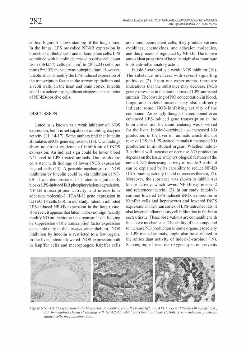
cortex. Figure 5 shows staining of the lung tissue. In the lungs, LPS provoked NF-kB expression in bronchial epithelial cells and infl ammation cells. LPS combined with luteolin decreased positive cell count from (364±54) cells per mm2 to (203±24) cells per mm2 (P=0.02) in the airway subepithelium. However, luteolin did not modify the LPS-induced expression of the transcription factor in the airway epithelium and alveoli walls. In the heart and brain cortex, luteolin could not induce any signifi cant changes in the number of NF-kB-positive cells.
DISCUSSION
Luteolin is known as a weak inhibitor of iNOS expression, but it is not capable of inhibiting enzyme activity (11, 14-17). Some authors fi nd that luteolin stimulates eNOS gene expression (18). Our fi ndings show no direct evidence of inhibition of iNOS expression. An indirect sign could be lower blood NO level in LPS-treated animals. Our results are consistent with fi ndings of lower iNOS expression in glial cells (15). A possible mechanism of iNOS inhibition by luteolin could be via inhibition of NF-kB. It was demonstrated that luteolin signifi cantly blocks LPS-induced IkB phosphorylation/degradation, NF-kB transcriptional activity, and intercellular adhesion molecule-1 (ICAM-1) gene expression in rat IEC-18 cells (30). In our study, luteolin inhibited LPS-induced NF-kB expression in the lung tissue. However, it appears that luteolin does not signifi cantly modify NO production at the organism level. Judging by suppression of the transcription factor expression detectable only in the airways subepithelium, iNOS inhibition by luteolin is restricted to a few organs. In the liver, luteolin lowered iNOS expression both in Kupffer cells and macrophages. Kupffer cells
are immunocompetent cells; they produce various cytokines, chemokines, and adhesion molecules, and this process is regulated by NF-kB. The known antioxidant properties of luteolin might also contribute to its anti-infl ammatory action.
Indole-3-carbinol is a weak iNOS inhibitor (19). The substance interferes with several signalling pathways (2). From our experiments, there are indications that the substance may decrease iNOS gene expression in the brain cortex of LPS-untreated animals. The lowering of NO concentration in blood, lungs, and skeletal muscles may also indirectly indicate some iNOS-inhibiting activity of the compound. Amazingly though, the compound even enhanced LPS-induced gene transcription in the brain cortex, and the same tendency was observed for the liver. Indole-3-carbinol also increased NO production in the liver of animals which did not receive LPS. In LPS-treated animals it increased NO production in all studied organs. Whether indole-3-carbinol will increase or decrease NO production depends on the tissue and physiological features of the animal. NO decreasing activity of indole-3-carbinol can be explained by its capability to reduce NF-kB DNA binding activity (2 and references therein, 12). Moreover, the substance was shown to inhibit Akt kinase activity, which lowers NF-kB expression (2 and references therein, 12). In our study, indole-3-carbinol lowered LPS-induced iNOS expression in Kupffer cells and hepatocytes and lowered iNOS expression in the brain cortex of LPS-untreated rats. It also lowered infl ammatory cell infi ltration in the brain cortex tissue. These observations are compatible with the above mechanisms. The ability of the compound to increase NO production in some organs, especially in LPS-treated animals, might also be attributed to the antioxidant activity of indole-3-carbinol (19). Scavenging of reactive oxygen species prevents
Figure 5 NF-kBp65 expression in the lung tissue. A - control, B - LPS (10 mg kg-1, i.p., 4 h), C - LPS+luteolin (50 mg kg-1, p.o., 4h). Immunohistochemical staining with NF-kBp65 rabbit polyclonal antibody (1:100). Arrow indicates positively stained cells, magnifi cation 200x.
Rostoka E, et al. EFFECTS OF NATURAL COMPOUNDS ON NO AND iNOSArh Hig Rada Toksikol 2010;61:275-285

283
their interaction with NO, which may increase its bioavailability. This effect is associated with several natural compounds, including cocoa polyphenols (31) and resveratrol (32). A similar effect is produced by kaempferol, also known as an inhibitor of iNOS expression; it enhances LPS-induced NO production in blood (33).
Our results suggest that the effects of natural compounds on NO metabolism are organ-specifi c. Indole-3-carbinol enhanced NO production in the liver of LPS-untreated rats, but reduced it in their blood, lungs, and skeletal muscles. It inhibited iNOS gene expression in the brain cortex, but not in the liver. Similarly, lycopene enhanced LPS-triggered iNOS gene transcription in the brain cortex, but not in the liver.
Moreover, the correlation between iNOS gene or protein expression and NO level in a given tissue is not always linear. It seems that iNOS expression does not limit NO production in the LPS model used in our study. Discrepancies can arise for several reasons, including the antioxidant activity of a compound. In this case scavenging of reactive oxygen species makes NO more available (31, 32). The ability of a compound to increase NOS expression or activity in vitro can be attenuated at the organism level. Excessive NO seems to inhibit NOS activity, but the drop in NOS activity does not necessarily affect NO level in tissues (34). Note that it is NO that mostly affects physiological processes in tissues. In our opinion, the effects, and therefore recommendations on the use of a natural compound should be based on direct tissue NO measurements after the compound’s administration. Our morphological analyses indicate that luteolin and indole-3-carbinol protect the liver and brain tissue against LPS-induced degenerative changes and confi rm their anti-degenerative and anti-infl ammatory potential.
Acknowledgements
This study was in part supported by the National Program “Novel drugs and biocorrection remedies: construction, transport forms and mechanisms of action” sub-project “Study of synergism and resistance phenomena of anti-cancer substances and creation of novel anti-cancer drugs” managed by I. Kalvinsh and the grant 04.1317 “Pathological production of nitric oxide, possibilities of its pharmacological correction” awarded to N. Sjakste by the Latvian Council of Science. We thank L. Lauberte for technical assistance. Participation of D. Meirena and A. Saleh in the experiment design and discussions is greatly appreciated.
REFERENCES
1. Ferguson LR. Role of plant polyphenols in genomic stability. Mutat Res 2001;475:89-111.
2. Sarkar FH, Li Y, Wang Z, Kong D. Cellular signalling perturbation by natural products. Cell Signal 2009;21:1541-7.
3. Olszanecki R, Gebska A, Kozlovski VI, Gryglewski RJ. Flavonoids and nitric oxide synthase. J Physiol Pharmacol 2002;53:571-84.
4. Benito S, Lopey D, Sáiz MP, Buxaderas S, Sánchez J, Puig-Parellada P, Mitjavila MT. A fl avonoid-rich diet increases nitric oxide production in rat aorta. Br J Pharmacol 2002;135:910-6.
5. Kim BH, Cho SM, Reddy AM, Kim YS, Min KR, Kim Y. Down-regulatory effect of quercitrin gallate on nuclear factor-kappa B-dependent inducible nitric oxide synthase expression in lipopolysaccharide-stimulated macrophages RAW 264.7. Biochem Pharmacol 2005;69:1577-83.
6. Wallerath T, Li H, Gödtel-Ambrust U, Schwarz PM, Förstermann U. A blend of polyphenolic compounds explains the stimulatory effect of red wine on human endothelial NO synthase. Nitric Oxide 2005;12:97-104.
7. Luo L, Sun Q, Mao YY, Lu YH, Tan RX. Inhibitory effects of fl avonoids from Hypericum perforatum on nitric oxide synthase. J Ethnopharmacol 2004;93:221-5.
8. Chen CK, Pace-Asciak CR. Vasorelaxing activity of resveratrol and quercetin in isolated rat aorta. Gen Pharmacol 1996;27:363-6.
9. Stoclet JC, Kleschyov A, Andriambeloson E, Diebolt M, Andriantsitohaina R. Endothelial NO release caused by red wine polyphenols. J Physiol Pharmacol 1999;50:535-40.
10. Shutenko YZ, Henry Y, Pinard E, Seylaz J, Potier P, Berthet F, Girard P, Sercombe R. Infl uence of the antioxidant quercetin in vivo on the level of nitric oxide determined by electron paramagnetic resonance in rat brain during global ischemia and reperfusion. Biochem Pharmacol 1999;57:199-208.
11. van Meeteren ME, Hendriks JJ, Dijkstra CD, van Tol EA. Dietary compounds prevent oxidative damage and nitric oxide production by cells involved in demyelinating disease. Biochem Pharmacol 2004;67:967-75.
12. Rahman KM, Ali S, Aboukameel A, Sarkar SH, Wang Z, Philip PA, Sakr WA, Raz A. Inactivation of NF-kappaB by 3,3’-diindolylmethane contributes to increased apoptosis induced by chemotherapeutic agent in breast cancer cells. Mol Cancer Ther 2007;6:2757-65.
13. Panasenko OM, Sharov VS, Briviba K, Sies H. Interaction of peroxynitrite with carotenoids in human low density lipoproteins. Arch Biochem Biophys 2000;373:302-5.
14. Kim SJ, Park H, Kim HP. Inhibition of nitric oxide production from lipopolysaccharide-treated RAW 264.7 cells by synthetic fl avones: structure-activity relationship and action mechanism. Arch Pharm Res 2004;27:937-43.
15. Kim JS, Lee HJ, Lee MH, Kim J, Jin C, Ryu JH. Luteolin inhibits LPS-stimulated inducible nitric oxide synthase expression in BV-2 microglial cells. Planta Med 2006;72:65-8.
16. Hu C, Kitts DD. Luteolin and luteolin-7-O-glucoside from dandelion fl ower suppress iNOS and COX-2 in RAW264.7 cells. Mol Cell Biochem 2004;265:107-13.
Rostoka E, et al. EFFECTS OF NATURAL COMPOUNDS ON NO AND iNOSArh Hig Rada Toksikol 2010;61:275-285

284
17. Scuro LS, Simioni PU, Grabriel DR, Saviani EE, Modolo LV, Tamashiro WM, Salgado I. Suppression of nitric oxide production in mouse macrophages by soybean fl avonoids accumulated in response to nitroprusside and fungal elicitation. BMC Biochem 2004;5:5.
18. Li H, Xia N, Brausch I, Yao Y, Förstermann U. Flavonoids from artichoke (Cynara scolymus L.) up-regulate endothelial-type nitric-oxide synthase gene expression in human endothelial cells. J Pharmacol Exp Ther 2004;310:926-32.
19. Gerhäuser C, Klimo K, Heiss E, Neumann I, Gamal-Eldeen A, Knauft J, Liu GY, Sitthimonchai S, Frank N. Mechanism-based in vitro screening of potential cancer chemopreventive agents. Mutat Res 2003;523-524:163-72.
20. Directive 86/609/EEC “European Convention for the Protection of Vertebrate Animals Used for Experimental and other Scientifi c Purposes” ECBR European Coalition for Biomedical Research http://www.ecbr.eu/directive-86609_2.htm
21. Kleschyov AL, Wenzel P, Münzel T. Electron paramagnetic resonance (EPR) spin trapping of biological nitric oxide. J Chromatogr B Analyt Technol Biomed Life Sci 2007;851:12-20.
22. Sjakste N, Baumane L, Meirena D, Lauberte L, Dzintare M, Kalvinsh I. Drastic increase in nitric oxide content in rat brain under halothane anesthesia, revealed by EPR method. Biochem Pharmacol 1999;58:1955-9.
23. Sjakste N, Kleschyov AL, Boucher JL, Baumane L, Dzintare M, Meirena D, Sjakste J, Sydow K, Münzel T, Kalvinsh I. Endothelium- and nitric oxide-dependent vasorelaxing activities of gamma-butyrobetaine esters: possible link to the antiischemic activities of mildronate. Eur J Pharmacol 2004;495:67-73.
24. Sjakste N, Sjakste J, Boucher JL, Baumane L, Sjakste T, Dzintare M, Meirena D, Sharipova J, Kalvinsh I. Putative role of nitric oxide synthase isoforms in the changes of nitric oxide concentration in rat brain cortex and cerebellum following sevofl urane and isofl urane anaesthesia. Eur J Pharmacol 2005;513:193-205.
25. Sjakste N, Andrianov VG, Boucher JL, Shestakova I, Baumane L, Dzintare M, Meirena D, Kalvinsh I. Paradoxical effects of two oximes on nitric oxide production by purifi ed
NO synthases, in cell culture and in animals. Nitric Oxide 2007;17:107-14.
26. Baumane L, Dzintare M, Zvejniece L, Meirena D, Lauberte L, Sile V, Kalvinsh I, Sjakste N. Increased synthesis of nitric oxide in rat brain cortex due to halogenated volatile anesthetics confi rmed by EPR spectroscopy. Acta Anaesthesiol Scand 2002;46:378-83.
27. Vandesompele J, de Preter K, Pattyn F, Poppe B, Van Roy N, De Paepe A, Speleman F. Accurate normalization of real-time quantitative RT-PCR data by geometric averaging of multiple internal control genes. Genome Biol 2002;3:research0034.1-0034.11
28. Ishak K, Baptista A, Bianchi L, Callea F, De Groote J, Gudat J, Gudat F, Denk H, Desmet V, Korb G, MacSween RN. Histological grading and staging of chronic hepatitis. J Hepatol 1995;22:696-9.
29. Di Stefano A, Caramori G, Oates T, Capelli A, Lusuardi M, Gnemmi I, Ioli F, Chung KF, Donner CF, Barnes PJ, Adcock IM. Increased expression of nuclear factor-kB in bronchial biopsies from smokers and patients with COPD. Eur Respir J 2000;20:556-63.
30. Kim JS, Jobin C. The flavonoid luteolin prevents lipopolysaccharide-induced NF-kappaB signalling and gene expression by blocking IkappaB kinase activity in intestinal epithelial cells and bone-marrow derived dendritic cells. Immunology 2005;115:375-87.
31. Sies H, Schewe T, Heiss C, Kelm M. Cocoa polyphenols and infl ammatory mediators. Am J Clin Nutr 2005;81(Suppl 1):304S-12S.
32. Orallo F, Alvarez E, Camiña M, Leiro JM, Gómez E, Fernández P. The possible implication of trans-Resveratrol in the cardioprotective effects of long-term moderate wine consumption. Mol Pharmacol 2002;61:294-302.
33. Rostoka E, Baumane L, Isajevs S, Line A, Dzintare M, Svirina D, Sharipova J, Silina K, Kalvinsh I, Sjakste N. Effects of kaempferol and myricetin on inducible nitric oxide synthase expression and nitric oxide production in rats. Basic Clin Pharmacol Toxicol 2010;106:461-6.
34. Salerno JC, Ghosh DK. Space, time and nitric oxide - neuronal nitric oxide synthase generates signal pulses. FEBS J 2009;276:6677-88.
Rostoka E, et al. EFFECTS OF NATURAL COMPOUNDS ON NO AND iNOSArh Hig Rada Toksikol 2010;61:275-285

285
Sažetak
DJELOVANJE LIKOPENA, INDOL-3-KARBINOLA I LUTEOLINA NA STVARANJE DUŠIKOVA MONOKSIDA I EKSPRESIJU iNOS-GENA U ŠTAKORA OVISI O ORGANU
Zna se da prirodni spojevi utječu na sadržaj NO u pojedinome tkivu. Ipak, biološka aktivnost hrane bogate polifenolima često ne odgovara djelovanju pojedinih polifenola na aktivnost NO-sintaze. Cilj je ovoga ispitivanja bio utvrditi kako prirodni spojevi luteolin, indol-3-karbinol i likopen utječu na stvaranje NO u tkivu štakora te kako utječu na ekspresiju gena i razine inducibilne sintaze dušikova monoksida (iNOS). Indol-3-karbinol višestruko je utjecao na razinu NO; značajno je snizio njegovu koncentraciju u krvi, plućima i skeletnim mišićima, ali ju je povisio u jetri. Također je povećao njegovo stvaranje u svim organima štakora koji su primili lipopolisaharide (LPS). U kori mozga životinja koje nisu primile LPS doveo je do smanjenja ekspresije iNOS-gena, a u onih koje su primile LPS do njegova povećanja. Likopen je doveo do ubrzane transkripcije iNOS-gena u kori mozga životinja koje su primile LPS, ali nije utjecao na stvaranje NO u životinja koje nisu primile LPS, odnosno na ekspresiju gena u jetri. U mozgu životinja koje nisu primile LPS doveo je do laganoga smanjenja ekspresije iNOS-gena. Luteolin je povećao stvaranje NO u krvi životinja koje su primile LPS te njihov broj iNOS-pozitivnih stanica. Naši rezultati upućuju na to da se promjene razina NO uzrokovane prirodnim spojevima ne daju predvidjeti na temelju ekspresije gena iNOS odnosno aktivnosti toga enzima dobivene na modelu. Stoga je veoma važno izravno mjeriti razine NO i ekspresiju NO-sintaze u tkivu životinja.
KLJUČNE RIJEČI: ESR spektroskopija, inducibilna sintaza dušikova monoksida, NO, prirodni spojevi
CORRESPONDING AUTHOR:
Nikolajs SjaksteLatvian Institute of Organic SynthesisAizkraukles Street 21, Riga, LV1006, LatviaE-mail: [email protected]
Rostoka E, et al. EFFECTS OF NATURAL COMPOUNDS ON NO AND iNOSArh Hig Rada Toksikol 2010;61:275-285
Related Documents