RESEARCH Open Access Effects of indole on drug resistance and virulence of Salmonella enterica serovar Typhimurium revealed by genome-wide analyses Eiji Nikaido 1,2,3† , Etienne Giraud 4,5† , Sylvie Baucheron 4,5 , Suguru Yamasaki 1,2,3 , Agnès Wiedemann 4,5 , Kousuke Okamoto 3 , Tatsuya Takagi 3 , Akihito Yamaguchi 2,3 , Axel Cloeckaert 4,5* and Kunihiko Nishino 1* Abstract Background: Many Gram-positive and Gram-negative bacteria produce large quantities of indole as an intercellular signal in microbial communities. Indole demonstrated to affect gene expression in Escherichia coli as an intra-species signaling molecule. In contrast to E. coli, Salmonella does not produce indole because it does not harbor tnaA, which encodes the enzyme responsible for tryptophan metabolism. Our previous study demonstrated that E. coli-conditioned medium and indole induce expression of the AcrAB multidrug efflux pump in Salmonella enterica serovar Typhimurium for inter-species communication; however, the global effect of indole on genes in Salmonella remains unknown. Results: To understand the complete picture of genes regulated by indole, we performed DNA microarray analysis of genes in the S. enterica serovar Typhimurium strain ATCC 14028s affected by indole. Predicted Salmonella phenotypes affected by indole based on the microarray data were also examined in this study. Indole induced expression of genes related to efflux-mediated multidrug resistance, including ramA and acrAB, and repressed those related to host cell invasion encoded in the Salmonella pathogenicity island 1, and flagella production. Reduction of invasive activity and motility of Salmonella by indole was also observed phenotypically. Conclusion: Our results suggest that indole is an important signaling molecule for inter-species communication to control drug resistance and virulence of S. enterica. Keywords: AcrAB, Indole, RamA, Salmonella, SPI-1 Background Bacteria communicate using small molecules by a process termed quorum sensing. Accumulation of quorum-sensing signals in growth medium indicates cell density. The use of chemical signals for bacterial communication is a widespread phenomenon [1-5]. In Gram-negative bacteria, these signals could be N-acyl derivatives of homoserine lactone, cyclic dipeptides, and quinolones [6-12]. These signals regulate various functions such as bioluminescence, differentiation, virulence, DNA transfer, and biofilm maturation [13-22]. The intestinal tract is colonized by approximately 10 12 commensal bacteria including those belonging to the genus Escherichia [23-25]. Among Enterobacteriaceae, indole is produced by E. coli and certain Proteeae such as Proteus vulgaris, Providencia spp., and Morganella spp. [26]. Indole production is commonly used for Escherichia coli identification [26]. Indole is generated from tryptophan by the enzyme tryptophanase, encoded by tnaA [27]. Extracellular indole is found at high con- centrations (over 600 μM) when E. coli is grown in enriched medium [28]. Furthermore, indole has also been found in human feces at comparable concentra- tions (~250–1100 μM) [29,30]. Recent studies have also revealed that indole is an extracellular signal in E. coli, since it has been demonstrated to regulate uptake, syn- thesis, and degradation of amino acids in the stationary phase of planktonic cells [31], multicopy plasmid * Correspondence: [email protected]; [email protected] † Equal contributors 1 Laboratory of Microbiology and Infectious Diseases, Institute of Scientific and Industrial Research, Osaka University, 8-1 Mihogaoka, Ibaraki, Osaka 567-00447, Japan 4 INRA, UMR1282 Infectiologie et Santé Publique, F-37380 Nouzilly, France Full list of author information is available at the end of the article © 2012 Nikaido et al.; licensee BioMed Central Ltd. This is an Open Access article distributed under the terms of the Creative Commons Attribution License (http://creativecommons.org/licenses/by/2.0), which permits unrestricted use, distribution, and reproduction in any medium, provided the original work is properly cited. Nikaido et al. Gut Pathogens 2012, 4:5 http://www.gutpathogens.com/content/4/1/5

Welcome message from author
This document is posted to help you gain knowledge. Please leave a comment to let me know what you think about it! Share it to your friends and learn new things together.
Transcript
Nikaido et al. Gut Pathogens 2012, 4:5http://www.gutpathogens.com/content/4/1/5
RESEARCH Open Access
Effects of indole on drug resistance and virulenceof Salmonella enterica serovar Typhimuriumrevealed by genome-wide analysesEiji Nikaido1,2,3†, Etienne Giraud4,5†, Sylvie Baucheron4,5, Suguru Yamasaki1,2,3, Agnès Wiedemann4,5,Kousuke Okamoto3, Tatsuya Takagi3, Akihito Yamaguchi2,3, Axel Cloeckaert4,5* and Kunihiko Nishino1*
Abstract
Background: Many Gram-positive and Gram-negative bacteria produce large quantities of indole as an intercellularsignal in microbial communities. Indole demonstrated to affect gene expression in Escherichia coli as anintra-species signaling molecule. In contrast to E. coli, Salmonella does not produce indole because it does notharbor tnaA, which encodes the enzyme responsible for tryptophan metabolism. Our previous study demonstratedthat E. coli-conditioned medium and indole induce expression of the AcrAB multidrug efflux pump in Salmonellaenterica serovar Typhimurium for inter-species communication; however, the global effect of indole on genes inSalmonella remains unknown.
Results: To understand the complete picture of genes regulated by indole, we performed DNA microarray analysisof genes in the S. enterica serovar Typhimurium strain ATCC 14028s affected by indole. Predicted Salmonellaphenotypes affected by indole based on the microarray data were also examined in this study. Indole inducedexpression of genes related to efflux-mediated multidrug resistance, including ramA and acrAB, and repressed thoserelated to host cell invasion encoded in the Salmonella pathogenicity island 1, and flagella production. Reduction ofinvasive activity and motility of Salmonella by indole was also observed phenotypically.
Conclusion: Our results suggest that indole is an important signaling molecule for inter-species communication tocontrol drug resistance and virulence of S. enterica.
Keywords: AcrAB, Indole, RamA, Salmonella, SPI-1
BackgroundBacteria communicate using small molecules by a processtermed quorum sensing. Accumulation of quorum-sensingsignals in growth medium indicates cell density. The use ofchemical signals for bacterial communication is a widespreadphenomenon [1-5]. In Gram-negative bacteria, these signalscould be N-acyl derivatives of homoserine lactone, cyclicdipeptides, and quinolones [6-12]. These signals regulatevarious functions such as bioluminescence, differentiation,virulence, DNA transfer, and biofilm maturation [13-22].
* Correspondence: [email protected]; [email protected]†Equal contributors1Laboratory of Microbiology and Infectious Diseases, Institute of Scientificand Industrial Research, Osaka University, 8-1 Mihogaoka, Ibaraki, Osaka567-00447, Japan4INRA, UMR1282 Infectiologie et Santé Publique, F-37380 Nouzilly, FranceFull list of author information is available at the end of the article
© 2012 Nikaido et al.; licensee BioMed CentralCommons Attribution License (http://creativecreproduction in any medium, provided the or
The intestinal tract is colonized by approximately 1012
commensal bacteria including those belonging to thegenus Escherichia [23-25]. Among Enterobacteriaceae,indole is produced by E. coli and certain Proteeae suchas Proteus vulgaris, Providencia spp., and Morganellaspp. [26]. Indole production is commonly used forEscherichia coli identification [26]. Indole is generatedfrom tryptophan by the enzyme tryptophanase, encodedby tnaA [27]. Extracellular indole is found at high con-centrations (over 600 μM) when E. coli is grown inenriched medium [28]. Furthermore, indole has alsobeen found in human feces at comparable concentra-tions (~250–1100 μM) [29,30]. Recent studies have alsorevealed that indole is an extracellular signal in E. coli,since it has been demonstrated to regulate uptake, syn-thesis, and degradation of amino acids in the stationaryphase of planktonic cells [31], multicopy plasmid
Ltd. This is an Open Access article distributed under the terms of the Creativeommons.org/licenses/by/2.0), which permits unrestricted use, distribution, andiginal work is properly cited.
Nikaido et al. Gut Pathogens 2012, 4:5 Page 2 of 13http://www.gutpathogens.com/content/4/1/5
maintenance, cell division [32], biofilm formation [28],acid resistance [33], and expression of multidrug expor-ters in E. coli [34-36] as well as to regulate the pathogen-icity island, including the locus of enterocyte effacementof pathogenic E. coli [37,38]. Indole has also beendemonstrated as an important cell-signaling molecule fora population-based antibiotic resistance mechanism [39].Salmonella enterica is a bacterial pathogen that causes
various diseases in humans including gastroenteritis,bacteremia, and typhoid fever [40]. In contrast to E. coli,S. enterica does not harbor tnaA; therefore, this organ-ism does not produce indole [41]. In our previous study,we demonstrated that an E. coli-conditioned mediumand indole induced expression of the acrAB–tolC multi-drug efflux system of Salmonella in a RamA regulator-dependent manner [35]. This suggests that indole is usedas a cell-signaling molecule in both intra- and inter-species communication. However, the global effect of in-dole on Salmonella remains to be elucidated.We hypothesized that indole controls expression of a
wide range of genes and plays a role in regulating thephysiological functions of S. enterica serovar Typhimur-ium. Therefore, to reveal the complete picture of indole-controlled genes, we conducted microarray analysis ofgenes affected by indole. Predicted Salmonella pheno-types affected by indole based on the microarray datawere also examined in this study.
MethodsBacterial strains and growth conditionsS. enterica serovar Typhimurium strains used in thisstudy were the wild-type strain ATCC14028s [42] and itsderivatives. These included strain NES114 which harborsa FLAG-tag fused at the chromosomal ramA gene(ramA-FLAG::KmR) and strain NES84 which carries aramA reporter plasmid (ATCC 14028s/pNNramA) [35].Derivatives also included various deletion mutants: ramAdeletion mutant 14028sΔramA::kanR, ramR deletion mu-tant 14028sΔramR::kanR and mutant 14028sΔram::kanR
deleted of the whole ram locus. Bacterial strains weregrown at 37 °C in Luria–Bertani (LB) broth supplemen-ted with indole (Sigma) where appropriate.
DNA microarray analysisThe ATCC 14028s strain was grown in the presence orabsence of 1 or 4 mM indole. The cells were rapidly col-lected for total RNA extraction when the culturereached an optical density (OD) of 0.6 at 600 nm. TotalRNA was extracted from the cells using the RNeasyMidi kit (Qiagen) and Turbo DNA-free™ kit (Ambion).After extraction of total RNA, fluorescent labeling ofcDNA was performed using the GeneChip DNA labelingreagents (Affimetrix). The fluorescent-labeled cDNA washybridized in cDNA microarray plates (NimbleExpress™
S. typhimurium array; NimbleGen Systems, Inc.). Thedegree of fluorescence in the plates was measured andquantified using the GeneChip Scanner 3000 (Affymetrix)and GeneChip Operating Software ver. 1.4 (Affymetrix),respectively. Measured values were compared to controlvalues, and p values of distribution of logged data wereobtained. A ranked conversion of p values was calculated,and values lying inside 2.5 % of the two extremes wereconsidered valid.
Semiquantitative RT-PCRThe ATCC 14028s strain was grown in the presence orabsence of 2 mM indole. The cells were rapidly collectedfor total RNA extraction when the culture reached anOD600 of 0.6. Total RNA from bacterial cultures wasextracted as described above. Semiquantitative RT-PCRwas used to measure the transcriptional expression of rrsand ramA. RNA was reverse-transcribed using randomhexamers and TaqMan reverse transcription reagents(Applied Biosystems), and PCR was performed usingTakara LA Taq DNA polymerase (Takara Bio, Inc.). Theprimers for rrs were rrs-F and rrs-R (Table 1) and thosefor ramA were ramA-F and ramA-R (Table 1).
β-Galactosidase assayThe NES84 strain [35] was grown in the presence of0–3 mM indole until an OD600 of 0.6 was reached.β-Galactosidase activity was determined as described byMiller [43]. All assays were performed in triplicate.
Construction of the ramA-FLAG strainInsertion of the FLAG-tag of the ramA gene 3' terminalwas performed as described by Datsenko and Wanner [44].The kanamycin resistance gene aph, flanked by Flp recog-nition sites, was amplified by PCR using the pri-mers ramA-FLAG-forward (GCCAGGCGCTTATCGTAAAGAAAAGCATGGCCGTACGCATGACTACAAGGACGACGATGACAAGTAGGTGTAGGCTGGAGCTGCTTC) and ramA-FLAG-reverse (CGATTAAACATTTCAATGCGTACGGCCATGCTTTTCTTTACATATGAATATCCTCCTTAG). The sequence of the FLAG-tagappears in bold in the sequence of the ramA-FLAG- for-ward primer. The resulting PCR products were used totransform the recipient ATCC 14028s strain harboring thepKD46 plasmid, which expresses Red recombinase [44].The chromosomal structure of the mutated loci was veri-fied by PCR.
Western blottingThe NES114 strain (ramA-FLAG::KmR) was grown inthe presence of 2 mM indole until an OD600 of 0.6 wasreached. Bacterial cells were washed with buffer [20 mMTris–HCl (pH 8.0), 200 mM NaCl, and 1 mM EDTA],resuspended in 1 ml of the same buffer, and disrupted by
Table 1 Primers used in this study
Primer name Oligonucleotide sequence (5′ to 3′)
For semiquantitative RT-PCR
rrs-F CCAGCAGCCGCGGTAAT
rrs-R TTTACGCCCAGTAATTCCGATT
ramA-F ATTTGAATCAGCCGTTACGTATTG
ramA-R TGCAGGTGCCACTTGGAAT
For quantitative RT-PCR
gmk-f TTGGCAGGGAGGCGTTT
gmk-r GCGCGAAGTGCCGTAGTAAT
gyrB-f TCTCCTCACAGACCAAAGATAAGCT
gyrB-r CGCTCAGCAGTTCGTTCATC
rrs-f CCAGCAGCCGCGGTAAT
rrs-r TTTACGCCCAGTAATTCCGATT
ramA-f GCGTGAACGGAAGCTAAAAC
ramA-r GGCCATGCTTTTCTTTACGA
acrB-f TCGTGTTCCTGGTGATGTACCT
acrB-r AACCGCAATAGTCGGAATCAA
mdtE-f AGTCGCTGGATACCACCATC
mdtE-r GATATTACGCACGCCGATTT
tolC-f GCCCGTGCGCAATATGAT
tolC-r CCGCGTTATCCAGGTTGTTG
flhC-f ATATCCAGTTGGCGATGGAG
flhC-r TTGCTCCCAGGTCATAAACC
hilA-f CATGGCTGGTCAGTTGGAG
hilA-r CGTAATTCATCGCCTAAACG
invF-f TGAAAGCCGACACAATGAAAAT
invF-r GCCTGCTCGCAAAAAAGC
invA-f GGCGCCAAGAGAAAAAGATG
invA-r CAAATATAACGCGCCATTGCT
sipA-f TTTGGCTGTACGTTAGATCCGTTA
sipA-r CCGCCGCTTTGTCAACA
Nikaido et al. Gut Pathogens 2012, 4:5 Page 3 of 13http://www.gutpathogens.com/content/4/1/5
sonication using the Branson Sonifier 200 (BransonSonic Power Co., Danbury, CT, USA) on ice for 2.5 min.Whole-cell lysate (10 μg of protein) was separated on15 % SDS-PAGE using Tris-glycine SDS as the runningbuffer. The gel was transferred to PVDF membranes,and analyzed by western blotting using a monoclonalanti-FLAG antibody (Sigma). The blot was developedusing anti-mouse IgG horseradish peroxidase-conjugatedantibody and analyzed using the ECL detection system(GE Healthcare).
Transmission electron microscopyOne hundred microliters of an overnight culture of thewild-type Salmonella strain was added to 5 ml of LBbroth, and the bacterial culture was grown in the pres-ence or absence of 1 mM indole until an OD600 of 0.6was reached. Bacterial cells were collected by centrifuga-tion, fixed in 2 % glutaraldehyde solution, and observed
using a transmission electron microscope JEM-2100(JEOL Ltd.).
Gene expression analysis by qRT-PCRBacteria were grown until mid-log phase (OD600 of 0.6)and harvested by centrifugation. Pelleted cultures werestabilized with RNAprotect Bacteria Reagent (Qiagen)and stored at −80 °C until use. Total RNA was extractedusing the RNeasy Mini kit. (Qiagen) following the manu-facturer’s instructions. Removal of residual genomicDNA was performed using the Turbo DNA-free kit(Ambion) and examined by negative PCR amplificationof a chromosomal sequence. RNA integrity was exam-ined by electrophoresis on 1 % agarose gel. Total RNAwas reverse-transcribed using random hexamers and theSuperscript III First Strand Synthesis System (AppliedBiosystems). Primers used for qRT-PCR are listed inTable 1. The cycling conditions were as follows: 95 °Cfor 5 min followed by 40 cycles of 95 °C for 10 s and 60 °Cfor 15 s. After each run, amplification specificity and theabsence of primer dimers were examined using a dissoci-ation curve acquired by heating the PCR products from 60to 95 °C. The relative quantities of transcripts were deter-mined using the standard curve method and normalizedagainst the geometric mean of three reference genes (gmk,gyrB, rrs). In qRT-PCR experiments performed to addressthe effect of 1 mM indole, the expression level of eachgene of interest was calculated as the average of three in-dependent RNA samples. A two-tailed Student’s t-test wasused to assess significance using a p value of <0.05 as acutoff.
Measurement of motility of SalmonellaAn overnight culture of the ATCC 14028s Salmonellastrain was diluted in LB broth and grown in the presenceor absence of 1 mM indole until an OD600 of 0.6 wasreached. Next, 1 μl of bacterial culture was spotted inthe center of a semi-solid agar plate containing 1 % tryp-tone peptone and 0.3 % BactoAgar and incubated at 37 °Cfor 3–5 h in a humidified incubator, after which strainswere assessed for motility.
Invasion assayInvasions assays were essentially performed as previouslydescribed [45]. Caco-2 cells were grown in Dulbecco’smodified Eagle medium (DMEM) supplemented with10 % inactivated fetal bovine serum, 1 % nonessentialamino acids, and 1 % antibiotic solution (Gibco, Invitro-gen). Cells harvested by trypsinization were seeded at2 × 105 cells/well in a 24-well plate (Falcon) and incu-bated for 4 days at 37 °C under 5 % CO2 in the mediumdescribed above, to obtain a confluent monolayer. Anti-biotic was removed 24 h before performing the invasionassays. Bacteria were grown to an OD600 of 0.6 in LB
Nikaido et al. Gut Pathogens 2012, 4:5 Page 4 of 13http://www.gutpathogens.com/content/4/1/5
broth in the presence or absence of 1 mM indole. Afterwashing with DMEM, bacteria were inoculated on Caco-2cells at a multiplicity of infection of 30, and the plates werefurther incubated for 30 min. The bacteria-containingmedium was removed from the wells, and the cells werewashed with PBS. Cells were incubated for 1.5 h withDMEM supplemented with 100 μg/ml gentamicin. Cellswere washed with PBS and lysed by the addition of sterileultrapure water for 30 min. Serial dilutions were plated onLB agar. The percentage of penetrating bacteria was calcu-lated on the basis of the ratio of the counted cfu to thebacterial inoculum. For each bacterial strain and for eachcondition, three replicates were used.
ResultsIndole affects gene expression in SalmonellaThe regulation of Salmonella genes in the wild-typestrain ATCC 14028s by 1 or 4 mM indole, was analyzedusing DNA microarray. To exclude noise data based onmicroarray analysis, we considered that genes for which
Table 2 Salmonella genes whose relative expression was incre
STM no. Gene Function
STM0521 ybbV Putative cytoplasmic protein
STM0581 ramA Transcriptional regulator (activator) of acrAB and tolC
STM0584 entD Enterochelin synthetase, component D (phoshpanteth
STM0707 kdpF Putative outer membrane protein
STM0823 ybiJ Putative periplasmic protein
STM1156 yceA Putative enzyme related to sulfurtransferases
STM1214 ycfR Putative outer membrane protein
STM1251 Putative molecular chaperone (small heat shock prote
STM1355 ydiP Putative transcription regulator, AraC family
STM1472 Putative periplasmic protein
STM1790 hyaE Putative thiol-disulfide isomerase and thioredoxins
STM1868A Putative protein
STM2103 wcaJ Putative UDP-glucose lipid carrier transferase/glucosecolanic acid gene cluster
STM2106 wcaI Putative glycosyl transferase in colanic acid biosynthe
STM2206 fruF Phosphoenolpyruvate-dependent sugar phosphotran
STM3028 stdB Putative outer membrane usher protein
STM3444 bfd Regulatory or redox component complexing with Bfr
STM3511 yhgI Putative thioredoxin-like proteins and domain
STM3606 yhjB Putative transcriptional regulator (LuxR/UhpA familiy)
STM3668 yiaK Putative malate dehydrogenase
STM3941 Putative inner membrane protein
STM4213 Putative phage tail sheath protein
STM4327 fxsA Suppresses F exclusion of bacteriophage T7
STM4548 bglJ Transcriptional regulator (activator) of bgl operon (Lux
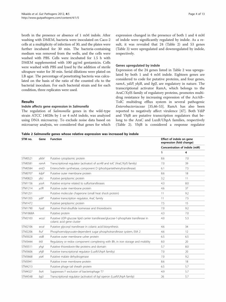
expression changed in the presence of both 1 and 4 mMof indole were significantly regulated by indole. As a re-sult, it was revealed that 24 (Table 2) and 53 genes(Table 3) were upregulated and downregulated by indole,respectively.
Genes upregulated by indoleExpression of the 24 genes listed in Table 2 was upregu-lated by both 1 and 4 mM indole. Eighteen genes areconsidered to code for putative proteins, and four genes,ramA, ydiP, yhjB, and bglJ, are regulatory in nature. Thetranscriptional activator RamA, which belongs to theAraC/XylS family of regulatory proteins, promotes multi-drug resistance by increasing expression of the AcrAB–TolC multidrug efflux system in several pathogenicEnterobacteriaceae [35,46-53]. RamA has also beenreported to negatively affect virulence [47]. Both YdiPand YhjB are putative transcription regulators that be-long to the AraC and LuxR/UhpA families, respectively(Table 2). YhjB is considered a response regulator
ased by indole
Effect of indole on geneexpression (fold change)
Concentration of indole (mM)
1 4
8.6 7.0
(AraC/XylS family) 7.0 39
einyltransferase) 6.1 7.5
8.6 18
3.2 11
4.3 8.0
4.6 37
in) 11 9.2
11 7.5
7.5 15
7.0 9.2
4.3 7.0
-1-phosphate transferase in 4.0 5.3
sis 4.6 34
sferase system, EIIA 2 4.6 12
6.5 6.5
, in iron storage and mobility 8.0 20
5.7 8.0
7.0 20
7.0 9.2
8.6 18
6.5 5.7
4.9 5.7
R/UhpA family) 26 5.7
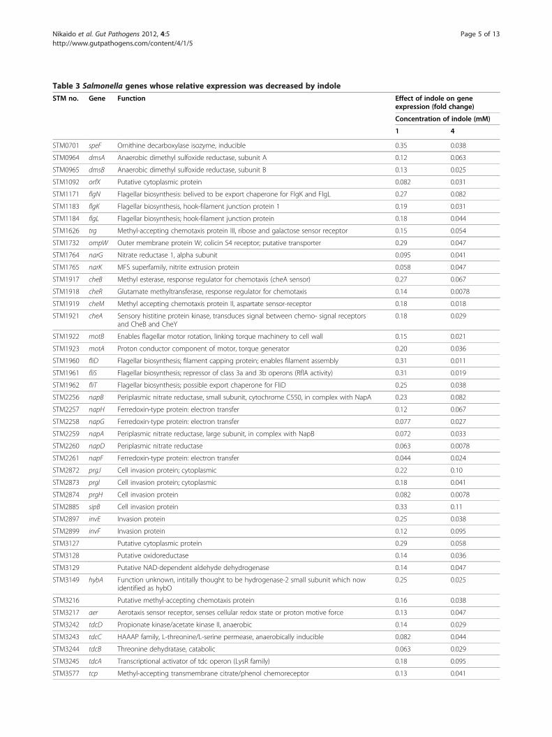
Table 3 Salmonella genes whose relative expression was decreased by indole
STM no. Gene Function Effect of indole on geneexpression (fold change)
Concentration of indole (mM)
1 4
STM0701 speF Ornithine decarboxylase isozyme, inducible 0.35 0.038
STM0964 dmsA Anaerobic dimethyl sulfoxide reductase, subunit A 0.12 0.063
STM0965 dmsB Anaerobic dimethyl sulfoxide reductase, subunit B 0.13 0.025
STM1092 orfX Putative cytoplasmic protein 0.082 0.031
STM1171 flgN Flagellar biosynthesis: belived to be export chaperone for FlgK and FlgL 0.27 0.082
STM1183 flgK Flagellar biosynthesis, hook-filament junction protein 1 0.19 0.031
STM1184 flgL Flagellar biosynthesis; hook-filament junction protein 0.18 0.044
STM1626 trg Methyl-accepting chemotaxis protein III, ribose and galactose sensor receptor 0.15 0.054
STM1732 ompW Outer membrane protein W; colicin S4 receptor; putative transporter 0.29 0.047
STM1764 narG Nitrate reductase 1, alpha subunit 0.095 0.041
STM1765 narK MFS superfamily, nitrite extrusion protein 0.058 0.047
STM1917 cheB Methyl esterase, response regulator for chemotaxis (cheA sensor) 0.27 0.067
STM1918 cheR Glutamate methyltransferase, response regulator for chemotaxis 0.14 0.0078
STM1919 cheM Methyl accepting chemotaxis protein II, aspartate sensor-receptor 0.18 0.018
STM1921 cheA Sensory histitine protein kinase, transduces signal between chemo- signal receptorsand CheB and CheY
0.18 0.029
STM1922 motB Enables flagellar motor rotation, linking torque machinery to cell wall 0.15 0.021
STM1923 motA Proton conductor component of motor, torque generator 0.20 0.036
STM1960 fliD Flagellar biosynthesis; filament capping protein; enables filament assembly 0.31 0.011
STM1961 fliS Flagellar biosynthesis; repressor of class 3a and 3b operons (RflA activity) 0.31 0.019
STM1962 fliT Flagellar biosynthesis; possible export chaperone for FliD 0.25 0.038
STM2256 napB Periplasmic nitrate reductase, small subunit, cytochrome C550, in complex with NapA 0.23 0.082
STM2257 napH Ferredoxin-type protein: electron transfer 0.12 0.067
STM2258 napG Ferredoxin-type protein: electron transfer 0.077 0.027
STM2259 napA Periplasmic nitrate reductase, large subunit, in complex with NapB 0.072 0.033
STM2260 napD Periplasmic nitrate reductase 0.063 0.0078
STM2261 napF Ferredoxin-type protein: electron transfer 0.044 0.024
STM2872 prgJ Cell invasion protein; cytoplasmic 0.22 0.10
STM2873 prgI Cell invasion protein; cytoplasmic 0.18 0.041
STM2874 prgH Cell invasion protein 0.082 0.0078
STM2885 sipB Cell invasion protein 0.33 0.11
STM2897 invE Invasion protein 0.25 0.038
STM2899 invF Invasion protein 0.12 0.095
STM3127 Putative cytoplasmic protein 0.29 0.058
STM3128 Putative oxidoreductase 0.14 0.036
STM3129 Putative NAD-dependent aldehyde dehydrogenase 0.14 0.047
STM3149 hybA Function unknown, intitally thought to be hydrogenase-2 small subunit which nowidentified as hybO
0.25 0.025
STM3216 Putative methyl-accepting chemotaxis protein 0.16 0.038
STM3217 aer Aerotaxis sensor receptor, senses cellular redox state or proton motive force 0.13 0.047
STM3242 tdcD Propionate kinase/acetate kinase II, anaerobic 0.14 0.029
STM3243 tdcC HAAAP family, L-threonine/L-serine permease, anaerobically inducible 0.082 0.044
STM3244 tdcB Threonine dehydratase, catabolic 0.063 0.029
STM3245 tdcA Transcriptional activator of tdc operon (LysR family) 0.18 0.095
STM3577 tcp Methyl-accepting transmembrane citrate/phenol chemoreceptor 0.13 0.041
Nikaido et al. Gut Pathogens 2012, 4:5 Page 5 of 13http://www.gutpathogens.com/content/4/1/5

Table 3 Salmonella genes whose relative expression was decreased by indole (Continued)
STM3626 dppF ABC superfamily (atp_bind), dipeptide transport protein 0.31 0.036
STM3628 dppC ABC superfamily (membrane), dipeptide transport protein 2 0.33 0.088
STM4258 Putative methyl-accepting chemotaxis protein 0.08 0.088
STM4300 fumB Fumarase B (fumarate hydratase class I), anaerobic isozyme 0.19 0.088
STM4305 Putative anaerobic dimethyl sulfoxide reductase, subunit A 0.14 0.082
STM4306 Putative anaerobic dimethyl sulfoxide reductase, subunit B 0.11 0.058
STM4452 nrdD Anaerobic ribonucleoside-triphosphate reductase 0.047 0.095
STM4465 Putative ornithine carbamoyltransferase 0.19 0.10
STM4466 Putative carbamate kinase 0.19 0.10
STM4467 Putative arginine deiminase 0.082 0.019
Nikaido et al. Gut Pathogens 2012, 4:5 Page 6 of 13http://www.gutpathogens.com/content/4/1/5
comprising a CheY-like receiver domain and a helix-turn-helix DNA-binding domain. YhjB in E. coli stimu-lates dephosphorylation of two histidine kinases, EnvZand NtrB, although the sensor kinase for YhjB phosphor-ylation has not yet been identified [54]. BglJ forms a het-erodimer with RcsB to relieve repression of the E. colibgl operon and allow arbutin and salicin transport andutilization [55,56]. Expression of all these regulatorygenes increased by more than 10-fold in the presence ofindole. Of particular interest, expression of ramAincreased by 39-fold in response to 4 mM indole. Notice-ably, higher indole concentration did not always lead toa higher expression level. Indeed, bglJ expression wasmore increased in response to 1 mM indole (26-fold)than to 4 mM indole (5.7-fold).Among other genes upregulated by indole, functions of
the gene products of entD, fruF, bfd, and fxsA have beencharacterized. EntD has phosphopantetheinyl transferaseactivity [57] and is involved in biosynthesis of the iron-acquiring siderophore enterobactin [58]. FruF is abifunctional PTS system fructose-specific transportersubunit IIA/HPr protein [41,59]. Bfd is a bacterioferritin-associated ferredoxin considered to be involved in Bfriron storage and release functions or in regulation of Bfr[41]. FxsA of E. coli was described as a suppressor of theF exclusion of phage T7 [60]. Expression of all thesegenes was more strongly induced by 4 mM indole thanby 1 mM indole.
Genes downregulated by indoleMicroarray analysis revealed that fifty-three genes wererepressed by both 1 and 4 mM indole (Table 3). Expres-sion of all these genes excluding STM4258 and nrdDwas reduced by 4 mM indole compared to that by 1 mMindole. Although there are 10 putative genes, functionsof most gene products have been characterized (Table 3).Microarray analysis revealed that indole represses ex-
pression of genes related to bacterial motility includingflagella biosynthesis (flgN/K/L and fliD/S/T), chemotaxis(cheB/R/M/A, aer [61], tcp, and trg), and flagella motor
activity (motB/A). Indole also decreased expression ofgenes related to cell invasion such as prgJ/I/H, sipB, andinvE/F, which are encoded by the Salmonella pathogen-icity island 1 (SPI-1). Expression of genes related to an-aerobic respiration was decreased by indole. The genesrepressed by indole included narG (nitrate reductase),narK (nitrate-nitrite antiporter), and genes in the napoperon encoding nitrate reductase (Nap) such as napB/H/G/A/D/F [62]. The tdc operon including tdcA/B/C/D,which are responsible for the anaerobic degradation ofthreonine [63], was also downregulated by indole. Inaddition to these genes, other genes related to anaerobicrespiration such as dmsA/B (anaerobic dimethyl sulfox-ide reductase), fumB (fumarase B, anaerobic isozyme),nrdD (anaerobic ribonucleoside-triphosphate reductase),and STM4305/4306 (putative anaerobic dimethyl sulfox-ide reductase, subunit A/B) were also repressed byindole. Indole also repressed membrane protein genessuch as ompW (outer membrane protein involved inosmoregulation that is also affected by environmentalconditions) and dppF/C (dipeptide transport protein).
Indole upregulates genes involved in efflux-mediatedmultidrug resistanceAs reported above, microarray analysis identified thatindole significantly increased expression of ramA, encod-ing a transcriptional activator of the multidrug trans-porter genes acrAB and tolC of Salmonella [35].To confirm this result, we performed reverse transcript-ase polymerase chain reaction (RT-PCR) and observedthat transcript levels of ramA increased when bacterialcells of the strain ATCC 14028s were treated with 2 mMindole (Figure 1A-1). To investigate whether indoleinduces production of RamA, we constructed a strainNES114 that harbors a FLAG-tag fused to chromosomally-encoded ramA. Western blotting revealed increased pro-duction of RamA in the presence of 2 mM indole (Figure1A-2). Microarray analysis demonstrated that expressionof ramA was more strongly induced by 4 mM indole (39-fold increase relative to untreated cells) than by 1 mM

Figure 1 Indole induces efflux-mediated multidrug resistance genes. (A-1) RT-PCR measurement of indole effect on expression of ramA.Expression of rrs, encoding rRNA, was measured as a control. The wild-type strain ATCC14028s was grown in the presence (+) or absence (−) of2 mM indole, and RT-PCR was performed after RNA isolation. (A-2) RamA production in the wild-type ATCC14028s derivative strain carrying theepitope-tagged ramA. NES114 (ramA-FLAG::KmR) was grown in the presence (+) or absence (−) of 2 mM indole. SDS-PAGE of lysates of NES114was followed by Western blotting with an anti-FLAG antibody (anti-FLAG, top) or by staining with Coomassie brilliant blue (CBB, bottom).(B) β-Galactosidase levels in the wild-type ATCC 14028s derivative strain carrying the ramA-lac transcriptional fusion (NES84) treated with differentindole concentrations. (C-1) qRT-PCR measurement of indole effect on expression of ramA. The wild-type strain (ATCC14028s) and its ramR::kanR
deletion mutant were grown in the presence (+) or absence (−) of 1 mM indole. (C-2) qRT-PCR measurement of indole effect on expression of acrB.The wild-type strain ATCC14028s and its ramR::kanR and ramA::kanR deletion mutants were grown in the presence (+) or absence (−) of 1 mMindole. (B and C-1, 2) The data correspond to mean values from three independent replicates. The bars indicate the standard deviation. (C-1, 2)ramA and acrB expression levels were expressed relative to that measured in the wild-type strain grown without indole, which was assigned theunit value. Asterisks indicate statistically significant difference (p< 0.05) according to a two-tailed Student’s t-test.
Nikaido et al. Gut Pathogens 2012, 4:5 Page 7 of 13http://www.gutpathogens.com/content/4/1/5
indole (7.0-fold increase), indicating that the effect of in-dole on expression of ramA may be concentrationdependent. To investigate the effect of different indoleconcentrations on the promoter activity of ramA, aβ-galactosidase assay with the NES84 strain [35] wasperformed, and it was found that indole activated theramA promoter in a concentration-dependent manner(Figure 1B). The finding of enhanced promoter activity oframA is in good agreement with our previous observation[35].
Confirming classical RT-PCR results, quantitativeRT-PCR (qRT-PCR) indicated that expression of ramAin the strain ATCC 14028s increases by 4-fold in thepresence of 1 mM indole (Figure 1C-1). As previouslyreported, RamR represses to the same extent the expres-sion of ramA [46,64]. Therefore, to determine a possiblecontribution of RamR to induction of expression oframA via indole, the effect of 1 mM indole on expressionof ramA in a ΔramR strain (14028sΔramR::kanR) wasexamined. At this concentration indole did not induce
Nikaido et al. Gut Pathogens 2012, 4:5 Page 8 of 13http://www.gutpathogens.com/content/4/1/5
expression of ramA suggesting indole-mediated ramAinduction is indeed dependent on the presence ofRamR (Figure 1C-1). However, whereas indole wasshown to induce expression of acrB in the wild-typestrain, this induction was neither observed in theΔramR (14028sΔramR::kanR) nor in the ΔramA strain(14028sΔramA::kanR) (Figure 1C-2). This suggested thatindole-mediated induction of acrB expression is notsolely dependent on RamA, but nevertheless alsorequires the presence of the RamR transcriptionalrepressor.
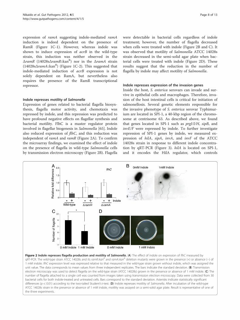
Indole represses motility of SalmonellaExpression of genes related to bacterial flagella biosyn-thesis, flagella motor activity, and chemotaxis wasrepressed by indole, and this repression was predicted tohave profound negative effects on flagellar synthesis andbacterial motility. FlhC is a master regulator proteininvolved in flagellar biogenesis in Salmonella [65]. Indolealso reduced expression of flhC, and this reduction wasindependent of ramA and ramR (Figure 2A). To confirmthe microarray findings, we examined the effect of indoleon the presence of flagella in wild-type Salmonella cellsby transmission electron microscopy (Figure 2B). Flagella
Figure 2 Indole represses flagella production and motility of SalmoneqRT-PCR. The wild-type strain ATCC 14028s and its ramR::kanR and ramA::ka1 mM indole. flhC expression level was expressed relative to that measuredunit value. The data corresponds to mean values from three independent relectron microscopy was used to detect flagella on the wild-type strain (ATnumber of flagella attached to a single cell was counted from images takenbacterial cells for both indole-treated and untreated cells. Bars corresponddifferences (p< 0.01) according to the two-tailed Student’s t-test. (D) IndoleATCC 14028s strain in the presence or absence of 1 mM indole, motility wathe three experiments.
were detectable in bacterial cells regardless of indoletreatment; however, the number of flagella decreasedwhen cells were treated with indole (Figure 2B and C). Itwas observed that motility of Salmonella ATCC 14028sstrain decreased in the semi-solid agar plate when bac-terial cells were treated with indole (Figure 2D). Theseresults suggest that the reduction in the number offlagella by indole may affect motility of Salmonella.
Indole represses expression of the invasion genesInside the host, S. enterica serovars can invade and sur-vive in epithelial cells and macrophages. Therefore, inva-sion of the host intestinal cells is critical for initiation ofsalmonellosis. Several genetic elements responsible forthe invasive phenotype of S. enterica serovar Typhimur-ium are located in SPI-1, a 40-kbp region of the chromo-some at centrisome 63. As described above, we foundthat genes located in SPI-1 such as prgJ/I/H, sipB, andinvE/F were repressed by indole. To further investigaterepression of SPI-1 genes by indole, we measured ex-pression of hilA, sipA, invA, and invF of the ATCC14028s strain in response to different indole concentra-tion by qRT-PCR (Figure 3). hilA is located on SPI-1,and it encodes the HilA regulator, which controls
lla. (A) The effect of indole on expression of flhC measured bynR deletion mutants were grown in the presence (+) or absence (−) ofin the wild-type strain grown without indole, which was assigned theeplicates. The bars indicate the standard deviation. (B) TransmissionCC 14028s) grown in the presence or absence of 1 mM indole. (C) Theusing transmission electron microscopy. Data were collected from 30
to the standard deviation. Asterisks indicate statistically significantrepresses motility of Salmonella. After incubation of the wild-types assayed on a semi-solid agar plate. Result is representative of one of

Figure 3 Indole represses expression of invasion genes encoded by SPI-1. The effect of indole on expression of SPI-1 genes including hilA(A), sipA (B), invA (C), and invF (D) was measured by qRT-PCR. The wild-type ATCC 14028s strain (open bars) and its ramA::kanR deletion mutant(solid bars) were grown with indole concentrations between 0 and 1 mM. Genes expression levels were expressed relative to that measured inthe wild-type strain grown without indole, which was assigned the unit value.
Nikaido et al. Gut Pathogens 2012, 4:5 Page 9 of 13http://www.gutpathogens.com/content/4/1/5
expression of SPI-1 genes, including the type III secretionsystem (T3SS). invF also encodes an invasion regulatoryprotein of SPI-1. sipA encodes an effector protein, the se-cretion of which is mediated by T3SS. invA encodes astructural component of T3SS. qRT-PCR revealed thatindole decreased expression of hilA, sipA, invA, and invFin a concentration-dependent manner (Figure 3). Since itwas reported that overexpression of ramA results indecreased expression of SPI-1 genes [47] and our studyindicated that indole induces ramA, we investigated theeffect of ramA deletion on expression of SPI-1 genesregulated by indole. As shown in Figure 3, indolerepressed expression of hilA, sipA, invA, and invF; how-ever, its repressive effect on those in the ramA-deletedmutant (14028sΔramA::kanR) was slightly lower than thatobserved in the wild-type strain (ATCC 14028s) whenbacterial cells were treated with 0.125 or 0.25 mM indole.In contrast, when bacterial cells were treated with con-centrations of indole of 0.35 mM or more, the repressiveeffect on SPI-1 genes was similar in the wild-type and in
the mutant strains. These data suggest that indolepartially represses SPI-1 genes in a RamA-dependentmanner when cells are treated with lower indole concen-trations; however, the repressive effect of indole on SPI-1may be RamA-independent at higher concentrations.A critical step in Salmonella pathogenesis is invasion of
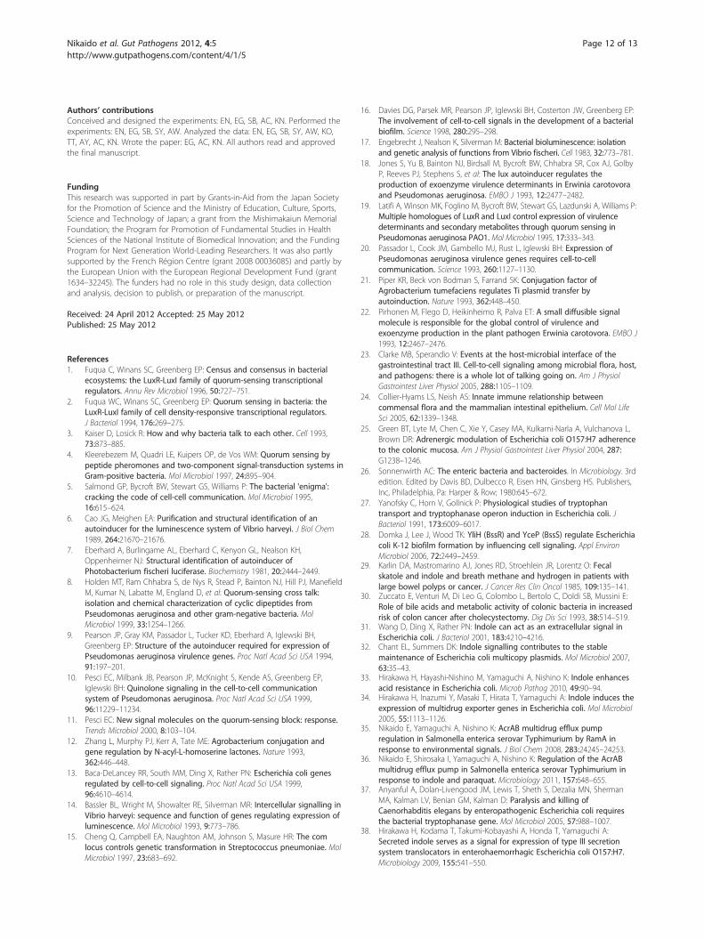
enterocytes and M cells of the small intestine via expres-sion of a type III secretion system encoded by SPI-1 thatsecretes effector proteins into host cells, leading toengulfment of bacteria within large membrane ruffles. Asindicated previously, indole represses expression of genesencoded by SPI-1, suggesting that indole reduces invasionof mammalian cells by Salmonella. To examine thispossibility, we investigated the effect of indole in an inva-sion assay using Caco-2 cells. When bacterial cells weretreated with 1 mM indole, the invasion rate of Salmonellawas reduced compared to that in untreated bacterial cells(Figure 4). We also examined the effect of deletions oframR and of the whole ram locus on invasive activity ofSalmonella treated with 1 mM indole. Indole repressed
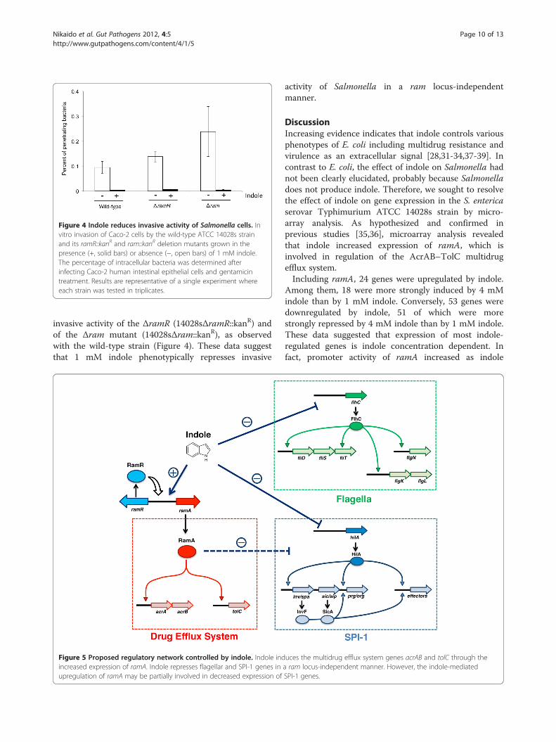
Figure 4 Indole reduces invasive activity of Salmonella cells. Invitro invasion of Caco-2 cells by the wild-type ATCC 14028s strainand its ramR::kanR and ram::kanR deletion mutants grown in thepresence (+, solid bars) or absence (−, open bars) of 1 mM indole.The percentage of intracellular bacteria was determined afterinfecting Caco-2 human intestinal epithelial cells and gentamicintreatment. Results are representative of a single experiment whereeach strain was tested in triplicates.
Nikaido et al. Gut Pathogens 2012, 4:5 Page 10 of 13http://www.gutpathogens.com/content/4/1/5
invasive activity of the ΔramR (14028sΔramR::kanR) andof the Δram mutant (14028sΔram::kanR), as observedwith the wild-type strain (Figure 4). These data suggestthat 1 mM indole phenotypically represses invasive
Figure 5 Proposed regulatory network controlled by indole. Indole indincreased expression of ramA. Indole represses flagellar and SPI-1 genes inupregulation of ramA may be partially involved in decreased expression of
activity of Salmonella in a ram locus-independentmanner.
DiscussionIncreasing evidence indicates that indole controls variousphenotypes of E. coli including multidrug resistance andvirulence as an extracellular signal [28,31-34,37-39]. Incontrast to E. coli, the effect of indole on Salmonella hadnot been clearly elucidated, probably because Salmonelladoes not produce indole. Therefore, we sought to resolvethe effect of indole on gene expression in the S. entericaserovar Typhimurium ATCC 14028s strain by micro-array analysis. As hypothesized and confirmed inprevious studies [35,36], microarray analysis revealedthat indole increased expression of ramA, which isinvolved in regulation of the AcrAB–TolC multidrugefflux system.Including ramA, 24 genes were upregulated by indole.
Among them, 18 were more strongly induced by 4 mMindole than by 1 mM indole. Conversely, 53 genes weredownregulated by indole, 51 of which were morestrongly repressed by 4 mM indole than by 1 mM indole.These data suggested that expression of most indole-regulated genes is indole concentration dependent. Infact, promoter activity of ramA increased as indole
uces the multidrug efflux system genes acrAB and tolC through thea ram locus-independent manner. However, the indole-mediatedSPI-1 genes.

Nikaido et al. Gut Pathogens 2012, 4:5 Page 11 of 13http://www.gutpathogens.com/content/4/1/5
concentration increased. Similarly, expression of SPI-1genes decreased as indole concentration increased.Indole induced ramA, and this induction is probably
responsible for induction of acrB, which encodes themultidrug efflux pump (Figure 5). Most of the genesupregulated by indole encode putative proteins. Severalof the genes, such as STM1251, STM1472, STM1868A,STM3941, and STM4213, have yet to be named. Amongthese, the only gene identified in E. coli is STM4213,which encodes a putative phage tail sheath protein. If thefunction of these putative genes is clarified, then otherphenotypes induced by indole in addition to multidrugresistance will also be understood.In this study, we found that indole repressed various
genes related to bacterial motility and virulence.Decreased motility and invasive activity of Salmonellawere also phenotypically observed. Recent studiesrevealed the coordinate regulation of flagellar and SPI-1genes by FliZ, encoded by a gene in the fliA operon[66,67]. It was demonstrated that FliZ posttranslationallycontrols HilD to positively regulate expression of hilA[67]. Our microarray data revealed reduced expression offliZ when bacterial cells were treated with indole (fliZ ex-pression was reduced to 0.47- and 0.038-fold of the levelin untreated cells by 1 and 4 mM indole, respectively),and that expression of hilA was also repressed by indole.Indole may coordinately repress flagellar and SPI-1 genesvia the regulatory network of FliZ. Because previousstudy indicated that both RamA and RamR are involvedin the control of SPI-1 genes [47], we examined theireffects on repression of SPI-1 genes and invasive activityof Salmonella. The results suggested that indolesuppresses SPI-1 genes in a RamA/RamR-independentmanner (Figure 5). Similarly, repression of flhC by indolewas also RamA/RamR independent. These data are sug-gestive of the presence of another pathway for indole torepress flagellar and SPI-1 genes, whereas acrAB/tolC isinduced by indole in a RamR/RamA-dependent manner.Bacterial adhesion to the Caco-2 cells, which is the pri-mary step of the cell invasion process, was not addressedin our experiments. However, since flagella wererepressed by indole, it cannot be excluded that the defect-ive invasion partially resulted from a lesser adhesion tothe cells, and not only to the repression of SPI-1 genes. Itshould also be noted that several of the indole-repressedgenes are related to anaerobic respiration in addition tomotility and SPI-1 genes. Because it is suggested that in-dole is a biological oxidant in bacteria [68], this oxidativeeffect may lead to repression of these genes.In conclusion, we identified that indole induces expres-
sion of genes related to efflux-mediated multidrug resist-ance and represses expression of genes related toinvasive activity and motility of S. enterica serovarTyphimurium. Reduction of invasive activity and motility
of Salmonella by indole was phenotypically observed.Because Salmonella itself does not produce indole, ourresults suggest that indole could also be an importantsignaling molecule for inter-species communication tocontrol drug resistance and virulence of Salmonella inaddition to its role in intra-species communication in E.coli. Indeed, it was previously demonstrated that E.coli-conditioned medium induced the AcrAB pump inSalmonella through the RamA regulator [35]. The typeof environment in which Salmonella experiences theeffect of indole is not well understood. It is believedthat Salmonella may be exposed to high indole con-centrations in the intestine, in which several speciesof indole-producing bacteria exist. In fact, indole isfound in human feces at comparable concentrations(~250–1100 μM) [29,30], and recent studies indicatedthe importance of indole in favorable inter-kingdom sig-naling interactions between the intestinal epithelial cellsand commensal bacteria [69]. In addition to indole itself,indole derivatives such as skatole (3-methylindole) alsooccur naturally in feces after being produced from trypto-phan in the mammalian digestive tract. Therefore, indoleand skatole may additively affect gene expression inSalmonella. In fact, when we examined the effect of ska-tole by a β-galactosidase assay, it significantly stimulatedthe promoter activity of ramA (unpublished data). Thus,there is a possibility that these molecules enhance drugresistance of Salmonella, while simultaneously repressingtheir motility and pathogenicity in the intestinal tract.Interestingly, the effect of indole on the pathogenicity ofE. coli is the opposite of that on Salmonella. In enterohe-morrhagic E. coli, it was suggested that indole can acti-vate expression of EspA and EspB as well as secretionand stimulate the ability of EHEC to form attaching andeffacing lesions in human cells [38]. Thus, although in-dole secreted by E. coli enhances the virulence of E. coli,it reduces the virulence of Salmonella, probably to theadvantage of E. coli. This finding suggests that the gastro-intestinal flora may affect regulation of virulence traits inSalmonella via the signaling of indole.
Competing interestsThe authors declare that they have no competing interests.
AcknowledgmentsWe thank Mitsuko Hayashi-Nishino for the protocol for the invasion assayand excellent advice and Ikue Shirosaka, Coralie Porte-Lebiguais, IsabelleMonchaux and Imane Boumart for excellent technical assistance.
Author details1Laboratory of Microbiology and Infectious Diseases, Institute of Scientificand Industrial Research, Osaka University, 8-1 Mihogaoka, Ibaraki, Osaka567-00447, Japan. 2Department of Cell Membrane Biology, Institute ofScientific and Industrial Research, Osaka University, Ibaraki, Osaka, Japan.3Graduate School of Pharmaceutical Sciences, Osaka University, Suita, Osaka,Japan. 4INRA, UMR1282 Infectiologie et Santé Publique, F-37380 Nouzilly,France. 5Université François Rabelais de Tours, UMR1282 Infectiologie etSanté Publique, F-37000 Tours, France.
Nikaido et al. Gut Pathogens 2012, 4:5 Page 12 of 13http://www.gutpathogens.com/content/4/1/5
Authors’ contributionsConceived and designed the experiments: EN, EG, SB, AC, KN. Performed theexperiments: EN, EG, SB, SY, AW. Analyzed the data: EN, EG, SB, SY, AW, KO,TT, AY, AC, KN. Wrote the paper: EG, AC, KN. All authors read and approvedthe final manuscript.
FundingThis research was supported in part by Grants-in-Aid from the Japan Societyfor the Promotion of Science and the Ministry of Education, Culture, Sports,Science and Technology of Japan; a grant from the Mishimakaiun MemorialFoundation; the Program for Promotion of Fundamental Studies in HealthSciences of the National Institute of Biomedical Innovation; and the FundingProgram for Next Generation World-Leading Researchers. It was also partlysupported by the French Région Centre (grant 2008 00036085) and partly bythe European Union with the European Regional Development Fund (grant1634–32245). The funders had no role in this study design, data collectionand analysis, decision to publish, or preparation of the manuscript.
Received: 24 April 2012 Accepted: 25 May 2012Published: 25 May 2012
References1. Fuqua C, Winans SC, Greenberg EP: Census and consensus in bacterial
ecosystems: the LuxR-LuxI family of quorum-sensing transcriptionalregulators. Annu Rev Microbiol 1996, 50:727–751.
2. Fuqua WC, Winans SC, Greenberg EP: Quorum sensing in bacteria: theLuxR-LuxI family of cell density-responsive transcriptional regulators.J Bacteriol 1994, 176:269–275.
3. Kaiser D, Losick R: How and why bacteria talk to each other. Cell 1993,73:873–885.
4. Kleerebezem M, Quadri LE, Kuipers OP, de Vos WM: Quorum sensing bypeptide pheromones and two-component signal-transduction systems inGram-positive bacteria. Mol Microbiol 1997, 24:895–904.
5. Salmond GP, Bycroft BW, Stewart GS, Williams P: The bacterial 'enigma':cracking the code of cell-cell communication. Mol Microbiol 1995,16:615–624.
6. Cao JG, Meighen EA: Purification and structural identification of anautoinducer for the luminescence system of Vibrio harveyi. J Biol Chem1989, 264:21670–21676.
7. Eberhard A, Burlingame AL, Eberhard C, Kenyon GL, Nealson KH,Oppenheimer NJ: Structural identification of autoinducer ofPhotobacterium fischeri luciferase. Biochemistry 1981, 20:2444–2449.
8. Holden MT, Ram Chhabra S, de Nys R, Stead P, Bainton NJ, Hill PJ, ManefieldM, Kumar N, Labatte M, England D, et al: Quorum-sensing cross talk:isolation and chemical characterization of cyclic dipeptides fromPseudomonas aeruginosa and other gram-negative bacteria. MolMicrobiol 1999, 33:1254–1266.
9. Pearson JP, Gray KM, Passador L, Tucker KD, Eberhard A, Iglewski BH,Greenberg EP: Structure of the autoinducer required for expression ofPseudomonas aeruginosa virulence genes. Proc Natl Acad Sci USA 1994,91:197–201.
10. Pesci EC, Milbank JB, Pearson JP, McKnight S, Kende AS, Greenberg EP,Iglewski BH: Quinolone signaling in the cell-to-cell communicationsystem of Pseudomonas aeruginosa. Proc Natl Acad Sci USA 1999,96:11229–11234.
11. Pesci EC: New signal molecules on the quorum-sensing block: response.Trends Microbiol 2000, 8:103–104.
12. Zhang L, Murphy PJ, Kerr A, Tate ME: Agrobacterium conjugation andgene regulation by N-acyl-L-homoserine lactones. Nature 1993,362:446–448.
13. Baca-DeLancey RR, South MM, Ding X, Rather PN: Escherichia coli genesregulated by cell-to-cell signaling. Proc Natl Acad Sci USA 1999,96:4610–4614.
14. Bassler BL, Wright M, Showalter RE, Silverman MR: Intercellular signalling inVibrio harveyi: sequence and function of genes regulating expression ofluminescence. Mol Microbiol 1993, 9:773–786.
15. Cheng Q, Campbell EA, Naughton AM, Johnson S, Masure HR: The comlocus controls genetic transformation in Streptococcus pneumoniae. MolMicrobiol 1997, 23:683–692.
16. Davies DG, Parsek MR, Pearson JP, Iglewski BH, Costerton JW, Greenberg EP:The involvement of cell-to-cell signals in the development of a bacterialbiofilm. Science 1998, 280:295–298.
17. Engebrecht J, Nealson K, Silverman M: Bacterial bioluminescence: isolationand genetic analysis of functions from Vibrio fischeri. Cell 1983, 32:773–781.
18. Jones S, Yu B, Bainton NJ, Birdsall M, Bycroft BW, Chhabra SR, Cox AJ, GolbyP, Reeves PJ, Stephens S, et al: The lux autoinducer regulates theproduction of exoenzyme virulence determinants in Erwinia carotovoraand Pseudomonas aeruginosa. EMBO J 1993, 12:2477–2482.
19. Latifi A, Winson MK, Foglino M, Bycroft BW, Stewart GS, Lazdunski A, Williams P:Multiple homologues of LuxR and LuxI control expression of virulencedeterminants and secondary metabolites through quorum sensing inPseudomonas aeruginosa PAO1. Mol Microbiol 1995, 17:333–343.
20. Passador L, Cook JM, Gambello MJ, Rust L, Iglewski BH: Expression ofPseudomonas aeruginosa virulence genes requires cell-to-cellcommunication. Science 1993, 260:1127–1130.
21. Piper KR, Beck von Bodman S, Farrand SK: Conjugation factor ofAgrobacterium tumefaciens regulates Ti plasmid transfer byautoinduction. Nature 1993, 362:448–450.
22. Pirhonen M, Flego D, Heikinheimo R, Palva ET: A small diffusible signalmolecule is responsible for the global control of virulence andexoenzyme production in the plant pathogen Erwinia carotovora. EMBO J1993, 12:2467–2476.
23. Clarke MB, Sperandio V: Events at the host-microbial interface of thegastrointestinal tract III. Cell-to-cell signaling among microbial flora, host,and pathogens: there is a whole lot of talking going on. Am J PhysiolGastrointest Liver Physiol 2005, 288:1105–1109.
24. Collier-Hyams LS, Neish AS: Innate immune relationship betweencommensal flora and the mammalian intestinal epithelium. Cell Mol LifeSci 2005, 62:1339–1348.
25. Green BT, Lyte M, Chen C, Xie Y, Casey MA, Kulkarni-Narla A, Vulchanova L,Brown DR: Adrenergic modulation of Escherichia coli O157:H7 adherenceto the colonic mucosa. Am J Physiol Gastrointest Liver Physiol 2004, 287:G1238–1246.
26. Sonnenwirth AC: The enteric bacteria and bacteroides. In Microbiology. 3rdedition. Edited by Davis BD, Dulbecco R, Eisen HN, Ginsberg HS. Publishers,Inc, Philadelphia, Pa: Harper & Row; 1980:645–672.
27. Yanofsky C, Horn V, Gollnick P: Physiological studies of tryptophantransport and tryptophanase operon induction in Escherichia coli. JBacteriol 1991, 173:6009–6017.
28. Domka J, Lee J, Wood TK: YliH (BssR) and YceP (BssS) regulate Escherichiacoli K-12 biofilm formation by influencing cell signaling. Appl EnvironMicrobiol 2006, 72:2449–2459.
29. Karlin DA, Mastromarino AJ, Jones RD, Stroehlein JR, Lorentz O: Fecalskatole and indole and breath methane and hydrogen in patients withlarge bowel polyps or cancer. J Cancer Res Clin Oncol 1985, 109:135–141.
30. Zuccato E, Venturi M, Di Leo G, Colombo L, Bertolo C, Doldi SB, Mussini E:Role of bile acids and metabolic activity of colonic bacteria in increasedrisk of colon cancer after cholecystectomy. Dig Dis Sci 1993, 38:514–519.
31. Wang D, Ding X, Rather PN: Indole can act as an extracellular signal inEscherichia coli. J Bacteriol 2001, 183:4210–4216.
32. Chant EL, Summers DK: Indole signalling contributes to the stablemaintenance of Escherichia coli multicopy plasmids. Mol Microbiol 2007,63:35–43.
33. Hirakawa H, Hayashi-Nishino M, Yamaguchi A, Nishino K: Indole enhancesacid resistance in Escherichia coli. Microb Pathog 2010, 49:90–94.
34. Hirakawa H, Inazumi Y, Masaki T, Hirata T, Yamaguchi A: Indole induces theexpression of multidrug exporter genes in Escherichia coli. Mol Microbiol2005, 55:1113–1126.
35. Nikaido E, Yamaguchi A, Nishino K: AcrAB multidrug efflux pumpregulation in Salmonella enterica serovar Typhimurium by RamA inresponse to environmental signals. J Biol Chem 2008, 283:24245–24253.
36. Nikaido E, Shirosaka I, Yamaguchi A, Nishino K: Regulation of the AcrABmultidrug efflux pump in Salmonella enterica serovar Typhimurium inresponse to indole and paraquat. Microbiology 2011, 157:648–655.
37. Anyanful A, Dolan-Livengood JM, Lewis T, Sheth S, Dezalia MN, ShermanMA, Kalman LV, Benian GM, Kalman D: Paralysis and killing ofCaenorhabditis elegans by enteropathogenic Escherichia coli requiresthe bacterial tryptophanase gene. Mol Microbiol 2005, 57:988–1007.
38. Hirakawa H, Kodama T, Takumi-Kobayashi A, Honda T, Yamaguchi A:Secreted indole serves as a signal for expression of type III secretionsystem translocators in enterohaemorrhagic Escherichia coli O157:H7.Microbiology 2009, 155:541–550.
Nikaido et al. Gut Pathogens 2012, 4:5 Page 13 of 13http://www.gutpathogens.com/content/4/1/5
39. Lee HH, Molla MN, Cantor CR, Collins JJ: Bacterial charity work leads topopulation-wide resistance. Nature 2010, 467:82–85.
40. Scherer CAaM SI: Principles of Bacterial Pathogenesis. Academic Press, NewYork 2001, :266–333.
41. McClelland M, Sanderson KE, Spieth J, Clifton SW, Latreille P, Courtney L,Porwollik S, Ali J, Dante M, Du F, et al: Complete genome sequence ofSalmonella enterica serovar Typhimurium LT2. Nature 2001, 413:852–856.
42. Fields PI, Swanson RV, Haidaris CG, Heffron F: Mutants of Salmonellatyphimurium that cannot survive within the macrophage are avirulent.Proc Natl Acad Sci USA 1986, 83:5189–5193.
43. Miller JH: Experiments in Molecular Genetics. Cold Spring Harbor LaboratoryPress, Cold Spring Harbor, NY 1972, :352–355.
44. Datsenko KA, Wanner BL: One-step inactivation of chromosomal genes inEscherichia coli K-12 using PCR products. Proc Natl Acad Sci USA 2000,97:6640–6645.
45. Rosselin M, Virlogeux-Payant I, Roy C, Bottreau E, Sizaret PY, Mijouin L,Germon P, Caron E, Velge P, Wiedemann A: Rck of Salmonella enterica,subspecies enterica serovar enteritidis, mediates zipper-likeinternalization. Cell Res 2010, 20:647–664.
46. Abouzeed YM, Baucheron S, Cloeckaert A: ramR mutations involved inefflux-mediated multidrug resistance in Salmonella enterica serovarTyphimurium. Antimicrob Agents Chemother 2008, 52:2428–2434.
47. Bailey AM, Ivens A, Kingsley R, Cottell JL, Wain J, Piddock LJ: RamA, amember of the AraC/XylS family, influences both virulence and efflux inSalmonella enterica serovar Typhimurium. J Bacteriol 2010, 192:1607–1616.
48. Chollet R, Chevalier J, Bollet C, Pages JM, Davin-Regli A: RamA is analternate activator of the multidrug resistance cascade in Enterobacteraerogenes. Antimicrob Agents Chemother 2004, 48:2518–2523.
49. Keeney D, Ruzin A, Bradford PA: RamA, a transcriptional regulator, andAcrAB, an RND-type efflux pump, are associated with decreasedsusceptibility to tigecycline in Enterobacter cloacae. Microb Drug Resist2007, 13:1–6.
50. Li XZ, Nikaido H: Efflux-mediated drug resistance in bacteria: an update.Drugs 2009, 69:1555–1623.
51. Ruzin A, Immermann FW, Bradford PA: Real-time PCR and statisticalanalyses of acrAB and ramA expression in clinical isolates of Klebsiellapneumoniae. Antimicrob Agents Chemother 2008, 52:3430–3432.
52. Zheng J, Cui S, Meng J: Effect of transcriptional activators RamA and SoxSon expression of multidrug efflux pumps AcrAB and AcrEF influoroquinolone-resistant Salmonella Typhimurium. J AntimicrobChemother 2009, 63:95–102.
53. Horiyama T, Nikaido E, Yamaguchi A, Nishino K: Roles of Salmonellamultidrug efflux pumps in tigecycline resistance. J Antimicrob Chemother2011, 66:105–110.
54. Yamamoto K, Hirao K, Oshima T, Aiba H, Utsumi R, Ishihama A: Functionalcharacterization in vitro of all two-component signal transductionsystems from Escherichia coli. J Biol Chem 2005, 280:1448–1456.
55. Venkatesh GR, Kembou Koungni FC, Paukner A, Stratmann T, Blissenbach B,Schnetz K: BglJ-RcsB heterodimers relieve repression of the Escherichiacoli bgl operon by H-NS. J Bacteriol 2010, 192:6456–6464.
56. Giel M, Desnoyer M, Lopilato J: A mutation in a new gene, bglJ, activatesthe bgl operon in Escherichia coli K-12. Genetics 1996, 143:627–635.
57. Lambalot RH, Gehring AM, Flugel RS, Zuber P, LaCelle M, Marahiel MA, ReidR, Khosla C, Walsh CT: A new enzyme superfamily - thephosphopantetheinyl transferases. Chem Biol 1996, 3:923–936.
58. Gehring AM, Mori I, Walsh CT: Reconstitution and characterization of theEscherichia coli enterobactin synthetase from EntB, EntE, and EntF.Biochemistry 1998, 37:2648–2659.
59. Feldheim DA, Chin AM, Nierva CT, Feucht BU, Cao YW, Xu YF, Sutrina SL,Saier MH Jr: Physiological consequences of the complete loss ofphosphoryl-transfer proteins HPr and FPr of the phosphoenolpyruvate:sugar phosphotransferase system and analysis of fructose (fru) operonexpression in Salmonella typhimurium. J Bacteriol 1990, 172:5459–5469.
60. Wang WF, Cheng X, Molineux IJ: Isolation and identification of fxsA, anEscherichia coli gene that can suppress F exclusion of bacteriophage T7.J Mol Biol 1999, 292:485–499.
61. Rebbapragada A, Johnson MS, Harding GP, Zuccarelli AJ, Fletcher HM,Zhulin IB, Taylor BL: The Aer protein and the serine chemoreceptor Tsrindependently sense intracellular energy levels and transduce oxygen,redox, and energy signals for Escherichia coli behavior. Proc Natl Acad SciUSA 1997, 94:10541–10546.
62. Brondijk TH, Fiegen D, Richardson DJ, Cole JA: Roles of NapF, NapG andNapH, subunits of the Escherichia coli periplasmic nitrate reductase, inubiquinol oxidation. Mol Microbiol 2002, 44:245–255.
63. Troxell B, Fink RC, Porwollik S, McClelland M, Hassan HM: The Fur regulon inanaerobically grown Salmonella enterica sv Typhimurium: identificationof new Fur targets. BMC Microbiol 2011, 11:236.
64. Baucheron S, Coste F, Canepa S, Maurel MC, Giraud E, Culard F, Castaing B,Roussel A, Cloeckaert A: Binding of the RamR repressor to wild-Type andmutated promoters of the ramA gene involved in efflux-mediatedmultidrug resistance in Salmonella enterica serovar Typhimurium.Antimicrob Agents Chemother 2012, 56:942–948.
65. Claret L, Hughes C: Functions of the subunits in the FlhD2C2transcriptional master regulator of bacterial flagellum biogenesis andswarming. J Mol Biol 2000, 303:467–478.
66. Saini S, Slauch JM, Aldridge PD, Rao CV: Role of cross talk in regulating thedynamic expression of the flagellar Salmonella pathogenicity island 1and type 1 fimbrial genes. J Bacteriol 2010, 192:5767–5777.
67. Chubiz JE, Golubeva YA, Lin D, Miller LD, Slauch JM: FliZ regulatesexpression of the Salmonella pathogenicity island 1 invasion locus bycontrolling HilD protein activity in Salmonella enterica serovartyphimurium. J Bacteriol 2010, 192:6261–6270.
68. Garbe TR, Kobayashi M, Yukawa H: Indole-inducible proteins in bacteriasuggest membrane and oxidant toxicity. Arch Microbiol 2000, 173:78–82.
69. Bansal T, Alaniz RC, Wood TK, Jayaraman A: The bacterial signal indoleincreases epithelial-cell tight-junction resistance and attenuatesindicators of inflammation. Proc Natl Acad Sci USA 2010, 107:228–233.
doi:10.1186/1757-4749-4-5Cite this article as: Nikaido et al.: Effects of indole on drug resistanceand virulence of Salmonella enterica serovar Typhimurium revealed bygenome-wide analyses. Gut Pathogens 2012 4:5.
Submit your next manuscript to BioMed Centraland take full advantage of:
• Convenient online submission
• Thorough peer review
• No space constraints or color figure charges
• Immediate publication on acceptance
• Inclusion in PubMed, CAS, Scopus and Google Scholar
• Research which is freely available for redistribution
Submit your manuscript at www.biomedcentral.com/submit
Related Documents