Effects of Cyclodextrins on Drug Delivery Through Biological Membranes THORSTEINN LOFTSSON, 1 STINE BYSKOV VOGENSEN, 1 MARCUS E. BREWSTER, 2 FI ´ FA KONRA ´ ðSDO ´ TTIR 1 1 Faculty of Pharmacy, University of Iceland, Hofsvallagata 53, IS-107 Reykjavik, Iceland 2 Chemical and Pharmaceutical Development, Johnson & Johnson Pharmaceutical Research and Development, Janssen Pharmaceutica, Turnhoutsweg 30, B-2340 Beerse, Belgium Received 29 January 2007; revised 21 February 2007; accepted 27 February 2007 Published online in Wiley InterScience (www.interscience.wiley.com). DOI 10.1002/jps.20992 ABSTRACT: Cyclodextrins have proven themselves to be useful functional excipients. Cyclodextrin derivatives can be hydrophilic or relatively lipophilic based on their substitution and these properties can give insight into their ability to act as permeability enhancers. Lipophilic cyclodextrins such as the methylated derivatives are thought to increase drug flux by altering barrier properties of the membrane through component extraction or fluidization. The hydrophilic cyclodextrin family also modulate drug flux through membranes but via different mechanisms. The current effort seeks to provide various explanations for these observations based on interactions of hydrophilic cyclo- dextrins with the unstirred water layer that separates the bulk media from biological membranes such as the gastric mucosa, cornea and reproductive tract. Theories on the serial nature of resistances to drug flux are used to explain why hydrophilic cyclodex- trins can enhance drug uptake in some situation (i.e., for lipophilic material) but not in others. In addition, the nature of secondary equilibria and competition between cyclo- dextrins and rheologically important biopolymers such as mucin are assessed to give a complete picture of the effect of these starch derivatives. This information can be useful not only in understanding the actions of cyclodextrin but also in expanding their application and uses. ß 2007 Wiley-Liss, Inc. and the American Pharmacists Association J Pharm Sci 96:2532–2546, 2007 Keywords: cyclodextrin; drug delivery; unstirred water layer; water structure; permeation; membrane INTRODUCTION Cyclodextrins (CDs) are natural cyclic oligosac- charides that are formed through enzymatic degradation of starch. 1 The three most common CDs, aCD, bCD and gCD, are composed of six, seven and eight a(1 ! 4)-linked a-D-glucopyra- nose units, respectively, with a hydrophilic outer surface and a somewhat lipophilic central cavity (Table 1). The hydroxyl functions are orientated to the exterior whereas the central cavity is lined by skeletal carbons and ethereal oxygens, which give it a lipophilic character. Although the naturally occurring CDs and their complexes are hydro- philic, their aqueous solubility is rather limited, especially that of bCD. Random substitution of the hydroxy groups located on the outer surface of the CD molecule, even by hydrophobic moieties such as methoxy functions, results in dramatic improvement in their solubility (Table 1). In an aqueous environment, CDs form inclusion com- plexes with many lipophilic drug molecules through a process in which water molecules Correspondence to: Thorsteinn Loftsson (Telephone: þ354- 525-4464; Fax: þ354-525-4071; E-mail: [email protected]) Journal of Pharmaceutical Sciences, Vol. 96, 2532–2546 (2007) ß 2007 Wiley-Liss, Inc. and the American Pharmicist Association 2532 JOURNAL OF PHARMACEUTICAL SCIENCES, VOL. 96, NO. 10, OCTOBER 2007

Welcome message from author
This document is posted to help you gain knowledge. Please leave a comment to let me know what you think about it! Share it to your friends and learn new things together.
Transcript
Effects of Cyclodextrins on Drug Delivery ThroughBiological Membranes
THORSTEINN LOFTSSON,1 STINE BYSKOV VOGENSEN,1 MARCUS E. BREWSTER,2 FIFA KONRAðSDOTTIR1
1Faculty of Pharmacy, University of Iceland, Hofsvallagata 53, IS-107 Reykjavik, Iceland
2Chemical and Pharmaceutical Development, Johnson & Johnson Pharmaceutical Research and Development,Janssen Pharmaceutica, Turnhoutsweg 30, B-2340 Beerse, Belgium
Received 29 January 2007; revised 21 February 2007; accepted 27 February 2007
Published online in Wiley InterScience (www.interscience.wiley.com). DOI 10.1002/jps.20992
Corresponde525-4464; Fax:
Journal of Pharm
� 2007 Wiley-Liss
2532 JOURN
ABSTRACT: Cyclodextrins have proven themselves to be useful functional excipients.Cyclodextrin derivatives can be hydrophilic or relatively lipophilic based on theirsubstitution and these properties can give insight into their ability to act as permeabilityenhancers. Lipophilic cyclodextrins such as the methylated derivatives are thought toincrease drug flux by altering barrier properties of the membrane through componentextraction or fluidization. The hydrophilic cyclodextrin family also modulate drug fluxthrough membranes but via different mechanisms. The current effort seeks to providevarious explanations for these observations based on interactions of hydrophilic cyclo-dextrins with the unstirred water layer that separates the bulk media from biologicalmembranes such as the gastric mucosa, cornea and reproductive tract. Theories on theserial nature of resistances to drug flux are used to explain why hydrophilic cyclodex-trins can enhance drug uptake in some situation (i.e., for lipophilic material) but not inothers. In addition, the nature of secondary equilibria and competition between cyclo-dextrins and rheologically important biopolymers such as mucin are assessed to give acomplete picture of the effect of these starch derivatives. This information can be usefulnot only in understanding the actions of cyclodextrin but also in expanding theirapplication and uses. � 2007 Wiley-Liss, Inc. and the American Pharmacists Association J
Pharm Sci 96:2532–2546, 2007
Keywords: cyclodextrin; drug deliver
y; unstirred water layer; water structure;permeation; membraneINTRODUCTION
Cyclodextrins (CDs) are natural cyclic oligosac-charides that are formed through enzymaticdegradation of starch.1 The three most commonCDs, aCD, bCD and gCD, are composed of six,seven and eight a(1! 4)-linked a-D-glucopyra-nose units, respectively, with a hydrophilic outersurface and a somewhat lipophilic central cavity
nce to: Thorsteinn Loftsson (Telephone: þ354-þ354-525-4071; E-mail: [email protected])
aceutical Sciences, Vol. 96, 2532–2546 (2007)
, Inc. and the American Pharmicist Association
AL OF PHARMACEUTICAL SCIENCES, VOL. 96, NO. 10, OC
(Table 1). The hydroxyl functions are orientated tothe exterior whereas the central cavity is lined byskeletal carbons and ethereal oxygens, which giveit a lipophilic character. Although the naturallyoccurring CDs and their complexes are hydro-philic, their aqueous solubility is rather limited,especially that of bCD. Random substitution of thehydroxy groups located on the outer surface of theCD molecule, even by hydrophobic moieties suchas methoxy functions, results in dramaticimprovement in their solubility (Table 1). In anaqueous environment, CDs form inclusion com-plexes with many lipophilic drug moleculesthrough a process in which water molecules
TOBER 2007
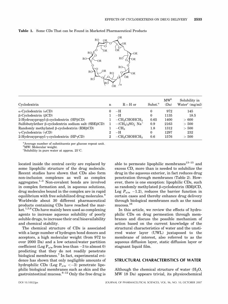
Table 1. Some CDs That can be Found in Marketed Pharmaceutical Products
O
O
O
O
O
O
O
O O
O
O
O
O
OR
OR
OR
OR
OROR
ORRO
RO
RO
RO
RO
RO OR
OROR
OR
ORRO
RO
RO
n
Cyclodextrin n R¼H or Subst.aMWb
(Da)Solubility in
Waterc (mg/ml)
a-Cyclodextrin (aCD) 0 �H 0 972 145b-Cyclodextrin (bCD) 1 �H 0 1135 18.52-Hydroxypropyl-b-cyclodextrin (HPbCD) 1 �CH2CHOHCH3 0.65 1400 > 600Sulfobutylether b-cyclodextrin sodium salt (SBEbCD) 1 �(CH2)4SO
�3 Naþ 0.9 2163 > 500
Randomly methylated b-cyclodextrin (RMbCD) 1 �CH3 1.8 1312 > 500g-Cyclodextrin (gCD) 2 �H 0 1297 2322-Hydroxypropyl-g-cyclodextrin (HPgCD) 2 �CH2CHOHCH3 0.6 1576 > 500
aAverage number of substituents per glucose repeat unit.bMW: Molecular weight.cSolubility in pure water at approx. 258C.
EFFECTS OF CYCLODEXTRINS ON DRUG DELIVERY 2533
located inside the central cavity are replaced bysome lipophilic structure of the drug molecule.Recent studies have shown that CDs also formnon-inclusion complexes as well as complexaggregates.1–3 Non-covalent bonds are involvedin complex formation and, in aqueous solutions,drug molecules bound in the complex are in rapidequilibrium with free solubilized drug molecules.4
Worldwide about 30 different pharmaceuticalproducts containing CDs have reached the mar-ket.1,5,6 CDs havemainly been used as complexingagents to increase aqueous solubility of poorlysoluble drugs, to increase their oral bioavailabilityand chemical stability.
The chemical structure of CDs is associatedwith a large number of hydrogen bond donors andacceptors, a high molecular weight (from 972 toover 2000 Da) and a low octanol/water partitioncoefficient (Log Po/w from less than�3 to almost 0)predicting that they do not readily penetratebiological membranes.7 In fact, experimental evi-dence has shown that only negligible amounts ofhydrophilic CDs (Log Po/w <�3) penetrate lipo-philic biological membranes such as skin and thegastrointestinal mucosa.8–12 Only the free drug is
DOI 10.1002/jps JOURN
able to permeate lipophilic membranes13–15 andexcess CD, more than is needed to solubilize thedrug in the aqueous exterior, in fact reduces drugpenetration through membranes (Table 2). How-ever, there is one exception: lipophilic CDs, suchas randomly methylated b-cyclodextrin (RMbCD,Log Po/w �1.2), reduces the barrier function incertain cases and thereby enhance drug deliverythrough biological membranes such as the nasalmucosa.16
In this article, we review the effects of hydro-philic CDs on drug permeation through mem-branes and discuss the possible mechanism ofaction based on the current knowledge of thestructural characteristics of water and the unsti-rred water layer (UWL) juxtaposed to themembrane of interest, also referred to as theaqueous diffusion layer, static diffusion layer orstagnant liquid film.
STRUCTURAL CHARACTERISTICS OF WATER
Although the chemical structure of water (H2O,MW 18 Da) appears trivial, its physicochemical
AL OF PHARMACEUTICAL SCIENCES, VOL. 96, NO. 10, OCTOBER 2007
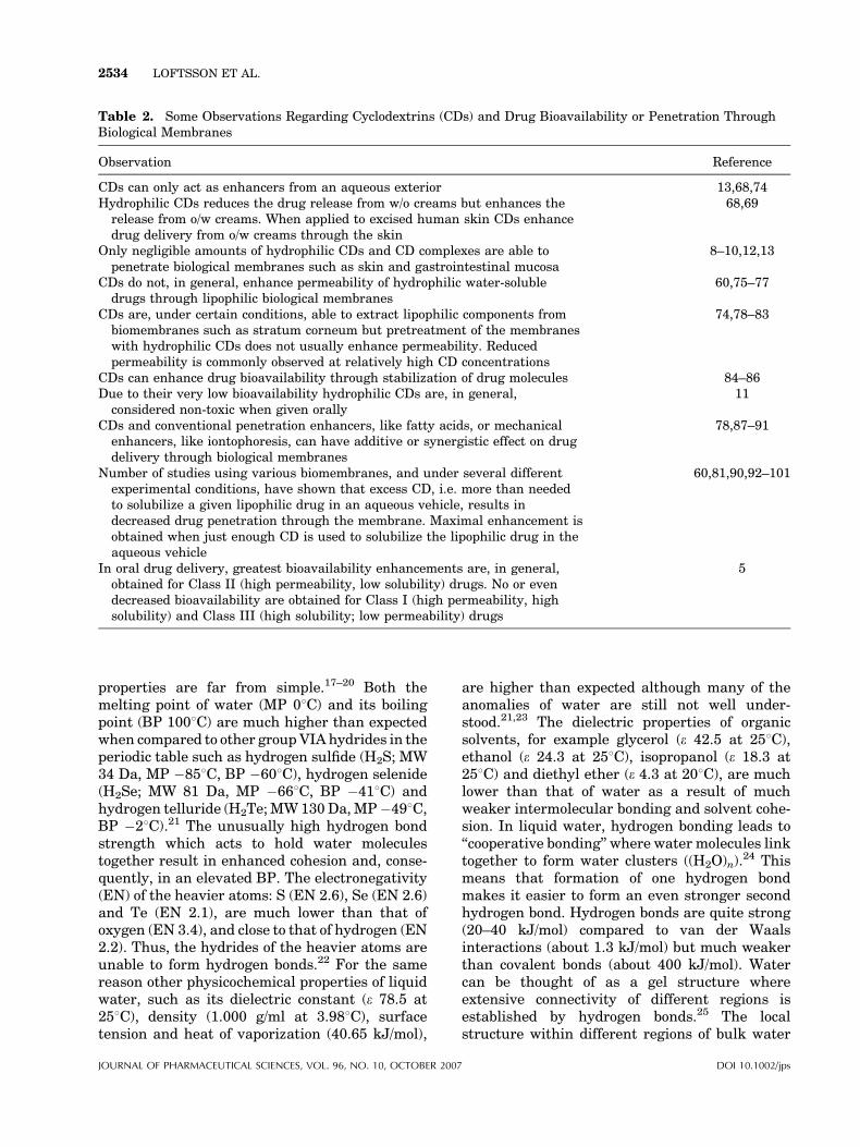
Table 2. Some Observations Regarding Cyclodextrins (CDs) and Drug Bioavailability or Penetration ThroughBiological Membranes
Observation Reference
CDs can only act as enhancers from an aqueous exterior 13,68,74Hydrophilic CDs reduces the drug release from w/o creams but enhances the
release from o/w creams. When applied to excised human skin CDs enhancedrug delivery from o/w creams through the skin
68,69
Only negligible amounts of hydrophilic CDs and CD complexes are able topenetrate biological membranes such as skin and gastrointestinal mucosa
8–10,12,13
CDs do not, in general, enhance permeability of hydrophilic water-solubledrugs through lipophilic biological membranes
60,75–77
CDs are, under certain conditions, able to extract lipophilic components frombiomembranes such as stratum corneum but pretreatment of the membraneswith hydrophilic CDs does not usually enhance permeability. Reducedpermeability is commonly observed at relatively high CD concentrations
74,78–83
CDs can enhance drug bioavailability through stabilization of drug molecules 84–86Due to their very low bioavailability hydrophilic CDs are, in general,
considered non-toxic when given orally11
CDs and conventional penetration enhancers, like fatty acids, or mechanicalenhancers, like iontophoresis, can have additive or synergistic effect on drugdelivery through biological membranes
78,87–91
Number of studies using various biomembranes, and under several differentexperimental conditions, have shown that excess CD, i.e. more than neededto solubilize a given lipophilic drug in an aqueous vehicle, results indecreased drug penetration through the membrane. Maximal enhancement isobtained when just enough CD is used to solubilize the lipophilic drug in theaqueous vehicle
60,81,90,92–101
In oral drug delivery, greatest bioavailability enhancements are, in general,obtained for Class II (high permeability, low solubility) drugs. No or evendecreased bioavailability are obtained for Class I (high permeability, highsolubility) and Class III (high solubility; low permeability) drugs
5
2534 LOFTSSON ET AL.
properties are far from simple.17–20 Both themelting point of water (MP 08C) and its boilingpoint (BP 1008C) are much higher than expectedwhen compared to other group VIAhydrides in theperiodic table such as hydrogen sulfide (H2S; MW34 Da, MP �858C, BP �608C), hydrogen selenide(H2Se; MW 81 Da, MP �668C, BP �418C) andhydrogen telluride (H2Te;MW130Da,MP�498C,BP �28C).21 The unusually high hydrogen bondstrength which acts to hold water moleculestogether result in enhanced cohesion and, conse-quently, in an elevated BP. The electronegativity(EN) of the heavier atoms: S (EN 2.6), Se (EN 2.6)and Te (EN 2.1), are much lower than that ofoxygen (EN 3.4), and close to that of hydrogen (EN2.2). Thus, the hydrides of the heavier atoms areunable to form hydrogen bonds.22 For the samereason other physicochemical properties of liquidwater, such as its dielectric constant (e 78.5 at258C), density (1.000 g/ml at 3.988C), surfacetension and heat of vaporization (40.65 kJ/mol),
JOURNAL OF PHARMACEUTICAL SCIENCES, VOL. 96, NO. 10, OCTOBER 2007
are higher than expected although many of theanomalies of water are still not well under-stood.21,23 The dielectric properties of organicsolvents, for example glycerol (e 42.5 at 258C),ethanol (e 24.3 at 258C), isopropanol (e 18.3 at258C) and diethyl ether (e 4.3 at 208C), are muchlower than that of water as a result of muchweaker intermolecular bonding and solvent cohe-sion. In liquid water, hydrogen bonding leads to‘‘cooperative bonding’’ where water molecules linktogether to form water clusters ((H2O)n).
24 Thismeans that formation of one hydrogen bondmakes it easier to form an even stronger secondhydrogen bond. Hydrogen bonds are quite strong(20–40 kJ/mol) compared to van der Waalsinteractions (about 1.3 kJ/mol) but much weakerthan covalent bonds (about 400 kJ/mol). Watercan be thought of as a gel structure whereextensive connectivity of different regions isestablished by hydrogen bonds.25 The localstructure within different regions of bulk water
DOI 10.1002/jps
EFFECTS OF CYCLODEXTRINS ON DRUG DELIVERY 2535
is continuously changing leading to homogeneousdensity, even at the nanoscale level.20,23 However,co-solvents, solutes and solid surfaces will alleffect the water structure. For example, ethanol–water binary mixtures have microscopic phaseseparation at the cluster level.26 Structure-break-ing solutes (chaotropes) destroy the hydrogen-bonded water network in a manner which issimilar to the effect of increased temperaturewhile structure-forming solutes (kosmotropes)increase the structural complexity. Sugars, suchas fructose, glucose and sucrose, behave aschaotropes at low concentrations, while at higherconcentrations they act as kosmotropes.27 Inaqueous solutions, water molecules form hydro-gen bonds with hydroxy groups on the CD surfaceforming a hydration shell around the dissolved CDmolecule.28–31 At aqueous concentrations belowabout 35% (w/v), CDs exert only small effects onviscosity but these effects can be significant athigher concentrations.32 This indicates that CDsbehave like sugars, as chaotropes at low concen-trations but as kosmotropes at high concentra-tions. Water-soluble polymers, such as cellulosederivatives and polyethylene glycols, form hydro-gen bonds with water that are stronger thanwater-water bonds, i.e. a positive hydrationcharacterized by low exchange rate of the watermolecules around the polymers, resulting inincreased viscosity even at concentrations below1% (w/v).33,34
Water structures at membrane surfaces arestrongly affected by the ability of the surface toform hydrogen bonds with water.35 Virtually nowater is adsorbed to graphitized carbon, ahydrophobic surface. The self-association of watermolecules is much stronger than interactions ofwater molecules with the hydrophobic surface, aphenomenon analogous to the hydrophobic effectobserved when non-polar solutes are dissolved inwater.20,36–38 Whereas water in contact withhydrophilic surfaces, e.g. some silica-based mate-rials, forms a water film where hydrogen bondingin the network of water molecules are partlysubstituted by bonds between the watermoleculesand the surface.36 These interactions result inreduction of the mobility of water moleculesdirectly adsorbed on the surface by more thanone order of magnitude. The water layer is only acouple of water molecules thick.20 At cell mem-branes, water molecules are bound to phospholi-pids, proteins and other membrane constituentsresulting in a water layer thickness of about1 nm.39 Water molecules are bound to the skin
DOI 10.1002/jps JOURN
surface as well as within the outermost layer ofthe skin, the stratum corneum.40 Mucosal epithe-lium (mucosa) contain mucosal cells that secretesmucus, a gel-like fluid containing mainly water(�95%) and mucin.41 Mucins are large glycopro-teins with MW ranging from 0.5 to 20 MDa. Someare membrane bound but others are not. Theviscous mucus forms a relatively thick (up toabout 100 mm) UWL in, for example, the gastro-intestinal tract, the respiratory tract, the ocular-rhino-otolaryngeal tracts and the reproductivetract.42 Under unstirred in vitro conditionthe UWL can be significantly thicker, even inabsence of mucus.43–46
Formation of water clusters, hydrophobic inter-actions and water structures at membranesurfaces can have profound implications on thebiological effects of drugs, including their abilityto permeate various membrane barriers.47
THEORETICAL BACKGROUND
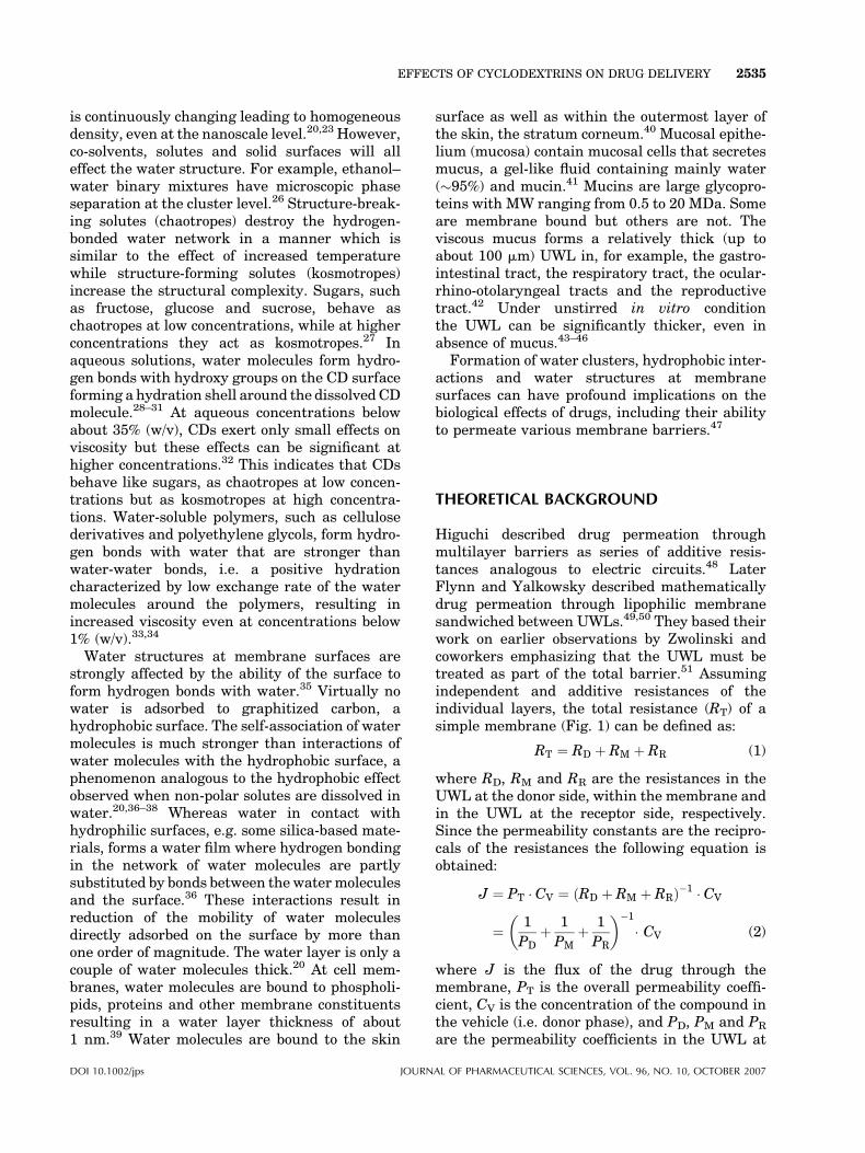
Higuchi described drug permeation throughmultilayer barriers as series of additive resis-tances analogous to electric circuits.48 LaterFlynn and Yalkowsky described mathematicallydrug permeation through lipophilic membranesandwiched between UWLs.49,50 They based theirwork on earlier observations by Zwolinski andcoworkers emphasizing that the UWL must betreated as part of the total barrier.51 Assumingindependent and additive resistances of theindividual layers, the total resistance (RT) of asimple membrane (Fig. 1) can be defined as:
RT ¼ RD þ RM þ RR (1)
where RD, RM and RR are the resistances in theUWL at the donor side, within the membrane andin the UWL at the receptor side, respectively.Since the permeability constants are the recipro-cals of the resistances the following equation isobtained:
J ¼ PT � CV ¼ ðRD þ RM þ RRÞ�1 � CV
¼ 1
PDþ 1
PMþ 1
PR
� ��1
� CV (2)
where J is the flux of the drug through themembrane, PT is the overall permeability coeffi-cient, CV is the concentration of the compound inthe vehicle (i.e. donor phase), and PD, PM and PR
are the permeability coefficients in the UWL at
AL OF PHARMACEUTICAL SCIENCES, VOL. 96, NO. 10, OCTOBER 2007
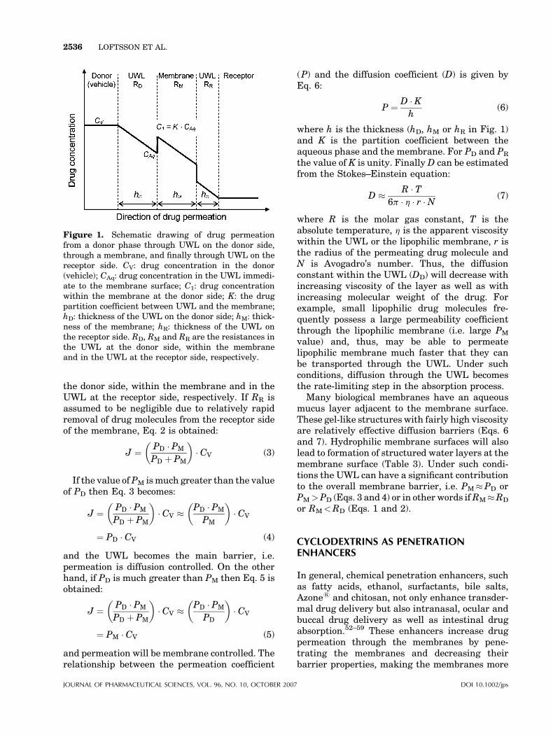
Figure 1. Schematic drawing of drug permeationfrom a donor phase through UWL on the donor side,through a membrane, and finally through UWL on thereceptor side. CV: drug concentration in the donor(vehicle); CAq: drug concentration in the UWL immedi-ate to the membrane surface; C1: drug concentrationwithin the membrane at the donor side; K: the drugpartition coefficient between UWL and the membrane;hD: thickness of the UWL on the donor side; hM: thick-ness of the membrane; hR: thickness of the UWL onthe receptor side. RD, RM and RR are the resistances inthe UWL at the donor side, within the membraneand in the UWL at the receptor side, respectively.
2536 LOFTSSON ET AL.
the donor side, within the membrane and in theUWL at the receptor side, respectively. If RR isassumed to be negligible due to relatively rapidremoval of drug molecules from the receptor sideof the membrane, Eq. 2 is obtained:
J ¼ PD � PM
PD þ PM
� �� CV (3)
If the value ofPM ismuch greater than the valueof PD then Eq. 3 becomes:
J ¼ PD � PM
PD þ PM
� �� CV � PD � PM
PM
� �� CV
¼ PD � CV (4)
and the UWL becomes the main barrier, i.e.permeation is diffusion controlled. On the otherhand, if PD is much greater than PM then Eq. 5 isobtained:
J ¼ PD � PM
PD þ PM
� �� CV � PD � PM
PD
� �� CV
¼ PM � CV (5)
and permeation will be membrane controlled. Therelationship between the permeation coefficient
JOURNAL OF PHARMACEUTICAL SCIENCES, VOL. 96, NO. 10, OCTOBER 2007
(P) and the diffusion coefficient (D) is given byEq. 6:
P ¼ D � Kh
(6)
where h is the thickness (hD, hM or hR in Fig. 1)and K is the partition coefficient between theaqueous phase and the membrane. For PD and PR
the value of K is unity. FinallyD can be estimatedfrom the Stokes–Einstein equation:
D � R � T6p � h � r �N (7)
where R is the molar gas constant, T is theabsolute temperature, h is the apparent viscositywithin the UWL or the lipophilic membrane, r isthe radius of the permeating drug molecule andN is Avogadro’s number. Thus, the diffusionconstant within the UWL (DD) will decrease withincreasing viscosity of the layer as well as withincreasing molecular weight of the drug. Forexample, small lipophilic drug molecules fre-quently possess a large permeability coefficientthrough the lipophilic membrane (i.e. large PM
value) and, thus, may be able to permeatelipophilic membrane much faster that they canbe transported through the UWL. Under suchconditions, diffusion through the UWL becomesthe rate-limiting step in the absorption process.
Many biological membranes have an aqueousmucus layer adjacent to the membrane surface.These gel-like structures with fairly high viscosityare relatively effective diffusion barriers (Eqs. 6and 7). Hydrophilic membrane surfaces will alsolead to formation of structured water layers at themembrane surface (Table 3). Under such condi-tions the UWL can have a significant contributionto the overall membrane barrier, i.e. PM�PD orPM>PD (Eqs. 3 and 4) or in other words ifRM�RD
or RM<RD (Eqs. 1 and 2).
CYCLODEXTRINS AS PENETRATIONENHANCERS
In general, chemical penetration enhancers, suchas fatty acids, ethanol, surfactants, bile salts,Azone1 and chitosan, not only enhance transder-mal drug delivery but also intranasal, ocular andbuccal drug delivery as well as intestinal drugabsorption.52–59 These enhancers increase drugpermeation through the membranes by pene-trating the membranes and decreasing theirbarrier properties, making the membranes more
DOI 10.1002/jps
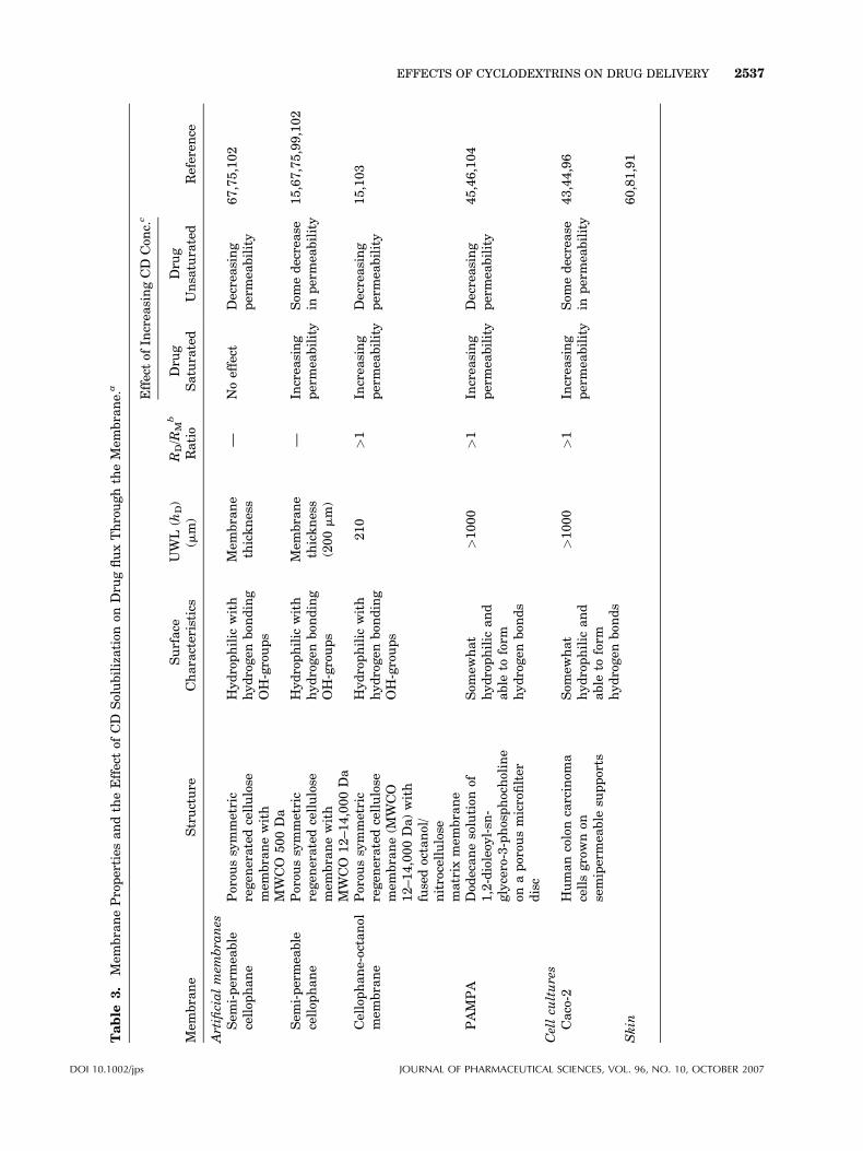
Table
3.
Mem
braneProperties
andtheEffectof
CD
Solubilization
onDru
gfluxThroughtheMem
brane.
a
Mem
brane
Structure
Surface
Characteristics
UWL
(hD)
(mm)
RD/R
Mb
Ratio
Effectof
Increa
singCD
Con
c.c
Referen
ceDru
gSaturated
Dru
gUnsa
turated
Artificialmem
bra
nes
Sem
i-permea
ble
cellop
hane
Porou
ssymmetric
regen
eratedcellulose
mem
branewith
MWCO
500Da
Hydrophilic
with
hydrogen
bon
ding
OH-groups
Mem
brane
thickness
—Noeffect
Decreasing
permea
bility
67,75,102
Sem
i-permea
ble
cellop
hane
Porou
ssymmetric
regen
eratedcellulose
mem
branewith
MWCO
12–14,000Da
Hydrophilic
with
hydrogen
bon
ding
OH-groups
Mem
brane
thickness
(200mm)
—In
crea
sing
permea
bility
Som
edecrease
inpermea
bility
15,67,75,99,102
Cellophane-octanol
mem
brane
Porou
ssymmetric
regen
eratedcellulose
mem
brane(M
WCO
12–14,000Da)with
fusedoctanol/
nitrocellulose
matrix
mem
brane
Hydrophilic
with
hydrogen
bon
ding
OH-groups
210
>1
Increa
sing
permea
bility
Decreasing
permea
bility
15,103
PAMPA
Dod
ecanesolution
of1,2-dioleoy
l-sn
-glycero-3-phosphocholine
onaporou
smicrofilter
disc
Som
ewhat
hydrophilic
and
able
toform
hydrogen
bon
ds
>1000
>1
Increa
sing
permea
bility
Decreasing
permea
bility
45,46,104
Cellcu
ltures
Caco-2
Humancoloncarcinom
acellsgrownon
semipermea
ble
supports
Som
ewhat
hydrophilic
and
able
toform
hydrogen
bon
ds
>1000
>1
Increa
sing
permea
bility
Som
edecrease
inpermea
bility
43,44,96
Skin
60,81,91
DOI 10.1002/jps JOURNAL OF PHARMACEUTICAL SCIENCES, VOL. 96, NO. 10, OCTOBER 2007
EFFECTS OF CYCLODEXTRINS ON DRUG DELIVERY 2537
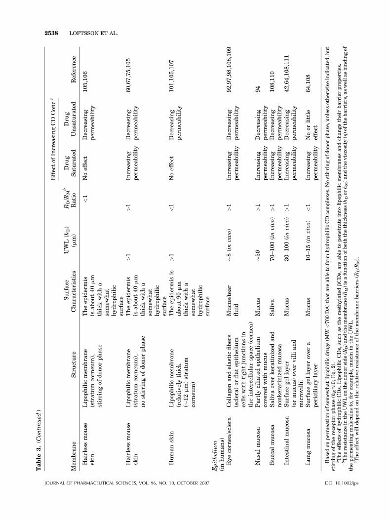
Table
3.(C
ontinued
)
Mem
brane
Structure
Surface
Characteristics
UWL
(hD)
(mm)
RD/R
Mb
Ratio
Effectof
Increa
singCD
Con
c.c
Referen
ceDru
gSaturated
Dru
gUnsa
turated
Hairless
mou
sesk
inLipop
hilic
mem
brane
(stratum
corn
eum),
stirringof
don
orphase
Theep
idermis
isabou
t40mm
thickwitha
somew
hat
hydrophilic
surface
<1
Noeffect
Decreasing
permea
bility
105,106
Hairless
mou
sesk
inLipop
hilic
mem
brane
(stratum
corn
eum),
nostirringof
don
orphase
Theep
idermis
isabou
t40mm
thickwitha
somew
hat
hydrophilic
surface
>1
>1
Increa
sing
permea
bility
Decreasing
permea
bility
60,67,75,105
Humansk
inLipop
hilic
mem
brane
(relativelythick
(�12mm)stratum
corn
eum)
Theep
idermis
isabou
t90mm
thickwitha
somew
hat
hydrophilic
surface
>1
<1
Noeffect
Decreasing
permea
bility
101,105,107
Epithelium
(inhumans)
Eyecorn
ea/sclera
Collagen
andelastic
fibers
(sclera)or
flatep
ithelium
cellswithtightjunctionsin
theintercellularsp
ace
(cornea
)
Mucu
s/tear
fluid
�8(invivo)
>1
Increa
sing
permea
bility
Decreasing
permea
bility
92,97,98,108,109
Nasa
lmucosa
Partly
ciliatedep
ithelium
covered
withmucu
sMucu
s�50
>1
Increa
sing
permea
bility
Decreasing
permea
bility
94
Buccalmucosa
Salivaov
erkeratinized
and
non
keratinized
mucosa
Saliva
70–100(invivo)
>1
Increa
sing
permea
bility
Decreasing
permea
bility
108,110
Intestinalmucosa
Surface
gel
layer
(ormucu
s)ov
ervilliand
microvilli.
Mucu
s30–100(invivo)
>1
Increa
sing
permea
bility
Decreasing
permea
bility
42,64,108,111
Lungmucosa
Surface
gel
layer
over
apericiliary
layer
Mucu
s10–15(invivo)
<1
Increa
sing
permea
bility
Noor
little
effect
64,108
Basedon
permea
tion
ofsomew
hatlipop
hilic
dru
gs(M
W<700DA)thatare
able
toform
hydrophilic
CD
complexes.Nostirringof
don
orphase,unless
otherwiseindicated,but
stirringof
thereceptorphase
(hR�0;Eq.2).
aTheeffectsof
hydrophilic
CDs.
Lipop
hilic
CDs,
such
asthemethylatedbCDs,
are
able
topen
etrate
into
lipop
hilic
mem
branes
andch
angetheirbarrierproperties.
bTheresistance
intheUWLon
thedon
orside(R
D)andthemem
brane(R
M)isafunctionof
boththethickness(h
Dor
hM)andtheviscosity
(h)of
thebarriers,a
swellasbindingof
thepermea
tingmoleculesto,forex
ample,mucinin
theUWL.
c Theeffect
willdep
endon
therelativeresistance
ofthemem
branebarriers
(RD/R
M).
JOURNAL OF PHARMACEUTICAL SCIENCES, VOL. 96, NO. 10, OCTOBER 2007 DOI 10.1002/jps
2538 LOFTSSON ET AL.
EFFECTS OF CYCLODEXTRINS ON DRUG DELIVERY 2539
permeable by, for example increasing theirhydration, modifying their intracellular lipiddomains or enhancing drug partition into themembranes by changing their solvent nature.These chemical penetration enhancers enhancemembrane permeation of both hydrophilic andlipophilic drugs, both from non-aqueous andaqueous donor phases. Hydrophilic CDs, suchas 2-hydroxypropyl-b-cyclodextrin (HPbCD) andsulfobutylether b-cyclodextrin sodium salt(SBEbCD), are on the other hand (Table 2):
(a) U
DOI 10.
nable to permeate biological membranessuch as skin and gastrointestinal mucosa toany significant extent.
(b) U
nable to enhance drug permeation fromlipophilic environments.(c) U
nable to enhance permeation of hydrophi-lic drugs.(d) A
ble to enhance permeation of lipophilicdrugs.(e) A
ble to reduce drug permeation throughlipophilic membranes by decreasing drugpartition from the exterior into the mem-brane.(f) A
ble to increase the chemical stability ofdrugs at the aqueous membrane exterior.Thus, for CDs the potential mechanism of actionmust be different from those of the chemicalpenetration enhancers.
The Chemical Potential
The chemical potential of drug in the donor phase(i.e. the vehicle or UWL on the donor side in Fig. 1)will also affect drug permeability through themembrane.48 If the barrier function of a mem-brane is unaffected by the vehicle composition, thepermeability will increase with increasing chemi-cal potential. The value of the partition coefficient(K in Eq. 6) is at its maximum when the chemicalpotential of the drug in the donor phase ismaximized thereby optimizing the tendency ofthe drug molecule to leave the donor phase andenter the membrane. The chemical potential of agiven lipophilic drug in aqueous CD solutionsaturated with the drug is constant and does notincrease with increasing CD concentration. Sinceonly the free drug and not the drug/CD complex isable to partition into the membrane, the observedincrease in drug flux with increasing CD concen-tration15,60 cannot be explained by an increase in
1002/jps JOURN
the chemical potential. Furthermore, excess CD,more than is needed to solubilize the drug in theaqueous donor phase, will decrease the chemicalpotential of the drug and that leads to a decreasein the partition coefficient and consequently adecrease in drug permeation.61
Physical Enhancers
Physical enhancers have been used to enhancetopical drug delivery, e.g. through the skin or intothe eye, in a controllable manner. For example, asmall electrical current can be applied to enhancemembrane penetration of ionized drugs. The waveenergy of ultrasound decreases the barrier poten-tial of biological membranes and in electropora-tion aqueous pathways are created in lipid bilayermembranes.62 These physical methods can beapplied to increase permeation of both lipophilicand hydrophilic drugs, and for general drugmolecules of diverse physicochemical propertiesand sizes. The permeation enhancing properties ofCDs are quite different from those of the physicalenhancement.
The UWL
The existence of an UWL during drug diffusionthrough membranes and drug dissolution is wellestablished.63 However, the contribution of anUWL to the overall barrier function of a mem-brane is frequently ignored.44 The thickness of theUWL can be significant (Table 3) and mucus andother surface structures can enhance its barrierfunction by increasing its viscosity (h) that leads toan overall decrease in the diffusion coefficient (Din Eq. 7). Studies have shown that drug diffusionthrough mucus is up to 100-times slower thanthrough pure water.64 It is possible that CDcomplexation of lipophilic, and somewhat water-insoluble, drug molecules enhances drug diffusionthrough the UWL. Increase in the concentrationgradient over the UWL will increase the rate ofdrug diffusion through the layer (Fig. 1). Drugpermeation through the UWL depends also on theUWL thickness, decreasing with increasing thick-ness (h in Eq. 6). Under in vitro conditions, thethickness of the UWL can be well above 1000 mmin the unstirred aqueous donor phase and in vivoits thickness is frequently 10–100 mm. However,the thickness of the UWL depends also on thephysicochemical properties of the permeatingdrug molecule, including their ability to form
AL OF PHARMACEUTICAL SCIENCES, VOL. 96, NO. 10, OCTOBER 2007
2540 LOFTSSON ET AL.
ionic and hydrogen bonds with mucin, and thusfixed UWL thickness for all drugs does not exist.65
The Membrane Barriers
The effects of hydrophilic CDs on drug fluxthrough various types of artificial and biologicalmembranes are summarized in Table 3 togetherwith a brief description of the membrane char-acteristics, including the approximate thicknessof the UWL on the donor side and the relativeresistance of the UWL (RD) and the membraneitself (RM). The resistance is a function of thethickness of the barriers (UWL or the membranebarrier) and their viscosity, increasing withincreasing thickness or increasing viscosity(Eqs. 6 and 7). The resistance will also increasewith increasing ability of the permeating mole-cules to bind to membrane constituents such aswater, mucin and collagen.
Intestinal Drug Absorption
It has been shown that for intestinal absorption,the main step in the absorption process isdiffusion through the stagnant mucus layer(UWL), together with transfer across themucus/membrane interface.66 Studies of therelationship between gastrointestinal drugabsorption and the biopharmaceutics classifica-tion system (BCS) have shown that CDs enhancethe bioavailability of Class II drugs that have lowaqueous solubility but show good absorption fromsolutions (low solubility, high permeability). Onthe other hand CDs do not enhance bioavailabilityof Class III drugs (high solubility, low perme-ability).5 Class III drugs are hydrophilic and donot, in general, form inclusion complexes withCDs. Furthermore, CD complexation of Class IIIdrugs can reduce their ability to partition from theaqueous exterior into the lipophilicmembrane (i.e.the CD complexation will lower their K-value inEq. 6).
Transdermal Drug Delivery
Skin is much less permeable than epithelium andsince it does not contain aqueous gel structureslike mucus, its UWL is usually much thinner.Thus, under normal conditions CDs do notenhance drug delivery through skin. However,under in vitro conditions using hairless mouseskin, which is more permeable than human skin,and when the aqueous donor phase is unstirred,
JOURNAL OF PHARMACEUTICAL SCIENCES, VOL. 96, NO. 10, OCTOBER 2007
the permeation resistance in the UWL (RD) can besufficient to make RD�RM. Under such condi-tions CDs can enhance dermal and transdermaldrug delivery (Table 3).67 Also, CDs enhancedermal and transdermal delivery of lipophilicdrugs from oil-in-water (o/w) creams where theUWL is extended into the cream layer on the skinsurface.68,69 Again, the CD complexation canreduce the ability of lipophilic compounds topartition from the exterior into the skin barrier,a property that sometimes is used to preventabsorption of, for example, sun screeners into andthrough skin.
Caco-2
The barrier properties of Caco-2 membranes arerate-limited (i.e. possess a relatively low RM) andunder such conditions the adjacent UWL on thedonor side has significant contribution to theoverall barrier function (Table 3). In the Caco-2model the aqueous donor phase is usuallyunstirred resulting in relatively thick UWL andconsequently to RD>RM. Under such conditionshydrophilic CDs can enhance delivery of lipophilicdrugs through the membrane.
PAMPA and other artificial membranes
In a previous study we have shown that in thePAMPA system, the thickness of the UWL and itscontribution to the overall membrane barrierdepends on the stirring rate.46 In absence ofHPbCD, drug permeability increased withdecreasing UWL thickness to a certain minimumvalues of about 40 mm. Addition of HPbCD tosystems exhibiting UWL thicknesses greater than40 mm significantly increased the drug fluxthrough PAMPA. The effect of HPbCD appearedalso to be related to stability constant (K) of thedrug/CD complex with flux increasing withincreasing K value.46 This suggests that hydro-philic CDs enhance flux when the UWL resistance(RD) has significant contribution to the overallbarrier resistance. CDs are able to enhance drugdelivery through artificial membranes if theexperimental conditions are such that RD�RM.Table 3 shows that CDs can, in general, enhancedrug permeation through membranes if thepermeation resistance of the UWL is about equalor greater than the resistance of membranebarrier (i.e. when RD�RM or PM�PD, Eq. 3and Fig. 1), but how do CDs enhance thepermeation?
DOI 10.1002/jps
EFFECTS OF CYCLODEXTRINS ON DRUG DELIVERY 2541
CDs AND UWLs
Excess CD in the UWL will decrease drugpartition from the UWL into the membrane (i.e.the value of K in Eq. 6) which explains why excessCD decreases drug permeation through themembranes (Table 3).61,70 However, since onlythe free drug is able to permeate the lipophilicmembranes, and not the CDmolecules or drug/CDcomplexes, it is difficult to explain why CDsenhance drug delivery through membranes fromdrug saturated CD solutions, especially since theconcentration of free drug does not increase withincreasing CD concentration. In aqueous CDsolutions saturated with drug, only the concen-tration of drug/CD complex increases withincreasing CD concentration.60 Since CDs onlyenhance permeation of relatively lipophilic drugsfrom aqueous donor phases (vehicles), theirenhancing effect must be associated with thephysicochemical properties of water and the drug/CD complexation. In aqueous solutions, thecooperative hydrogen bonding leads to formationof water clusters that can decrease mobility ofdissolved drug molecules. At membrane surfaces,the cooperative hydrogen bonding leads to forma-tion of a relatively thin (�1 mm) UWL wheremobility of the water molecules is significantlyreduced. Water-soluble polymers, like mucin, canincrease the thickness of this layer to 100 mm(in vivo) or even more (>1000 mm) under somein vitro conditions. These relatively thick andviscous UWLs, where mobility of dissolved mole-cules is severely limited compared to pureaqueous solutions, can induce permeation resis-tance (RD) that is greater than the permeationresistance of the membrane itself (RM) (seeTable 3). Interactions of the permeating drugmolecules with, for example, mucin will result ineven further increase in RD. Recently it wasshown that by eliminating hydrophobic interac-tions between mucin fibers and hydrophobicsurfaces resulted in enhanced particle permeationand that large nano-particles can permeate fasterthrough mucosa than smaller particles.71 The CDmolecules are three to four times larger than thedrug molecules and, thus, should permeate UWLat a slower rate than the drug molecules (Eq. 7)but since CDs behave as chaotropes at CDconcentrations used in the donor phases (vehi-cles), the effect should be less than expected basedon their MW. Also, it should be remembered thatthe rates for formation and dissociation of drug/CD complexes are very close to the diffusion
DOI 10.1002/jps JOURN
controlled limits and that drug/cyclodextrin com-plexes are continuously being formed and brokendown.4 Based on extensive review of the litera-ture, it appears that CDs enhance drug deliverythrough the UWL by one or more of the followingmechanisms:
(a) C
AL OF PH
D complexation increases the totalamount of dissolved drug molecules in theaqueous donor phase. This increases theconcentration gradient of the drug overthe UWL leading to more rapid drug deliv-ery to the membrane surface. Since drugrelease from the CD complex is more rapidthan the flux of drug molecules through theUWL, this will increase the availability offree drug molecules immediate to the lipo-philic membrane surface.
(b) S
ince low MW carbohydrates behave aschaotropes at relatively low concentrations,CDs will disrupt the hydrogen-bondedwater network in the UWL. This could facil-itate penetration of drug/CD complexesthrough the UWL and increase drug avail-ability at the membrane surface. However,there are no indications that CDs enhancepermeation of free drug molecules throughthe UWL.(c) C
D complexation of drug molecules couldpossibly prevent them from interactingwith molecules in the UWL, such as mucin,increasing their overall delivery rate to themembrane surface.It is possible that CDs may have other addi-tionalmechanisms of action that have not yet beenelucidated. Also it is still not clear how otherexcipients in the aqueous donor phase, such aswater-soluble polymers, can enhance drug deliv-ery from aqueous CD solutions.72,73
CONCLUSION
It is clear that hydrophilic CDs can only enhancedrug delivery when the permeation resistance ofthe UWL on the donor side is about equal orgreater than the resistance of membrane barrier.This is frequently the case when drugs permeatemucosal epithelium where mucosa forms theUWL. Hydrophilic CDs do not enhance drugdelivery through membranes if the lipophilicmembrane barrier is themain permeation barrier.When aqueous vehicles, such as hydrogels and o/w
ARMACEUTICAL SCIENCES, VOL. 96, NO. 10, OCTOBER 2007
2542 LOFTSSON ET AL.
creams, are applied to membranes, the UWL isextended into the vehicle and under such condi-tions CDs can increase drug delivery from thevehicle through the membrane.
CDs only enhance permeation of drugmoleculesthat are able to permeate given membrane withrelative ease once the dissolved drug moleculesare in contact with the membrane surface, forexample absorption of BCSClass II drugs from thegastrointestinal tract. In the case of lipophilicmembranes, the greatest enhancement is usuallyobtained with relatively small, lipophilic drugmolecules. Finally, CDs are only able to enhancepermeation of drugs that readily form CD com-plexes.
REFERENCES
1. Loftsson T, Duchene D. 2007. Cyclodextrins andtheir pharmaceutical applications. Int J Pharm329:1–11.
2. Loftsson T, Masson M, Brewster ME. 2004. Self-association of cyclodextrins and cyclodextrin com-plexes. J Pharm Sci 93:1091–1099.
3. Bonini M, Rossi S, Karlsson G, Almgren M,Lo Nostro P, Baglioni P. 2006. Self-assembly ofb-cyclodextrin in water. Part 1. Cryo-TEM anddynamic and static light scattering. Langmuir22:1478–1484.
4. Stella VJ, Rajewski RA. 1997. Cyclodextrins: theirfuture in drug formulation and delivery. PharmRes 14:556–567.
5. Loftsson T, Brewster ME, Masson M. 2004. Role ofcyclodextrins in improving oral drug delivery.Am J Drug Deliv 2:261–275.
6. Loftsson T, Jarho P, Masson M, Jarvinen T. 2005.Cyclodextrins in drug delivery. Expert Opin DrugDeliv 2:335–351.
7. Lipinski CA, Lombardo F, Dominy BW, Feeney PJ.2001. Experimental and computatorial approachesto estimate solubility and permeability in drugdiscovery and development settings. Adv DrugDeliv Rev 46:3–26.
8. Uekama K, Arimori K, Sakai A, Masaki K, Irie T,Otagiri M. 1987. Improvement of percutaneousabsorption of prednisolone by b- and g-cyclodex-trin complexations. Chem Pharm Bull 35:2910–2913.
9. Gerloczy A, Antal S, Szejtli J. 1988. Percutaneousabsorption of heptakis-(2,6-di-O-14C-methyl)-b-cyclodextrin in rats. In: Huber O, Szejtli J, editors.Proceedings of the Fourth International Sympo-sium on Cyclodextrins. Dordrecht: Kluwer Aca-demic Publishers. p 415–420.
JOURNAL OF PHARMACEUTICAL SCIENCES, VOL. 96, NO. 10, OCTOBER 2007
10. Tanaka M, Iwata Y, Kouzuki Y, Taniguchi K,Matsuda H, Arima H, Tsuchiya S. 1995. Effectof 2-hydroxypropyl-b-cyclodextrin on percuta-neous absorption of methyl paraben. J PharmPharmacol 47:897–900.
11. Irie T, Uekama K. 1997. Pharmaceutical applica-tions of cyclodextrins. III. Toxicological issues andsafety evaluation. J Pharm Sci 86:147–162.
12. Matsuda H, Arima H. 1999. Cyclodextrins intransdermal and rectal delivery. Adv Drug DelivRev 36:81–99.
13. UekamaK,HirayamaF, Irie T. 1998. Cyclodextrindrug carrier systems. Chem Rev 98:2045–2076.
14. Loftsson T, Konraðsdottir F, Masson M. 2006.Influence of aqueous diffusion layer on passivedrug diffusion from aqueous cyclodextrin solutionsthrough biological membranes. Pharmazie 61:83–89.
15. Loftsson T, Konradsdottir F, Masson M. 2006.Development and evaluation of an artificial mem-brane for determination of drug availability inpharmaceutical formulations. Int J Pharm326:60–68.
16. Marttin E, Verhoef JC, Merkus FWHM. 1998.Efficiency, safety and mechanism of cyclodex-trins as absorption enhancers in nasal deliveryof peptide and protein drugs. J Drug Target 6:17–36.
17. Chaplin MF. 1999. A proposal for the structuringof water. Biophys Chem 93:211–221.
18. Griffith JH, Scheraga HA. 2004. Statistical ther-modynamics of aqueous solutions. I. Water struc-ture, solutions with non-polar solutes, andhydrophobic interactions. J Mol Struct 682:97–113.
19. Schmid R. 2001. Recent advances in the descrip-tion of the structure of water, the hydrophobiceffect, and the like-dissolves-like rule. ChemMonth 132:1295–1326.
20. Michot LJ, Villieras F, Francois M, Bihannic I,Pelltier M, Cases JM. 2002. Water organisation atthe solid-aqueous solution interface. C R Geosci334:611–631.
21. 2001. The Merck Index. 13th ed. WhitehouseStation: Merck & Co.
22. Pauling L. 1967. The nature of the chemical bond.New York: Cornell University Press.
23. Cabane B, Vuilleumier R. 2005. The physics ofliquid water. C R Geosc 337:159–171.
24. Xantheas SS. 2000. Cooperativity and hydrogenbonding network in water clusters. Chem Phys258:225–231.
25. Dore J. 2000. Structural studies of water in con-fined geometry by neutron diffraction. Chem Phys258:327–347.
26. Wakisaka A, Matsuura K. 2006. Microheterogene-ity of ethanol–water binary mixtures observed atthe cluster level. J Mol Liq 129:25–32.
DOI 10.1002/jps
EFFECTS OF CYCLODEXTRINS ON DRUG DELIVERY 2543
27. Giangiacomo R. 2006. Study of water–sugar inter-actions at increasing sugar concentration by NIRspectroscopy. Food Chem Toxicol 96:371–379.
28. Winkler RG, Fioravanti S, Ciccotti G, MargheritisC, Villa M. 2000. Hydration of b-cyclodextrin: amolecular dynamics simulation study. J Comp-AidMol Des 14:659–667.
29. Bone S. 2006. Dielectric studies of water clustersin cyclodextrins: relevence to the transitionbetween slow and fast forms. J Phys Chem B110:20609–20614.
30. Hakkarainen B, Fujita K, Immel S, Kenne L,Sandstrom C. 2005. 1H NMR studies on the hydro-gen-bonding network inmono-altro-b-cyclodextrinand its complex with adamantane-1-carboxylicacid. Carbohydr Res 340:1539–1545.
31. Lichtenthaler FW, Immel S. 1996. On the hydro-phobic characteristics of cyclodextrins: computer-aided visualization of molecular lipophilicity pat-terns. Liebigs Ann Chem 27–37.
32. Loftsson T, Masson M, Sigurjonsdottir JF. 1999.Methods to enhance the complexation efficiency ofcyclodextrins. STP Pharma Sci 9:237–242.
33. McBrierty VJ, Martin SJ, Karasz FE. 1999.Understanding hydrated polymers: the perspec-tive of NMR. J Mol Liq 80:179–205.
34. Branca C, Magazu S, Maisano G, Migliardo P,Migliardo F, Romeo G. 2002. Hydration para-meters of aqueous solutions of poly(ethyleneglycol)s by viscosity data. Phys Scrip 66:175–179.
35. Gun’ko VM, Turov VV, Bogatyrev VM, Zarko VI,Leboda R, Goncharuk EV, Novza AA, Turov AV,Chuiko AA. 2005. Unusual properties of water athydrophilic/hydrophobic interfaces. Adv ColloidInterf Sci 118:125–172.
36. Vogler EA. 1998. Structure and reactivity of waterat biomaterial surfaces. Adv Colloid Interf Sci74:69–117.
37. Yaminsky VV, Vogler EA. 2001. Hydrophobichydration. Curr Opin Colloid Interf Sci 6:342–349.
38. Chandler D. 2005. Interfaces and the driving forceof hydrophobic assembly. Nature 437:640–647.
39. Disalvo EA, Lairion F, Martini F, Almaleck H,Diaz S, Gordillo G. 2004. Water in biological mem-branes at interfaces: does it play a functional role?J Argent Chem Soc 92:1–22.
40. Wertz PW. 2004. Stratum corneum lipids andwater. Exog Dermatol 3:53–56.
41. Bansil R, Turner BS. 2006. Mucin structure,aggregation, physiological functions and biomedi-cal applications. Curr Opin Colloid Interf Sci11:164–170.
42. Lennernas H. 1998. Human intestinal permeabil-ity. J Pharm Sci 87:403–410.
43. Karlsson JP, Artursson P. 1991. A method for thedetermination of cellular permeability coefficientsand aqueous boundary layer thickness in mono-
DOI 10.1002/jps JOURN
layers of intestinal epithelia (Caco-2) cells grownin permeable filter chambers. Int J Pharm 71:55–64.
44. Youdim KA, Avdeef A, Abbott NJ. 2003. In vitrotrans-monolayer permeability calculations: oftenforgotten assumptions. Drug Discov Today 8:997–1003.
45. Avdeef A, Nielsen PE, Tsinman O. 2004. PAMPA -a drug absorption in vitro model 11. Matching thein vivo unstirred water layer thickness by indivi-dual-well stirring in microtitre plates. Eur JPharm Sci 22:365–374.
46. Brewster ME, Noppe M, Peeters J, Loftsson T.2007. Effect of the unstirred water layer on perme-ability enhancement by hydrophilic cyclodextrins.Int J Pharm (manuscript).
47. Plumridge TH, Waigh RD. 2002. Water structuretheory and some implications for drug design.J Pharm Pharmacol 54:1155–1179.
48. Higuchi T. 1960. Physical chemical analysis ofpercutaneous absorption process from creamsand ointments. J Soc Cosmetic Chemists 11:85–97.
49. Flynn GL, Carpender OS, Yalkowsky SH. 1972.Total mathematical resolution of diffusion layercontrol of barrier flux. J Pharm Sci 61:312–314.
50. Flynn GL, Yalkowsky SH. 1972. Correlation andprediction of mass transport across membranes Iinfluence of alkyl chain length on flux-determiningproperties of barrier and diffusant. J Pharm Sci61:838–852.
51. Zwolinski BJ, Eyring H, Reese CE. 1949. Diffusionand membrane permeability. I. J Phys Coll Chem53:1426–1453.
52. Chattaraj SC, Walker RB. 1995. Penetrationenhancer classification. In: Smith EW, MaibachHI, editors. Percutaneous penetration enhancers.Boca Raton: CRC Press. p 5–20.
53. Williams AC, Barry BW. 2004. Penetration enhan-cers. Adv Drug Deliv Rev 56:603–618.
54. Nicolazzo JA, Reed BL, Finnin BC. 2005. Buccalpenetration enhancers—how do they really work?J Control Rel 105:1–15.
55. Sasaki H, Yamamura K, Nishida K, Nakamura J,Ichikawa M. 1996. Delivery of drugs to the eyeby topical application. Prog Ret Eye Res 15:583–620.
56. Kaur IP, Smitha R. 2002. Penetration enhacersand ocular bioadhesives: two new avenues forophthalmic drug delivery. Drug Dev Ind Pharm24:353–369.
57. Ross BP, Toth I. 2005. Gastrointestinal absorptionof heparin by lipidization or coadministration withpenetration enhancers. Curr Drug Deliv 2:277–287.
58. Behl CR, Pimplaskar HK, Sileno AP, DemeirelesJ, Romeo VD. 1998. Effects of physicochemical
AL OF PHARMACEUTICAL SCIENCES, VOL. 96, NO. 10, OCTOBER 2007
2544 LOFTSSON ET AL.
properties and other factors on systemic nasaldrug delivery. Adv Drug Deliv Rev 29:89–116.
59. Benson HAF. 2005. Transdermal drug delivery:penetration enhancement techniques. Curr DrugDeliv 2:23–33.
60. Loftsson T, Masson M. 2001. Cyclodextrins intopical drug formulations: theory and practice.Int J Pharm 225:15–30.
61. Loftsson T, Konradsdottir F, Masson M. 2007.Development of octanol membranes for drugscreening. J Incl Phenom Macroc Chem 57:613–617.
62. Smith EW, Maibach HI, editors. 1995. Percuta-neous penetration enhancers. Boca Raton: CRCPress.
63. Sinko PJ, editor. 2006. Martin’s Physical Phar-macy and Pharmaceutical Sciences. Philadelphia:Lippincott Williams & Wilkins.
64. Khanvilkar K, Donovan MD, Flanagan DR. 2001.Drug transfer throughmucus. AdvDrug Deliv Rev48:173–193.
65. Pohl P, Saparov SM, Antonenko YN. 1998. Thesize of the unstirred layer as a function of thesolute diffusion coefficient. Biophys J 75:1403–1409.
66. Abraham MH, Zhao YH, Le J , Hersey A, Lus-combe CN, Reynolds DP, Beck G, Sherborne B,Cooper I. 2002. On the mechanism of humanintestinal absorption. Eur J Med Chem 37:595–605.
67. Masson M, Loftsson T, Masson G, Stefansson E.1999. Cyclodextrins as permeation enhancers:some theoretical evaluations and in vitro testing.J Control Rel 59:107–118.
68. Preiss A, Mehnert W, Fromming KH. 1994. In-vitro hydrocortisone release from ointments inpresence of cyclodextrins. Pharmazie 49:902–906.
69. Preiss A, Mehnert W, Fromming K-H. 1995. Pene-tration of hydrocortisone into excised human skinunder the influence of cyclodextrins. Pharmazie50:121–126.
70. Masson M, Sigurdardottir BV, Matthıasson K,Loftsson T. 2005. Investigation of drug–cyclodex-trin complexes by a phase-distribution method:some theoretical and practical considerations.Chem Pharm Bull 53:958–964.
71. Lai SK, O’Hanlon DE, Harrold S, Man ST, WangY-Y, Cone R, Hanes J. 2007. Rapid transport oflarge polymeric nanoparticles in fresh undilutedhuman mucus. Proc Natl Acad Sci USA (PNAS)104:1482–1487.
72. Loftsson T. 1998. Increasing the cyclodextrin com-plexation of drugs and drug biovailability throughaddition of water-soluble polymers. Pharmazie53:733–740.
73. Loftsson T, Masson M. 2004. The effects of water-soluble polymers on cyclodextrins and cyclodextrin
JOURNAL OF PHARMACEUTICAL SCIENCES, VOL. 96, NO. 10, OCTOBER 2007
solubilization of drugs. J Drug Deliv Sci Tech14:35–43.
74. ArimaH,Miyaji T, Irie T, Hirayama F, UekamaK.1998. Enhancing effect of hydroxypropyl-b-cyclo-dextrin on cutaneous penetration activation ofethyl 4-biphenylyl acetate in hairless mouse skin.Eur J Pharm Sci 6:53–59.
75. Loftsson T, Sigfusson SD, Sigurðsson HH,Masson M. 2003. The effects of cyclodextrinson topical delivery of hydrocortisone: theaqueous diffusion layer. STP Pharma Sci 13:125–131.
76. Siefert B, Keipert S. 1997. Influence of a-cyclodex-trin and hydroxyalkylated b-cyclodextrin deriva-tives on the in vitro corneal uptake andpermeation of aqueous pilocarpine-HCl solutions.J Pharm Sci 86:716–720.
77. Loftsson T, Stefansson E. 1997. Effect of cyclodex-trins on topical drug delivery to the eye. DrugDeliv Ind Pharm 23:473–481.
78. Legendre JY, Rault I, Petit A, Luijten W,Demuynck I, Horvath S, Ginot YM, Cuine A.1995. Effects of b-cyclodextrins on skin: implica-tions for the transdermal delivery of piribedil anda novel cognition enhancing-drug, S-9977. Eur JPharm Sci 3:311–322.
79. Vitoria M, Bentley B, Renata F, Wilson S, CollettJH. 1997. Characterization of the influence ofsome cyclodextrins on the stratum corneum fromthe hairless mouse. J Pharm Pharmacol 49:397–402.
80. ArimaH,Miyaji T, Irie T, Hirayama F, UekamaK.1996. Possible enhancing mechanism of the cuta-neous permeation of 4-biphenylylacetic acid by b-cyclodextrin derivatives in hydrophilic ointment.Chem Pharm Bull 44:582–586.
81. Babu RJ, Pandit JK. 2004. Effect of cyclodextrinson the complexation and transdermal delivery ofbupranolol through rat skin. Int J Pharm 271:155–165.
82. Ventura CA, Giannone I, Paolino D, Pistara V,Corsaro A, Puglisi G. 2005. Preparation of cele-coxib-dimethyl-b-cyclodextrin inclusion complex:characterization and in vitro permeation study.Eur J Med Chem 40:624–631.
83. Zheng Y, Zou Z, Chow AHL. 2006. Lack of effect ofb-cyclodextrin and its water-soluble derivatives onin vitro drug transport across rat intestinal epithe-lium. Int J Pharm 309:123–128.
84. Irie T, Wakamatsu K, Arima H, Aritomi H,Uekama K. 1992. Enhancing effects of cyclodex-trins on nasal absorption of insulin in rats. Int JPharm 84:129–139.
85. Schonfelder U, Radestock A, Elsner P, Hipler U-C.2006. Cyclodextrin-induced apoptosis in humankeratinocytes is caspase-8 dependent and accom-panied bymitochondrial cytochrome c release. ExpDermatol 15:883–890.
DOI 10.1002/jps
EFFECTS OF CYCLODEXTRINS ON DRUG DELIVERY 2545
86. Martins PS, Ochoa R, Pimenta AMC, FerreiraLAM, Melo AL, da Silva JBB, Sinisterra RD,Demicheli C, Frezard F. 2006. Mode of action ofb-cyclodextrin as an absorption enhancer of thewater-soluble drug meglumine antimonate. Int JPharm 325:39–47.
87. Loftsson T, Masson M, Sigurdsson HH, Magnus-son P, Goffic FL. 1998. Cyclodextrins as co-enhan-cers in dermal and transdermal drug delivery.Pharmazie 53:137–139.
88. Adachi H, Irie T, Uekama K, Manako T, Yano T,Saita M. 1992. Inhibitory effect of prostaglandinE1 on laurate-induced peripheral vascular occlu-sive sequelae in rabbits; optimized topical for-mulation with b-cyclodextrin derivative andpenetration enhancer HPE-101. J Pharm Pharma-col 44:1033–1035.
89. Adachi H, Irie T, Uekama K, Manako T, Yano T,Saita M. 1993. Combination effects of O-carboxy-methyl-O-ethyl-b-cyclodextrin and penetrationenhancer HPE-101 on transdermal delivery ofprostaglandin-E(1) in hairless mice. Eur J PharmSci 1:117–123.
90. Sinha VR, Bindra S, Kumria R, Nanda A. 2003.Cyclodextrin as skin-penetration enhancers.Pharm Technol 27:120–138.
91. Uekama K, Adachi H, Irie T, Yano T, Saita M,Noda K. 1992. Improved transdermal delivery ofprostaglandin E1 through hairless mouse skin:combined use of carboxymethyl-ethyl-b-cyclodex-trin and penetration enhancers. J Pharm Phar-macol 44:119–121.
92. Jarho P, Urtti A, Pate DW, Suhonen P, Jarvinen T.1996. Increase in aqueous solubility, stability andin vitro corneal permeability of anandamide byhydroxypropyl-b-cyclodextrin. Int J Pharm 137:209–217.
93. Cho MJ, Chen FJ, Huczek DL. 1995. Effects ofinclusion complexation on the transepithelialtransport of a lipophilic substance in vitro. PharmRes 12:560–564.
94. Kublik H, Bock TK, Schreier H, Muller BW. 1996.Nasal absorption of 17b-estradiol from differentcyclodextrin inclusion formulations in sheep. EurJ Pharm Biopharm 42:320–324.
95. Chang SL, Banga AK. 1998. Transdermal ionto-phoretic delivery of hydrocortisone from cyclodex-trin solutions. J Pharm Pharmacol 50:635–640.
96. Zuo Z, Kwon G, Stevenson B, Diakur J, Wiebe LI.2000. Flutamide–hydroxypropyl-b-cyclodextrincomplex: formulation, physical characterization,and absorption using the caco-2 in vitro model.J Pharm Pharmaceut Sci 3:220–227.
97. Bary AR, Tucker IG, Davies NM. 2000. Considera-tions in the use of hydroxypropyl-b-cyclodextrin inthe formulation of aqueous ophthalmic solutions ofhydrocortisone. Eur J Pharm Biopharm 50:237–244.
DOI 10.1002/jps JOURN
98. Richter T, Keipert S. 2004. In vitro permeationstudies comparing bovine nasal mucosa, porcinecornea and artificial membrane: androstenedionein microemulsions and their components. Eur JPharm Biopharm 58:137–143.
99. Manosroi J, Apriyani MG, Foe K, Manosroi A.2005. Enhancement of the release of azelaic acidthrough the synthetic membranes by inclusioncomplex formation with hysroxypropyl-b-cyclo-dextrin. Int J Pharm 293:235–240.
100. Felton LA, Wiley CJ, Godwin DA. 2002. Influenceof hydroxypropyl-b-cyclodextrin on transdermalpermeation and skin accumulation of oxybenzone.Drug Devel Ind Pharm 28:1117–1124.
101. Ventura CA, Tommasini S, Falcone A, Giannone I,Paolino D, Sdrafkakis V, Mondello MR, Puglisi G.2006. Influence of modified cyclodextrins on solu-bility and percutaneous absorption of celecoxibthrough human skin. Int J Pharm 314:37–45.
102. Loftsson T, Masson M, Sigurdsson HH. 2002.Cyclodextrins and drug permeability throughsemi-permeable cellophane membranes. Int JPharm 232:35–43.
103. Loftsson T, Konradsdottir F, Masson M. 2006.Influence of aqueous diffusion layer on passivedrug diffusion from aqueous cyclodextrin solutionsthrough biological membranes. Pharmazie 61:83–89.
104. Bermejo M, Avdeef A, Ruiz A, Nalda R, Ruell JA,Tsinman O, Gonzalez I, Fernandez C, Sanchez G,Garrigues TM, Merino V. 2004. PAMPA–a drugabsorption in vitro model 7. Comparing rat in situ,Caco-2, and PAMPA permeability of fluoroquino-lones. Eur J Pharm Sci 21:429–441.
105. Hikima T, Yamada K, Kimura T, Maibach HI,Tojo K. 2002. Comparison of skin distributionof hydrophilic activity for bioconversion of b-estradiol 17-acetate between man and severalanimals in vitro. Eur J Pharm Biopharm 54:155–160.
106. Shaker DS, Ghanem A-H, Li SK, Warner KS,Hashem FM, Higuchi WI. 2003. Mechanistic stu-dies of the effect of hydroxypropyl-b-cyclodextrinon in vitro transdermal permeation of corticoster-one through hairless mouse skin. Int J Pharm253:1–11.
107. Pirot F, Berardesca E, Kalia YN, Singh M, Mai-bach HI, Guy RH. 1998. Stratum corneum thuck-ness and apparent water diffusivity: facile andnoninvasive quantitation in vivo. Pharm Res15:492–494.
108. Washington N, Washington C, Wilson CG.2001. Physiological pharmaceutics; barriers todrug absorption. 2nd ed. London: Taylor &Francis.
109. Mannermaa E, Vellonen K-S, Urtti A. 2006. Drugtransport in corneal epithelium and blood–retinabarrier: emerging role of transporters in ocular
AL OF PHARMACEUTICAL SCIENCES, VOL. 96, NO. 10, OCTOBER 2007
2546 LOFTSSON ET AL.
pharmacokinetics. Adv Drug Deliv Rev 58:1136–1163.
110. Cappello B, De Rosa G, Giannini L, La RotondaMI, Mensitieri G, Miro A, Quaglia F, Russo R.2006. Cyclodextrin-containing poly(ethyleneox-ide) tablets for the delivery of poorly soluble drugs:
JOURNAL OF PHARMACEUTICAL SCIENCES, VOL. 96, NO. 10, OCTOBER 2007
potential as buccal delivery system. Int J Pharm319:63–70.
111. Somogyi G, Posta J, Buris L, Varga M. 2006.Cyclodextrin (CD) complexes of cholesterol–theirpotential use in reducing dietary cholesterolintake. Pharmazie 61:154–156.
DOI 10.1002/jps
Related Documents