1 23 The Journal of Physiological Sciences ISSN 1880-6546 Volume 62 Number 4 J Physiol Sci (2012) 62:333-341 DOI 10.1007/s12576-012-0209-8 Effects of the AMP-activated protein kinase inhibitor compound C on the postconditioned rat heart R. Hermann, M. G. Marina Prendes, M. E. Torresin, D. Vélez, E. A. Savino & A. Varela

Welcome message from author
This document is posted to help you gain knowledge. Please leave a comment to let me know what you think about it! Share it to your friends and learn new things together.
Transcript
1 23
The Journal of Physiological Sciences ISSN 1880-6546Volume 62Number 4 J Physiol Sci (2012) 62:333-341DOI 10.1007/s12576-012-0209-8
Effects of the AMP-activated proteinkinase inhibitor compound C on thepostconditioned rat heart
R. Hermann, M. G. Marina Prendes,M. E. Torresin, D. Vélez, E. A. Savino &A. Varela
1 23
Your article is protected by copyright
and all rights are held exclusively by The
Physiological Society of Japan and Springer.
This e-offprint is for personal use only
and shall not be self-archived in electronic
repositories. If you wish to self-archive your
work, please use the accepted author’s
version for posting to your own website or
your institution’s repository. You may further
deposit the accepted author’s version on
a funder’s repository at a funder’s request,
provided it is not made publicly available until
12 months after publication.
ORIGINAL PAPER
Effects of the AMP-activated protein kinase inhibitorcompound C on the postconditioned rat heart
R. Hermann • M. G. Marina Prendes •
M. E. Torresin • D. Velez •
E. A. Savino • A. Varela
Received: 27 February 2012 / Accepted: 3 May 2012 / Published online: 22 May 2012
� The Physiological Society of Japan and Springer 2012
Abstract Ischemic postconditioning (IPOC) protects the
myocardium from ischemic–reperfusion injury, improving
functional recovery and cell viability. This protection is
concurrent with stimulation of glycogen breakdown,
increased mitochondrial ATP synthesis and content, main-
tenance of reduced-to-oxidized glutathione ratio (GSH/
GSSG), and decreased oxidative damage. The present
study’s objective was to assess whether these effects are
associated with increased resistance to mitochondrial
permeability transition pore (MPTP) opening. The effects of
the AMP-activated protein kinase (AMPK) inhibitor, com-
pound C (CC), were measured to investigate association with
AMPK. Mitochondria removed from postconditioned hearts
required higher calcium levels to induce MPTP opening.
Improved functional recovery, increased glycogen mobili-
zation, maintenance of the GSH/GSSG ratio, decreased
oxidative damage, and increased resistance to MPTP open-
ing were abrogated when the hearts were postconditioned in
the presence of CC, without affecting preservation of cell
viability. Although AMPK appears to play a role in IPOC, it
would not be the major cellular mediator.
Keywords Ischemia � Postconditioning � Heart �Compound C � AMP-activated protein kinase
Introduction
Ischemic postconditioning (IPOC) describes a phenome-
non whereby rapid intermittent interruptions of blood
flow in the early phase of reperfusion protect the myo-
cardium from ischemia–reperfusion injury. Previous
findings provide evidence that in the Langendorff-per-
fused rat heart this protection is concurrent with stimu-
lation of glycogen breakdown, increased rate of
mitochondrial ATP synthesis, increased ATP content,
maintenance of reduced-to-oxidized glutathione ratio
(GSH/GSSG), and subsequent protection against oxida-
tive damage [1], effects that might prevent the mito-
chondrial permeability transition. On the other hand,
inhibition of the mitochondrial permeability transition
pore (MPTP), whose irreversible opening at the onset of
myocardial reperfusion is a critical mediator of ischemia–
reperfusion injury, has also been proposed to underlie the
protection mechanism induced by IPOC [2–4]. MPTP
opening at the time of reperfusion is believed to be
precipitated by several different factors, including cal-
cium and phosphate overload, ATP depletion, oxidative
stress, and rapid correction of intracellular pH from the
acidification induced by myocardial ischemia [5–7]. On
this basis, any intervention capable of counteracting any
or all of these factors can be expected to prevent or at
least reduce the extent of MPTP opening. In this respect,
the beneficial effects of IPOC on the preservation of ATP
levels and the reduction of oxidative stress [1] can be
expected to impact on the susceptibility to MPTP opening
triggered by mitochondrial calcium overload.
R. Hermann � M. G. Marina Prendes � M. E. Torresin �D. Velez � E. A. Savino � A. Varela
Physiology Unit, Department of Biological Sciences,
School of Pharmacy and Biochemistry, Universidad de Buenos
Aires and IQUIMEFA-CONICET, Buenos Aires, Argentina
A. Varela (&)
Catedra de Fisiologıa, Facultad de Farmacia y Bioquımica,
School of Pharmacy and Biochemistry, Universidad de Buenos
Aires, Junın 956, C1113AAD Buenos Aires, Argentina
e-mail: [email protected]
123
J Physiol Sci (2012) 62:333–341
DOI 10.1007/s12576-012-0209-8
Author's personal copy
The molecular mechanisms responsible for IPOC are
complex and implicate the activation of a diverse array of
protein kinase cascades, including the reperfusion injury
salvage kinase (RISK) pathway [3, 8, 9]. However, recent
evidence suggests that AMP-activated protein kinase
(AMPK), which plays an important role in regulating both
fatty acid and glucose metabolism by switching on cata-
bolic pathways that generate ATP [10–12], is up-regulated
at the onset of IPOC [13]. Furthermore, it has been shown
that administration of the AMPK activators metformin or
5-amino-4-imidazolecarboxamide-riboside (AICAR) dur-
ing the first minutes of reperfusion provides a significant
reduction in myocardial infarction in Langendorff-perfused
rat hearts, a protection that can be abolished in the presence
of the AMPK inhibitor compound C (CC) [14].
Accordingly, it seemed appropriate to investigate whe-
ther the protection afforded by IPOC is associated with
increased resistance to MPTP opening in mitochondria
isolated from the Langendorff-perfused rat heart. The
effects of CC on functional recovery, glycogen breakdown,
the GSH/GSSG ratio, oxidative damage, and the suscepti-
bility to MPTP opening triggered by calcium were mea-
sured in control and postconditioned hearts in order to
investigate the association with AMPK.
Materials and methods
Experimental protocol
This study conformed to the Guide for the Care and Use of
Laboratory Animals published by the US National Insti-
tutes of Health (NIH Publication No. 85-23, revised 1996;
http://acu.od.nih.gov/regs/guide.pdf) and Argentine Law
No. 14346 concerning animal protection. Female Wistar
rats, weighing 250–350 g, maintained on a 12-h dark:light
cycle, fed ad libitum, were used in the study. Rats were
anaesthetized with diethylether, and then heparin (250 IU)
was injected into the jugular vein. Hearts were excised
quickly and cooled in ice-cold saline until contractions
stopped. Hearts were then mounted on a modified Lange-
ndorff apparatus (Hugo Sachs Elektronik, March-Hugstet-
ten, Germany) and perfused at a constant pressure of
70 mmHg with a non-recirculating Krebs–Ringer bicar-
bonate solution of the following composition (mM): NaCl
120, NaHCO3 25, KCl 4.8, MgSO4 1.33, KH2PO4 1.2,
CaCl2 1.6, Na2EDTA 0.02, glucose 10. The perfusate was
gassed with 95 % O2 and 5 % CO2 (pH 7.4), and kept at a
constant temperature of 37 �C. In the conventional
Langendorff preparation, oxygen is provided by gassing
the perfusion solution with a sintered glass bubbling device
with high concentrations of oxygen because of the low
oxygen-carrying capacity of crystalloid buffers. Typically,
a mixture of 95 % oxygen and 5 % carbon dioxide is used
to ensure adequate O2 delivery to the cells. After a 25-min
equilibration period, hearts were subjected to 25 min of
global ischemia, followed by 30 min of reperfusion (RP).
Ischemia was started by shutting off the flow of perfusate.
IPOC was induced by six cycles of 10-s reperfusion
interspersed by 10-s no-flow ischemia immediately after
sustained ischemia. CC (10 lM) was added to the perfu-
sion medium during the first 5 min of reperfusion with or
without postconditioning cycles.
Only hearts with left ventricular developed pressure
(LVDP)[60 mmHg and heart rate (HR)[200 beats/min at
the end of the equilibration period were included in the
study.
It is worth noting that Langendorff-perfused rat hearts
subjected to 25 min of total global ischemia followed by
30 min of reperfusion have been extensively used for the
evaluation of cardioprotective interventions on necrosis,
functional recovery, and the study of metabolic pathways [1].
Measurement of heart function
The left atrium was removed, and a latex balloon con-
nected to a pressure transducer was inserted into the left
ventricle through the mitral valve in order to measure left
ventricular pressures. The volume of the balloon was
adjusted to obtain an initial left ventricular end diastolic
pressure (LVEDP) of 10 mmHg. This allowed continuous
measurement of end diastolic and systolic pressure changes
during ischemia and reperfusion. Values for LVDP, peak
rate of contraction (?dP/dt), and peak rate of relaxation
(-dP/dt) were obtained using a digital data acquisition
system (Unkel Scope Configuration Program for the PC-
LabCard Data Acquisition Boards from Advantec, USA.
This program was adapted and modified by the technical
assistant). Heart rate was measured by means of a counter
that was triggered by the LVDP pulse. Rate-pressure product
(RPP) was determined by multiplying HR by LVDP.
Measurement of cell viability
At the end of the RP period, the hearts were removed,
frozen, and cut into six to eight slices of approximately 0.8
up to 1 mm of thickness. Following defrosting, the slices
were incubated at room temperature with 1 % triphenyl-
tetrazolium chloride in phosphate buffer (100 mM, pH 7.4)
for 90 min and fixed in 10 % formaldehyde solution to
distinguish clearly stained viable tissue and unstained
necrotic tissue. The areas of viable tissue were determined
by computer morphometry (Scion Image B 4, Frederick,
MD, USA). The risk area was the sum of total ventricular
area minus cavities. The cellular viability was calculated as
percentage of risk area.
334 J Physiol Sci (2012) 62:333–341
123
Author's personal copy
Concentration of compounds that react
with thiobarbituric acid (TBARS) and GSH/GSSG
assay
TBARS and GSH/GSSG were measured from parallel
experiments in separate hearts treated according to the
above protocols. Frozen heart tissue was homogenized in
5 mL of 50 mM cold phosphate buffer (pH 7.4). An aliquot
was taken for measurement of TBARS as a marker of lipid
peroxidation. The rest of the homogenate was centrifuged
at 10,000 rpm for 10 min at 0 �C, and the supernatant
separated and used for measurement of GSH/GSSG.
GSH/GSSG was determined using a commercially
available kit (Calbiochem, La Jolla, CA, USA). The tech-
nique is based on the enzymatic recycling method descri-
bed in [15].
Levels of TBARS were determined using a commer-
cially available kit (Cayman Chemical, Ann Arbor, MI,
USA) based on the spectrophotometric method described in
[16].
ATP and glycogen assay
Tissue ATP and glycogen content were measured from
parallel experiments in separate hearts treated according to
the above protocols. A sample of approximately 60 mg of
wet tissue was used to determine the dry-to-wet ratio and to
calculate the total dry weight (g) of the heart.
Tissue levels of ATP were determined by luciferin–
luciferase luminometry (Sigma bioluminescent assay kit)
in *200 mg neutralized HClO4 extracts of frozen ven-
tricular tissue according to a standard technique [17].
Glycogen was determined in *200-mg samples of
frozen ventricular tissue according to the method of Walaas
and Walaas [18] with the use of amyloglucosidase.
Mitochondria swelling assay
At the end of reperfusion, the ventricles were removed
rapidly from the hearts, weighed, and homogenized in ice-
cold sucrose buffer solution [300 mmol/L sucrose,
10 mmol/L Tris–Cl, 2 mmol/L EGTA, 5 mg/mL bovine
serum albumin (BSA), pH 7.4]. The homogenate was
centrifuged at 2,000g for 2 min to remove cell debris and
the supernatant was centrifuged at 10,000g for 5 min to
sediment the mitochondria.
Fresh mitochondria were used for each experiment.
MPTP opening was assessed spectrophotometrically fol-
lowing changes in mitochondrial volume by monitoring the
classic decrease in absorbance at 540 nm [19] up to 5 min
at 25 �C. Isolated mitochondria (0.5 mg) were added to
1 mL of buffer (200 mM sacarosa, 5 mM Tris, 10 mM
Mops, 10 lM EGTA, 5 mM KH2PO4, 4 lM rotenone,
0,2 lg/mL antimycin, 8 mM succinate). After a basal line
was established, Ca2? (100–500 lM) was added.
Since cyclosporin A (CsA) is considered to be a potent
direct inhibitor of MPTP, mitochondria incubated in the
presence of CsA 1 lM were used as negative controls.
Statistical analysis
All data are presented as mean ± SEM. Changes in the
ventricular contractile function were statistically compared
using a three-factor ANOVA for repeated measurements in
one factor, followed by Tukey’s test. Differences between
the same biochemical measurements at different times
were assessed using factorial ANOVA followed by
Tukey’s test. Statistical significance was set at p \ 0.05.
Results
Exposure to 25 min of global ischemia led to complete
cessation of spontaneous contractions and, over the 30 min
of RP, HR gradually returned to pre-ischemic values (pre-
ischemic: 242.75 ± 14.32; RP, 222.63 ± 12.69; expressed
as beats/min). In addition, there was no significant differ-
ence in HR between control and postconditioned hearts
during reperfusion (30-min RP: postconditioned 231.24 ±
10.40 beats/min). CC did not exert any effect on HR
in either control or postconditioned hearts (30-min RP:
control, 230 ± 17.82; postconditioned, 243.23 ± 20.43
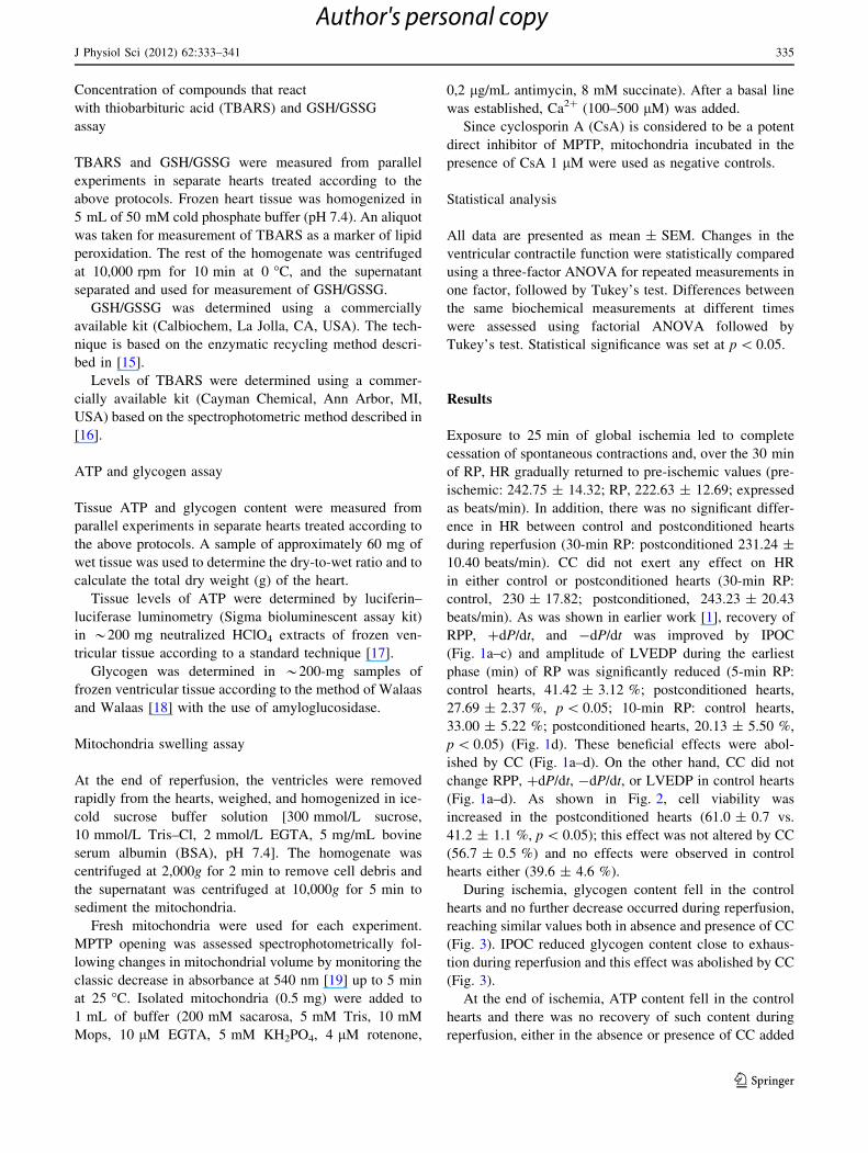
beats/min). As was shown in earlier work [1], recovery of
RPP, ?dP/dt, and -dP/dt was improved by IPOC
(Fig. 1a–c) and amplitude of LVEDP during the earliest
phase (min) of RP was significantly reduced (5-min RP:
control hearts, 41.42 ± 3.12 %; postconditioned hearts,
27.69 ± 2.37 %, p \ 0.05; 10-min RP: control hearts,
33.00 ± 5.22 %; postconditioned hearts, 20.13 ± 5.50 %,
p \ 0.05) (Fig. 1d). These beneficial effects were abol-
ished by CC (Fig. 1a–d). On the other hand, CC did not
change RPP, ?dP/dt, -dP/dt, or LVEDP in control hearts
(Fig. 1a–d). As shown in Fig. 2, cell viability was
increased in the postconditioned hearts (61.0 ± 0.7 vs.
41.2 ± 1.1 %, p \ 0.05); this effect was not altered by CC
(56.7 ± 0.5 %) and no effects were observed in control
hearts either (39.6 ± 4.6 %).
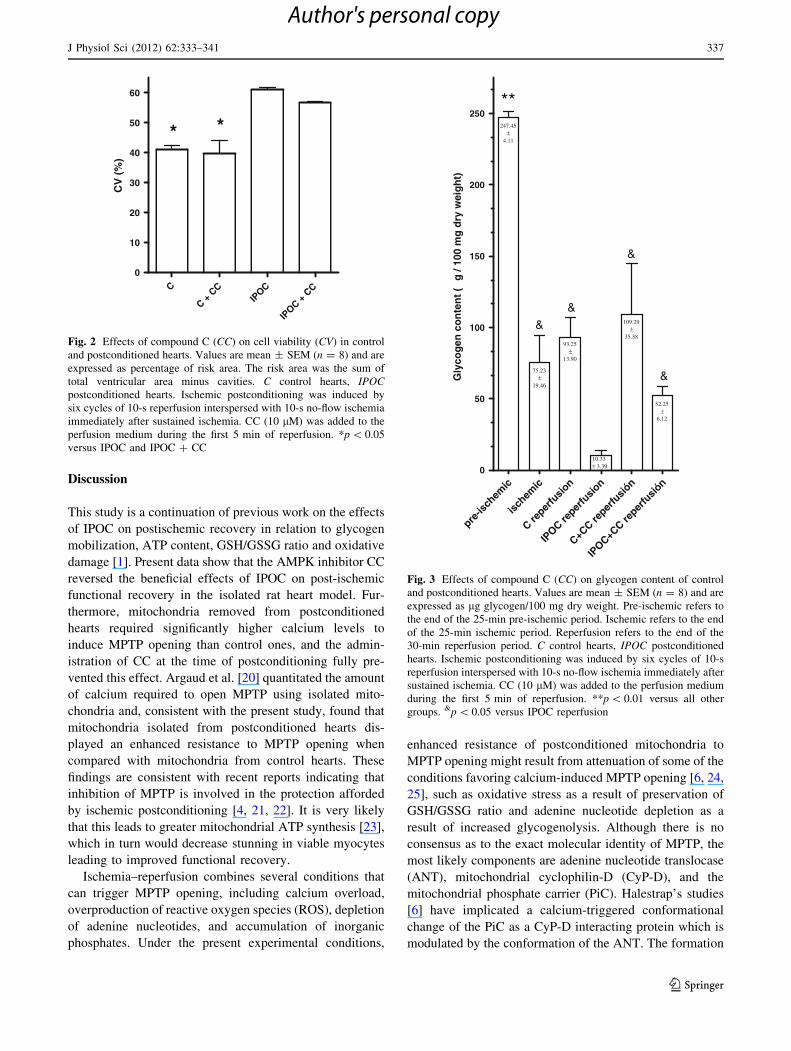
During ischemia, glycogen content fell in the control
hearts and no further decrease occurred during reperfusion,
reaching similar values both in absence and presence of CC
(Fig. 3). IPOC reduced glycogen content close to exhaus-
tion during reperfusion and this effect was abolished by CC
(Fig. 3).
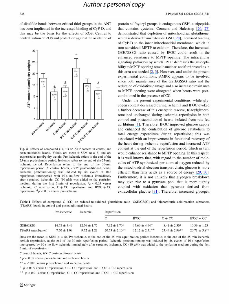
At the end of ischemia, ATP content fell in the control
hearts and there was no recovery of such content during
reperfusion, either in the absence or presence of CC added
J Physiol Sci (2012) 62:333–341 335
123
Author's personal copy
to the perfusion medium during the first 5 min of reper-
fusion (Fig. 4). IPOC markedly raised myocardial ATP
content and this beneficial effect was abolished by CC
(Fig. 4).
As shown in Table 1, ischemia–reperfusion decreased
the GSH/GSSG ratio by approximately 50 % and increased
tissue levels of TBARS by approximately 169 % both in
the absence and presence of CC. In the reperfused hearts,
IPOC significantly raised the GSH/GSSG ratio and reduced
TBARS content, and these beneficial effects elicited by
IPOC were abolished by CC (Table 1).
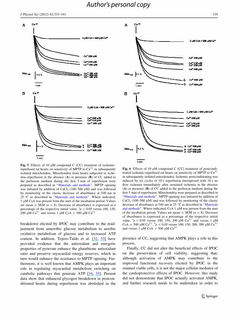
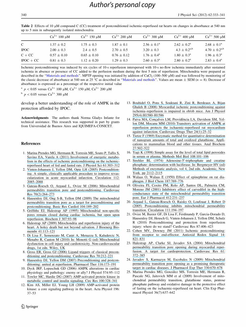
The sensitivity of the MPTP opening to calcium in
mitochondria isolated from ischemia–reperfused hearts was
studied by measuring the changes in the suspension
absorbance at 540 nm in a sucrose-based medium. Typical
traces are shown in Figs. 5 and 6, and demonstrate that
final change in absorbance up to 5 min, which reflects that
mitochondrial swelling and, thus, the sensitivity of the
mitochondrial pore, increased significantly at 400 lM
[Ca2?] in mitochondria from postconditioned hearts
(Fig. 6a) and at 300 lM [Ca2?] in mitochondria from
control hearts (Fig. 5a), suggesting that mitochondria from
postconditioned hearts were less sensitive to MPTP open-
ing than those from control hearts. Even though CC per se
did not have any effect against [Ca2?]-induced MPTP
opening (Fig. 5b), it reversed the beneficial effects of IPOC
(Fig. 6b). Quantitative and statistical analysis of final
changes in the suspension absorbance are also presented in
Table 2. The decrease in absorbance induced by 500 lM
[Ca2?] was greatly inhibited in the presence of 1 lM CsA,
suggesting that this decrease was induced by MPTP
opening.
0 10 20 30 40 50 600
10
20
30
40
50
60
70
80
90
100
*
* *
*
**
time (min)
RP
P (
%)
0 10 20 30 40 50 600
10
20
30
40
50
60
70
80
90
100
*
*
time (min)
+dP
/dt
(%)
0 10 20 30 40 50 600
10
20
30
40
50
60
70
80
90
100
110
*
*
time (min)
-dP
/dt
(%)
0 10 20 30 40 50 600
10
20
30
40
50
*
*
time (min)
LV
ED
P (
%)
ischemia reperfusion ischemia reperfusionA B
C D
Fig. 1 Effects of compound C
(CC) on a rate-pressure product
(RPP), b peak rate of
contraction (?dP/dt), c peak
rate of relaxation (-dP/dt),d left ventricular end-diastolic
pressure (LVEDP) due to
ischemia–reperfusion in control
and postconditioned hearts.
Values are expressed as a
percentage of the respective
basal value at the end of the
25-min equilibration period.
Squares control hearts. Circlespostconditioned hearts. Filledsymbols hearts perfused with
CC added to the perfusion
medium during the first 5 min
of reperfusion. Open symbolshearts perfused in the absence of
CC. Ischemic postconditioning
was induced by six cycles of
10-s reperfusion interspersed
with 10-s no-flow ischemia
immediately after sustained
ischemia. Values are
mean ± SEM (n = 8).
*p \ 0.05 versus all other
groups
336 J Physiol Sci (2012) 62:333–341
123
Author's personal copy
Discussion
This study is a continuation of previous work on the effects
of IPOC on postischemic recovery in relation to glycogen
mobilization, ATP content, GSH/GSSG ratio and oxidative
damage [1]. Present data show that the AMPK inhibitor CC
reversed the beneficial effects of IPOC on post-ischemic
functional recovery in the isolated rat heart model. Fur-
thermore, mitochondria removed from postconditioned
hearts required significantly higher calcium levels to
induce MPTP opening than control ones, and the admin-
istration of CC at the time of postconditioning fully pre-
vented this effect. Argaud et al. [20] quantitated the amount
of calcium required to open MPTP using isolated mito-
chondria and, consistent with the present study, found that
mitochondria isolated from postconditioned hearts dis-
played an enhanced resistance to MPTP opening when
compared with mitochondria from control hearts. These
findings are consistent with recent reports indicating that
inhibition of MPTP is involved in the protection afforded
by ischemic postconditioning [4, 21, 22]. It is very likely
that this leads to greater mitochondrial ATP synthesis [23],
which in turn would decrease stunning in viable myocytes
leading to improved functional recovery.
Ischemia–reperfusion combines several conditions that
can trigger MPTP opening, including calcium overload,
overproduction of reactive oxygen species (ROS), depletion
of adenine nucleotides, and accumulation of inorganic
phosphates. Under the present experimental conditions,
enhanced resistance of postconditioned mitochondria to
MPTP opening might result from attenuation of some of the
conditions favoring calcium-induced MPTP opening [6, 24,
25], such as oxidative stress as a result of preservation of
GSH/GSSG ratio and adenine nucleotide depletion as a
result of increased glycogenolysis. Although there is no
consensus as to the exact molecular identity of MPTP, the
most likely components are adenine nucleotide translocase
(ANT), mitochondrial cyclophilin-D (CyP-D), and the
mitochondrial phosphate carrier (PiC). Halestrap’s studies
[6] have implicated a calcium-triggered conformational
change of the PiC as a CyP-D interacting protein which is
modulated by the conformation of the ANT. The formation
C
C + C
CIP
OC
IPOC +
CC
0
10
20
30
40
50
60
* *C
V (
%)
Fig. 2 Effects of compound C (CC) on cell viability (CV) in control
and postconditioned hearts. Values are mean ± SEM (n = 8) and are
expressed as percentage of risk area. The risk area was the sum of
total ventricular area minus cavities. C control hearts, IPOCpostconditioned hearts. Ischemic postconditioning was induced by
six cycles of 10-s reperfusion interspersed with 10-s no-flow ischemia
immediately after sustained ischemia. CC (10 lM) was added to the
perfusion medium during the first 5 min of reperfusion. *p \ 0.05
versus IPOC and IPOC ? CC
pre-is
chem
ic
ischem
ic
C reper
fusio
n
IPOC re
perfu
sion
C+CC re
perfu
sión
IPOC+C
C reper
fusió
n0
50
100
150
200
250
&
**
&
&
&
10.33± 3.39
247.45±
4.11
75.23±
19.46
93.25±
13.90
109.29±
35.38
52.25±
6.12
Gly
cog
en c
on
ten
t ( µ
g /
100
mg
dry
wei
gh
t)
Fig. 3 Effects of compound C (CC) on glycogen content of control
and postconditioned hearts. Values are mean ± SEM (n = 8) and are
expressed as lg glycogen/100 mg dry weight. Pre-ischemic refers to
the end of the 25-min pre-ischemic period. Ischemic refers to the end
of the 25-min ischemic period. Reperfusion refers to the end of the
30-min reperfusion period. C control hearts, IPOC postconditioned
hearts. Ischemic postconditioning was induced by six cycles of 10-s
reperfusion interspersed with 10-s no-flow ischemia immediately after
sustained ischemia. CC (10 lM) was added to the perfusion medium
during the first 5 min of reperfusion. **p \ 0.01 versus all other
groups. &p \ 0.05 versus IPOC reperfusion
J Physiol Sci (2012) 62:333–341 337
123
Author's personal copy
of disulfide bonds between critical thiol groups in the ANT
has been implicated in the increased binding of CyP-D, and
this may be the basis for the effects of ROS. Central to
neutralization of ROS and protection against the oxidation of
protein sulfhydryl groups is endogenous GSH, a tripeptide
that contains cysteine. Connern and Halestrap [26, 27]
demonstrated that depletion of mitochondrial glutathione,
which is derived from cytosolic GSH [28], increased binding
of CyP-D to the inner mitochondrial membrane, which in
turn sensitized MPTP to calcium. Therefore, the increased
GSH/GSSG ratio caused by IPOC could result in the
enhanced resistance to MPTP opening. The intracellular
signaling pathways by which IPOC decreases the suscepti-
bility to MPTP opening remain unclear, and further studies in
this area are needed [2, 3]. However, and under the present
experimental conditions, AMPK appears to be involved
since both maintenance of the GSH/GSSG ratio and the
reduction of oxidative damage and also increased resistance
to MPTP opening were abrogated when hearts were post-
conditioned in the presence of CC.
Under the present experimental conditions, while gly-
cogen content decreased during ischemia and IPOC evoked
a further decrease of this energetic reserve, triacylglycerol
remained unchanged during ischemia–reperfusion in both
control and postconditioned hearts isolated from rats fed
ad libitum [1]. Therefore, IPOC improved glucose supply
and enhanced the contribution of glucose catabolism to
total energy expenditure during reperfusion; this was
associated with an improvement in functional recovery of
the heart during ischemia–reperfusion and increased ATP
content at the end of the reperfusion period, which in turn
would enhance resistance to MPTP opening. In this respect,
it is well known that, with regard to the number of mole-
cules of ATP synthesized per atom of oxygen reduced by
the mitochondrial electron transport chain, glucose is more
efficient than fatty acids as a source of energy [29, 30].
Furthermore, it is not unlikely that glycogen breakdown
may give rise to a pyruvate pool that is more tightly
coupled with oxidation than pyruvate derived from
extracellular glucose [31]. Therefore, increased glycogen
pre-is
chem
ic
ischem
ic
Cre
perfu
sion
IPOC
reper
fusio
n
C+CC
reper
fusio
n
IPOC+C
Cre
perfu
sion
0
5
10
15
20
25
30
*
&& &17.95
±2.04
12.00±
1.25
13.80±
0.95
22.45±
4.10
15.76±
1.90
12.56±
1.27
AT
P c
on
ten
t (µ
mo
l/g d
ry w
eig
ht)
Fig. 4 Effects of compound C (CC) on ATP content in control and
postconditioned hearts. Values are mean ± SEM (n = 8) and are
expressed as lmol/g dry weight. Pre-ischemic refers to the end of the
25-min pre-ischemic period. Ischemic refers to the end of the 25-min
ischemic period. Reperfusion refers to the end of the 30-min
reperfusion period. C control hearts, IPOC postconditioned hearts.
Ischemic postconditioning was induced by six cycles of 10-s
reperfusion interspersed with 10-s no-flow ischemia immediately
after sustained ischemia. CC (10 lM) was added to the perfusion
medium during the first 5 min of reperfusion. *p \ 0.05 versus
ischemic, C reperfusion, C ? CC reperfusion and IPOC ? CC
reperfusion. &p \ 0.05 versus pre-ischemic
Table 1 Effects of compound C (CC) on reduced-to-oxidized glutathione ratio (GSH/GSSG) and thiobarbituric acid-reactive substances
(TBARS) levels in control and postconditioned hearts
Pre-ischemic Ischemic Reperfusion
C IPOC C ? CC IPOC ? CC
GSH/GSSG 14.58 ± 3.49 12.76 ± 1.77 7.92 ± 1.70* 17.69 ± 4.64? 8.41 ± 2.30* 10.39 ± 1.23
TBARS (nmol/gww) 7.70 ± 1.09 9.72 ± 1.23 20.73 ± 2.10** 12.12 ± 2.51?? 23.49 ± 2.96** 20.71 ± 3.8**
Data are the mean ± SEM (n = 8). Pre-ischemic, at the end of the 25 min equilibration period; ischemic, at the end of the 25 min ischemic
period; reperfusion, at the end of the 30 min reperfusion period. Ischemic postconditioning was induced by six cycles of 10-s reperfusion
interspersed by 10-s no-flow ischemia immediately after sustained ischemia. CC (10 lM) was added to the perfusion medium during the first
5 min of reperfusion
C control hearts, IPOC postconditioned hearts
* p \ 0.05 versus pre-ischemic and ischemic hearts
** p \ 0.01 versus pre-ischemic and ischemic hearts? p \ 0.05 versus C reperfusion, C ? CC reperfusion and IPOC ? CC reperfusion?? p \ 0.01 versus C reperfusion, C ? CC reperfusion and IPOC ? CC reperfusion
338 J Physiol Sci (2012) 62:333–341
123
Author's personal copy
breakdown elicited by IPOC may contribute to the read-
justment from anaerobic glucose metabolism to aerobic
oxidative metabolism of glucose and to increased ATP
content. In addition, Tejero-Taldo et al. [32, 33] have
provided evidence that the antioxidant and energetic
properties of pyruvate enhance the glutathione antioxidant
ratio and preserve myocardial energy reserves, which in
turn would enhance the resistance to MPTP opening. Fur-
thermore, it is well known that AMPK plays an important
role in regulating myocardial metabolism switching on
catabolic pathways that generate ATP [34, 35]. Present
data show that enhanced glycogen breakdown in postcon-
ditioned hearts during reperfusion was abolished in the
presence of CC, suggesting that AMPK plays a role in this
process.
Finally, CC did not alter the beneficial effects of IPOC
on the preservation of cell viability, suggesting that,
although activation of AMPK may contribute to the
improved functional recovery elicited by IPOC in the
stunned viable cells, it is not the major cellular mediator of
the cardioprotective effects of IPOC. However, this study
did not demonstrate that IPOC actually activated AMPK,
and further research needs to be undertaken in order to
Fig. 5 Effects of 10 lM compound C (CC) treatment of ischemic-
reperfused rat hearts on sensitivity of MPTP to Ca2? in subsequently
isolated mitochondria. Mitochondria from hearts subjected to ische-
mia–reperfusion in the absence (A) or presence (B) of CC added to
the perfusion medium during the first 5 min of reperfusion were
prepared as described in ‘‘Materials and methods’’. MPTP opening
was initiated by addition of CaCl2 (100–500 lM) and was followed
by monitoring of the classic decrease of absorbance at 540 nm at
25 �C as described in ‘‘Materials and methods’’. Where indicated,
1 lM CsA was present from the start of the incubation period. Values
are mean ± SEM (n = 8). Decrease of absorbance is expressed as a
percentage of the respective initial value. ap \ 0.05 versus 100, 150,
200 lM Ca2? and versus 1 lM CsA ? 500 lM Ca2?
Fig. 6 Effects of 10 lM compound C (CC) treatment of postcondi-
tioned ischemic-reperfused rat hearts on sensitivity of MPTP to Ca2?
in subsequently isolated mitochondria. Ischemic postconditioning was
induced by six cycles of 10-s reperfusion interspersed with 10-s no
flow ischemia immediately after sustained ischemia in the absence
(A) or presence (B) of CC added to the perfusion medium during the
first 5 min of reperfusion. Mitochondria were prepared as described in
‘‘Materials and methods’’. MPTP opening was initiated by addition of
CaCl2 (100–500 lM) and was followed by monitoring of the classic
decrease of absorbance at 540 nm at 25 �C as described in ‘‘Materials
and methods’’. Where indicated, CsA 1 lM was present from the start
of the incubation period. Values are mean ± SEM (n = 8). Decrease
of absorbance is expressed as a percentage of the respective initial
value. ap \ 0.05 versus 100, 150, 200 lM Ca2? and versus 1 lM
CsA ? 500 lM Ca2?. bp \ 0.05 versus 100, 150, 200, 300 lM Ca2?
and versus 1 lM CsA ? 500 lM Ca2?
J Physiol Sci (2012) 62:333–341 339
123
Author's personal copy
develop a better understanding of the role of AMPK in the
protection afforded by IPOC.
Acknowledgments The authors thank Norma Gladys Infante for
technical assistance. This research was supported in part by grants
from Universidad de Buenos Aires and IQUIMEFA-CONICET.
References
1. Marina Prendes MG, Hermann R, Torresin ME, Souto P, Tallis S,
Savino EA, Varela A (2011) Involvement of energetic metabo-
lism in the effects of ischemic postconditioning on the ischemic-
reperfused heart of fed and fasted rats. J Physiol Sci 61:303–312
2. Vinten-Johansen J, Yellon DM, Opie LH (2005) Postcondition-
ing. A simple, clinically applicable procedure to improve revas-
cularization in acute myocardial infarction. Circulation 112:
2085–2088
3. Gateau-Roesch O, Argaud L, Ovize M (2006) Mitochondrial
permeability transition pore and postconditioning. Cardiovasc
Res 70(2):264–273
4. Hausenloy DJ, Ong S-B, Yellon DM (2009) The mitochondrial
permeability transition pore as a target for preconditioning and
postconditioning. Basic Res Cardiol 104:189–202
5. Griffiths EJ, Halestrap AP (1995) Mitochondrial non-specific
pores remain closed during cardiac ischemia, but open upon
reperfusion. Biochem J 307:93–98
6. Halestrap AP (2009) Mitochondria and reperfusion injury of the
heart. A holey death but not beyond salvation. J Bioenerg Bio-
membr 41:113–121
7. Di Lisa F, Semenzato M, Carpi A, Menazza S, Kaludercic N,
Menabo R, Canton M (2010) In: Monotti G (ed) Mitochondrial
dysfunction in cell injury and cardiotoxicity. Non-cardiovascular
drugs, 1st edn. Wiley, UK
8. Gross ER, Gross GJ (2006) Ligand triggers of classical precon-
ditioning and postconditioning. Cardiovasc Res 70:212–221
9. Hausenloy DJ, Yellon DM (2007) Preconditioning and postcon-
ditioning: united at reperfusion. Pharmacol Ther 116:173–191
10. Dyck JRP, Lopaschuk GD (2006) AMPK alterations in cardiac
physiology and pathology: enemy or ally? J Physiol 574:95–112
11. Towler MC, Hardie DG (2007) AMP-activated protein kinase in
metabolic control and insulin signaling. Circ Res 100:328–341
12. Kim AS, Miller EJ, Young LH (2009) AMP-activated protein
kinase: a core signaling pathway in the heart. Acta Physiol 196:
37–53
13. Bouhidel O, Pons S, Souktani R, Zini R, Berdeaux A, Bijan
Ghaleh B (2008) Myocardial ischemic postconditioning against
ischemia–reperfusion is impaired in ob/ob mice. Am J Physiol
295(4):H1580–H1586
14. Paiva MA, Concalves LM, Providencia LA, Davidson SM, Yel-
lon DM, Mocanu MM (2010) Transitory activation of AMPK at
reperfusion protects the ischaemic-reperfused rat myocardium
against infarction. Cardiovasc Drugs Ther 24(1):25–32
15. Tietze F (1969) Enzymatic method for quantitative determination
of nanogram amounts of total and oxidized glutathione. Appli-
cations to mammalian blood and other tissues. Anal Biochem
27:502–522
16. Yagi K (1998) Simple assay for the level of total lipid peroxides
in serum or plasma. Methods Mol Biol 108:101–106
17. Strehler BL (1974) Adenosine-50-triphosphate and creatine
phosphate: determination with luciferase. In: Bergmeyer HV (ed)
Methods of enzymatic analysis, vol 4, 2nd edn. Academic, New
York. pp 2112–2115
18. Walaas O, Walaas E (1950) Effect of epinephrine on rat dia-
phragm. J Biol Chem 187:769–776
19. Oliveira PJ, Coxito PM, Rolo AP, Santos DL, Palmeira CM,
Moreno JM (2001) Inhibitory effect of carvedilol in the high-
conductance state of the mitochondrial permeability transition
pore. Eur J Pharmacol 412:231–237
20. Argaud L, Gateau-Roesch O, Raisky O, Loufouat J, Robert D
(2005) Postconditioning inhibits mitochondrial permeability
transition. Circulation 111:194–197
21. Ovize M, Baxter GF, Di Lisa F, Ferdinandy P, Garcia-Dorado D,
Hausenloy DJ, Heusch G, Vinten-Johansen J, Yellon DM, Schulz
R (2010) Postconditioning and protection from reperfusion
injury: where do we stand? Cardiovasc Res 87:406–423
22. Cohen MV, Downey JM (2011) Ischemic postconditioning:
from receptor to end-effector. Antioxid Redox Signal 14:
821–831
23. Halestrap AP, Clarke SJ, Javadov SA (2004) Mitochondrial
permeability transition pore opening during myocardial reper-
fusion. A target for cardioprotection. Cardiovasc Res 61:
372–385
24. Javadov S, Karmazyn M, Escobales N (2009) Mitochondrial
permeability transition pore opening as a promising therapeutic
target in cardiac diseases. J Pharmacol Exp Ther 330:670–678
25. Marina Prendes MG, Gonzalez MS, Torresin ME, Hermann R,
Pascale NG, Jaitovich MM et al (2009) Involvement of mito-
chondrial permeability transition, glutathione status, pentose
phosphate pathway and oxidative damage in the protective effect
of fasting on the ischaemic-reperfused rat heart. Clin Exp Phar-
macol Physiol 36(7):637–642
Table 2 Effects of 10 lM compound C (CC) treatment of postconditioned ischemic-reperfused rat hearts on changes in absorbance at 540 nm
up to 5 min in subsequently isolated mitochondria
Ca2? 100 lM Ca2? 150 lM Ca2? 200 lM Ca2? 300 lM Ca2? 400 lM Ca2? 500 lM
C 1.37 ± 0.2 1.75 ± 0.3 1.87 ± 0.1 2.56 ± 0.1a 2.62 ± 0.2a 2.68 ± 0.1a
IPOC 2.00 ± 0.3 2.4 ± 0.5 2.70 ± 0.5 3.20 ± 0.3 4.3 ± 0.2a,b 4.70 ± 0.2a,b
C ? CC 0.57 ± 0.10 0.65 ± 0.10 0.76 ± 0.12 1.76 ± 0.4a 1.80 ± 0.3a 1.96 ± 0.3a
IPOC ? CC 0.81 ± 0.3 1.12 ± 0.20 1.29 ± 0.3 2.60 ± 0.3a 2.80 ± 0.2a 2.83 ± 0.4a
Ischemic postconditioning was induced by six cycles of 10-s reperfusion interspersed with 10-s no-flow ischemia immediately after sustained
ischemia in absence or presence of CC added to the perfusion medium during the first 5 min of reperfusion. Mitochondria were prepared as
described in the ‘‘Materials and methods’’. MPTP opening was initiated by addition of CaCl2 (100–500 lM) and was followed by monitoring of
the classic decrease of absorbance at 540 nm at 25 �C as described in ‘‘Materials and methods’’. Values are mean ± SEM (n = 8). Decrease of
absorbance is expressed as a percentage of the respective initial valuea p \ 0.05 versus Ca2? 100 lM, Ca2? 150 lM, Ca2? 200 lMb p \ 0.05 versus Ca2? 300 lM
340 J Physiol Sci (2012) 62:333–341
123
Author's personal copy
26. Connern CP, Halestrap AP (1994) Recruitment of mitochon-
drial cyclophilin to the mitochondrial inner membrane under
conditions of oxidative stress that enhance the opening of a cal-
cium-sensitive non-specific channel. Biochem J 302:321–324
27. Connern CP, Halestrap AP (1996) Chaotropic agents and
increased matrix volume enhance binding of mitochondrial
cyclophilin to the inner mitochondrial membrane and sensitize
the mitochondrial permeability transition to [Ca2?]. Biochemistry
35:8172–8180
28. Chen Z, Laser LH (1998) Evidence for mitochondrial uptake of
glutathione dicarboxylate and 2-oxoglutarate carriers. J Pharma-
col Exp Ther 285:608–618
29. Stanley WC, Lopaschuk GD, Hall JL, McCormack JG (1997)
Regulation of myocardial carbohydrate metabolism under normal
and ischaemic conditions. Potential for pharmacological inter-
ventions. Cardiovasc Res 33:243–257
30. Ussher JR, Lopaschuk GD (2009) Targeting malonyl CoA inhi-
bition of mitochondrial fatty acid uptake as an approach to treat
cardiac ischemia/reperfusion. Basic Res Cardiol 104:203–210
31. Goodwin GW, Taylor CS, Taegtmeyer H (1998) Regulation of
energy metabolism of the heart during acute increase in heart
work. J Biol Chem 273:29530–29539
32. Tejero-Taldo MI, Caffrey JL, Sun J, Mallet RT (1998) Pyruvate
potentiates b-adrenergic inotropism of stunned guinea-pig myo-
cardium. J Mol Cell Cardiol 30:2327–2339
33. Tejero-Taldo MI, Caffrey JL, Sun J, Mallet RT (1999) Antioxi-
dant properties of pyruvate mediates its potentiation of b-adren-
ergic inotropism in stunned myocardium. J Mol Cell Cardiol
31:1863–1872
34. Arad M, Seidman CE, Seidman JG (2007) AMP-activated protein
kinase in the heart. Role during health and disease. Circ Res
100:474–488
35. Carling D, Hardie DG (1989) The substrate and sequence spec-
ificity of the AMP-activated protein kinase. Phosphorylation of
glycogen synthase and phosphorylase kinase. Biochim Biophys
Acta 1012:81–86
J Physiol Sci (2012) 62:333–341 341
123
Author's personal copy
Related Documents


