Effect of Strain Magnitude on the Tissue Properties of Engineered Cardiovascular Constructs RALF A. BOERBOOM,MIRJAM P. RUBBENS,NIELS J. B. DRIESSEN,CARLIJN V. C. BOUTEN, and FRANK P. T. BAAIJENS Department of Biomedical Engineering, Soft Tissue Biomechanics and Engineering, Eindhoven University of Technology, PO Box 513, 5600 MB, Eindhoven, The Netherlands (Received 24 April 2007; accepted 26 November 2007; published online 8 December 2007) Abstract—Mechanical loading is a powerful regulator of tissue properties in engineered cardiovascular tissues. To ultimately regulate the biochemical processes, it is essential to quantify the effect of mechanical loading on the properties of engineered cardiovascular constructs. In this study the Flexercell FX-4000T (Flexcell Int. Corp., USA) straining system was modified to simultaneously apply various strain magnitudes to individual samples during one experiment. In addition, porous polyglycolic acid (PGA) scaffolds, coated with poly-4-hydroxybutyrate (P4HB), were partially embed- ded in a silicone layer to allow long-term uniaxial cyclic mechanical straining of cardiovascular engineered constructs. The constructs were subjected to two different strain mag- nitudes and showed differences in biochemical properties, mechanical properties and organization of the microstructure compared to the unstrained constructs. The results suggest that when the tissues are exposed to prolonged mechanical stimulation, the production of collagen with a higher fraction of crosslinks is induced. However, straining with a large strain magnitude resulted in a negative effect on the mechanical properties of the tissue. In addition, dynamic straining induced a different alignment of cells and collagen in the superficial layers compared to the deeper layers of the construct. The presented model system can be used to systematically optimize culture protocols for engineered cardiovascular tissues. Keywords—Tissue engineering, Collagen organization, Cell orientation, Biochemical properties, Mechanical properties. INTRODUCTION Load bearing (soft) tissues are composed of a highly organized extracellular matrix (ECM), which pri- marily consists of collagen, elastin and proteoglycans. Predominantly, the collagen architecture (i.e., content, crosslinks, and orientation) determines the mechanical behavior of these tissues. 3,8 In addition, model studies show that the collagen organization is strongly cou- pled to the mechanical loading condition of these tissues. 9,16 Several load bearing tissues become dys- functional and need replacement. Frequently replaced load bearing cardiovascular soft tissues include coro- nary arteries and heart valves. 49 The currently used replacement strategies all have several shortcom- ings 35,45 and the most important drawback of the replacement tissue is the inability to remodel in re- sponse to the dynamic biological environment. Tissue engineering (TE) is a promising technique that has the potential to overcome these shortcomings. However, engineered tissues often lack sufficient amounts of properly organized ECM and consequently do not meet mechanical demands. An often applied technique in TE comprises the use of autologous cell sources and biodegradable carrier materials (synthetic or biological). These tissue engi- neered constructs are most often mechanically condi- tioned and are ultimately placed in the human body. Successful attempts have been performed to create strong vessels by engineering vessels using the self assembled vascular graft approach, 34 growing vessels in the recipients peritoneal cavity 12 and using a porous biodegradable scaffold seeded with cells. 26,41 Tissue engineered heart valves have been cultured using either a biological scaffold that will attract endogenous cells 1,43 or using a biodegradable synthetic scaffold on which cells are seeded. 19,25 Recently, Mol et al. 38 have cultured tissue engineered heart valves that demon- strated sufficient mechanical strength for placement at the aortic position. The valves were cultured using mechanical stimulation of cell seeded scaffolds in a bioreactor by mimicking the diastolic phase of the heart cycle. However, the mechanical stimulation Address correspondence to Ralf A. Boerboom, Department of Biomedical Engineering, Soft Tissue Biomechanics and Engineering, Eindhoven University of Technology, PO Box 513, 5600 MB, Eind- hoven, The Netherlands. Electronic mail: [email protected] Annals of Biomedical Engineering, Vol. 36, No. 2, February 2008 (Ó 2007) pp. 244–253 DOI: 10.1007/s10439-007-9413-8 0090-6964/08/0200-0244/0 Ó 2007 The Author(s) 244

Welcome message from author
This document is posted to help you gain knowledge. Please leave a comment to let me know what you think about it! Share it to your friends and learn new things together.
Transcript
Effect of Strain Magnitude on the Tissue Properties of Engineered
Cardiovascular Constructs
RALF A. BOERBOOM, MIRJAM P. RUBBENS, NIELS J. B. DRIESSEN, CARLIJN V. C. BOUTEN, andFRANK P. T. BAAIJENS
Department of Biomedical Engineering, Soft Tissue Biomechanics and Engineering, Eindhoven University of Technology,PO Box 513, 5600 MB, Eindhoven, The Netherlands
(Received 24 April 2007; accepted 26 November 2007; published online 8 December 2007)
Abstract—Mechanical loading is a powerful regulator oftissue properties in engineered cardiovascular tissues. Toultimately regulate the biochemical processes, it is essential toquantify the effect of mechanical loading on the properties ofengineered cardiovascular constructs. In this study theFlexercell FX-4000T (Flexcell Int. Corp., USA) strainingsystem was modified to simultaneously apply various strainmagnitudes to individual samples during one experiment. Inaddition, porous polyglycolic acid (PGA) scaffolds, coatedwith poly-4-hydroxybutyrate (P4HB), were partially embed-ded in a silicone layer to allow long-term uniaxial cyclicmechanical straining of cardiovascular engineered constructs.The constructs were subjected to two different strain mag-nitudes and showed differences in biochemical properties,mechanical properties and organization of the microstructurecompared to the unstrained constructs. The results suggestthat when the tissues are exposed to prolonged mechanicalstimulation, the production of collagen with a higher fractionof crosslinks is induced. However, straining with a largestrain magnitude resulted in a negative effect on themechanical properties of the tissue. In addition, dynamicstraining induced a different alignment of cells and collagenin the superficial layers compared to the deeper layers of theconstruct. The presented model system can be used tosystematically optimize culture protocols for engineeredcardiovascular tissues.
Keywords—Tissue engineering, Collagen organization,
Cell orientation, Biochemical properties, Mechanical
properties.
INTRODUCTION
Load bearing (soft) tissues are composed of a highlyorganized extracellular matrix (ECM), which pri-marily consists of collagen, elastin and proteoglycans.
Predominantly, the collagen architecture (i.e., content,crosslinks, and orientation) determines the mechanicalbehavior of these tissues.3,8 In addition, model studiesshow that the collagen organization is strongly cou-pled to the mechanical loading condition of thesetissues.9,16 Several load bearing tissues become dys-functional and need replacement. Frequently replacedload bearing cardiovascular soft tissues include coro-nary arteries and heart valves.49 The currently usedreplacement strategies all have several shortcom-ings35,45 and the most important drawback of thereplacement tissue is the inability to remodel in re-sponse to the dynamic biological environment. Tissueengineering (TE) is a promising technique that has thepotential to overcome these shortcomings. However,engineered tissues often lack sufficient amounts ofproperly organized ECM and consequently do notmeet mechanical demands.
An often applied technique in TE comprises the useof autologous cell sources and biodegradable carriermaterials (synthetic or biological). These tissue engi-neered constructs are most often mechanically condi-tioned and are ultimately placed in the human body.Successful attempts have been performed to createstrong vessels by engineering vessels using the selfassembled vascular graft approach,34 growing vesselsin the recipients peritoneal cavity12 and using a porousbiodegradable scaffold seeded with cells.26,41 Tissueengineered heart valves have been cultured using eithera biological scaffold that will attract endogenouscells1,43 or using a biodegradable synthetic scaffold onwhich cells are seeded.19,25 Recently, Mol et al.38 havecultured tissue engineered heart valves that demon-strated sufficient mechanical strength for placement atthe aortic position. The valves were cultured usingmechanical stimulation of cell seeded scaffolds in abioreactor by mimicking the diastolic phase of theheart cycle. However, the mechanical stimulation
Address correspondence to Ralf A. Boerboom, Department of
Biomedical Engineering, Soft Tissue Biomechanics and Engineering,
Eindhoven University of Technology, PO Box 513, 5600 MB, Eind-
hoven, The Netherlands. Electronic mail: [email protected]
Annals of Biomedical Engineering, Vol. 36, No. 2, February 2008 (� 2007) pp. 244–253
DOI: 10.1007/s10439-007-9413-8
0090-6964/08/0200-0244/0 � 2007 The Author(s)
244
protocol for cardiovascular engineered constructs canbe further improved to ultimately approach themechanical properties of the native tissue (e.g.,anisotropy) and reduce culture times.
The effect of mechanical conditioning on thebiosynthetic activity of cells has been studied both intwo- and three-dimensional culture conditions and thiseffect showed to be dependent on the nature of theECM.48 Cells have been stimulated by using a varietyof culture systems such as longitudinal stretchingdevices and vacuum driven devices.11 Several studieshave been performed looking at the effect of mechan-ical stimulation on tissue remodeling in complexgeometries.25,38 Similar experiments in well definedsimple geometries were performed.2,19,36,46 Seliktaret al.46,47 cultured fibroblast populated collagen gels(tubular constructs) in the presence of accuratelycontrolled deformation. Only a limited amount ofstudies have focused on the effect of well definedmechanical loading on the properties of engineeredcardiovascular tissues,36,41 in which de novo formationof ECM is studied (e.g., polyglycolic acid (PGA)scaffolds seeded with cells). Mechanical control oftissue properties in engineered constructs is desired.This requires a detailed study on the effects of welldefined loading conditions on ECM synthesis andECM organization in order to optimize the loadingregimen.
The first aim of this paper was to develop a modelsystem, which allowed long term dynamic loading ofcardiovascular engineered constructs. Previously,PGA in combination with cells deformed plasticallyand straining of the constructs required continuousadjustment of the applied strain.29,36 By supportingthe scaffolds with a silicone layer, a technique wasdeveloped that enabled continuous straining of engi-neered cardiovascular constructs. Local tissue strainswere measured during culture using digital imagecorrelation (DIC). These constructs, consisting ofPGA coated with poly-4-hydroxybutyrate (P4HB)and seeded with human saphenous vein cells (HSVC),were attached to flexible membranes, which werestrained using a Flexercell FX-4000T (Flexcell Int.Corp., USA) straining system.4,21 The system wasmodified to apply various strain magnitudes to indi-vidual samples. The second aim of this paper was toinvestigate the influence of different strain magnitudeson ECM organization. Different strain magnitudeswere applied to cardiovascular engineered constructsin order to demonstrate the effect of mechanicalloading on the ECM organization. The culturedconstructs were analyzed biochemically and themicrostructure was visualized using multiphotonmicroscopy.
MATERIALS AND METHODS
Straining System
The straining system consisted of a modified versionof a Flexercell FX-4000T (Flexcell International,USA). This setup allowed easy access to straining ofcells and tissue constructs using a vacuum controlleddeformation of Bioflex (Flexcell International, USA)sixwell plates.21 The original Flexercell system con-trolled the strain magnitude by the amount of vacuumthat was applied. This system was modified in order toapply various strain magnitudes to individual samplesby controlling the amount of membrane displacementwhen a vacuum was applied.
In Fig. 1a a schematic overview of the modifiedstraining setup is shown. When a vacuum is applied tothe flexible membrane of the Bioflex plate, the mem-brane will deform at the locations where it is notsupported by the loading post. Polycarbonate rings ofvarying height were placed around the original loadingposts. When a maximum vacuum is applied in thepresence of the polycarbonate rings, the rings limit thedeformation of the flexible silicone membrane. Byvarying the height of these rings the deformation of themembrane can be varied.
The applied strain fields were validated by usingDIC. A random dot pattern was sprayed on the flexiblesilicone membrane of the Bioflex wells. These mem-branes were deformed by applying a square waveformwith a magnitude of maximum vacuum pressure at afrequency of 1 Hz. During loading, images of the de-formed state were recorded at 60 frames per secondusing a Phantom v5.1 high speed camera (Vision Re-search Inc., USA). The recorded images were analysedusing Aramis DIC software (Gom mbh., Germany).Strain fields were calibrated for three specific ringheights (8.16, 7.47, and 7.05 mm), corresponding toapproximately 4, 8, and 12% strain (n = 6).
Straining PGA Based Constructs
The scaffold consisted of non-woven PGA scaffold(density 72.76 mg/cm3; Cellon, Luxemburg), whichwas coated with 1% (w/v) poly-4-hydroxybutyrate(P4HB; Symetis Inc., Switzerland) dissolved in tetra-hydrofuran (THF; Merck, Germany).25 The scaffoldwas supported by a 0.5 mm thick layer of elastic sili-cone (Fig. 1b), which was created by partly pressingthe PGA scaffold into uncured silicone (SilasticMDX4-4210, Dow Corning, USA). After curing,rectangular scaffolds were cut (34 · 5 · 1 mm3) andthe constructs were attached at the outer 5 mm to theflexible membrane by using Silastic MDX4-4210(Fig. 1b). The strain applied to the constructs was
Effect of Strain Magnitude on the Tissue Properties 245
quantified (n = 6) after 2 weeks of dynamic cultureusing a similar protocol as was used for the membranestrains in the section ‘‘Straining System’’.
Cell and Tissue Culture
Human saphenous vein cells (HSVC, myofibro-blasts) were acquired from a 44-year-old woman andwere grown using regular cell culture methods.44 Thecells were expanded in medium consisting of advancedDulbecco’s Modified Eagle Medium (DMEM; Gibco,USA) supplemented with 10% fetal bovine serum(FBS; Greiner Bio one, The Netherlands), 1%l-glutamax (Gibco, USA) and 0.1% gentamycin (Bio-chrom, Germany).
The scaffolds were vacuum dried for 48 h, followedby exposure to ultra violet light for 1 h and weresubsequently placed in 70% ethanol for 4–5 h to ob-tain sterility. The scaffolds were allowed to dry over-night, followed by washing three times with phosphatebuffered saline (PBS; Sigma, USA). Prior to cell seed-ing, tissue culture medium was added to the scaffoldsfor 16 h to facilitate cell attachment. HSVC cells atpassage 7 were seeded on these scaffolds using fibringel (Sigma, Germany) as a cell carrier.37 The cells wereseeded at a concentration of approximately 20 millioncells per cm3 and the tissue constructs were subse-quently cultured in tissue culture medium (at 37 �Cand 5% CO2). The tissue culture medium was changedevery 3–4 days and consisted of advanced DMEM(Gibco, USA) supplemented with 10% FBS, 1% l-glutamax, 0.3% gentamycin and L-ascorbic acid 2-phosphate (0.25 mg/L; Sigma, USA).
Straining Protocol
The engineered cardiovascular constructs werecultured for 3 weeks, comprising 6 days of no appliedloading to allow the cells to adapt after seeding,
followed by 2 weeks of dynamic straining at a fre-quency of 1 Hz. Three different strain conditions wereapplied: 0% strain, 4% dynamic strain and 8% dy-namic strain (n = 8). The measurements of the appliedstrain were performed on separate samples strained at4 and 8% strain for 2 and 3 weeks (n = 6).
Mechanical Testing
The constructs were sacrificed after 3 weeks of cul-ture and were tested for their mechanical propertieswithin 1 h (n = 6). The silicone layer was removedfrom the samples and the remaining tissues were placedin tissue culture medium to moisturize the samples.Thickness and width of the samples were determinedusing a Pll 2300 non contact optical image profiler(Sensofar Tech S.L., Spain). A representative areameasuring 8.35 · 7.85 mm2 was scanned using a 5·objective (Nikon, Japan) at a scanspeed of 1·. Thick-ness and width were obtained by averaging over therepresentative area.
Uniaxial tensile tests in the longitudinal directionwere performed on the engineered tissues using a ten-sile stage (Kammrath & Weiss Gmbh, Germany)equipped with a 20 N loadcell. The tissues were testedat a constant strainrate of 1/60 s-1 and were testeduntil break. Simultaneously, the force and elongationwere measured, which were converted to Cauchy stressand strain, respectively. The linear slope of theresulting curve was defined as the tangent stiffness ofthe sample.
Biochemical Assays on Tissue Formation
The tissues that were used for mechanical testing(n = 6) were subsequently analyzed with biochemicalassays for the amount of DNA, glycosaminoglycan(GAG), collagen and hydroxylysyl pyridinoline (HP)cross-links. The amount GAG and hydroxyproline
FIGURE 1. (a) Schematic cross-section of modified Flexcell system. This schematic shows the polycarbonate rings (gray) placedaround the loading post, which limit the deformation of the membrane when a vacuum is applied. (b) Reinforcement of thepolyglycolic acid scaffold with an elastic silicone layer. In the upper part a longitudinal cross-section of the PGA/P4HB scaffoldembedded in an elastic silicone layer. A layer of 0.5 mm thick, consisting of MDX4-4210 (Dow Corning, USA). In the lower partrectangular scaffolds (34 · 5 mm2 · ±1–1.2 mm) were attached to Bioflex culture wells (Flexcell Int. Corp., USA) by applying MDX4-4210 to the outer 5 mm of these constructs.
BOERBOOM et al.246
(Hyp) were expressed per lg DNA and the amount ofDNA was expressed per mg dryweight. Furthermore,the amount of collagen cross-links was normalized percollagen triple helix.
The lyophelized samples were digested in papainsolution (100 mM phosphate buffer, 5 mM L-cystein,5 mM ethylenediaminetetraacetic acid (EDTA) and125–140 lg papain per mL) at 60 �C for 16 h. Thesamples were then centrifuged and subsequentlyanalysed. A portion of the supernatant was used forthe DNA assay, a portion was used for the GAGassay and another portion was used for Hyp andcross-link analysis. The amount of DNA was deter-mined using the Hoechst dye method.13 In short, TEbuffer (10 mM Tris, 1 mM EDTA, pH 7.4) was usedto dilute the samples and 100 lL was pipetted into ablack 96 wells plate (Corning, USA). An equalamount of working solution containing the Hoechstdye (10 mM Tris, 1 mM EDTA, 2 M NaCl and2.5 lg Hoechst dye per mL) was added to each well.To allow binding of the Hoechst dye to the DNA, theplate was incubated at room temperature for 10 minand was protected from light. Subsequently, thefluorescence was measured (excitation wavelength355 nm, emission wavelength 460 nm). The amountof DNA in the samples was determined from astandard curve prepared from calf thymus DNA(Sigma, USA).
The GAG content was determined using a modifi-cation of the assay described by Farndale et al.20
Briefly, 40 lL of each sample was pipetted in duplointo a flat bottom 96 wells plate. 150 lL of DMMBcolor reagens (46 lM dimethylmethylene blue,40.5 mM glycin, 40.5 mM NaCl, pH 3.0) was added toeach well and the plate was gently shaken. Theabsorbencies at 540 and 595 nm were read within5–10 min and extracted from one another. The amountof GAG in the samples was determined from a stan-dard curve prepared from chrondroitin sulfate of sharkcartilage (Sigma, USA).
Digested tissue samples were hydrolyzed by adding25% HCl (Merck, Germany) to 200 lL of samplevolume to obtain a final concentration of 6 M HCl.These samples were hydrolysed at 110 �C for 22 h andwere subsequently used for amino acid and cross-linkanalyses. Hyp residues were measured on the acidhydrolysates by reverse-phase high-performance liquidchromatography (HPLC) after derivatization with9-fluorenylmethyl chloroformate (FMOC, Fluka,Switzerland) as previously described by Bank et al.6
Residues of the same samples were used to measurethe number of HP cross-links using HPLC as des-cribed by Bank et al.5 The amount of Hyp was con-verted to the amount of collagen using a conversionfactor of 7.46.40
Histology and Multiphoton Microscopy
One sample per group was used for histology. Tis-sues were fixed in phosphate-buffered formalin (Fluka,USA) and embedded in paraffin. 10 lm transversesections were cut and these sections were stained withHaematoxylin and Eosin (H&E) to study general tissueformation.
In addition, one living sample per group was stainedfor multiphoton microscopy. Cell Tracker BlueCMAC (CTB; Invitrogen, the Netherlands) andCNA35-OG48810,32 were used as specific fluorescentmarkers for cell cytoplasm and collagen, respectively.CTB and CNA35-OG488 were excitable with two-photon microscopy and exhibited broad emissionspectra at 466 and 520 nm. Labeling of the tissue wasperformed with tissue culture medium, which wassupplemented with CTB (15.0 lM) and CNA35-OG488 (2.0 lM). The CTB solution was applied for5 h, followed by CNA35-OG488 (3.0 lM) for 16 h.The samples were then placed in tissue culture med-ium. An inverted Zeiss Axiovert 200 microscope (CarlZeiss, Germany) coupled to an LSM 510 Meta (CarlZeiss, Germany) laser scanning microscope was usedto image the tissue engineered construct. A chameleonultra 140 fs pulsed Ti:Sapphire laser (Coherent, USA),was tuned to 760 nm to image the applied fluorescentprobes. A 63· water-immersion objective (1.2 N.A.;Carl Zeiss, Germany) was used and the channels forthe two photo multiplier tubes (PMT) were defined asfollows: 435–485 nm, CTB (PMT1) and 500–530 nm,CNA35-OG488 (PMT2). Separate images wereobtained from each PMT (coded blue and green,respectively) and combined into a single image.
Statistics
Quantitative data are represented as mean ±standard deviation (Table 1), except for graphical rep-resentations, where data are depicted as mean ±standard error of the mean (SEM, n = 6). The datawere analysed with an analysis of variance (ANOVA),followed by a Bonferroni post-hoc test to indicate sig-nificant differences between experimental groups (SPSS12.01, SPSS Inc., USA).
RESULTS
Strain Field Characterization
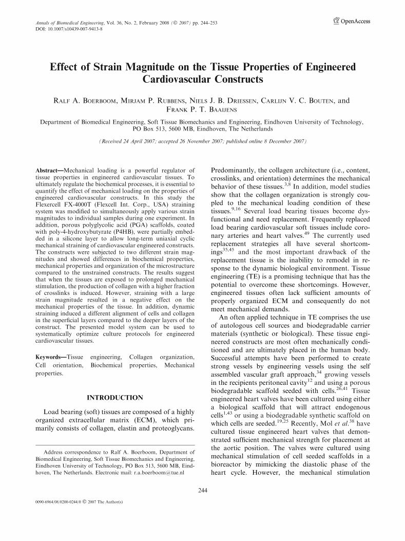
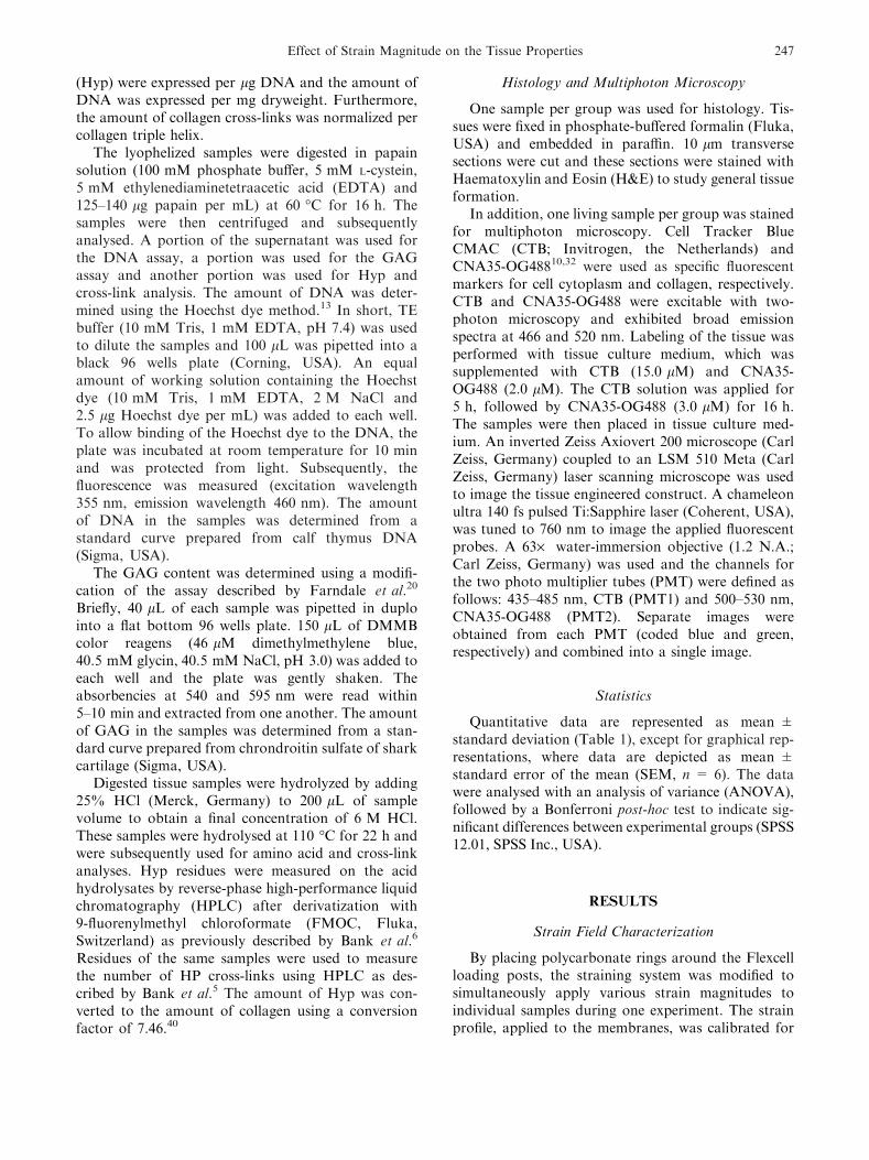
By placing polycarbonate rings around the Flexcellloading posts, the straining system was modified tosimultaneously apply various strain magnitudes toindividual samples during one experiment. The strainprofile, applied to the membranes, was calibrated for
Effect of Strain Magnitude on the Tissue Properties 247
three different ring heights (8.16, 7.47, and 7.05 mm)using round loading post geometries and showedreproducible homogeneous strain fields (Fig. 2).The average strains (%) and standard deviationwere 3.84 ± 0.61, 8.08 ± 0.72 and 12.48 ± 0.45,respectively.
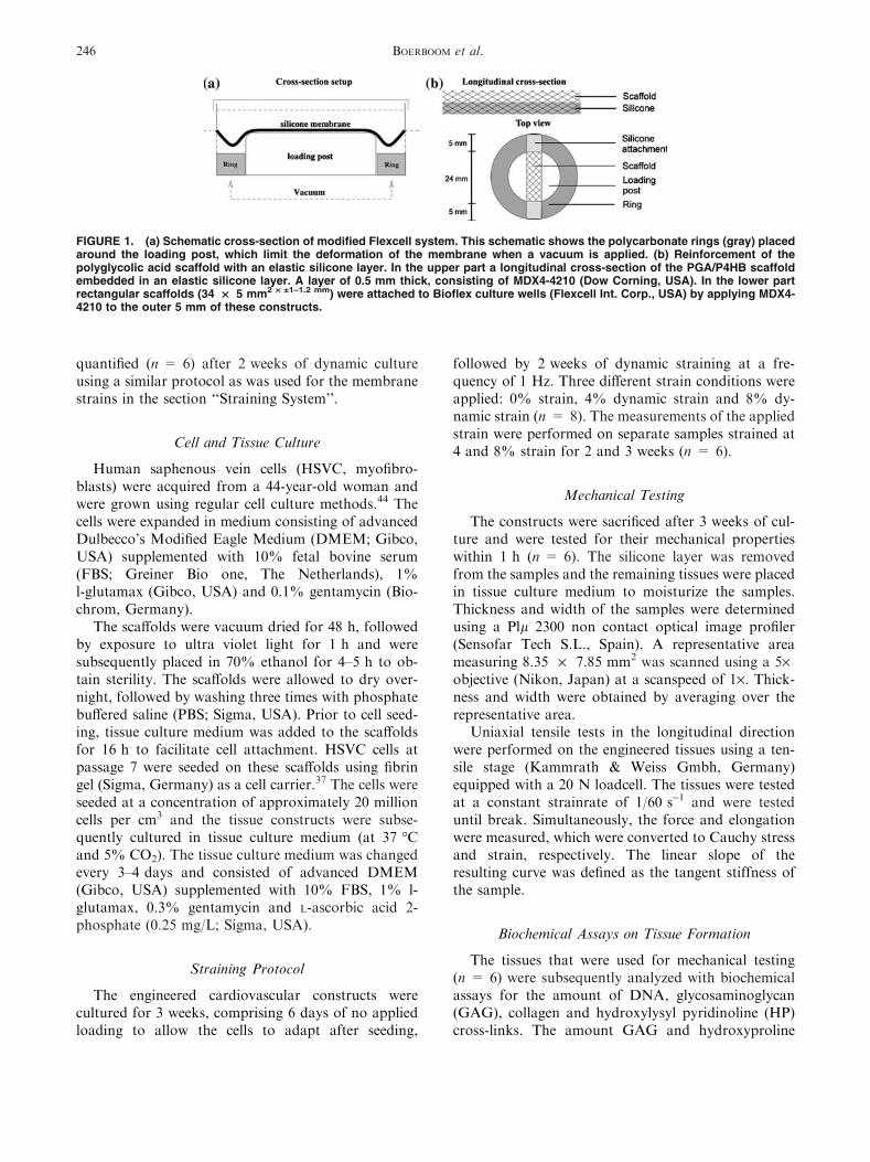
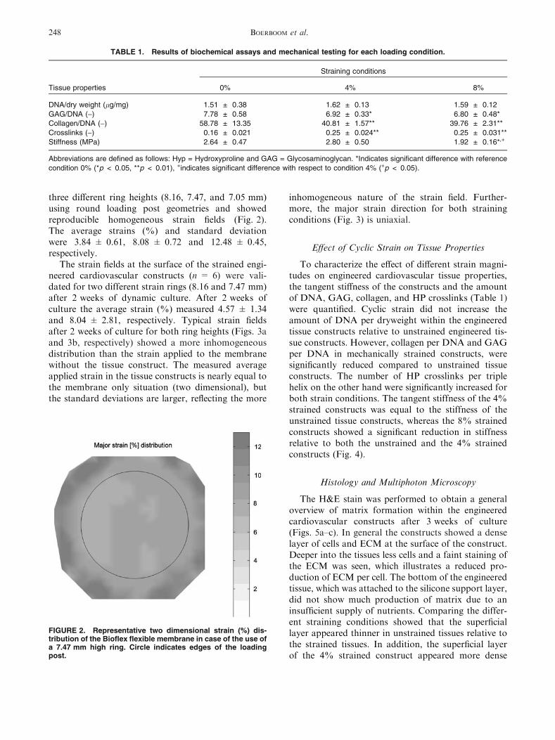
The strain fields at the surface of the strained engi-neered cardiovascular constructs (n = 6) were vali-dated for two different strain rings (8.16 and 7.47 mm)after 2 weeks of dynamic culture. After 2 weeks ofculture the average strain (%) measured 4.57 ± 1.34and 8.04 ± 2.81, respectively. Typical strain fieldsafter 2 weeks of culture for both ring heights (Figs. 3aand 3b, respectively) showed a more inhomogeneousdistribution than the strain applied to the membranewithout the tissue construct. The measured averageapplied strain in the tissue constructs is nearly equal tothe membrane only situation (two dimensional), butthe standard deviations are larger, reflecting the more
inhomogeneous nature of the strain field. Further-more, the major strain direction for both strainingconditions (Fig. 3) is uniaxial.
Effect of Cyclic Strain on Tissue Properties
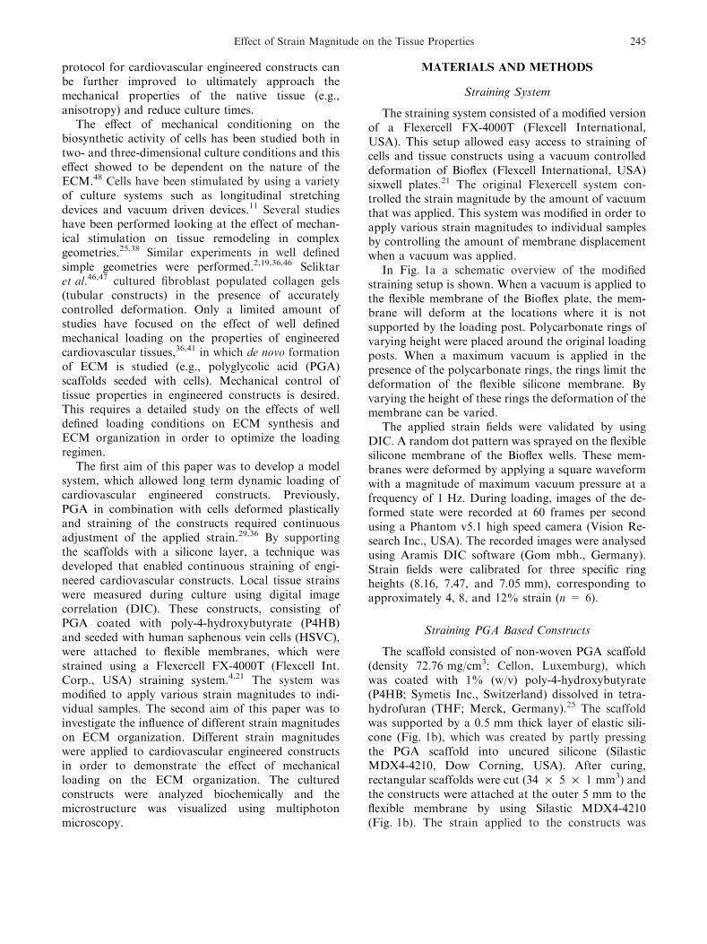
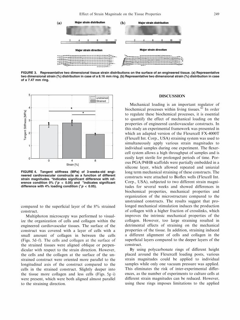
To characterize the effect of different strain magni-tudes on engineered cardiovascular tissue properties,the tangent stiffness of the constructs and the amountof DNA, GAG, collagen, and HP crosslinks (Table 1)were quantified. Cyclic strain did not increase theamount of DNA per dryweight within the engineeredtissue constructs relative to unstrained engineered tis-sue constructs. However, collagen per DNA and GAGper DNA in mechanically strained constructs, weresignificantly reduced compared to unstrained tissueconstructs. The number of HP crosslinks per triplehelix on the other hand were significantly increased forboth strain conditions. The tangent stiffness of the 4%strained constructs was equal to the stiffness of theunstrained tissue constructs, whereas the 8% strainedconstructs showed a significant reduction in stiffnessrelative to both the unstrained and the 4% strainedconstructs (Fig. 4).
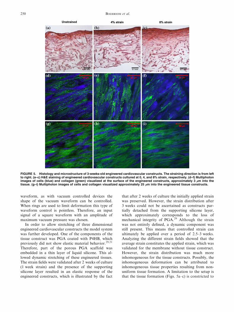
Histology and Multiphoton Microscopy
The H&E stain was performed to obtain a generaloverview of matrix formation within the engineeredcardiovascular constructs after 3 weeks of culture(Figs. 5a–c). In general the constructs showed a denselayer of cells and ECM at the surface of the construct.Deeper into the tissues less cells and a faint staining ofthe ECM was seen, which illustrates a reduced pro-duction of ECM per cell. The bottom of the engineeredtissue, which was attached to the silicone support layer,did not show much production of matrix due to aninsufficient supply of nutrients. Comparing the differ-ent straining conditions showed that the superficiallayer appeared thinner in unstrained tissues relative tothe strained tissues. In addition, the superficial layerof the 4% strained construct appeared more dense
TABLE 1. Results of biochemical assays and mechanical testing for each loading condition.
Tissue properties
Straining conditions
0% 4% 8%
DNA/dry weight (lg/mg) 1.51 ± 0.38 1.62 ± 0.13 1.59 ± 0.12
GAG/DNA (-) 7.78 ± 0.58 6.92 ± 0.33* 6.80 ± 0.48*
Collagen/DNA (-) 58.78 ± 13.35 40.81 ± 1.57** 39.76 ± 2.31**
Crosslinks (-) 0.16 ± 0.021 0.25 ± 0.024** 0.25 ± 0.031**
Stiffness (MPa) 2.64 ± 0.47 2.80 ± 0.50 1.92 ± 0.16*,+
Abbreviations are defined as follows: Hyp = Hydroxyproline and GAG = Glycosaminoglycan. *Indicates significant difference with reference
condition 0% (*p < 0.05, **p < 0.01), +indicates significant difference with respect to condition 4% (+p < 0.05).
FIGURE 2. Representative two dimensional strain (%) dis-tribution of the Bioflex flexible membrane in case of the use ofa 7.47 mm high ring. Circle indicates edges of the loadingpost.
BOERBOOM et al.248
compared to the superficial layer of the 8% strainedconstruct.
Multiphoton microscopy was performed to visual-ize the organization of cells and collagen within theengineered cardiovascular tissues. The surface of theconstruct was covered with a layer of cells with asmall amount of collagen in between the cells(Figs. 5d–f). The cells and collagen at the surface ofthe strained tissues were aligned oblique or perpen-dicular with respect to the strain direction. However,the cells and the collagen at the surface of the un-strained construct were oriented more parallel to thelongitudinal axis of the construct compared to thecells in the strained construct. Slightly deeper intothe tissue more collagen and less cells (Figs. 5g–i)were present, which were both aligned almost parallelto the straining direction.
DISCUSSION
Mechanical loading is an important regulator ofbiochemical processes within living tissues.42 In orderto regulate these biochemical processes, it is essentialto quantify the effect of mechanical loading on theproperties of engineered cardiovascular constructs. Inthis study an experimental framework was presented inwhich an adapted version of the Flexercell FX-4000T(Flexcell Int. Corp., USA) straining system was used tosimultaneously apply various strain magnitudes toindividual samples during one experiment. The flexer-cell system allows a high throughput of samples and iseasily kept sterile for prolonged periods of time. Por-ous PGA/P4HB scaffolds were partially embedded in asilicone layer, which allowed repeated and uniaxiallong term mechanical straining of these constructs. Theconstructs were attached to Bioflex wells (Flexcell Int.Corp., USA), subjected to two different strain magni-tudes for several weeks and showed differences inbiochemical properties, mechanical properties andorganization of the microstructure compared to theunstrained constructs. The results suggest that pro-longed mechanical stimulation induces the productionof collagen with a higher fraction of crosslinks, whichimproves the intrinsic mechanical properties of thecollagen. However, too large straining resulted indetrimental effects of straining on the mechanicalproperties of the tissue. In addition, straining induceda different alignment of cells and collagen in thesuperficial layers compared to the deeper layers of theconstruct.
By using polycarbonate rings of different heightplaced around the Flexercell loading posts, variousstrain magnitudes could be applied to individualsamples while only one vacuum pressure was applied.This eliminates the risk of inter-experimental differ-ences, as the number of experiments to culture cells atdifferent strain magnitudes can be reduced. However,using these rings imposes limitations to the applied
FIGURE 3. Representative two dimensional tissue strain distributions on the surface of an engineered tissue. (a) Representativetwo dimensional strain (%) distribution in case of a 8.16 mm ring. (b) Representative two dimensional strain (%) distribution in caseof a 7.47 mm ring.
0 4 80
1
2
3
4
5
Strain [%]
Tan
gent
Stif
fnes
s [M
Pa]
unstrainedStrained
*+
FIGURE 4. Tangent stiffness (MPa) of 3-weeks-old engi-neered cardiovascular constructs as a function of differentstrain magnitudes. *Indicates significant difference with ref-erence condition 0% (*p < 0.05) and +indicates significantdifference with 4% loading condition (+p < 0.05).
Effect of Strain Magnitude on the Tissue Properties 249
waveform, as with vacuum controlled devices theshape of the vacuum waveform can be controlled.When rings are used to limit deformation this type ofwaveform control is pointless. Therefore, an inputsignal of a square waveform with an amplitude ofmaximum vacuum pressure was chosen.
In order to allow stretching of three dimensionalengineered cardiovascular constructs the model systemwas further developed. One of the components of thetissue construct was PGA coated with P4HB, whichpreviously did not show elastic material behavior.29,51
Therefore, part of the porous PGA scaffold wasembedded in a thin layer of liquid silicone. This al-lowed dynamic stretching of these engineered tissues.The strain fields were validated after 2 weeks of culture(1 week strain) and the presence of the supportingsilicone layer resulted in an elastic response of theengineered constructs, which is illustrated by the fact
that after 2 weeks of culture the initially applied strainwas preserved. However, the strain distribution after3 weeks could not be ascertained as constructs par-tially detached from the supporting silicone layer,which approximately corresponds to the loss ofmechanical integrity of PGA.29 Although the strainwas not entirely defined, a dynamic component wasstill present. This means that controlled strain canultimately be applied over a period of 2.5–3 weeks.Analyzing the different strain fields showed that theaverage strain constitutes the applied strain, which wasvalidated for the membrane without tissue construct.However, the strain distribution was much moreinhomogeneous for the tissue constructs. Possibly, theinhomogeneous deformation can be attributed toinhomogeneous tissue properties resulting from non-uniform tissue formation. A limitation to the setup isthat the tissue formation (Figs. 5a–c) is constricted to
FIGURE 5. Histology and microstructure of 3-weeks-old engineered cardiovascular constructs. The straining direction is from leftto right. (a–c) H&E staining of engineered cardiovascular constructs cultured at 0, 4, and 8% strain, respectively. (d–f) Multiphotonimages of cells (blue) and collagen (green) visualized at the surface of the engineered constructs, approximately 3 lm into thetissue. (g–i) Multiphoton images of cells and collagen visualized approximately 25 lm into the engineered tissue constructs.
BOERBOOM et al.250
the surface of the construct due to the reduced supplyof nutrients in the presence of the silicone layer.
In the present study dynamic straining did not havean effect on proliferation. The amount of DNA perdryweight did not differ between the dynamicallystrained constructs and the unstrained constructs.Previously, cell proliferation was not influenced bymechanical loading in relatively long term studies.2,28,38
However, Mol et al.38 did show an elevated level ofproliferation in free floating engineered cardiovascularconstructs, which were not constrained mechanically. Itis essential to realize that in this study the unstrainedconstructs were not entirely stress free. The HSVCbelong to the myofibroblast phenotype 25,38 and thesemyofibroblasts produce a continuous isometric ten-sion.24 An internal tension is generated in the constructdue to the fact that the engineered construct is restrictedto contract by the supporting silicone layer. The inter-nal stress generated by (myo)fibroblast contraction issufficient to limit cell proliferation.
In general, studies on biosynthesis of collagen andGAG most often show upregulation in response tomechanical straining.31,33 However, these results areoften observed in short term studies, whereas forprolonged straining of engineered tissue constructsan increase in collagen production is not alwaysobserved.28,38 Our results show that the productionof both GAGs and collagen per cell was even re-duced in response to strain. This reduced collagenproduction per DNA was similar to Mol et al.,36
who also observed a decrease in collagen productionper DNA. In contrast, the fraction of HP crosslinksper collagen triple helix were increased compared tothe unstrained culture condition. Crosslinks are oftenrelated to the mechanical properties of tissues.3,14
The mechanical properties of the 4% strainedconstructs were equal to the unstrained constructs,whereas the tangent stiffness of the 8% strainedconstructs was significantly reduced. Equal mechani-cal properties combined with an increased fraction ofcrosslinks and a decreased production of collagenindicates that the cells produced collagen withdifferent intrinsic mechanical properties in order toresist the effect of mechanical straining more effec-tively. Increasing the strain even further resulted inequal collagen production and collagen fractioncompared to the 4% strained sample. However, adecrease in mechanical properties was observed,which possibly indicates that damage was introducedto the immature collagen fibers within the engineeredconstruct. The upregulation of crosslink formationby mechanical loading is potentially explained by anincreased TGF-b secretion due to mechanical load-ing. TGF-b upregulates lysyl hydroxylase 2 (LH2)expression, which in its turn facilitates HP crosslink
formation.50 In contrast to previous studies,30,36
dynamic mechanical loading did not result in anincrease of the mechanical properties. Similar to Molet al.38 the tangent stiffness of the constructs, createdusing fibrin as a cell carrier, became high in a rela-tively short period of time. Mol et al.38 showed thatthe effect of mechanical loading only came toexpression after 4 weeks of culture.
The microstructure of the strained engineered car-diovascular constructs showed a striking difference incell and collagen orientation between the superficiallayers and the deeper layers, whereas in the unstrainedconstructs this typical orientation was almost absent(Fig. 5). An oblique and perpendicular cell orientationhas also been observed in studies where two dimen-sional substrates seeded with cells were subjected tostrain.15,23,27,39 Similar to the superficial layers of theengineered construct, a relatively low amount of ECMwas present in the 2D cultures. However, cellsembedded in a 3D environment subjected to strainaligned parallel to the direction of strain.7,18,22,46 Thiscorresponds to the alignment of cells deeper into theengineered constructs (Fig. 5). The orientation re-sponse of cells has been attributed to a strain avoid-ance mechanism (present in superficial layer of theconstruct) and cell contact guidance (present in deeperlayers of the construct).7,39 Potentially the orientationresponse is determined by the amount of collagensurrounding the cells. In addition, in the strainedsamples the characteristic waviness of collagen seemedto be more present. The waviness might be related tothe appearance of the non-linear behavior in engi-neered cardiovascular tissues (i.e., uncrimping of col-lagen fibers).17 As can be observed from the histology,the amount of collagen and cells in the layer of theconstruct close to the silicone is much lower. When thelayer is observed with two photon microscopy (datanot shown), much more scaffold compared to thesuperficial layer is observed. In addition, the limitedamount of collagen is both randomly oriented andoriented in the direction of the scaffold fibers (contactguidance). Furthermore, only a very limited amount ofcells could be detected, most likely due to the limitedsupply of nutrients.
In this study an adapted form of the Flexercellstraining system was developed, which allows simul-taneous straining of individual engineered cardiovas-cular constructs at various strain magnitudes duringone experiment. The used system and the used anal-yses techniques are valuable tools to obtain an im-proved understanding of the effects of mechanicalloading on tissue formation. This will help us to elu-cidate the effect of mechanical strain on tissueremodeling and ultimately optimize loading protocolsfor TE.
Effect of Strain Magnitude on the Tissue Properties 251
ACKNOWLEDGMENT
The authors wish to thank Jessica Snabel and RuudBank of TNO Leiden for analyzing our samples forcollagen content and crosslink fraction.
OPEN ACCESS
This article is distributed under the terms ofthe Creative Commons Attribution NoncommercialLicense which permits any noncommercial use, distri-bution, and reproduction in any medium, provided theoriginal author(s) and source are credited.
REFERENCES
1Bader, A., T. Schilling, O. E. Teebken, G. Brandes, T.Herden, G. Steinhoff, and A. Haverich. Tissue engineeringof heart valves–human endothelial cell seeding of detergentacellularized porcine valves. Eur. J. Cardiothorac. Surg.14(3):279–284, 1998.2Balestrini, J. L., and K. L. Billiar. Equibiaxial cyclic stretchstimulates fibroblasts to rapidly remodel fibrin. J. Biomech.39(16):2983–2990, 2006.3Balguid, A., M. P. Rubbens, A. Mol, R. A. Bank, A. J. J.C. Bogers, J. P. van Kats, B. A. J. M. de Mol, F. P. T.Baaijens, and C. V. C. Bouten. The role of collagen cross-links in biomechanical behavior of human aortic heartvalve leaflets: relevance for tissue engineering. Tissue Eng.13:1501–1511, 2007.4Banes, A. J., J. Gilbert, D. Taylor, and O. Monbureau.A new vacuum-operated stress-providing instrumentthat applies static or variable duration cyclic tensionor compression to cells in vitro. J. Cell Sci. 75:35–42,1985.5Bank, R. A., B. Beekman, N. Verzijl, J. A. de Roos, A. N.Sakkee, and J. M. TeKoppele. Sensitive fluorimetricquantitation of pyridinium and pentosidine crosslinks inbiological samples in a single high-performance liquidchromatographic run. J. Chromatogr. B Biomed. Sci. Appl.703(1–2):37–44, 1997.6Bank, R. A., E. J. Jansen, B. Beekman, and J. M. teKoppele. Amino acid analysis by reverse-phase high-per-formance liquid chromatography: improved derivatizationand detection conditions with 9-fluorenylmethyl chloro-formate. Anal. Biochem. 240(2):167–176, 1996.7Barocas, V. H., and R. T. Tranquillo. An anisotropic bi-phasic theory of tissue-equivalent mechanics: the interplayamong cell traction, fibrillar network deformation, fibrilalignment, and cell contact guidance. J. Biomech. Eng.119(2):137–145, 1997.8Billiar, K. L., and M. S. Sacks. Biaxial mechanical prop-erties of the natural and glutaraldehyde treated aortic valvecusp: part I—experimental results. J. Biomech. Eng.122(1):23–30, 2000.9Boerboom, R. A., N. J. B. Driessen, C. V. C. Bouten,J. M. R. J. Huyghe, and F. P. T. Baaijens. Finite elementmodel of mechanically induced collagen fiber synthesis anddegradation in the aortic valve. Ann. Biomed. Eng.31(9):1040–1053, 2003.
10Boerboom, R. A., K. Nash Krahn, R. A. Megens, M. A.M. J. van Zandvoort, M. Merkx, and C. V. C. Bouten.High resolution imaging of collagen organisation andsynthesis using a versatile collagen specific probe. J. Struct.Biol. 159(3):392–399, 2007.
11Brown, T. D. Techniques for mechanical stimulation ofcells in vitro: a review. J. Biomech. 33(1):3–14, 2000.
12Campbell, J. H., J. L. Efendy, and G. R. Campbell. Novelvascular graft grown within recipient’s own peritonealcavity. Circ. Res. 85(12):1173–1178, 1999.
13Cesarone, C. F., C. Bolognesi, and L. Santi. Improvedmicrofluorometric DNA determination in biologicalmaterial using 33258 Hoechst. Anal. Biochem. 100(1):188–197, 1979.
14Dahl, S. L. M., R. B. Rucker, and L. E. Niklason. Effectsof copper and cross-linking on the extracellular matrix oftissue-engineered arteries. Cell Transplant. 14(10):861–868,2005.
15Dartsch, P. C., and H. Hammerle. Orientation response ofarterial smooth muscle cells to mechanical stimulation. Eur.J. Cell Biol. 41(2):339–346, 1986.
16Driessen,N. J.,R.A.Boerboom, J.M.R. J.Huyghe,C.V.C.Bouten, and F. P. T. Baaijens. Computational analyses ofmechanically induced collagen fiber remodeling in the aorticheart valve. J. Biomech. Eng. 125(4):549–557, 2003.
17Driessen, N. J. B., A. Mol, C. V. C. Bouten, and F. P. T.Baaijens. Modeling the mechanics of tissue-engineered hu-man heart valve leaflets. J. Biomech. 40(2):325–334, 2007.
18Eastwood, M., V. C. Mudera, D. A. McGrouther, andR. A. Brown. Effect of precise mechanical loading onfibroblast populated collagen lattices: morphologicalchanges. Cell Motil. Cytoskeleton 40(1):13–21, 1998.
19Engelmayr, G. C., Jr., E. Rabkin, F. W. H. Sutherland, F. J.Schoen, J. E. Mayer, Jr., and M. S. Sacks. The independentrole of cyclic flexure in the early in vitro development of anengineered heart valve tissue. Biomaterials 26(2):175–187,2005.
20Farndale, R. W., D. J. Buttle, and A. J. Barrett. Improvedquantitation and discrimination of sulphated glycosami-noglycans by use of dimethylmethylene blue. Biochim.Biophys. Acta. 883(2):173–177, 1986.
21Garvin, J., J. Qi, M. Maloney, and A. J. Banes. Novelsystem for engineering bioartificial tendons and applicationof mechanical load. Tissue Eng. 9(5):967–979, 2003.
22Grenier, G., M. Remy-Zolghadri, D. Larouche, R.Gauvin, K. Baker, F. Bergeron, D. Dupuis, E. Langelier,D. Rancourt, F. A. Auger, and L. Germain. Tissuereorganization in response to mechanical load increasesfunctionality. Tissue Eng. 11(1–2):90–100, 2005.
23Hamilton, D. W., T. M. Maul, and D. A. Vorp. Character-ization of the response of bone marrow-derived progenitorcells to cyclic strain: implications for vascular tissue-engi-neering applications. Tissue Eng. 10(3–4):361–369, 2004.
24Hinz, B., and G. Gabbiani. Mechanisms of force genera-tion and transmission by myofibroblasts. Curr. Opin. Bio-technol. 14(5):538–546, 2003.
25Hoerstrup, S. P., R. Sodian, S. Daebritz, J. Wang, E. A.Bacha, D. P. Martin, A. M. Moran, K. J. Guleserian, J. S.Sperling, S. Kaushal, J. P. Vacanti, F. J. Schoen, and J. E.Mayer, Jr.. Functional living trileaflet heart valves grown invitro. Circulation 102(Suppl 3):44–49, 2000.
26Hoerstrup, S. P., G. Zund, R. Sodian, A. M. Schnell, J.Grunenfelder, and M. I. Turina. Tissue engineering ofsmall caliber vascular grafts. Eur. J. Cardiothorac. Surg.20(1):164–169, 2001.
BOERBOOM et al.252
27Iba, T., and B. E. Sumpio. Morphological response ofhuman endothelial cells subjected to cyclic strain in vitro.Microvasc. Res. 42(3):245–254, 1991.
28Isenberg, B. C., and R. T. Tranquillo. Long-term cyclicdistention enhances the mechanical properties of collagen-based media-equivalents. Ann. Biomed. Eng. 31(8):937–949,2003.
29Kim, B. S., and D. J. Mooney. Scaffolds for engineeringsmooth muscle under cyclic mechanical strain conditions.J. Biomech. Eng. 122(3):210–215, 2000.
30Kim, B. S., J. Nikolovski, J. Bonadio, and D. J. Mooney.Cyclic mechanical strain regulates the development ofengineered smooth muscle tissue. Nature Biotech.17(10):979–983, 1999.
31Kolpakov, V., M. D. Rekhter, D. Gordon, W. H. Wang,and T. J. Kulik. Effect of mechanical forces on growth andmatrix protein synthesis in the in vitro pulmonary artery.Analysis of the role of individual cell types. Circ. Res.77(4):823–831, 1995.
32Krahn, K. N., C. V. C. Bouten, S. van Tuijl, M. A. M. J.van Zandvoort, and M. Merkx. Fluorescently labeledcollagen binding proteins allow specific visualization ofcollagen in tissues and live cell culture. Anal. Biochem.350(2):177–185, 2006.
33Lee, A. A., T. Delhaas, A. D. McCulloch, and F. J.Villarreal. Differential responses of adult cardiac fibro-blasts to in vitro biaxial strain patterns. J. Mol. Cell Car-diol. 31(10):1833–1843, 1999.
34L’Heureux, N., S. Paquet, R. Labbe, L. Germain, and F.A. Auger. A completely biological tissue-engineered humanblood vessel. FASEB J. 12(1):47–56, 1998.
35Mayer, J. E., Jr. Uses of homograft conduits for right ven-tricle to pulmonary artery connections in the neonatal peri-od. Semin. Thorac. Cardiovasc. Surg. 7(3):130–132, 1995.
36Mol, A., C. V. C. Bouten, G. Zund, C. I. Gunter, J. F.Visjager, M. I. Turina, F. P. T. Baaijens, and S. P. Ho-erstrup. The relevance of large strains in functional tissueengineering of heart valves. Thorac. Cardiovasc. Surg.51(2):78–83, 2003.
37Mol, A., N. J. B. Driessen, M. C. M. Rutten, S. P.Hoerstrup, C. V. C. Bouten, and F. P. T. Baaijens. Tissueengineering of human heart valve leaflets: a novel biore-actor for a strain-based conditioning approach. Ann. Bio-med. Eng. 33(12):1778–1788, 2005.
38Mol, A., M. C. M. Rutten, N. J. B. Driessen, C. V. C.Bouten, G. Zund, F. P. T. Baaijens, and S. P. Hoerstrup.Autologous human tissue-engineered heart valves: pros-pects for systemic application. Circulation 114(Suppl.1):I152–I158, 2006.
39Neidlinger-Wilke, C., E. S. Grood, R. A. Brand, and L.Claes. Cell alignment is induced by cyclic changes in celllength: studies of cells grown in cyclically stretched sub-strates. J. Orthop. Res. 19(2):286–293, 2001.
40Neuman, R. E., and M. A. Logan. The determination ofhydroxyproline. J. Biol. Chem. 184(1):299–306, 1950.
41Niklason, L. E., J. Gao, W. M. Abbott, K. K. Hirschi, S.Houser, R. Marini, and R. Langer. Functional arteriesgrown in vitro. Science 284(5413):489–493, 1999.
42Sarasa-Renedo, A., and M. Chiquet. Mechanical signalsregulating extracellular matrix gene expression in fibro-blasts. Scand. J. Med. Sci. Sports 15(4):223–230, 2005.
43Schenke-Layland, K., F. Opitz, M. Gross, C. Doring, K. J.Halbhuber, F. Schirrmeister, Th. Wahlers, and U. A.Stock. Complete dynamic repopulation of decellularizedheart valves by application of defined physical signals—anin vitro study. Cardiovasc. Res. 60(3):497–509, 2003.
44Schnell, A. M., S. P. Hoerstrup, G. Zund, S. Kolb, R.Sodian, J. F. Visjager, J. Grunenfelder, A. Suter, and M.Turina. Optimal cell source for cardiovascular tissue engi-neering: venous vs. aortic human myofibroblasts. Thorac.Cardiovasc. Surg. 49(4):221–225, 2001.
45Schoen F. J., and R. J. Levy. Founder’s Award, 25thAnnual Meeting of the Society for Biomaterials, perspec-tives. Providence, RI, April 28–May 2, 1999. Tissue heartvalves: current challenges and future research perspectives.J. Biomed. Mater. Res. 47(4):439–465, 1999.
46Seliktar, D., R. A. Black, R. P. Vito, and R. M. Nerem.Dynamic mechanical conditioning of collagen–gel bloodvessel constructs induces remodeling in vitro. Ann. Biomed.Eng. 28(4):351–362, 2000.
47Seliktar, D., R. M. Nerem, and Z. S. Galis. Mechanicalstrain-stimulated remodeling of tissue-engineered bloodvessel constructs. Tissue Eng. 9(4):657–666, 2003.
48Stegemann, J. P., and R. M. Nerem. Altered response ofvascular smooth muscle cells to exogenous biochemicalstimulation in two- and three-dimensional culture. Exp.Cell Res. 283(2):146–155, 2003.
49Thom, T., N. Haase, W. Rosamond, V. J. Howard, J.Rumsfeld, T. Manolio, Z. Zheng, K. Flegal, C. O’Donnell,S. Kittner, D. Lloyd-Jones, D. C. Goff, Jr., Y. Hong, R.Adams, G. Friday, K. Furie, P. Gorelick, B. Kissela, J.Marler, J. Meigs, V. Roger, S. Sidney, P. Sorlie, J.Steinberger, S. Wasserthiel-Smoller, M. Wilson, and P.Wolf. Heart disease and stroke statistics—2006 update: areport from the American Heart Association StatisticsCommittee and Stroke Statistics Subcommittee. Circula-tion 113(6):e85–e151, 2006.
50van der Slot, A. J., A. M. Zuurmond, A. J. van denBogaerdt, M. M. Ulrich, E. Middelkoop, W. Boers, H.Karel Ronday, J. De Groot, R. A. Bank, and T. W. Hu-izinga. Increased formation of pyridinoline cross-links dueto higher telopeptide lysyl hydroxylase levels is a generalfibrotic phenomenon. Matrix Biol. 23(4):251–257, 2004.
51Webb, A. R., J. Yang, and G. A. Ameer. Biodegradablepolyester elastomers in tissue engineering. Expert Opin.Biol. Ther. 4(6):801–812, 2004.
Effect of Strain Magnitude on the Tissue Properties 253
Related Documents