..................................................................................................................................................................................... ..................................................................................................................................................................................... CLINICAL RESEARCH Effect of renal denervation on left ventricular mass and function in patients with resistant hypertension: data from a multi-centre cardiovascular magnetic resonance imaging trial Felix Mahfoud 1 * , Daniel Urban 1,2 , Desiree Teller 2 , Dominik Linz 1 , Philipp Stawowy 2 , Jan-Hendrik Hassel 2 , Peter Fries 3 , Stephan Dreysse 2 , Ernst Wellnhofer 2 , Gu ¨ nther Schneider 3 , Arno Buecker 3 , Christopher Schneeweis 2 , Adelina Doltra 2 , Markus P. Schlaich 4 , Murray D. Esler 4 , Eckart Fleck 2 , Michael Bo ¨ hm 1 , and Sebastian Kelle 2 1 Klinik fu ¨r Innere Medizin III, Universita ¨tsklinikum des Saarlandes, Homburg/Saar, Germany; 2 Klinik fu ¨r Innere Medizin/Kardiologie, Deutsches Herzzentrum Berlin, Berlin, Germany; 3 Klinik fu ¨r Diagnostische und Interventionelle Radiologie, Universita ¨tsklinikum des Saarlandes, Homburg/Saar, Germany; and 4 Baker IDI Heart & Diabetes Institute, Melbourne, Australia Received 27 November 2013; revised 5 February 2014; accepted 10 February 2014; online publish-ahead-of-print 6 March 2014 See page 2205 for the editorial comment on this article (doi:10.1093/eurheartj/ehu127) Aims Sympathetic stimulation induces left ventricular hypertrophy and is associated with increased cardiovascular risk. Catheter-based renal denervation (RDN) has been shown to reduce sympathetic outflow and blood pressure (BP). The present multi-centre study aimed to investigate the effect of RDN on anatomic and functional myocardial parameters, assessed by cardiac magnetic resonance (CMR), in patients with resistant hypertension. Methods and results Cardiac magnetic resonance was performed in 72 patients (mean age 66 + 10 years) with resistant hypertension (55 patients underwent RDN, 17 served as controls) at baseline and after 6 months. Clinical data and CMR results were analysed blindly. Renal denervation significantly reduced systolic and diastolic BP by 22/8 mm Hg and left ventricular mass index (LVMI) by 7.1% (46.3 + 13.6 g/m 1.7 vs. 43.0 + 12.6 g/m 1.7 , P , 0.001) without changes in the control group (41.9 + 10.8 g/m 1.7 vs. 42.0 + 9.7 g/m 1.7 , P ¼ 0.653). Ejection fraction (LVEF) in patients with impaired LVEF at baseline ( ,50%) significantly increased after RDN (43% vs. 50%, P , 0.001). Left ventricular circumferential strain as a surrogate of diastolic function in the subgroup of patients with reduced strain at baseline increased by 21% only in the RDN group ( 214.8 vs. 217.9; P ¼ 0.001) and not in control patients ( 215.5 vs. 216.4, P ¼ 0.508). Conclusions Catheter-based RDN significantly reduced BP and LVMI and improved EF and circumferential strain in patients with resistant hypertension, occurring partly BP independently. ----------------------------------------------------------------------------------------------------------------------------------------------------------- Keywords Resistant hypertension † Left ventricular hypertrophy † Circumferential strain † Cardiovascular magnetic resonance † Renal nerve ablation Introduction Hypertension is the most popular cardiovascular risk factor world- wide being closely associated with stroke and heart failure. 1 Resistant hypertension is defined as blood pressure (BP) above goal despite the concurrent use of three or more different antihypertensive drugs, one ideally being a diuretic, with all agents prescribed at maximum or maximum tolerated doses. 2 Patients with resistant hypertension are at high risk for cardiovascular events, in particular when hyperten- sive end organ damage is present. 3 Hypertension induces left ven- tricular hypertrophy (LVH) and predicts incident chronic heart failure. 4 Regression of LVH has been associated with favourable * Corresponding author. Tel: +49 6841 16 21346, Fax +49 6841 16 13211, Email: [email protected] Published on behalf of the European Society of Cardiology. All rights reserved. & The Author 2014. For permissions please email: [email protected] European Heart Journal (2014) 35, 2224–2231 doi:10.1093/eurheartj/ehu093 by guest on February 18, 2016 http://eurheartj.oxfordjournals.org/ Downloaded from

Welcome message from author
This document is posted to help you gain knowledge. Please leave a comment to let me know what you think about it! Share it to your friends and learn new things together.
Transcript

. . . . . . . . . . . . . . . . . . . . . . . . . . . . . . . . . . . . . . . . . . . . . . . . . . . . . . . . . . . . . . . . . . . . . . . . . . . . . . . . . . . . . . . . . . . . . . . . . . . . . . . . . . . . . . . . . . . . . . . . . . . . . . . . . . . . . . . . . . . . . . . . . . . . . . . . . . . . . . . . . . . . . . . . . . . . . . . . . . . . .
. . . . . . . . . . . . . . . . . . . . . . . . . . . . . . . . . . . . . . . . . . . . . . . . . . . . . . . . . . . . . . . . . . . . . . . . . . . . . . . . . . . . . . . . . . . . . . . . . . . . . . . . . . . . . . . . . . . . . . . . . . . . . . . . . . . . . . . . . . . . . . . . . . . . . . . . . . . . . . . . . . . . . . . . . . . . . . . . . . . . .
CLINICAL RESEARCH
Effect of renal denervation on left ventricularmass and function in patients with resistanthypertension: data from a multi-centrecardiovascular magnetic resonance imaging trialFelix Mahfoud1*, Daniel Urban1,2, Desiree Teller2, Dominik Linz1, Philipp Stawowy2,Jan-Hendrik Hassel2, Peter Fries3, Stephan Dreysse2, Ernst Wellnhofer2,Gunther Schneider3, Arno Buecker3, Christopher Schneeweis2, Adelina Doltra2,Markus P. Schlaich4, Murray D. Esler4, Eckart Fleck2, Michael Bohm1,and Sebastian Kelle2
1Klinik fur Innere Medizin III, Universitatsklinikum des Saarlandes, Homburg/Saar, Germany; 2Klinik fur Innere Medizin/Kardiologie, Deutsches Herzzentrum Berlin, Berlin, Germany;3Klinik fur Diagnostische und Interventionelle Radiologie, Universitatsklinikum des Saarlandes, Homburg/Saar, Germany; and 4Baker IDI Heart & Diabetes Institute, Melbourne, Australia
Received 27 November 2013; revised 5 February 2014; accepted 10 February 2014; online publish-ahead-of-print 6 March 2014
See page 2205 for the editorial comment on this article (doi:10.1093/eurheartj/ehu127)
Aims Sympathetic stimulation induces left ventricular hypertrophy and is associated with increased cardiovascular risk.Catheter-based renal denervation (RDN) has been shown to reduce sympathetic outflow and blood pressure (BP).The present multi-centre study aimed to investigate the effect of RDN on anatomic and functional myocardialparameters, assessed by cardiac magnetic resonance (CMR), in patients with resistant hypertension.
Methodsand results
Cardiac magnetic resonance was performed in 72 patients (mean age 66+ 10 years) with resistant hypertension(55 patients underwent RDN, 17 served as controls) at baseline and after 6 months. Clinical data and CMR resultswere analysed blindly. Renal denervation significantly reduced systolic and diastolic BP by 22/8 mm Hg and left ventricularmass index (LVMI) by 7.1% (46.3+13.6 g/m1.7 vs. 43.0+ 12.6 g/m1.7, P , 0.001) without changes in the control group(41.9+10.8 g/m1.7 vs. 42.0+ 9.7 g/m1.7, P ¼ 0.653). Ejection fraction (LVEF) in patients with impaired LVEF at baseline(,50%) significantly increased after RDN (43% vs. 50%, P , 0.001). Left ventricular circumferential strain as a surrogateof diastolic function in the subgroup of patients with reduced strain at baseline increased by 21% only in the RDN group(214.8 vs. 217.9; P ¼ 0.001) and not in control patients (215.5 vs. 216.4, P ¼ 0.508).
Conclusions Catheter-based RDN significantly reduced BP and LVMI and improved EF and circumferential strain in patients withresistant hypertension, occurring partly BP independently.
- - - - - - - - - - - - - - - - - - - - - - - - - - - - - - - - - - - - - - - - - - - - - - - - - - - - - - - - - - - - - - - - - - - - - - - - - - - - - - - - - - - - - - - - - - - - - - - - - - - - - - - - - - - - - - - - - - - - - - - - - - - - - - - - - - - - - - - - - - - - - - - - - - - - - - - - - - -Keywords Resistant hypertension † Left ventricular hypertrophy † Circumferential strain † Cardiovascular magnetic
resonance † Renal nerve ablation
IntroductionHypertension is the most popular cardiovascular risk factor world-wide being closely associated with stroke and heart failure.1 Resistanthypertension is defined asblood pressure (BP) abovegoal despite theconcurrent use of three or more different antihypertensive drugs,
one ideally being a diuretic, with all agents prescribed at maximumor maximum tolerated doses.2 Patients with resistant hypertensionareathigh risk for cardiovascularevents, in particularwhen hyperten-sive end organ damage is present.3 Hypertension induces left ven-tricular hypertrophy (LVH) and predicts incident chronic heartfailure.4 Regression of LVH has been associated with favourable
* Corresponding author. Tel: +49 6841 16 21346, Fax +49 6841 16 13211, Email: [email protected]
Published on behalf of the European Society of Cardiology. All rights reserved. & The Author 2014. For permissions please email: [email protected]
European Heart Journal (2014) 35, 2224–2231doi:10.1093/eurheartj/ehu093
by guest on February 18, 2016http://eurheartj.oxfordjournals.org/
Dow
nloaded from

outcomes and varies between different antihypertensive drugclasses.5 Sympathetic activation promotes hypertension andinduces cardiac hypertrophy.6 –8 Catheter-based renal denervation(RDN) in patients with resistant hypertension decreases BP andcentral sympathetic activity.9,10 Two recently published pilotstudies demonstrated reductions in left ventricular mass and intra-ventricular septum thickness following RDN.11,12 Given the limitedreproducibilityof echocardiography these studies wereof borderlinepower to assess changes in cardiac systolic and diastolic function andvolumes.13 Cardiac magnetic resonance (CMR) is more precise andsensitive than echocardiography and provides highly reproducibleassessments of cardiac volumes, function, and mass,14 –16 allowing aconsiderable reduction in the patient numbers compared with echo-cardiography to prove changes in remodelling parameters afterRDN.13 Therefore, the present multi-centre, blinded, controlledCMR study aimed to investigate the effects of RDN on myocardialparameters in 72 patients with resistant hypertension.
Methods
Study population and designThe present study was designed as a prospective, multi-centre study andwas implemented at two investigational sites in Germany and one inAustralia. Patients underwent RDN and received CMR at baseline andafter 6 months. The study was approved by the local ethics committeeand was conducted in accordance with the ethical standards defined bylocal law. All patients provided written informed consent.
Enrolled subjects were aged ≥18 years with an office systolic bloodpressure (SBP) above goal (≥140 mm Hg) or mean ambulatory 24-hSBP .135 mm Hg despite the use of ≥3 antihypertensive agents of dif-ferent classes, including a diuretic at maximum or highest tolerateddoses.2 Office BP readings were taken in a seated position with an auto-matic oscillometric Omron HEM-705 monitor (Omron Healthcare,Vernon Hills, IL, USA) after 5 min of rest according to the StandardJoint National Committee VII Guidelines.17 At baseline, BP was measuredat each arm and the arm with the higher BP was used for all subsequentreadings. Averages of the triplicate measures were calculated and usedfor analysis. According to recent recommendations on RDN18,19 patientswith pseudo-resistant hypertension defined as mean ambulatory 24-hSBP ,130 mm Hg were excluded.2 Patients with GFR ,45 mL/min/1.73 m2 and patients on haemodialysis were excluded. Measurementswere performed as an extension to the Symplicity protocols(NCT00664638, NCT00888433, and NCT01888315). Patients withgeneral contraindications for CMR (implantable devices, cerebral aneur-ysm clips, severe claustrophobia, and cochlear implants) were excluded.Patients with resistant hypertension not undergoing RDN served as con-trols. Only patients with stable antihypertensive drug regimen wereincluded and patients with known, treatable secondary causes of hyper-tension were excluded. All patients underwent a complete history andphysical examination, assessment of vital signs, and review of medication.Patients were interviewed whether they had taken their complete medi-cation at defined doses. Patients and physicians were instructed not tochange antihypertensive medication during the study period exceptwhen medically required. All patients were treated using the SymplicityFlex system (Medtronic, MN, USA) as described elsewhere.20
Magnetic resonance imagingCardiac magnetic resonance images were performed in all subjectsbefore and 6 months after RDN using a 1.5 T Achieva MRI scanner
(Philips Healthcare, Best, the Netherlands) or 1.5 T Siemens Symphonyor a 1.5 T Siemens Aera MRI system (Siemens Healthcare Sector, Erlan-gen, Germany). Cine images were acquired using a balanced steady-statefree precession sequence during breath-holds of �10–15 s using VCGgating with patients being positioned in supine position. Whole-heartcoverage from apex to base was performed as previously reported.21
Additionally, late gadolinium enhancement (LGE) has been performedin a subgroup of 24 patients to evaluate the impact of RDN on scartissue and 16 control patients. Late gadolinium-enhanced imaging wasperformed 10–15 min after injection of 0.2 mmol/kg gadolinium DTPAusing an inversion-recovery3D spoiled gradient echo sequence. The pre-pulse delay was individually adjusted according to apre-pulse-delayfinder(Look-Locker sequence). All CMR examinations were performed byoperators, whowereblinded topatient’s treatment and time of the meas-urement (pre- or post-RDN).
Cardiac magnetic resonance analysisLeft ventricular mass measurementsCardiac magnetic resonance image analyses were performed accordingto the recommendations of the task force for post-processing of theSociety for Cardiovascular MR.22 Offline CMR analyses were performedusing the softwareQmass MR Enterprise Solution (version 7.4, Medis, theNetherlands). Endocardial and epicardial borders were traced automat-ically and corrected manually at end-diastole and end-systole, while thepapillary muscles were excluded from LVM to achieve better reproduci-bility.23 Left ventricular volumes and mass were calculated using the sum-mation of slices method.24 Left ventricular end-systolic (LVESVI) andend-diastolic volume index (LVEDVI) were normalized in every patientfor sex, age, height, and weight, and LVESVI and LVEDVI have beenassessed.25 Left ventricular mass was then normalized indexing to bodysurface area and height (g/m1.7).26 Left ventricular measurements includ-ing wall thickness and internal dimensions were obtained using the SAXview basal to the tips of the papillary muscles.27 Wall thickness wasdefined as the radial distance between endocardium and epicardiumfor septum and lateral wall on basal short-axis, end-diastolic cineimages.28 In addition, relative wall thickness has been calculated as 2 ×posterior wall thickness divided by LV internal diameter at diastole.29
Left ventricular wall stress was evaluated using the Laplace equation re-lating wall stress to intracavitary pressure, wall curvature, and wall thick-ness as follows: S ¼ P(r/2)h, where S is wall stress, P is LV pressure, r is aradius of curvature, and h is wall thickness (modified by using brachial BPmeasurements from Yin30 and Grossman31). These non-invasivelyassessed parameters have been demonstrated to be highly comparablewith measurements using invasive hemodynamics.32
Feature trackingAnalysis of myocardial circumferential strain (Ecc) was performed usingthe software Image Arena 4.6 (TomTec). The mid-ventricular SAXslice containing both papillary muscles was chosen, and endocardialborders were manually drawn by setting six to eight contour points be-ginning clockwise from the anterior septum. A reference point was seton the anterior interventricular septum to allow segmentation accordingto standard model.33 Circumferential strain and peak systolic strain ratewas calculated for each myocardial segment.
Left atriumThe size of the left atrium (LA) was assessed by four-chamber planimetrywith Qmass software, using a four-chamber view at the level of the mem-branous septum. Endocardial contour was drawn manually at an end-systolic phase and a size of .24 cm2 was recognized as dilated.34
CMR imaging after renal denervation 2225
by guest on February 18, 2016http://eurheartj.oxfordjournals.org/
Dow
nloaded from

Scar tissueTo evaluate the impact of RDN on myocardial fibrosis LGE MRI wasapplied using the inversion-recovery gradient-echo sequence. Lategadolinium-enhanced images were scored visually by two experiencedobservers (blinded to other MRI and clinical data) at the time of the acqui-sition using a 17-segment model.33 Each segment was graded usingfollowing -point score: 0, absence of enhancement; 1, enhancement of1–25% transmurality; 2, enhancement of 26–50% transmurality; 3, en-hancement of 51–75% transmurality, and 4, enhancement of 76–100%transmurality.35 The score per segment was then calculated by dividingtotal score by 17 referring to the 17-segment model.33
Statistical analysisAll data are presented as mean+ SD. Differences in mean values werecompared using a x2 test for unpaired tests and McNemar’s for pairedcomparisons. For the evaluation of intra- and interobserver variability,12 measurements (for LVM, myocardial strain, and LA size) wererepeated both by the first observer and a second observer after 3months. Intra- and interobserver variability is displayed in Bland-AltmanPlots. Additionally, the intraclass correlation coefficient (ICC) were con-sidered as good with a value of .0.6 and excellent with a value of .0.7.36
All P-values of ,0.05 were considered as statistically significant. Both, theregression to the mean phenomenon and the confounding by indicationissue were addressed by general linear models for repeated measure-ments including covariates (ANCOVA) using SPSS Version 21. The treat-ment modality was investigated as an intergroup factor. Serial BPmeasurements (with LV mass at baseline as covariate) and serial LVmass determinations (with SBP at baseline as covariate) were explored
separately. Moreover, a model including two inner-subject factors LVmass and SBP was implemented. Further covariates included in the ana-lysis were age, diabetes, prevalence of coronary artery disease, numberof antihypertensive drugs, weight, and height.
Results
Study populationSeventy-two patients with resistant hypertension were enrolled inthe study. Fifty-five subjects were treated with RDN and 17 subjectsserved as controls (medical treatment only). No patient was lost tofollow-up during the study period of 6 months. All clinical data arepresented in Table 1.
Blood pressureOffice systolic and diastolic BP decreased significantly from 170/90+ 21/15 mm Hg at baseline to 148/82+ 19/14 mm Hg (P ,
0.001) 6 months after RDN. SBP/diastolic BP (DBP) in the controlgroup did change during follow-up (156/84+ 17/11 vs. 145/77+23/15 mm Hg; P ¼ 0.044 for SBP and P ¼ 0.034 for DBP).
Antihypertensive drugsPatients and physicians were instructed not to change antihyperten-sive medication during the study period. The average number of anti-hypertensive drugs remained constant in the RDN group (4.6+1.6
. . . . . . . . . . . . . . . . . . . . . . . . . . . . . . . . . . . . . . . . . . . . . . . . . . . . . . . . . . . . . . . . . . . . . . . . . . . . . . . . . . . . . . . . . . . . . . . . . . . . . . . . . . . . . . . . . . . . . . . . . . . . . . . . . . . . . . . . . . . . . . . . . . . . . . . . . . . . . . . . . . . . . . . . . . . . . . .
. . . . . . . . . . . . . . . . . . . . . . . . . . . . . . . . . . . . . . . . . . . . . . . . . . . . . . . . . . . . . . . . . . . . . . . . . . . . . . . . . . . . . . . . . . . . . . . . . . . . . . . . . . . . . . . . . . . . . . . . . . . . . . . . . . . . . . . . . . . . . . . . . . . . . . . . . . . . . . . . . . . . . . . . . . . . . . .
. . . . . . . . . . . . . . . . . . . . . . . . . . . . . . . . . . . . . . . . . . . . . . . . . . . . . . . . . . . . . . . . . . . . . . . . . . . . . . . . . . . . . . . . . . . . . . . . . . . . . . . . . . . . . . . . . . . . . . . . . . . . . . . . . . . . . . . . . . . . . . . . . . . . . . . . . . . . . . . . . . . . . . . . . . . . . . .
Table 1 Patient’s baseline characteristics
Variables RDN (n 5 55) Control (n 5 17) P-value
Characteristics
Age (years) 65+10 (67) 70+9 (74) 0.058
Male (%) 39 (71%) 10 (59%) 0.350
BMI (kg/m2) 29.2+4.3 (29) 28.6+5.3 (28) 0.646
Atrial fibrillation 7 (13%) 4 (24%) 0.279
Stroke 7 (13%) 2 (12%) 0.946
Type 2 diabetes 26 (47%) 7 (41%) 0.659
Hypercholesterolaemia 34 (62%) 13 (76%) 0.578
Smoking 9 (16%) 2 (12%) 0.645
Medication
Number of antihypertensive drugs 4.6+1.6 (4) 4.5+1.2 (4) 0.704
ACE inhibitors/ARBs 49 (89%) 17 (100%) 0.763
Direct renin inhibitors 14 (26%) 1 (6%) 0.082
Beta-blockers 48 (87%) 16 (93%) 0.433
Calcium-channel blockers 41 (75%) 15 (88%) 0.235
Diuretics 46 (84%) 17 (100%) 0.642
Sympatholytics 21 (38%) 5 (29%) 0.477
Haemodynamics
Systolic BP (mm Hg) 170.0+21.4 (170) 157.4+15.3 (159) 0.027
Diastolic BP (mm Hg) 89.9+14.8 (90) 83.8+10.9 (82) 0.117
Pulse pressure (mm Hg) 79.6+15.0 (77) 74.6+14.6 (72) 0.225
Values are given either as total number, percentage or mean+ SD; median is given in parenthesis. BMI, body mass index; ACE, angiotensin converting enzyme; ARB, angiotensinreceptor blocker; BP, blood pressure.
F. Mahfoud et al.2226
by guest on February 18, 2016http://eurheartj.oxfordjournals.org/
Dow
nloaded from

vs. 4.5+1.7; P ¼ 0.287) and the control group (4.5+ 1.2 vs. 4.3+1.3; P ¼ 0.395).
Cardiac magnetic resonance variablesLeft ventricular measurementsNo significant changes between baseline and 6 months were evidentfor LVESV, however for LVEDV in the RDN group (LVEDV: 177+54 mL vs. 176+ 53; P ¼ 0.246 and LVESV: 81+ 40 mL vs. 77+35 mL; P ¼ 0.038). No significant changes between baseline and6 months were evident for LVESV and LVEDV in the controls(EDV: 172+42 vs. 172+29 mL, P ¼ 0.795 and ESV: 78+ 31 mLvs. 78+27 mL, P ¼ 0.868). Normalized LVESVI and LVEDVI aregiven in Table 2. There were no significant changes of LV internal di-mension measures in both groups.
Ejection fraction increased significantly after RDN (55.7+ 11.1 vs.57.6+9.3%, P ¼ 0.048) and remained unchanged in the controlgroup (55.9+8.2 vs. 55.5+8.4%, P ¼ 0.723). In patients withreduced systolic LVEF at baseline (defined as ≤50%, n ¼ 19) LVEFsignificantly increased by 7.3% after RDN (43.2+5.5 vs. 50.5+7.0%, P , 0.001). There was no improvement in the control group(42.9+5.7 vs. 44.3+ 8.4%, P ¼ 0.537; n ¼ 3) (Figure 1).
Left ventricular mass indexed (LVMI) to height1.7 significantlydecreased by 7.1% 6 months after RDN (from 46.3+13.6 to43.0+12.6 g/m1.7, P , 0.001) (Table 2, Figure 2). In the controlgroup, LV mass remained unchanged (41.9+ 10.8 vs. 42.0+ 9.7g/m1.7, P ¼ 0.653) (Figure 2). In total, 18 (33%) patients after RDNshowed a SBP reduction of ,10 mm Hg and were subsequentlydefined as ‘non-responders’.37 Interestingly, in 15/18 (83%) non-responders LVMI significantly decreased from 52.1+14.9 vs.
47.8+ 14.4 g/m2 (P ¼ 0.001) (Figure 3, Table 3). Left ventricularwall stress was reduced after RDN (105+46 vs. 95+43 dyn/mm2, P ¼ 0.033), whereas no changes occurred in the controlgroup (120+44 vs. 105+38 dyn/mm2, P ¼ 0.149).
Circumferential strainMean values of Ecc remained constant in both study groups (Table 2).Patients were segregated in two subgroups depending on their con-tractile function at baseline. A circumferential strain of ≥220% was
. . . . . . . . . . . . . . . . . . . . . . . . . . . . . . . . . . . . . . . . . . . . . . . . . . . . . . . . . . . . . . . . . . . . . . . . . . . . . . . . . . . . . . . . . . . . . . . . . . . . . . . . . . . . . . . . . . . . . . . . . . . . . . . . . .
. . . . . . . . . . . . . . . . . . . . . . . . . . . . . . . . . . . . . . . . . . . . . . . . . . . . . . . . . . . . . . . . . . . . . . . . . . . . . . . . . . . . . . . . . . . . . . . . . . . . . . . . . . . . . . . . . . . . . . . . . . . . . . . . . . . . . . . . . . . . . . . . . . . . . . . . . . . . . . . . . . . . . . . . . . . . . . .
. . . . . . . . . . . . . . . . . . . . . . . . . . . . . . . . . . . . . . . . . . . . . . . . . . . . . . . . . . . . . . . . . . . . . . . . . . . . . . . . . . . . . . . . . . . . . . . . . . . . . . . . . . . . . . . . . . . . . . . . . . . . . . . . . . . . . . . . . . . . . . . . . . . . . . . . . . . . . . . . . . . . . . . . . . . . . . .
Table 2 Cardiac magnetic resonance parameters at baseline and 6-month follow-up in patients undergoing renaldenervation and control patients
RDN (n 5 55) Control (n 5 17)
Baseline 6 months P-value Baseline 6 months P-value
Anatomy
LVEDVI (mL/m2) 85.8+22.1 (85) 84.1+22.7 (83) 0.146 88.9+20.3 (89) 90.2+13.9 (88) 0.570
LVESVI (mL/m2) 39.8+16.8 (37) 36.1+15.2 (32) 0.019 40.2+15.9 (37) 41.6+14.0 (38) 0.422
IVSTd (mm) 12.3+3.5 (12) 11.9+3.2 (12) 0.265 10.6+2.7 (10) 10.5+2.8 (10) 0.587
LVIDd (mm) 57.0+7.0 (57) 56.8+6.8 (57) 0.774 58.0+6.3 (59) 56.7+6.2 (57) 0.266
PWTd (mm) 8.7+1.9 (8) 8.4+2.1 (8) 0.083 7.6+1.8 (7) 7.7+1.8 (7) 0.740
RWT 0.31+0.1 (0.29) 0.30+0.1 (0.28) 0.150 0.26+0.07 (0.25) 0.28+0.08 (0.25) 0.501
LV mass/body surface area (g/m2) 58.7+16.5 (59) 54.9+15.9 (55) ,0.001 54.6+13.5 (53) 53.6+12.6 (54) 0.245
LV mass/height1.7 (g/m1.7) 46.3+13.6 (46) 43.0+12.6 (43) ,0.001 41.9+10.8 (40) 42.0+9.7 (40) 0.653
LA size (cm2) 25.7+7.1 (25) 25.3+6.4 (24) 0.251 23.5+5.9 (24) 24.4+4.5 (23) 0.492
Function
EF (%) 55.7+11.1 (56) 57.6+9.3 (58) 0.048 55.9+8.2 (59) 55.5+8.4 (57) 0.723
Circumferential strain (%) 221.3+7.7 (219) 221.7+7.0 (221) 0.392 218.5+5.0 (219) 219.8+6.4 (218) 0.246
Peak systolic strain rate (s21) 21.32+0.49 (21.32) 21.37+0.46 (21.37) 0.088 21.40+0.94 (21.12) 21.44+0.90 (21.21) 0.356
LV wall stress (dyn/mm2) 105+46 (108) 95+43 (92) 0.033 120+44 (129) 105+38 (120) 0.149
LVEDVI, left ventricular end-diastolic volume index; LVESVI, left ventricular end-systolic volume index; IVSTd, interventricular septal thickness at diastole; LVIDd, left ventricularinternal diameter at diastole; PWTd, posterior wall thickness at diastole; RWT, relative wall thickness; LV, left ventricle; LA, left atrium; EF, ejection fraction.
Figure1 Subgroupof patients with reduced systolic left ventricu-lar ejection fraction (defined as ≤50%) undergoing renal denerv-ation (n ¼ 19) and controls (n ¼ 3) at baseline and at 6-monthfollow-up. Values are given as mean+ standard deviation.
CMR imaging after renal denervation 2227
by guest on February 18, 2016http://eurheartj.oxfordjournals.org/
Dow
nloaded from

considered as a contractile dysfunction.38 In the subgroup of patientswith reduced myocardial contractility at baseline (diastolic dysfunc-tion, n ¼ 27), Ecc improved after RDN by 21% (214.8+3.9 to217.9+4.6%, P ¼ 0.001). In the control group (n ¼ 10), Eccremained unchanged (215.5+ 4.3 vs. 216.4+ 5.0%, P ¼ 0.508)(Figure 4).
Left atriumMean values of LA size did not significantly change (Table 2).However, following RDN the number of patients with left-atrial en-largement (LAE) was decreased from 62 to 55% and increased from47 to 53% in the control group. Notably, in the subgroup of patientswith LAE at baseline (n ¼ 34; 62%) RDN resulted in a significantreduction of LA size from 29.7+5.8 to 28.6+5.3 cm2 (P ¼ 0.026).
Late gadolinium enhancementThere was no significant difference in LGE score between RDNpatients and controls at baseline (0.22+0.6 vs. 0.25+ 0.8;
Figure 2 Left ventricular mass index at baseline and 6-month follow-up in patients undergoing renal denervation (n ¼ 55) and controls (n ¼ 17),depicted as individual changes and average values+ standard deviation.
Figure 3 Correlation of absolute change in systolic bloodpressure and left ventricular mass index after renal denervation.Response to renal denervation was defined as a ≥10 mm Hgdecrease in systolic blood pressure.
Figure 4 Subgroup of patients with reduced myocardial con-tractility (diastolic dysfunction, renal denervation: n ¼ 27; control:n ¼ 10) at baseline and 6-month follow-up evaluated by measure-ments of circumferential strain. Values are given as mean+standard deviation.
F. Mahfoud et al.2228
by guest on February 18, 2016http://eurheartj.oxfordjournals.org/
Dow
nloaded from

. . . . . . . . . . . . . . . . . . . . . . . . . . . . . . . . . . . . . . . . . . . . . . . . . . . . . . . . . . . . . . . . . . . . . . . . . . . . . . . . . . . . . . . . . . . . . . . . . . . . . . . . . . . . . . . . . . . . . . . . . . . . . . . . . . . . . . . . . . . . . . . . . . . .
. . . . . . . . . . . . . . . . . . . . . . . . . . . . . . . . . . . . . . . . . . . . . . . . . . . . . . . . . . . . . . . . . . . . . . . . . . . . . . . . . . . . . . . . . . . . . . . . . . . . . . . . . . . . . . . . . . . . . . . . . . . . . . . . . . . . . . . . . . . . . . . . . . . . . . . . . . . . . . . . . . . . . . . . . . . . . . . . . . . . . . . . . . . . . . . . . . . . . . . . . . . . . . . . . . . . . . . . . . . . . . . . . . . . . . . . . . . . .
. . . . . . . . . . . . . . . . . . . . . . . . . . . . . . . . . . . . . . . . . . . . . . . . . . . . . . . . . . . . . . . . . . . . . . . . . . . . . . . . . . . . . . . . . . . . . . . . . . . . . . . . . . . . . . . . . . . . . . . . . . . . . . . . . . . . . . . . . . . . . . . . . . . . . . . . . . . . . . . . . . . . . . . . . . . . . . . . . . . . . . . . . . . . . . . . . . . . . . . . . . . . . . . . . . . . . . . . . . . . . . . . . . . . . . . . . . . . .
. . . . . . . . . . . . . . . . . . . . . . . . . . . . . . . . . . . . . . . . . . . . . . . . . . . . . . . . . . . . . . . . . . . . . . . . . . . . . . . . . . . . . . . . . . . . . . . . . . . . . . . . . . . . . . . . . . . . . . . . . . . . . . . . . . . . . . . . . . . . . . . . . . . . . . . . . . . . . . . . . . . . . . . . . . . . . . . . . . . . . . . . . . . . . . . . . . . . . . . . . . . . . . . . . . . . . . . . . . . . . . . . . . . . . . . . . . . . .
. . . . . . . . . . . . . . . . . . . . . . . . . . . . . . . . . . . . . . . . . . . . . . . . . . . . . . . . . . . . . . . . . . . . . . . . . . . . . . . . . . . . . . . . . . . . . . . . . . . . . . . . . . . . . . . . . . . . . . . . . . . . . . . . . . . . . . . . . . . . . . . . . . . . . . . . . . . . . . . . . . . . . . . . . . . . . . . . . . . . . . . . . . . . . . . . . . . . . . . . . . . . . . . . . . . . . . . . . . . . . . . . . . . . . . . . . . . . .
Table3 Comparisonof baseline and6-months follow-upparameters inpatientsundergoingrenaldenervationdivided inresponder (systolicbloodpressurereductionat 6 months follow-up ≥10 mm Hg) and non-responder (systolic blood pressure reduction at 6 months <10 mm Hg)
Responder (n 5 37) Non-responder (n 5 18) Responder vs. non-responder at baseline
Baseline 6 months P-value Baseline 6 months P-value P-value
Characteristics
Age (years) 64+11 (66) 67+15 (69) 0.268
Number of antihypertensive drugs 4.7+1.4 (5) 4.3+1.8 (4) 0.388
BMI (kg/m2) 28.9+4.6 (29) 29.8+3.6 (29) 0.455
Hemodynamics
Systolic BP (mm Hg) 174+21 (171) 142+16 (138) ,0.001 161+21 (163) 159+21 (163) 0.132 0.028
Diastolic BP (mm Hg) 92+16 (90) 79+15 (82) ,0.001 86+12 (89) 87+10 (89) 0.760 0.177
Pulse pressure (mm Hg) 82+14 (81) 63+13 (63) ,0.001 75+17 (73) 72+20 (67) 0.124 0.103
Anatomy
IVSTd (mm) 12.1+3.5 (12) 11.8+3.3 (12) 0.481 12.7+3.6 (12) 12.2+3.2 (12) 0.326 0.586
LVIDd (mm) 56.1+5.7 (56) 55.7+5.8 (57) 0.683 59.0+9.0 (59) 59.0+8.2 (59) 0.962 0.155
PWTd (mm) 8.6+1.9 (8) 8.4+2.3 (8) 0.313 8.9+1.9 (9) 8.4+1.6 (8) 0.121 0.590
LVEDVI (mL/m2) 81.1+19.7 (84) 81.9+22.3 (79) 0.783 95.6+23.4 (97) 88.8+23.4 (94) 0.019 0.021
LVESVI (mL/m2) 35.6+13.0 (35) 35.1+14.5 (31) 0.226 45.4+21.6 (47) 38.6+16.9 (39) 0.012 0.041
LV mass/height1.7 (g/m1.7) 43.4+12.1 (44) 41.7+11.2 (38) 0.001 52.1+14.9 (53) 47.8+14.4 (47) 0.001 0.025
LA size (cm2) 24.1+6.4 (24) 23.6+5.9 (23) 0.280 29.1+7.4 (26) 28.8+6.1 (28) 0.594 0.014
Myocardial scar score 0.32+0.8 0.05+0.14 0.311
Function
EF (%) 56.2+10.2 (56) 57.7+8.6 (56) 0.151 54.7+12.9 (53) 57.5+10.8 (61) 0.157 0.660
Circumferential strain (%) 221.5+7.2 (221) 221.8+7.0 (222) 0.489 220.9+8.2 (216) 221.5+7.2 (219) 0.879 0.784
Peak systolic strain rate (s21) 21.34+0.47 (21.32) 21.38+0.40 (21.42) 0.112 21.29+0.54 (21.05) 21.34+0.56 (21.23) 0.528 0.756
BMI, body mass index; BP, blood pressure; IVSTd, interventricular septal thickness at diastole; LVIDd, left ventricular internal diameter at diastole; PWTd, posterior wall thickness at diastole; LVEDVI, left ventricular end-diastolic volume index;LVESVI, left ventricular end-systolic volume index; LV, left ventricle; LA, left atrium; EF, ejection fraction.
CM
Rim
agingafter
renaldenervation2229
by guest on February 18, 2016 http://eurheartj.oxfordjournals.org/ Downloaded from
P ¼ 0.877). The score per segment remained constant in the RDNgroup (0.22+0.6 vs. 0.24+0.6; P ¼ 0.317) and in the controlgroup (0.25+ 0.8 vs. 0.27+ 0.8; P ¼ 0.414).
ReproducibilityBland-Altman plots and ICC analysis are shown in Supplementarymaterial online, Figure S1A–E and Table S1 for intra- and interobser-ver variability measurements for LVMI, myocardial strain, and LA size.The intra- and interobserver concordance (95% confidence interval)for LV mass, circumferential strain, and LA size were 0.95 (0.53–0.99), 0.61 (20.04–0.90), and 0.99 (0.96–0.99) as well as 0.91(0.72–0.97), 0.65 (0.04–0.91), and 0.90 (0.66–0.98), respectively, in-dicating good or excellent consistency of repeated evaluations.
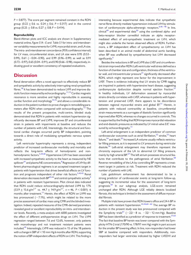
DiscussionRenal denervation offers a novel approach to effectively reduce BPand sympathetic activity by selectively interrupting renal sympatheticfibres.18 It has been demonstrated to reduce LVM and improve dia-stolic function measuredbyechocardiography.11,12 Cardiac magneticresonance is more sensitive and highly reproducible in assessingcardiac function and morphology14,15 and allows a considerable re-duction in the patient numbers to prove changes in remodelling para-meters after RDN when compared with echocardiography.13 Theresults of the present prospective, multi-centre, blinded studydemonstrated that RDN in patients with resistant hypertension sig-nificantly decreases BP and LVMI, improves EF and circumferentialstrain in patients with impairments at baseline and reduces thenumber of patients with LAE. Interestingly, the structural and func-tional cardiac changes occurred partly BP independent, pointingtowards a direct role of modulating sympathetic nervous systemactivity.
Left ventricular hypertrophy represents a strong, independentpredictor of increased cardiovascular morbidity and mortality andreflects the long-term effects of hemodynamic and non-hemodynamic factors.5,39,40 Hypertensive LVH has been associatedwith increased sympathetic activity to the heart as measured by NEspillover41 and plasma NE concentrations.8 Regression of LVH by dif-ferent pharmacological regimens is an accepted treatment target inpatients with hypertension that drives beneficial effects on LV func-tion and prognosis independent of other risk factors.5,40,42 Renaldenervation decreases both BP43,44 and central sympathetic activity9
in patients with resistant hypertension. Pilot clinical data indicatedthat RDN could reduce echocardiography-derived LVMI by 17%(53.9+ 15.6 g/m2.7 vs. 44.7+ 14.9 g/m2.7, n ¼ 46, P , 0.001) 6months after treatment.11 Herein, the effect of RDN on LVMI wasless pronounced (27.1%), which might be related to the moreprecise assessment of cardiac mass using CMR and the blinded inves-tigation. Indeed, repeated measures of the CMR-derived parametersrevealed good or excellent reproducibility on intra- and interobser-ver levels. Recently, a meta-analysis with 6000 patients investigatedthe effect of different antihypertensive drugs on LVH. The LVMIregression ranged between 7.6 and 12.6%, however, only patientswith monotherapy and not treatment resistant patients wereincluded.45 Interestingly, LVMI was reduced in 15 of the 18 patientswith a change in SBP of ,10 mm Hg 6 months after RDN, supportingthe notion of BP-independent effects of RDN on LVH. This finding is
interesting because experimental data indicate that sympatheticnerve fibres directly mediate hypertension-induced LVH by stimula-tion of cardiomyocyte alpha-adrenergic receptors.6,46 –48 In line,clinical49 and experimental data50 using the combined alpha- andbeta-receptor blocker carvedilol indicate an alpha receptor-mediated effect of anti-sympathetic treatment on LVH, whichmight also underlie the herein observed effects of RDN. Further-more, a BP independent effect of sympathectomy on LVH hasbeen described in an animal model of abdominal aortic banding,when BP was unaffected by sympathectomy but LVH decreasedsignificantly.6
Besides the reductions in BP and LVMI also LVEF and circumferen-tial strain improvedafterRDN.Left ventricular wall stress definedas afunction of chamber size and configuration, thickness of the ventricu-lar wall, and intraventricular pressure31 significantly decreased afterRDN, which might represent one factor for the improvement inLVEF. There is evidence indicating that LV strains by CMR taggingare impaired in patients with hypertensive heart disease, suggestingcardiomyocyte dysfunction despite normal ejection fraction.51
In healthy individuals, LV deformation assessed by myocardialstrains directly correlates with LVEF, whereas in patients with hyper-tension and preserved LVEF, there appears to be discordancebetween regional myocardial strains and global EF.51 Herein, inpatients with reduced contractility at baseline, circumferentialstrain measurements as a surrogate of diastolic function significantlyimproved after RDN, whereas no changes occurred in controls. Thisis supported by the finding that RDN improves myocardial relaxationand end-diastolic pressures as indicated by LV mitral valve E/E′ mea-sured by echocardiography.11
Left-atrial enlargement is an independent predictor of commoncardiovascular outcomes such as atrial fibrillation,52 stroke,53 heartfailure,54 and death.55 Increased LA volume usually reflects ventricu-lar filling pressure, as it is exposed to LV pressure during ventriculardiastole.56 Left-atrial enlargement may therefore represent thechronicity exposure of the LA to abnormal LV filling pressure,mainly by high arterial BP.57 The left atrium possesses structural fea-tures that contribute to the pathogenesis of atrial fibrillation.58
Reverse remodelling of the LA by controlling BP represents a treat-ment target in patients at risk. Treatment with RDN reduced thenumber of patients with LAE.
Late gadolinium enhancement has demonstrated to be astrong predictor of cardiovascular events at long-term follow-up,suggesting its incremental value for the assessment of long-termprognosis.59 In our subgroup analysis, LGE-score remainedunchanged after RDN. Although LGE reliably detects localizedfibrosis, this technique is limited in the assessment of diffuse myocar-dial fibrosis.
Multiple trials have proven that RDN lowers office and 24-h BP inpatients with resistant hypertension.43,44,60– 62 The average BP re-duction in the present study was less pronounced compared withthe Symplicity trials61 (222/28 vs. 232/212 mm Hg). BaselineSBP has been identified as a predictor of response to treatment.37,63
The fact that baseline BP herein was lower compared with the Sym-plicity HTN-2 trial (170/90 vs. 178/97 mm Hg) might in part accountfor the smaller BP lowering effect. In line, non-responders had lowerSBP at baseline compared with responders. Additionally, non-responders had larger ventricles indicated by higher LVEDVI and
F. Mahfoud et al.2230
by guest on February 18, 2016http://eurheartj.oxfordjournals.org/
Dow
nloaded from

LVESVI, dilated left atria and significantly increased LVMI, which po-tentially indicate longstanding resistant hypertension with severestructural hypertensive end organ damage. Blood pressure was alsoreduced in the control group (211/27 mm Hg; P ¼ 0.044 for SBPand P ¼ 0.034 for DBP), interestingly, this did not correspond to animprovement in LVMI or EF. Although, some reports suggest thatdiffuse renal artery constriction and local tissue damage at the abla-tion site with oedema and thrombus formation may occur afterRDN,64,65 none of the patients included in the study developed a sig-nificant renal artery stenosis or clinical apparent renal embolismduring follow-up of 6 months.
LimitationsThe non-randomized study design and the small sample size arelimitations of this study that might also limit the evaluation and in-terpretation of subgroups among the cohort, although it representsthe largest cohort of patients undergoing RDN and followed byCMR. Furthermore, CMR is a highly reproducible method, resultingin a considerable reduction in sample sizes of 80–90% when com-pared with echocardiography.13 The control group consisted of 17patients with resistant hypertension on stable antihypertensivemedication, which were not anatomical eligible for the procedureor denied an invasive treatment. We carefully checked whetherthese patients were principally different from those treated withRDN. Except for SBP all other visible potential confounders havenot reached statistical significance between both groups. Due tothe relative small number of patients minor differences betweenthe treatment group and the control group might not havereached statistical significance probably due to power. In consider-ation of the relatively small number of patients propensity scorematching with respect to these characteristics was not done.However, general linear modelling was performed to assess the re-gression to the mean and the confounding by indication issue. Theresults are given in the statistical supplement. Future studies needto address the issue of imbalance between the groups in largercohorts of patients. Patients with an SBP reduction of ≥10 mmHg were subsequently defined as responders to RDN. Althoughthis threshold was not provided by guidelines, it represents a clin-ical relevant BP reduction and was used in the trials.10,43,60 Patientsand physicians were instructed not to change antihypertensivemedication during the study period. However, antihypertensivedrug regimen was reduced in one patient (1%), due to confirmedBP levels below respective target BP and the development of symp-tomatic hypotension. Antihypertensive treatment was increased intwo patients (3%) who remained above target BP during follow-up.Censoring for these post-procedural medication changes did notaffect the improvements in LVMI or function, making a relevant in-fluence of treatment intensification unlikely. Furthermore, it is pos-sible that patients changed their medication themselves, asnon-adherence is a major problem in patients with resistant hyper-tension.66 Adherence to prescribed drug regimen was checkedbefore study entrance and at each visit, making a self-reductionof drug treatment unlikely, although not impossible. However,urine or plasma toxicological analysis of antihypertensive drugsor corresponding metabolites was not part of the study protocol.
ConclusionRenal denervation reduced BP and significantly improved LVH andmyocardial function, as diagnosed by CMR, in patients with resistanthypertension. The beneficial effects of RDN on cardiac remodellingdocumented herein occurred partly BP independent, suggesting aprognosticbenefit ofRDN in patients athigh cardiovascular risk.Ran-domized controlled studies are needed to investigate whether thesechanges correlate to improved outcomes.
Supplementary materialSupplementary material is available at European Heart Journal online.
AcknowledgementsWe thank Alexander Berger, MD, Rolf Gebker, MD, and ThomasHucko, MD for their outstanding help with patient managementand performing the blinded CMR examinations. We also thank thestudy nurses and study coordinators at both sites for their help.
FundingM.B. is supported by Deutsche Forschungsgemeinschaft (KFO 196). F.M.is supported by Deutsche Hochdruckliga. F.M. and M.B. are supported byDeutsche Gesellschaft fur Kardiologie. The DHZB is supported by Foun-dation German Heart Institute Berlin. A.D. is supported by a research fel-lowship of the European Society of Cardiology (ESC).Conflict of interest: All institutions received scientific support fromMedtronic/Ardian. F.M., M.P.S., M.D.E., and M.B. were investigators ofSymplicity HTN-1 and HTN-2 trial. F.M., M.P.S., M.D.E., and M.B. havereceived speaker honorarium and consultancy fees from Medtronic/Ardian, St. Jude, Boston Scientific, and/or Cordis.
References1. Lim SS, Vos T, Flaxman AD, Danaei G, Shibuya K, Adair-Rohani H, Amann M,
Anderson HR, Andrews KG, Aryee M, Atkinson C, Bacchus LJ, Bahalim AN,Balakrishnan K, Balmes J, Barker-Collo S, Baxter A, Bell ML, Blore JD, Blyth F,Bonner C, Borges G, Bourne R, Boussinesq M, Brauer M, Brooks P, Bruce NG,Brunekreef B, Bryan-Hancock C, Bucello C, Buchbinder R, Bull F, Burnett RT,Byers TE, Calabria B, Carapetis J, Carnahan E, Chafe Z, Charlson F, Chen H,Chen JS, Cheng AT, Child JC, Cohen A, Colson KE, Cowie BC, Darby S, Darling S,Davis A, Degenhardt L, Dentener F, Des Jarlais DC, Devries K, Dherani M,Ding EL, Dorsey ER, Driscoll T, Edmond K, Ali SE, Engell RE, Erwin PJ, Fahimi S,Falder G, Farzadfar F, Ferrari A, Finucane MM, Flaxman S, Fowkes FG,Freedman G, Freeman MK, Gakidou E, Ghosh S, Giovannucci E, Gmel G,Graham K, Grainger R, Grant B, Gunnell D, Gutierrez HR, Hall W, Hoek HW,Hogan A, Hosgood HD 3rd, Hoy D, Hu H, Hubbell BJ, Hutchings SJ, Ibeanusi SE,Jacklyn GL, Jasrasaria R, Jonas JB, Kan H, Kanis JA, Kassebaum N, Kawakami N,Khang YH, Khatibzadeh S, Khoo JP, Kok C, Laden F, Lalloo R, Lan Q, Lathlean T,Leasher JL, Leigh J, Li Y, Lin JK, Lipshultz SE, London S, Lozano R, Lu Y, Mak J,Malekzadeh R, Mallinger L, Marcenes W, March L, Marks R, Martin R, McGale P,McGrath J, Mehta S, Mensah GA, Merriman TR, Micha R, Michaud C, Mishra V,Hanafiah KM, Mokdad AA, Morawska L, Mozaffarian D, Murphy T, Naghavi M,Neal B, Nelson PK, Nolla JM, Norman R, Olives C, Omer SB, Orchard J,Osborne R, Ostro B, Page A, Pandey KD, Parry CD, Passmore E, Patra J,Pearce N, Pelizzari PM, Petzold M, Phillips MR, Pope D, Pope CA 3rd, Powles J,Rao M, Razavi H, Rehfuess EA, Rehm JT, Ritz B, Rivara FP, Roberts T, Robinson C,Rodriguez-Portales JA, Romieu I, Room R, Rosenfeld LC, Roy A, Rushton L,Salomon JA, Sampson U, Sanchez-Riera L, Sanman E, Sapkota A, Seedat S, Shi P,Shield K, Shivakoti R, Singh GM, Sleet DA, Smith E, Smith KR, Stapelberg NJ,Steenland K, Stockl H, Stovner LJ, Straif K, Straney L, Thurston GD, Tran JH, VanDingenen R, van Donkelaar A, Veerman JL, Vijayakumar L, Weintraub R,Weissman MM, White RA, Whiteford H, Wiersma ST, Wilkinson JD,Williams HC, Williams W, Wilson N, Woolf AD, Yip P, Zielinski JM, Lopez AD,Murray CJ, Ezzati M, AlMazroa MA, Memish ZA. A comparative risk assessment ofburden of disease and injury attributable to 67 risk factors and risk factor clusters
CMR imaging after renal denervation 2231
by guest on February 18, 2016http://eurheartj.oxfordjournals.org/
Dow
nloaded from
in 21 regions, 1990–2010: a systematic analysis for the Global Burden of DiseaseStudy 2010. Lancet 2012;380:2224–2260.
2. Mancia G, Fagard R, Narkiewicz K, Redon J, Zanchetti A, Bohm M, Christiaens T,Cifkova R, De Backer G, Dominiczak A, Galderisi M, Grobbee DE, Jaarsma T,Kirchhof P, Kjeldsen SE, Laurent S, Manolis AJ, Nilsson PM, Ruilope LM,Schmieder RE, Sirnes PA, Sleight P, Viigimaa M, Waeber B, Zannad F, Burnier M,Ambrosioni E, Caufield M, Coca A, Olsen MH, Tsioufis C, van de Borne P,Zamorano JL, Achenbach S, Baumgartner H, Bax JJ, Bueno H, Dean V, Deaton C,Erol C, Ferrari R, Hasdai D, Hoes AW, Knuuti J, Kolh P, Lancellotti P, Linhart A,Nihoyannopoulos P, Piepoli MF, Ponikowski P, Tamargo JL, Tendera M,Torbicki A, Wijns W, Windecker S, Clement DL, Gillebert TC, Rosei EA,Anker SD, Bauersachs J, Hitij JB, Caulfield M, De Buyzere M, De Geest S,Derumeaux GA, Erdine S, Farsang C, Funck-Brentano C, Gerc V, Germano G,Gielen S, Haller H, Jordan J, Kahan T, Komajda M, Lovic D, Mahrholdt H,Ostergren J, Parati G, Perk J, Polonia J, Popescu BA, Reiner Z, Ryden L, Sirenko Y,Stanton A, Struijker-Boudier H, Vlachopoulos C, Volpe M, Wood DA. 2013 ESH/ESC Guidelines for the management of arterial hypertension: The Task Force forthe management of arterial hypertension of the European Society of Hypertension(ESH) and of the European Society of Cardiology (ESC). Eur Heart J 2013;34:2159–2219.
3. Daugherty SL, Powers JD, Magid DJ, Tavel HM, Masoudi FA, Margolis KL,O’Connor PJ, Selby JV, Ho PM. Incidence and prognosis of resistant hypertensionin hypertensive patients. Circulation 2012;125:1635–1642.
4. Levy D, Larson MG, Vasan RS, Kannel WB, Ho KK. The progression from hyperten-sion to congestive heart failure. JAMA 1996;275:1557–1562.
5. Okin PM, Devereux RB, Jern S, Kjeldsen SE, Julius S, Nieminen MS, Snapinn S,Harris KE, Aurup P, Edelman JM, Wedel H, Lindholm LH, Dahlof B. Regression ofelectrocardiographic left ventricular hypertrophy during antihypertensive treat-ment and the prediction of major cardiovascular events. JAMA 2004;292:2343–2349.
6. Perlini S, Palladini G, Ferrero I, Tozzi R, Fallarini S, Facoetti A, Nano R, Clari F,Busca G, Fogari R, Ferrari AU. Sympathectomy or doxazosin, but not propranolol,blunt myocardial interstitial fibrosis in pressure-overload hypertrophy. Hypertension2005;46:1213–1218.
7. DiBona GF. Physiology in perspective: The Wisdom of the Body. Neural control ofthe kidney. Am J Physiol Regul Integr Comp Physiol 2005;289:R633–R641.
8. Corea L, Bentivoglio M, Verdecchia P. Echocardiographic left ventricular hyper-trophy as related to arterial pressure and plasma norepinephrine concentration inarterial hypertension. Reversal by atenolol treatment. Hypertension 1983;5:837–843.
9. Hering D, Lambert EA, Marusic P, Walton AS, Krum H, Lambert GW, Esler MD,Schlaich MP. Substantial reduction in single sympathetic nerve firing after renal de-nervation in patients with resistant hypertension. Hypertension 2013;61:457–464.
10. Esler MD, Krum H, Schlaich M, Schmieder RE, Bohm M, Sobotka PA. Renal sympa-thetic denervation for treatment of drug-resistant hypertension: one-year resultsfrom the symplicity HTN-2 randomized, controlled trial. Circulation 2012;126:2976–2982.
11. Brandt MC, Mahfoud F, Reda S, Schirmer SH, Erdmann E, Bohm M, Hoppe UC. Renalsympathetic denervation reduces left ventricular hypertrophy and improves cardiacfunction in patients with resistant hypertension. J Am Coll Cardiol 2012;59:901–909.
12. Schirmer SH, Sayed MM, Reil JC, Ukena C, Linz D, Kindermann M, Laufs U,Mahfoud F, Bohm M. Improvements of left-ventricular hypertrophy and diastolicfunction following renal denervation – Effects beyond blood pressure and heartrate reduction. J Am Coll Cardiol 2013; doi: 10.1016/j.jacc.2013.10.073.
13. Bellenger NG, Davies LC, Francis JM, Coats AJ, Pennell DJ. Reduction in sample sizefor studies of remodeling in heart failure by the use of cardiovascular magnetic res-onance. J Cardiovasc Magn Reson 2000;2:271–278.
14. Hendel RC, Patel MR, Kramer CM, Poon M, Carr JC, Gerstad NA, Gillam LD,Hodgson JM, Kim RJ, Lesser JR, Martin ET, Messer JV, Redberg RF, Rubin GD,Rumsfeld JS, Taylor AJ, Weigold WG, Woodard PK, Brindis RG, Douglas PS,Peterson ED, Wolk MJ, Allen JM. ACCF/ACR/SCCT/SCMR/ASNC/NASCI/SCAI/SIR 2006 appropriateness criteria for cardiac computed tomography and cardiacmagnetic resonance imaging: a report of the American College of Cardiology Foun-dation Quality Strategic Directions Committee Appropriateness Criteria WorkingGroup, American College of Radiology, Society of Cardiovascular Computed Tom-ography, Society for Cardiovascular Magnetic Resonance, American Society ofNuclear Cardiology, North American Society for Cardiac Imaging, Society for Car-diovascular Angiography and Interventions, and Society of Interventional Radiology.J Am Coll Cardiol 2006;48:1475–1497.
15. Maceira AM, Mohiaddin RH. Cardiovascular magnetic resonance in systemic hyper-tension. J Cardiovasc Magn Reson 2012;14:28.
16. Achenbach S, Barkhausen J, Beer M, Beerbaum P, Dill T, Eichhorn J, Fratz S,Gutberlet M, Hoffmann M, Huber A, Hunold P, Klein C, Krombach G,Kreitner KF, Kuhne T, Lotz J, Maintz D, Marholdt M, Merkle N, Messroghli D,Miller S, Paetsch I, Radke P, Steen H, Thiele H, Sarikouch S, Fischbach R. Consensus
recommendations of the German Radiology Society (DRG), the German CardiacSociety (DGK) and the German Society for Pediatric Cardiology (DGPK) on theuse of cardiac imaging with computed tomography and magnetic resonanceimaging. Kardiologe 2012;6:105–125.
17. Chobanian AV, Bakris GL, Black HR, Cushman WC, Green LA, Izzo JL Jr, Jones DW,Materson BJ, Oparil S, Wright JT Jr, Roccella EJ. The Seventh Report of the Joint Na-tional Committee on Prevention, Detection, Evaluation, and Treatment of HighBlood Pressure: the JNC 7 report. JAMA 2003;289:2560–2572.
18. Mahfoud F, Luscher TF, Andersson B, Baumgartner I, Cifkova R, Dimario C,Doevendans P, Fagard R, Fajadet J, Komajda M, Lefevre T, Lotan C, Sievert H,Volpe M, Widimsky P, Wijns W, Williams B, Windecker S, Witkowski A, Zeller T,Bohm M. Expert consensus document from the European Society of Cardiologyon catheter-based renal denervation. Eur Heart J 2013;34:2149–2157.
19. Schmieder RE, Redon J, Grassi G, Kjeldsen SE, Mancia G, Narkiewicz K, Parati G,Ruilope L, van de Borne P, Tsioufis C. Updated ESH position paper on interventionaltherapy of resistant hypertension. EuroIntervention 2013;9:R58–R66.
20. Tsioufis C, Mahfoud F, Mancia G, Redon J, Damascelli B, Zeller T, Schmieder RE.What the interventionalist should know about renal denervation in hypertensivepatients: a position paper by the ESH WG on the interventional treatment of hyper-tension. EuroIntervention 2014;9:1027–1035.
21. Kelle S, RoesSD, Klein C, Kokocinski T, de Roos A, Fleck E, Bax JJ, Nagel E. Prognosticvalue of myocardial infarct size and contractile reserve using magnetic resonanceimaging. J Am Coll Cardiol 2009;54:1770–1777.
22. Schulz-Menger J, Bluemke DA, Bremerich J, Flamm SD, Fogel MA, Friedrich MG,Kim RJ, von Knobelsdorff-Brenkenhoff F, Kramer CM, Pennell DJ, Plein S, Nagel E.Standardized image interpretation and post processing in cardiovascular magneticresonance: Society for Cardiovascular Magnetic Resonance (SCMR) board of trus-tees task force on standardized post processing. J Cardiovasc Magn Reson 2013;15:35.
23. Vogel-Claussen J, Finn JP, Gomes AS, Hundley GW, Jerosch-Herold M, Pearson G,Sinha S, Lima JA, Bluemke DA. Left ventricular papillary muscle mass: relationshipto left ventricular mass and volumes by magnetic resonance imaging. J ComputAssist Tomogr 2006;30:426–432.
24. Papavassiliu T, Kuhl HP, van Dockum W, Hofman MB, Bondarenko O, Beek IA, vanRossum AC. Accuracy of one- and two-dimensional algorithms with optimal imageplane position for the estimation of left ventricular mass: a comparative study usingmagnetic resonance imaging. J Cardiovasc Magn Reson 2004;6:845–854.
25. Maceira AM, Prasad SK, Khan M, Pennell DJ. Normalized left ventricular systolic anddiastolic functionby steadystate freeprecessioncardiovascularmagnetic resonance.J Cardiovasc Magn Reson 2006;8:417–426.
26. Armstrong AC, Gidding S, Gjesdal O, Wu C, Bluemke DA, Lima JA. LV mass assessedby echocardiography and CMR, cardiovascular outcomes, and medical practice.JACC Cardiovasc Imaging 2012;5:837–848.
27. Puntmann VO, Gebker R, Duckett S, Mirelis J, Schnackenburg B, Graefe M, Razavi R,Fleck E, Nagel E. Left ventricular chamber dimensions and wall thickness bycardiovascular magnetic resonance: comparison with transthoracic echocardiog-raphy. Eur Heart J Cardiovasc Imaging 2013;14:240–246.
28. Nakatani S, White RD, Powell KA, Lever HM, Thomas JD. Dynamic magnetic reson-ance imaging assessment of the effect of ventricular wall curvature on regional func-tion in hypertrophic cardiomyopathy. Am J Cardiol 1996;77:618–622.
29. Foppa M, Duncan BB, Rohde LE. Echocardiography-based left ventricular mass esti-mation. How should we define hypertrophy? Cardiovasc Ultrasound 2005;3:17.
30. Yin FC. Ventricular wall stress. Circ Res 1981;49:829–842.31. Grossman W, Jones D, McLaurin LP. Wall stress and patterns of hypertrophy in the
human left ventricle. J Clin Invest 1975;56:56–64.32. Holtzman DG, Duncanson L, McLaughlin J, Reichek N, Kadiyala M, Cao JJ. Abstract
12155: Analysis of Stress-Strain Relationships Using Cardiac MRI. Circulation 2013;128:A12155.
33. Cerqueira MD, Weissman NJ, Dilsizian V, Jacobs AK, Kaul S, Laskey WK, Pennell DJ,Rumberger JA, Ryan T, Verani MS. Standardized myocardial segmentation and no-menclature for tomographic imaging of the heart. A statement for healthcare profes-sionals fromthe Cardiac Imaging Committeeof the Council on Clinical Cardiologyofthe American Heart Association. Circulation 2002;105:539–542.
34. Caudron J, Fares J, Bauer F, Dacher JN. Evaluation of left ventricular diastolic functionwith cardiac MR imaging. Radiographics 2011;31:239–259.
35. Wu E, Judd RM, Vargas JD, Klocke FJ, Bonow RO, Kim RJ. Visualisation of presence,location, and transmural extent of healed Q-wave and non-Q-wave myocardialinfarction. Lancet 2001;357:21–28.
36. Oppo K, Leen E, Angerson WJ, Cooke TG, McArdle CS. Dopplerperfusion index: aninterobserver and intraobserver reproducibility study. Radiology 1998;208:453–457.
37. Ukena C, Cremers B, Ewen S, Bohm M, Mahfoud F. Response and non-response torenal denervation: who is the ideal candidate? EuroIntervention 2013;9(Suppl. R):R54–R57.
38. Epstein FH. MRI of left ventricular function. J Nucl Cardiol 2007;14:729–744.
F. Mahfoud et al.2231a
by guest on February 18, 2016http://eurheartj.oxfordjournals.org/
Dow
nloaded from

39. Yeboah J, Rodriguez CJ, Stacey B, Lima JA, Liu S, Carr JJ, Hundley WG,Herrington DM. Prognosis of individuals with asymptomatic left ventricular systolicdysfunction in the multi-ethnic study of atherosclerosis (MESA). Circulation 2012;126:2713–2719.
40. Okin PM, Devereux RB, Harris KE, Jern S, Kjeldsen SE, Julius S, Edelman JM, Dahlof B.Regression of electrocardiographic left ventricular hypertrophy is associated withless hospitalization for heart failure in hypertensive patients. Ann Intern Med 2007;147:311–319.
41. Schlaich MP, Kaye DM, Lambert E, Sommerville M, Socratous F, Esler MD. Relationbetween cardiac sympathetic activity and hypertensive left ventricular hypertrophy.Circulation 2003;108:560–565.
42. Verdecchia P, Sleight P, Mancia G, Fagard R, Trimarco B, Schmieder RE, Kim JH,Jennings G, Jansky P, Chen JH, Liu L, Gao P, Probstfield J, Teo K, Yusuf S. Effects oftelmisartan, ramipril, and their combination on left ventricular hypertrophy in indi-viduals at high vascular risk in the Ongoing Telmisartan Alone and in CombinationWith Ramipril Global End Point Trial and the Telmisartan Randomized AssessmentStudy in ACE Intolerant Subjects With Cardiovascular Disease. Circulation 2009;120:1380–1389.
43. Worthley SG, Tsioufis CP, Worthley MI, Sinhal A, Chew DP, Meredith IT,Malaiapan Y, Papademetriou V. Safety and efficacy of a multi-electrode renal sympa-thetic denervation system in resistant hypertension: the EnligHTN I trial. Eur Heart J2013;34:2132–2140.
44. Mahfoud F, Ukena C, Schmieder RE, Cremers B, Rump LC, Vonend O, Weil J,Schmidt M, Hoppe UC, Zeller T, Bauer A, Ott C, Blessing E, Sobotka PA, Krum H,Schlaich M, Esler M, Bohm M. Ambulatory blood pressure changes after renal sym-pathetic denervation in patients with resistant hypertension. Circulation 2013;128:132–140.
45. Fagard RH, Celis H, Thijs L, Wouters S. Regression of left ventricular mass by anti-hypertensive treatment: a meta-analysis of randomized comparative studies. Hyper-tension 2009;54:1084–1091.
46. Bohm M, Mende U, Schmitz W, Scholz H. Cardiac alpha-receptors and cardiachypertrophy in genetic predisposition to hypertension. Am Heart J 1986;112:1347–1349.
47. Bohm M, Mende U, Schmitz W, Scholz H. Increased sensitivity toalpha-adrenoceptor stimulation but intact purinergic and muscarinergic effects inprehypertensive cardiac hypertrophy of spontaneously hypertensive rats. NaunynSchmiedebergs Arch Pharmacol 1986;333:284–289.
48. Sen S, Tarazi RC, Khairallah PA, Bumpus FM. Cardiac hypertrophy in spontaneouslyhypertensive rats. Circ Res 1974;35:775–781.
49. Metra M, Torp-Pedersen C, Swedberg K, Cleland JG, Di Lenarda A, Komajda M,Remme WJ, Lutiger B, Scherhag A, Lukas MA, Charlesworth A, Poole-Wilson PA.Influence of heart rate, blood pressure, and beta-blocker dose on outcome andthe differences in outcome between carvedilol and metoprolol tartrate in patientswith chronic heart failure: results from the COMET trial. Eur Heart J 2005;26:2259–2268.
50. Hanada K, Asari K, Saito M, Kawana J, Mita M, Ogata H. Comparison of pharmaco-dynamics between carvedilol and metoprolol in rats with isoproterenol-inducedcardiac hypertrophy: effects of carvedilol enantiomers. Eur J Pharmacol 2008;589:194–200.
51. Ahmed MI, Desai RV, Gaddam KK, Venkatesh BA, Agarwal S, Inusah S, Lloyd SG,Denney TS Jr, Calhoun D, Dell’italia LJ, Gupta H. Relation of torsion and myocardialstrains to LV ejection fraction in hypertension. JACC Cardiovasc Imaging 2012;5:273–281.
52. Verdecchia P, Reboldi G, Gattobigio R, Bentivoglio M, Borgioni C, Angeli F,Carluccio E, Sardone MG, Porcellati C. Atrial fibrillation in hypertension: predictorsand outcome. Hypertension 2003;41:218–223.
53. Nagarajarao HS, Penman AD, Taylor HA, Mosley TH, Butler K, Skelton TN,Samdarshi TE, Aru G, Fox ER. The predictive value of left atrial size for incident is-chemic stroke and all-cause mortality in African Americans: the AtherosclerosisRisk in Communities (ARIC) Study. Stroke 2008;39:2701–2706.
54. Gerdts E, Oikarinen L, Palmieri V, Otterstad JE, Wachtell K, Boman K, Dahlof B,Devereux RB. Correlates of left atrial size in hypertensive patients with left ventricu-lar hypertrophy: the Losartan Intervention For Endpoint Reduction in Hypertension(LIFE) Study. Hypertension 2002;39:739–743.
55. Laukkanen JA, Kurl S, Eranen J,Huttunen M,Salonen JT. Left atriumsize and the riskofcardiovascular death in middle-aged men. Arch Intern Med 2005;165:1788–1793.
56. AbhayaratnaWP, Seward JB, Appleton CP, Douglas PS, Oh JK, TajikAJ,Tsang TS. Leftatrial size: physiologic determinants and clinical applications. J Am Coll Cardiol 2006;47:2357–2363.
57. Raman SV. The hypertensive heart. An integrated understanding informed byimaging. J Am Coll Cardiol 2010;55:91–96.
58. Iwasaki YK, Nishida K, Kato T, Nattel S. Atrial fibrillation pathophysiology: implica-tions for management. Circulation 2011;124:2264–2274.
59. Kelle S, Nagel E, Voss A, Hofmann N, Gitsioudis G, Buss SJ, Chiribiri A, Wellnhofer E,Klein C, Schneeweis C, Egnell C, Vierecke J, Berger A, Giannitsis E, Fleck E, Katus HA,Korosoglou G. A bi-center cardiovascular magnetic resonance prognosis study fo-cusing on dobutamine wall motion and late gadolinium enhancement in 3,138 con-secutive patients. J Am Coll Cardiol 2013;61:2310–2312.
60. Krum H, Schlaich MP, Bohm M, Mahfoud F, Rocha-Singh K, Katholi R, Esler MD.Percutaneous renal denervation in patients with treatment-resistant hypertension:final 3-year report of the Symplicity HTN-1 study. Lancet 2014;383:622–629.
61. Esler MD, Krum H, Sobotka PA, Schlaich MP, Schmieder RE, Bohm M. Renal sympa-thetic denervation inpatientswith treatment-resistant hypertension (The SymplicityHTN-2 Trial): a randomised controlled trial. Lancet 2010;376:1903–1909.
62. Vogel B, Kirchberger M, Zeier M, Stoll F, Meder B, Saure D, Andrassy M, Mueller OJ,Hardt S, Schwenger V, Strothmeyer A, Katus HA, Blessing E. Renal sympathetic de-nervation therapy in the real world: results from the Heidelberg registry. Clin ResCardiol 2014;103:117–124.
63. Mahfoud F, Cremers B, Janker J, Link B, Vonend O, Ukena C, Linz D, Schmieder R,Rump LC, Kindermann I, Sobotka PA, Krum H, Scheller B, Schlaich M, Laufs U,Bohm M. Renal hemodynamics and renal function after catheter-based renal sympa-thetic denervation in patients with resistant hypertension. Hypertension 2012;60:419–424.
64. Templin C, Jaguszewski M, Ghadri JR, Sudano I, Gaehwiler R, Hellermann JP,Schoenenberger-Berzins R, Landmesser U, Erne P, Noll G, Luscher TF. Vascularlesions induced by renal nerve ablation as assessed by optical coherence tomog-raphy: pre- and post-procedural comparison with the Simplicity(R) cathetersystem and the EnligHTN multi-electrode renal denervation catheter. Eur Heart J2013;34:2141–2148.
65. Stabile E, Ambrosini V, Squarcia R, Salemme L, Popusoi G, Esposito G, Trimarco B,Rubino P. Percutaneous sympathectomy of the renal arteries: the OneShot RenalDenervation System is not associated with significant vessel wall injury. EuroInterven-tion 2013;9:694–699.
66. Jung O, Gechter JL, Wunder C, Paulke A, Bartel C, Geiger H, Toennes SW. Resistanthypertension? Assessment of adherence by toxicological urine analysis. J Hypertens2013;31:766–774.
CMR imaging after renal denervation 2231b
by guest on February 18, 2016http://eurheartj.oxfordjournals.org/
Dow
nloaded from
Related Documents