International Journal of Clinical and Developmental Anatomy 2016; 2(3): 17-23 http://www.sciencepublishinggroup.com/j/ijcda doi: 10.11648/j.ijcda.20160203.11 ISSN: 2469-7990 (Print); ISSN: 2469-8008 (Online) Effect of Pirfenidone on Bleomycin Induced Pulmonary Alveolar Fibrosis in Adult Male Rats (Histological, Immunohistochemical, Morphometrical and Biochemical Study) Ayman M. Mousa 1, 2 1 Department of Histology and Cell Biology, Benha Faculty of Medicine, Benha University, Cairo, Egypt 2 Department of Basic Health Sciences, College of Applied Medical Sciences, Qassim University, KSA Email address: [email protected] To cite this article: Ayman M. Mousa. Effect of Pirfenidone on Bleomycin Induced Pulmonary Alveolar Fibrosis in Adult Male Rats (Histological, Immunohistochemical, Morphometrical and Biochemical Study). International Journal of Clinical and Developmental Anatomy. Vol. 2, No. 3, 2016, pp. 17-23. doi: 10.11648/j.ijcda.20160203.11 Received: May 26, 2016; Accepted: June 3, 2016; Published: June 12, 2016 Abstract: Introduction: Bleomycin is a chemotherapeutic agent commonly used to treat curable diseases such as Hodgkin’s lymphoma. The major limitation of bleomycin therapy is the pulmonary toxicity. Pirfenidone is a modified phenyl pyridine that has an antioxidant, anti-transforming growth factor and anti-platelet derived growth factor effects. Aim of the study: to evaluate the histological, immunohistochemical and biochemical changes in the pulmonary alveoli of adult male albino rats after intake of bleomycin and the possible role of pirfenidone in minimizing these changes. Material and Methods: Forty adult male albino rats were used in this study. They were divided equally into 4 equal groups; the first group (control), the second group that received bleomycin for 10 days, the third group that received pirfenidone for 10 days and the fourth group that received pirfenidone & bleomycin for 10 days. The lungs were dissected out, processed and lung sections were stained with Hx&E, Masson's trichrome and immunohistochemicaly. Then they were examined by light microscope for histological and immuno- histochemical study to evaluate the structure of pulmonary alveoli. Biochemical measurement of malondialdehyde (MDA), glutathione peroxidase (GSH-Px) and tumor necrosis factor-α (TNF-α) were also performed. Results: Bleomycin treatment in the second group induced alveolar inflammation, interstitial pulmonary inflammation and pulmonary alveolar fibrosis, while pirfenidone significantly reduced these induced lung injuries in the fourth group rats that treated with pirfenidone and bleomycin. These protective effects were associated with a significant (P<0.05) reduction in the levels of MDA, and TNF-α associated with a significant (P<0.05) increase in the levels of GSH-P in the homogenate of lung tissue compared with the second group. Conclusion: The present study showed a protective effect of pirfenidone on the structure of pulmonary alveoli subjected to bleomycin intake. So intake of pirfenidone with bleomycin is advised for treatment of pulmonary alveolar toxicity. Keywords: Bleomycin, Pirfenidone, Pulmonary Fibrosis, Inflammatory Cytokines 1. Introduction Pulmonary fibrosis is a chronic and serious lung disease, of unknown etiology limited to the lungs that can be developed as a complication of many respiratory and systemic diseases.[1] It causes replacement of normal lung tissue with scar tissue or excess fibrous connective tissue. It is also characterized by alveolar epithelial cell injury, interstitial inflammation, fibroblast proliferation and impairment of lung function.[2] Bleomycin is the most widely used experimental model of lung fibrosis, because the pathology in rats is very similar to human. 1 It is a chemotherapeutic antibiotic, produced by the bacterium “Streptomyces Verticillus” that is used as an anticancer drug mainly in treatment of Hodgkin, non-Hodgkin lymphomas and testicular carcinoma.[3] Bleomycin reduces molecular oxygen to superoxide and hydroxyl radicals which cause DNA strand cleavage or breakdown.[4]

Welcome message from author
This document is posted to help you gain knowledge. Please leave a comment to let me know what you think about it! Share it to your friends and learn new things together.
Transcript
International Journal of Clinical and Developmental Anatomy 2016; 2(3): 17-23
http://www.sciencepublishinggroup.com/j/ijcda
doi: 10.11648/j.ijcda.20160203.11
ISSN: 2469-7990 (Print); ISSN: 2469-8008 (Online)
Effect of Pirfenidone on Bleomycin Induced Pulmonary Alveolar Fibrosis in Adult Male Rats (Histological, Immunohistochemical, Morphometrical and Biochemical Study)
Ayman M. Mousa1, 2
1Department of Histology and Cell Biology, Benha Faculty of Medicine, Benha University, Cairo, Egypt 2Department of Basic Health Sciences, College of Applied Medical Sciences, Qassim University, KSA
Email address:
To cite this article: Ayman M. Mousa. Effect of Pirfenidone on Bleomycin Induced Pulmonary Alveolar Fibrosis in Adult Male Rats (Histological,
Immunohistochemical, Morphometrical and Biochemical Study). International Journal of Clinical and Developmental Anatomy.
Vol. 2, No. 3, 2016, pp. 17-23. doi: 10.11648/j.ijcda.20160203.11
Received: May 26, 2016; Accepted: June 3, 2016; Published: June 12, 2016
Abstract: Introduction: Bleomycin is a chemotherapeutic agent commonly used to treat curable diseases such as Hodgkin’s
lymphoma. The major limitation of bleomycin therapy is the pulmonary toxicity. Pirfenidone is a modified phenyl pyridine that
has an antioxidant, anti-transforming growth factor and anti-platelet derived growth factor effects. Aim of the study: to evaluate
the histological, immunohistochemical and biochemical changes in the pulmonary alveoli of adult male albino rats after intake
of bleomycin and the possible role of pirfenidone in minimizing these changes. Material and Methods: Forty adult male albino
rats were used in this study. They were divided equally into 4 equal groups; the first group (control), the second group that
received bleomycin for 10 days, the third group that received pirfenidone for 10 days and the fourth group that received
pirfenidone & bleomycin for 10 days. The lungs were dissected out, processed and lung sections were stained with Hx&E,
Masson's trichrome and immunohistochemicaly. Then they were examined by light microscope for histological and immuno-
histochemical study to evaluate the structure of pulmonary alveoli. Biochemical measurement of malondialdehyde (MDA),
glutathione peroxidase (GSH-Px) and tumor necrosis factor-α (TNF-α) were also performed. Results: Bleomycin treatment in
the second group induced alveolar inflammation, interstitial pulmonary inflammation and pulmonary alveolar fibrosis, while
pirfenidone significantly reduced these induced lung injuries in the fourth group rats that treated with pirfenidone and
bleomycin. These protective effects were associated with a significant (P<0.05) reduction in the levels of MDA, and TNF-α
associated with a significant (P<0.05) increase in the levels of GSH-P in the homogenate of lung tissue compared with the
second group. Conclusion: The present study showed a protective effect of pirfenidone on the structure of pulmonary alveoli
subjected to bleomycin intake. So intake of pirfenidone with bleomycin is advised for treatment of pulmonary alveolar toxicity.
Keywords: Bleomycin, Pirfenidone, Pulmonary Fibrosis, Inflammatory Cytokines
1. Introduction
Pulmonary fibrosis is a chronic and serious lung disease, of
unknown etiology limited to the lungs that can be developed
as a complication of many respiratory and systemic
diseases.[1] It causes replacement of normal lung tissue with
scar tissue or excess fibrous connective tissue. It is also
characterized by alveolar epithelial cell injury, interstitial
inflammation, fibroblast proliferation and impairment of lung
function.[2]
Bleomycin is the most widely used experimental model of
lung fibrosis, because the pathology in rats is very similar to
human.1 It is a chemotherapeutic antibiotic, produced by the
bacterium “Streptomyces Verticillus” that is used as an
anticancer drug mainly in treatment of Hodgkin, non-Hodgkin
lymphomas and testicular carcinoma.[3] Bleomycin reduces
molecular oxygen to superoxide and hydroxyl radicals which
cause DNA strand cleavage or breakdown.[4]

18 Ayman M. Mousa: Pirfenidone Effect on Induced Lung Injury
Currently, there are no approved medical antifibrotic
therapies for pulmonary fibrosis.[5] Pirfenidone is an orally
active small molecule comprising a modified phenyl pyridine
that is able to move through cell membranes without
requiring a receptor. It is easily absorbed from the
gastrointestinal tract after oral administration with a peak
blood level after 1–2 h. It crosses the blood-brain barrier and
is eliminated in urine within 6 hours. Modulation of
fibrogenesis by pirfenidone is still unclear in detail, but its
effects are probably multi-targeted because it has antioxidant,
anti-transforming growth factor (anti-TGF) and anti-platelet
derived growth factor effects.[6] The most common adverse
effects of pirfenidone include gastrointestinal symptoms
(nausea, dyspepsia, diarrhea, abdominal discomfort, and
vomiting), anorexia, fatigue, sedation and photosensitivity
rash. Overall, pirfenidone appears to be reasonably safe in
various patient populations with chronic fibrotic disorders,
multiple sclerosis, chronic hepatitis C and chronic allograft
rejection.[7]
The aim of the present study was to evaluate the possible
protective effect of pirfenidone on bleomycin induced
pulmonary fibrosis in the lung of adult male albino rats.
2. Materials and Methods
In this study, forty adult male albino rats weighing 150–250
gm were used. The animals were obtained and housed at the
animal laboratory house, Moshtohor faculty of Veterinary
Medicine, Benha University, Egypt. Strict care and cleaning
measures were utilized to keep the animals in a normal
healthy state. The animals were housed in animal cages at
room temperature (25±1°C), relative humidity (55±5) with
12h light/12h dark cycle, fed standard balanced diet and water
ad-libitum. All ethical protocols for animal treatment were
followed and the experimental protocol was approved by the
Ethical Committee of Benha Faculty of Medicine.
Used drugs: Bleomycin hydrochloride (Nippon Kayaku,
Japan), 15 mg powder per vial was dissolved in normal sterile
saline as a vehicle. Pirfenidone (Licheng Chemical Co. Ltd.
Shanghai, China) was dissolved in a 0.5% carboxy-
methylcellulose solution as a vehicle.
Experimental design: Rats were divided into four equal
groups (10 rats for each).
Group 1 (Control group= G1): The animals of this group
were further subdivided into 2 subgroups each one included 5
rats.
Subgroup 1A: Rats were injected intraperitonealy (IP) with
saline as a vehicle once daily for 10 days, and then sacrificed
at the same time as the corresponding experimental groups.
Subgroup 1B: Rats were received 0.5% carboxy-
methylcellulose solution as a vehicle orally by gastric tube for
10 days, and then sacrificed at the same time as the corre-
sponding experimental groups.
Group 2 (Bleomycin group= G2): Rats were received
bleomycin hydrochloride IP in a dose of (10 mg/kg of body
weight/ once daily for10 days) to induce lung fibrosis.[8]
Group 3 (Pirfenidone group= G3): Rats were received
pirfenidone orally by a gastric tube in a dose of (500 mg/kg of
body weight once daily for10 days). [9]
Group 4 (Bleomycin and Pirfenidone group= G4): Rats
were received pirfenidone and bleomycin as in the previous
groups for10 days.
At the end of the experiment, the rats were anaesthetized
by inhalation of ether, sacrificed and then the lungs were
exposed and excised. The lung biopsies were divided and
fixed immediately in 10 % neutral buffered formalin. Paraffin
sections were prepared and stained with hematoxylin and
eosin (Hx&E) to verify the general histological details of the
lungs and Masson’s trichrome to assess the sub-epithelial
collagen deposition.[10]
Immunohistochemical study: was performed to detect α-
smooth muscle actin (α-SMA) as a marker of myofibroblast
differentiation and fibrosis. Anti α-SMA immuno-
histochemical staining was done as follow: Paraffin sections
were deparaffinized in xylene for 1-2 minutes, rehydrated in
descending grades of ethanol and then brought to distilled
water for 5 minutes. Sections were incubated in hydrogen
peroxide for 30 minutes then rinsed in PBS (3 times, 2
minutes each). Each section was incubated for 60 minutes
with 2 drops (100 µl) of the primary antibody (α-SMA mouse
monoclonal antibody, (Lab. Vision Corporation laboratories,
CA 94538, USA, catalogue number MS-113-R7). Slides were
rinsed well in PBS (3 times, 2 minutes each), incubated for 20
minutes with 2 drops of biotinylated secondary antibody for
each section then rinsed well with PBS. Each section was
incubated with 2 drops (100 µl) of enzyme conjugate
"Streptavidin-Horseradish peroxidase" for 10 minutes at room
temperature then washed in PBS. Two drops of the substrate-
chromogen mixture diaminobenzidine (DAB) were applied to
each section and incubated at room temperature for 5-10
minutes then rinsed well with distilled water. The sections
were counterstained with Mayer’s hematoxylin (Sigma-
Aldrich Co., St Louis, MO, USA) then dehydrated and
mounted. α-SMA +ve cells showed brown cytoplasmic
deposits and the primary antibody was omitted for negative
control sections.[11]
Biochemical measurements: Portions of lung tissues were
homogenized in a saline solution (0.9%), centrifuged at 3000
rpm for 15 min, and the supernatant was stored at -20°C until
they were analyzed for:
1. Malondialdehyde (MDA) which is the breakdown
product of lipid peroxidation that was analyzed to
determine lipid peroxidation.[12]
2. Glutathione peroxidase (GSH-Px) which is a lung
content that was determined by using a commercial kit
(Biodiagnostic, Egypt).[13]
3. Tumor necrosis factor-α (TNF-α) which is a lung
proinflammatory cytokine that was measured by using
the commercially available sandwich enzyme-linked
immunosorbent assay (ELISA) kits for rats according to
manufacturer’s instructions (Sigma-Aldrich Co., St
Louis, MO, USA) The results were expressed as
picograms per milligram of tissue protein (pg/mg).[14]
Morphometric analysis: The Image-Pro Plus program

International Journal of Clinical and Developmental Anatomy 2016; 2(3): 17-23 19
version 6.0 (Media Cybernetics Inc., Bethesda, Maryland,
USA) was used to determine the following:
1- The mean area % of the stained collagen fibers in the
lungs of different experimental groups.
2- The mean area % of α-SMA immunohistohemical
expression in the lungs of different experimental groups.
Statistical analysis: The histological and
immunohistochemical data were analyzed by using the
statistical package SPSS version 20 (SPSS Inc., Chicago,
Illinois, USA). Data were expressed as mean ± SD. The
statistical significance in differences between groups was
analyzed by using one-way analysis of variance (ANOVA)
test, followed by the post-hoc test of Tukey’s to compare
the mean area % of collagen fibers and α-SMA immuno-
histohemical expression in the lungs of different
experimental groups.
P values < 0.01 were considered a highly significant and
< 0.05 were considered significant.
3. Results
1. Histological results:
A. Hematoxylin and eosin:
Sections of G1 showed normal histological architecture
of the lung with many alveoli, alveolar sacs, alveolar ducts,
bronchioles and small blood vessels (Fig.1). While G2
showed a various histological changes in the form of many
collapsed alveoli, dilated or ruptured alveoli and
extravasated RBCs with a multiple thick interalveolar septa
between the alveoli (Fig. 2). Other sections from G2
revealed multiple thick interalveolar septa between
collapsed alveoli and were studded with mononuclear
cellular infiltration (Fig. 3).
On the other hand G3 showed a histological picture
nearly similar to the normal histological architecture of the
lung (Fig. 4) while, G4 rats showed a picture more or less
similar to that of G1 that had many alveoli with apparently
thin interalveolar septa, while few interalveolar septa were
thick and studded with mononuclear cellular infiltration
(Fig. 5).
B. Masson’s trichrome stain:
Sections of G1 revealed a minimal amount of collagen
fibers around the alveoli or within the interalveolar septa with
small blood vessels (Fig.6). However, G2 rats showed an
extensive accumulation of collagen fibers around alveoli,
bronchioles and small blood vessels or within the
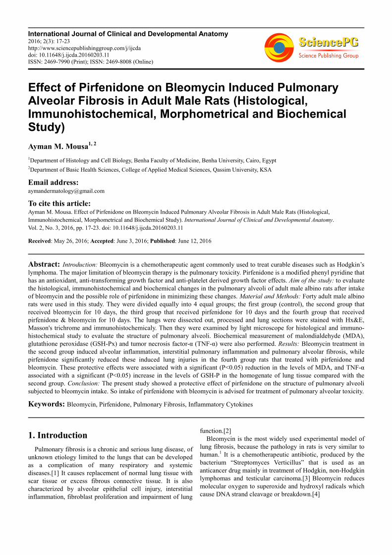
interalveolar septa (Fig.7). On the other hand G3 sections
showed a minimal amount of collagen fibers (Fig. 8) while,
G4 rats showed a moderate amount of collagen fibers around
the alveoli and small blood vessels or within the interalveolar
septa (Fig. 9).
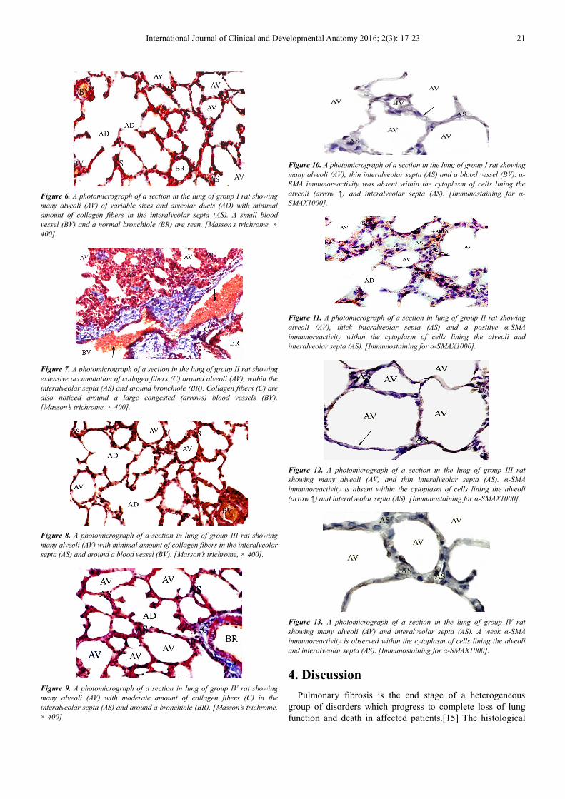
1. Immunohistochemical results: Sections of G1 revealed
absence of α-SMA immuno-reactivity (Fig.10) while, G2
rats showed a positive immuno-reactivity of α-SMA within
the cytoplasm of cells lining the alveoli and interalveolar
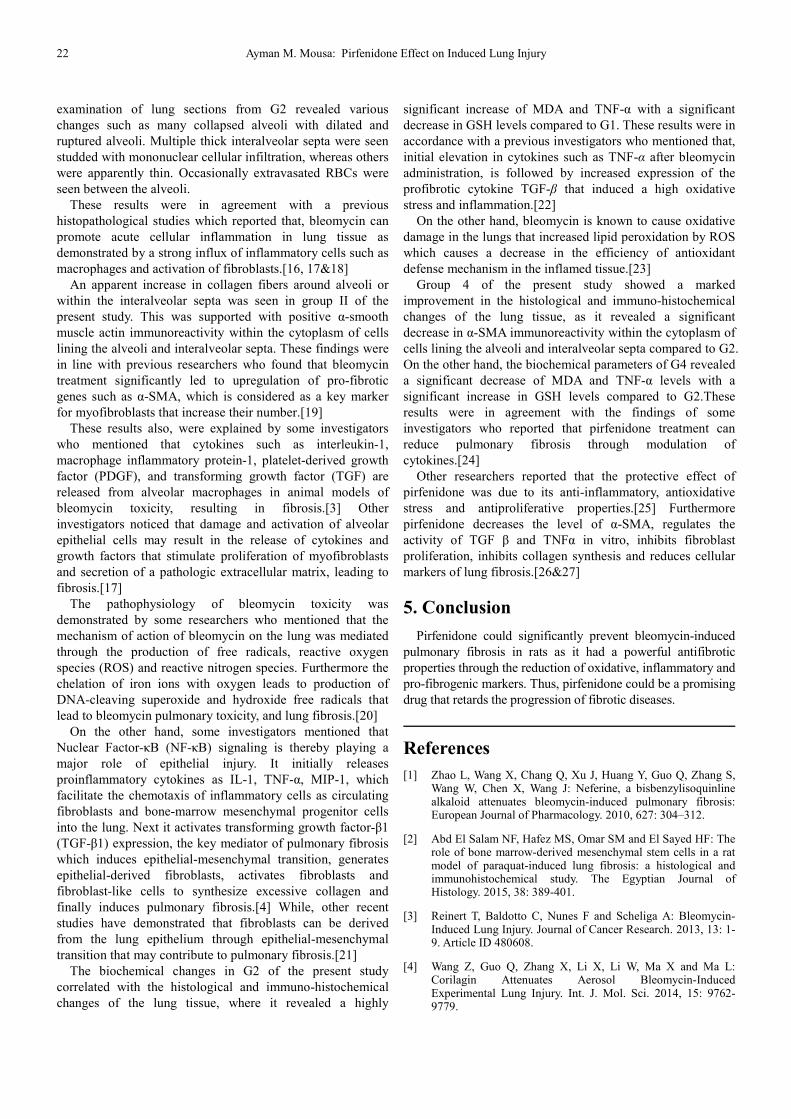
septa (Fig.11). On the other hand G3 sections showed
absence of α-SMA immunoreactivity (Fig.12) while, G4 rats
showed a weak immunoreactivity of α-SMA within the
cytoplasm of cells lining the alveoli and interalveolar septa
(Fig. 13).
2. Morphometric results: The mean area % of collagen fibers
and α-SMA immunoexpression for all groups were
presented in table 1 and histogram1. The mean area % of
collagen fibers and α-SMA immunoreactivity showed a
highly significant increase in G2 compared with G1
(P<0.01) while, they were significantly decreased in G3 and
G4 compared with G2 (P<0.05).
Table 1. Showing the mean area % of collagen fibers ± SD and the mean area % of smooth muscle actin (α-SMA) immuno-expression ± SD in all experimental
groups.
Groups G1 G2 G3 G4
The mean area% of
collagen fibers
Mean ± SD 1.02±0.89 11.35±2.37 2.21±1.42 3.38±1.79
P value 0.00** 0.17* 0.14*
The mean area% of
α-SMA immuno-expression
Mean ± SD 0.47±0.17 19.22±0.26 1.40±0.27 3.13±0.35
P value 0.00** 0.17* 0.21*
SD = Standard deviation, highly significant** (P<0.01) for G2 compared with G1, and significant* (P<0.05) for G3 and G4 compared with G2. (ANOVA test).
Histogram 1. Showing the mean area % of collagen fibers and the mean area % of α-SMA immuno-expression in all experimental groups.

20 Ayman M. Mousa: Pirfenidone Effect on Induced Lung Injury
3. Biochemical Results: As shown in table 2 and histogram
2, the MDA and TNF-α showed a highly significant
increase (P < 0.01) in G2 compared to G1, while they
were significantly decreased in G3 and G4 compared to
G2 (P < 0.05). On the other hand, GSH-Px activity
showed a highly significant decrease (P < 0.01) in G2
compared to G1, while they were significantly increased
in G3 and G4 compared to G2 (P < 0.05).
Table 2. Showing changes in MDA, GSH-Px and TNF-α levels in all experimental groups.
Groups G1 G2 G3 G4
MDA (nmol/g tissue) Mean ± SD 14.6 ± 1.82 65.76 ±5.7** 16.24 ± 1.06* 20.25±2.41*
P value 0.000** 0.212* 0.147*
GSH-Px (u/mg tissue) Mean ± SD 8.8 ± 0.36 1.9 ± 0.3 7.9 ± 0.36 6.8 ± 0.36
P value 0.000** 0.110* 0.145*
TNF-α (Pg/mg tissue) Mean ± SD 19.36± 3.7 80.1± 7.6** 25.4± 4.1* 28.7± 5.6*
P value 0.000** 0.110* 0.145*
Malondialdehyde (MDA), glutathione peroxidase (GSH-Px), tumor necrosis factor α (TNF-α), SD = standard deviation, highly significant** for G2 compared
with G1 and significant* for G3 and G4 compared with G2. (ANOVA test).
Histogram (2). Showing changes in MDA, TNF-α and GSH-Px levels
in all experimental groups.
Figure 1. A photomicrograph of a section in the lung of group I rat showing
many alveoli (AV), pneumocytes (arrow ↑), alveolar ducts (AD), thin
interalveolar septa (AS), bronchiole (BR) and a small blood vessel (BV).
[Hx&E, X 640].
Figure 2. A photomicrograph of a section in lung of group II rat showing many collapsed alveoli (CAV), dilated alveoli (DAV) and ruptured alveoli
(AV). Also, there is thick interalveolar septa (As) studded with mononuclear
cellular infiltrations (↑), few thin interalveolar septa (arrowhead▲) and
extravasated RBCs.[Hx&E, X 640].
Figure 3. A photomicrograph of a section in the lung of group II rat showing
many collapsed alveoli (CAV), thick interalveolar septa (AS), studded with
diffuse mononuclear cellular infiltration (arrow↑) and a small blood vessel
(BV). [Hx&E, X 640].
Figure 4. A photomicrograph of a section in the lung of group III rat showing
many normal alveoli (AV) of variable sizes, alveolar ducts (AD), thin
interalveolar septa (arrow ↑) and a bronchiole (BR). [Hx&E, X 640].
Figure 5. A photomicrograph of a section in lung of group IV rat showing
many normal alveoli (AV) of variable sizes and alveolar ducts (AD), few thick
interalveolar septa (AS) studded with mononuclear cellular infiltration
( arrow↑), many thin interalveolar septa (arrowhead▲) and a bronchiole
(BR). [Hx&E, X 640].
International Journal of Clinical and Developmental Anatomy 2016; 2(3): 17-23 21
Figure 6. A photomicrograph of a section in the lung of group I rat showing
many alveoli (AV) of variable sizes and alveolar ducts (AD) with minimal
amount of collagen fibers in the interalveolar septa (AS). A small blood
vessel (BV) and a normal bronchiole (BR) are seen. [Masson’s trichrome, ×
400].
Figure 7. A photomicrograph of a section in the lung of group II rat showing
extensive accumulation of collagen fibers (C) around alveoli (AV), within the
interalveolar septa (AS) and around bronchiole (BR). Collagen fibers (C) are
also noticed around a large congested (arrows) blood vessels (BV).
[Masson’s trichrome, × 400].
Figure 8. A photomicrograph of a section in lung of group III rat showing
many alveoli (AV) with minimal amount of collagen fibers in the interalveolar
septa (AS) and around a blood vessel (BV). [Masson’s trichrome, × 400].
Figure 9. A photomicrograph of a section in lung of group IV rat showing
many alveoli (AV) with moderate amount of collagen fibers (C) in the
interalveolar septa (AS) and around a bronchiole (BR). [Masson’s trichrome,
× 400]
Figure 10. A photomicrograph of a section in the lung of group I rat showing
many alveoli (AV), thin interalveolar septa (AS) and a blood vessel (BV). α-
SMA immunoreactivity was absent within the cytoplasm of cells lining the
alveoli (arrow ↑) and interalveolar septa (AS). [Immunostaining for α-
SMAX1000].
Figure 11. A photomicrograph of a section in lung of group II rat showing
alveoli (AV), thick interalveolar septa (AS) and a positive α-SMA
immunoreactivity within the cytoplasm of cells lining the alveoli and
interalveolar septa (AS). [Immunostaining for α-SMAX1000].
Figure 12. A photomicrograph of a section in the lung of group III rat
showing many alveoli (AV) and thin interalveolar septa (AS). α-SMA
immunoreactivity is absent within the cytoplasm of cells lining the alveoli
(arrow ↑) and interalveolar septa (AS). [Immunostaining for α-SMAX1000].
Figure 13. A photomicrograph of a section in the lung of group IV rat
showing many alveoli (AV) and interalveolar septa (AS). A weak α-SMA
immunoreactivity is observed within the cytoplasm of cells lining the alveoli
and interalveolar septa (AS). [Immunostaining for α-SMAX1000].
4. Discussion
Pulmonary fibrosis is the end stage of a heterogeneous
group of disorders which progress to complete loss of lung
function and death in affected patients.[15] The histological
22 Ayman M. Mousa: Pirfenidone Effect on Induced Lung Injury
examination of lung sections from G2 revealed various
changes such as many collapsed alveoli with dilated and
ruptured alveoli. Multiple thick interalveolar septa were seen
studded with mononuclear cellular infiltration, whereas others
were apparently thin. Occasionally extravasated RBCs were
seen between the alveoli.
These results were in agreement with a previous
histopathological studies which reported that, bleomycin can
promote acute cellular inflammation in lung tissue as
demonstrated by a strong influx of inflammatory cells such as
macrophages and activation of fibroblasts.[16, 17&18]
An apparent increase in collagen fibers around alveoli or
within the interalveolar septa was seen in group II of the
present study. This was supported with positive α-smooth
muscle actin immunoreactivity within the cytoplasm of cells
lining the alveoli and interalveolar septa. These findings were
in line with previous researchers who found that bleomycin
treatment significantly led to upregulation of pro-fibrotic
genes such as α-SMA, which is considered as a key marker
for myofibroblasts that increase their number.[19]
These results also, were explained by some investigators
who mentioned that cytokines such as interleukin-1,
macrophage inflammatory protein-1, platelet-derived growth
factor (PDGF), and transforming growth factor (TGF) are
released from alveolar macrophages in animal models of
bleomycin toxicity, resulting in fibrosis.[3] Other
investigators noticed that damage and activation of alveolar
epithelial cells may result in the release of cytokines and
growth factors that stimulate proliferation of myofibroblasts
and secretion of a pathologic extracellular matrix, leading to
fibrosis.[17]
The pathophysiology of bleomycin toxicity was
demonstrated by some researchers who mentioned that the
mechanism of action of bleomycin on the lung was mediated
through the production of free radicals, reactive oxygen
species (ROS) and reactive nitrogen species. Furthermore the
chelation of iron ions with oxygen leads to production of
DNA-cleaving superoxide and hydroxide free radicals that
lead to bleomycin pulmonary toxicity, and lung fibrosis.[20]
On the other hand, some investigators mentioned that
Nuclear Factor-κB (NF-κB) signaling is thereby playing a
major role of epithelial injury. It initially releases
proinflammatory cytokines as IL-1, TNF-α, MIP-1, which
facilitate the chemotaxis of inflammatory cells as circulating
fibroblasts and bone-marrow mesenchymal progenitor cells
into the lung. Next it activates transforming growth factor-β1
(TGF-β1) expression, the key mediator of pulmonary fibrosis
which induces epithelial-mesenchymal transition, generates
epithelial-derived fibroblasts, activates fibroblasts and
fibroblast-like cells to synthesize excessive collagen and
finally induces pulmonary fibrosis.[4] While, other recent
studies have demonstrated that fibroblasts can be derived
from the lung epithelium through epithelial-mesenchymal
transition that may contribute to pulmonary fibrosis.[21]
The biochemical changes in G2 of the present study
correlated with the histological and immuno-histochemical
changes of the lung tissue, where it revealed a highly
significant increase of MDA and TNF-α with a significant
decrease in GSH levels compared to G1. These results were in
accordance with a previous investigators who mentioned that,
initial elevation in cytokines such as TNF-α after bleomycin
administration, is followed by increased expression of the
profibrotic cytokine TGF-β that induced a high oxidative
stress and inflammation.[22]
On the other hand, bleomycin is known to cause oxidative
damage in the lungs that increased lipid peroxidation by ROS
which causes a decrease in the efficiency of antioxidant
defense mechanism in the inflamed tissue.[23]
Group 4 of the present study showed a marked
improvement in the histological and immuno-histochemical
changes of the lung tissue, as it revealed a significant
decrease in α-SMA immunoreactivity within the cytoplasm of
cells lining the alveoli and interalveolar septa compared to G2.
On the other hand, the biochemical parameters of G4 revealed
a significant decrease of MDA and TNF-α levels with a
significant increase in GSH levels compared to G2.These
results were in agreement with the findings of some
investigators who reported that pirfenidone treatment can
reduce pulmonary fibrosis through modulation of
cytokines.[24]
Other researchers reported that the protective effect of
pirfenidone was due to its anti-inflammatory, antioxidative
stress and antiproliferative properties.[25] Furthermore
pirfenidone decreases the level of α-SMA, regulates the
activity of TGF β and TNFα in vitro, inhibits fibroblast
proliferation, inhibits collagen synthesis and reduces cellular
markers of lung fibrosis.[26&27]
5. Conclusion
Pirfenidone could significantly prevent bleomycin-induced
pulmonary fibrosis in rats as it had a powerful antifibrotic
properties through the reduction of oxidative, inflammatory and
pro-fibrogenic markers. Thus, pirfenidone could be a promising
drug that retards the progression of fibrotic diseases.
References
[1] Zhao L, Wang X, Chang Q, Xu J, Huang Y, Guo Q, Zhang S, Wang W, Chen X, Wang J: Neferine, a bisbenzylisoquinline alkaloid attenuates bleomycin-induced pulmonary fibrosis: European Journal of Pharmacology. 2010, 627: 304–312.
[2] Abd El Salam NF, Hafez MS, Omar SM and El Sayed HF: The role of bone marrow-derived mesenchymal stem cells in a rat model of paraquat-induced lung fibrosis: a histological and immunohistochemical study. The Egyptian Journal of Histology. 2015, 38: 389-401.
[3] Reinert T, Baldotto C, Nunes F and Scheliga A: Bleomycin-Induced Lung Injury. Journal of Cancer Research. 2013, 13: 1-9. Article ID 480608.
[4] Wang Z, Guo Q, Zhang X, Li X, Li W, Ma X and Ma L: Corilagin Attenuates Aerosol Bleomycin-Induced Experimental Lung Injury. Int. J. Mol. Sci. 2014, 15: 9762-9779.
International Journal of Clinical and Developmental Anatomy 2016; 2(3): 17-23 23
[5] Madala SK, Schmidt S, Davidson C, Ikegami M, Wert S and Hardie WD: MEK-ERK pathway modulation ameliorates pulmonary fibrosis associated with epidermal growth factor receptor activation. Am. J Respir Cell Mol Biol. 2012; 46:380-8.
[6] Hilberg O, Simonsen U, du Bois R and Bendstrup E.: Pirfenidone: significant treatment effects in idiopathic pulmonary fibrosis. Clin. Respir.J.2012; 6: 131–143.
[7] Lopez DA, SanchezRC, Montoya B M, Sanchez ES, Lucano LS, Macias BJ, Armendariz B J: Role and New Insights of Pirfenidone in Fibrotic Diseases. Int J Med Sci 2015; 12(11): 840-847.
[8] El-Gamal MA, Zaitone SA and Moustafa YM: Role of irbesartan in protection against pulmonary toxicity induced by bleomycin in rats. IOSR Journal Of Pharmacy. 2013; 3: 38-47.
[9] Chen JF, Ni HF, Pan MM, Liu H, Xu M, Zhang MH, Liu BC: Pirfenidone inhibits macrophage infiltration in 5/6 nephrectomized rats. Am J Physiol Renal Physiol (2012). 14(6):676-85 doi:10.1152/ajprenal.00507.2012.
[10] Bancroft JD, Layton C. The hematoxylin and eosin, connective and mesenchymal tissues with their stains In: Suvarna SK, Layton C and Bancroft JD, editors. Bancroft’s Theory and practice of histological techniques. 7th edition. Churchill Livingstone: Philadelphia; 2013. pp. 173-212.
[11] Simon G. Royce, Matthew Shen, Krupesh P. Patel, BrookeM. Huuskes,Sharon D. Ricardo, and Chrishan S. Samuel: Mesenchymal stem cells and serelaxin synergistically abrogate established airway fibrosis in an experimental model of chronic allergic airways disease. Stem Cell Research 15 (2015) 495–505.
[12] Valenzuela A. The biological significance of malondialdehvde determination in the assessment of tissue oxidative stress?. Life Sci., 1991:48:301-309.
[13] Ovize M., Baxter G. F., Di L. F., Ferdinandy P., Garcia-Dorado D., and Hausenloy D. J. Postconditioning and protection from reperfusion injury: where do we stand? Position paper from the Working Group of Cellular Biology of the Heart of the Euro-pean Society of Cardiology. Cardiovasc. Res. (2010); 87(1): 406–423. http://dx.doi.org/10.1093/cvr/cvq129
[14] Francis J., Chu Y., Johnson A. K., Weiss R. M., Felder R. B. Acute myocardial infarction induces hypothalamic cytokine synthesis. Am. J. Physiol. Heart Circ. Physiol. (2004); 286(1):2264–2271.
[15] El-Morsy AA, Al-Shathly MR: Evaluation of the anti-proliferative and immuomodulatory effect of sallyl cysteine on pulmonary fibrosis induced-rats. International Journal of Plant, Animal and Environmental Sciences. 2014, 4(2): 357-371.
[16] Skurikhin EG, Pershina OV, Reztsova AM, Ermakova NN, Khmelevskaya ES, Krupin VA, Stepanova IE, Artamonov AV, Bekarev AA, Madonov PG, Dygai AM.: Modulation of bleomycin-induced lung fibrosis by pegylated hyaluronidase and dopamine receptor antagonist in mice. PLoS One. 2015;10(4): 0125065.
[17] Salem MY, El-Azab NE-E and Faruk EM: Modulatory effects of green tea and aloe vera extracts on experimentally-induced lung fibrosis in rats: histological and immunohistochemical study. Journal of Histology & Histopathology 2014, 1(6): 1-7.
[18] Sabry MM, Elkalawy SA, Abo-Elnour RK, El-Maksod DF: Histological and immunohistochemical study on the effect of stem cell therapy on bleomycin induced pulmonary fibrosis in albino rat. International Journal of Stem Cells 2014; 7: 33-42.
[19] Arafat EM, Ghoneim FM, Elsamanoudy AZ: Fibrogenic gene expression in the skin and lungs of animal model of systemic sclerosis: A histological, immunohistochemical and molecular study. The Egyptian Journal of Histology. 2015, 38: 21-31.
[20] Zhu B, Ma AQ, Yang L and Dang XM: Atorvastatin Attenuates Bleomycin-Induced Pulmonary Fibrosis via suppressing iNOS Expression and the CTGF (CCN2)/ERK Signaling Pathway. Int. J. Mol. Sci. 2013, 14, 24476-24491.
[21] Nergiz HT, Haki K, Sahende E, Koksal D, Huseyin G, and Emre A: The protective effect of naringin against bleomycin-induced pulmonary fibrosis in Wistar rats. Pulmonary Medicine. 2016; 16(1):1-12. Article ID 7601393.
[22] Ermis H, Parlakpinar H, Gulbas G: Protective effect of dexpanthenol on bleomycin induced pulmonary fibrosis in rats. Naunyn-Schmiedeberg's Archives of Pharmacology, 2013. 386(12): 1103–1110.
[23] Moore BB, Lawson WE, Oury TD, Sisson TS, Raghavendran K, and Hogaboam CM: Animal models of fibrotic lung disease. American Journal of Respiratory Cell and Molecular Biology. 2013, 49(2): 167–179.
[24] Schaefer CJ, Ruhrmund DW, Pan L, Seiwert SD and Kossen K: Antifibrotic activities of pirfenidone in animal models. Eur. Respir. Rev. 2011; 20(120): 85–97.
[25] Barragán JM, Rodríguez AS, Partida JN, Borunda JA: The multifaceted role of pirfenidone and its novel targets. Fibrogenesis & Tissue Repair. 2010; 3(16):1-11.
[26] Hilberg O, Simonsen U, Bois R and Bendstrup E: Pirfenidone: significant treatment effects in idiopathic pulmonary fibrosis. The Clinical Respiratory Journal. 2012; 131-143.
[27] Myllarniemi M and Kaarteenaho R: Pharmacological treatment of idiopathic pulmonary fibrosis − preclinical and clinical studies of pirfenidone, nintedanib, and N-acetylcysteine. European Clinical Respiratory Journal. 2015; 26 (2): 1-10.
Related Documents


![Extracorporeal Membrane Oxygenation in Presence of Severe … · 2018. 11. 6. · Bleomycin-induced pulmonary toxicity will also be increased [13]. This risk factor is relevant for](https://static.cupdf.com/doc/110x72/60d6cc8fe659d54d8539f44d/extracorporeal-membrane-oxygenation-in-presence-of-severe-2018-11-6-bleomycin-induced.jpg)